hchapter 9--bio 163 class notes transmit impulses along nerve fibers to other neurons ......
TRANSCRIPT

Chapter 9
Nervous System

Central Nervous System (CNS)
vs. Peripheral Nervous System(PNS)CNS
•Brain
•Spinal cord
PNS
•Peripheral nerves connecting CNS to the body
•Cranial nerves
•Spinal nerves
Neurons transmit impulses along nerve fibers to other neurons
Nerves are bundles of nerve fibers
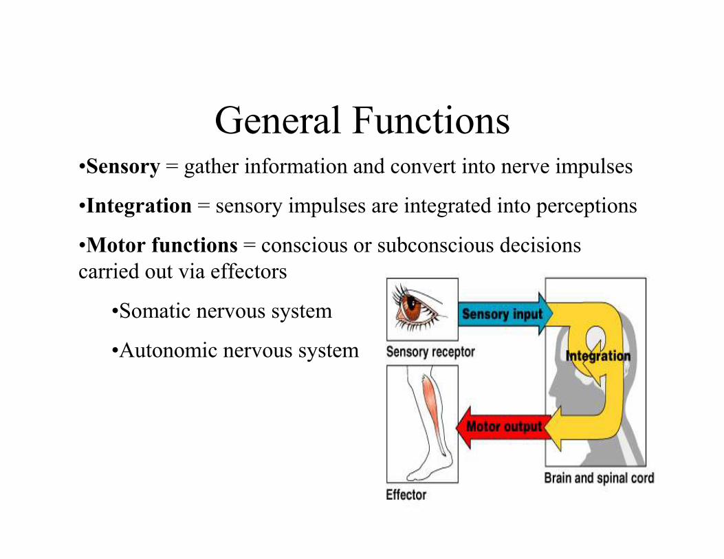
General Functions•Sensory = gather information and convert into nerve impulses
•Integration = sensory impulses are integrated into perceptions
•Motor functions = conscious or subconscious decisions
carried out via effectors
•Somatic nervous system
•Autonomic nervous system

Organization of the Nervous System

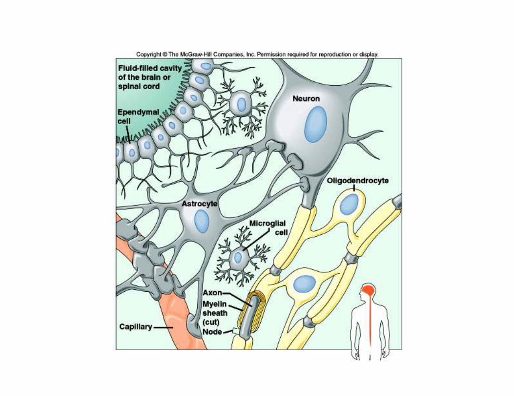
Supporting Cells
Neuroglial cells
CNS PNS
1.Microglial 1.Schwann
2.Oligodendrocytes 2.Satellite
3.Astrocytes
4.Ependyma


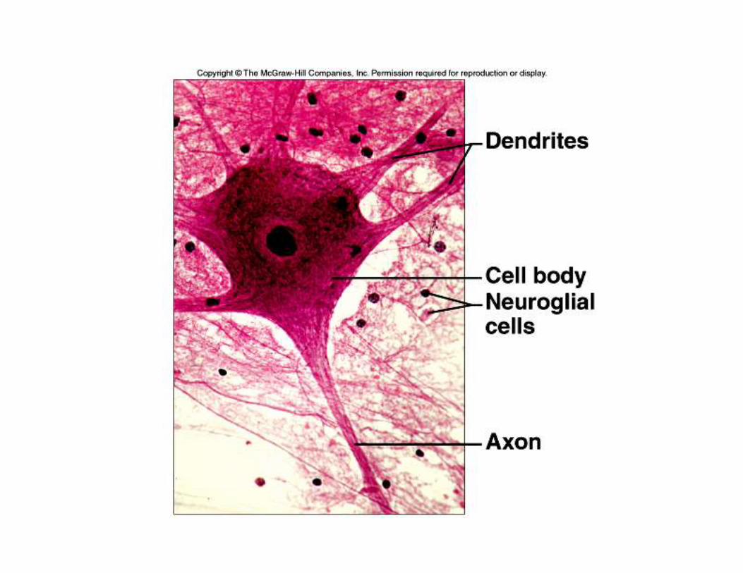
Neuron Structure = Nerve Cell
1. Cell Body
• Typical organelles
• Nissl bodies (chromatophilic substance) = Rough ER
• Neurofibrils
2. Processes
• Dendrites
• Axon
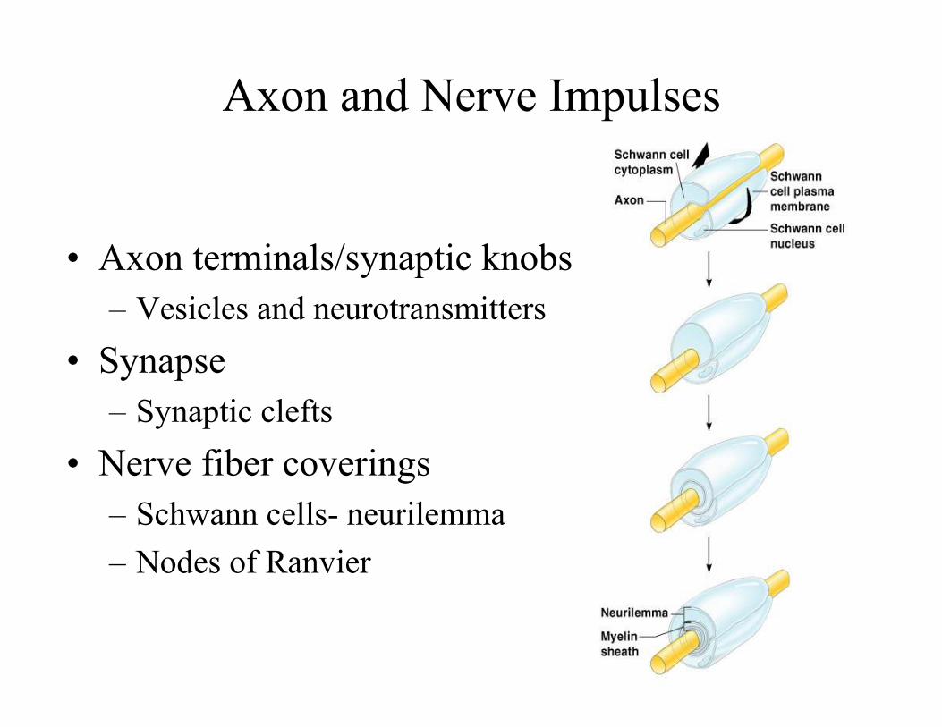
Axon and Nerve Impulses
• Axon terminals/synaptic knobs
– Vesicles and neurotransmitters
• Synapse
– Synaptic clefts
• Nerve fiber coverings
– Schwann cells- neurilemma
– Nodes of Ranvier

Cell Body Location
• Gray matter
• White matter
• Ganglia


Classification of NeuronsWays of classification:
1. Structure
2. Function
• Bipolar neurons
• Unipolar neurons
• Multipolar neurons
• Sensory neurons (afferent neurons)
• Interneurons
• Motor neurons (efferent neurons)


Cell Membrane Potential
•Irritability
•Conductivity
A cell membrane is usually polarized, with an excess of
negative charges on the inside of the membrane;
polarization is important to the conduction of nerve
impulses.

Distribution of IonsThe distribution of ions is determined by the membrane channel
proteins that are selective for certain ions.
Potassium ions pass through the membrane readily than do
sodium ions, making potassium ions a major contributor to
membrane polarization.

Resting Potential
1. Due to active transport, the cell membrane maintains a
greater concentration of sodium ions outside and a
greater concentration of potassium ions inside the
membrane.
2. Inside of the membrane has excess negative charges,
while the outside has more positive charges.
3. The separation of charge, potential difference, is called
the resting potential.

Potential Changes1. Stimulation of a membrane locally affect its resting
potential.
2. When the membrane potential becomes less negative, the
membrane is depolarized.
3. If sufficiently strong depolarization occurs, a threshold
potential is achieved as ion channels open.

Action Potential
At threshold potential, membrane permeability to sodium suddenly
changes in the region of stimulation.—Action potential
• As sodium channels open, sodium ions rush in, and the
membrane potential changes and becomes depolarized.
• At the same time, potassium channels open to allow
potassium ions to leave the cell—the membrane is
repolarized and resting potential is reestablished.
• Active transport works to maintain the original concentration
of sodium and potassium ions.

Nerve Impulse
A nerve impulse is conducted as action potential is
reached at the trigger zone and spreads by a local
current flowing down the fiber, and adjacent areas of
the membrane reach action potential. Nerve impulse
is conducted as a series of action potentials occurring
along the length of the axon.


Impulse Conduction1. Unmyelinated fibers
2. Myelinated fibers
***All-or-None Response***
• If a nerve fiber responds at all to a stimulus, it responds
completely by conducting an impulse.
• Greater intensity of stimulation triggers more impulses per
second, not stronger impulses.

SynapseSynapse = junction between two communicating neurons
Synaptic Transmission = Presynaptic neuron transmits impulse
across the synaptic cleft to the postsynaptic neuron
When an impulse reaches the synaptic knobs of an axon, the
synaptic vesicles release neurotransmitter into the synaptic
cleft. The neurotransmitter react with the receptors on the
postsynaptic membrane.
•Excitatory Action
•Inhibitory Action


Neurotransmitters•50 kinds of neurotransmitters
•Synthesized in the synaptic knobs
•Stored in vesicles
•Presence of calcium ions are needed to allow vesicles to
fuse to the membrane and release their contents into the
synaptic cleft.
•Enzymes in the cleft and on postsynaptic membrane
decompose the neurotransmitter in order to prevent
continuous stimulation of the postsynaptic neuron

Impulse ProcessingNeuronal Pools
•Facilitation
•Convergence
•Divergence

Nerve Pathways
•Reflex arc
•Reflex behavior
•Knee-jerk reflex
•Withdrawal reflex

Meninges
The spinal cord and
brain are surrounded by
membranes called
meninges that lie
between the bone and
soft tissues.
•Dura mater
•Arachnoid mater
•Subarachnoid space
•Pia mater


Spinal Cord
•31 segments, each gives rise to a pair
of spinal nerves
•Cervical enlargement = nerves leading
to the upper limbs
•Lumbar enlargement = nerves leading
to the lower limbs
•Anterior median fissure
•Posterior median sulcus
•White matter
•Gray matter
•Central canal

Functions of the Spinal Cord
•Transmit impulses to and from the brain
•House spinal reflexes
•Ascending tracts vs. descending tracts

Brain•Largest, most complex portion of the nervous system
•Cerebrum (high mental functions)
•Diencephalon (processes sensory input)
•Cerebellum (coordinates muscular activity)
•Brain stem (coordinates and regulates visceral activities)

Cerebrum•Largest portion of the brain, 2 cerebral hemispheres
•Corpus callosum
•Convolutions, sulci, fissures
•Divided into lobes, named according to the bones they underlie
•Functions of cerebrum
•Higher brain functions
•Integrating information for reasoning
•Memory
•Interpretation of sensory input
•Initiating voluntary muscular movements
•Cerebral cortex

Functions of the Cerebral Cortex•Motor, sensory, and association areas
•Primary motor areas = frontal lobe
•Broca’s area = coordinates muscular activity, speech
•Frontal eye field = voluntary movements of the eyes and
eyelids
•Sensory areas = interpret sensory input, feelings, sensations
•Association areas = analyze and interpret sensory impulses,
reasoning, judgment, emotions, verbalizing ideas, storing
memory
•General interpretive area = junction of the lobes, complex
thought processing


Ventricles and Cerebrospinal
Fluid•Ventricles
•Choroid plexuses
•Cerebrospinal fluid

Diencephalon•Thalamus
•Sorting and directing sensory information
•Hypothalamus
•Maintains homeostasis
•Regulates visceral activities
•Linking endocrine system to this system
•Limbic system
•Controls emotional experience and expression

Brain Stem•Midbrain
•Centers for auditory and visual reflexes
•Pons
•Centers that regulate rate and depth of breathing
•Medulla oblongata
•Control visceral functions
•Cardiac center for heart rate and blood pressure
regulation
•Assists pons with breathing regulation

Cerebellum
•2 hemispheres
•Integrate sensory
information about the
position of body parts
•Coordinates skeletal
muscle activity
•Maintains posture

Peripheral Nervous System= consists of the cranial and spinal nerves that arise from
the CNS and travel to the remainder of the body
= somatic nervous system that oversees voluntary activities
12 pairs of cranial nerves:
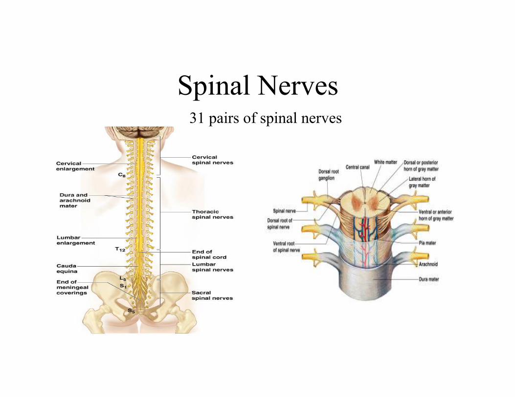
Spinal Nerves31 pairs of spinal nerves

Structure of a Nerve

Autonomic Nervous System= maintaining homeostasis of visceral activities without
conscious effort
2 divisions:
•Sympathetic division
•Parasympathetic division
Autonomic nerve fibers:
•Preganglionic fibers that leaves CNS
•Postganglionic fibers that extends to the visceral effectors

Sympathetic Division= operates under
conditions of stress or
emergency
•Fibers arise from the
thoracic and lumbar
regions of the spinal
cord
•Synapse in ganglia
close to the vertebral
column
•Postganglionic axons
lead to the effector
organ

Parasympathetic Division
= operates under
normal conditions
•Fibers arise from
brainstem and
sacral region of the
spinal cord
•Synapse in the
ganglia close to the
effector organ
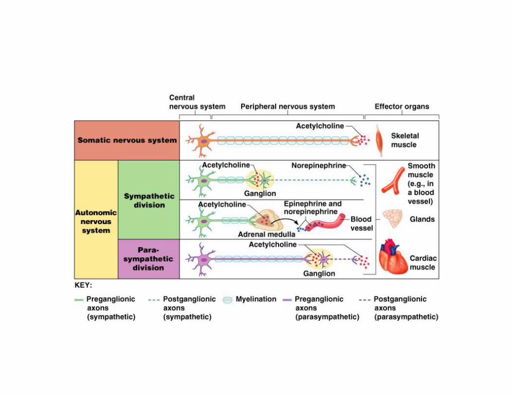

Autonomic NeurotransmittersPreganglionic fibers release acetylcholine
Postganglionic fibers:
Parsympathetic: acetylcholine
Sympathetic: norepinephrine
Control of Autonomic Activity:
•Reflex centers in the brain and spinal cord
•Limbic system and cerebral cortex