nervous system: general principles pa 544 clinical anatomy tony serino. ph.d
TRANSCRIPT
Nervous System: General Principles
PA 544
Clinical Anatomy
Tony Serino. Ph.D.
Nervous System
• Controls and/or modifies all other systems
• Rapid response time
• Usually short duration

Functional Areas

Divisions of the Nervous System

Nervous Tissue
• Non-excitable Tissue (Supportive cells)– Neuroglia –present in CNS– Schwann and Satellite cells –present in PNS
• Neurons (excitable tissue)– Initiate and conduct electrical signals (action potentials)

Neuroglia (glial cells)
•Form BBB•Regulate microenvironment•Pass on nutrients; get rid of waste
Phagocytic, protective

Neuroglia
•Line cavities•Create CSF
Secrete myelin in CNS

PNS Supportive Cells
• Schwann cells –secrete myelin in PNS• Satellite cells –surround neuron cell bodies in PNS

Neuron Anatomy
Axonal terminalNerve endingSynaptic boutonsSynaptic knobs

Functional Zones of a Neuron
Receptor Zone
Initial segment of Axon(trigger zone)
Axon
Nerve endings
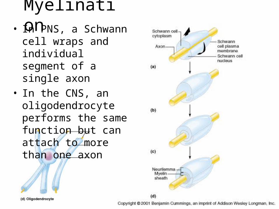
Myelination• In PNS, a Schwann cell
wraps and individual segment of a single axon
• In the CNS, an oligodendrocyte performs the same function but can attach to more than one axon
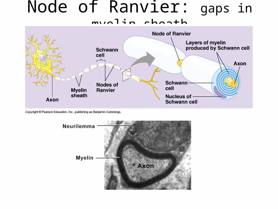
Node of Ranvier: gaps in myelin sheath

Types of Neurons
• Anatomical classification– Based on number of process projecting from
cell body
• Functional Classification– Based on location of neuron and direction of
information flow


General Terms
• Ganglia vs. Nuclei– Areas of densely packed nerve cell bodies– Ganglia are usually found in PNS– Nuclei are found in CNS
• Nerve vs. nerve fiber– A nerve is a dissectible structure containing
hundreds of axons– A nerve fiber is a single axon
• CT sheaths covering peripheral nerves:

Nerve CT sheaths

Synapses
• Areas where neurons communicate with other cells
• Can be chemical (with neurotransmitters) or electrical (gap junctions)

Anatomy of Synapse (chemical)
Neurotransmission ends when NT diffuses away,re-absorbed by presynaptic neuron, or NT metabolized(degraded) by enzymes in cleft
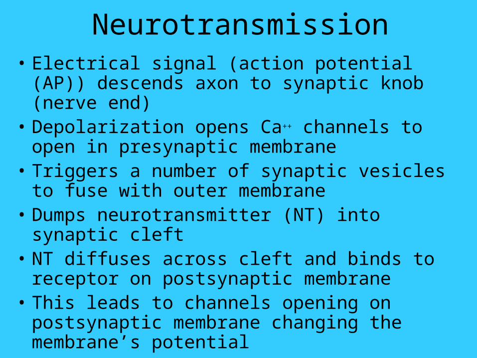
Neurotransmission• Electrical signal (action potential (AP)) descends
axon to synaptic knob (nerve end)• Depolarization opens Ca++ channels to open in
presynaptic membrane• Triggers a number of synaptic vesicles to fuse
with outer membrane• Dumps neurotransmitter (NT) into synaptic cleft• NT diffuses across cleft and binds to receptor on
postsynaptic membrane• This leads to channels opening on postsynaptic
membrane changing the membrane’s potential

Types of Anatomical Synapses
Regeneration of Nerve Fibers•Damage to nerve tissue is serious because mature neurons are post-mitotic cells•If the soma of a damaged nerve remains intact, damage may be repaired •Regeneration involves coordinated activity among:
– remove debris–form regeneration tube and secrete growth factors–regenerate damaged part

Response to Injury • Anterograde degeneration with some retrograde; phagocytic cells (from Schwann cells, microglia or monocytes) remove fragments of axon and myelin sheath
• Cell body swells, nucleus moves peripherally
• Loss of Nissl substance (chromatolysis)
• In the PNS, some Schwann cells remain and form a tubular structure distal to injury; if gap or scarring is not great axon regeneration may occur with growth down tube
• In the CNS, glial scar tissue seems to prevent regeneration
If contact with tube is not established then no regeneration and a traumatic neuroma forms

Regeneration in PNS

Drug Intervention Possibilities
A. Increase leakage and breakdown of NT from vesicles
B. Agonize NT releaseC. Block NT releaseD. Inhibit NT synthesisE. Block NT uptakeF. Block degradative enzymes in
cleftG. Bind to post-synaptic receptorH. Stimulate or inhibit second
messengers in post-synaptic cell