natural products k-26 and anthramycin...
TRANSCRIPT

BIOSYNTHETIC INVESTIGATIONS OF THE PEPTIDE
NATURAL PRODUCTS K-26 AND ANTHRAMYCIN
By
Vanessa Victoria Phelan
Dissertation
Submitted to the Faculty of the
Graduate School of Vanderbilt University
in partial fulfillment of the requirements
for the degree of
DOCTOR OF PHILOSOPHY
in
Chemistry
May, 2010
Nashville, Tennessee
Approved by:
Professor Brian O. Bachmann
Professor Gary A. Sulikowski
Professor John A. McLean
Professor Richard N. Armstrong

ii
TABLE OF CONTENTS
PAGE
DEDICATION……………………………………………………………………..….…iv
ACKNOWLEDGEMENTS………………………………………………………….…..v
LIST OF TABLES……………………………………………………….……………..viii
LIST OF FIGURES……………………………………………………………………..ix
LIST OF SCHEMES……………………………………………………………………xii
LIST OF ABBREVIATIONS…………………………………………………………..xiii
CHAPTER
I. INTRODUCTION .................................................................................. 1 Case Study I: K-26 .......................................................................... 2 Case Study II: Anthramycin ............................................................. 9 Peptide Bond Biosynthesis ........................................................... 19
Non-ribosomal Peptide Natural Product Biosynthesis ........ 19 Ribosomal Peptide Natural Product Biosynthesis .............. 25
Lantibiotics ............................................................... 28 Microcins ................................................................. 29 Cyanobactins ........................................................... 31 Thiopeptides ............................................................ 33
Ligase-based Peptide Biosynthesis .................................... 34 Biosynthetic Hypotheses for K-26 and Anthramycin ..................... 37
K-26 .................................................................................... 37 Anthramycin ....................................................................... 38
Dissertation Goals ......................................................................... 39 References .................................................................................... 41
II. γ-18O4-ATP PYROPHOSPHATE EXCHANGE ASSAY ...................... 60
Introduction ................................................................................... 60 Results .......................................................................................... 62 Discussion ..................................................................................... 71 Materials and Methods .................................................................. 73 References .................................................................................... 77
III. BIOSYNTHETIC STUDIES OF K-26 .................................................. 80
Introduction ................................................................................... 80 Results .......................................................................................... 82

iii
Incorporation of isotopically-labeled precursors ................. 82 Biosynthetic hypothesis ...................................................... 89 Genetic Analysis ................................................................. 90 Reverse Genetic Analysis .................................................. 92
N-acetyltransferase Isolation ................................... 92 Adenylation Enzyme Isolation .................................. 94
Discussion ................................................................................... 103 Materials and Methods ................................................................ 108 References .................................................................................. 128
IV. BIOSYNTHETIC INVESTIGATIONS OF ANTHRAMYCIN ............... 131
Introduction ................................................................................. 131 Results ........................................................................................ 134
Biosynthetic hypothesis .................................................... 134 Genetic Identification of Anthramycin Gene Cluster ......... 134 Proposed MHA biosynthesis ............................................ 138 Chemical complementation studies .................................. 140 Orf21 amino acid activation .............................................. 143 Proposed dehydroproline acrylamide biosynthesis .......... 152 Synthesis of dehydroproline acrylamide Precursor .......... 153
Discussion ................................................................................... 155 Materials and Methods ................................................................ 158 References .................................................................................. 169
V. FUTURE DIRECTIONS…………………………………………………..175

iv
To my parents:
Words can never express how thankful I am for all that you have given me.

v
ACKNOWLEDGEMENTS
It has been my privilege to study at Vanderbilt University in the
Department of Chemistry and I am eternally grateful to both the university and
department for allowing me to pursue my education in such a supportive and
collaborative environment. Without the supportive, collaborative and scientific
environment at Vanderbilt, my research and subsequent dissertation would not
have been possible.
I would like to thank my advisor, Dr. Brian Bachmann, for his support and
guidance throughout my graduate career. His scientific insight, support and
openness allowed me to follow my scientific curiosity, has provided me with vast
knowledge and piqued my interest in further scientific pursuits. I would also like
to thank my Ph.D. committee members Dr. Gary Sulikowski, Dr. John McLean
and Dr. Richard Armstrong for providing the opportunity for collaboration and
support in both the academic and research aspects of my journey at Vanderbilt.
In addition to my advisor and committee members, it is necessary for me
to acknowledge and thank my many collaborators at Vanderbilt without whom my
research would have never progressed. To Dawn Overstreet, Dr. M. Wade
Calcutt and Dr. David Hachey in the Mass Spectrometry Research Core, thank
you for your constant willingness to help me accomplish my goals with your
insightful suggestions, willingness to share and endless patience. To Dr. John
McLean and his entire research group, specifically Randi Gant-Branum and
Michal Kliman, thank you for your help and patience during my endeavors into

vi
MALDI. To Dr. Tony Forster, thank you for opening your laboratory to me and
taking the time to teach me. And to my “unofficial” collaborators in the Sulikowski
lab, thank you for sharing both your knowledge and your chemicals.
Of course, this research would not have been possible without funding. I
am grateful for funding from the National Institutes of Health, the Petroleum
Research Fund and the Vanderbilt Institute for Chemical Biology for research
funding. The Vanderbilt Institute for Chemical Biology, National Institutes of
Health and Graduate School also provided me with support through fellowships
and assistantships.
To my colleagues in the Bachmann laboratory, both past and present, I
have to give my sincerest thanks. It has been a joy working with all of you.
Thank you for the scientific input, friendship and camaraderie that you have
provided over the years. I am eternally grateful for you accepting me as the
dinosaur loving, color coordinating person that I am. I have to specifically thank
Rob Scism and Glenna Kramer as my desk-mates for putting up with my never-
ending external monologue.
To my many friends, thank you. My time at Vanderbilt has been difficult
and without you, I would not have made it. I especially have to thank Brian
Turner, Easton Selby, Victor Ghidu, Bill Evans and Kevin Perzynski for holding
me up when the ground disappeared from underneath me. I am eternally
grateful for your friendship. There are some things that can never be repaid. To
Steve Townsend, Melissa Carter, Anh Hoang, the entire Cliffel lab and my
Thursday morning and Monday evening soccer buddies, it has been a pleasure.

vii
You made graduate school bearable. Thank you for both your scientific insight
and your friendship.
To my family, thank you for everything. Thank you for your faith, love,
support and everything else that you have ever given and instilled in me. Thank
you for letting me be who I am without question. Thank you for shaping me into
the person I have become.

viii
LIST OF TABLES
Table Page
III-1. Bioactive Phosphonate Containing Compounds ......................................... 3
III-2. Activity of Amino Acid Activating Enzymes ................................................ 70
III-3. Prospective K-26 NRPS Gene Clusters .................................................... 91
III-4. Summary of NAT Activity ........................................................................... 93
IV-1. ORFs Found Within the Anthramycin Gene Cluster ................................ 137
IV-2. Results from Chemical Complementation Experiments .......................... 142
IV-3. Specificity Code of the A-domain of Orf21 .............................................. 143
IV-4. Equilibrium and Kinetic Parameters of Orf21………………………………150
IV-5. Specificity Code of Orf21, SibE and TomA ............................................. 158

ix
LIST OF FIGURES
Figure Page
I-1. Chemical Structures of K-26 and Anthramycin .............................................. 2
I-2. Phosphonate Natural Products. ..................................................................... 4
I-3. K-26 and analogues ...................................................................................... 5
I-4. Binding Mechanism of K-26 in AnCE ............................................................ 7
I-5. Biosynthetic Route to Phosphonate Moiety. .................................................. 8
I-6. Possible Mechanisms for PEP mutase. ......................................................... 9
I-7. Classes of 1,4-benzodiazepine. .................................................................. 11
I-8. Examples of Bacterial Pyrrolobenzodiazepines ........................................... 12
I-9. Proposed Mechanism for PDB Bioactivity. .................................................. 14
I-10 Anthramycin Bound in the Minor Groove of DNA ........................................ 15
I-11. Precursor Incorporation Studies with Bacterial PBDs. ............................... 16
I-12. A-domain Reactivity and Specificity. ......................................................... 21
I-13. The Role of the T-Domain in NRPS Systems ............................................ 22
I-14. C-Domain Reaction Mechanism ................................................................ 23
I-15. NRPS Biosynthetic Strategies.. ................................................................. 25
I-16. General Ribosomal Peptide Biosynthesis. ................................................. 27
I-17. Post-translational Modifications Found in Lantibiotics. .............................. 29
I-18. Microcin Ribosomal Peptides .................................................................... 30
I-19. Proposed Biosynthesis of Patellamides A and C. ...................................... 32
I-20. Biosynthesis of Thiostrepton ..................................................................... 33
I-21. Dapdiamide Biosynthesis. ......................................................................... 35

x
I-22. ATP-independent Peptide Bond Formation ............................................... 36
I-23. Proposed Peptide Bond Formation in K-26. .............................................. 38
I-24. Proposed Peptide Bond Formation in Anthramycin. .................................. 39
II-1. Role of A-domains in NRPS. ...................................................................... 61
II-2. ATP-Pyrophosphate exchange. .................................................................. 64
II-3. Limit of Exchange Detection. ...................................................................... 66
II-4. 18O ATP Lability. ......................................................................................... 68
II-5. Amino Acid Activation by TycA.. ................................................................. 69
II-6. TycA L-phenylalanine Substrate Dependence. ............................................ 71
III-1. K-26 and Related Analogues ..................................................................... 81
III-2. C-P Bond Forming Pathway ...................................................................... 82
III-3. Precursor Incorporation into K-26. ............................................................. 83
III-4. Tyramine Incorporation. ............................................................................. 83
III-5. SRM Detection of Precursor Incorporation. ............................................... 87
III-6. Dipeptide Incorporation Studies. ............................................................... 88
III-7 Possible biosynthetic route for K-26 formation ........................................... 90
III-8. Kinetic Characterization of ORF6213. ....................................................... 94
III-9. Overview of Adenylation Enzyme Purification Methods ............................ 96
III-10. Ammonium Sulfate Fractionation ............................................................ 97
III-11. Q-sepharose Fractionation. ..................................................................... 98
III-12. DEAE-sepharose Fractionation ............................................................... 98
III-13. Hydrophobicity and Size Exclusion Fractionation .................................... 99
III-14. SDS-PAGE Adenylation Enzymes. ....................................................... 100

xi
III-15. Pyrophosphate Exchange Analysis of Fractions. .................................. 101
III-16. tRNA Charging Assay............................................................................ 102
III-17. SDS-PAGE of NAT 97747 ..................................................................... 102
III-18. K-26 and Analogue Production by S. rubeum ....................................... 106
IV-1. Representative Examples of Bacterial PBDs. ......................................... 132
IV-2. Precursor Incorporation Studies of PBDs ................................................ 133
IV-3. The Anthramycin Gene Cluster. .............................................................. 135
IV-4. Proposed Peptide Biosynthesis in Anthramycin. ..................................... 138
IV-5. Amino Acid Concentration Dependence of Orf21. .................................. 144
IV-6. Orf21 T-loading Assay with MHA. ........................................................... 146
IV-7. Possible Biochemical Mechanisms for A-domains. ................................. 148
IV-8. Equilibrium Kinetics of Orf21 with MHA................................................... 150
IV-9. Secondary Plots of Orf21 Equilibrium Kinetics…………………………….150
IV-10. Proposed Mechanism of A-domain. ...................................................... 156

xii
LIST OF SCHEMES
Scheme Page
I-1. Proposed MHA Biosynthetic Pathway. ........................................................ 17
I-2. Proposed Dehydroproline Acrylamine Biosynthesis. ................................... 19
III-1. Synthesis of Stable Labeled Dipeptides .................................................... 85
IV-1. Possible Biosynthetic Pathway for MHA ................................................. 139
IV-2. Proposed Biosynthetic Pathways for MHA. ............................................. 141
IV-3. Proposed Biosynthetic Pathway for Dehydroproline Acrylamide.. ........... 153
IV-4. Proposed Synthesis of Dehydroproline Acrylamide and Intermediates. .. 154
xiii
LIST OF ABBREVIATIONS
ACE Angiotensin converting enzyme
A-domain Adenylation domain
ADP Adenosine diphosphate
AEP 2-aminoethylphosphonic acid
AHEP (R)-1-amino-2-(4-hydroxyphenyl)ethylphosphonic acid
AMP Adenosine monophosphate
AnCE ACE homologue in Drosophila melanogaster
A-site Acceptor site
ATP Adenosine triphosphate
C-domain Condensation domain
CoA Coenzyme A
C-P Phosphonate bond
C-P-C Phosphinate bond
Cy-domain Cyclization domain
Da Dalton
DAP 2,3-diaminopropionate
DEAE Diethylaminoethyl
dK-26 Des-acetyl-K-26
DNA Deoxyribonucleic acid
E-domain Epimerization domain
EF-Tu Elongation factor Tu

xiv
ESI Electrospray ionization
E-site Exit site
GEB General enzyme buffer
GTP Guanosine triphosphate
HA 3-hydroxyanthranilic acid
IC50 Half maximal inhibitory concentration
LB Luria broth
LC Liquid chromatography
L-DOPA L-3,4-dihydroxyphenylalanine
LOD Limit of detection
LysRS Lysine tRNA synthetase
m/z Mass to charge ratio
MALDI Matrix-assisted laser desorption/ionization
MHA 4-methyl-3-hydroxyanthranilic acid
mRNA Messenger ribonucleic acid
MS Mass spectrometry
MT-domain Methyltransferasae domain
NAT N-acetyltransferase
NCBI National Center for Biotechnology Information
NCE New chemical entity
NMR Nuclear magnetic resonance
NRPS Non-ribosomal peptide synthetase
NRRL Northern Regional Research Laboratory

xv
ORF Open reading frame
Ox-domain Oxidation domain
PBD Pyrrolobenzodiazepine
PCR polymerase chain reaction
PEP Phosphoenolpyruvate
polydG Poly deoxyguanine
PPi Pyrophosphate
PPTase Phosphopantetheinyl transferase
PPyr Phosphonopyruvate
P-site Peptidyl site
Re-domain Reductase domain
RNA Ribonucleic acid
SAM S-adenosyl methionine
SDS-PAGE Sodium dodecylsulfide polyacrylamide gel
electrophoresis
SRM Selected reaction monitoring
T-domain Thiolation domain
Te-domain Thioesterase domain
TIGR The Institute for Genomic Research
TLC Thin layer chromatography
TOF Time-of-flight
tRNA Transfer ribonucleic acid
TrpRS Tryptophan tRNA synthetase

1
CHAPTER I
INTRODUCTION
Natural products have been an invaluable source of therapeutic agents1, 2.
Of 1024 small molecule new chemical entities (NCEs) introduced between 1981
and 2008, over half were natural products or natural product inspired synthetic
derivatives.2 During this time period, natural products and their derivatives
accounted for more than 68% of all anti-infectives (anti-bacterial, -fungal,
-parasitic and -viral) and approximately 63% of all anticancer compounds. An
additional 225 natural product derived molecules are currently in clinical trials or
preclinical development including 86 for cancer treatment and 40 for the
treatment of infectious diseases.3, 4 While natural products are clearly vital for the
treatment of human disease, the study of microbial ecosystems has been
severely limited by the ability to cultivate less than 1% of the occupying
microorganisms leaving a vast untapped resource for drug discovery.5-8
Over the past few years, sequencing technologies have advanced
substantially, lowering the cost of genome sequencing.9 Total genome
sequencing of a variety of actinomycetes (soil-dwelling bacteria) revealed large
numbers of previously unidentified biosynthetic cassettes involved in the
biosynthesis of natural products10-12. In Streptomyces avermitilis alone, over 30
putative gene clusters were identified.10, 11 In addition to revealing the untapped
potential of actinomycetes for possible therapeutics, these genomic

2
advancements have allowed researchers to correlate previously identified
bioactive natural products with their biosynthetic origins. By correlating known
natural products to their biosynthetic cassettes, researchers can tap into nature’s
chemical repertoire by deconstructing the mechanisms required for natural
product biosynthesis and harnessing that knowledge to generate novel
therapeutics through synthetic biology.13 Herein, our investigations into
understanding the biosynthesis of two previously uninvestigated peptide natural
products from actinomycete species: K-26, a potent naturally produced
phosphonate containing hypotensive agent, and anthramycin, an anti-cancer
pyrrolobenzodiazepine, are described (Figure I-1).14, 15
Figure I-1. Chemical Structures of K-26 and Anthramycin K-26 is a unique phosphonate containing tripeptide capable of modulating ACE activity, while anthramycin in an anti-tumor, antibacterial pyrrolobenzodiazepine
Case Study I: K-26
During the last several decades, both natural and synthetic phosphonate
and phosphinate containing compounds have been used extensively in medicine
and agriculture.16 (Table I-1) These compounds are similar to phosphate esters
and anhydrides, but are distinguished by carbon-phosphorus bonds replacing

3
one or more oxygen-phosphorus bonds (C-P in phosphonates and C-P-C in
phosphinates). The structural similarity of phosphonates and phosphinates to
labile phosphate esters and carboxylic acids imparts their ability to act as
substrate and transition state analogues of enzyme substrates, inhibiting enzyme
catalysis.17
Table I-1. Bioactive Phosphonate Containing Compounds
In 1959, the first naturally occurring phosphonate,
2-aminoethylphosphonic acid (AEP), was identified.18 AEP is widely distributed
in biological systems as a polar head group of membrane lipids. Subsequently,
additional phosphonylated macromolecules including exopolysaccharides and
glycoproteins have been described19, 20. Furthermore, a variety of phosphonate
containing small molecules has been reported. These include bialaphos, an
herbicide; fosfomycin, an antibacterial; fosmidomycin, an anti-parasitic compound
and K-26, a hypotensive agent.21 (Figure I-2)

4
Figure I-2. Phosphonate Natural Products. Representative examples of phosponate containing natural products.
K-26, isolated from Astrosporangium hypotensionis (NRRL 12379), is a
representative member of an uninvestigated class of phosphonate containing
natural products which incorporate a phosphonic acid analogue of tyrosine.14
NMR, mass spectrometry, degradation and synthetic investigations have
demonstrated that K-26 is comprised of N-acetylated isoleucine, tyrosine and the
non-proteinogenic amino acid (R)-1-amino-2-(4-hydroxyphenyl)ethylphosphonic
acid (AHEP).14, 22 AHEP is shared among several related compounds produced
by Streptosporangium and Actinomadura species.23, 24 (Figure I-3)

5
Figure I-3. K-26 and analogues. Highlighted in red is a unique non-proteinogenic amino acid, AHEP. shared among a group of hypotensive tripeptides
K-26 was initially discovered by bioactivity guided fractionation of A.
hypotensionis culture extracts testing for inhibition of angiotensin converting
enzyme (ACE).14 ACE is a zinc-dependent dipeptidyl carboxypeptidase which
catalyzes the cleavage of carboxy-terminal dipeptides from numerous peptide
substrates of the Renin-Angiotensin-Aldosterone system.25 All analogues of
K-26 show some inhibition of ACE with K-26 possessing an IC50 value of 14.4
nM, comparable to the widely prescribed anti-hypertension drug Captopril (7.7
nM).26 Initial inhibition studies showed that K-26 is a non-competitive inhibitor of
ACE with possible interactions between the phosphonate of AHEP and an active
site zinc.14 Recent structure-activity relationship studies have revealed that the
phosphonate moiety of K-26 is required for its inhibitory activity.26 K-26 was
found to be 1500-fold more potent than its carboxylate congener. Additionally,
6
the absolute configuration of AHEP and N-acetylation were found to play an
important role in modulating ACE inhibitory activity.
While it may seem unusual to isolate a compound capable of modulating
mammalian ACE from a bacterial source, the presence of ACE homologues has
been reported in non-vertebrates and unicellular microorganisms.27-46 Six ACE-
like enzymes have been identified in Drosophila melanogaster.28-46 Recent
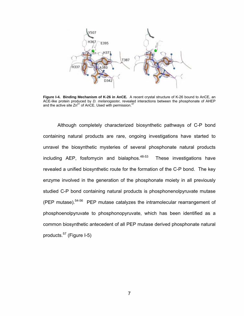
crystallography of one of these enzymes, AnCE, in complex with K-26 revealed
that K-26 occupies two of the four available substrate binding pockets.27, 47 K-26
binds within the S1 and S2 substrate binding sites with two of the phosphonic
oxygen atoms coordinating to the active site Zn2+ ion, while the third oxygen
hydrogen bonds with a histidine and tyrosine within the active site. (Figure I-4)
Additional hydrogen bonds mediate and stabilize the binding of K-26 to AnCE.
While the inhibition of AnCE by K-26 is lower (160 nM) than the inhibition of ACE
(14.4 nM), this crystal structure provides insight into the mechanism of ACE
inhibition by K-26 and the potential to develop new investigative tools and
therapeutics. It is important to note, however, that the role of K-26 in its
environmental interactions, whether endogenous or exogenous, has not been
established. Despite the potent hypotensive activity of K-26 and related
compounds, the biosynthetic pathway of K-26 has remained cryptic.
7
Figure I-4. Binding Mechanism of K-26 in AnCE. A recent crystal structure of K-26 bound to AnCE, an ACE-like protein produced by D. melanogaster, revealed interactions between the phosphonate of AHEP and the active site Zn2+ of AnCE. Used with permission.47
Although completely characterized biosynthetic pathways of C-P bond
containing natural products are rare, ongoing investigations have started to
unravel the biosynthetic mysteries of several phosphonate natural products
including AEP, fosfomycin and bialaphos.48-53 These investigations have
revealed a unified biosynthetic route for the formation of the C-P bond. The key
enzyme involved in the generation of the phosphonate moiety in all previously
studied C-P bond containing natural products is phosphonenolpyruvate mutase
(PEP mutase).54-56 PEP mutase catalyzes the intramolecular rearrangement of
phosphoenolpyruvate to phosphonopyruvate, which has been identified as a
common biosynthetic antecedent of all PEP mutase derived phosphonate natural
products.57 (Figure I-5)

8
Figure I-5. Biosynthetic Route to Phosphonate Moiety. All previously studied phosphonate natural products derive their phosphonate moiety from the conversion of phosphoenolpyruvate (PEP) to phosphonopyruvate (Ppyr) by PEP mutase.
While the catalytic mechanism of PEP mutase has been widely studied, a
unified mechanism has remained elusive. There are two possible mechanisms
consistent with the experimentally confirmed intramolecular phosphoryl transfer
retaining absolute stereochemistry at the phosphorus.54, 57-59 (Figure I-6) In
mechanism I, the phosphoryl group is transferred to an active site residue of PEP
mutase which is then attacked by the C(3) of the pyruvate enol intermediate.
While in mechanism II, metaphosphate dissociates from PEP and is attacked by
the C(3) of the pyruvate enol intermediate. It is prosposed that in mechanism II,
interactions with active site residues hold the metaphosphate in place, while
rotation around the C1-C2 bond of the pyruvate enolate to allow attack of the
C(3) onto the metaphosphate, which would account for the retention of
stereochemistry at the phosphorus as well as the stereochemistry of the C1
position of phosphonopyruvate.

9
Figure I-6. Possible Mechanisms for PEP mutase. There are two plausible mechanisms for PEP mutase. In mechanism I, the phosphate of PEP is transferred to the active site of PEP mutase which is the attacked by the C(3) of the pyruvate enol intermediate. In mechanism II, the phosphate dissociates from PEP mutase as metaphosphate which is then attacked by the C(3) of the pyruvate enol intermediate.
Although PEP mutase is the biosynthetic route of all previously
investigated C-P bond containing natural products, it is difficult to rationalize a
possible biosynthetic pathway through phosphonopyruvate to generate AHEP.
Comparison of AHEP to known C-P bond containing natural products makes it
apparent that the mechanism of AHEP formation may be radically different than
that of PEP mutase or an analogous enzyme. Initial labeled precursor
incorporation investigations into K-26 biosynthesis have revealed AHEP is a
discrete intermediate of K-26 and is derived from tyrosine without the formation
of a β-elimination intermediate.22, 60
Case Study II: Anthramycin
Anthramycin is representative of a class of peptide natural products
characterized by a 1,4-benzodiazepine ring system.15, 61 This class of
compounds is composed of four pharmacophore groups: 1,4-benzodiazepine-2-
ones, 1,4-benzodiazepines-2,5-diones, dibenzodiazepinones and

10
pyrrolobenzodiazepines (PBDs). (Figure I-7) Over 50
1,4-benzodiazepine-2-ones are currently in clinically use for treatment of
psychological diseases including insomnia, alcohol dependency and anxiety.61
The most prominent member of this group is diazepam (Valium).
1,4-benzodiazepene-2,5-ones have been tested as anticonvulsants,
peptidomemetic inhibitors and herbicides.62-66 Within this group, only cyclopenin
has been isolated from nature, identified as an intermediate to viridicatin, an
inhibitor of HIV replication.67 The sole member of the dibenzodiazepinone group
is the naturally produced TLN-4601 (ECO-4601) and is characterized by broad
spectrum antitumor activity and is currently under phase I/II clinical trials.68, 69
The final group, PBDs, includes both natural and synthetic compounds. These
compounds, including anthramycin, are potent alkylating agents reacting in the
minor groove of DNA, imparting both antibacterial and antitumor activities.70-74

11
N
HN
Cl
O
Diazapam (Valium)1,4-benzodiazepine-2-one
N
HN
O
O
OH
CH3
O
OH
Cyclopenin1,4-benzodiazepine-2,5-one
N
HN
O
OHHO
OH
Diazepinomycin (TLN-4601/ECO-4601)Dibenzodiazepinone
N
HN
O
OHH3C
NH2
O
OHH
AnthramycinPyrrolobenzodiazepines
Figure I-7. Classes of 1,4-benzodiazepine. Representative members of the 1,4-benzodiazepine family of compounds. The core 1,4-benzodiazepine structure is highlighted in red.
PBDs are structurally defined by a tricyclic ring system consisting of an
aromatic A-ring, a seven-membered diazepine B-ring and a hydropyrrolidine
C-ring.75 Chemical diversity of PBDs is introduced by varied substitution patterns
of the A-ring and unsaturation at the C2-C3 bond and/or an exocyclic moiety at
the C2 position of the C-ring (Figure I-8). Currently, 19 natural PBDs have been
isolated from bacterial cultures, including anthramycin. While 16 natural PBDs
show moderate antimicrobial properties, their antitumor activities are the most
intriguing.15

12
Figure I-8. Examples of Bacterial Pyrrolobenzodiazepines. Chemical diversity of PBDs is introduced by varied substitution patterns of the A-ring and unsaturation at the C2-C3 bond and/or an exocyclic moiety at the C2 position of the C-ring. Inactive PDBs have a carbonyl at the C11 of the B-ring, exemplified here by RK1441B
Anthramycin has shown broad spectrum antitumor activity in vitro
including against HeLa, Sarcoma 180, Walker 256 carcinosarcoma and leukemia
P388 cell lines.76 In initial human studies, of 219 patients with unresponsive
tumors, including lymphomas and sarcomas, approximately half saw a significant
decrease in tumor size.76 At the concentrations used, 0.02 mg/kg, no side effects
were observed. However at concentrations between 0.1 and 0.5 mg/kg,

13
cardiotoxicity was observed in rats.77 Structure-activity relationship studies
suggest that the C9 hydroxyl of the A-ring may be the source of these cardiotoxic
side effects and synthetic analogues have been created reducing this activity.73,
74, 77, 78
Initial experiments investigating the mechanism of inhibition of replication
and transcription in cells by anthramycin revealed that anthramycin showed
preferred binding to double stranded DNA over single stranded DNA and RNA.79
Co-purification of anthramycin with double stranded DNA indicated covalent
modification of DNA by anthramycin, while experiments with single stranded
polydG suggested that guanine may be the reactive DNA residue.80, 81 Structural
studies including NMR and crystallography illuminated the role of a covalent
bond between the C11 position of the B-ring and an exocyclic amino group of a
guanine base within the DNA binding sequence74, 82-88. Anthramycin, sibiromycin
and tomaymycin alkylate DNA in a site-specific manner preferring the reactive
guanine to be situated between two purine nucleobases rather than
pyrimidines.85, 89, 90 In terms of DNA affinity, the binding order is as follows:
sibiromycin > anthramycin > tomaymycin > DC-81 > neothramycin.61, 72 The
conformational flexibility of the PBD and degree of right-handed twist may be
responsible for the differences in binding affinity.91
Currently, there are two plausible mechanisms for the bioactivity of PBDs
consistent with experimental data.92, 93 In solution, PBDs can exist in three
interconverting forms: the imine form, the carbinolamine form and in methanol,
the carbinolamine methyl ether form. In mechanism I, a direct SN2 reaction of the

14
guanine amine on the carbinolamine carbon with water elimination leads to
alkylation. In mechanism II, alkylation results from a direct reaction of the
guanine amine with the imine form. (Figure I-9)
Figure I-9. Proposed Mechanism for PDB Bioactivity. Research strongly suggests that DNA alkylation occurs from a direction reaction of a guanine amine within the minor groove of B-DNA with the imine form of PBDs.
Mechanism II is strongly supported by the formation of two diastereomers
of tomaymycin-DNA adducts (11S, 11aS and11R, 11aS) in a proportion different
than while free in solution and increased reactivity of PBDs when electron-
donating substitutions are present on the A-ring.82, 87, 94, 95 The S configuration at
the C11a position allows the PBD scaffold to adopt a right-handed twist permits
binding within the minor groove of B-DNA. While there are four possible binding
configurations of PBDs (two for each diasteromer: 5’ to 3’ or 3’ to 5’ orientations),
anthramycin mainly binds with the C-ring on the 5’-end of the modified DNA
strand resulting only in the 11S, 11aS adduct, likely due to the 35o twist of
anthramycin causing steric clashing in the other three binding configurations.87, 96
(Figure I-10) Hydrogen bonding of the A-ring substituents and the C-ring
acrylamide tail may increase the stability of this adduct.

15
Figure I-10 Anthramycin Bound in the Minor Groove of DNA. The duplex is represented in CPK in (A) and sticks in (B). Used with permission.15
Despite the potent biological activities of PBDs, little is known about their
biosynthesis. The structural similarities of PBDs indicate that they may be
derived from common precursors through parallel biosynthetic pathways.
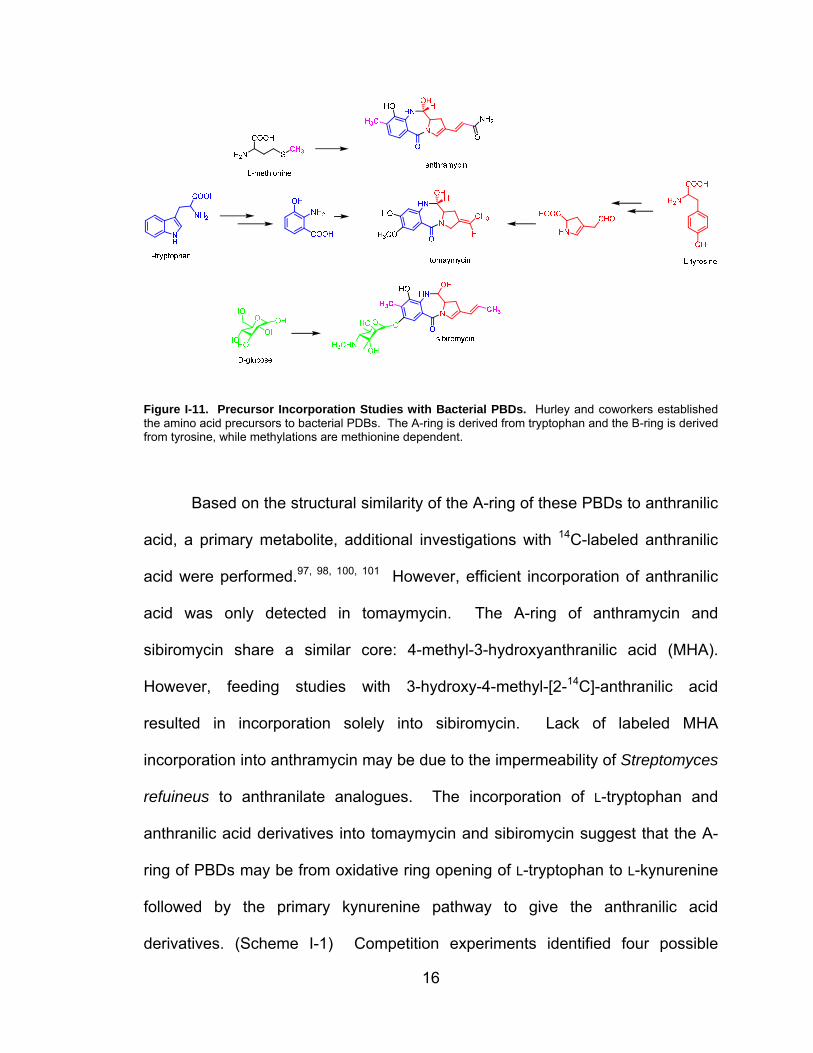
Labeled precursor feeding studies with the producers of anthramycin, sibiromycin
and tomaymycin by Hurley and coworkers identified the amino acid precursors of
these PBDs97-99. (Figure I-11) The anthrinilate A-ring of all three PBDs is derived
from L-tryptophan with retention of the indole nitrogen, while the dehydroproline
C-ring is derived from L-tyrosine. Labeled L-methionine was incorporated into the
4-position of the A-ring and the C-1 position of the exocyclic side chain of the
C-ring. Glucose provides the precursor of the sugar incorporated into
sibiromycin.
16
Figure I-11. Precursor Incorporation Studies with Bacterial PBDs. Hurley and coworkers established the amino acid precursors to bacterial PDBs. The A-ring is derived from tryptophan and the B-ring is derived from tyrosine, while methylations are methionine dependent.
Based on the structural similarity of the A-ring of these PBDs to anthranilic
acid, a primary metabolite, additional investigations with 14C-labeled anthranilic
acid were performed.97, 98, 100, 101 However, efficient incorporation of anthranilic
acid was only detected in tomaymycin. The A-ring of anthramycin and
sibiromycin share a similar core: 4-methyl-3-hydroxyanthranilic acid (MHA).
However, feeding studies with 3-hydroxy-4-methyl-[2-14C]-anthranilic acid
resulted in incorporation solely into sibiromycin. Lack of labeled MHA
incorporation into anthramycin may be due to the impermeability of Streptomyces
refuineus to anthranilate analogues. The incorporation of L-tryptophan and
anthranilic acid derivatives into tomaymycin and sibiromycin suggest that the A-
ring of PBDs may be from oxidative ring opening of L-tryptophan to L-kynurenine
followed by the primary kynurenine pathway to give the anthranilic acid
derivatives. (Scheme I-1) Competition experiments identified four possible
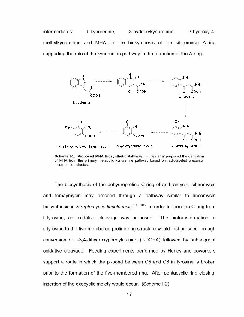
17
intermediates: L-kynurenine, 3-hydroxykynurenine, 3-hydroxy-4-
methylkynurenine and MHA for the biosynthesis of the sibiromycin A-ring
supporting the role of the kynurenine pathway in the formation of the A-ring.
Scheme I-1. Proposed MHA Biosynthetic Pathway. Hurley et al proposed the derivation of MHA from the primary metabolic kynurenine pathway based on radiolabeled precursor incorporation studies.
The biosynthesis of the dehydroproline C-ring of anthramycin, sibiromycin
and tomaymycin may proceed through a pathway similar to lincomycin
biosynthesis in Streptomyces lincolnensis.102, 103 In order to form the C-ring from
L-tyrosine, an oxidative cleavage was proposed. The biotransformation of
L-tyrosine to the five membered proline ring structure would first proceed through
conversion of L-3,4-dihydroxyphenylalanine (L-DOPA) followed by subsequent
oxidative cleavage. Feeding experiments performed by Hurley and coworkers
support a route in which the pi-bond between C5 and C6 in tyrosine is broken
prior to the formation of the five-membered ring. After pentacyclic ring closing,
insertion of the exocyclic moiety would occur. (Scheme I-2)

18
Characterization of a strain of S. lincolnensis unable to produce lincomycin
led to the isolation and identification of 4-propylidene-3,4-dihydropyrrole-2-
carboxylic acid, a proposed intermediate in the biosynthetic pathway of
lincomycin.103 Genomic analysis of the lincomycin gene cluster revealed that this
intermediate is due to the inability of this strain to synthesize a cofactor required
for an F450 reductase, a participant in lincomycin biosynthesis.104 Limited
functional assignment could be generated for other genes within this biosynthetic
cassette due to little to no similarity to characterized proteins within the Protein
Data Base (PDB).
Currently, only the activities of two enzymes within this pathway have
been assigned.105, 106 LmbB1 contains the catechol-2,3-dioxygenase signature
sequence Hx7FYx2DPxGx3E and catalyzes the conversion of L-DOPA to
4-(3-carboxy-3-oxopropenyl)2,3-dihydropyrrole-2-carboxylic acid through a
transient intermediate with an absorbance of 378nm.107 Structural studies
support the enzymatic formation of a semialdehyde followed by non-enzymatic
cyclization to the α-hydroxyacid. The activity of LmbB2 was indirectly assigned
by observing to 4-(3-carboxy-3-oxopropenyl)2,3-dihydropyrrole-2-carboxylic acid
only in cells transformed with a plasmid containing both lmbB1 and lmbB2.108
Based on the structural similarity of PDBs to lincomycin, the C-ring of the PBDs
may follow a similar biosynthetic route.
19
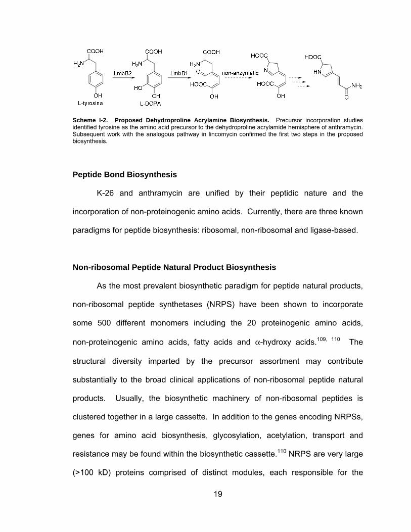
Scheme I-2. Proposed Dehydroproline Acrylamine Biosynthesis. Precursor incorporation studies identified tyrosine as the amino acid precursor to the dehydroproline acrylamide hemisphere of anthramycin. Subsequent work with the analogous pathway in lincomycin confirmed the first two steps in the proposed biosynthesis.
Peptide Bond Biosynthesis
K-26 and anthramycin are unified by their peptidic nature and the
incorporation of non-proteinogenic amino acids. Currently, there are three known
paradigms for peptide biosynthesis: ribosomal, non-ribosomal and ligase-based.
Non-ribosomal Peptide Natural Product Biosynthesis
As the most prevalent biosynthetic paradigm for peptide natural products,
non-ribosomal peptide synthetases (NRPS) have been shown to incorporate
some 500 different monomers including the 20 proteinogenic amino acids,
non-proteinogenic amino acids, fatty acids and α-hydroxy acids.109, 110 The
structural diversity imparted by the precursor assortment may contribute
substantially to the broad clinical applications of non-ribosomal peptide natural
products. Usually, the biosynthetic machinery of non-ribosomal peptides is
clustered together in a large cassette. In addition to the genes encoding NRPSs,
genes for amino acid biosynthesis, glycosylation, acetylation, transport and
resistance may be found within the biosynthetic cassette.110 NRPS are very large
(>100 kD) proteins comprised of distinct modules, each responsible for the

20
incorporation of one discrete monomer into the final peptide.111-114 Each module
minimally consists of at least three domains: an adenylation (A)-domain, a
thiolation (T)-domain and a condensation (C)-domain.110, 111
The A-domain selects the cognate amino acid and catalyzes the formation
of a reactive aminoacyl adenylate intermediate utilizing ATP and releasing
pyrophosphate.115 (Figure I-12) Using biochemical, structural and phylogenic
information, the amino acids of the A-domain involved in amino acid substrate
binding were revealed.116, 117 The crystal structure of PheA identified the amino
acids involved in L-phenylalanine binding. This 8- to 10- amino acid specificity-
conferring code can be extracted from primary sequence data of A-domains by
sequence alignment to PheA. It is possible to predict the substrate specificity of
a given A-domain based on identity of the specificity-conferring code to
previously characterized A-domains.118

21
Figure I-12. A-domain Reactivity and Specificity. Top: An NRPS A-domain catalyzes the formation of an aminoacyl adenylate by utilizing ATP and subsequently reacts the aminoacyl adenylate with the free thiol of the phosphopantetheinyl moiety of the T-domain. Middle: Based on the crystal structure of PheA, 10 amino acids were identified involved in substrate discrimination. These amino acids confer a substrate specificity code (Lysine 517 not shown). Bottom Left: Representative examples A-domains and their respective specificity code. Bottom Right: A schematic of substrate binding within the amino acid binding pocket of PheA.
After formation of the reactive aminoacyl adenylate intermediate, the
A-domain tethers the amino acid to a post-translationally modified T-domain by
thioesterification.119, 120 (Figure I-13) All T-domains must be post-translationally
modified prior to fulfilling their role as transport proteins during peptide bond
formation. Formation of the active holo-T-domain requires transfer of the
4’phosphopantetheinylate of coenzyme A (CoA) to an active site serine residue
of the apo-T-domain by a phosphopantetheinyl transferase (PPTase). The
activated aminoacyl adenylate reacts with the terminating sulfhydryl group of the

22
phosphopantetheinyl moiety resulting in an energy-rich thioester primed for
condensation into the growing peptide product.
Figure I-13. The Role of the T-Domain in NRPS Systems. After post-translational modification of an active site serine residue with phosphopantethiene, the A-domain activates the cognate amino acid substrate as the aminoacyl adenylate and catalyzes the thioesterification of the phosphopantetheinyl moiety with the aminoacyl adenylate to give the tethered amino acid primed for condensation into the peptide.
Peptide bond formation is catalyzed by the C-domain by promoting
nucleophilic attack of the amino group of the activated amino acid bound to the
downstream (of the C-domain) module onto the acyl group of the amino acid
tethered to the upstream module.121, 122 (Figure I-14) Based on the recent crystal
structure of a T-C didomain from tyrocidine biosynthesis, the C-domain has a
V-shaped architecture, possessing sites for both the upstream and downsteam
T-domain tethered amino acids.123, 124 Additional substrate specificity for the
nucleophilic amino acid substrate is imparted within the C-domain by the

23
structure of the respective binding pocket preventing incorporation of an incorrect
amino acid into the peptide.
Figure I-14. C-Domain Reaction Mechanism. Two T-domains of neighboring modules are loaded with their cognate amino acid. The electrophile amino acid enters the acceptor site (1) and the nucleophile amino acid enters the donor site (2) of the C-domain. After peptide bond formation, the resulting dipeptide can now act as the electrophile in the active site of the subsequent C-domain.
In most NRPS, the peptide is released by the activity of a thioesterase
(Te)-domain.125, 126 The growing peptide chain is handed from one module to the
next until it reaches the last T-domain on the final module. Release of the final
peptide is catalyzed by a two-step process through an acyl-O-Te-enzyme
intermediate that is attacked by either an internal nucleophile on the peptide or
water, resulting in either a cyclic or linear peptide, respectively.127, 128
Alternatively, a reductase domain can release the peptide with a terminal
aldehyde or alcohol.
Additional structural diversity is incorporated into non-ribosomal peptides
by tailoring domains.129 These domains fall into two categories: integral parts of
the NRPS acting in cis on the growing peptide or distinct enzymes acting in trans.
These domains include cyclization (Cy)-domains, which form thiazoline and
oxazoline rings from cysteine, serine and threonine residues; oxidation
(Ox)-domains, responsible for oxidation of the thiazoline and oxadoline rings to

24
thiazoles and oxazoles, respectively; epimerization (E)-domains epimerize an L
amino acid to a D amino acid and methyltransferase (MT)-domains insert a
methyl group on an amino acid substrate using S-adenosyl methionine as the
methyl donor.
Initial investigations into the biosynthesis of non-ribosomal peptides
identified a direct link between the order of the modules of the NRPS, including
the domains within those modules, and the incorporation of the amino acid into
the growing peptide.112, 130-132 This hierarchy of reactions was coined the
co-linearity rule. (Figure I-15) For example, the cyclic lipopeptide surfactin
follows this biosynthetic route.133 A total of 24 domains are organized in seven
modules over three NRPS that act linearly, starting at module 1 and ending at
module 7. However, as more gene clusters were identified, deviations from the
co-linearity rule were identified. Two types of deviations are iterative, exemplified
by gramicidin S, and non-linear, exemplified by vibriobactin.112, 134 In gramicidin
S biosynthesis, a pentapeptide monomer is formed and stalled on the final
module.127, 135 The regenerated NRPS then engages in a second round of
synthesis and is released by a head-to-tail cyclization to give the final product. In
non-linear biosynthesis of an NRPS, the domains are arranged non-linearly,
often missing expected domains, repeat use of single domains or include inactive
domains.112
25
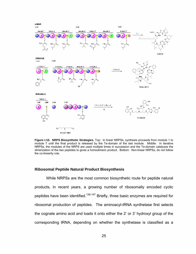
Figure I-15. NRPS Biosynthetic Strategies. Top: In linear NRPSs, synthesis proceeds from module 1 to module 7 until the final product is released by the Te-domain of the last module. Middle: In iterative NRPSs, the modules of the NRPS are used multiple times in succession and the Te-domain catalyzes the dimerization of the two peptides to gives a homodimeric product. Bottom: Non-linear NRPSs, do not follow the co-linearity rule.
Ribosomal Peptide Natural Product Biosynthesis
While NRPSs are the most common biosynthetic route for peptide natural
products, in recent years, a growing number of ribosomally encoded cyclic
peptides have been identified.136-147 Briefly, three basic enzymes are required for
ribosomal production of peptides. The aminoacyl-tRNA synthetase first selects
the cognate amino acid and loads it onto either the 2’ or 3’ hydroxyl group of the
corresponding tRNA, depending on whether the synthetase is classified as a

26
Class I or II, respectively.148, 149 The ribosome is the catalytic machine
responsible for generating the amino acid sequence of a peptide, based on the
sequence of the bound mRNA.150 The ribosome selects the correct aminoacyl-
tRNA, with the help of elongation factor (EF)-Tu. The aminoacyl-tRNA, EF-Tu
and GTP enter the acceptor (A)-site of the ribosome. The large ribosomal
subunit stimulates hydrolysis of GTP committing the aminoacyl-tRNA to peptide
bond formation, releasing EF-Tu. The amino acid-tRNA complex is bound via
codon-anticodon recognition to the ribosome. Peptide bond formation occurs
through nucleophilic attack of the amino group of the aminoacyl-tRNA in the
A-site on the carbonyl of the aminoacyl-tRNA, with the attached peptide, in the
peptidyl (P)-site. The tRNA with the attached polypeptide is now pushed from
the A-site to the P-site, while the unloaded tRNA in the P-site shifts to the exit
(E)-site. Subsequent cycles elongate the polypeptide.
Unlike non-ribosomal peptides, ribosomal peptides are limited by the 20
proteinogenic amino acids. However, extensive posttranslational modification of
the precursor peptide imparts structural diversity to the peptide scaffold. For a
majority of ribosomal peptide natural products, a series of genes encode a
precursor peptide containing an N-terminal leader peptide and a C-terminal core
peptide that is processed into the final compound.151 (Figure I-16) Variably, the
N-terminus of the leader peptide may be appended with a signal peptide
governing subcellular localization and/or a recognition sequence may be
attached to the C-terminal end of the core peptide. After post-translational
modification, the precursor peptide is usually cleaved by at least one protease

27
freeing the core peptide. The gene cluster containing the precursor peptide also
encompasses genes encoding modifying enzymes, resistance and secretion.
Figure I-16. General Ribosomal Peptide Biosynthesis. The precursor peptide usually consists of an N-terminal leader peptide and a C-terminal core peptide. A signal peptide and/or recognition sequence may be appended to the main peptide structure. The precursor peptide is ribosomally synthesized and post-translationally modified to the active compounds. Used with permission151
Several roles have been proposed for the N-terminal leader peptide,
though none have been completely substantiated experimentally.151 In fact, the
role of the leader peptide may differ between families of ribosomal peptides or
even within an individual class. Most often, it is proposed that the leader peptide
plays a role in signaling secretion of the core peptide; however, most of the
leader peptides in these gene clusters display no homology to proteins involved
in bacterial secretion pathways. Additional proposed roles for the leader peptide

28
include acting as a recognition motif for post-translational modification enzymes
including perhaps acting as chaperones for enzymes assisting in folding of the
precursor peptide and stabilizing the peptide against degradation and premature
activation of its biological function. Supporting the role of the leader peptide as a
preventative to premature activation, the protease involved in cleaving the
precursor peptide resides on the outside of the producing cell.
Despite the variability of the precursor peptides, both between and within
classes, they remain substrates for post-translational modification. Both the side
chains and main peptide chain can undergo post-translational modifications.
Herein, I provide an overview of several families of ribosomal peptide natural
products produced by bacteria and the modifications of those peptides as
representative examples of their biosynthesis. These four families were chosen
based on their prevalence in nature and the inclusion of post-translational
modifications within their structure.
Lantibiotics
Lantibiotics are divided into two classes based on their leader peptide
sequences and biosynthetic enzymes. Class I lantibiotics have leader peptides
approximately 25 amino acids in length, rich in aspartic acid residues.152 In this
class, the leader peptide greatly reduces the antimicrobial activities of the core
peptide and appears to be involved in the recruitment of enzymes for peptide
modification and excretion.153, 154 Class II lantibiotics have leader peptides rich in
aspartate and glutamate residues, contain an ELXXBX motif (where B = V, L or I)

29
and usually end in two glycine residues.155 The double glycine motif is required
for proteolytic cleavage of the precursor peptide, while the leader peptide itself
may be involved in secretion and guidance of additional biosynthetic enzymes
involved in producing the final natural products.156-158 A total of 15
post-translational modifications have been documented within the lantibiotic
family.159 (Figure I-17) Structurally, the lantibiotic family of ribosomal peptide
natural products is distinguished by thioether crosslinks introduced by the
dehydration of serine and threonine to dehydroalanine and dehydrobutyrine,
respectively, followed by addition of the thiol of cysteine.
Figure I-17. Post-translational Modifications Found in Lantibiotics. A total of 15 post-translational modifications have been identified in the lantibiotics family. This family of ribosomal peptide natural products is distinguished by the incorporation of a variety of thioether crosslinks.
Microcins
Microcins are small, structurally diverse peptides produced by
enterobacteria.160 As opposed to the lantibiotics, only a few different types of

30
post-translational modifications have been reported for microcins.161-164 One
class of peptides within this family is exemplified by microcin B17, which contains
oxazole and thiazole heterocycles.161 (Figure I-18) The formation of these
heterocycles requires three proteins including a cyclodehydratase, which
generates the oxazoline and thiazoline structures from Gly-Ser and Gly-Cys
sequences; a flavin-dependent dehydrogenase, responsible for oxidizing these
intermediates to the oxazole and thiazole structures and a scaffolding protein,
required for catalysis.165 The leader peptide for these types of microcins has
been shown to interact with the scaffolding proteins and reduce the antimicrobial
activity of the core peptide.166, 167
Figure I-18. Microcin Ribosomal Peptides. Two classes found within the Microcin family of peptide natural products are the thiazole and oxazole containing peptides and the lasso peptides. Used with permission.151
Another class of peptides within the microcin family is the lasso peptides,
exemplified by microcin J25.162-164 (Figure I-18) In these peptides, the
distinguishing feature is a lactam bond. The N-terminal amino group reacts with
the carboxylate side chain of an aspartate or glutamate residue at position 8,
essentially trapping the C-terminus of the peptide chain threaded through the
ring. The leader peptide of microcin J25 has been shown to be required for the

31
recruitment of two enzymes involved in post-translational modification of the core
peptide.146, 168 One protein catalyzes the adenylation of the aspartate/glutamate
carboxylate at position 8, while a second cleaves off the leader peptide and
installs the new lactam bond.
Cyanobactins
Produced by diverse cyanobacteria, over 100 unique cyclic peptides
belong to the cyanobactin family, including the patellamides.146 (Figure I-19)
Members of this family of ribosomal peptide natural products share similar
structural features, like oxazoles and thiazoles, as well as, similar biosynthetic
origins. Two enzymes, a cyclodehydratase and a bifunctional enzyme with an
oxidase domain, are required for the cyclization of serine, threonine and
cysteine.169 Proteolytic cleavage of the precursor peptide releases the core
peptide which is then cyclized to give the final product.

32
NH
HN
O
NH
HN
O
NH
HN
O
OOH
OSH
OOH
NH
O
HN
SH
AYDGVEPS
O
NH
O
HN
OH
NH
O
OHN
O
SH
NH
O
HN
O
OH
NH
HN
O
O
SH
AYDGE
GLEASLeaderPeptide
NH
N
O
NH
N
O
NH
N
O
O S O
NH
S
NAYDGVEPS
O
NH
O
NNH
S
O
N
O
NH
O
N
O
NH
N
S
O
AYDGE
GLEASLeaderPeptide
NH
N
O
NH
N
O
NH
N
O
O S O
NH
S
NAYDGVEPS
O
H2N
O
NNH
S
O
N
O
NH
O
N
O
NH
N
S
O
AYDGEGLEASLeader
Peptide
H2NN
O
NH
N
O
NH
N
O
O S O
NH
S
NAYDGVEPS
O
H2N
O
NNH
S
O
N
O
NH
O
N
O
NH
N
S
O
AYDGE
N
NH N
O
O
S
NHHN
O
O
ONN
S
HN
O
N
NH N
O
O
S
NHHN
O
O
ONN
S
HN
O
PatD/PatG
PatA
PatA
PatG PatG
Patellamide CProchloron didemni
Patellamide AProchloron didemni
Figure I-19. Proposed Biosynthesis of Patellamides A and C. Cyanbactins differ from other types of ribosomal peptide natural products by incorporating two core peptides in the precursor peptide.
Interestingly, most cyanobactin precursor peptides consist of a leader
peptide, whose activities have not been substantiated in vitro, and two highly
variable core peptides flanked between recognition sequences responsible for
directing post-translational modification of the core peptides.146 The N-terminal
region flanking the first core peptide typically contains a conserved
G(L/V)E(A/P)S sequence, while the C-terminal region contains a conserved
AYDG(E) sequence, which act as recognition sequences for the excision and
cyclization of the core peptides.169

33
Thiopeptides
Thiopeptides are another large class of ribosomally encoded natural
products containing a string of thiazoles/thiazolines which are conjoined by a six-
member heterocyclic ring.136, 139, 142, 144, 170 (Figure I-20) This heterocycle can be
a pyridine, hydroxypyridine or a dehydropiperadine. Common modifications
within the thiopeptide family include dehydrations of threonine and serine,
thiazole and thiazoline formation and proposed cyclization of two
dehydroalanines to form the six-membered ring structure.
Figure I-20. Biosynthesis of Thiostrepton. Thiostrepton is initially encoded as the precursor peptide TsrH. Subsequent post-translational modifications install the heterocycle rings and dehydrated amino acid. This conformationally restrained peptide is further modified to give the characteristic central hexacycle of thiopeptides. Although represented in a particular order, the timing of the biosynthetic steps is not known. Used with permission.151

34
Ligase-based Peptide Biosynthesis
While non-ribsomal and ribosomal natural products make up the majority
of peptide natural products, increasing examples of ligase-based biosyntheses
have been reported in the literature, including non-ribosomal independent
pathways, aSerRS homologues amino acid:[carrier protein] ligases and the
recently identified ATP-independent strategy.171-175 The biosynthesis of the
dapdiamide antibiotics provides one example of the role of ATP-dependent
ligases in peptide natural product biosynthesis.172 (Figure I-21) ATP-dependent
ligases are soluble enzymes that couple amino acid monomers to the growing
peptide chain. There are two types of ATP-dependent ligases involved in
dapdiamide biosynthesis. DdaG activates fumarate, as the aminoacyl adenylate
using ATP and releasing pyrophosphate, and forms the first peptide bond
between fumarate and 2,3-diaminopropionate (DAP) to create the
Nβ-fumaroyl-DAP dipeptide intermediate. Activity similar to DdaG occurs during
the ATP-dependent biosynthesis of the siderophore ferrioxamine E by the non-
ribosomal synthetase independent pathway.173, 174 After amination of
Nβ-fumaroyl-DAP to Nβ-fumaramoyl-DAP, DdaF activates the dipeptide as the
acyl phosphate by using ATP and releasing ADP, similar to the ligases involved
in glutathione and bacterial cell wall pentapeptide biosynthesis.176, 177

35
Figure I-21. Dapdiamide Biosynthesis. The biosynthesis of the dapdiamides requires two ATP-dependent ligases, DdaG, which employs an aminoacyl adenylate and DdaF, which utilizes an aminoacylphosphate, to activate amino acids for peptide bond formation.
More recently, two other biosynthetic pathways for peptides have been
identified. Homologues to serine tRNA synthetase (aSerRS) have been
identified in a variety of bacterial species including Alpha- and
Betaproteobacteria, Clostridium, Bacillus and Streptomyces species.175 While
showing homology to serine tRNA synthetases, these proteins, exemplified by
two homologues from Bradyrhizobium japonicum (BII0957 and BII6282) and one
homologue from Agrobacterium tumefaciens (Atu2573), activate amino acids in
the same manner as tRNA synthetases, but do not load amino acids onto tRNA.
As the genes for these proteins are often near genes for small hypothetical
proteins with a 4’phosphopantetheine attachment site, it was proposed that

36
aSerRS may be capable of loading these T-domain-like proteins called carrier
proteins (CP). The three aSerRS (BII0957, BII6282 and Atu2573) were shown to
tether alanine or glycine to these neighboring CPs; however, the peptide product
of this route of biosynthesis has not been identified.
Unlike the other ligase-based biosynthetic pathways, another recently
identified route for peptide bond formation is ATP-independent.171
Capuramycins, produced by Streptomyces sp SANK 62799 and Streptomyces
griseus SANK 60196, are nucleotide containing peptide natural products. (Figure
I-22) The structure of the capuramycins consists of three distinct portions: a
5’-C-carbamoyl-uridine, an unsaturated hexuronic acid and an aminocaprolactam
ring. The gene cluster for capuramycin A-503083 (A and B) contains two NRPS
genes believed to be involved in the biosynthesis of the L-aminocaprolactam ring,
while, two additional genes are required for the installation of the
aminocaprolactam ring into the final capuramycin structure. CapS activates the
carboxylic acid of the hexuronic acid-carbamoyl uridine complex by S-adenosyl
methionine-dependent methylation. CapW is responsible for the condensation of
the hexuronic acid-carbamoyl uridine complex with the amino caprolactam ring.
Figure I-22. ATP-independent Peptide Bond Formation. The biosynthetic pathway of the capuramycins includes a unique method for peptide bond formation. CapS installs a methyl on the free carboxylate of the hexuronic acid-carbamoyl uridine complex. This activated complex is then condensed with L-aminocaprolactam by CapW, releasing methanol and creating the final peptide.

37
Biosynthetic Hypotheses for K-26 and Anthramycin
Based on the chemical structures, we propose both K-26 and anthramycin
are produced by NRPS:
K-26
The intermediacy of AHEP suggests that K-26 may be biosynthesized by
the NRPS paradigm. In this case, L-isoleucine, L-tyrosine and AHEP would be
activated by individual A-domains, loaded onto the corresponding T-domains and
peptide bond formation would be catalyzed by the C-domain resulting in the
formation of des-acetyl-K-26 (dK-26). (Figure I-23) Then either on the final
T-domain or after release of the peptide by the Te-domain, N-acetylation would
occur resulting in formation of K-26. Our proposed gene cluster would consist of
an N-acetyltransferase, NRPS with a domain string of A-T-C-A-T-C-A-T-Te and
hypothetical proteins of unknown function responsible for AHEP biosynthesis.

38
Figure I-23. Proposed Peptide Bond Formation in K-26. Based on the non-proteinogenic nature of AHEP, the most logical biosynthetic route for K-26 formation is via the NRPS paradigm. In this case, the discrete precursors would be activated and loaded onto individual T-domains by the corresponding A-domains. Peptide bond formation would be catalyzed by the C-domains and the peptide would be released by a thioesterase. N-acetylation could occur prior to peptide bond formation or afterward as depicted.
Anthramycin
Based on the non-proteinogenic nature of the two amino acids proposed
in the formation anthramycin, evidenced by the aforementioned isotopic
incorporation experiments, we propose an NRPS based biosynthetic hypothesis
with a domain string of A-T-C-A-T-Re. (Figure I-24) The first A-domain of the
NRPS would activate either 3-hydroxyanthranilic acid (HA) or MHA and the

39
second A-domain would activate the dehydroproline acrylamide hemisphere for
loading on to their respective T-domains. A specialized reductase domain would
provide the final cyclization to form the final benzodiazepine ring of anthramycin.
Figure I-24. Proposed Peptide Bond Formation in Anthramycin. Based on the non-proteinogenic nature of the two proposed precursors of anthramycin, peptide bond formation by an NRPS is the most logical hypothesis. In this case, the discrete amino acid precursors would be activated by individual A-domains and loaded onto the corresponding T-domains. The lone C-domain would catalyze peptide bond formation and a unique reductase domain would release anthramycin from the megasynthetase by installing the benzodiazepine ring.
Dissertation Goals
Within this dissertation the first biosynthetic investigations of two natural
product classes are described.
Chapter II describes the development of the mass-based pyrophosphate
exchange assay for characterization of the proteins involved in the biosynthesis
of K-26 and anthramycin.
Chapter III describes the biosynthetic investigations of K-26, including our
attempts at isolating the peptide bond forming machinery. This work is currently
the only research being performed investigating this unique phosphonate
containing natural product.

40
Chapter IV describes the biosynthetic investigation of anthramycin,
including the first biochemical characterization of the enzymes involved in
anthramycin biosynthesis. The work described herein is the first identification and
characterization of a PBD gene cluster. It is important to note that after
publication of our investigations into the biosynthesis of anthramycin, biosynthetic
investigations of both sibiromycin and tomaymycin were published by the
Gerratana laboratory.
Chapter V describes the possible future biosynthetic investigations of
K-26 and anthramycin.
41
REFERENCES
1. Newman, D. J. and Cragg, G. M. (2007) Natural products as sources of new drugs over the last 25 years. Journal of Natural Products, 70, 461-477.
2. Newman, D. J. and Cragg, G. M. (2009) Microbial antitumor drugs: natural
products of microbial origin as anticancer agents. Current Opinion in Investigational Drugs, 10, 1280-1296.
3. Koehn, F. E. and Carter, G. T. (2005) The evolving role of natural products
in drug discovery. Nature Reviews Drug Discovery, 4, 206-220. 4. Newman, D. and Cragg, G. (2009) Natural products in medicinal
chemistry. Bioorganic and Medicinal Chemistry, 17, 2120. 5. Cragg, G. M., Grothaus, P. G. and Newman, D. J. (2009) Impact of natural
products on developing new anti-cancer agents. Chemical Reviews, 109, 3012-3043.
6. Gross, H. (2010) Genomic mining - a concept for the discovery of new
bioactive natural products. Planta Medica, 76, 1169-1169. 7. Bode, H. B. and Muller, R. (2005) The impact of bacterial genomics on
natural product research. Angewandte Chemie-International Edition, 44, 6828-6846.
8. Pace, N. R. (1997) A molecular view of microbial diversity and the
biosphere. Science, 276, 734-740. 9. Mira, A., Martin-Cuadrado, A. B., D'Auria, G. and Rodriguez-Valera, F.
(2010) The bacterial pan-genome: a new paradigm in microbiology. International Microbiology, 13, 45-57.
10. Ikeda, H., Ishikawa, J., Hanamoto, A., Shinose, M., Kikuchi, H., Shiba, T.,
Sakaki, Y., Hattori, M. and Omura, S. (2003) Complete genome sequence and comparative analysis of the industrial microorganism Streptomyces avermitilis. Nature Biotechnology, 21, 526-531.

42
11. Omura, S., Ikeda, H., Ishikawa, J., Hanamoto, A., Takahashi, C., Shinose, M., Takahashi, Y., Horikawa, H., Nakazawa, H., Osonoe, T., Kikuchi, H., Shiba, T., Sakaki, Y. and Hattori, M. (2001) Genome sequence of an industrial microorganism Streptomyces avermitilis: Deducing the ability of producing secondary metabolites. Proceedings of the National Academy of Sciences of the United States of America, 98, 12215-12220.
12. Bentley, S. D., Chater, K. F., Cerdeno-Tarraga, A. M., Challis, G. L.,
Thomson, N. R., James, K. D., Harris, D. E., Quail, M. A., Kieser, H., Harper, D., Bateman, A., Brown, S., Chandra, G., Chen, C. W., Collins, M., Cronin, A., Fraser, A., Goble, A., Hidalgo, J., Hornsby, T., Howarth, S., Huang, C. H., Kieser, T., Larke, L., Murphy, L., Oliver, K., O'Neil, S., Rabbinowitsch, E., Rajandream, M. A., Rutherford, K., Rutter, S., Seeger, K., Saunders, D., Sharp, S., Squares, R., Squares, S., Taylor, K., Warren, T., Wietzorrek, A., Woodward, J., Barrell, B. G., Parkhill, J. and Hopwood, D. A. (2002) Complete genome sequence of the model actinomycete Streptomyces coelicolor A3(2). Nature, 417, 141-147.
13. Keasling, J. D. (2010) Manufacturing Molecules Through Metabolic
Engineering. Science, 330, 1355-1358. 14. Yamato, M., Koguchi, T., Okachi, R., Yamada, K., Nakayama, K., Kase,
H., Karasawa, A. and Shuto, K. (1986) K-26, a novel inhibitor of angiotensin I converting enzyme produced by an actinomycete K-26. Journal of Antibiotics (Tokyo), 39, 44-52.
15. Gerratana, B. (2010) Biosynthesis, synthesis, and biological activities of
pyrrolobenzodiazepines. Medicinal Research Reviews. 16. Kafarski, P. and Lejczak, B. (2001) Aminophosphonic acids of potential
medical importance. Current Medicinal Chemistry - Anticancer Agents, 1, 301-312.
17. Wanke, C. and Amrhein, N. (1993) Evidence that the reaction of the UDP-
N-acetylglucosamine 1-carboxyvinyltransferase proceeds through the O-phosphothioketal of pyruvic acid bound to Cys115 of the enzyme. European Journal of Biochemistry, 218, 861-870.
18. Horiguchi, M. and Kandatsu, M. (1959) Isolation of 2-aminoethane
phosphonic acid from rumen protozoa. Nature, 184(Suppl 12), 901-902. 19. Hilderbrand, R. L. The Role of phosphonates in living systems, CRC
Press: Boca Raton, Fla., 1983. 20. Moschidis, M. C. (1984) Phosphonolipids. Progress in Lipid Research, 23,
223-246.

43
21. Metcalf, W. W. and van der Donk, W. A. (2009) Biosynthesis of
Phosphonic and Phosphinic Acid Natural Products. Annual Review of Biochemistry, 78, 65-94.
22. Ntai, I., Manier, M. L., Hachey, D. L. and Bachmann, B. O. (2005)
Biosynthetic origins of C-P bond containing tripeptide K-26. Organic Letters, 7, 2763-2765.
23. Koguchi, T., Yamada, K., Yamato, M., Okachi, R., Nakayama, K. and
Kase, H. (1986) K-4, a novel inhibitor of angiotensin I converting enzyme produced by Actinomadura spiculosospora. Journal of Antibiotics (Tokyo), 39, 364-371.
24. Kido, Y., Hamakado, T., Anno, M., Miyagawa, E., Motoki, Y., Wakamiya,
T. and Shiba, T. (1984) Isolation and characterization of I5B2, a new phosphorus containing inhibitor of angiotensin I converting enzyme produced by Actinomadura sp. Journal of Antibiotics (Tokyo), 37, 965-969.
25. Acharya, K. R., Sturrock, E. D., Riordan, J. F. and Ehlers, M. R. (2003)
Ace revisited: a new target for structure-based drug design. Nature Reviews Drug Discovery, 2, 891-902.
26. Ntai, I. and Bachmann, B. O. (2008) Identification of ACE pharmacophore
in the phosphonopeptide metabolite K-26. Bioorganic & Medicinal Chemistry Letters, 18, 3068-3071.
27. Riviere, G., Michaud, A., Corradi, H. R., Sturrock, E. D., Acharya, K. R.,
Cogez, V., Bohin, J. P., Vieau, D. and Corvol, P. (2007) Characterization of the first angiotensin-converting like enzyme in bacteria: Ancestor ACE is already active. Gene, 399, 81-90.
28. Appleford, P. J., Griffiths, M., Yao, S. Y. M., Ng, A. M. L., Chomey, E. G.,
Isaac, R. E., Coates, D., Hope, I. A., Cass, C. E., Young, J. D. and Baldwin, S. A. (2004) Functional redundancy of two nucleoside transporters of the ENT family (CeENT1, CeENT2) required for development of Caenorhabditis elegans. Molecular Membrane Biology, 21, 247-259.
29. Coates, D., Siviter, R. and Isaac, R. E. (2000) Exploring the
Caenorhabditis elegans and Drosophila melanogaster genomes to understand neuropeptide and peptidase function. Biochemical Society Transactions, 28, 464-469.

44
30. Coates, K. J., Haines, H., Bobbitt, J., Kondekas, N. and Isaac, J. (2000) Investigation of the pork supply chain into Singapore. Asian-Australasian Journal of Animal Sciences, 13, 87-90.
31. Cornel, M. J., Williams, T. A., Lamango, N. S., Coates, D., Corvol, P.,
Soubrier, F., Hoheisel, J., Lehrach, H. and Isaac, R. E. (1995) Cloning and Expression of an Evolutionary Conserved Single-Domain Angiotensin-Converting Enzyme from Drosophila-Melanogaster. Journal of Biological Chemistry, 270, 13613-13619.
32. Ekbote, U., Coates, D. and Isaac, R. E. (1999) A mosquito (Anopheles
stephensi) angiotensin I-converting enzyme (ACE) is induced by a blood meal and accumulates in the developing ovary. Febs Letters, 455, 219-222.
33. Houard, X., Williams, T. A., Michaud, A., Dani, P., Isaac, R. E., Shirras, A.
D., Coates, D. and Corvol, P. (1998) The Drosophila melanogaster-related angiotensin-I-converting enzymes Acer and Ance - Distinct enzymic characteristics and alternative expression during pupal development. European Journal of Biochemistry, 257, 599-606.
34. Isaac, R. E., Ekbote, U., Coates, D. and Shirras, A. D. (1999) Insect
angiotensin-converting enzyme - A processing enzyme with broad substrate specificity and a role in reproduction. Neuropeptides: Structure and Function in Biology and Behavior, 897, 342-347.
35. Isaac, R. E., Macgregor, D. and Coates, D. (1996) Metabolism and
inactivation of neurotransmitters in nematodes. Parasitology, 113, S157-S173.
36. Isaac, R. E., Michaud, A., Keen, J. N., Williams, T. A., Coates, D., Wetsel,
W. C. and Corvol, P. (1999) Hydrolysis by somatic angiotensin-I converting enzyme of basic dipeptides from a cholecystokinin gastrin and a LH-RH peptide extended at the C-terminus with Gly-Arg/Lys-Arg, but not from diarginyl insulin. European Journal of Biochemistry, 262, 569-574.
37. Isaac, R. E., Schoofs, L., Williams, T. A., Corvol, P., Veelaert, D., Sajid, M.
and Coates, D. (1998) Toward a role for angiotensin-converting enzyme in insects. Trends in Comparative Endocrinology and Neurobiology, 839, 288-292.
38. Isaac, R. E., Schoofs, L., Williams, T. A., Veelaert, D., Sajid, M., Corvol, P.
and Coates, D. (1998) A novel peptide-processing activity of insect peptidyl-dipeptidase A (angiotensin I-converting enzyme): the hydrolysis of lysyl-arginine and arginyl-arginine from the C-terminus of an insect prohormone peptide. Biochemical Journal, 330, 61-65.

45
39. Isaac, R. E., Siviter, R. J., Stancombe, P., Coates, D. and Shirras, A. D.
(2000) Conserved roles for peptidases in the processing of invertebrate neuropeptides. Biochemical Society Transactions, 28, 460-464.
40. Isaac, R. E., Williams, T. A., Sajid, M., Corvol, P. and Coates, D. (1997)
Cleavage of arginyl-arginine and lysyl-arginine from the C-terminus of pro-hormone peptides by human germinal angiotensin I-converting enzyme (ACE) and the C-domain of human somatic ACE. Biochemical Journal, 328, 587-591.
41. Laurent, V., Brooks, D. R., Coates, D. and Isaac, R. E. (2001) Functional
expression and characterization of the cytoplasmic aminopeptidase P of Caenorhabditis elegans. European Journal of Biochemistry, 268, 5430-5438.
42. Lee, A. J., Huntley, J., Van den Broek, A., Coates, D. and Isaac, R. E.
(2002) Expression and characterisation of a Psoroptes ovis glutathione S-transferase. Veterinary Parasitology, 105, 49-63.
43. Lee, A. J., Isaac, R. E. and Coates, D. (1999) The construction of a cDNA
expression library for the sheep scab mite Psoroptes ovis. Veterinary Parasitology, 83, 241-252.
44. Meyer, D. J., Muimo, R., Thomas, M., Coates, D. and Isaac, R. E. (1996)
Purification and characterization of prostaglandin-H E-isomerase, a sigma-class glutathione S-transferase, from Ascaridia galli. Biochemical Journal, 313, 223-227.
45. Siviter, R. J., Coast, G. M., Winther, A. M. E., Nachman, R. J., Taylor, C.
A. M., Shirras, A. D., Coates, D., Isaac, R. E. and Nassel, D. R. (2000) Expression and functional characterization of a Drosophila neuropeptide precursor with homology to mammalian preprotachykinin A. Journal of Biological Chemistry, 275, 23273-23280.
46. Turner, A. J., Isaac, R. E. and Coates, D. (2001) The neprilysin (NEP)
family of zinc metalloendopeptidases: genomics and function. Bioessays, 23, 261-269.
47. Akif, M., Ntai, I., Sturrock, E. D., Isaac, R. E., Bachmann, B. O. and
Acharya, K. R. (2010) Crystal structure of a phosphonotripeptide K-26 in complex with angiotensin converting enzyme homologue (AnCE) from Drosophila melanogaster. Biochemical and Biophysical Research Communications, 398, 532-536.
46
48. Seto, H. and Kuzuyama, T. (1999) Bioactive natural products with carbon-phosphorus bonds and their biosynthesis. Natural Products Reports, 16, 589-596.
49. Warren, W. A. (1968) Biosynthesis of phosphonic acids in Tetrahymena.
Biochimica Et Biophysica Acta, 156, 340-346. 50. Hidaka, T., Goda, M., Kuzuyama, T., Takei, N., Hidaka, M. and Seto, H.
(1995) Cloning and Nucleotide-Sequence of Fosfomycin Biosynthetic Genes of Streptomyces-Wedmorensis. Molecular & General Genetics, 249, 274-280.
51. Thompson, C. J. and Seto, H. (1995) Bialaphos. Biotechnology, 28, 197-
222. 52. Schwartz, D., Berger, S., Heinzelmann, E., Muschko, K., Welzel, K. and
Wohlleben, W. (2004) Biosynthetic gene cluster of the herbicide phosphinothricin tripeptide from Streptomyces viridochromogenes Tu494. Applied and Environmental Microbiology, 70, 7093-7102.
53. Blodgett, J. A., Thomas, P. M., Li, G., Velasquez, J. E., van der Donk, W.
A., Kelleher, N. L. and Metcalf, W. W. (2007) Unusual transformations in the biosynthesis of the antibiotic phosphinothricin tripeptide. Nature Chemical Biology, 3, 480-485.
54. Seidel, H. M., Freeman, S., Seto, H. and Knowles, J. R. (1988)
Phosphonate biosynthesis: isolation of the enzyme responsible for the formation of a carbon-phosphorus bond. Nature, 335, 457-458.
55. Bowman, E. D., McQueney, M. S., Scholten, J. D. and Dunaway-Mariano,
D. (1990) Purification and characterization of the Tetrahymena pyriformis P-C bond forming enzyme phosphoenolpyruvate phosphomutase. Biochemistry, 29, 7059-7063.
56. Hidaka, T., Seto, H. and Imai, S. (1989) Biosynthetic Mechanism of C-P
Bond Formation - Isolation of Carboxyphosphonoenolpyruvate and Its Conversion to Phosphinopyruvate. Journal of the American Chemical Society, 111, 8012-8013.
57. Bowman, E., Mcqueney, M., Barry, R. J. and Dunaway-mariano, D. (1988)
Catalysis and Thermodynamics of the Phosphoenolpyruvate Phosphonopyruvate Rearrangement - Entry into the Phosphonate Class of Naturally-Occurring Organo-Phosphorus Compounds. Journal of the American Chemical Society, 110, 5575-5576.

47
58. Freeman, S., Seidel, H. M., Schwalbe, C. H. and Knowles, J. R. (1989) Phosphonate Biosynthesis - the Stereochemical Course of Phosphoenolpyruvate Phosphomutase. Journal of the American Chemical Society, 111, 9233-9234.
59. Mcqueney, M. S., Lee, S. L., Swartz, W. H., Ammon, H. L., Mariano, P. S.
and Dunaway-mariano, D. (1991) Evidence for an Intramolecular, Stepwise Reaction Pathway for Pep Phosphomutase Catalyzed P-C Bond Formation. Journal of Organic Chemistry, 56, 7121-7130.
60. Ntai, I., Phelan, V. V. and Bachmann, B. O. (2006) Phosphonopeptide K-
26 biosynthetic intermediates in Astrosporangium hypotensionis. Chemical Communications, 4518-4520.
61. Puvvada, M. S., Forrow, S. A., Hartley, J. A., Stephenson, P., Gibson, I.,
Jenkins, T. C. and Thurston, D. E. (1997) Inhibition of bacteriophage T7 RNA polymerase in vitro transcription by DNA-binding pyrrolo[2,1-c][1,4]benzodiazepines. Biochemistry, 36, 2478-2484.
62. Cummings, M. D., Schubert, C., Parks, D. J., Calvo, R. R., LaFrance, L.
V., Lattanze, J., Milkiewicz, K. L. and Lu, T. (2006) Substituted 1,4-benzodiazepine-2,5-diones as alpha-helix mimetic antagonists of the HDM2-p53 protein-protein interaction. Chemistry and Biolology of Drug Design, 67, 201-205.
63. Demartino, G., Massa, S., Corelli, F., Pantaleoni, G., Fanini, D. and
Palumbo, G. (1983) Cns Agents - Neuropsychopharmacological Effects of 5h-Pyrrolo[2,1-C] [1,4]Benzodiazepine Derivatives. European Journal of Medicinal Chemistry, 18, 347-350.
64. Joseph, C. G., Wilson, K. R., Wood, M. S., Sorenson, N. B., Phan, D. V.,
Xiang, Z., Witek, R. M. and Haskell-Luevano, C. (2008) The 1,4-benzodiazepine-2,5-dione small molecule template results in melanocortin receptor agonists with nanomolar potencies. Journal of Medicinal Chemistry, 51, 1423-1431.
65. Karp, G. M., Manfredi, M. C., Guaciaro, M. A., Ortlip, C. L., Marc, P. and
Szamosi, I. T. (1997) Synthesis and herbicidal activity of 1H-1,4-benzodiazepine-2,5-diones. Journal of Agricultural and Food Chemistry, 45, 493-500.
66. Wright, W. B., Jr., Brabander, H. J., Greenblatt, E. N., Day, I. P. and
Hardy, R. A., Jr. (1978) Derivatives of 1,2,3,11a-tetrahydro-5H-pyrrolo[2,1-c][1,4]benzodiazepine-5,11(10H)-dione as anxiolytic agents. Journal of Medicinal Chemistry, 21, 1087-1089.

48
67. Heguy, A., Cai, P., Meyn, P., Houck, D., Russo, S., Michitsch, R., Pearce, C., Katz, B., Bringmann, G., Feineis, D., Taylor, D. L. and Tyms, A. S. (1998) Isolation and characterization of the fungal metabolite 3-O-methylviridicatin as an inhibitor of tumour necrosis factor alpha-induced human immunodeficiency virus replication. Antiviral Agents and Chemotherapy, 9, 149-155.
68. Gourdeau, H., McAlpine, J. B., Ranger, M., Simard, B., Berger, F.,
Beaudry, F., Farnet, C. M. and Falardeau, P. (2008) Identification, characterization and potent antitumor activity of ECO-4601, a novel peripheral benzodiazepine receptor ligand. Cancer Chemotherary and Pharmacology, 61, 911-921.
69. McAlpine, J. B., Bachmann, B. O., Piraee, M., Tremblay, S., Alarco, A. M.,
Zazopoulos, E. and Farnet, C. M. (2005) Microbial Genomics as a guide to drug discovery and structural elucidation: ECO-02301, a novel antifungal agent, as an example. Journal of Natural Products, 68, 493-496.
70. Kumar, R. and Lown, J. W. (2003) Recent developments in novel
pyrrolo[2,1-c][1,4]benzodiazepine conjugates: synthesis and biological evaluation. Mini Reviews in Medicinal Chemistry, 3, 323-339.
71. Pepper, C. J., Hambly, R. M., Fegan, C. D., Delavault, P. and Thurston, D.
E. (2004) The novel sequence-specific DNA cross-linking agent SJG-136 (NSC 694501) has potent and selective in vitro cytotoxicity in human B-cell chronic lymphocytic leukemia cells with evidence of a p53-independent mechanism of cell kill. Cancer Research, 64, 6750-6755.
72. Thurston, D. E., Bose, D. S., Howard, P. W., Jenkins, T. C., Leoni, A.,
Baraldi, P. G., Guiotto, A., Cacciari, B., Kelland, L. R., Foloppe, M. P. and Rault, S. (1999) Effect of A-ring modifications on the DNA-binding behavior and cytotoxicity of pyrrolo[2,1-c][1,4]benzodiazepines. Journal of Medicinal Chemistry, 42, 1951-1964.
73. Petrusek, R. L., Anderson, G. L., Garner, T. F., Fannin, Q. L., Kaplan, D.
J., Zimmer, S. G. and Hurley, L. H. (1981) Pyrrol[1,4]benzodiazepine antibiotics. Proposed structures and characteristics of the in vitro deoxyribonucleic acid adducts of anthramycin, tomaymycin, sibiromycin, and neothramycins A and B. Biochemistry, 20, 1111-1119.
74. Petrusek, R. L., Uhlenhopp, E. L., Duteau, N. and Hurley, L. H. (1982)
Reaction of Anthramycin with DNA - Biological Consequences of DNA Damage in Normal and Xeroderma Pigmentosum Cell-Lines. Journal of Biological Chemistry, 257, 6207-6216.

49
75. Hurley, L. H. (1977) Pyrrolo(1,4)benzodiazepine antitumor antibiotics. Comparative aspects of anthramycin, tomaymycin and sibiromycin. Journal of Antibiotics (Tokyo), 30, 349-370.
76. Korman, S. and Tendler, M. D. (1965) Clinical investigation of cancer
chemotherapeutic agents for neoplastic disease. Journal of New Drugs, 5, 275-285.
77. Cargill, C., Bachmann, E. and Zbinden, G. (1974) Effects of daunomycin
and anthramycin on electrocardiogram and mitochondrial metabolism of the rat heart. Journal of the National Cancer Institute, 53, 481-486.
78. Lubawy, W. C., Dallam, R. A. and Hurley, L. H. (1980) Protection against
Anthramycin-Induced Toxicity in Mice by Coenzyme-Q10. Journal of the National Cancer Institute, 64, 105-109.
79. Kohn, K. W. and Spears, C. L. (1970) Reaction of Anthramycin with
Deoxyribonucleic Acid. Journal of Molecular Biology, 51, 551-&. 80. Kohn, K. W., Bono, V. H. and Kann, H. E. (1968) Anthramycin a New Type
of DNA-Inhibiting Antibiotic - Reaction with DNA and Effect on Nucleic Acid Synthesis in Mouse Leukemia Cells. Biochimica Et Biophysica Acta, 155, 121-&.
81. Kohn, K. W., Glaubige.D and Spears, C. L. (1974) Reaction of
Anthramycin with DNA .2. Studies of Kinetics and Mechanism. Biochimica Et Biophysica Acta, 361, 288-302.
82. Barkley, M. D., Cheatham, S., Thurston, D. E. and Hurley, L. H. (1986)
Pyrrolo[1,4]benzodiazepine antitumor antibiotics: evidence for two forms of tomaymycin bound to DNA. Biochemistry, 25, 3021-3031.
83. Boyd, F. L., Stewart, D., Remers, W. A., Barkley, M. D. and Hurley, L. H.
(1990) Characterization of a Unique Tomaymycin-D(Cicgaattcicg)2 Adduct Containing 2 Drug Molecules Per Duplex by Nmr, Fluorescence, and Molecular Modeling Studies. Biochemistry, 29, 2387-2403.
84. Graves, D. E., Pattaroni, C., Krishnan, B. S., Ostrander, J. M., Hurley, L.
H. and Krugh, T. R. (1984) The Reaction of Anthramycin with DNA - Proton and Carbon Nuclear Magnetic-Resonance Studies on the Structure of the Anthramycin-DNA Adduct. Journal of Biological Chemistry, 259, 8202-8209.

50
85. Hertzberg, R. P., Hecht, S. M., Reynolds, V. L., Molineux, I. J. and Hurley, L. H. (1986) DNA-Sequence Specificity of the Pyrrolo[1,4]Benzodiazepine Antitumor Antibiotics - Methidiumpropyl-Edta-Iron(Ii) Footprinting Analysis of DNA-Binding Sites for Anthramycin and Related Drugs. Biochemistry, 25, 1249-1258.
86. Hurley, L. H. and Petrusek, R. (1979) Proposed Structure of the
Anthramycin-DNA Adduct. Nature, 282, 529-531. 87. Kopka, M. L., Goodsell, D. S., Baikalov, I., Grzeskowiak, K., Cascio, D.
and Dickerson, R. E. (1994) Crystal structure of a covalent DNA-drug adduct: anthramycin bound to C-C-A-A-C-G-T-T-G-G and a molecular explanation of specificity. Biochemistry, 33, 13593-13610.
88. Krugh, T. R., Graves, D. E. and Stone, M. P. (1989) 2-Dimensional Nmr-
Studies on the Anthramycin-D(Atgcat)2 Adduct. Biochemistry, 28, 9988-9994.
89. Kizu, R., Draves, P. H. and Hurley, L. H. (1993) Correlation of DNA-
Sequence Specificity of Anthramycin and Tomaymycin with Reaction-Kinetics and Bending of DNA. Biochemistry, 32, 8712-8722.
90. Konrad, F., Schwalbe, B., Heeg, K., Wagner, H., Wiedeck, H., Kilian, J.
and Ahnefeld, F. W. (1989) Frequency of Bacterial-Colonization and Respiratory-Tract Infections and Resistance Behavior in Patient Subjected to Long-Term Ventilation Patients with Selective Decontamination of the Digestive Tract. Anaesthesist, 38, 99-109.
91. Arora, S. K. (1979) Structural Investigations of Mode of Action of Drugs.1.
Molecular-Structure of Mitomycin-C. Life Sciences, 24, 1519-1526. 92. Hurley, L. H. (1977) Pyrrolo(1,4)Benzodiazepine Antitumor Antibiotics -
Comparative Aspects of Anthramycin, Tomaymycin and Sibiromycin. Journal of Antibiotics, 30, 349-370.
93. Hurley, L. H., Gairola, C. and Zmijewski, M. (1977)
Pyrrolo(1,4)Benzodiazepine Antitumor Antibiotics - Invitro Interaction of Anthramycin, Sibiromycin and Tomaymycin with DNA Using Specifically Radiolabeled Molecules. Biochimica Et Biophysica Acta, 475, 521-535.
94. Hurley, L. H., Reck, T., Thurston, D. E., Langley, D. R., Holden, K. G.,
Hertzberg, R. P., Hoover, J. R., Gallagher, G., Jr., Faucette, L. F., Mong, S. M. and et al. (1988) Pyrrolo[1,4]benzodiazepine antitumor antibiotics: relationship of DNA alkylation and sequence specificity to the biological activity of natural and synthetic compounds. Chemical Research in Toxicology, 1, 258-268.
51
95. Vargiu, A. V., Ruggerone, P., Magistrato, A. and Carloni, P. (2006)
Anthramycin-DNA binding explored by molecular simulations. Journal of Physical Chemistry B, 110, 24687-24695.
96. Boyd, F. L., Cheatham, S. F., Remers, W., Hill, G. C. and Hurley, L. H.
(1990) Characterization of the Structure of the Anthramycin-D(Atgcat)2 Adduct by Nmr and Molecular Modeling Studies - Determination of the Stereochemistry of the Covalent Linkage Site, Orientation in the Minor Groove of DNA, and Effect on Local DNA-Structure. Journal of the American Chemical Society, 112, 3279-3289.
97. Hurley, L. H., Zmijewski, M. and Chang, C. J. (1975) Biosynthesis of
Anthramycin - Determination of Labeling Pattern by Use of Radioactive and Stable Isotope Techniques. Journal of the American Chemical Society, 97, 4372-4378.
98. Hurley, L. H. and Gairola, C. (1979) Pyrrolo (1,4) benzodiazepine
antitumor antibiotics: Biosynthetic studies on the conversion of tryptophan to the anthranilic acid moieties of sibiromycin and tomaymycin. Antimicrobial Agents and Chemotherapy, 15, 42-45.
99. Hurley, L. H., Lasswell, W. L., Malhotra, R. K. and Das, N. V. (1979)
Pyrrolo[1,4]benzodiazepine antibiotics. Biosynthesis of the antitumor antibiotic sibiromycin by Streptosporangium sibiricum. Biochemistry, 18, 4225-4229.
100. Hurley, L. H., Zmijewski, M. and Chang, C. J. (1975) Biosynthesis of
anthramycin. Determination of the labeling pattern by the use of radioactive and stable isotope techniques. Journal of the American Chemical Society, 97, 4372-4378.
101. Hurley, L. H. (1980) Elucidation and Formulation of Novel Biosynthetic
Pathways Leading to the Pyrrolo[1,4]Benzodiazepine Antibiotics Anthramycin, Tomaymycin, and Sibiromycin. Accounts of Chemical Research, 13, 263-269.
102. Brahme, N. M., Gonzalez, J. E., Rolls, J. P., Hessler, E. J., Mizsak, S. and
Hurley, L. H. (1984) Biosynthesis of the Lincomycins .1. Studies Using Stable Isotopes on the Biosynthesis of the Propyl-L-Hygric and Ethyl-L-Hygric Acid Moieties of Lincomycin-a and Lincomycin-B. Journal of the American Chemical Society, 106, 7873-7878.

52
103. Kuo, M. S., Yurek, D. A., Coats, J. H., Chung, S. T. and Li, G. P. (1992) Isolation and Identification of 3-Propylidene-Delta(1)-Pyrroline-5-Carboxylic Acid, a Biosynthetic Precursor of Lincomycin. Journal of Antibiotics, 45, 1773-1777.
104. Peschke, U., Schmidt, H., Zhang, H. Z. and Piepersberg, W. (1995)
Molecular Characterization of the Lincomycin-Production Gene-Cluster of Streptomyces Lincolnensis-78-11. Molecular Microbiology, 16, 1137-1156.
105. Neusser, D., Schmidt, H., Spizek, J., Novotna, J., Peschke, U.,
Kaschabeck, S., Tichy, P. and Piepersberg, W. (1998) The genes lmbB1 and lmbB2 of Streptomyces lincolnensis encode enzymes involved in the conversion of L-tyrosine to propylproline during the biosynthesis of the antibiotic lincomycin A. Archives of Microbiology, 169, 322-332.
106. Harayama, S. and Rekik, M. (1989) Bacterial Aromatic Ring-Cleavage
Enzymes Are Classified into 2 Different Gene Families. Journal of Biological Chemistry, 264, 15328-15333.
107. Novotna, J., Honzatko, A., Bednar, P., Kopecky, J., Janata, J. and Spizek,
J. (2004) L-3,4-Dihydroxyphenyl alanine-extradiol cleavage is followed by intramolecular cyclization in lincomycin biosynthesis. European Journal of Biochemistry, 271, 3678-3683.
108. Colabroy, K. L., Hackett, W. T., Markham, A. J., Rosenberg, J., Cohen, D.
E. and Jacobson, A. (2008) Biochemical characterization of L-DOPA 2,3-dioxygenase, a single-domain type I extradiol dioxygenase from lincomycin biosynthesis. Archives of Biochemistry and Biophysics, 479, 131-138.
109. Caboche, S., Pupin, M., Leclere, V., Fontaine, A., Jacques, P. and
Kucherov, G. (2008) NORINE: a database of nonribosomal peptides. Nucleic Acids Research, 36, D326-331.
110. Fischbach, M. A. and Walsh, C. T. (2006) Assembly-line enzymology for
polyketide and nonribosomal Peptide antibiotics: logic, machinery, and mechanisms. Chemical Reviews, 106, 3468-3496.
111. Marahiel, M. A. (1997) Protein templates for the biosynthesis of peptide
antibiotics. Chemistry & Biology, 4, 561-567. 112. Mootz, H. D., Schwarzer, D. and Marahiel, M. A. (2002) Ways of
assembling complex natural products on modular nonribosomal peptide synthetases. Chembiochem, 3, 490-504.

53
113. Schwarzer, D. and Marahiel, M. A. (2001) Multimodular biocatalysts for natural product assembly. Naturwissenschaften, 88, 93-101.
114. vonDohren, H., Keller, U., Vater, J. and Zocher, R. (1997) Multifunctional
peptide synthetases. Chemical Reviews, 97, 2675-2705. 115. Dieckmann, R., Lee, Y. O., Vanliempt, H., Vondohren, H. and Kleinkauf,
H. (1995) Expression of an Active Adenylate-Forming Domain of Peptide Synthetases Corresponding to Acyl-Coa-Synthetases. Febs Letters, 357, 212-216.
116. Challis, G. L., Ravel, J. and Townsend, C. A. (2000) Predictive, structure-
based model of amino acid recognition by nonribosomal peptide synthetase adenylation domains. Chemistry & Biology, 7, 211-224.
117. Stachelhaus, T., Mootz, H. D. and Marahiel, M. A. (1999) The specificity-
conferring code of adenylation domains in nonribosomal peptide synthetases. Chemistry & Biology, 6, 493-505.
118. Du, L. C., Sanchez, C., Chen, M., Edwards, D. J. and Shen, B. (2000) The
biosynthetic gene cluster for the antitumor drug bleomycin from Streptomyces verticillus ATCC15003 supporting functional interactions between nonribosomal peptide synthetases and a polyketide synthase. Chemistry & Biology, 7, 623-642.
119. Ehmann, D. E., Shaw-Reid, C. A., Losey, H. C. and Walsh, C. T. (2000)
The EntF and EntE adenylation domains of Escherichia coli enterobactin synthetase: Sequestration and selectivity in acyl-AMP transfers to thiolation domain cosubstrates. Proceedings of the National Academy of Sciences of the United States of America, 97, 2509-2514.
120. Stachelhaus, T., Huser, A. and Marahiel, M. A. (1996) Biochemical
characterization of peptides carrier protein (PCP), the thiolation domain of multifunctional peptide synthetases. Chemistry & Biology, 3, 913-921.
121. Stachelhaus, T., Mootz, H. D., Bergendahl, V. and Marahiel, M. A. (1998)
Peptide bond formation in nonribosomal peptide biosynthesis - Catalytic role of the condensation domain. Journal of Biological Chemistry, 273, 22773-22781.
122. Bergendahl, V., Linne, U. and Marahiel, M. A. (2002) Mutational analysis
of the C-domain in nonribosomal peptide synthesis. European Journal of Biochemistry, 269, 620-629.

54
123. Keating, T. A., Marshall, C. G., Walsh, C. T. and Keating, A. E. (2002) The structure of VibH represents nonribosomal peptide synthetase condensation, cyclization and epimerization domains. Nature Structural Biology, 9, 522-526.
124. Stein, T., Vater, J., Kruft, V., Otto, A., WittmannLiebold, B., Franke, P.,
Panico, M., McDowell, R. and Morris, H. R. (1996) The multiple carrier model of nonribosomal peptide biosynthesis at modular multienzymatic templates. Journal of Biological Chemistry, 271, 15428-15435.
125. Sieber, S. A. and Marahiel, M. A. (2003) Learning from nature's drug
factories: Nonribosomal synthesis of macrocyclic peptides. Journal of Bacteriology, 185, 7036-7043.
126. Schneider, A. and Marahiel, M. A. (1998) Genetic evidence for a role of
thioesterase domains, integrated in or associated with peptide synthetases, in non-ribosomal peptide biosynthesis in Bacillus subtilis. Archives of Microbiology, 169, 404-410.
127. Kohli, R. M., Trauger, J. W., Schwarzer, D., Marahiel, M. A. and Walsh, C.
T. (2001) Generality of peptide cyclization catalyzed by isolated thioesterase domains of nonribosomal peptide synthetases. Biochemistry, 40, 7099-7108.
128. Miller, D. A., Luo, L. S., Hillson, N., Keating, T. A. and Walsh, C. T. (2002)
Yersiniabactin synthetase: A four-protein assembly line producing the nonribosomal peptide/polyketide hybrid siderophore of Yersinia pestis. Chemistry & Biology, 9, 333-344.
129. Walsh, C. T., Chen, H. W., Keating, T. A., Hubbard, B. K., Losey, H. C.,
Luo, L. S., Marshall, C. G., Miller, D. A. and Patel, H. M. (2001) Tailoring enzymes that modify nonribosomal peptides during and after chain elongation on NRPS assembly lines. Current Opinion in Chemical Biology, 5, 525-534.
130. Guenzi, E., Galli, G., Grgurina, I., Pace, E., Ferranti, P. and Grandi, G.
(1998) Coordinate transcription and physical linkage of domains in surfactin synthetase are not essential for proper assembly and activity of the multienzyme complex. Journal of Biological Chemistry, 273, 14403-14410.
131. Keating, T. A. and Walsh, C. T. (1999) Initiation, elongation, and
termination strategies in polyketide and polypeptide antibiotic biosynthesis. Current Opinion in Chemical Biology, 3, 598-606.
55
132. Marahiel, M. A., Stachelhaus, T. and Mootz, H. D. (1997) Modular peptide synthetases involved in nonribosomal peptide synthesis. Chemical Reviews, 97, 2651-2673.
133. Peypoux, F., Bonmatin, J. M. and Wallach, J. (1999) Recent trends in the
biochemistry of surfactin. Applied Microbiology and Biotechnology, 51, 553-563.
134. Gehring, A. M., Mori, I. and Walsh, C. T. (1998) Reconstitution and
characterization of the Escherichia coli enterobactin synthetase from EntB, EntE, and EntF. Biochemistry, 37, 2648-2659.
135. Shaw-Reid, C. A., Kelleher, N. L., Losey, H. C., Gehring, A. M., Berg, C.
and Walsh, C. T. (1999) Assembly line enzymology by multimodular nonribosomal peptide synthetases: the thioesterase domain of E-coli EntF catalyzes both elongation and cyclolactonization. Chemistry & Biology, 6, 385-400.
136. Brown, L. C. W., Acker, M. G., Clardy, J., Walsh, C. T. and Fischbach, M.
A. (2009) Thirteen posttranslational modifications convert a 14-residue peptide into the antibiotic thiocillin. Proceedings of the National Academy of Sciences of the United States of America, 106, 2549-2553.
137. Cotter, P. D., Draper, L. A., Lawton, E. M., Daly, K. M., Groeger, D. S.,
Casey, P. G., Ross, R. P. and Hill, C. (2008) Listeriolysin S, a novel peptide haemolysin associated with a subset of lineage I Listeria monocytogenes. Plos Pathogens, 4, -.
138. Hallen, H. E., Luo, H., Scott-Craig, J. S. and Walton, J. D. (2007) Gene
family encoding the major toxins of lethal Amanita mushrooms. Proceedings of the National Academy of Sciences of the United States of America, 104, 19097-19101.
139. Kelly, W. L., Pan, L. and Li, C. X. (2009) Thiostrepton Biosynthesis:
Prototype for a New Family of Bacteriocins. Journal of the American Chemical Society, 131, 4327-4334.
140. Lawton, E. M., Cotter, P. D., Hill, C. and Ross, R. P. (2007) Identification
of a novel two-peptide lantibiotic, Haloduracin, produced by the alkaliphile Bacillus halodurans C-125. FEMS Microbiology Letters, 267, 64-71.
141. Lee, S. W., Mitchell, D. A., Markley, A. L., Hensler, M. E., Gonzalez, D.,
Wohlrab, A., Dorrestein, P. C., Nizet, V. and Dixon, J. E. (2008) Discovery of a widely distributed toxin biosynthetic gene cluster. Proceedings of the National Academy of Sciences of the United States of America, 105, 5879-5884.

56
142. Liao, R., Duan, L., Lei, C., Pan, H., Ding, Y., Zhang, Q., Chen, D., Shen,
B., Yu, Y. and Liu, W. (2009) Thiopeptide biosynthesis featuring ribosomally synthesized precursor peptides and conserved posttranslational modifications. Chemistry & Biology, 16, 141-147.
143. McClerren, A. L., Cooper, L. E., Quan, C., Thomas, P. M., Kelleher, N. L.
and van der Donk, W. A. (2006) Discovery and in vitro biosynthesis of haloduracin, a two-component lantibiotic. Proceedings of the National Academy of Sciences of the United States of America, 103, 17243-17248.
144. Morris, R. P., Leeds, J. A., Naegeli, H. U., Oberer, L., Memmert, K.,
Weber, E., LaMarche, M. J., Parker, C. N., Burrer, N., Esterow, S., Hein, A. E., Schmitt, E. K. and Krastel, P. (2009) Ribosomally synthesized thiopeptide antibiotics targeting elongation factor Tu. Journal of the American Chemical Society, 131, 5946-5955.
145. Philmus, B., Christiansen, G., Yoshida, W. Y. and Hemscheidt, T. K.
(2008) Post-translational Modification in Microviridin Biosynthesis. Chembiochem, 9, 3066-3073.
146. Schmidt, E. W., Nelson, J. T., Rasko, D. A., Sudek, S., Eisen, J. A.,
Haygood, M. G. and Ravel, J. (2005) Patellamide A and C biosynthesis by a microcin-like pathway in Prochloron didemni, the cyanobacterial symbiont of Lissoclinum patella. Proceedings of the National Academy of Sciences of the United States of America, 102, 7315-7320.
147. Ziemert, N., Ishida, K., Liaimer, A., Hertweck, C. and Dittmann, E. (2008)
Ribosomal synthesis of tricyclic depsipeptides in bloom-forming cyanobacteria. Angewandte Chemie-International Edition, 47, 7756-7759.
148. Eriani, G., Delarue, M., Poch, O., Gangloff, J. and Moras, D. (1990)
Partition of Transfer-Rna Synthetases into 2 Classes Based on Mutually Exclusive Sets of Sequence Motifs. Nature, 347, 203-206.
149. Sankaranarayanan, R., Dock-Bregeon, A. C., Romby, P., Caillet, J.,
Springer, M., Rees, B., Ehresmann, C., Ehresmann, B. and Moras, D. (1999) The structure of threonyl-tRNA synthetase-tRNA(Thr) complex enlightens its repressor activity and reveals an essential zinc ion in the active site. Cell, 97, 371-381.
150. Ogle, J. M., Carter, A. P. and Ramakrishnan, V. (2003) Insights into the
decoding mechanism from recent ribosome structures. Trends in Biochemical Sciences, 28, 259-266.

57
151. Oman, T. J. and van der Donk, W. A. (2010) Follow the leader: the use of leader peptides to guide natural product biosynthesis. Nature Chemical Biology, 6, 9-18.
152. Lubelski, J., Rink, R., Khusainov, R., Moll, G. N. and Kuipers, O. P. (2008)
Biosynthesis, immunity, regulation, mode of action and engineering of the model lantibiotic nisin. Cellular and Molecular Life Sciences, 65, 455-476.
153. Li, B., Yu, J. P., Brunzelle, J. S., Moll, G. N., van der Donk, W. A. and
Nair, S. K. (2006) Structure and mechanism of the lantibiotic cyclase involved in nisin biosynthesis. Science, 311, 1464-1467.
154. van der Meer, J. R., Rollema, H. S., Siezen, R. J., Beerthuyzen, M. M.,
Kuipers, O. P. and de Vos, W. M. (1994) Influence of amino acid substitutions in the nisin leader peptide on biosynthesis and secretion of nisin by Lactococcus lactis. The Journal of Biological Chemistry, 269, 3555-3562.
155. Havarstein, L. S., Holo, H. and Nes, I. F. (1994) The leader peptide of
colicin V shares consensus sequences with leader peptides that are common among peptide bacteriocins produced by gram-positive bacteria. Microbiology, 140 ( Pt 9), 2383-2389.
156. Chen, P., Qi, F. X., Novak, J., Krull, R. E. and Caufield, P. W. (2001)
Effect of amino acid substitutions in conserved residues in the leader peptide on biosynthesis of the lantibiotic mutacin II. FEMS Microbiology Letters, 195, 139-144.
157. Furgerson Ihnken, L. A., Chatterjee, C. and van der Donk, W. A. (2008) In
vitro reconstitution and substrate specificity of a lantibiotic protease. Biochemistry, 47, 7352-7363.
158. Xie, L., Miller, L. M., Chatterjee, C., Averin, O., Kelleher, N. L. and van der
Donk, W. A. (2004) Lacticin 481: in vitro reconstitution of lantibiotic synthetase activity. Science, 303, 679-681.
159. Willey, J. M. and van der Donk, W. A. (2007) Lantibiotics: peptides of
diverse structure and function. Annual Review of Microbiology, 61, 477-501.
160. Duquesne, S., Destoumieux-Garzon, D., Peduzzi, J. and Rebuffat, S.
(2007) Microcins, gene-encoded antibacterial peptides from enterobacteria. Natural Products Reports, 24, 708-734.

58
161. Bayer, A., Freund, S., Nicholson, G. and Jung, G. (1993) Posttranslational Backbone Modifications in the Ribosomal Biosynthesis of the Glycine-Rich Antibiotic Microcin-B17. Angewandte Chemie-International Edition in English, 32, 1336-1339.
162. Bayro, M. J., Mukhopadhyay, J., Swapna, G. V. T., Huang, J. Y., Ma, L.
C., Sineva, E., Dawson, P. E., Montelione, G. T. and Ebright, R. H. (2003) Structure of antibacterial peptide microcin J25: A 21-residue lariat protoknot. Journal of the American Chemical Society, 125, 12382-12383.
163. Rosengren, K. J., Clark, R. J., Daly, N. L., Goransson, U., Jones, A. and
Craik, D. J. (2003) Microcin J25 has a threaded sidechain-to-backbone ring structure and not a head-to-tail cyclized backbone. Journal of the American Chemical Society, 125, 12464-12474.
164. Wilson, K. A., Kalkum, M., Ottesen, J., Yuzenkova, J., Chait, B. T.,
Landick, R., Muir, T., Severinov, K. and Darst, S. A. (2003) Structure of microcin J25, a peptide inhibitor of bacterial RNA polymerase, is a lassoed tail. Journal of the American Chemical Society, 125, 12475-12483.
165. Milne, J. C., Roy, R. S., Eliot, A. C., Kelleher, N. L., Wokhlu, A., Nickels, B.
and Walsh, C. T. (1999) Cofactor requirements and reconstitution of Microcin B17 synthetase: A multienzyme complex that catalyzes the formation of oxazoles and thiazoles in the antibiotic Microcin B17. Biochemistry, 38, 4768-4781.
166. Li, Y. M., Milne, J. C., Madison, L. L., Kolter, R. and Walsh, C. T. (1996)
From peptide precursors to oxazole and thiazole-containing peptide antibiotics: Microcin B17 synthase. Science, 274, 1188-1193.
167. Madison, L. L., Vivas, E. I., Li, Y. M., Walsh, C. T. and Kolter, R. (1997)
The leader peptide is essential for the post-translational modification of the DNA-gyrase inhibitor microcin B17. Molecular Microbiology, 23, 161-168.
168. Duquesne, S., Destoumieux-Garzon, D., Zirah, S., Goulard, C., Peduzzi,
J. and Rebuffat, S. (2007) Two enzymes catalyze the maturation of a lasso peptide in Escherichia coli. Chemistry & Biology, 14, 793-803.
169. Lee, J., McIntosh, J., Hathaway, B. J. and Schmidt, E. W. (2009) Using
Marine Natural Products to Discover a Protease that Catalyzes Peptide Macrocyclization of Diverse Substrates. Journal of the American Chemical Society, 131, 2122-+.
170. Bagley, M. C., Dale, J. W., Merritt, E. A. and Xiong, A. (2005) Thiopeptide
antibiotics. Chemical Reviews, 105, 685-714.

59
171. Funabashi, M., Yang, Z. Y., Nonaka, K., Hosobuchi, M., Fujita, Y., Shibata, T., Chi, X. L. and Van Lanen, S. G. (2010) An ATP-independent strategy for amide bond formation in antibiotic biosynthesis. Nature Chemical Biology, 6, 581-586.
172. Hollenhorst, M. A., Clardy, J. and Walsh, C. T. (2009) The ATP-dependent
amide ligases DdaG and DdaF assemble the fumaramoyl-dipeptide scaffold of the dapdiamide antibiotics. Biochemistry, 48, 10467-10472.
173. Kadi, N., Oves-Costales, D., Barona-Gomez, F. and Challis, G. L. (2007)
A new family of ATP-dependent oligomerization-macrocyclization biocatalysts. Nature Chemical Biology, 3, 652-656.
174. Oves-Costales, D., Kadi, N. and Challis, G. L. (2009) The long-overlooked
enzymology of a nonribosomal peptide synthetase-independent pathway for virulence-conferring siderophore biosynthesis. Chemical Communications, 6530-6541.
175. Mocibob, M., Ivic, N., Bilokapic, S., Maier, T., Luic, M., Ban, N. and
Weygand-Durasevic, I. (2010) Homologs of aminoacyl-tRNA synthetases acylate carrier proteins and provide a link between ribosomal and nonribosomal peptide synthesis. Proceedings of the National Academy of Sciences of the United States of America, 107, 14585-14590.
176. Mooz, E. D. and Meister, A. (1967) Tripeptide (glutathione) synthetase.
Purification, properties, and mechanism of action. Biochemistry, 6, 1722-1734.
177. Walsh, C. T. (1989) Enzymes in the D-Alanine Branch of Bacterial-Cell
Wall Peptidoglycan Assembly. Journal of Biological Chemistry, 264, 2393-2396.
60
CHAPTER II
γ-18O4-ATP PYROPHOSPHATE EXCHANGE ASSAY
Introduction
Most peptide natural products, especially those containing
non-proteinogenic amino acids, such as those proposed in the biosynthesis of
K-26 (AHEP) and anthramycin (MHA and (E)-4-(3-amino-3-oxoprop-1-enyl)-2,3-
dihydro-1H-pyrrole-2-carboxylic acid), are formed via the NRPS paradigm.1 In
order to investigate the biosynthesis of the peptide natural products K-26 and
anthramycin, we required an assay to test for amino acid activation necessary for
peptide bond formation in NRPS based peptide biosynthesis. First developed by
Lee and Lipmann in 1975, the pyrophosphate exchange assay takes advantage
of the non-catalytic reversible nature of isolated synthetases (both tRNA
synthetases and NRPS A-domains) to reveal the substate specificity of the
synthetase of interest.2 These isolated synthetases perform half reactions of
cognate amino acid with ATP creating tightly bound aminoacyl adenylates
releasing pyrophosphate. Subsequent amino acid esterification to the respective
tRNA or T-domain prepares the amino acid substrate for peptide bond formation
(Figure II-1).

61
Figure II-1. Role of A-domains in NRPS. NRPS A-domains catalyze the reversible formation of aminoacyl adenylates for subsequent thiolation reactions to tether the amino acid on T-domains. The reversible nature of aminoacyl adenylate formation can be exploited to identify the amino acid substrate of the A-domain.
Classically, pyrophosphate exchange activity is assayed using the
ATP-32PPi isotope exchange assay where the synthetase of interest is incubated
with an excess of ATP, amino acid and 32PPi and the reversible back-exchange
of labeled 32PPi into ATP is monitored.2, 3 In order to monitor the exchange
activity, the ATP is captured on activated charcoal and the amount of
radioactivity incorporated is determined by scintillation counting. This assay has
provided a highly sensitive assay for identifying the substrate specificity of a
variety of synthetases and has been developed for high-throughput screening
and kinetic measurements.4 However, it is limited by the relatively high amount
of radioactive substrate used in each experiment, extensive liquid handling of
radioactive materials and speed of analysis. In order to circumvent the use of
radioactive materials and to increase the speed of analysis, we developed a
mass-based pyrophosphate exchange assay allowing for direct exchange
analysis and reduction of background interference. To validate this method, we
tested a panel of previously characterized synthetases including TycA, an
A-domain responsible for L-phenylalanine activation during tyrocidine
biosynthesis; ValA, an orphan A-domain responsible for valine activation and
tRNA synthetases for tryptophan (TrpRS) and lysine (LysRS).5-8

62
Results
Hypothetically, mass spectrometry (MS) based pyrophosphate exchange
can detect mass shifts on either side of the exchange equation. We decided to
introduce a heavy-atom label into the ATP starting material as γ-18O4-ATP based
on its commercial availablility and literature precedent for its chemical synthesis.9
An additional advantage of incorporating the label into ATP is that back
exchange is favored by incubating the synthetase with an excess of unlabeled
pyrophosphate, reducing the amount of label required for each reaction.
Conditions for this assay were based on previously reported radioactive assay
methods and simultaneously developed for both MALDI-TOF and ESI-LC mass
spectrometry analyses.4, 10 Adenylation enzymes (200 nM) were incubated with
1 mM γ-18O4-ATP, 1 mM amino acid, 5 mM MgCl2 and 5 mM pyrophosphate. For
MALDI-TOF MS analysis, reactions were quenched by mixing with an equal
volume of 9-aminoacridine in acetone, identified as the optimal matrix for the
detection of ATP in negative mode.11 For ESI-LC/MS analysis, reactions were
quenched with an equal volume of acetone and separated from contaminating
salts by using a Hypercarb graphitic matrix column (Thermo, Inc.), which
dependably retains charged metabolites by charge quadrupole and hydrophobic
interactions.12, 13 ATP was eluted from the column with an isocratic gradient of
17.5% ACN in 20 mM ammonium acetate buffer (pH 6) and detected in negative
ion mode.
Observation of the 8 Da mass shift, caused by back exchange of
unlabeled pyrophosphate leading to the formation of γ-16O4-ATP and
63
consumption by γ-18O4-ATP, allows for the quantification of enzyme activity.
(Figure II-2) The enzyme activity was quantified as the integrated peak ratio of
γ-16O4-ATP to all ATP species in the reaction mixture. As the species being
compared in the reaction mixture are isotopologues of ATP, peak integration and
the subsequent signal ratio is quantitative in mass spectrometric analysis, as
there should be no difference in ionization efficiency due to differences in
chemical composition. The observed mass ratios correlated to the fraction of
ATP-pyrophosphate exchange activity. Using this strategy, 6 μL reaction
incubations can be analyzed via MALDI-TOF MS in as little as 30 seconds and,
with higher sensitivity, ESI-LC/MS in 5 minutes.

64
Figure II-2. ATP-PPi exchange. A: The exchange reaction, performed in the absence of thiolation activity, measures equilibrium exchange of γ-18O4-ATP with 16O4-pyrophosphate. B: Time dependent formation of γ-16O4-ATP and disappearance of γ-18O4-ATP with TycA, measured by MALDI-TOFMS. Intermediary masses correspond to 18O3,
18O2, and 18O1 peaks.14
In order to show the utility of the mass based pyrophosphate exchange
assay, we determined the limit of detection (LOD) for both MALDI-TOF and
ESI-LC mass spectrometry (MS) analysis. (Figure II-3) Known amounts of
labeled and unlabeled ATP were added to TycA reaction mixtures with no amino
acid present. For ESI-LC/MS, the LOD was determined to be 3.4 μM (0.34%
exchange), and for MALDI-TOF MS, a higher LOD of 10 μM (1% exchange) was
observed. A possible source for the higher LOD in MALDI-TOF MS samples is
the ion suppression effects in MALDI-TOF MS caused by a large amount of salt

65
within the MALDI samples, which is eliminated in the ESI-LC/MS sample
preparation by the use of the Hypercarb column. Background levels of γ-16O4-
ATP found within commercial γ-18O4-ATP were estimated to be 3.4 μM by
ESI-LC/MS and fall below the LOD threshold for MALDI-TOF MS analysis. Both
detection methods showed comparable sensitivity to reported radioactive
pyrophosphate exchange assays. Under optimized radioactive conditions, as
little as 50 pmol exchange (0.01%) has been detected.15 With the rapid
MALDI-TOF method, 1% (60 pmol) exchange was detected, while with full scan
ESI-LC/MS detection 0.1% (6 pmol) exchange was observed and with selected
reaction monitoring ESI-LC/MS detection, the sensitivity was enhance by
monitoring as little as 0.01% (600 fmol) exchange.

66
Figure II-3. Limit of Exchange Detection for MALDI-TOFMS, ESI-LC/MS and ESI-LC/MS/MS detection methods. Top: MALDI-TOFMS detection (in triplicate) of γ-16O4-ATP in 1 mM γ-18O4-ATP in reaction mixture demonstrated reliable detection above 1 % exchange. Middle: Similar measurement using ESI-LC/MS detection permitted detection levels above 0.1% exchange. Bottom: Similar measurement using ESI-LC/MS/MS detection permitted detection levels above 0.01% exchange.14
67
As some NRPS A-domains are not particularly soluble or active when
heterologously expressed, longer incubation times may be necessary to identify
the correct amino acid substrate. Of particular concern in these cases is the
hydrolytic stability of γ-18O4-ATP under typical assay conditions. To test this,
γ-18O4-ATP was incubated under assay conditions with 1) no enzyme (TycA), 2)
no amino acid and 3) incorrect amino acid for up to 15 hours. No loss of label
was observed at up to 15 hours in assay buffer in the absence of enzyme,
consistent with literature reports showing the relative stability of 18O-substituted
phosphates in buffered solutions.16 (Figure II-4) However, in the presence of
enzyme or incorrect amino acid, slow exchange of the 18O labeled was observed
after 2-5 hours. Incubation of γ-18O4-ATP with enzyme for 5 hours resulted in a
decrease in γ-18O4-ATP of 45 - 60 %, and an increase of partially labeled ATP,
whereas only a 14 - 24% increase in the formation of γ-16O4-ATP, corresponding
to the loss of the bridging β−γ 18O, was observed under the same conditions.
This indicates that non β−γ−bridging 18O atoms exchanged more rapidly than
bridging 18O atoms. As pyrophosphate exchange is only indicated by the
complete loss of the bridging β−γ 18O, the slow loss of non-bridging labels can be
compensated for by calculating the exchange as the ratio of unlabeled ATP
divided by the sum of all ATP species normalized to the theoretical equilibrium
16O/18O molar ratio (83.33% apparent exchange = 100% exchange). Therefore,
% exchange = (100/0.833) * 16O/(18O + 16O).

68
Figure II-4. 18O ATP lability in 15 hour time course with No TycA (), no Substrate (), glutamate () and phenylalanine (). Over 15 hours γ-18O4-ATP is stable when no enzyme is present. γ-18O4-ATP only becomes significantly depleted after two hours when γ-18O4-ATP is incubated with TycA without amino acid or with incorrect amino acid present. However, γ-16O4-ATP created by loss of all four 18O labels is still only 25% while exchange of 1, 2 or 3 18O labels accounts for the decrease of γ-18O4-ATP detected. The loss of 18O labels can be corrected for by calculating the ratio of the area of γ-16O4-ATP to the area of total ATP and
subtracting the substrate free control.14
TycA, from tyrocidine biosynthesis, was chosen as the model synthetase
to validate the mass spectrometry based pyrophosphate exchange assay. TycA
specificity has been extensively studied by the traditional radioactive
pyrophosphate exchange assay.2, 4 To demonstrate amino acid selectivity, we

69
tested the full panel of proteinogenic amino acids with the addition of
D-phenylalanine, a previously identified substrate of TycA. Both L- and
D-phenylalanine were identified as substrates of TycA, showing 70 – 100%
exchange using both MALDI-TOF and ESI-LC/MS analysis. (Figure II-5) While
lower rates of exchange were effectively measured by ESI-LC/MS analysis, due
to the LOD for MALDI-TOF analysis (1% exchange), no exchange was observed
for all other amino acids tested. However, the signal could be enhanced by
increasing the enzyme concentration and/or incubation times.
Figure II-525. Amino Acid Activation by TycA. Top: ESI-LC/MS () and MALDI-TOFMS () detection of TycA substrate activation by ATP-PPi exchange. Bottom: Enhanced sensitivity of MALDI-TOFMS detection of TycA substrate activation by γ−18O4-ATP-PPi exchange. Activities were monitored via MALDI-TOFMS by incubating 1μM TycA for two hours in assay reaction mixture.14

70
To further validate the mass-based assay, an additional ‘orphan’
synthetase, ValA, a valine activating A-domain, identified and verified by Shen
and coworkers, was tested.5 The MALDI-TOF assay demonstrated appreciable
levels of exchange for valine in accordance with previous studies. (Table II-1)
Again, for all other amino acids tested, levels of exchange fell below the LOD for
MALDI-TOF MS analysis, but low levels of exchange were easily detectable
using either ESI-LC/MS analysis or longer incubation times with higher enzyme
concentrations for the MALDI-TOF method.
Table II-1. Activity of Amino Acid Activating Enzymes14
In addition to the aforementioned NRPS adenylation domains, two
previously characterized tRNA synthetases from E. coli, TrpRS and LysRS,
were also assayed using mass-based pyrophosphate exchange. As shown in
Table II-1, TrpRS and LysRS activate their cognate amino acids under standard
assay conditions. No activation was observed for non-substrates.
To demonstrate the applicability of the mass-based exchange assay for
kinetics measurements, the exchange velocities for TycA were plotted versus

71
L-phenylalanine concentration. (Figure III-6) The apparent Michaelis-Menten
kinetic parameters were calculated for the purpose of comparison to previously
reported TycA apparent parameters. ESI-LC/MS analysis yielded an apparent KM
of 67 ± 2 μM and apparent kcat of 92 ± 2 min-1. These values are consistent with
previously described measurements which report KM values between 40 μM to
13 μM.4, 7
0 25 50 75 100 125 150 1750123456789
101112
Concentation of Phe (μM)
Vel
oci
ty (
μM/m
in)
0 10 20 30 40 50 60 70 80 900.0
0.1
0.2
0.3
0.4
0.5
1/[Phe] mM-1
1/V
Figure II-6. TycA L-phenylalanine substrate dependence Substrate dependence of TycA with L-Phe. Data were measured in triplicate using the ESI-LC/MS method.
Discussion
The conventional radioactive pyrophosphate exchange assay has been an
indispensible tool in identifying substrate specificity of isolated synthetases. The
assay described herein improves on the existing assay as a complement to the
ever expanding repertoire of technologies available to investigate the
biosynthesis of peptide natural products.3, 17-21 Mass based pyrophosphate
exchange directly measures the mass isotopologue ratio and permits quantitative
72
analysis using both MALDI and ESI ionization allowing substrate identification
and easy determination of exchange kinetic parameters.
Additionally, our method provides practical advantages compared to the
conventional assay. The use of stable isotopes circumvents the labor and
regulatory expenses related to the safe handling of radioactive materials while
eliminating the potential for false results due to radioactive artifacts. The speed
of the mass-based assay compares favorably to conventional exchange methods
which employ solid phase capture, centrifugation or TLC steps followed by liquid
scintillation counting, typically requiring continuous monitoring of β−emission for
up to 48 hours for maximal sensitivity. γ-18O4-ATP is indefinitely stable at -80 °C
in buffered solutions, while the low reaction volume used in our method (6 μL)
permits over 3000 exchange reactions to be performed using 10 mg of
γ-18O4-ATP, limiting the cost of the assay to 33 cents using commercial
γ-18O4-ATP. Additionally, the assay was designed to allow easy scale up of the
reaction mixture, if necessary. Mass-based detection can be performed in as
little as 30 seconds per sample in our MALDI-TOF MS implementation, which
has not yet been optimized for speed. Furthermore, the MALDI-based assay
described herein requires little sample clean-up and handling prior to analysis.
Recently, microbial genomics initiatives have identified staggering
numbers of gene clusters containing cryptic putative NRPS. NRPS A-domain
substrate selectivity can be estimated by primary sequence analysis using
homology modeling approaches or neural network algorithms.22, 23 However,
subsequent to these in silico analyses, it is often necessary to provide

73
biochemical evidence supporting A-domain selectivity. The γ−18O4-ATP-PPi
exchange system provides a rapid, sensitive and reproducible means to measure
adenylation domain specificity.
Materials and Methods
Chemicals and general methods
All chemicals were purchased from Sigma-Aldrich unless otherwise noted.
γ−18O4-ATP was purchased from Cambridge Isotope Labs (Cambridge, MA).
Protein concentrations were measured with Pierce® BCA Protein Assay Kit
(Thermo Scientific).
Mass Spectrometry
MALDI-TOF MS analyses were performed on a Voyager-DE™ STR
(Applied Biosystems, Inc.) using a nitrogen laser (337 nm). Prior to analysis, 1
μL of analyte/matrix mixture was spotted on to a stainless steel MALDI target.
External mass calibration was performed in the reflectron mode using a mixture
of γ-18O4-ATP and γ−16O4-ATP. Mass spectra were acquired in negative ion mode
over a range of 450 to 1200 m/z. Mass spectra for ATP-PPi exchange analysis
were obtained by averaging 100 consecutive laser shots. Data acquisition and
quantitative spectral analysis was conducted using Applied Biosystems
DataExplorer software, version 4.5.
ESI-LC/MS analyses were performed on a ThermoFinnigan LTQ linear ion
trap mass spectrometer (Thermo Fisher Scientific, Waltham, MA) equipped with
an ESI interface in negative ion mode. Nitrogen was used both for the auxiliary

74
and sheath gas. The auxiliary and sheath gases were set to 20 psi and 36 psi,
respectively. The following instrumental parameters were used: capillary
temperature 300 C; source voltage 4.5 kV; source current 100 μA capillary
voltage -49.0 V; tube lens -148.30 V; skimmer offset 0.00 V; activation time
50 ms with an isolation width of 1 m/z. The sensitivity of the mass spectrometer
was tuned by infusion of γ-16O4-ATP at a flowrate of 0.01 mL/min. Samples were
introduced by a Waters Acquity UPLC system (Waters, Milford, MA) with an
injection volume of 5 μL. γ-18O4-ATP was separated from contaminating salts on
a 5 μm Hypercarb column (3 x 50mm, ThermoFisher Scientific) with an isocratic
method of 82.5% 20 mM ammonium acetate pH 6 containing 0.1% diethylamine
and 17.5% acetonitrile over 5 minutes with a flow rate of 0.2 mL/min. A divert
valve was used for the first two minutes to avoid introducing salts into the source
and the retention time of ATP species under these conditions was 3 minutes. For
ESI-LC/MS/MS studies, the selection reaction monitoring mode was used with
collision energy of 20eV. The mass transitions of (514 408, 410, 412, 414,
416), (512 408, 410, 412, 414), (510 408, 410, 414), (508 408. 410) and
(506 408) corresponding to unlabeled, partially labeled and fully labeled ATP
were monitored. Data acquisition and quantitative spectral analysis was
conducted using the Thermo-Finnigan Xcaliber software, version 2.0 Sur 1.
Data Analysis
Monoisotopic peak area was determined using the respective software for
each method. For MALDI-TOFMS analysis, the ratio of the area of γ−16O4-ATP
(m/z 506) to the area of total ATP including unlabeled, partially labeled, fully

75
labeled and monosodium-coordinated ions (m/z 506, 508, 510, 512, 514, 528,
530, 532, 534, 536) was calculated using Microsoft excel and adjusted to reflect
actual incorporation based on maximum theoretical incorporation to yield percent
exchange. For ESI-LC/MS analysis, the ratio of the area of γ−16O4-ATP to the
area of total ATP including unlabeled, partially labeled, fully labeled,
monosodium-coordinated and sodium acetate adduct ions was calculated using
Microsoft excel and adjusted to reflect actual incorporation based on maximum
theoretical incorporation to yield percent exchange. For ESI-LC/MS/MS, the
ratio of the area of γ−16O4-ATP, taken as the area of the product ion (m/z 408), to
the area of total ATP including unlabeled, partially labeled, and fully labeled ions,
taken as the sum of the area of their respective product ions ((514 408, 410,
412, 414, 416), (512 408, 410, 412, 414), (510 408, 410, 414), (508 408.
410) and (506 408)), was calculated using Microsoft excel and adjusted to
reflect actual incorporation based on maximum theoretical incorporation to yield
percent exchange.
Cloning, Expression and Purification of A-domains
TycA Expression and Purification
Overproduction of the 110kD His-tagged TycA PheATE was performed in
E. coli BL21(DE3) bearing the vector pSU18-tycAPheATE-His. In a 2.8-L baffled
flask, 500 mL LB medium containing 30 μg/mL chloramphenicol was inoculated
(1:100) with an overnight culture made from a fresh colony of the above strain
and grown at 37°C in a 2.8L baffled flask until OD600 ~ 0.6. IPTG was then added
to give a final concentration of 0.5 mM and the culture was incubated for an

76
additional 20 h at 30 °C. The cells were pelleted (30 min, 3750 rpm, 4 °C) and
resuspended in binding buffer (500 mM NaCl, 20 mM NaH2PO4, 20 mM
imidazole, pH 7.4). For cell lysis, DNase I (NEB, 0.2 U/mL) was added, the cells
were disrupted using a French pressure cell and filtered through a 0.45 μm filter.
The protein was purified on a HisTrap FF column (GE Healthcare) on an ÄKTA
chromatography system (GE Healthcare) using binding buffer with linearly
increasing imidazole concentration (20–500 mM). The pure protein was then
desalted with a HiTrap Desalting column using 20 mM Tris, pH 7.5 and stored in
aliquots at –80°C in storage buffer containing 5% glycerol and 1 mM DTT.
ValA Expression and Purification
Production of the 56.6 kDa his-tagged Val-A was performed in E. coli
BL21(DE3) bearing the vector pBS3. The conditions for expression and
purification are similar to those described above except 50 μg/ml kanamycin was
added to the medium instead of chloramphenicol.
Kinetic Parameters for TycA
For TycA kinetic measurements, the reactions were performed as
described with the following modifications: % exchange was measured over a
range of L-Phe concentrations (0.118 – 0.15 mM) and rates were measured as
single time points at 3 minutes. In all cases the % exchange of reactions did not
exceed 12%. The ‘apparent’ substrate dependence parameters, KM and kcat,
were calculated from initial velocity time curves with varied L-Phe concentrations
using a non-linear least square fit to the Michaelis-Menten equation.
77
REFERENCES
1. Caboche, S., Pupin, M., Leclere, V., Fontaine, A., Jacques, P. and Kucherov, G. (2008) NORINE: a database of nonribosomal peptides. Nucleic Acids Research, 36, D326-331.
2. Lee, S. G. and Lipmann, F. (1975) Tyrocidine synthetase system.
Methods in Enzymology, 43, 585-602. 3. Linne, U. and Marahiel, M. A. (2004) Reactions catalyzed by mature and
recombinant nonribosomal peptide synthetases. Methods in Enzymology, 388, 293-315.
4. Otten, L. G., Schaffer, M. L., Villiers, B. R., Stachelhaus, T. and Hollfelder,
F. (2007) An optimized ATP/PP(i)-exchange assay in 96-well format for screening of adenylation domains for applications in combinatorial biosynthesis. Biotechnology Journal, 2, 232-240.
5. Du, L. and Shen, B. (1999) Identification and characterization of a type II
peptidyl carrier protein from the bleomycin producer Streptomyces verticillus ATCC 15003. Chemistry & Biology, 6, 507-517.
6. Joseph, D. R. and Muench, K. H. (1971) Tryptophanyl transfer ribonucleic
acid synthetase of Escherichia coli. II. Molecular weight, subunit structure, sulfhydryl content, and substrate-binding properties. The Journal of Biological Chemistry, 246, 7610-7615.
7. Pfeifer, E., Pavela-Vrancic, M., von Dohren, H. and Kleinkauf, H. (1995)
Characterization of tyrocidine synthetase 1 (TY1): requirement of posttranslational modification for peptide biosynthesis. Biochemistry, 34, 7450-7459.
8. Stern, R., DeLuca, M., Mehler, A. H. and McElroy, W. D. (1966) Role of
sulfhydryl groups in activating enzymes. Properties of Escherichia coli lysine-transfer ribonucleic acid synthetase. Biochemistry, 5, 126-130.
9. Hoard, D. E. and Ott, D. G. (1965) Conversion of Mono- and
Oligodeoxyribonucleotides to 5-Triphosphates. Journal of the American Chemical Society, 87, 1785-1788.
10. Linne, U., Schwarzer, D., Schroeder, G. N. and Marahiel, M. A. (2004)
Mutational analysis of a type II thioesterase associated with nonribosomal peptide synthesis. European Journal of Biochemistry, 271, 1536-1545.

78
11. Sun, G., Yang, K., Zhao, Z., Guan, S., Han, X. and Gross, R. W. (2007) Shotgun metabolomics approach for the analysis of negatively charged water-soluble cellular metabolites from mouse heart tissue. Analytical Chemistry, 79, 6629-6640.
12. Xing, J., Apedo, A., Tymiak, A. and Zhao, N. (2004) Liquid
chromatographic analysis of nucleosides and their mono-, di- and triphosphates using porous graphitic carbon stationary phase coupled with electrospray mass spectrometry. Rapid Communications in Mass Spectrometry, 18, 1599-1606.
13. Hu, Y., Phelan, V., Ntai, I., Farnet, C. M., Zazopoulos, E. and Bachmann,
B. O. (2007) Benzodiazepine biosynthesis in Streptomyces refuineus. Chemistry & Biology, 14, 691-701.
14. Phelan, V. V., Du, Y., McLean, J. A. and Bachmann, B. O. (2009)
Adenylation Enzyme Characterization Using gamma-O-18(4)-ATP Pyrophosphate Exchange. Chemistry & Biology, 16, 473-478.
15. Eigner, E. A. and Loftfield, R. B. (1974) Kinetic techniques for the
investigation of amino acid: tRNA ligases (aminoacyl-tRNA synthetases, amino acid activating enzymes). Methods in Enzymology, 29, 601-619.
16. Cohn, M. and Hu, A. (1978) Isotopic (18O) shift in 31P nuclear magnetic
resonance applied to a study of enzyme-catalyzed phosphate--phosphate exchange and phosphate (oxygen)--water exchange reactions. Proceedings of the National Academy of Sciences of the United States of America, 75, 200-203.
17. Dorrestein, P. C., Bumpus, S. B., Calderone, C. T., Garneau-Tsodikova,
S., Aron, Z. D., Straight, P. D., Kolter, R., Walsh, C. T. and Kelleher, N. L. (2006) Facile detection of acyl and peptidyl intermediates on thiotemplate carrier domains via phosphopantetheinyl elimination reactions during tandem mass spectrometry. Biochemistry, 45, 12756-12766.
18. Francklyn, C. S., First, E. A., Perona, J. J. and Hou, Y. M. (2008) Methods
for kinetic and thermodynamic analysis of aminoacyl-tRNA synthetases. Methods, 44, 100-118.
19. Hicks, L. M., O'Connor, S. E., Mazur, M. T., Walsh, C. T. and Kelleher, N.
L. (2004) Mass spectrometric interrogation of thioester-bound intermediates in the initial stages of epothilone biosynthesis. Chemistry & Biology, 11, 327-335.

79
20. La Clair, J. J., Foley, T. L., Schegg, T. R., Regan, C. M. and Burkart, M. D. (2004) Manipulation of carrier proteins in antibiotic biosynthesis. Chemistry & Biology, 11, 195-201.
21. Zou, Y. and Yin, J. (2008) Alkyne-functionalized chemical probes for
assaying the substrate specificities of the adenylation domains in nonribosomal peptide synthetases. Chembiochem, 9, 2804-2810.
22. Challis, G. L., Ravel, J. and Townsend, C. A. (2000) Predictive, structure-
based model of amino acid recognition by nonribosomal peptide synthetase adenylation domains. Chemistry & Biology, 7, 211-224.
23. Stachelhaus, T., Mootz, H. D. and Marahiel, M. A. (1999) The specificity-
conferring code of adenylation domains in nonribosomal peptide synthetases. Chemistry & Biology, 6, 493-505.

80
CHAPTER III
BIOSYNTHETIC STUDIES OF K-26
Introduction
K-26, isolated from Astrosporangium hypotensionis (NRRL 12379), is a
member of a unique class of naturally occurring phosphonate containing peptides
which incorporate a phosphonic acid analogue of tyrosine (Figure III-1).1 K-26
was originally discovered via bioassay guided fractionation testing for inhibitors of
angiotensin converting enzyme (ACE). K-26 possesses potent ACE inhibition
with an IC50 of 14.4 nM, which is comparable to the antihypertensive drug
Captopril.2 NMR, mass spectrometry and synthetic studies have demonstrated
that K-26 is a tripeptide consisting of N-acetylated L-isoleucine, L-tyrosine and the
non-proteinogenic amino acid (R)-1-amino-2-(hydroxyphenyl)ethylphosphonic
acid (AHEP).1, 2 AHEP is shared among several other K-26 analogues isolated
from Streptosporangium and Actinomadura species, all with some ACE inhibitory
activity.3 Despite the potent hypotensive capability of K-26 and related
analogues, little is known about the biosynthetic pathway of K-26 and its AHEP
moiety.
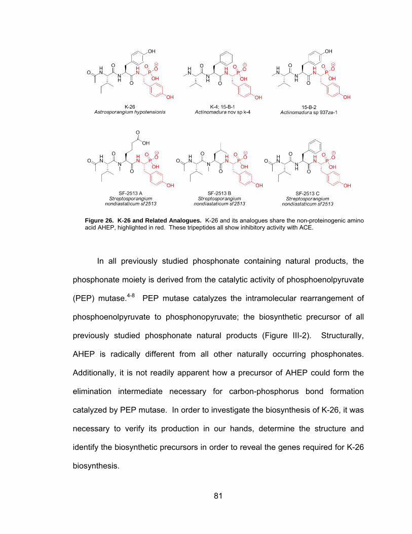
81
Figure 26. K-26 and Related Analogues. K-26 and its analogues share the non-proteinogenic amino acid AHEP, highlighted in red. These tripeptides all show inhibitory activity with ACE.
In all previously studied phosphonate containing natural products, the
phosphonate moiety is derived from the catalytic activity of phosphoenolpyruvate
(PEP) mutase.4-8 PEP mutase catalyzes the intramolecular rearrangement of
phosphoenolpyruvate to phosphonopyruvate; the biosynthetic precursor of all
previously studied phosphonate natural products (Figure III-2). Structurally,
AHEP is radically different from all other naturally occurring phosphonates.
Additionally, it is not readily apparent how a precursor of AHEP could form the
elimination intermediate necessary for carbon-phosphorus bond formation
catalyzed by PEP mutase. In order to investigate the biosynthesis of K-26, it was
necessary to verify its production in our hands, determine the structure and
identify the biosynthetic precursors in order to reveal the genes required for K-26
biosynthesis.

82
Figure 27. C-P Bond Forming Pathway. All previously investigated carbon-phosphorus bond containing natural products are derived from phosphonoacetaldehyde. Phosphonoacetaldehyde is formed subsequent to the PEP catalyzed transformation of PEP to phosphonopyruvate.
Results
Incorporation of isotopically-labeled precursors
From the original structural analysis performed by Seto and coworkers in
1986, K-26 was determined to consist of N-acetyl L-isoleucine, L-tyrosine and the
non-proteinogenic amino acid AHEP.1 To validate these findings and to shed
light on the biosynthetic pathway of K-26 and AHEP, isotopically labeled
precursors were pulse-fed to growing cultures of A. hypotensionis.9 The purpose
of these experiments was to reveal the timing of precursor incorporation. Initial
studies by Ntai et al revealed that tyrosine is a precursor of AHEP and AHEP is a
discrete precursor of K-26. Additional studies revealed that each amino acid is
incorporated independently and N-acetylation occurs after at least the first
peptide bond is formed (Figure III-3).

83
Figure 28. Precursor Incorporation into K-26. Incorporation of tyrosine into AHEP indicates that AHEP is derived from tyrosine. AHEP is incorporated intact into K-26 as is most likely a discrete precursor. N-acetylation occurs after the first peptide bond is formed.
Decarboxylation of tyrosine is formally required for AHEP formation,
therefore, tyramine was proposed as a logical biosynthetic intermediate. D4-
tyramine was produced from D4-tyrosine by enzymatic decarboxylation catalyzed
by tyrosine decarboxylase from Streptococcus faecalis. Four different pulse-
feeding experiments of 1 mM D4-tyramine failed to show any incorporation into
AHEP and K-2610 (Figure III-4).
Figure 29. Tyramine Incorporation. Although decarboxylation is formally required for AHEP biosynthesis, D4-tyramine was not incorporated into K-26.

84
One possible biosynthetic route for K-26 consistent with the precursor
incorporation studies includes a series of ligases. To investigate this possibility,
[1-13C]-N-acetyl-L-isoleucine-[ring-D4]-L-tyrosine and [1-13C]-L-isoleucine-
[ring-D4]-L-tyrosine were synthesized using traditional peptide methods.11-16
Briefly, the amine of [ring-D4]-L-tyrosine was reacted with Boc anhydride to give
the protected amine. Boc-[ring-D4]-L-tyrosine was further protected by addition of
benzyl bromide. Subsequent reaction with TFA yielded the free amine. The
amine group of [1-13C]-L-isoleucine was protected by Cbz and coupled with the
Bn-protected [ring-D4]-L-tyrosine under standard conditions. The protecting
groups were then removed using catalytic hydrogenation resulting in the fully
deprotected peptide, [1-13C]-L-isoleucine-[ring-D4]-L-tyrosine. L-isoleucine was
reacted with [1-13C]-acetyl chloride and coupled with the benzyl-protected
[ring-D4]-L-tyrosine as above. The benzyl groups were removed by catalytic
hydrogenation to give [1-13C]-N-acetyl-L-isoleucine-[ring-D4]-L-tyrosine.

85
Scheme III-1. Synthesis of Stable Labeled Dipeptides
After synthetic preparation of the stable-labeled dipeptide precursors,
separate production cultures of A. hypotensionis were supplemented with 1 mM
of each dipeptide or with water vehicle for four days. A. hypotensionis was
cultured in K26 production media following the production methods established
by Yamato et al.1 After six days of incubation, the production cultures were
harvested by centrifugation, separating the culture broth supernantant and the
cell paste. The culture broth was extracted by Diaion HP-20 polystyrene resin at
pH 3. K-26 was eluted from the resin with a 50% methanol solution. The
methanolic solution was evaporated in vacuo and the residue redissolved in
methanol and subjected to mass spectrometric analysis. Cultures that were
supplemented with only water were used for reference.

86
The specific incorporation of the labeled dipeptides into K-26 was
determined using the Selected Reaction Monitoring (SRM) method described by
Ntai et al9. This tandem mass spectrometry method fragments the [M-H]
precursor ions of K-26 by collision-induced dissociation. By prudently selecting
the precursor and product isotopomer masses in a given experiment, it is
possible to quantitate the isotopic enrichment in both the charged product ion
and the neutral loss fragments. K-26 (m/z 534) characteristically fragments to
AHEP (m/z 216) and Tyr-AHEP (m/z 379) under tandem mass spectrometry
conditions. Figure III-5 shows a chromatographic example of these mass
spectrometry experiments. Incorporation of the labeled dipeptide, intact, would
shift the precursor mass of K-26 by 5 amu. However, if the dipeptide undergoes
proteolytic cleavage, the possibility exists for the precursor mass of K-26 to shift
by 4 amu or 1 amu depending on the incorporation of [ring-D4]-L-tyrosine or
[1-13C]-L-isoleucine/[1-13C]-N-acetyl-L-isoleucine, respectively.

87
Figure 30. SRM Detection of Precursor Incorporation. To detect incorporation of a proposed biosynthetic precursor into K-26, an SRM method was used. Judicious selection of the precursor and product ion masses allows for calculation of label incorporation. This example is from a control sample from a culture of A. hypotensionis without administration of a labeled precursor. Top: K-26 (m/z 534) fragmentation to AHEP (m/z 216). Middle: K-26 + 5 (m/z 539) fragmentation to AHEP (m/z 216). Bottom: K-26 + 5 (m/z 539) fragmentation to AHEP + 4 (m/z 220).
To monitor incorporation of the labeled dipeptides, we selected a specific
precursor mass for K-26 (m/z 534, 539) and monitored the characteristic product
ions m/z 216 and m/z 220, which correspond to AHEP and AHEP+5. If the
dipeptides are incorporated intact, we would expect to observe fragmentation of
the K-26+5 peak (m/z 539) to AHEP (m/z 216). Fragmentation of the K-26+5
(m/z 539) peak to AHEP+4 (m/z 220) would indicate that the dipeptide was

88
hydrolyzed and D4-tyrosine was incorporated into AHEP. Additional experiments
monitored the possible incorporation of inidividual labeled amino acids, if
hydrolysis occurred. Least squares fitting of labeled isotopomer data permitted
the extraction of the relative isotopic enrichments of each amino acid in K-26.
Based on the isotopic incorporation data, the dipeptides were not incorporated
intact. (Figure III-6)
HN
O
O
NH
O
HN PO3H
OH
OH
HN
O
O
NH
O
OH
OH
D
D
D
D
H2N
O
NH
O
OH
OH
D
D
D
D(+1) 0%(+4) 3%(+5) 0%
(+1) 4%(+4) 2%(+5) 0%
Figure 31. Dipeptide Incorporation Studies. Precursor Incorporation studies with stable-labeled didpeptide substrate indicate that the dipeptides were not incorporated intact.
The labeled tyrosine and AHEP incorporation studies revealed that
tyrosine is a precursor to AHEP and AHEP is a discrete precursor to K-26.
However, tyramine is not incorporated indicating that decarboxylation and
phosphonylation of tyrosine may occur in concert or that the tyramine
intermediate is not released before phosphonylation. Moreover, the labeled
isoleucine and dipeptide experiments indicate that each amino acid is
incorporated individually and that N-acetylation occurs after peptide bond
formation.

89
Biosynthetic hypothesis
Based on the stable-labeled precursor incorporation studies, we
hypothesized that K-26 is biosynthesized via the NRPS paradigm.17 As described
in Chapter I, NRPS are modular multidomain proteins capable of synthesizing
small peptides containing non-proteinogenic amino acids. As feeding studies
indicate that AHEP is a discrete precursor of K-26, we hypothesize that AHEP is
biosynthesized prior to peptide bond formation. In this case, shown in Figure
III-7, L-isoleucine, L-tyrosine and AHEP would be activated by individual
A-domains, loaded onto the corresponding T-domains and peptide bond
formation would be catalyzed by the C-domain resulting in the formation of
des-acetyl-K-26 (dK-26). Then either on the final T-domain or after release of the
peptide by the Te-domain, N-acetylation would occur resulting in formation of
K-26. Our proposed gene cluster would consist of an N-acetyltransferase, NRPS
with a domain string of A-T-C-A-T-C-A-T-Te and hypothetical proteins of
unknown function responsible for AHEP biosynthesis.
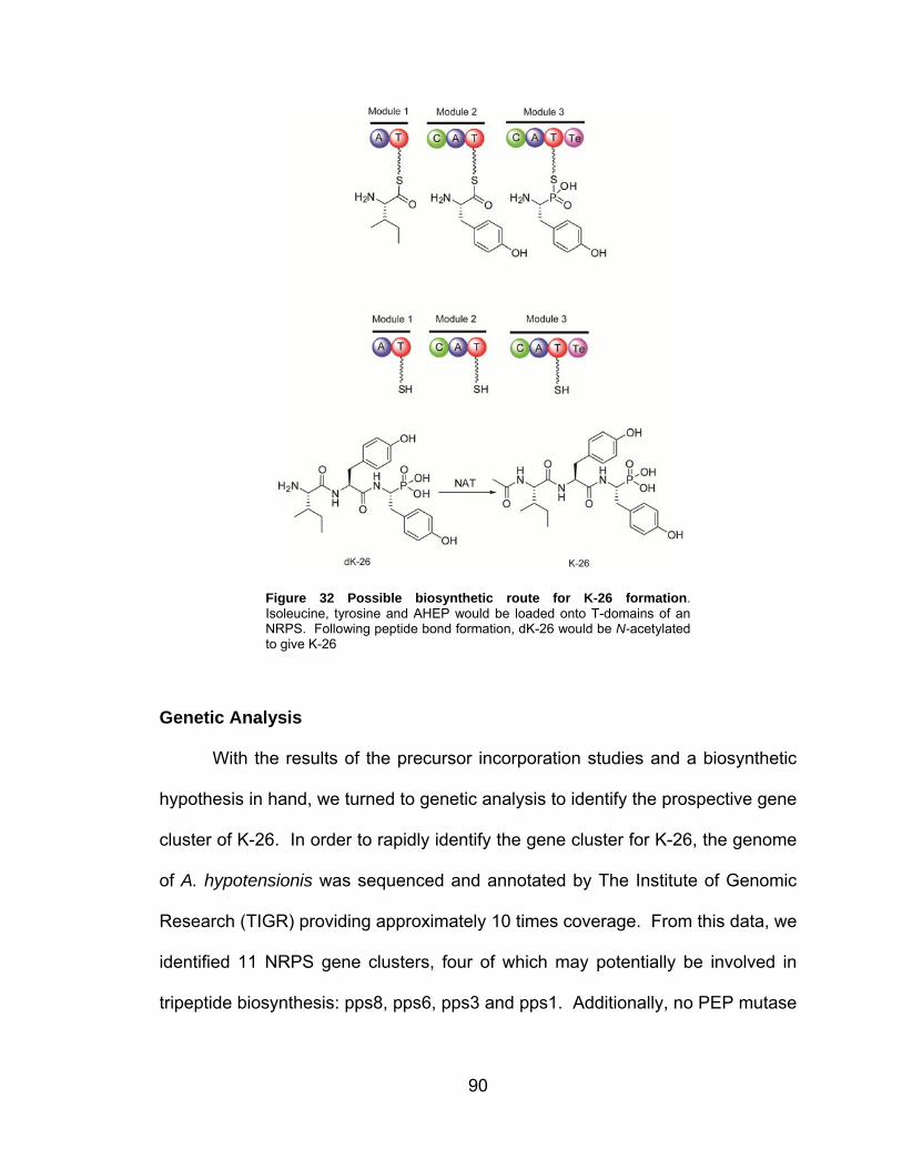
90
Figure 32 Possible biosynthetic route for K-26 formation. Isoleucine, tyrosine and AHEP would be loaded onto T-domains of an NRPS. Following peptide bond formation, dK-26 would be N-acetylated to give K-26
Genetic Analysis
With the results of the precursor incorporation studies and a biosynthetic
hypothesis in hand, we turned to genetic analysis to identify the prospective gene
cluster of K-26. In order to rapidly identify the gene cluster for K-26, the genome
of A. hypotensionis was sequenced and annotated by The Institute of Genomic
Research (TIGR) providing approximately 10 times coverage. From this data, we
identified 11 NRPS gene clusters, four of which may potentially be involved in
tripeptide biosynthesis: pps8, pps6, pps3 and pps1. Additionally, no PEP mutase

91
was found within the A. hypotensionis genome, supporting our hypothesis that
AHEP is formed via a novel biosynthetic route for phosphonate formation.
In the hope that substrate specificity of the A-domains of prospective K-26
gene clusters would provide evidence to determine which tripeptide biosynthetic
cassette is responsible for K-26 biosynthesis, we extracted the 8- to 10- amino
acid specificity code involved in discrimination of amino acid binding within the A-
domain binding pocket, as described by Challis et al and Stachelhaus et al.18, 19
The prospective gene clusters and their A-domain specificity codes are shown in
Table III-1. Out of these four gene clusters, pps3 seems like the most promising
gene cluster, however, no N-acetyltransferase nor proteins of unknown function
are clustered with this NRPS. As no clear gene cluster for K-26 biosynthesis was
revealed by genomic evaluation, we turned to traditional biochemical methods to
isolate of the protein machinery responsible for K-26 biosynthesis from cell-free
extract of A. hypotensionis.
Table III-1. Prospective K-26 NRPS Gene Clusters

92
Reverse Genetic Analysis
As genomic analysis did not provide us with sufficient evidence to identify
the biosynthetic gene cluster of K-26, we were forced to attempt to identify the
gene cluster using traditional biochemical methods. Based on the structure of K-
26, there are three possible handles for biochemical assay development: N-
acetylation, peptide bond formation and AHEP biosynthesis. To begin
biochemical investigations, N-acetylation of dK-26 was first investigated, as most
acetyltransferases only require acetyl-Coenzyme A (Ac-CoA) and substrate for
activity. Second, the mass-based pyrophosphate exchange assay was used to
test for L-isoleucine, L-tyrosine and AHEP activation required for peptide bond
formation in nonribosomal peptide biosynthesis.
N-acetyltransferase Isolation
Based on the precursor incorporation experiments, N-acetylation occurs
after peptide bond formation in K-26 biosynthesis. To identify the
N-acetyltransferase (NAT) involved in K-26 biosynthesis, Ioanna Ntai developed
a mass spectrometry based assay.20 Protein fractions generated from A.
hypotensionis cells shown to produce K-26 were incubated with dK-26 and
acetyl-CoA. After incubation, the reaction mixture was subjected to mass
spectrometry utilizing selected reaction monitoring (SRM) specifically monitoring
the fragmentation of the precursor, dK-26 ([M-H] 492); the product, K-26 ([M-H]
534) and the internal standard D3-K26 ([M-H] 537) to AHEP ([M-H] 216). From
proteomic analyses of SDS-PAGE gel slices of partially purified protein mixtures,
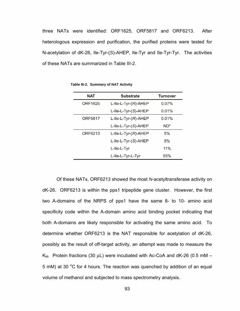
93
three NATs were identified: ORF1625, ORF5817 and ORF6213. After
heterologous expression and purification, the purified proteins were tested for
N-acetylation of dK-26, Ile-Tyr-(S)-AHEP, Ile-Tyr and Ile-Tyr-Tyr. The activities
of these NATs are summarized in Table III-2.
Table III-2. Summary of NAT Activity
Of these NATs, ORF6213 showed the most N-acetyltransferase activity on
dK-26. ORF6213 is within the pps1 tripeptide gene cluster. However, the first
two A-domains of the NRPS of pps1 have the same 8- to 10- amino acid
specificity code within the A-domain amino acid binding pocket indicating that
both A-domains are likely responsible for activating the same amino acid. To
determine whether ORF6213 is the NAT responsible for acetylation of dK-26,
possibly as the result of off-target activity, an attempt was made to measure the
KM. Protein fractions (30 μL) were incubated with Ac-CoA and dK-26 (0.5 mM –
5 mM) at 30 oC for 4 hours. The reaction was quenched by addition of an equal
volume of methanol and subjected to mass spectrometry analysis.

94
The KM of ORF6213 was determined to be higher than 5mM dK-26 (Figure
III-8). Higher concentrations of dK-26 could not be tested due to the limited
solubility of dK-26. These results indicate that the acetylation of dK26 may be
due to promiscuous NATs and not a dedicated NAT clustered with the peptide
bond forming machinery. Since these experiments did not unambiguously
identify the gene cluster of K-26, we proceeded to use our second handle,
peptide bond formation, in an attempt to isolate the proteins responsible for K-26
biosynthesis.
Figure 33. Kinetic Characterization of ORF6213. The KM of Orf6213 is greater than 5mM. Higher dK-26 concentrations could not be tested due to the low solubility of dK-26.
Adenylation Enzyme Isolation
Based on our hypothesis, K-26 is biosynthesized via the NRPS paradigm
where A-domains are involved in amino acid selectivity and promote the reaction
of the cognate amino acid with ATP to create the aminoacyl adenylate and
release of inorganic pyrophosphate.17 This reaction is reversible, in most cases,
and can be followed by the pyrophosphate exchange assay. To purify the

95
proteins responsible for amino acid activation in K-26 biosynthesis, protein
fractions were tested for exchange activity with Ile, Tyr and AHEP, with a
negative control performed concurrently.21-25 To test for this activity, we used the
mass-based pyrophosphate exchange assay as described in Chapter II. Briefly,
partially purified protein fractions were incubated with 1 mM γ-18O4-ATP, 5 mM
pyrophosphate and 1 mM amino acid substrate in the presence of magnesium.
After incubation, the reaction was quenched and the mixture was subjected to
mass spectrometry analysis. Activity was measured as the percentage of
unlabeled ATP, from the back exchange of pyrophosphate, to total ATP, adjusted
to reflect the probability of incorporation.
With a sensitive and accurate assay in hand, we began our initial
purification of adenylation enzymes from A. hypotensionis cell-free extract with
the goal of proteomic identification of the enzymes responsible for activity and
subsequent correlation of the proteins to the biosynthetic gene cluster of K-26.
Figure III-9 is a comprehensive chart of all the purification methods used. The
following represents the final attempt at purification:

96
Figure 34. Overview of adenylation enzyme purification methods. The methods used for final
purification are highlighted in light red.
A. hypotensionis was cultured in K26 production media following the
production methods established by Yamato et al. The production cultures were
harvested by centrifugation, separating the culture broth and the cell paste. The
broth was adjusted to pH 3 and extracted with Diaion HP-20 resin. K-26 was
eluted from the HP-20 resin with a 50% methanol solution. The methanolic
solution was evaporated in vacuo and the residue redissolved in methanol and
subjected to mass spectrometric analysis, confirming the production of K-26.
The cell paste, of A. hypotensionis cultures verified to produce K-26, was washed
with 20% glycerol. The mycelia were frozen in liquid nitrogen for storage at
-80oC. Protein purification was initiated by suspending approximately 60 g of
frozen mycelia in general enzyme buffer (GEB) and rupturing the cells using a
French pressure cell. Nucleic acids were precipitated by streptomycin sulfate
addition followed by centrifugation. After removal of the precipitated nucleic
acids, crude protein fractionation was performed on the supernatant by

97
ammonium sulfate precipitation at 30%, 55% and 75% saturation. After
centrifugation, each ammonium sulfate pellet was dialyzed overnight against
GEB to remove excess salt and the dialysate was tested for activity with L-
isoleucine, L-tyrosine and AHEP. Initial testing of the dialysate revealed that
most of the activity precipitated in the 55% ammonium sulfate fraction. (Figure III-
10)
Figure III-10. Ammonium Sulfate Fractionation. Pyrophosphate exchange analysis of ammonium sulfate fractions of the A. hypotensionis proteome revealed that the 55% saturation fraction contained most of the proteins responsible for activating the amino acid precursors. Tyr = red, Ile = green, AHEP = purple and no amino acid control = light blue
After ammonium sulfate precipitation, further fractionation of the 55%
saturation fraction was performed by anion exchange chromatography using
Q-sepharose resin. (Figure III-11) Pyrophosphate activity for tyrosine and
isoleucine co-eluted, while no activity was detected for AHEP or the negative
control.
0
20
40
60
80
100
75% AS 55% AS 30% AS
Per
cen
t E
xch
ang
e

98
Figure 35. Q-sepharose Fractionation. After ammonium sulfate precipitation, the 55% saturation fraction was further fractionated by Q-sepharose anion exchange. Pyrophosphate activity for tyrosine and isoleucine co-eluted, while no activity was detected for AHEP or the negative control. Tyr = red, Ile= green, AHEP = purple, no amino acid control = light blue, absorbance at 280 = dark blue.
The active fractions from the Q-sepharose column were combined,
concentrated and applied to a DEAE sepharose anion exchange column, in an
attempt to further purify the proteins responsible for the observed pyrophosphate
exchange activity. (Figure III-12)
Figure 36. DEAE-sepharose Fractionation. After separation by Q-sepharose resin, the active fractions were further fractionated by DEAE anion exchange. Pyrophosphate activity for tyrosine and isoleucine co-eluted, while no activity was detected for AHEP or the negative control. Tyr = red, Ile= green, AHEP = purple, no amino acid control = light blue, absorbance at 280 = dark blue
-0.1
6E-16
0.1
0.2
0.3
0.4
0.5
0.6
0
10
20
30
40
50
60
1 9 17 25 33 41 49 57 65 73 81 89
Ab
sorb
ance
(A
U)
Per
cen
t E
xch
ang
e
Fraction Number
-0.16E-160.10.20.30.40.50.60.7
0102030405060708090
A1
A3
A5
A7
A9
A11
B12
B10 B8
B6
B4
B2
C1
C3
Ab
sorb
ance
(A
U)
Per
cen
t E
xch
ang
e
Fraction Number

99
The active fractions were combined, concentrated and divided in half to
continue purification using two methods: hydrophobic interaction, using Butyl
Fast Flow resin, and size exclusion, using Sephacryl S-200 resin. (Figure III-13)
Figure 37. Hydrophobicity and Size Exclusion Fractionation. After DEAE anion exchange fractionation, half of the pooled fractions were applied to either a Butyl Fast Flow column ( hydrophobic separation, top graph) or a Sephacryl S-200 column (size exclusion, bottom graph). Some separation of the pyrophosphate exchange activity for tyrosine and isoleucine was observed. Tyr = red, Ile= green, AHEP = purple, no amino acid control = light blue, absorbance at 280 = dark blue
00.010.020.030.040.050.060.070.080.090.1
05
101520253035404550
A1
A3
A5
A7
A9
A11
B12
B10 B8
B6
B4
B2
C1
C3
Ab
sorb
ance
(A
U)
Per
cen
t E
xch
ang
e
Fraction Number
00.010.020.030.040.050.060.070.080.090.1
05
101520253035404550
A1
A3
A5
A7
A9
A11
B12
B10 B
8
B6
B4
B2
C1
C3
Ab
sorb
ance
(A
U)
Per
cen
t E
xch
ang
e
Fraction Number

100
The active fractions were subjected to SDS-PAGE analysis and two
fractions were sent for proteomics analysis (Figure III-14): Sephacryl S-200
fraction 11, which showed 42% activity for L-isoleucine and 0% activity for
L-tyrosine, and Butyl Fast Flow fraction B9, which showed 30% activity for
L-isoleucine and 21% activity for L-tyrosine.
Figure 38. SDS-PAGE of active fractions for isoleucine and/or tyrosine adenylation. Lane 1: Marker, Lane 2: Sephacryl S-200 fraction 11, Lane 3: Sephacryl S-200 fraction 14, Lane 4: ButylFF fraction B8, Lane 5: Butyl FF fraction B9, Lane 6: ButylFF fraction B11, Lane 7: Marker. Sephacryl S-200 fraction 11 and ButylFF fraction B9 were subjected to proteomics analysis.
Proteomics analysis of these two samples revealed 101 proteins, two of
which may correspond to adenylation activity: isoleucyl tRNA synthetase and a
hypothetical protein containing an AMP-binding domain. To verify the proteomics
results and to test whether the tyrosine adenylation activity was also due to a
tyrosyl tRNA synthetase, a radioactive tRNA loading assay was performed using
active fractions from a duplicate purification where instead of using
hydrophobicity or size exclusion, the protein mixture was subjected to anion

101
exchange fractionation using MonoQ resin.26 It should be noted that pooled
active fractions were tested for pyrophosphate exchange against a panel of
amino acids to ensure our isolation of isoleucine and tyrosine activating
enzymes. (Figure III-15)
Figure 39. Pyrophosphate Exchange Analysis of Fractions. To verify the isolation of isoleucine and tyrosine activating enzymes, at each step, pooled fractions were tested for pyrophosphate activity against a panel of amino acids.
The MonoQ fraction active for both L-isoleucine and L-tyrosine catalyzed
the loading of tritiated L-isoleucine and L-tyrosine onto E. coli tRNA, in separate
experiments, confirming the isolation of tRNA synthetases from A. hypotensionis.
(Figure III-16)

102
Figure 40. tRNA Charging Assay. To test for the presence of tRNA synthetases, the traditional radioactive tRNA charging assay can be performed. For example, radiolabeled L-tyrosine and tyrosine specific tRNA would be incubated with a protein mixture to test for the presence of TyrRS in a protein mixure. If TyrRS is present, the Tyr tRNA would be loaded with the labeled L-tyrosine.
Additionally from genomic analysis, the hypothetical protein identified via
proteomics was part of a small gene cluster containing an N-acetyltransferase.
To investigate whether this cluster was responsible for K-26 biosynthesis, we
heterologously expressed and purified the N-acetyltransferase (NAT 97747) and
tested for N-acetylation of dK-26. (Figure III-17) Neither dK-26 nor Ile-Tyr were
acetylated.
Figure 41. SDS-PAGE of NAT 97747. Lane 1: Marker; Lane 2: Uninduced; Lane 3: Induced; Lane 4 Flow-through; Lane 5: His-Trap Purified; Lane 6: Desalted; Lane 7: Marker.

103
Discussion
Identification of the biosynthetic genes of K-26 presents a unique
challenge. In our hands, K-26 is produced by A. hypotensionis at approximately
10 μg/L.9 At such low levels of production, the only method for verifying K-26
production is through mass spectrometry. The tripeptide nature of K-26 and the
unique pharmacophore of AHEP limit the handles available to exploit for
biosynthetic investigations. The required genes for K-26 biosynthesis are limited
to an N-acetyltransferase, peptide bond forming machinery and those required
for AHEP biosynthesis. Traditional precursor incorporation experiments with
stable-labeled biosynthetic precursors allowed us to propose a NRPS
biosynthetic mechanism for K-26 formation and a possible pathway for AHEP
formation from tyrosine.9, 10 Although the genome of A. hypotensionis has been
sequenced, the biosynthetic cassette for K-26 remains cryptic.
As an N-acetyltransferase is formally required for K-26 formation, it was
selected as the initial handle to examine K-26 biosynthesis.20 The results of the
precursor incorporation studies suggest that N-acetylation occurs after formation
of the peptide bond between L-isoleucine and L-tyrosine. Activity guided protein
purification and subsequent proteomics analysis identified three
N-acetyltranferases, which were heterologously expressed in E. coli: ORF 1625,
which was isolated multiple times; ORF5817, which had high sequence coverage
in proteomics analysis and ORF6213, which is situated neighboring pps1, an
NRPS tripeptide gene cluster. While heterologously expressed ORF1625 and
ORF5817 showed limited turnover of dK-26, ORF6213 showed moderate

104
turnover for dK-26 and L-Ile- L-Tyr and good turnover for L-Ile-L-Tyr-L-Tyr. The KM
of ORF6213 indicates that ORF6213 may not be dedicated to K-26 biosynthesis;
however, it may be the N-acetyltransferase responsible for acetylation of dK-26
through off-target activity. Further analysis of the 8- to 10- amino acid specificity
code within the A-domains of pps1 revealed that the first and second A-domains
within the cluster have identical specificity codes and predict glycine
incorporation while the third A-domain shows homology to aspartate activating
A-domains making pps1 an unlikely candidate gene cluster for K-26 biosynthesis.
As investigation of N-acetyltransferase activity did not reveal the
biosynthetic genes of K-26, peptide formation was selected as the second handle
to analyze. Typically, small peptides containing non-proteinogenic amino acids
are biosynthesized by the NRPS paradigm. To investigate peptide bond
formation, we chose to use the mass-based pyrophosphate exchange assay as
NRPS A-domains, tRNA synthetases and AMP-binding peptide ligases usually
catalyze the formation of an aminoacyl adenylate. Pyrophosphate exchange
experiments were carried out with L-isoleucine, L-tyrosine and AHEP.
L-isoleucine and L-tyrosine showed pyrophosphate exchange activity, while
AHEP did not. The lack of exchange activity was not wholly unexpected as there
are some A-domains and tRNA synthetases that do not undergo reversible
exchange. By following the L-isoleucine and L-tyrosine pyrophosphate exchange
activity, an isoleucyl tRNA synthetase and one hypothetical protein with an
AMP-binding domain were identified by proteomics analysis. However, no NRPS
or L-tyrosine activating enzymes were identified. To confirm the isolation of

105
isoleucyl tRNA synthetase and to explore the possibile isolation of tyrosyl tRNA
synthetase, we performed the traditional tRNA charging assays for both
L-isoleucine and L-tyrosine. Both E. coli isoleucyl and tyrosyl tRNAs were
charged with the corresponding amino acid indicating the pyrophosphate
exchange activity observed with both L-isoleucine and L-tyrosine was caused by
tRNA synthetases. To determine whether the protein of unknown function with
an AMP-binding motif was involved in K-26 biosynthesis, NAT 97747 within the
same gene cluster was heterologously expressed and purified. The purified
protein showed no acetylation of dK-26 or L-Ile- L-Tyr.
Unfortunately, we have not been able to unambiguously identify the gene
cluster responsible for K-26 biosynthesis. The ability to identify the proteins
responsible for K-26 biosynthesis is limited by the production levels of K-26.
Correspondingly, the proteins involved in K-26 are presumably also produced at
extremely low levels. In order to isolate the proteins responsible for K-26
biosynthesis, it will be necessary to increase the production level of K-26 in A.
hypotensionis or identify another producer with higher production levels.
We have identified another producer of K-26, and two additional
analogues, Sphaerosporangium rubeum. However, the production levels of S.
rubeum are similar to those of A. hypotensionis. Briefly, seed culture of S.
rubeum was grown in K26S media. After seed culture, S. rubeum was grown in
6 different media for K-26 analogue production. These cultures were extracted
using the methods previously described. AHEP containing peptide natural
products characteristically fragment to AHEP when subjected to tandem mass

106
spectrometry analysis. Using precursor ion scanning, we identified K-26,
SF5213B and SF5213C in S. rubeum culture broth. (Figure III-18)
Figure 42. K-26 and Analogue Production by S. rubeum. Precursor ion scanning mass spectrometry experiments of S. rubeum production cultures identified production of K-26, SF-2513B and SF-22513C.
The results of the aforementioned experiments seem to indicate that the
N-acetyltransferase responsible for acetylating dK-26 may not be clustered with
the rest of the genes responsible for K-26 biosynthesis. Speculatively, if

107
acetylation is occurring through off-target activity of a variety of
N-acetyltransferases and peptide bond formation is through a promiscuous
ligase, the only genes required for K-26 biosynthesis are those encoding the
proteins responsible for AHEP formation. This lack of clustering may limit the
handles available for investigation to AHEP formation. Additional difficulties lie in
the lack of information about the precursors of AHEP. From the stable-labeled
precursor incorporation experiments, we know that AHEP is derived from
L-tyrosine and AHEP is a discrete precursor of K-26, however, during the above
experiments, at no time was dK-26 or K-26 formation observed when
L-isoleucine, L-tyrosine and AHEP were incubated with cell-free extract of A.
hypotensionis. In order to identify the proteins required for AHEP formation, the
phosphate source and additional cofactors will have to be identified. Additionally,
as AHEP formation is expected to be the rate limiting step in K-26 biosynthesis, it
will be necessary to develop an extremely sensitive assay for AHEP formation.
Complementary to reverse genetic experiments, heterologous expression
of A-domains within prospective K-26 gene clusters has been undertaken. While
this has been an ongoing route to identify of the K-26 biosynthetic cassette,
expression of K-26 A-domains in heterologous hosts has proved to be
challenging, limiting the success of this endeavor. Recent reports have indicated
that small MbtH-like proteins often found near or within NRPS gene clusters may
play a role in increasing the solubility and expression levels of A-domains when
co-expressed in heterologous hosts. Future work co-expressing these proteins

108
may help to identify and/or eliminate prospective gene clusters involved in K-26
biosynthesis.
Materials and Methods
Chemicals and general methods
All non-aqueous reactions were performed under ultra high purity argon in
flame-dried glassware. All chemicals were purchased from Sigma-Aldrich at the
highest purity available unless otherwise noted. γ-18O4-ATP, [ring-D4]-L-tyrosine,
[1-13C]-acyl chloride and [1-13C]-L-isoleucine were purchased from Cambridge
Isotope Labs (Cambridge, MA). Reactions were monitored by thin-layer
chromatography (TLC) using E. Merck precoated silica gel 60 F254 plates.
Visualization was accomplished by UV light and aqueous stain followed by
charring on a hot plate. Flash chromatography was performed using the
indicated solvents and silica gel (230-400 mesh). 1H and 13C NMR were
recorded on Bruker 300, 400 or 500 MHz spectrometers.
Synthesis of [ring-D4]-tyramine
[ring-D4]-L-tyrosine (100 mg, 0.54 mmol) was dissolved in H2O (100 mL) by
microwave heating. A suspension of tyrosine decarboxylase from Streptococcus
faecalis (2.0 mg, 0.05 unit/mg) and pyridoxal-5-phosphate (7 mg, 0.026 mmol) in
acetate buffer (4 mL of 0.1 M, pH 5.5) was added to the cooled tyrosine solution.
The solution was incubated at 37oC for 3 hr. The reaction was stopped by
heating to boiling. Tyramine was purified using Millipore Ultra 5000 MWCO
109
centrifugal filter devices (3450g, 30 min). The filtrate was concentrated in vacuo
and passed through a Dowex 1-x8 anion exchange column. Evaporation of the
eluent gave [ring-D4]-tyamine as a tan powder (98% D, 97% yield). 1H NMR (300
MHz, D2O): δ2.747 (t, J = 7.2Hz, 2H), 3.058 (t, J = 7.2Hz, 2H). 13C NMR (100
MHz, D2O): δ155.33, 129.80 (t, J= 19Hz), 127.68, 115.66 (t, J= 19Hz), 40.79,
31.89
Synthesis of [1-13C]-N-Ac-L-Ile-[ring-D4]-L-Tyr
N-Boc-[ring-D4]-L-Tyr. A solution of 100 mg [ring-D4]-L-tyrosine (1 eq, 100 mg,
0.54 mmol) in dioxane (7.5 mL), H2O (7.5 mL) and NaOH (1M, 1 eq, 540 μL, 0.54
mmol) was stirred and cooled in an ice-water bath. Di-tert-butyl dicarbonate (1.1
eq, 136 μL, 0.59 mmol) was added and the solution was stirred overnight. The
solution was concentrated in vacuo and the aqueous solution was acidified to pH
2. The acidified aqueous solution was extracted with EtOAc (3 x 20 mL). The
organic layers were pooled, washed with brine (3 x 100 mL), dried (MgSO4) and
evaporated to give a white crystalline powder. (0.48 mmol, 138.9 mg, 89.5%) 1H
NMR (400 MHz, DMSO): δ9.15 (s, 1H), 3.95 (m, 1H), 2.85 (m, 1H), 2.69 (s, 1H),
1.36 (s, 9H). 13C NMR (100 MHz, DMSO): δ174.13, 155.83, 128.18, 78.38,
55.90, 35.95, 28.55.
N-Boc-[ring-D4]-L-Tyr(OBn)-COOBn. A solution of N-Boc-[ring-D4]-L-Tyrosine
(1 eq, 100 mg, 0.35 mmol), in dry DMF (3 mL) at room temperature was treated
with potassium carbonate (2.5 eq, 26.7 mg, 0.70 mmol), benzyl bromide (2.5 eq,
83 μL, 0.70 mmol), and tetrabutylammonium iodide (0.125 eq, 16 mg, 0.044

110
mmol). The reaction mixture was stirred at room temperature overnight. The
aqueous mixture was extracted with ether (3 x 10 mL). The organic layers were
combined washed with 1 N HCl (3 x 30 mL) and brine (3 x 30 mL), dried (MgSO4)
and concentrated in vacuo leaving a yellow solid. (0.24 mmol, 110.8 mg, 68.6%)
1H NMR (500 MHz, DMSO): δ9.19 (s, 1H), 7.43-7.26 (m, 10H), 5.07 (s, 2H), 4.12
(s, 1H), 3.33 (s, 2H), 2.85 (m, 1H), 2.69 (m, 1H), 1.32 (s, 9H). 13C NMR (125
MHz, DMSO): δ172.53, 156.27, 155.80, 137.57, 136.28, 128.79, 128.71, 128.48,
128.34, 128.11, 128.00, 127.61, 117.31, 78.65, 69.52, 66.17, 56.20, 35.93,
28.51.
[ring-D4]-L-Tyr(OBn)-COOBn. Boc-[ring-D4]-L-Tyr(OBn)-COOBn (1 eq, 50 mg,
0.11 mmol) was treated with TFA (236eq, 2 mL, 26 mmol) in the presence of
anisole (16 eq, 0.2 mL, 1.8 mmol) in an ice-bath for 30 min. TFA was then
removed by evaporation to give white crystals. (.095 mmol, 34.5 mg, 85.9%) 1H
NMR (500 MHz, DMSO): δ8.446 (s, 2H), 7.441-7.255 (m, 10H), 5.061 (s, 2H),
4.318 (t, J = 6.5Hz, 1H), 3.110-3.001 (m, 4H). 13C NMR (125 MHz, DMSO):
δ171.32, 157.51, 137.23, 136.14, 131.64, 128.77, 128.56, 128.42, 128.23,
128.09, 127.89, 127.53, 116.27, 77.83, 69.73, 53.78, 38.44.
[1-13C]-N-Ac-L-Isoleucine. To a solution of L-Isoleucine (1 eq, 300 mg, 2.3
mmol) in NaOH (4M, 3 eq, 1.75 mL, 7 mmol) at 0oC, [1-13C]-acyl chloride (1.1 eq,
178 μL, 2.5 mmol) was added in 5 portions over 50 min. The mixture was
acidified to pH 1.5 with HCl and extracted with EtOAc (3 x 10 mL). The organic
layers were combined, dried (MgSO4) and concentrated in vacuo to give a white
powder. (0.58 mmol, 100 mg, 25%) 1H NMR (500 MHz, DMSO): δ7.98 (t, J =

111
5Hz, 1H), 4.17-4.13 (m, 1H), 1.85 (s, 3H), 1.75 (m, 1H), 1.37 (m, 1H), 1.16 (m,
1H), 0.90-0.81 (m, 5H). 13C NMR (125 MHz, DMSO): δ173.55, 169.76, 56.60,
36.67, 25.10, 22.89, 15.96, 11.67.
[1-13C]-N-Ac-L-Ile-[ring-D4]-L-Tyr(OBn)-COOBn. [1-13C]-N-Ac-L-Isoleucine
(1.05 eq, 18.0 mg, 0.104 mmol), [ring-D4]-L-Tyr(OBn)-COOBn (1 eq, 36.1 mg,
0.099 mmol) and HOBt (1.1 eq, 14.7 mg, 0.109 mmol) were dissolved in DMF
(10 mL). The solution was cooled in an ice-bath and treated with EDC (1.1 eq,
20.9 mg, 0.109 mmol) and 2,4,6-Trimethylpyridine (1 eq, 13 μL, 0.099 mmol).
The mixture was stirred at 0 oC for 1 hr, allowed to warm to room temperature
and stirred overnight. The reaction was diluted with EtOAc, washed with 1N HCl
(3 x 30 mL), 1N NaHCO3 (3 x 30 mL) and brine (3 x 30 mL), dried (MgSO4) and
concentrated in vacuo to give white crystals. (46.3 mg, 0.089 mmol, 89.6%) 1H
NMR (500 MHz, DMSO): δ8.42 (d, J = 7Hz, 1H), 7.80 (dd, J = 8.75Hz, 1H), 7.43-
7.22 (m, 10H), 5.04-4.88 (m, 4H), 4.44 (q, J = 7.5Hz, 1H), 4.21 (t, J = 6Hz, 1H),
2.96-2.86 (m, 2H), 1.62 (m, 1H), 1.34 (m, 1H), 0.99 (m, 1H), 0.78-0.71 (m, 5H)
13C NMR (125 MHz, DMSO): δ169.29, 157.42, 149.20, 137.57, 128.80, 128.67,
128.21, 128.16, 127.99, 70.51, 69.51, 66.24, 56.67, 54.26, 36.06, 32.81, 24.59,
23.02, 15.49, 11.29.
[1-13C]-N-Ac-L-Ile-[ring-D4]-L-Tyr 1-13C-N-Ac-L-Ile-[ring-D4]-L-Tyr(OBn)-COOBn
(1 eq, 46.3 mg, 0.089 mmol) was dissolved in ethanol (2 mL) in a degassed flask
with 10% wt Pd/C. The suspension was stirred under a H2 atmosphere at room
temperature. The resulting mixture was filtered through a pad of celite and
concentrated to produce a white solid. (26.3 mg, 0.078 mmol, 88%) 1H NMR

112
(500 MHz, D2O): δ4.71 (q, J = 6Hz, 1H), 3.81-3.71 (m, 1H), 2.96-2.61 (m, 2H),
2.06 (m, 1H), 2.04 (s, 3H), 1.70 (m, 1H), 1.29 (m, 1H), 1.08 – 1.02 (m, 5H) 13C
NMR (125 MHz, D2O): δ177.34, 170.92, 170.72, 153.00, 136.77, 58.44, 56.92,
33.78, 32.21, 23.78, 14.58, 10.32.
Synthesis of [1-13C]-L-Ile-[ring-D4]-L-Tyr
N-Cbz-[1-13C]-L-Isoleucine. [1-13C]-L-isoleucine (1 eq, 32.1 mg, 0.24 mmol) was
dissolved in ddH2O (75 μL) and NaOH (5M, 50 μL). The reaction mixture was
cooled to 0oC while stirring. Simultaneously, benzyl chloroformate (1.12 eq, 38.5
μL, 0.27 mmol) and NaOH (2M, 1.12 eq, 135 μL, 0.27 mmol) were added
drop-wise under argon. The reaction was allowed to warm to room temperature
and stirred overnight. After adjusting the pH of the reaction to 10 (saturated
aqueous Na2CO3), it was extracted with ether (3 x 10 mL). The layers were
separated and the aqueous layer was adjusted to pH 3 and again extracted with
ether (3 x 10 mL). The organic layers were combined, dried (MgSO4) and
concentrated in vacuo to give a clear oil. (50.6 mg, 0.20 mmol, 85%) 1H NMR
(500 MHz, DMSO): δ7.46 (d, J = 8.5Hz, 1H), 7.37 (s, 5H), 5.02 (s, 2H), 3.89 (q, J
= 6Hz, 1H), 1.76 (m, 1H), 1.38 (m, 1H), 1.19 (m, 1H), 0.86-0.82 (m, 5H). 13C
NMR (125 MHz, DMSO): δ173.67, 156.69, 137.42, 128.71, 128.41, 128.17,
128.11, 126.99, 126.76, 65.78, 63.26, 36.44, 25.02, 15.99, 11.65.
N-Cbz-[1-13C]-L-Ile-[ring-D4]-L-Tyr(OBn)-COOBn. N-Cbz-[1-13C]-L-Isoleucine
(1.05 eq, 25.8 mg, 1.04 mmol), [ring-D4]-L-Tyr(OBn)-COOBn (1 eq, 36.1 mg,
0.099 mmol) and HOBt (1.1 eq, 14.7 mg, 0.109 mmol) were dissolved in DMF
(10 mL). The solution was cooled in an ice-bath and treated with EDC (1.1 eq,

113
20.9 mg, 0.109 mmol) and 2,4,6-Trimethylpyridine (1 eq, 13 μL, 0.099 mmol).
The mixture was stirred at 0 oC for 1 hr, allowed to warm to room temperature
and stirred overnight. The reaction was diluted with EtOAc, washed with 1N HCl
(3 x 30 mL), 1N NaHCO3 (3 x 30 mL) and brine (3 x 30 mL), dried (MgSO4) and
concentrated in vacuo to give a white powder. (52.8 mg, 0.089 mmol, 89.6%) 1H
NMR (500 MHz, DMSO): δ8.15 (d, J = 8.5Hz, 1H), 7.94 (d, J = 8Hz, 1H), 7.70-
7.20 (m, 15H), 5.16-4.99 (m, 6H), 4.97 (q, J = 6Hz, 1H), 3.93-3.89 (m, 1H), 2.97-
2.86 (m, 2H), 2.06 (m, 1H), 1.63 (m, 1H), 1.39 (m, 1H), 1.09-1.02 (m, 5H) 13C
NMR (125 MHz, DMSO): δ170.81, 166.76, 155.93, 136.72, 135.88, 131.13,
128.93, 128.88, 128.73, 128.69, 128.48, 128.13, 70.51, 69.51, 55.95, 54.23,
33.78, 22.21, 15.48, 11.79.
[1-13C]-L-Ile-[ring-D4]-L-Tyr N-Cbz-[1-13C]-L-Ile-[ring-D4]-L-Tyr(OBn)-COOBn (1
eq, 52.8 mg, 0.089 mmol) was dissolved in ethanol (2 mL) in a degassed flask
with 10% wt Pd/C. The suspension was stirred under a H2 atmosphere at room
temperature. The resulting mixture was filtered through a pad of celite and
concentrated to produce a white solid. (23.4 mg, 0.078 mmol, 88%) 1H NMR
(500 MHz, D2O): δ4.70 (q, J = 6Hz, 1H), 3.81-3.71 (m, 1H), 2.96-2.61 (m, 2H),
2.06 (m, 1H), 1.70 (m, 1H), 1.30 (m, 1H), 1.08 – 1.02 (m, 5H) 13C NMR (125
MHz, D2O): δ177.34, 170.92, 153.00, 136.77, 58.44, 56.92, 33.78, 32.21, 23.78,
14.58, 10.32.

114
Fermentation
The K-26 producing strain, Astrosporangium hypotensionis, was obtained from
the Agricultural Research Service (NRRL 12379). The production protocol of
Yamato and coworkers was followed with minor modifications. Seed medium
consisted of dextrose, 0.1 g/L; Difco soluble starch, 0.1 g/L; Bacto beef extract,
0.05 g/L; Bacto yeast extract, 0.05 g/L; Bacto tryptone, 0.05 g/L; and CaCO3,
0.02 g/L dissolved in distilled water and was adjusted to pH 7.2 before
autoclaving. Fermentation was initiated by aseptically inoculating one loop of
mycelia grown on an agar plate into a sterile 50 mL Falcon tube containing 10
mL seed medium (Phase I). The Falcon tube was incubated for 10-12 days at 28
oC in a shaker incubator. For Phase II, 3 mL of the Phase I seed culture were
transferred into a 250 mL flask containing 30 mL seed medium. The Phase II
seed culture was incubated for 3-4 days at 28 oC in a shaker incubator. For
Production, 30 mL of Phase II seed culture was inoculated into a 2800 mL
Fernbach flask containing 300 mL production medium. The flask was incubated
for 5-6 days at 28oC in a shaker incubator. The production medium consisted of
Difco soluble starch, 0.4 g/L; soybean meal (Whole Foods), 0.3 g/L; corn steep
liquor, 0.05 g/L; K2HPO4, 5 mg/L; MgSO4, 2.4 mg/mL; KCl, 3 mg/L and CaCO3,
0.03 g/L dissolved in distilled water and was adjusted to pH 7.8 before
autoclaving.
115
Pulse feeding experiments
1 mM of each precursor (dipeptides and tyramine) was dissolved in 5 mL water
and administered separately to Phase III culture through a sterile syringe filter in
every 24 hours for 4 days.
K-26 Extraction
The production culture was centrifuged. The cell paste was washed with 20%
glycerol and the mycelia were frozen in liquid nitrogen for storage at -80 oC, while
the supernatant was acidified to pH 3.0. To one liter of supernatant was added
thirty grams of previously activated Diaion HP-20 resin. The suspension was
stirred for 30 minutes. The resin was filtered and washed with water. The
washed resin beads were then stirred in 50% methanol-50% water solution for 10
minutes. The suspension was filtered, washed with 50% methanol and
discarded. The filtrate was concentrated to 40 mL and neutralized. The filtrate
was then additionally concentrated to approximately 1 mL.
K-26 Precursor Incorporation Mass Spectrometry
General
Mass spectrometry was performed using ThermoFinnigan (San Jose, CA)
TSQ® Quantum triple quadrupole mass spectrometer equipped with a standard
electrospray ionization source outfitted with a 100-μm I.D. deactivated fused Si
capillary. Data acquisition and spectral analysis were conducted with Xcalibur™
Software, version 1.3, from ThermoFinnigan (San Jose, CA), on a Dell Optiplex

116
GX270 computer running the Microsoft® Windows 2000 operating system. The
source spray head was oriented at an angle of 90o to the ion transfer tube.
Nitrogen was used for both the sheath and auxiliary gas. The sheath and
auxiliary gases were set to 33 and 14 (arbitrary units) respectively. Samples were
introduced by HPLC. A Surveyor® Autosampler and a Surveyor® MS Pump from
ThermoFinnigan (San Jose, CA) were used. The injection volume was 10 μL.
K-26 was separated from co-metabolites using a Jupiter™ minibore 5μm C18
column (2.0 mm × 15 cm) with a linear water-acetonitrile gradient (ranging from
95:5 to 5:95 H2O:CH3CN) containing 10 mM ammonium acetate. The flow rate
was 0.2 mL/min.
Ring-D4-tyramine
The mass spectrometer was operated in the negative ion mode and the
electrospray needle was maintained at 4200 V. The ion transfer tube was
operated at -35V and 350oC. The tube lens voltage was set to -150 V. Source
CID (offset voltage between skimmer and the first ion guide, Q00) was used at
15 V. The selected reaction monitoring (SRM) mode was used. Ions were
collisionally activated with argon at an indicated pressure of 1.4 mT. The mass
spectral resolution was set to a peak width (full width at half maximum, FWHM)
of 0.50 u and 0.50 u for precursor and product ions respectively. Mass transitions
at the specified collision energy (m/z 534216; 35 eV), (m/z 538216; 35 eV)
and (m/z 538220; 35 eV) were monitored for unenriched K-26, enrichment in
the central tyrosine and enrichment in AHEP, respectively. The scan width for

117
product ions was 1.000 u and the cycle time for each ion was 0.25 seconds. The
electron multiplier gain was set to 2 × 10⁶. Data were acquired in profile mode.
Both a ring-D4-tyramine enriched sample and an unenriched sample were
scanned. The mass spectral data for the two samples was almost identical,
indicating no D4 enrichment of K-26.
[1-13C]-N-Ac-L-Ile-[ring-D4]-L-Tyr and [1-13C]-L-Ile-[ring-D4]-L-Tyr.
The mass spectrometer was operated in the same manner as for the
[ring-D4]-tyramine experiments. Transitions (m/z 534216; 35 eV), (m/z
539216; 35 eV) and (m/z 539220; 35 eV) were monitored for unenriched K-
26, +5 enrichment in N-Ac-Ile-Tyr and +1 enrichment in N-Ac-Ile-Tyr/+4
enrichment in AHEP. To verify +1 and +4 enrichment, additional transitions were
monitored. Transitions (m/z 538216; 35 eV) and (m/z 538220; 35 eV) were
monitored for +4 enrichment in N-Ac-Ile-Tyr and AHEP, respectively, while
transitions (m/z 534379; 35 eV), (m/z 535379; 35 eV) and (m/z 535380; 35
eV) were monitored for unenriched K-26, +1 enrichment in N-Ac-Ile and +1
enrichment in Tyr-AHEP. An unenriched sample was used to create theoretical
curves of ratios of mass isotopomer abundance in K-26 to correct the isotopomer
distribution of the enriched sample as described previously.
118
Cloning, Expression and Purification of N-Acetyltransferases
ORF6213
N-acetyltransferase ORF6213 was cloned from the genomic DNA of A.
hypotensionis by PCR using the primers 5'-GCATATGCGGGTCTCGTTCATC-3'
and 5’-CGTTCGAAGATTAACAAG-3’. The 546 bp PCR product was cloned into
the vector pCR®2.1-TOPO® using TOPO TA Cloning® Kit (Invitrogen). The gene
was then subcloned into the expression vector pET28a(+) using NdeI/HindIII
restriction sites to express N-His6-tagged protein. The resulting plasmid was
introduced into E.coli BL21(DE3) (Novagen) and the protein was expressed as
following: in a 2.8-L baffled flask, 2x 500 mL LB medium containing 50 μg/mL
Kanamycin was inoculated from 2x 5mL overnight cultures made from a fresh
colony of the above strain and grown at 37°C until OD600 = ~0.6. IPTG was then
added (final concentration of 0.5 mM) and the cultures were grown overnight at
25 °C.
The 20.0kD His6-tagged protein was purified as following: the cells were
pelleted (30 min, 3750 rpm, 4 °C), resuspended in binding buffer (50 mM Tris-
HCl pH 8 containing 300 mM NaCl and 10 mM imidazole) . For cell lysis, DNase I
(NEB, 0.2 U/mL) was added. The cells were broken using French pressure cell
(at 1500 psi). The protein was affinity purified on a HisTrap FF column on an
ÄKTA chromatography system (GE Healthcare) using binding buffer with linearly
increasing imidazole concentration (10–500 mM). The pure protein was then
desalted with a HiTrap Desalting column on the same ÄKTA chromatography
119
system using the assay buffer (20 mM Tris, pH7.5) and stored in aliquots at -80
°C with 5% glycerol and 1 mM DTT added.
NAT97747
NAT97747 was cloned from the genomic DNA of A. hypotensionis by PCR
using the primers 5'-CATATGCGTTCTGATGTCACCCTGC-3' and
5’-AAGCTTCAGCCGGCGAGGTCCAC-3’. The 489 bp PCR product was cloned
into the vector pCR®2.1-TOPO® using TOPO TA Cloning® Kit (Invitrogen). The
gene was then subcloned into the expression vector pET28a(+) using
NdeI/HindIII restriction sites to express N-His6-tagged protein. The resulting
plasmid was introduced into E.coli BL21(DE3) (Novagen) and the ~20kD protein
was expressed and purified similarly to ORF6213.
N-Acetyltransferase Assay
Protein fractions (30μL) were incubated with 1 mM AcCoA and 1 mM dK-26 in 20
mM Tris-HCl pH 8 at a total volume of 90 μL at 30 oC for 4 hours. After
incubation, D3-K-26 was added at a 10 nM final concentration. For kinetic
experiments, dK-26 concentrations ranged from 0.5 mM to 5 mM. The reaction
was quenched by addition of an equal volume of methanol and subjected to
mass spectrometry analysis.

120
N-Acetyltransferase Assay Mass Spectrometry
Mass spectrometry was performed using a ThermoFinnigan (San Jose, CA) TSQ
Quantum triple quadrupole mass spectrometer equipped with a standard
electrospray ionization source outfitted with a 100-μm I.D. deactivated fused Si
capillary. The mass spectrometer was operated as above. A Surveyor
Autosampler and a Surveyor MS Pump from ThermoFinnigan were used. The
injection volume was 10 μL. Peptides were separated using a Luna minibore 3
μm C18(2) column (2.0 mm x 5 cm) with an isocratic mobile phase of 90% Buffer
A, 10% Buffer B (Buffer A: 95% water, 5% acetonitrile 10mM ammonium acetate;
Buffer B: 5% water, 95% acetonitrile 10 mM ammonium acetate) with a flow rate
of 0.2 mL/min. Mass transitions at the specified collision energy (m/z 492216;
20 eV), (m/z 534216; 20 eV) and (m/z 537216; 35 eV) were monitored for
dK-26, K-26, and D3-K-26, respectively.
Adenylating Enzyme Purification
Protein Purification
Purification was initiated by suspending flash frozen mycelia in general enzyme
buffer (GEB: 50 mM Tris-HCl pH 7 containing 10% glycerol, 1mM DTT and 0.2
mM PMSF). Cells were ruptured at 0 oC using a French pressure cell. The
ruptured cells were centrifuged (16300 x g, 30 min, 4 oC). The nucleic acids
were precipitated from the supernatant by adding 5% streptomycin sulfate at 1/5
volume, stirring at 4 oC for 15 min and centrifuging. Ammonium sulfate was
added to the supernatant in portions over 10 min to the desired final saturation

121
and stirred for 1 hr at 4 oC and centrifuged. The protein pellets were
resuspended in a minimum volume of GEB and dialyzed overnight versus 3 L of
GEB at 4 oC. Protein concentrations were measured with Pierce® BCA Protein
Assay Kit (Thermo Scientific).
Q-sepharose. The dialysed protein solution was loaded on a Q-sepharose
(Sigma-Aldrich) column equilibrated with 50 mM Tris, pH 7.5 at 5.0 mL/min using
a peristaltic pump and eluted using a linear salt gradient 0.0 - 0.5 M NaCl in 50
mM Tris at pH 7.5. Active fractions were combined and concentrated using an
Amicon stirred cell.
Hydrophobic Interaction chromatography. A high salt protein solution was loaded
on a HiTrap ButylFF column (GE) equilibrated with 50 mM Tris, 1M (NH4)2SO4,
pH 7.5 using an Akta FPLC at a 1 mL/min flow rate. Proteins were eluted using a
linear salt gradient 1.0 – 0.0 M (NH4)2SO4 in 50 mM Tris at pH 7.5.
Size exclusion chromatography. Protein solutions were loaded on a Sephacryl
S-200 (GE) column equilibrated with GEB at 0.5 mL/min and eluted with GEB.
DEAE Sepharose. Desalted protein solutions were loaded on a DEAE sepharose
(GE) column equilibrated with 50 mM Tris, pH 7.2 buffer at 1 mL/min using an
Akta FPLC. Proteins were eluted using a linear gradient 0.0 - 0.5 M NaCl in 50
mM Tris at pH 7.2.
MonoQ. Desalted protein solutions were loaded on MonoQ (GE) column
equilibrated with 50 mM Tris, pH 7.2 buffer at 1 mL/min using an Akta FPLC.
Proteins were eluted using a linear gradient 0.0 - 0.5 M NaCl in 50 mM Tris at pH
7.2.

122
γ-18O4-ATP-PPi Exchange
Assay Conditions
In order to avoid precipitation of magnesium pyrophosphate, assay components
were divided into stock solutions comprising 1) 3 mM amino acids containing 15
mM PPi in 20 mM Tris pH 7.5, 2) 3 mM γ−18O4-ATP containing 15 mM MgCl2 in
20 mM Tris pH 7.5 and 3) protein fractions. Exchange reactions containing 2 μL
of each component were initiated by the addition of enzyme solution. 6 μL
reactions contained final concentrations of 5 mM MgCl2, 5 mM PPi, 1 mM γ-18O4-
ATP, 1mM amino acid and 20 mM Tris-HCl pH 7.5. After incubation at 25 oC, the
reactions were stopped by the addition of 6 μL 9-aminoacridine in acetone (10
mg/mL) for MALDI-TOF MS analysis or 6 μL acetone for ESI-LC/MS analysis.
γ-18O4-ATP-PPi Exchange Assay Mass Spectrometry
MALDI-TOF MS analyses were performed on a Voyager-DE™ STR (Applied
Biosystems, Inc.) using a nitrogen laser (337 nm). Prior to analysis, 1 μL of
analyte/matrix mixture was spotted on to a stainless steel MALDI target. External
mass calibration was performed in the reflectron mode using a mixture of γ−18O4-
ATP and γ−16O4-ATP. Mass spectra were acquired in the negative ion mode over
a range of 450 to 1200 m/z. Mass spectra for ATP-PPi exchange analysis were
obtained by averaging 100 consecutive laser shots. Data acquisition and
quantitative spectral analysis was conducted using Applied Biosystems
DataExplorer software, version 4.5.

123
ESI-LC/MS analyses were performed on a ThermoFinnigan LTQ linear ion trap
mass spectrometer (Thermo Fisher Scientific, Waltham, MA) equipped with an
ESI interface in negative ion mode. Nitrogen was used both for the auxiliary and
sheath gas. The auxiliary and sheath gases were set to 20 psi and 36 psi,
respectively. The following instrumental parameters were used: capillary
temperature 300 °C; source voltage 4.5 kV; source current 100 μA capillary
voltage -49.0 V; tube lens -148.30 V; skimmer offset 0.00 V; activation time
50 ms with an isolation width of 1 m/z. The sensitivity of the mass spectrometer
was tuned by infusion of γ-16O4-ATP at a flowrate of 0.1 mL/min. Samples were
introduced by a Waters Acquity UPLC system (Waters, Milford, MA) with an
injection volume of 5 μL. γ-18O4-ATP was separated from contaminating salts on
a 5 μm Hypercarb column (3 x 50mm, ThermoFisher Scientific) with an isocratic
method of 82.5% 20 mM ammonium acetate pH 6 containing 0.1% diethylamine
and 17.5% acetonitrile over 5 minutes with a flow rate of 0.2 mL/min.
tRNA Charging Assay
Each assay contained 50 mM HEPES-KOH pH 7, 15 mM MgCl2, 15 mM DTT, 5
mM ATP, 2.5 U Roche total tRNA. For TyrRS charging, the reaction mixture
contained 50 μM Tyr with 2 μM L-ring-3,5-3H-Tyr (100 Ci/mmol; Amersham).
For IleRS charging, the reaction mixture contained 50 μM Ile with 2.4 μM L-[4,5]-
3H-Ile (40 Ci/mmol; PerkinElmer). To initiate the reaction, 3 μL of MonoQ purified
protein was added to each reaction. Reactions were incubated at 37 oC for 25
min. The reactions were placed on ice, 25 μL of a 10 mg/mL BSA solution (New

124
England Biosciences) was added followed by 1 mL of cold 10% TCA. After a 10
min incubation at 0voC, the solution was filtered through nitrocellulose filters
(Millipore). The filters were washed twice with 10% TCA and dissolved in
FiltronX (NationalDiagnostics, Atlanta, GA) for analysis.
K-26 analogue production by Sphaerosporangium rubeum
Culture Conditions
S. rubeum, was obtained from the Agricultural Research Service (NRRL B-2636).
The production protocol of K-26 by A. hypotensionis was followed. Seed medium
consisted of dextrose, 0.1 g/L; Difco soluble starch, 0.1 g/L; Bacto beef extract,
0.05 g/L; Bacto yeast extract, 0.05 g/L; Bacto tryptone, 0.05 g/L; and CaCO3,
0.02 g/L dissolved in distilled water and was adjusted to pH 7.2 before
autoclaving. Fermentation was initiated by aseptically inoculating one loop of
mycelia grown on an agar plate into a sterile 50 mL Falcon tube containing 10
mL seed medium (Phase I). The Falcon tube was incubated for 10-12 days at
28oC in a shaker incubator. For Phase II, 3 mL of the Phase I seed culture were
transferred into a 250 mL flask containing 30 mL seed medium. The Phase II
seed culture was incubated for 3-4 days at 28 oC in a shaker incubator. For
Production, 30 mL of Phase II seed culture was inoculated into a 2800 mL
Fernbach flask containing 300 mL of production media. Individual production
cultures, containing one of six possible production media, were incubated for 5-6
days at 28oC in a shaker incubator. The production media included K26P: Difco
soluble starch, 0.4 g/L; soybean meal (Whole Foods), 0.3 g/L; corn steep liquor,

125
0.05 g/L; K2HPO4, 5 mg/L; MgSO4, 2.4 mg/mL; KCl, 3 mg/L and CaCO3, 0.03 g/L
dissolved in distilled water and adjusted to pH 7.8 before autoclaving; BA:
Soybean powder, 15 g/L; Glucose, 10 g/L; Difco soluble starch, 10 g/L; NaCl, 3
g/L; MgSO4*7H2O, 1 g/L; K2HPO4, 1 g/L and 1 mL trace element solution
(FeSO4*7H2O, 0.01 g/L; MnCl2*4H2O, 0.08 g/L; CuSO4*5H2O, 0.07 g/L;
ZnSO4*7H2O, 0.02 g/L; 1 drop H2SO4) dissolved in distilled water and adjusted to
pH 7.2 before autoclaving; EA: lactose, 50 g/L; corn steep solids, 5 g/L; glucose,
5 g/L; glycerol, 15 g/L; soybean flour, 10 g/L; Bacto peptone, 5 g/L; CaCO3, 3
g/L; (NH4)2SO4, 2 g/L; FeSO4*7H2O, 0.1 g/L; ZnCl2, 0.1 g/L; MnCl2*4H2O, 0.1 g/L
and MgSO4*7H2O, 0.5 g/L dissolved in distilled water and adjusted to pH 7.2
before autoclaving; OA: glucose, 10 g/L; glycerol, 5 g/L; corn steep liquor, 3 g/L;
beef extract, 3 g/L; malt extract, 3 g/L; yeast extract, 3 g/L and CaCO3, 2 g/L are
dissolved in distilled water, adjusted to pH 7.2 and after autoclaving, 0.1 g/L
thiamine is added; KA: glucose, 10 g/L; corn steep liquor, 10 g/L; soybean
powder, 10 g/L; glycerol, 5 g/L; dry yeast, 5 g/L and NaCl, 5 g/L are dissolved in
distilled water, the pH is adjust to 5.7 and 2 g/L CaCO3 is added before
autoclaving; QB: soluble starch, 5 g/L; glucose, 6 g/L; corn steep liquor, 2.5 g/L;
Proflo powder, 5 g/L and Proflo oil, 2 mL/L are dissolved in distilled water, the pH
is adjusted to 7.2 and autoclaved.
K-26 analogue extraction
The production culture was centrifuged. The cell paste was discarded and
the supernatant was acidified to pH 3.0. To one liter of supernatant was added

126
thirty grams of previously activated Diaion HP-20 resin. The suspension was
stirred for 30 minutes. The resin was filtered and washed with water. The
washed resin beads were then stirred in 50% methanol-50% water solution for 10
minutes. The suspension was filtered, washed with 50% methanol and
discarded. The filtrate was concentrated to 40mL and neutralized. The filtrate
was then additionally concentrated to approximately 1mL.
K-26 analogue mass spectrometry
Mass spectrometry was performed using FisherScientific (San Jose, CA)
TSQ® Quantum Access triple quadrupole mass spectrometer equipped with a
standard electrospray ionization source outfitted with a 100-μm I.D. deactivated
fused Si capillary. Data acquisition and spectral analysis were conducted with
Xcalibur™ Software, The source spray head was oriented at an angle of 90o to
the ion transfer tube. Nitrogen was used for both the sheath and auxiliary gas.
The sheath and auxiliary gases were set to 33 and 14 (arbitrary units)
respectively. Samples were introduced by HPLC. A Surveyor® Autosampler and
a Surveyor® MS Pump from ThermoFinnigan (San Jose, CA) were used. The
injection volume was 10 μL. K-26 and analogues were separated from co-
metabolites using a Jupiter™ minibore 5μm C18 column (2.0 mm × 15 cm) with a
linear water-acetonitrile gradient (ranging from 95:5 to 5:95 H2O:CH3CN)
containing 10 mM ammonium acetate. The flow rate was 0.2 mL/min.
The mass spectrometer was operated in the negative ion mode and the
electrospray needle was maintained at 4200 V. The ion transfer tube was

127
operated at -35 V and 350 oC. The tube lens voltage was set to -150 V. Source
CID (offset voltage between skimmer and the first ion guide, Q00) was used at
15 V. Precursor ion scanning was used. Ions were collisionally activated with
argon at an indicated pressure of 1.4 mT and a product ion of m/z 216 was
chosen. The mass spectral resolution was set to a peak width (full width at half
maximum, FWHM) of 0.50 u and 0.50 u for precursor and product ions
respectively. The scan width for product ions was 1.000 u and the cycle time for
each ion was 0.25 seconds. The electron multiplier gain was set to 2 × 10⁶. Data
were acquired in profile mode. Three molecules were identified corresponding to
K-26, SF2513B and SF2513C in BA and EA media, while only K-26 was
identified in OA and KA. Production cultures in QB and K26P media did not
produce any K-26 or analogues.

128
REFERENCES
1. Yamato, M., Koguchi, T., Okachi, R., Yamada, K., Nakayama, K., Kase, H., Karasawa, A. and Shuto, K. (1986) K-26, a novel inhibitor of angiotensin I converting enzyme produced by an actinomycete K-26. Journal of Antibiotics (Tokyo), 39, 44-52.
2. Ntai, I. and Bachmann, B. O. (2008) Identification of ACE pharmacophore
in the phosphonopeptide metabolite K-26. Bioorganic & Medicinal Chemistry Letters, 18, 3068-3071.
3. Koguchi, T., Yamada, K., Yamato, M., Okachi, R., Nakayama, K. and
Kase, H. (1986) K-4, a novel inhibitor of angiotensin I converting enzyme produced by Actinomadura spiculosospora. Journal of Antibiotics (Tokyo), 39, 364-371.
4. Liu, S. J., Lu, Z. B., Jia, Y., Dunaway-Mariano, D. and Herzberg, O. (2002)
Dissociative phosphoryl transfer in PEP mutase catalysis: Structure of the enzyme/sulfopyruvate complex and kinetic properties of mutants. Biochemistry, 41, 10270-10276.
5. Mcqueney, M. S., Lee, S. L., Swartz, W. H., Ammon, H. L., Mariano, P. S.
and Dunaway-mariano, D. (1991) Evidence for an Intramolecular, Stepwise Reaction Pathway for Pep Phosphomutase Catalyzed P-C Bond Formation. Journal of Organic Chemistry, 56, 7121-7130.
6. Metcalf, W. W. and van der Donk, W. A. (2009) Biosynthesis of
Phosphonic and Phosphinic Acid Natural Products. Annual Review of Biochemistry, 78, 65-94.
7. Bowman, E., Mcqueney, M., Barry, R. J. and Dunaway-mariano, D. (1988)
Catalysis and Thermodynamics of the Phosphoenolpyruvate Phosphonopyruvate Rearrangement - Entry into the Phosphonate Class of Naturally-Occurring Organo-Phosphorus Compounds. Journal of the American Chemical Society, 110, 5575-5576.
8. Bowman, E. D., McQueney, M. S., Scholten, J. D. and Dunaway-Mariano,
D. (1990) Purification and characterization of the Tetrahymena pyriformis P-C bond forming enzyme phosphoenolpyruvate phosphomutase. Biochemistry, 29, 7059-7063.
9. Ntai, I., Manier, M. L., Hachey, D. L. and Bachmann, B. O. (2005)
Biosynthetic origins of C-P bond containing tripeptide K-26. Organic Letters, 7, 2763-2765.
129
10. Ntai, I., Phelan, V. V. and Bachmann, B. O. (2006) Phosphonopeptide K-
26 biosynthetic intermediates in Astrosporangium hypotensionis. Chemical Communications, 4518-4520.
11. Boger, D. L. and Yohannes, D. (1987) Selectively Protected L-Dopa
Derivatives - Application of the Benzylic Hydroperoxide Rearrangement. Journal of Organic Chemistry, 52, 5283-5286.
12. Carpino, L. A., Elfaham, A. and Albericio, F. (1995) Efficiency in Peptide
Coupling - 1-Hydroxy-7-Azabenzotriazole Vs 3,4-Dihydro-3-Hydroxy-4-Oxo-1,2,3-Benzotriazine. Journal of Organic Chemistry, 60, 3561-3564.
13. Chen, C., Zhu, Y. F. and Wilcoxen, K. (2000) An improved synthesis of
selectively protected L-Dopa derivatives from L-tyrosine. Journal of Organic Chemistry, 65, 2574-2576.
14. Chenault, H. K., Dahmer, J. and Whitesides, G. M. (1989) Kinetic
Resolution of Unnatural and Rarely Occurring Amino-Acids - Enantioselective Hydrolysis of N-Acyl Amino-Acids Catalyzed by Acylase-I. Journal of the American Chemical Society, 111, 6354-6364.
15. Ecija, M., Diez, A., Rubiralta, M., Casamitjana, N., Kogan, M. J. and Giralt,
E. (2003) Synthesis of 3-aminolactams as X-Gly constrained pseudodipeptides and conformational study of a Trp-Gly surrogate. Journal of Organic Chemistry, 68, 9541-9553.
16. Walker, D. M., Mcdonald, J. F., Franz, J. E. and Logusch, E. W. (1990)
Design and Synthesis of Gamma-Oxygenated Phosphinothricins as Inhibitors of Glutamine-Synthetase. Journal of the Chemical Society-Perkin Transactions 1, 659-666.
17. Finking, R. and Marahiel, M. A. (2004) Biosynthesis of nonribosomal
peptides. Annual Review of Microbiology, 58, 453-488. 18. Challis, G. L., Ravel, J. and Townsend, C. A. (2000) Predictive, structure-
based model of amino acid recognition by nonribosomal peptide synthetase adenylation domains. Chemistry & Biology, 7, 211-224.
19. Stachelhaus, T., Mootz, H. D. and Marahiel, M. A. (1999) The specificity-
conferring code of adenylation domains in nonribosomal peptide synthetases. Chemistry & Biology, 6, 493-505.
20. Ntai, I., Vanderbilt University, Nashville, 2007.
130
21. Fujikawa, K., Sakamoto, Y. and Kurahash.K (1971) Biosynthesis of Tyrocidine by a Cell-Free Enzyme System of Bacillus-Brevis Atcc 8185 .3. Further Purification of Components I and Ii and Their Functions in Tyrocidine Synthesis. Journal of Biochemistry, 69, 869-&.
22. Keller, U. and Schlumbohm, W. (1992) Purification and Characterization of
Actinomycin Synthetase-I, a 4-Methyl-3-Hydroxyanthranilic Acid-Amp Ligase from Streptomyces-Chrysomallus. Journal of Biological Chemistry, 267, 11745-11752.
23. Lee, S. G. and Lipmann, F. (1975) Tyrocidine synthetase system.
Methods in Enzymology, 43, 585-602. 24. Schwecke, T., Aharonowitz, Y., Palissa, H., Vondohren, H., Kleinkauf, H.
and Vanliempt, H. (1992) Enzymatic Characterization of the Multifunctional Enzyme Delta-(L-Alpha-Aminoadipyl)-L-Cysteinyl-D-Valine Synthetase from Streptomyces-Clavuligerus. European Journal of Biochemistry, 205, 687-694.
25. Janc, J. W., Egan, L. A. and Townsend, C. A. (1995) Purification and
characterization of clavaminate synthase from Streptomyces antibioticus. A multifunctional enzyme of clavam biosynthesis. The Journal of Biological Chemistry, 270, 5399-5404.
26. Forster, A. C. (2009) Low modularity of aminoacyl-tRNA substrates in
polymerization by the ribosome. Nucleic Acids Research, 37, 3747-3755.
131
CHAPTER IV
BIOSYNTHETIC INVESTIGATIONS OF ANTHRAMYCIN
Introduction
First described in the early 1940s and structurally characterized in 1963,
anthramycin is an antitumor and antibiotic peptide natural product.1 Produced by
the thermophilic actinomycete Streptomyces refuineus sbsp thermotolerans,
anthramycin is a member of the pyrrolobenzodiazepine (PBD) class of natural
products2. (Figure IV-1) These natural products are distinguished by a tricyclic
ring system formed by an anthranilate (A), a diazepine (B) and a hydropyrrole (C)
moiety. Chemical diversity between members of this class is established by the
varied substitution patterns of the A- and C- rings. Additional diversity can be
found within the C-ring including unsaturation at the C2-C3 bond and/or exocyclic
unsaturated moiety at the C2 position. The chemical diversity decorating the
pyrrolobenzodiazepine core modulates the biological potency of these
compounds, where non-covalent interactions with TA-rich regions in the minor
groove of double stranded DNA and the irreversible covalent modification of the
N2-amino group of a neighboring guanine provide the general mechanism for
both the antitumor and antibacterial activities.3-12

132
N
HNHO
H3C
O
OHH
NH2
O
anthramycinStreptomyces refuineus
N
HNHO
O
OHH
CH3
tomaymycinStreptomyces achromogenes
H3CO HN
NHO
O
neothramycin A: R1=H, R2=OHneothramycin B: R1=OH, R2=H
Streptomyces MC916-C4
H3CO
H
R1R2
N
HNHO
H3C
Osibiromycin
Streptomyces sibiricum
OH
CH3
OOHOH3C
H3CHNH3C OH
N
NHO
O
DC-18Streptomyces roseiscleroticus
H3CO
N
HNHO
O
chicanmycin AStreptomyces sp. J576-99
H3CO
H
OH
OCH3
AB
C
Figure 43. Representative Examples of Bacterial PBDs. These natural products are distinguished by a tricyclic ring system formed by an anthranilate (A), a diazepine (B) and a hydropyrrole (C) moiety. Chemical diversity between members of this class is established by the varied substitution patterns of the A- and C- rings.
Before our investigations into the biosynthesis of anthramycin, little was
known about the biosynthesis of PBD natural products. Previous investigations
by Hurley and coworkers established the amino acid precursors to both the
anthranilate and hydropyrrole hemispheres of anthramycin, sibiromycin and
tomaymycin.2, 13-16 (Figure IV-2) Both hemispheres of anthramycin are derived
from oxidative ring opening of amino acids: the 3-hydroxyanthranilic acid (HA)
portion of 4-methyl-3-hydroxyanthranilic acid (MHA) hemisphere is derived from
tryptophan while the dehydropyrrole hemisphere is derived from tyrosine.
Methylation of HA and the hydropyrrole moiety are both through methionine.

133
Figure IV-2. Precursor Incorporation Studies of PBDs. The amino acid precursors of the PBDs have been established. The A-ring of the PBDs is derived from tryptophan, while the C-ring is derived from tyrosine. Methylation of the core structure is methionine dependent.
From these results, Hurley proposed that the MHA portion of anthramycin
is derived from tryptophan via the kynurenine primary metabolic pathway to give
HA followed by S-adenosylmethionine (SAM) dependent aromatic C-methylation.
Additionally, it was proposed that the dehydropyrrole hemisphere is derived from
tyrosine via a biosynthetic pathway analogous to the biosynthesis of the antibiotic
lincomycin, which contains a 4-propyl-4,5-dehydroproline moiety, through
oxidative ring opening of L-DOPA. Incorporation studies with CH3-labeled

134
methionine into the C-terminal carboxamide of anthramycin and the terminal
methyl group of the propyl side chain of lincomycin provide additional evidence of
parallel biosynthesis of anthramycin and lincomycin.
Results
Biosynthetic hypothesis
Based on the non-proteinogenic nature of the two amino acids proposed
in the formation anthramycin, evidenced by the aforementioned isotopic
incorporation experiments, we proposed an NRPS based biosynthetic hypothesis
with a proposed domain string of A-T-C-A-T-Re. The first A-domain of the NRPS
would activate either HA or MHA and the second A-domain would activate a
dehydroproline acrylamide moiety for loading on to their respective T-domains. A
specialized reductase domain would catalyze the final cyclization to form the
benzodiazepine ring of anthramycin. Genes homologous to those found with the
kynurenine primary metabolic pathway will provide the biosynthetic route for HA
biosynthesis, while those homologous to the biosynthesis 4-propyl-4,5-
dehydroproline moiety of lincomycin will synthesize the dehydroproline moiety
found in anthramycin.
Genetic Identification of Anthramycin Gene Cluster
The complete gene cluster for anthramycin biosynthesis was identified by
our collaborators, Ecopia Biosciences (Thallion Pharmaceuticals), by rapid
genome-scanning.17 Briefly, a two-tiered genomic DNA library of 1.5-3 kb and 30-

135
50 kb fragments was constructed from genomic DNA isolated from S. refuineus
sbsp thermotolerans. The short insert library was cloned to give a number of
genomic sequence tags by sequencing random clones. These clones were
analyzed for sequence similarity to verified secondary metabolism gene products
in the National Center for Biotechnology Information (NCBI) nonredundant
protein database. A single genome sequence tag was identified containing
sequence similarity to the predicted requisite NRPS and used in colony
hybridization, identifying two cosmids, 024CA and 024CO, generated from the
large fragment DNA library. These two overlapping cosmids were sequenced
providing a putative gene cluster for anthramycin biosynthesis harboring 25
genes including those consistent with HA biosynthesis, dehydropyrrole moiety
biosynthesis, a bimodular NRPS terminating with a reductase domain and gene
candidates consistent with regulation, resistance and transport. (Figure IV-3)
Figure IV-344. The Anthramycin Gene Cluster. The scientists at Ecopia Biosciences identified the gene cluster of anthramycin on two cosmids. This gene cluster contains an NRPS (red), genes for MHA biosynthesis (blue), the dehydroproline acrylamide moiety (green) and regulatory genes (yellow). Gene disruptions of the genes indicated by red boxes were generated. Anthramycin production was not observed in production cultures of the strains with gene disruptions within the cluster. Used with permission.17
To establish the boundaries of this gene cluster and to validate its role in
anthramycin biosynthesis, a series of PCR-targeted gene replacement
experiments were performed by Hu et al.17, 18 Developing and using a variation

136
of the λ-RED recombination system, a series of mutants was created including
disruptions within orfAS10, orf1, orf12, orf19, orf21/orf22, orf23, orf24 and
orfS11. (Figure IV-3) Orf1 and orf12 are proposed to be involved in
dehydropyrrole moiety biosynthesis, while orf19, orf23 and orf24 are proposed to
be involved in MHA biosynthesis. (Table IV-1) Orf21 and orf22 encode the NRPS
modules of the biosynthetic pathway with a domain string of A-T (orf21)
C-A-T-Re (orf22) and orfAS10 and orfS11 are genes flanking the anthramycin
gene cluster. (Figure IV-3) Production of anthramycin via TLC-bioautography
(anti-Bacillus) and LC/MS was observed in replacement mutants of orfAS10 and
orfS11, the genes flanking either side of the anthramycin gene cluster, while
anthramycin production was not observed in mutants within the proposed gene
cluster. This verified the identification and isolation of the anthramycin
biosynthetic gene cluster.

137
Table IV-1. ORFs Found Within the Anthramycin Gene Cluster
Based on genomic analysis, the NRPS within this cluster consists of a
domain string of A-T-C-A-T-Re with an AT didomain on one module (Orf21) and
C-A-T-Re (Orf22) on a second module. (Figure IV-4) To begin studying the
biochemical mechanisms within anthramycin biosynthesis, we endeavored to
identify the amino acid substrates of the A-domains within the NRPS. Based on
the 8- to 10- amino acid specificity code involved in amino acid discrimination
within the binding pocket of A-domains, we established no similarity between the
binding pockets of the A-domains in anthramycin biosynthesis and the specificity
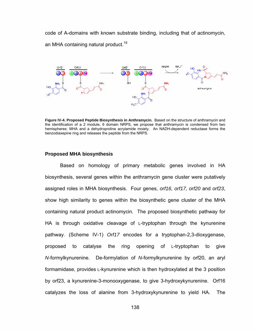
138
code of A-domains with known substrate binding, including that of actinomycin,
an MHA containing natural product.19
Figure IV-4. Proposed Peptide Biosynthesis in Anthramycin. Based on the structure of anthramycin and the identification of a 2 module, 6 domain NRPS, we propose that anthramycin is condensed from two hemispheres: MHA and a dehydroproline acrylamide moiety. An NADH-dependent reductase forms the benzodiaxepine ring and releases the peptide from the NRPS.
Proposed MHA biosynthesis
Based on homology of primary metabolic genes involved in HA
biosynthesis, several genes within the anthramycin gene cluster were putatively
assigned roles in MHA biosynthesis. Four genes, orf16, orf17, orf20 and orf23,
show high similarity to genes within the biosynthetic gene cluster of the MHA
containing natural product actinomycin. The proposed biosynthetic pathway for
HA is through oxidative cleavage of L-tryptophan through the kynurenine
pathway. (Scheme IV-1) Orf17 encodes for a tryptophan-2,3-dioxygenase,
proposed to catalyse the ring opening of L-tryptophan to give
N-formylkynurenine. De-formylation of N-formylkynurenine by orf20, an aryl
formamidase, provides L-kynurenine which is then hydroxylated at the 3 position
by orf23, a kynurenine-3-monooxygenase, to give 3-hydroxykynurenine. Orf16
catalyzes the loss of alanine from 3-hydroxykynurenine to yield HA. The

139
presence of a C-methyl substituent at the 4 position of the anthranilate ring,
shown to be derived from methionine, indicates that a methionine-dependent
C-methyltransferase should be present within the anthramycin biosynthetic gene
cluster. Two genes within the gene cluster have been putatively assigned as
methyltransferases: orf5 and orf19. Orf5 shows high sequence similarity to a
C-methyltransferase within the lincomycin biosynthetic pathway, which has a
similar dehydroproline subunit, indicating that orf5 is probably involved in the
biosynthesis of the dehydroproline acrylamide moiety in anthramycin. Based on
deductive reasoning, we speculate that Orf19 is the C-methyltransferase
responsible for appending the methyl group to the 4 position of the anthranilate
ring. An additional gene, orf24, is clustered with orf23 and proposed to encode a
flavin-dependent or amine oxidase, however, the role of Orf24 remains
speculative.
Scheme IV-1. Possible Biosynthetic Pathway for MHA. Previous precursor incorporation studies coupled with the sequence homology of proteins within the anthramycin biosynthetic cassette to proteins involved in primary metabolic kynurenine pathway suggest that MHA may be derived from tryptophan through kynurenine.

140
Chemical complementation studies to elucidate MHA biosynthesis
While primary metabolic formation of HA as a precursor to nicotinate-
derived cofactors is through the linear kynurenine pathway from tryptophan, the
order and substrate specificity of the homologous genes within the anthramycin
gene cluster may deviate from this paradigm. In order to establish the timing of
each step of MHA biosynthesis a series of chemical complementation studies
was undertaken. Of particular interest is the timing of C-methylation at the 4
position. Isotopic incorporation studies by Hurley and coworkers demonstrated
incorporation of HA into anthramycin, while MHA was not incorporated.2, 13, 14
This may be due to lack of transport of MHA across the cell membrane of S.
refuineus. However, this hypothesis has not been substantiated experimentally.20
There are two general biosynthetic possibilities for the formation of MHA.
(Scheme IV-2) In pathway A, C-methylation is proposed to occur subsequent to
HA formation, via the primary metabolic paradigm. In this case, C-methylation
can occur directly after HA formation to give MHA before incorporation into the
dipeptide or, alternatively, the aryl C-methyl can be inserted during or after
dipeptide assembly. In pathway B, methylation occurs at an unknown position in
the primary anthranilic acid pathway resulting in enzyme substrate divergence
downstream.

141
Scheme IV-2. Possible Biosynthetic Pathways for MHA. Formation of MHA may follow the primary metabolic kynurenine pathway to HA followed by C-methylation either before or after incorporation (Pathway A). Alternatively, the proteins involved in MHA may function in a sequence divergent from primary metabolism if C-methylation occurs before HA formation (Pathway B).
Using S. refuineus strains harboring gene disruptions in orf19, orf23 and
orf24, genes homologous to those of HA biosynthesis, precursor incorporation
studies were performed.17 Four possible biosynthetic intermediates: L-
kynurenine, 3-hydroxy- L-kynurenine, HA and MHA were added to growing
cultures of the S. refuineus mutants to test for their ability to complement the
three gene-replacement mutants to produce anthramycin. Butanolic extracts
prepared from production cultures of the gene-replacement mutants grown were
subjected to TLC-bioautography and LC/MS analysis. Both 3-hydroxy- L-
kynurenine and HA were shown to restore benzodiazepine production when
exogenously supplementing all three mutants, while neither L-kynurenine nor
MHA were able to complement any of the mutants to produce anthramycin.
(Table IV-2)

142
Table IV-2. Results from Chemical Complementation Experiments. Anthramycin = m/z 316; desmethylanthramycin = m/z 302
For pathway A to be operative and encompass the experimental data,
transport of L-kynurenine must be unfavorable under our fermentation conditions
or aryl oxidation may occur earlier in the biosynthesis of HA, slightly diverging
from primary metabolism. Additionally, for pathway B to be operative and agree
with our data, MHA transport must be unfavorable under our fermentation
conditions, as our data suggests that MHA is not a discrete precursor of
anthramycin. As the intermediacy of 3-hydroxy-L-kynurenine is established
based on its ability to complement anthramycin biosynthesis, we favor pathway
A. Chemical complementation of 3-hydroxy-L-kynurenine and HA with the orf23
and orf24 mutants produced anthramycin. However, chemical complementation
of the orf19 mutant with these proposed intermediates led to the accumulation of
a major shunt metabolite, desmethylanthramycin. (Table IV-2) This data indicates
Orf19 is the C-methyltransferase responsible for insertion of the C-methyl at the
4 position of MHA, but the aromatic substrate of this enzyme remains elusive.
These experiments reveal two possible substrates for incorporation into
anthramycin: HA and MHA.

143
Orf21 amino acid activation
As discussed above, an NRPS was identified with in the anthramycin gene
cluster. This dimodular NRPS consists of two genes containing six catalytic
domains in co-linear order on two separate modules orf21 (A-T) and orf22 (C-A-
T-Re). Based on the linear order of the domains within these modules, we
hypothesized that the first A-domain residing within Orf21 was responsible for
activating either MHA or HA for incorporation into anthramycin. Sequence
analysis of the putative A-domain peptide sequence indicates that it is highly
divergent from previously studied A-domains.19 (Table IV-3)
Table IV-3. Specificity Code of the A-domain of Orf21
Analysis of the residues in the substrate binding region indicate Asp-235
(PheA numbering), essential for binding α-amino functionality, is substituted by
alanine in Orf21. Furthermore, the 8- to 10- amino acid selectivity conferring code
bears no similarity to previously described A-domains including, notably,
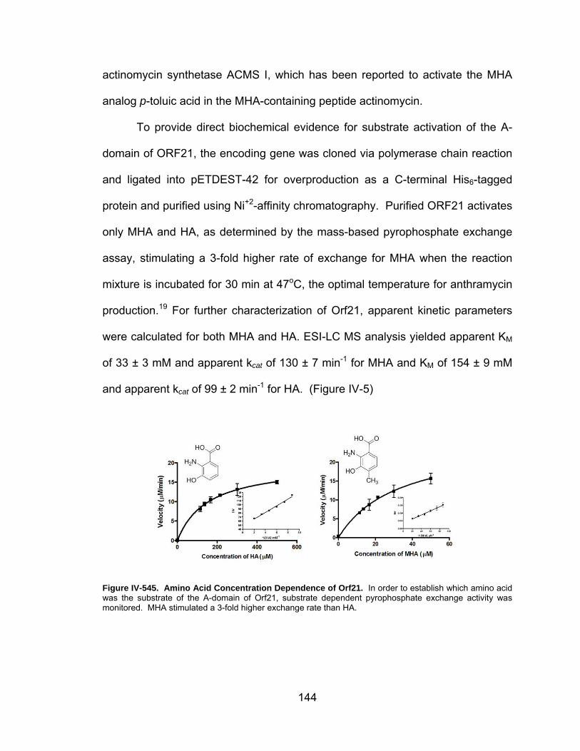
144
actinomycin synthetase ACMS I, which has been reported to activate the MHA
analog p-toluic acid in the MHA-containing peptide actinomycin.
To provide direct biochemical evidence for substrate activation of the A-
domain of ORF21, the encoding gene was cloned via polymerase chain reaction
and ligated into pETDEST-42 for overproduction as a C-terminal His6-tagged
protein and purified using Ni+2-affinity chromatography. Purified ORF21 activates
only MHA and HA, as determined by the mass-based pyrophosphate exchange
assay, stimulating a 3-fold higher rate of exchange for MHA when the reaction
mixture is incubated for 30 min at 47oC, the optimal temperature for anthramycin
production.19 For further characterization of Orf21, apparent kinetic parameters
were calculated for both MHA and HA. ESI-LC MS analysis yielded apparent KM
of 33 ± 3 mM and apparent kcat of 130 ± 7 min-1 for MHA and KM of 154 ± 9 mM
and apparent kcat of 99 ± 2 min-1 for HA. (Figure IV-5)
Figure IV-545. Amino Acid Concentration Dependence of Orf21. In order to establish which amino acid was the substrate of the A-domain of Orf21, substrate dependent pyrophosphate exchange activity was monitored. MHA stimulated a 3-fold higher exchange rate than HA.

145
Additionally, as an additional layer of substrate discrimination is involved
in peptide bond formation through the binding pocket of the C-domain of an
NRPS, MHA loading onto holo-Orf21 was performed.21-24 Using a modified
version of the T-loading assay, apo-Orf21 was phosphopantetheinylated by Sfp,
a promiscuous phosphopantetheinyl transferase, and incubated with excess
MHA. MHA was successfully loaded onto the T-domain of Orf21. This indicates
that MHA is most likely a discrete precursor to anthramycin. (Figure IV-6)

146
Figure IV-6. Orf21 T-loading Assay with MHA. To verify that MHA is the immediate precursor to anthramycin, the T-loading assay was performed. As expected, changes in m/z correlate to the addition of the phosphopantetheinyl group (B) and MHA (C).

147
While the apparent kinetic parameters indicate that MHA is a better
substrate for the A-domain of Orf21, decoupled A-domains are not formally
catalytic. Therefore, concentration-response curves of pyrophosphate exchange
assays cannot be strictly interpreted in terms of Michaelis-Menten steady-state
kinetics. In order to fully biochemically characterize Orf21 with MHA, we
comprehensively derived kinetic constants using exchange kinetics based on
previously described methods. 25-31
Like tRNA synthetases, ATP-PPi exchange catalyzed by A-domains
proceeds through an enzyme-bound aminoacyl adenylate intermediate and the
liberated pyrophosphate product and a modified enzyme form are generated in
equimolar amounts.32 The binding of amino acid and ATP may proceed by an
ordered mechanism (mechanism I and II) or by a random mechanism
(mechanism III) as shown in Figure IV-7.30 Extensive derivations and
interpretations of these mechanisms have been done by Cleland, Cole and
Schimmel.27, 28, 30 Briefly, in mechanism I, a product of the enzymatic reaction is
released before all reactants bind producing two or more stable enzyme forms.
However, in mechanisms II and III, all reactants bind, the enzymatic reaction
occurs and all products are released producing only one stable enzyme form. In
order to distinguished between these mechanisms, initial velocity is measured as
a function of the concentration of the variable substrate, while the secondary
substrate remains constant.28 It is possible to distinguish between possible
mechanisms when several experiments at different fixed concentrations for the
secondary substrate are plotted on the same graph.30 For mechanism I, a

148
double-reciprocal plot will yield parallel lines, while mechanisms II and III will
yield intersecting lines. For mechanism II, the intersection will occur left of the
vertical axis when amino acid concentration is varied and on the vertical axis
when ATP concentration is varied. For mechanism III, the lines will intersect left
of the vertical axis when both amino acid and ATP concentration are varied
Figure IV-7. Possible Biochemical Mechanisms for A-domains. A bimolecular enzymatic reaction has three possible mechanisms: Bi-Bi Ping-Pong, where substrate A binds, releasing product P to create a bound intermediate F, followed by addition of substrate B and subsequent release of product Q to give the free enzyme; Ordered Bi-Bi, where substrate A binds, followed by substrate B subsequently, the products P and Q are released in a defined order; and Random Bi-Bi where substrates A and B bind in no defined order and products P and Q are released at random.

149
The mass based pyrophosphate exchange assay was used following the
guidelines suggested by Cleland.30 For these experiments, the concentration
dependence of the exchange rate was investigated by varying the concentrations
of ATP, pyrophosphate and MHA. The range of concentrations investigated
were 0.25 mM - 4 mM ATP, 0.25 mM-4 mM PPi and 10-100 μM MHA. ATP-PPi
exchange rates were calculated by standard equations derived by Cole and
Schimmel28:
[ ] = 1 1[ ] + 1[ ] + 2[ ] + 3[ ][ ] + 4[ ][ ][ ] + 5
Where:
1 = ; 2 = ; 3 = ; 4 = ; 5 = 1
Data was analyzed by plotting V-1 versus [MHA]-1 at different ATP and
pyrophosphate concentrations and V-1 versus [ATP]-1 at different MHA and
pyrophosphate concentrations. (Figure IV-8)

150
Figure IV-8. Equilibrium Kinetics of Orf21 with MHA. In order to extract equilibrium kinetics, pyrophosphate exchange reactions were performed varying ATP and MHA concentrations and PPi and MHA concentration. Plots of this data yield mechanistic information, as described in the text. A: 1/V versus 1/[MHA] for five [ATP] (0.25, 0.50, 1.0, 2.0 and 4.0 mM from top to bottom). B: 1/V versus 1/[MHA] for five [PP] (0.25, 0.50, 1.0, 2.0 and 4.0 mM from top to bottom). C: 1/V versus 1/[ATP] for seven [MHA] (10, 25, 40, 56, 71, 83, 100 μM from bottom to top). C: 1/V versus 1/[PP] for seven [MHA] (10, 25, 40, 56, 71, 83, 100 μM from bottom to top – only 10, 40, 71 and 100 μM are shown for clarity).
Extrapolations of the primary plots varying both MHA and ATP intersected
left of the Y-axis. Based on the work of Cleland and coworkers, this intersection
pattern is consistent with a random bi-bi mechanism (mechanism III) and is
distinguished for mechanism I, a non-rapid equilibrium mechanism by lack of
substrate inhibition by high levels MHA.30 (Figure IV-7)
To calculate the equilibrium constants, secondary plots were constructed
plotting the slopes and y-intercepts of the lines versus the changing variable.27
For instance, in A of Figure IV-8, five different ATP concentrations were used
over a range of MHA concentrations to yield five lines. The y-intercepts and

151
slopes of these lines were then plotted against 1/[ATP]. (Figure IV-9) The same
holds true for the pyrophosphate concentration plots.
Figure IV-9. Secondary Plots of Orf21 Equilibrium Kinetics. The y-intercept of the slope replot versus 1/[ATP] (Left) gives the KMHA constant, while the x-intercept of the y-intercept replot versus 1/[ATP] (Right) give the KATP constant.
The y-intercept of the slope plot versus 1/B, where B is [ATP] or [PP] and
the slope is equal to (Ka/V) where Ka is equal to KAA, while the x-intercept of the y-
intercept replot is equal to -1/Kb. This yields KAA, KATP and KPP. To calculate Keq
and kapp, double reciprocal plots of 1/V and 1/[AA] are created to yield apparent
kinetic parameters. These values can then be applied to the equation derived by
Cole and Schimmel to give Kx, where x is all enzyme forms.28 (Table IV-4)
Table IV-4. Equilibrium and Kinetic Parameters of Orf21
152
Proposed dehydroproline acrylamide biosynthesis
The biosynthetic pathway for the dehydroproline acrylamide hemisphere
of anthramycin is evidenced by high sequence similarity of a group of genes
found in the anthramycin gene cluster to those involved in 4-propyl-4,5-
dehydroproline biosynthesis in lincomycin.33 Precursor experiments identified
tyrosine as the precursor to the dehydropyrrole portion of both natural products
while methionine dependent methylation is required for formation of the
acrylamide carbonyl and propyl methyl group of anthramycin and lincomycin,
respectfully.34-37 While the precise order of biotransformations leading to the
dehydroproline hemisphere is mostly speculation, the high similarity of orf12 and
orf13, of the anthramycin gene cluster, to lmbB1 and lmbB2, of the lincomycin
gene cluster, suggest their involvement in biosynthesis of this moiety. (Table
IV-1) LmbB1 and lmbB2 have been demonstrated to convert L-tyrosine to
L-DOPA and catalyze 2,3-extradiol cleavage via an Fe2+-dependent
mechanism.38-40 (Scheme IV-3) Based on labeled precursor experiments with
lincomycin, after oxidative ring opening, glycoxylate (or a chemical equivalent) is
lost.34 We propose Orf5, a putative methyl transferase with 79% sequence
identity to lincomycin methyltransferase LmbA, methylates the vinylogous
dehydroproline intermediate providing a possible branch point for anthramycin
and lincomycin biosynthesis. Tautomerization of the imine provides a possible
substrate for hydroxylation by Orf4 which shows high similarity to cytochrome P-
450 hydroxylases. Subsequent transformations by Orf3, a putative alcohol
dehydrogenase; orf2, a putative aldehyde dehydrogenase and orf1, a putative

153
amidotransferase provide a possible route for biosynthesis of the dehydroproline
acrylamide hemisphere of anthramycin.
Scheme IV-3. Proposed Biosynthetic Pathway for Dehydroproline Acrylamide Hemisphere. Based on the precursor incorporation studies of Hurley et al and the similarity of genes within the anthramycin biosynthetic cassette to lincomycin biosynthesis, oxidative ring opening of L-DOPA followed by tailoring steps will yield the dehydroproline acrylamide moiety found within anthramycin.
Synthesis of dehydroproline acrylamide Precursor
While the biosynthetic precursors of the MHA hemisphere of anthramycin
were readily available from commercial sources or through simple chemical
reactions, the biosynthetic precursors of the dehydroproline acrylamide
hemisphere have to be synthesized. Recently, Vanderwal and coworkers
developed a new synthetic route for the synthesis of porothramycins A and B,
close PBD relatives of anthramycin, through a Zincke pyridinium ring-
opening/ring-closing cascade.41, 42 Modification of this synthetic route could
possibly provide us with three of the proposed intermediates in the biosynthesis
of the dehydroproline acrylamide hemisphere of anthramycin.
In order to synthesize the proposed dehydroproline acrylamide substrate
of Orf22, our synthetic route began with Boc-3-(3-pyridyl)-Ala-OH. (Scheme IV-4)
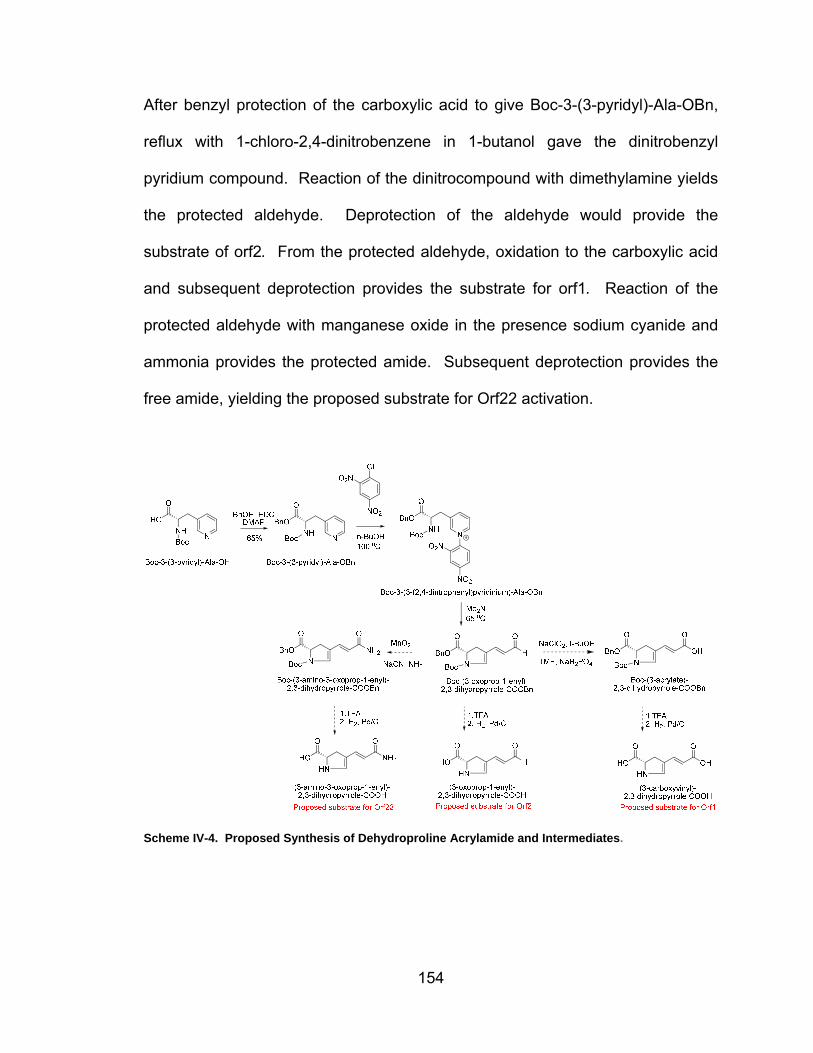
154
After benzyl protection of the carboxylic acid to give Boc-3-(3-pyridyl)-Ala-OBn,
reflux with 1-chloro-2,4-dinitrobenzene in 1-butanol gave the dinitrobenzyl
pyridium compound. Reaction of the dinitrocompound with dimethylamine yields
the protected aldehyde. Deprotection of the aldehyde would provide the
substrate of orf2. From the protected aldehyde, oxidation to the carboxylic acid
and subsequent deprotection provides the substrate for orf1. Reaction of the
protected aldehyde with manganese oxide in the presence sodium cyanide and
ammonia provides the protected amide. Subsequent deprotection provides the
free amide, yielding the proposed substrate for Orf22 activation.
Scheme IV-4. Proposed Synthesis of Dehydroproline Acrylamide and Intermediates.

155
Discussion
Unlike the biosynthetic pathway of K-26, the gene cluster for anthramycin
was readily identified and verified. Our collaborators at Ecopia Biosciences (now
Thallion Pharmaceuticals) were able to isolate the cosmids containing the entire
biosynthetic cassette for anthramycin biosynthesis by using a two-tiered genetic
approach. Genetic experiments by Yunfeng Hu in our lab generated a series of
mutant strains unable to produce anthramycin establishing the role of this gene
cluster in the biosynthesis. While the role of a variety of gene products within this
cluster can be inferred through comparative genetics to similar biosynthetic
pathways, the biochemical roles of these proteins remained completely
uninvestigated. Based on the chemical and genetic similarity of the MHA
hemisphere of anthramycin to primary metabolic production of HA and the
dehydroproline acrylamide to the dehydroproline moiety in lincomycin, we were
able to propose a possible biosynthetic route for both halves.
For MHA biosynthesis, chemical complementation studies provided us
with two possible substrates for the NRPS: HA and MHA. Using the mass-based
pyrophosphate exchange assay, we were able to show that MHA is most likely
the immediate precursor to anthramycin through both activation by the A-domain
of Orf21 and the subsequent loading of the T-domain on the same module.
Additionally, we determined that the mechanism of an isolated A-domain under
reversible conditions is random bi-bi. While we were completing this work,
Aldrich and coworkers established using a terminal assay that adenylate forming

156
enzymes proceed through a bi-uni-uni-bi ping-pong kinetic mechanism when an
activated T-domain is present.43 (Figure IV-10) This corresponds well with the
mechanism of tRNA synthetases with (bi-uni-uni-bi) and without (bi-bi) tRNA.25
Figure IV-10. Proposed Mechanism of A-domain. Recent literature suggests that when a T-domain is present, the mechanism of an A-domain is a Bi-Uni-Uni-Bi Ping-Pong mechanism where the amino acid binds first, followed by ATP. The aminoacyl adenylate is formed releasing pyrophosphate. Subsequently, the T-domain binds and is loaded with the amino acid. The primed T-domain is then released. Lastly, AMP is released from the A-domain.
With the substrate of the MHA hemisphere of anthramycin in hand, we
turned our investigations to the identification of the substrate of Orf22. Unlike the
possible intermediates of the MHA biosynthetic pathway, the substrates
necessary to investigate the biosynthesis of the dehydroproline acrylamide
hemisphere were not readily available. A short synthetic route provides us with
the amide. Currently, orf22 is being synthesized by GenScript as cloning of the
approximately 4kb was unsuccessful. Once gene synthesis is complete, Orf22
will be tested for activity with the dehydroproline acrylamide substrate with both
pyrophosphate exchange and the T-loading assay. Additionally, investigations of
the biosynthesis of the amide can be investigated using gene knockouts and
chemical complementation. Through slight adjustments of the synthetic pathway

157
for the amide, proposed substrates for orf2 (aldehyde) and orf1 (carboxylic acid),
can be synthesized.
In addition to assay based biochemical investigations, the small dimodular
nature of the NRPS within the anthramycin gene cluster provides the perfect
system for protein crystallography of an entire NRPS. The large size of the total
NRPS systems limits the ability to crystallize the protein complex successfully.44-
50 Co-expression of Orf21 and Orf22 and in vitro production of anthramycin will
provide the groundwork for crystallography work. Future crystallization of the
NRPS of anthramycin may provide mechanistic insight into the protein-protein
interactions within these complexes. Additionally, as more and more genome
sequences for PBD producers are becoming accessible, it may be possible to
genetically modify the A-domains to bind non-native substrates to produce novel
molecules with superior or novel biological activities.51
After identification and functional determination of the anthramycin gene
cluster, the bacterial gene clusters of sibiromycin, tomaymycin and
diazepinomicin/ECO-4601, a novel farnesylated dibenzodiazepinone, were
identified.52-54 Comparative genomics of the sibiromycin, tomaymycin,
anthramycin and lincomycin producers provided additional evidence, through
high similarity and homology of analogous proposed proteins within the cluster,
that our gene assignments were correct. Additionally, the 8- to 10- amino acid
code conferring A-domain specificity of the A-domains activation HA derivatives
in sibiromycin, tomaymycin, anthramycin and a series of fungal gene clusters
were extracted. This information revealed that bacterial substituted anthranilic

158
acid activating A-domains cluster separately from fungal anthranilic acid
activating A-domains.55 Additionally, all of these β-amino acid have a
hydrophobic residue, alanine for bacterial A-domains and glycine for fungal A-
domains, at position 235 (GrsA numbering), replacing aspartate. (Table IV-4).
Table IV-4. Specificity Code of Orf21, SibE and TomA
Materials and Methods
Bacterial Strains and Culture Conditions
Streptomyces refuineus var. thermotolerans (NRRL 3143) and its derivatives
were maintained and grown on either ISP4 medium or TSB medium with
appropriate antibiotics at 37°C. For anthramycin production, S. refuineus and its
derivatives were cultured in production medium (1% corn starch, 2% peptonized
milk, and 0.3% yeast extract at pH 7.0) at 47 °C. Bacillus sp. TA (NRRL B-3167)
was cultured in nutrient agar and used as a test organism for the antibacterial
activity of S. refuineus and its derivatives. DH10B (Invitrogen) served as host for
S. refuineus genomic library construction. E. coli BW25113 containing plasmid
pIJ790 was used for targeted gene disruption in S. refuineus. E. coli ET12567

159
containing the RP4 derivative pUZ8002 was used for intergenetic conjugation
between E. coli and S. refuineus. E. coli strains were grown in LB medium
supplemented with appropriate antibiotics for selection of plasmids.
Preparation of Biosynthetic Intermediates of Anthramycin
L-kynurenine, 3-hydroxy-L-kynurenine, hydroxyanthranilic acid were purchased
directly from Sigma Corporation. MHA was prepared by catalytic hydrogenation
of 4-methyl-3-hydroxy-2-nitrobenzoic acid (Aldrich, Inc.) with Pd/C and H2 at 1
atm in ethanol.
Chemical Complementation of Anthramycin
For chemical complementation, corresponding compounds were added directly
to the production medium at the beginning of fermentation for a final
concentration of 2 mM.
Production and Detection of Anthramycin
S. refuineus and its mutant derivatives were cultured in 50 ml seed medium at
47°C for 24 hr. A 5% inoculum was then added to 50 ml production medium and
cultured at 47°C for 24 hr. Anthramycin was extracted from the production
medium with 50 mL butanol. Butanol extracts were concentrated in vacuo and
redissolved in MeOH. Antibacterial activity of anthramycin was detected by thin-
layer chromatography bioautography. Anthramycin butanolic extracts (dissolved
in MeOH) were run on 25DC-Alufolien Kieselgel plates with solvent MeOH:CHCl3
(1:9); LB agar containing indicator strain Bacillus sp. TA was overlaid on TLC

160
plates and cultured at 37 °C for 20 hr to detect anti-Bacillus activity (evidenced by
growth inhibition zones) of anthramycin.
Detection of Anthramycin by LC/MS
Anthramycin production was further confirmed by HPLC/MS. Mass spectrometry
was performed by using ThermoFinnigan (San Jose, CA) TSQ Quantum triple
quadrupole mass spectrometer equipped with a standard electrospray ionization
source outfitted with a 100 mm I.D. deactivated fused Si capillary. The injection
volume was 10 ml. Anthramycin was separated from co-metabolites by using a
Jupiter minibore 5 μm C18 column (2.0 mm x 15 cm) with a linear gradient (0-
100% Buffer B; Buffer A 95% water 5% acetonitrile 10mM ammonium acetate;
Buffer B 5% water 95% acetonitrile 10 mM ammonium acetate). The flow rate
was 0.2 ml/min. The mass spectrometer was operated in the positive (or
negative) ion mode, and the electrospray needle was maintained at 4,200 V. The
ion transfer tube was operated at 35 V and 342 °C (~35 V and 300 °C for
negative). The tube lens voltage was set to 85 V (~220 V for negative). Source
CID (offset voltage between skimmer and the first ion guide, Q00) was used at
15 V. The mass spectrometer was operated in full scan mode with Quad 1. The
mass spectral resolution was set to a peak width of 0.70 u (full width at half
maximum, FWHM). Full scan spectra were acquired from m/z 150.0 to 700.0
(m/z 150.0 to 1200.0 for negative) over 1.0 s. Data were acquired in profile
mode. The electron multiplier gain was set to 3 x 10⁵.
161
Cloning and Expression of ORF21
ORF21 was cloned from the Streptomyces refuineus genomic DNA via PCR
using the primers 5’-CACCATGACAGTACGCAGCACCGCC-3’ and
5’-GCCTCGGGAACGCTTGGTG-3’ (synthesized by Sigma-Aldrich). The 1.8 kb
PCR product was cloned into the vector pENTRTM/SD/D-TOPO® using the
pENTR™ directional TOPO cloning kit (Invitrogen). The gene was subcloned into
an expression vector pET-DEST42 via LR-recombination using Gateway LR
Clonase enzyme mix (Invitrogen). The resulting plasmid was introduced into the
ROSETTA E. coli strain (Novagen). The culture was inoculated (1:100) with an
overnight culture made from a fresh colony of the above strain and grown at 37
°C in a 2.8 L baffled flask containing 500 mL LB medium with 100 μg/mL OD600 =
~ 0.6. IPTG was then added (final concentration of 0.5 mM) and the culture
grown at °C. The cells were pelleted (30 min, 3750 rpm, 4°C) and resuspended
in binding buffer (500 mM NaCl, 20 mM NaH2PO4, 20 mM imidazole, pH 7.4). For
cell lysis, DNase I (NEB, 0.2 U/mL) was added, the cells were disrupted using a
French pressure cell and filtered through a 0.45-μm filter. The 63 kD protein was
purified on a HisTrap FF column (GE Healthcare) on an ÄKTA chromatography
system (GE Healthcare) using binding buffer with linearly increasing imidazole
concentration (20–500 mM). The pure protein was then desalted with a HiTrap
Desalting column using 20 mM Tris, pH 7.5 and stored in aliquots at –80°C in
storage buffer containing 5% glycerol and 1 mM DTT.

162
ATP-PPi Exchange Assay Conditions
In order to avoid precipitation of magnesium pyrophosphate, assay components
were divided into stock solutions comprising 1) 3 mM amino acids containing 15
mM PPi in 20 mM Tris pH 7.5, 2) 3 mM γ−18O4-ATP containing 15mM MgCl2 in
20 mM Tris pH 7.5 and 3) 600 nM enzyme in 20 mM Tris pH 7.5 containing 5%
glycerol and 1mM DTT. Exchange reactions containing 2 μL of each component
were initiated by the addition of enzyme solution. 6 μL reactions therefore
contained final concentrations of 5 mM MgCl2, 5 mM PPi, 1 mM γ-18O4-ATP, 1mM
amino acid and 20 mM Tris-HCl pH 7.5. After an incubation period (30 min at
47 oC), the reactions were stopped by the addition of 6 μL acetone for ESI-
LC/MS analysis.
Orf21 Michaelis-Menten Kinetic Parameters
For Orf21 kinetic measurements, the reactions were performed as described with
the following modifications: % exchange was measured over a range of MHA
concentrations (11.5 – 50.0 μM) and HA concentrations (0.115-0.5 mM) and
rates were measured as single time points at 10 minutes and 15 minutes,
respectively, corresponding to a maximal % exchange of 15%. The ‘apparent’
substrate dependence parameters, KM and kcat, were calculated from initial
velocity time curves with varied MHA or HA concentrations using a non-linear
least square fit to the Michaelis-Menten equation.
163
Orf21 Equilibrium Kinetic Parameters
For Orf21 kinetic measurements, the reactions were performed as described with
the following modifications: % exchange was measured over a range of MHA
concentrations (10 – 100 μM), ATP (0.25 – 4mM) and pyrophosphate (0.25-
4mM) and rates were measured as single time points at 10 minutes. The
equilibrium constants were derived from double reciprocal plots and secondary
plots of slopes and intercepts versus substrate concentration. Slope and
intercept were determined by applying linear regressions using GraphPad Prism.
Mass Spectrometry
ESI-LC/MS analyses were performed on a ThermoFinnigan LTQ linear ion trap
mass spectrometer (Thermo Fisher Scientific, Waltham, MA) equipped with an
ESI interface in negative ion mode. Nitrogen was used both for the auxiliary and
sheath gas. The auxiliary and sheath gases were set to 20 psi and 36 psi,
respectively. The following instrumental parameters were used: capillary
temperature 300 °C; source voltage 4.5 kV; source current 100 μA capillary
voltage -49.0 V; tube lens -148.30 V; skimmer offset 0.00 V; activation time
50 ms with an isolation width of 1 m/z. The sensitivity of the mass spectrometer
was tuned by infusion of γ-16O4-ATP at a flowrate of 0.01 mL/min. Samples were
introduced by a Waters Acquity UPLC system (Waters, Milford, MA) with an
injection volume of 5 μL. γ-18O4-ATP was separated from contaminating salts on
a 5 μm Hypercarb column (3 x 50 mm, ThermoFisher Scientific) with a isocratic
method of 82.5% 20 mM ammonium acetate pH 6 containing 0.1% diethylamine

164
and 17.5% acetonitrile over 5 minutes with a flow rate of 0.2 mL/min. A divert
valve was used for the first two minutes to avoid introducing salts into the source
and the retention time of ATP species under these conditions was 3 minutes.
Data acquisition and quantitative spectral analysis was conducted using the
Thermo-Finnigan Xcaliber software, version 2.0 Sur 1.
Data Analysis
Monoisotopic peak area was determined using the respective software for each
method. For MALDI-TOFMS analysis, the ratio of the area of γ−16O4-ATP (m/z
506) to the area of total ATP including unlabeled, partially labeled, fully labeled
and monosodium-coordinated ions (m/z 506, 508, 510, 512, 514, 528, 530, 532,
534, 536) was calculated using Microsoft excel and adjusted to reflect actual
incorporation based on maximum theoretical incorporation to yield percent
turnover. For ESI-LC/MS analysis, the ratio of the area of γ−16O4-ATP to the area
of total ATP including unlabeled, partially labeled, fully labeled, monosodium-
coordinated and sodium acetate adduct ions was calculated using Microsoft
excel and adjusted to reflect actual incorporation based on maximum theoretical
incorporation to yield percent turnover.
Sfp cloning and expression
Sfp was cloned as previously described. The resulting plasmid was
introduced into the ROSETTA E. coli strain (Novagen). The culture was
inoculated (1:100) with an overnight culture made from a fresh colony of the
above strain and grown at 37°C in a 2.8 L baffled flask containing 500 mL LB
165
medium with 50 μg/mL kanamycin until OD600 = ~ 0.6 was reached. IPTG was
then added (final concentration of 1 mM) and the culture grown overnight at
15°C. The cells were pelleted (30 min, 3750 rpm, 4°C) and resuspended in
binding buffer (500 mM NaCl, 20 mM NaH2PO4, 20 mM imidazole, pH 7.4). For
cell lysis, DNase I (NEB, 0.2 U/mL) was added, the cells were disrupted using a
French pressure cell and filtered through a 0.45-μm filter. The 26 kD His-tagged
protein protein was purified on a HisTrap FF column (GE Healthcare) on an
ÄKTA chromatography system (GE Healthcare) using binding buffer with linearly
increasing imidazole concentration (20–500 mM). The pure protein was then
desalted with a HiTrap Desalting column using 20 mM Tris, pH 7.5, flash frozen
in liquid nitrogen and stored in aliquots at –80°C in storage buffer containing 10%
glycerol and 1 mM EDTA.
T-loading assay
Holo-Orf21 was generated in situ by incubating 25 μM apo-Orf21 with 4
μM Sfp in the presence of 10 mM MgCl2 and 250 μM Coenzyme-A. After 1hr, the
reaction mix was supplemented with 5 mM ATP and 1 mM MHA. The reaction
was allowed to incubate for 30 minutes and extraneous salts were removed by
filtration by centrifugation (10 kD MW cutoff).
Mass Spectrometry for T-domain Loading
ESI-LC/MS analyses were performed on a ThermoFinnigan LTQ linear ion trap
mass spectrometer (Thermo Fisher Scientific, Waltham, MA) equipped with an

166
ESI interface in positive ion mode. Nitrogen was used both for the auxiliary and
sheath gas. The auxiliary and sheath gases were set to 20 psi and 36 psi,
respectively. The following instrumental parameters were used: capillary
temperature 300 °C; source voltage 4.5 kV; source current 100 μA capillary
voltage -49.0 V; tube lens -148.30 V; skimmer offset 0.00 V; activation time
50 ms with an isolation width of 1 m/z. Samples were introduced by a Waters
Acquity UPLC system (Waters, Milford, MA) with an injection volume of 10 μL.
Orf21 was separated from contaminating co-purified proteins on a Waters
Acquity UPLC HEB C8 column (1.0 x 150 mm, 1.7 μm), with a gradient method
of 30% B to 100% B over 10 minutes (Buffer A: 80% water, 10% acetonitrile,
10% isopropanol, 2% acetic acid, 0.2% trifluoroacetic acid; Buffer B: 20% water,
70% acetonitrile, 10% isopropanol, 2% acetic acid, 0.2% trifluoroacetic acid).
Data acquisition and spectral analysis was conducted using the BioMass
software.
Synthesis of dehydroproline acrylamide derivatives
Boc-3-(3-pyridyl)-Ala-OBn
To a stirred solution of Boc-3-(3-pyridyl)-Ala-OH (1 eq, 300 mg, 1.13 mmol) in
anhydrous CH2Cl2 (10 mL) was added DMAP (0.1 eq, 13.8 mg, .113 mmol) and
benzyl alcohol (4 eq, 468 μL, 4.52 mmol). The reaction was cooled to 0oC and
EDC (3 eq, 238 mg, 3.39 mmol) was added.. After stirring for 5 minutes, the
reaction was allowed to warm to room temperature and stirred overnight. The
reaction mixture was washed with HCl (0.5N, 2 x 30 mL) followed by saturated
167
NaHCO3 (3 x 30 mL) and dried (MgSO4). The solvent was removed in vacuo to
yield the crude ether. The ether was purified by column chromotography (100%
DCM followed by 4% MeOH in DCM) to give the pure compound (230 mg, 0.64
mmol, 57%) 1H NMR (400 MHz, CDCl3): δ8.49 (s, 1H), 8.36 (s, 1H), 7.58-7.33
(m, 4H), 7.16 (s, 1H), 5.07 (d, 2H), 4.98 (s, 1H), 4.55 (s, 1H), 3.19 (m, 1H), 3.06
(m, 1H), 1.44 (s, 9H). 13C NMR (100 MHz, CDCl3): δ171.10, 150.52, 148.37,
136.70, 134.86, 131.52, 128.60, 123.26, 67.33, 54.03, 35.48, 28.18.
Boc-3-(3-(2,4-dintrophenyl)pyridinium)-Ala-OBn
To a solution of Boc-3-(3-pyridyl)-Ala-OBn (1 eq, 50 mg, 0.14 mmol) in 1-butanol
(0.1 mL) was added 1-chloro-2,4-dinitrobenzene (1 eq, 28.4 mg, 0.14 mmol).
The resulting solution was heated to 100oC and stirred for 3 hours. After cooling
to room temperature, the solvent was removed in vacuo. The material was
purified by silica gel chromatography (20% MeOH in DCM). (36.8 mg, 0.066
mmol ,47%) 1H NMR (400 MHz, DMSO): δ9.400 (s, 1H), 9.322 (m, 1H), 9.129 (s,
1H), 8.989 (m, 1H), 8.840 (m, 1H), 8.380 (m, 2H), 7.368 (s, 5H), 5.168 (s, 1H),
4.540 (broad s, 1H), 3.223 (m, 2H) 1.326 (s, 9H) 13C NMR (100 MHz, DMSO):
δ162.70, 155.85, 149.57, 143.29, 138.95, 136.11, 132.26, 130.50, 128.79,
128.52, 128.24, 121.92, 79.10, 66.72, 53.99, 36.17, 33.42, 31.15, 28.42.
Boc-(3-oxoprop-1-enyl)-2,3-dihydropyrrole-COOBn
To a solution of Boc-3-(3-(2,4-dinitrophenyl)pyridinium)-Ala-OBn (1 eq, 10 mg,
0.018mmol) in ethanol (100 proof, 471μL) at 50oC, under anhydrous condtions,

168
was added dimethylamine (2.0 M in MeOH, 5 eq, 45μL, 0.09 mmol) and the
reaction vessel was opened to atmosphere. The solution was heated to 65oC
and stirred until the solution turned from a deep maroon to red. The mixture was
allowed to cool to room temperature and concentrated in vacuo. Water was
added and the aqueous solution was extracted with DCM (3 x 10 mL). The
organic extracts were combined, washed with brine (3 x 30 mL), dried (MgSO4),
filtered and concentrated in vacuo. The aldehyde was purified via silica gel
chromatography (1:1 EtOAc:hexanes) followed by phenylhexyl LC/MS. (2.95 mg,
0.008 mmol, 46%).
169
REFERENCES
1. Tendler, M. D. and Korman, S. (1963) 'Refuin': A Non-Cytotoxic Carcinostatic Compound Proliferated by a Thermophilic Actinomycete. Nature, 199, 501.
2. Hurley, L. H., Zmijewski, M. and Chang, C. J. (1975) Biosynthesis of
Anthramycin - Determination of Labeling Pattern by Use of Radioactive and Stable Isotope Techniques. Journal of the American Chemical Society, 97, 4372-4378.
3. Hurley, L. H. and Petrusek, R. (1979) Proposed Structure of the
Anthramycin-DNA Adduct. Nature, 282, 529-531. 4. Kizu, R., Draves, P. H. and Hurley, L. H. (1993) Correlation of DNA-
Sequence Specificity of Anthramycin and Tomaymycin with Reaction-Kinetics and Bending of DNA. Biochemistry, 32, 8712-8722.
5. Kohn, K. W., Bono, V. H. and Kann, H. E. (1968) Anthramycin a New Type
of DNA-Inhibiting Antibiotic - Reaction with DNA and Effect on Nucleic Acid Synthesis in Mouse Leukemia Cells. Biochimica Et Biophysica Acta, 155, 121-&.
6. Kohn, K. W., Glaubige.D and Spears, C. L. (1974) Reaction of
Anthramycin with DNA .2. Studies of Kinetics and Mechanism. Biochimica Et Biophysica Acta, 361, 288-302.
7. Kohn, K. W. and Spears, C. L. (1968) Reaction of Anthramycin with DNA -
Kinetics and Specificity. Federation Proceedings, 27, 800-&. 8. Kohn, K. W. and Spears, C. L. (1970) Reaction of Anthramycin with
Deoxyribonucleic Acid. Journal of Molecular Biology, 51, 551-&. 9. Kopka, M. L., Goodsell, D. S., Baikalov, I., Grzeskowiak, K., Cascio, D.
and Dickerson, R. E. (1994) Crystal structure of a covalent DNA-drug adduct: anthramycin bound to C-C-A-A-C-G-T-T-G-G and a molecular explanation of specificity. Biochemistry, 33, 13593-13610.
10. Krugh, T. R., Graves, D. E. and Stone, M. P. (1989) 2-Dimensional Nmr-
Studies on the Anthramycin-D(Atgcat)2 Adduct. Biochemistry, 28, 9988-9994.
11. Kumar, R. and Lown, J. W. (2003) Recent developments in novel
pyrrolo[2,1-c][1,4]benzodiazepine conjugates: synthesis and biological evaluation. Mini Reviews in Medicinal Chemistry, 3, 323-339.

170
12. Pepper, C. J., Hambly, R. M., Fegan, C. D., Delavault, P. and Thurston, D. E. (2004) The novel sequence-specific DNA cross-linking agent SJG-136 (NSC 694501) has potent and selective in vitro cytotoxicity in human B-cell chronic lymphocytic leukemia cells with evidence of a p53-independent mechanism of cell kill. Cancer Research, 64, 6750-6755.
13. Hurley, L. H. (1980) Elucidation and Formulation of Novel Biosynthetic
Pathways Leading to the Pyrrolo[1,4]Benzodiazepine Antibiotics Anthramycin, Tomaymycin, and Sibiromycin. Accounts of Chemical Research, 13, 263-269.
14. Hurley, L. H. and Gairola, C. (1979) Pyrrolo (1,4) benzodiazepine
antitumor antibiotics: Biosynthetic studies on the conversion of tryptophan to the anthranilic acid moieties of sibiromycin and tomaymycin. Antimicrobial Agents and Chemotherapy, 15, 42-45.
15. Hurley, L. H., Lasswell, W. L., Malhotra, R. K. and Das, N. V. (1979)
Pyrrolo[1,4]benzodiazepine antibiotics. Biosynthesis of the antitumor antibiotic sibiromycin by Streptosporangium sibiricum. Biochemistry, 18, 4225-4229.
16. Hurley, L. H., Zmijewski, M. and Chang, C. J. (1975) Biosynthesis of
anthramycin. Determination of the labeling pattern by the use of radioactive and stable isotope techniques. Journal of the American Chemical Society, 97, 4372-4378.
17. Hu, Y., Phelan, V., Ntai, I., Farnet, C. M., Zazopoulos, E. and Bachmann,
B. O. (2007) Benzodiazepine biosynthesis in Streptomyces refuineus. Chemistry & Biology, 14, 691-701.
18. Hu, Y., Phelan, V. V., Farnet, C. M., Zazopoulos, E. and Bachmann, B. O.
(2008) Reassembly of anthramycin biosynthetic gene cluster by using recombinogenic cassettes. Chembiochem, 9, 1603-1608.
19. Phelan, V. V., Du, Y., McLean, J. A. and Bachmann, B. O. (2009)
Adenylation enzyme characterization using gamma -(18)O(4)-ATP pyrophosphate exchange. Chemistry & Biology, 16, 473-478.
20. Rokem, J. S. and Hurley, L. H. (1981) Sensitivity and Permeability of the
Anthramycin Producing Organism Streptomyces-Refuineus to Anthramycin and Structurally Related Antibiotics. Journal of Antibiotics, 34, 1171-1174.
21. Dorrestein, P. C. and Kelleher, N. L. (2006) Dissecting non-ribosomal and
polyketide biosynthetic machineries using electrospray ionization Fourier-Transform mass spectrometry. Natural Products Reports, 23, 893-918.

171
22. Dorrestein, P. C., Bumpus, S. B., Calderone, C. T., Garneau-Tsodikova,
S., Aron, Z. D., Straight, P. D., Kolter, R., Walsh, C. T. and Kelleher, N. L. (2006) Facile detection of acyl and peptidyl intermediates on thiotemplate carrier domains via phosphopantetheinyl elimination reactions during tandem mass spectrometry. Biochemistry, 45, 12756-12766.
23. Hicks, L. M., Mazur, M. T., Miller, L. M., Dorrestein, P. C., Schnarr, N. A.,
Khosla, C. and Kelleher, N. L. (2006) Investigating nonribosomal peptide and polyketide biosynthesis by direct detection of intermediates on >70 kDa polypeptides by using Fourier-transform mass spectrometry. Chembiochem, 7, 904-907.
24. Dorrestein, P. C., Blackhall, J., Straight, P. D., Fischbach, M. A., Garneau-
Tsodikova, S., Edwards, D. J., McLaughlin, S., Lin, M., Gerwick, W. H., Kolter, R., Walsh, C. T. and Kelleher, N. L. (2006) Activity screening of carrier domains within nonribosomal peptide synthetases using complex substrate mixtures and large molecule mass spectrometry. Biochemistry, 45, 1537-1546.
25. Freist, W., Sternbach, H. and Cramer, F. (1982) Isoleucyl-tRNA
synthetase from Escherichia coli MRE 600. Different pathways of the aminoacylation reaction depending on presence of pyrophosphatase, order of substrate addition in the pyrophosphate exchange, and substrate specificity with regard to ATP analogs. European Journal of Biochemistry, 128, 315-329.
26. Boyer, P. D. (1959) Uses and limitations of measurements of rates of
isotopic exchange and incorporation in catalyzed reactions. Archives of Biochemistry and Biophysics, 82, 387-410.
27. Cleland, W. W. (1967) Enzyme kinetics. Annual Review of Biochemistry,
36, 77-112. 28. Cole, F. X. and Schimmel, P. R. (1970) On the rate law and mechanism of
the adenosine triphosphate--pyrophosphate isotope exchange reaction of amino acyl transfer ribonucleic acid synthetases. Biochemistry, 9, 480-489.
29. Midelfort, C. F. and Mehler, A. H. (1974) Applications of kinetic methods to
aminoacyl-tRNA synthetases. Methods in Enzymology, 29, 627-642. 30. Santi, D. V., Webster, R. W., Jr. and Cleland, W. W. (1974) Kinetics of
aminoacyl-tRNA synthetases catalyzed ATP-PPi exchange. Methods in Enzymology, 29, 620-627.

172
31. Yagil, G. and Hoberman, H. D. (1969) Rate of isotope exchange in enzyme-catalyzed reactions. Biochemistry, 8, 352-360.
32. Finking, R. and Marahiel, M. A. (2004) Biosynthesis of nonribosomal
peptides. Annual Review of Microbiology, 58, 453-488. 33. Peschke, U., Schmidt, H., Zhang, H. Z. and Piepersberg, W. (1995)
Molecular Characterization of the Lincomycin-Production Gene-Cluster of Streptomyces Lincolnensis-78-11. Molecular Microbiology, 16, 1137-1156.
34. Brahme, N. M., Gonzalez, J. E., Rolls, J. P., Hessler, E. J., Mizsak, S. and
Hurley, L. H. (1984) Biosynthesis of the Lincomycins .1. Studies Using Stable Isotopes on the Biosynthesis of the Propyl-L-Hygric and Ethyl-L-Hygric Acid Moieties of Lincomycin-a and Lincomycin-B. Journal of the American Chemical Society, 106, 7873-7878.
35. Hurley, L. H., Lasswell, W. L., Ostrander, J. M. and Parry, R. (1979)
Pyrrolo[1,4]benzodiazepine antibiotics. Biosynthetic conversion of tyrosine to the C2- and C3-proline moieties of anthramycin, tomaymycin, and sibiromycin. Biochemistry, 18, 4230-4237.
36. Ostrander, J. M., Hurley, L. H., McInnes, A. G., Smith, D. G., Walter, J. A.
and Wright, J. L. (1980) Proof for the biosynthetic conversion of L-[indole-15N]tryptophan to [10-15N]anthramycin using (13C, 15N) labelling in conjunction with 13C-NMR and mass spectral analysis. Journal of Antibiotics (Tokyo), 33, 1167-1171.
37. Witz, D. F., Hessler, E. J. and Miller, T. L. (1971) Bioconversion of
Tyrosine into Propylhygric Acid Moiety of Lincomycin. Biochemistry, 10, 1128-&.
38. Colabroy, K. L., Hackett, W. T., Markham, A. J., Rosenberg, J., Cohen, D.
E. and Jacobson, A. (2008) Biochemical characterization of L-DOPA 2,3-dioxygenase, a single-domain type I extradiol dioxygenase from lincomycin biosynthesis. Archives of Biochemistry and Biophysics, 479, 131-138.
39. Harayama, S. and Rekik, M. (1989) Bacterial Aromatic Ring-Cleavage
Enzymes Are Classified into 2 Different Gene Families. Journal of Biological Chemistry, 264, 15328-15333.
40. Novotna, J., Honzatko, A., Bednar, P., Kopecky, J., Janata, J. and Spizek,
J. (2004) L-3,4-Dihydroxyphenyl alanine-extradiol cleavage is followed by intramolecular cyclization in lincomycin biosynthesis. European Journal of Biochemistry, 271, 3678-3683.

173
41. Kearney, A. M. and Vanderwal, C. D. (2006) Synthesis of nitrogen heterocycles by the ring opening of pyridinium salts. Angewandte Chemie-International Edition, 45, 7803-7806.
42. Michels, T. D., Kier, M. J., Kearney, A. M. and Vanderwal, C. D. (2010)
Concise Formal Synthesis of Porothramycins A and B via Zincke Pyridinium Ring-Opening/Ring-Closing Cascade. Organic Letters, 12, 3093-3095.
43. Neres, J., Wilson, D. J., Celia, L., Beck, B. J. and Aldrich, C. C. (2008)
Aryl acid adenylating enzymes involved in siderophore biosynthesis: fluorescence polarization assay, ligand specificity, and discovery of non-nucleoside inhibitors via high-throughput screening. Biochemistry, 47, 11735-11749.
44. Drake, E. J., Cao, J., Qu, J., Shah, M. B., Straubinger, R. M. and Gulick,
A. M. (2007) The 1.8 A crystal structure of PA2412, an MbtH-like protein from the pyoverdine cluster of Pseudomonas aeruginosa. The Journal of Biological Chemistry, 282, 20425-20434.
45. Reger, A. S., Carney, J. M. and Gulick, A. M. (2007) Biochemical and
crystallographic analysis of substrate binding and conformational changes in acetyl-CoA synthetase. Biochemistry, 46, 6536-6546.
46. Samel, S. A., Wagner, B., Marahiel, M. A. and Essen, L. O. (2006) The
thioesterase domain of the fengycin biosynthesis cluster: a structural base for the macrocyclization of a non-ribosomal lipopeptide. Journal of Molecular Biology, 359, 876-889.
47. Drake, E. J., Nicolai, D. A. and Gulick, A. M. (2006) Structure of the EntB
multidomain nonribosomal peptide synthetase and functional analysis of its interaction with the EntE adenylation domain. Chemistry & Biology, 13, 409-419.
48. May, J. J., Kessler, N., Marahiel, M. A. and Stubbs, M. T. (2002) Crystal
structure of DhbE, an archetype for aryl acid activating domains of modular nonribosomal peptide synthetases. Proceedings of the National Academy of Sciences of the United States of America, 99, 12120-12125.
49. Keating, T. A., Marshall, C. G., Walsh, C. T. and Keating, A. E. (2002) The
structure of VibH represents nonribosomal peptide synthetase condensation, cyclization and epimerization domains. Nature Structural Biology, 9, 522-526.
50. Bruner, S. D., Weber, T., Kohli, R. M., Schwarzer, D., Marahiel, M. A.,
Walsh, C. T. and Stubbs, M. T. (2002) Structural basis for the cyclization

174
of the lipopeptide antibiotic surfactin by the thioesterase domain SrfTE. Structure, 10, 301-310.
51. Keasling, J. D. (2010) Manufacturing Molecules Through Metabolic
Engineering. Science, 330, 1355-1358. 52. Li, W., Chou, S., Khullar, A. and Gerratana, B. (2009) Cloning and
characterization of the biosynthetic gene cluster for tomaymycin, an SJG-136 monomeric analog. Applied and Environmental Microbiology, 75, 2958-2963.
53. Li, W., Khullar, A., Chou, S., Sacramo, A. and Gerratana, B. (2009)
Biosynthesis of sibiromycin, a potent antitumor antibiotic. Applied and Environmental Microbiology, 75, 2869-2878.
54. McAlpine, J. B., Banskota, A. H., Charan, R. D., Schlingmann, G.,
Zazopoulos, E., Piraee, M., Janso, J., Bernan, V. S., Aouidate, M., Farnet, C. M., Feng, X., Zhao, Z. and Carter, G. T. (2008) Biosynthesis of diazepinomicin/ECO-4601, a Micromonospora secondary metabolite with a novel ring system. Journal of Natural Products, 71, 1585-1590.
55. Ames, B. D. and Walsh, C. T. (2010) Anthranilate-activating modules from
fungal nonribosomal peptide assembly lines. Biochemistry, 49, 3351-3365.

175
CHAPTER V
FUTURE DIRECTIONS
The research described within the previous chapters has been the initial
biosynthetic investigations of both K-26 and anthramycin. As these are initial and
preliminary investigations more experiments will be required to fully investigate
the biosynthesis of these molecules. Herein, I will discuss possible paths of
investigations.
K-26
Unfortunately, we have not been able to unambiguously identify the gene
cluster responsible for K-26 biosynthesis by genomic analysis. The ability to
identify the proteins responsible for K-26 biosynthesis is limited by the production
levels of K-26. Correspondingly, the proteins involved in K-26 are presumably
also produced at extremely low levels. In order to isolate the proteins
responsible for K-26 biosynthesis, it will be necessary to increase the production
level of K-26 in A. hypotensionis or identify another producer with higher
production levels. To increase production levels of K-26 in A. hypotensionis or
another producer, mutagenesis experiments were begun by Dr. Jaeheon Lee
using UV-radiation.
The results of the aforementioned experiments seem to indicate that the
N-acetyltransferase responsible for acetylating dK-26 may not be clustered with
the rest of the genes responsible for K-26 biosynthesis. Speculatively, if

176
acetylation is occurring through off-target activity of a variety of
N-acetyltransferases and peptide bond formation is through a promiscuous
ligase, the only genes required for K-26 biosynthesis are those encoding the
proteins responsible for AHEP formation. This lack of clustering may limit the
handles available for investigation to AHEP formation. Additional difficulties lie in
the lack of information about the precursors of AHEP. From the stable-labeled
precursor incorporation experiments, we know that AHEP is derived from
L-tyrosine and AHEP is a discrete precursor of K-26, however, during the
previously described experiments, at no time was dK-26 or K-26 formation
observed when L-isoleucine, L-tyrosine and AHEP were incubated with cell-free
extract of A. hypotensionis. In order to identify the proteins required for AHEP
formation, the phosphate source and additional cofactors will have to be
identified. Additionally, as AHEP formation is expected to be the rate limiting
step in K-26 biosynthesis, it will be necessary to develop an extremely sensitive
assay for AHEP formation.
In order to do this, the best course of action would require the ability to
detect AHEP in cell-free extract. AHEP is easily separated from contaminating
salts by using a Hypercarb column. Initial experiments with a full gradient from
0% acetonitrile (100% 20 mM ammonium acetate pH 6 with 0.1% TEA) to 100 %
acetonitrile will provide a good starting point for method development for LC/MS
methodology. After development of the LC/MS method, limit of detection
experiments must be undertaken. In order to detect AHEP formation, it will be
necessary to detect 0.01% AHEP in cell-free extract. To do this, L-tyrosine, a

177
phosphate mixture and AHEP of known concentrations are added to cell-free
extract. The cell-free extract is heated to 95 oC, to denature the proteins and
centrifuged to remove the precipitated proteins. The supernatant would then be
subjected to LC/MS analysis. After limits of detection experiments in both full
scan and SRM (AHEP readily fragments, verify by product ion scanning), initial
cell-free experiments can be undertaken. If AHEP still cannot be detected, it may
be necessary to further concentrate AHEP from the cell-free extract. To do this, I
would recommend using either a gallium or zinc affinity column, similar to nickel
affinity used for protein purification. This would necessitate identifying another
compound capable of eluting AHEP from the column.
Once the detection method is developed and AHEP can be detected in
cell-free extraction, identification of the phosphate source can be undertaken. As
mentioned in Chapter III, L-tyrosine is an immediate precursor of AHEP,
however, the phosphate source and any required cofactors remain unidentified.
Under cell-free extract conditions, no supplementation with phosphate or
cofactors should be necessary to observe conversion of tyrosine to AHEP. To
identify the phosphate source, incubation of a co-factor mix containing all
possible co-factors with a mix of possible phosphate sources should increase the
conversion of tyrosine to AHEP in cell-free extract. After formation of AHEP can
be detected, process of elimination can first identify the phosphate source and
then identify the required co-factors.
Subsequent to the identification of the phosphate source and the required
co-factors, protein fractionation can be performed using traditional bioactivity

178
guided fractionation similar to that discussed in Chapter III. Proteomics analysis
of active protein fractions will identify prospective proteins. Hopefully, possible
candidates can be eliminated by correlating identified prospective proteins with a
gene cluster with either an NRPS or genes for isoleucine or tyrosine
biosynthesis.
Complementary to reverse genetic experiments, heterologous expression
of A-domains within prospective K-26 gene clusters has been undertaken. While
this has been an ongoing route to identify of the K-26 biosynthetic cassette,
expression of K-26 A-domains in heterologous hosts has proved to be
challenging, limiting the success of this endeavor. Recent reports have indicated
that small MbtH-like proteins often found near or within NRPS gene clusters may
play a role in increasing the solubility and expression levels of A-domains when
co-expressed in heterologous hosts. Future work co-expressing these proteins
may help to identify and/or eliminate prospective gene clusters involved in K-26
biosynthesis.
Anthramycin
Unlike the possible intermediates of the MHA biosynthetic pathway, the
substrates necessary to investigate the biosynthesis of the dehydroproline
acrylamide hemisphere are not readily available. A short synthetic route will
provide the amide. Our synthetic route also yields the aldehyde and acid
proposed biosynthetic precursors of the amide (See Chapter IV). Orf22 was
successfully synthesized by GenScript and is currently subcloned into the

179
pET28(a) vector. Successful expression and purification of Orf22 will allow for
biochemical characterization of the enzyme. Orf22 will be tested for activity with
the dehydroproline acrylamide substrate with both pyrophosphate exchange and
the T-loading assay. Additionally, investigations of the biosynthesis of the amide
can be investigated using gene knockouts and chemical complementation.
Through slight adjustments of the synthetic pathway for the amide, proposed
substrates for orf2 (aldehyde) and orf1 (carboxylic acid), can be synthesized.
In addition to assay based biochemical investigations, the small dimodular
nature of the NRPS within the anthramycin gene cluster provides the perfect
system for protein crystallography of an entire NRPS. The large size of the total
NRPS systems limits the ability to crystallize the protein complex successfully.1-7
Co-expression of Orf21 and Orf22 and in vitro production of anthramycin will
provide the groundwork for crystallography work. Future crystallization of the
NRPS of anthramycin may provide mechanistic insight into the protein-protein
interactions within these complexes. Additionally, as more and more genome
sequences for PBD producers are becoming accessible, it may be possible to
genetically modify the A-domains to bind non-native substrates to produce novel
molecules with superior or novel biological activities.8

180
REFERENCES
1. Drake, E. J., Cao, J., Qu, J., Shah, M. B., Straubinger, R. M. and Gulick, A. M. (2007) The 1.8 A crystal structure of PA2412, an MbtH-like protein from the pyoverdine cluster of Pseudomonas aeruginosa. The Journal of Biological Chemistry, 282, 20425-20434.
2. Reger, A. S., Carney, J. M. and Gulick, A. M. (2007) Biochemical and
crystallographic analysis of substrate binding and conformational changes in acetyl-CoA synthetase. Biochemistry, 46, 6536-6546.
3. Samel, S. A., Wagner, B., Marahiel, M. A. and Essen, L. O. (2006) The
thioesterase domain of the fengycin biosynthesis cluster: a structural base for the macrocyclization of a non-ribosomal lipopeptide. Journal of Molecular Biology, 359, 876-889.
4. Drake, E. J., Nicolai, D. A. and Gulick, A. M. (2006) Structure of the EntB
multidomain nonribosomal peptide synthetase and functional analysis of its interaction with the EntE adenylation domain. Chemistry & Biology, 13, 409-419.
5. May, J. J., Kessler, N., Marahiel, M. A. and Stubbs, M. T. (2002) Crystal
structure of DhbE, an archetype for aryl acid activating domains of modular nonribosomal peptide synthetases. Proceedings of the National Academy of Sciences of the United States of America, 99, 12120-12125.
6. Keating, T. A., Marshall, C. G., Walsh, C. T. and Keating, A. E. (2002) The
structure of VibH represents nonribosomal peptide synthetase condensation, cyclization and epimerization domains. Nature Structural Biology, 9, 522-526.
7. Bruner, S. D., Weber, T., Kohli, R. M., Schwarzer, D., Marahiel, M. A.,
Walsh, C. T. and Stubbs, M. T. (2002) Structural basis for the cyclization of the lipopeptide antibiotic surfactin by the thioesterase domain SrfTE. Structure, 10, 301-310.
8. Keasling, J. D. (2010) Manufacturing Molecules Through Metabolic
Engineering. Science, 330, 1355-1358.