nanostructures and nanomaterials for … maria céu... · maria do céu esteves amaral teixeira...
TRANSCRIPT

Julho de 2016
Dissertação de Mestrado em Tecnologias do Medicamento orientada pela Professora Doutora Eliana Maria Barbosa Souto e apresentada à Faculdade de Farmácia da Universidade de Coimbra
Maria do Céu Esteves Amaral Teixeira
NANOSTRUCTURES AND NANOMATERIALS FOR ANTIMICROBIAL PEPTIDES (AMPS) DELIVERY

Maria do Céu Esteves Amaral Teixeira
Nanostructures and nanomaterials for antimicrobial peptides (AMPs) delivery
Dissertação de Mestrado em Tecnologias do Medicamento orientada pela Professora Doutora Eliana Maria Barbosa Souto
e apresentada à Faculdade de Farmácia da Universidade de Coimbra
Julho de 2016

ii
AGRADECIMENTOS
Finda esta etapa, dedico o resultado destes dois anos de aprendizagem a todos aqueles que
acompanharam e apoiaram este percurso.
Em primeiro lugar, o meu agradecimento à Professora Doutora Eliana Souto, orientadora
deste trabalho, com quem muito aprendi a nível profissional e pessoal. Agradeço todo o apoio,
motivação, disponibilidade e amizade que me ofereceu durante este último ano, e pelas
oprtunidades e experiências que me proporcionou no mundo da ciência.
De seguida um obrigado às minhas colegas de curso, que partilharam esta etapa, bem como à
Professora Doutora Maria Eugénia Pina, na qualidade de Coordenadora do Mestrado em
Tecnologias do Medicamento, e a todos os Professores e Especialistas que nos transmitiram
os seus conhecimentos durante o primeiro ano deste curso.
A título mais pessoal gostaria de agradecer aos amigos e família, que a outro nível, sempre
acompanharam e apoiaram esta aventura, nos momentos bons e menos bons, e cujo
contributo foi igualmente precioso.
Ao José, companheiro de vida, pelo apoio incondicional e pelo amor e carinho.
Aos meus Pais, que, cada um à sua maneira, sempre me fizeram acreditar que tudo na vida é
possível, e que, contra todas as adversidades, os sonhos se concretizam.

iii
RESUMO
A resistência antimicrobiana é, actualmente, uma das principais preocupações económicas e
de saúde pública a nível global e, não obstante os esforços na investigação e desenvolvimento
de novos fármacos, estes têm-se revelado pouco eficazes na resolução deste problema. Uma
abordagem tecnológica baseada na Nanomedicina para o desenvolvimento de novas
formulações pode ajudar a superar algumas limitações terapêuticas, tanto de fármacos antigos
como daqueles mais inovadores, criando novas adições ao arsenal terapêutico de agentes
antimicrobianos.
A descoberta de novas moléculas antimicrobianas, no início do século XX, foi um
marco histórico no campo da farmacologia permitindo a redução da taxa de morbidade e
mortalidade por doenças infecciosas, que eram, ao mesmo tempo, a principal causa de morte
a nível mundial. O uso generalizado e indiscriminado de antibióticos potentes nas últimas
décadas conduziu, todavia, a um aumento dramático no nível de resistência microbiana, sendo
hoje uma das principais ameaças à saúde pública mundial. Há uma longa lista de bactérias
resistentes a medicamentos, que inclui a resistência a e.g. sulfonamidas, penicilinas, macrólidos,
meticilina, vancomicina, ou até mesmo aqueles resistentes a múltiplos fármacos. As infecções
bacterianas resistentes a fármacos podem, por conseguinte, conduzir ao aumento da dose
administrada, com risco aumentado de toxicidade, períodos de hospitalização mais longos,
traduzindo-se no aumento da mortalidade. Os antibióticos são geralmente classificados de
acordo com seu mecanismo de acção, designadamente, mediante a interferência na síntese da
parede celular, no ciclo de reprodução da célula, e/ou na estrutura da membrana bacteriana.
Alguns microorganismos podem ser intrinsecamente resistentes a alguns medicamentos
antimicrobianos, influenciando o seu espectro de acção, ou adquirir esta resistência em
consequência da exposição excessiva a esses tipos de fármacos. Os mecanismos específicos
de resistência adquirida antimicrobiana são multifactoriais, e estes incluem a diminuição da
absorção e aumentou o efluxo de fármaco a partir da célula microbiana, a expressão de genes
de resistência de codificação bombas de efluxo ou modificação do substrato para o agente
antimicrobiano, a modificação covalente da molécula do fármaco antimicrobiano provocando
inactivação, aumento da produção de um inibidor competitivo de antibiótico, tolerância das
células a fármacos que se mantêm metabolicamente inactivas, ou a formação de biofilmes.
Devido aos recentes avanços no campo das nanotecnologias, bem como a síntese de novos
biomateriais, uma das principais estratégias da resistência antimicrobiana parece ser o
iv
desenvolvimento de novas tecnologias farmacêuticas e sistemas de distribuição de fármacos
baseados em nanopartículas – designada por “Nanomedicina”. Estes sistemas visam melhorar
e/ou modificar as características físico-químicas das moléculas conhecidas com propriedades
antimicrobianas, que também podem oferecer uma solução para ultrapassar estes mecanismos
de resistência.
O desenvolvimento de novos sistemas de administração e cedência de fármacos
permite melhorar e/ou modificar as características físico-químicas de moléculas com
propriedades antimicrobianas conhecidas. Com características físico-químicas únicas, os
nanomateriais são sensíveis e selectivos para a detecção de sinalização bacteriana podendo,
também, exibir propriedades antimicrobianas intrínsecas. Além disso, a utilização de
nanopartículas para a administração e cedência de fármacos antimicrobianos, e a incorporação
de nanomateriais antimicrobianos em dispositivos médicos e em implantes, pode prevenir a
adesão microbiana e, por conseguinte, a infecção. Todos estes factos são importantes no
combate à resistência farmacológica, comprometendo os mecanismos de resistência
antimicrobiana.
Os péptidos antimicrobianos (AMPs, do inglês “Antimicrobial Peptides”) são
moléculas pequenas, com ca. 5-100 aminoácidos de comprimento, e com potente e largo
espectro de acção antimicrobiana. Eles são parte do sistema imune inato, o que pode
contribuir para um risco mínimo de desenvolvimento de resistência. Estas características
contribuem para o reconhecimento destas moléculas como sendo novas moléculas,
promissoras quanto ao desenvolvimento de novos fármacos antimicrobianos. Devido à sua
natureza, estas moléculas são, contudo, dispendiosas, apresentando muitas vezes propriedades
antigénicas. Também a sua estabilidade é limitada causando a diminuição da biodisponibilidade.
O uso de nanoestruturas e nanomateriais para a cedência de AMPs parece ser uma abordagem
promissora, com vista ao aumento da sua biodisponibilidade e a diminuição dos efeitos
colaterais e, por conseguinte, risco de citotoxicidade.
O objectivo deste trabalho consiste na revisão do estado da arte sobre as vantagens
da concepção de novos sistemas de cedência e distribuição de AMPs, visando a melhoria da
biodisponibilidade antimicrobiana, tendo em conta os mais recentes desenvolvimentos em
nanotecnologia. Além de uma abordagem conceptual e da exposição dos conceitos teóricos,
também é proposta uma avaliação dos avanços mais recentes sobre esta temática.
Palavras-chave: Resistência antimicrobiana, Péptidos antimicrobianos; Biodisponibilidade
antimicrobiana, Nanotecnologia, Nanoestruturas, Nanomateriais, Nanomedicina

v
LIST OF PUBLICATIONS
This work was partially published (Chapters I, III and IV) in:
Teixeira, M.C., Vazzana, M., Silva, A.M., Santini, A., Souto, E.B., Delivery of Antimicrobials by
Chitosan Composed Therapeutic Nanostructures, in: Therapeutic Nanostructures (Multi-
Volume SET I-V) (Alexandru Mihai Grumezescu, Editor), Elsevier (2016) in press.

vi
ABSTRACT
Antimicrobial resistance is, nowadays, one of the major global economic and healthcare
concern and, despite the efforts in research and development of new molecular entities, the
pipeline for new drugs tends to grow on empty. A Nanomedicine based technological
approach on the development of new formulations may overcome some therapeutic
limitations of both old and innovative drugs, creating new additions to the antimicrobial
therapeutic arsenal.
The discovery of new antimicrobial molecules, in the early 20ies, was a landmark in
the field of pharmacology allowing the reduction of morbidity and mortality from infectious
diseases, which were at the same time, the main cause of death worldwide. The widespread
and indiscriminate use of powerful antibiotics in recent decades has led, however, to a
dramatic increase in microbial resistance, being nowadays a major threat to global public
health. There is a long list of drug-resistant bacteria, including the resistance to e.g.
sulfonamides, penicillin, macrolides, methicillin, vancomycin, or even those resistant to
multiple drugs. The drug-resistant bacterial infections may therefore lead to an increase of the
dose with an increased risk of toxicity, longer periods of hospitalization, resulting in increased
mortality. Antibiotics are usually classified according to their mechanism of action, in particular,
by interfering with cell wall synthesis, the reproduction of the cell cycle, and/or with the
bacterial membrane structure. Some microorganisms may be intrinsically resistant to some
antimicrobial drugs, influencing their action spectrum of acquired resistance as a result of
excessive exposure to these types of drugs. The antimicrobial acquired resistance specific
mechanisms are multifactorial, and include decreased absorption and increased the drug efflux
from the microbial cell, the expression of coding genes of resistance efflux pumps or
modification of the substrate to the antimicrobial agent, covalent modification of the
antimicrobial drug molecule causing inactivation, increased production of a competitive
inhibitor of antibiotic, cell tolerance to drugs which remain metabolically inactive, or the
formation of biofilms. Due to recent advances in nanotechnology, as well as the synthesis of
new biomaterials, one of the major strategies of antimicrobial resistance seems to be the
development of new pharmaceutical technologies and distribution of nanoparticle-based drug
delivery systems – so-called “Nanomedicine”. These systems are aimed to improve and/or
modify the physicochemical characteristics of the molecules with known antimicrobial
properties, which can also offer a solution to overcome these mechanisms of drug resistance.

vii
The development of new drug delivery systems aims to improve and/or modify
physicochemical characteristics of known molecules with antimicrobial properties. With
unique physicochemical characteristics, nanomaterials are sensitive and selective in the
detection of bacterial signaling and may also possess intrinsic antimicrobial properties. In
addition, nanocarriers can be used for antimicrobial drug delivery and also for the
incorporation of antimicrobial nanomaterials in medical devices and implants can prevent
microbial adhesion and infection. All these facts are important against antimicrobial resistance
by compromising bacterial mechanisms of resistance.
Antimicrobial peptides (AMPs) are small peptide based molecules, 5 to 100 amino
acids length, with potent and broad-spectrum antimicrobial properties. They are part of the
innate immune system which can represent minimal risk of resistance development. These
characteristics contribute to the description of these molecules as promising new molecules
in the development of new antimicrobial drugs. Due to their nature these drugs are, however,
expensive and often antigenic. Also their stability is limited causing a decreased bioavailability.
The use of nanostructures and nanomaterials for the delivery of AMPs seems to be an
excellent approach to increase their bioavailability and decrease side effects and cytotoxicity.
The aim of this work is to revise the state of the art on the approach that combines
the advantages of the design of new drug delivery systems for the improvement on
antimicrobial bioavailability, taking into account the recent developments in nanotechnology
for antimicrobial peptides delivery. In addition to a conceptual definition and clarification, a
review of recent advances on this topic is also proposed.
Keywords: Antimicrobial resistance, Antimicrobial peptides, Antimicrobial Bioavailability,
Nanotechnology, Nanostructures, Nanomaterials, Nanomedicina

viii
TABLE OF CONTENTS
AGRADECIMENTOS II
RESUMO III
LIST OF PUBLICATIONS V
ABSTRACT VI
TABLE OF CONTENTS VIII
LIST OF ABBREVIATIONS XI
LIST OF FIGURES XVI
LIST OF TABLES XVII
CHAPTER 1 - INTRODUCTION 1
CHAPTER II - NANOTECHNOLOGY AS A TOOL AGAINST
ANTIMICROBIAL RESISTANCE 5
2.1. Antimicrobial Resistance 6
2.1.1 Mechanisms of antimicrobial drug resistance 7
2.1.1.1. Decreased uptake and increased efflux of drug from the microbial cell 8
2.1.1.2. Resistance genes that codify for an altered version of the antimicrobial substrate
binding site 9
2.1.1.3. Covalent modification of antimicrobial drug molecules 10
2.1.1.4. Increased production of competitive inhibitor 11
2.1.1.5. Drug tolerance of metabolically inactive persisters 12
2.1.1.6. Biofilms 12

ix
2.1.1.7. Swarming 13
2.1.1.8. Obligate and facultative intracellular microbes 14
2.2. Nanoantibiotics: Nanostructures and Nanomaterials for infection control 14
2.2.1. Antimicrobial Nanostructures and Nanomaterials 14
2.3. How can nanoantibiotics help to bypass bacterial drug resistance? 17
2.3.1. Alteration of bacteria’s efflux pump activity 17
2.3.2. Antibiofilm activity 17
2.3.3. Enhanced penetration through biofilms 18
2.3.4. Protection against enzymatic degradation and inactivation 20
2.3.5. Intracellular bacterial killing 21
2.3.6. Specific targeting and sustained-release 21
2.3.7. Downregulation of bacteria oxidative-stress resistance genes 22
2.4. Nanoantibiotics delivery 23
2.4.1. Systemic versus local antibiotic delivery 23
2.4.2. Nanoparticles against intracellular bacteria 23
2.4.3. Nanoparticles that target antimicrobial agents to the site of infection 24
2.4.4. Synergistic effect of multidrug complexes 25
2.4.5. Infection-activated delivery systems 26
2.5. Advantages and disadvantages of nanoantibiotics 26
CHAPTER III - ANTIMICROBIAL PEPTIDES (AMPs) – NEW ADD ON THE
THERAPEUTIC ARSENAL 30
3.1. Classification of AMP’s 33
3.1.1. Biochemical classification 33
3.1.2. Structural classification 34
3.1.3. Biological activity classification 34
3.2. Mechanisms of action 35
3.2.1. Membrane disruption and permeation 35
3.2.2. Intracellular activity 37
3.2.3. Inducing immune response 38
3.3. Future perspectives 38

x
CHAPTER IV - POLYMERIC NANOSTRUCTURES FOR AMPs DELIVERY 40
4.1. Chitosan: a highly recognized biopolymer 41
4.1.1. Production of Chitosan and Chitosan Derivatives 43
4.1.2. Chitosan recognized properties 45
4.2. Recent advances on the development of polymeric based nanostructures for
AMPs delivery 46
4.2.1. Recent advances in the development of chitosan-based nanostructures 46
4.2.2. Recent advances in other polymeric nanostructures 50
CHAPTER V - INORGANIC OR METALLIC NANOSTRUCTURES FOR AMPs
DELIVERY 54
5.1. Gold nanoparticles. 55
5.2. Silver nanoparticles 57
5.3. Silicon nanostructures 57
CHAPTER VI - CONCLUSIONS AND FUTURE PROSPECTS 59
REFERENCES 61

xi
LIST OF ABBREVIATIONS
A
AAMPs – Anionic antimicrobial peptides
Ag – Silver
AgNPs – Silver nanoparticles
AIDS – Adquired immunodeficiency syndrome
AMPs – Antimicrobial peptides
AP-57-NPs-H – AP-57 nanoparticles hydrogel
Au – Gold
AuNDs – Gold nanodots
AuNPs – Gold nanoparticles
B
BBB – Blood-brain barrier
C
CAMPs – Cationic antimicrobial peptides
CDC – Center of Disease Control
c-di-GMP – Cyclic di-guanosine monophosphate
CF-patients – Cystic fibrosis patients
CFU – Colony forming units
CLSM – Confocal laser scanning microscopy
CNS – Central nervous system
CS – Chitosan
CS-NPs – Chitosan nanoparticles
CS-TPP – Chitosan tripolyphosphate
CW – Continuous-wave

xii
D
DMPG – Dipalmitoyphosphatidylglycerol
DNA – Desoxyribonuceic acid
DPPC – Dipalmitoyphosphatidylcholine
DT – 1-dodecanethiol
E
EPR – Electron paramagnetic resonance
EtBr – Ethidium bromide
F
FDA – Food and Drug Administration
G
GRAS – Generally recognized as safe
H
HPMC – Hydroxypropyl methylcellulose
I
ISMN – Isosorbide mononitrate
J
K
L
LPS – Lipopolysaccharide
LTA – Lipoteichoic acid
LTP-NPs – L-Tyrosine polyphosphate nanoparticles

xiii
M
MBC – Minimum bactericidal concentration
MDCAMPs – Multidomain cationic antimicrobial peptides
MDR – Multidrug resistant
MFS – Major facilitator superfamily
MIC – Minimum inhibitory concentration
MLV – Multilamellar vehsicles
MRAB – Multidrug resistant A. baumannii
MRSA – Methicillin-resistant Staphylococcus aureus
MSN – Mesoporous silica nanoparticles
N
NaTPP – Sodium tripolyphosphate
NDA – New drug approval
Ni – Nickel
NIR – Near-infrared
NLP-NPs – Nisin-loaded pectin nanoparticles
NMR – Nuclear magnetic ressonance
NO – Nitric oxide
NPs – Nanoparticles
NRAMPs – Non-ribosomally synthesized peptides
O
P
P(X)s – Dmyristoylphospholipids
PABA – Para-aminobenzoic acid
PBP – Penicillin binding protein
PCA – Poly (cyanoacrylate)

xiv
PCL – Poly (carprolactone)
PEG – Polietileneglycol
PELDOR – Pulsed electron-electron double resonance
PGA – Poly (glycolic acid)
PLA – Poly (lactic acid)
PLA-NPs – Poly (lactic acid) nanoparticles
PLGA – Poly (lactide-co-glycolide)
PLGA-NPs – Poly (lactide-co-glycolide) nanoparticles
PLLA – Poly (L-lactic acid)
PLLA-L35-PLLA – Poly (L-lactic acid)-Pluronic L35-poly (L-lactic acid)
PVP – Polyvinyl pyrrolidone
Q
R
RAMPs –Ribosomally synthesized peptides
RNA – Ribonucleic acid
RND – Resistance nodulation cell division family
RNS – Reactive nitrogen species
ROS – Reactive oxygen species
S
SC – Sodium caseinate
SCC – Silver carbine complexes
SDS – Sodium dodecyl sulphate;
SFT – Surfactin
Si – Silicon; Silica
SiNPs – Silica nanoparticles
SiNS – Silica nanospheres
SLN – Solid lipid nanoparticles

xv
SMR – Small multidrug resistance family
T
TATFAR – Transatlantic Taskforce on Antimicrobial Resistance
TC – Transition temperature
TEM – Transmission electron microscopy
TLN – Triple layerd nanogel
U
V
VRE – Vancomycin-resistant Enterococcus
VRSA – Vancomycin-resistant S. aureus
W
WHO – World Health Organization
X
Y
Z
ZnONPs – Zinc oxide-nanoparticles

xvi
LIST OF FIGURES
Figure 1 – Number of antibacterial New Drug Application (NDA) in the past 30 years.
Figure 2 – Three of the main antibiotic resistance strategies used by bacteria
Figure 3 – Schematic representation of the biofilm formation process.
Figure 4 – Mechanisms of action of nantibiotics.
Figure 5 – Schematic representation of (A) nanomaterials with inherent antimicrobial
properties, and (B) nanoparticle-based antimicrobial drug
Figure 6 – Mechanisms of Action of AMPs.
Figure 7 – Membrane permeabilization models.
Figure 8 – Chemical structure of CS polymeric unit.

xvii
LIST OF TABLES
Table 1 – Marketed nanotechnology-based products for antimicrobial management.
Table 2 – Antimicrobial nanostructures and nanomaterials.
Table 3 – Advantages and disadvantages of antimicrobial nanodelivery systems over free
antimicrobial agents.
Table 4 – Potential toxicity of therapeutically used NPs.
Table 5 – Currently available AMPs databases.
Table 6 – Properties of AMPs and conventional antibiotics.
Table 7 – AMPs examples based on biochemical properties.
Table 8 – Commercial development of AMPs.
Table 9 – Examples of commercial medical devices and oral nutraceuticals with CS.
Table 10 – Recent studies on CS based nanostrucutures for AMP delivery.
Table 11 – Recent studies on other polmeric nanostrucutures for AMP delivery.
Table 12 – Recent studies on inorganic nanostrucutures for AMP delivery.
1
CHAPTER 1
Introduction
The discovery of antimicrobial drugs or antibiotics, in the early 20th century, was an historical
milestone in pharmacology allowing the decrease of morbidity and mortality from infectious
disease, which were, at the same time, the main cause of death worldwide [1]. Despite this
glorious fact, the widespread and indiscriminate use of potent antibiotics in the past decades,
has led to a dramatic increase on the microbial resistance rates, causing one of the major
concerns nowadays, and listed by the World Health Organization (WHO) as a top 3 threats
to global public health [2]. There is a long list of identified drug-resistant bacteria, including
sulfonamide-resistant, penicillin-resistant, methicillin-resistant, macrolide-resistant and
vancomycin-resistant, or even multidrug-resistant. As a result, drug-resistant bacterial
infections can cause a use of higher drug dosage, higher toxicity treatments, and longer
hospitalization periods, ultimately translated in an increased mortality. This impacts negatively
both medicine and society in general [3]. The natural step on fighting these issues would be
the discovery and development of new antimicrobial molecules to add to the current
therapeutic arsenal. Despite the efforts in research, antimicrobial resistance has receiving
particular attention, and as a consequence of low return of investment, the pipeline for new
drugs tends to grow on empty [1] (Figure 1).
Antibiotics are generally classified according to their mechanism of action on
eradicating microbes, for instance, interference on cell wall synthesis, interference on cell

2
reproduction cycle, and bacterial membrane structure disruption. Some pathogens may be
intrinsically resistant to some antimicrobial drugs, influencing their spectrum of action, or
acquire this resistance in consequence of overexposure to these types of agents [4]. Specific
mechanisms of acquired resistance are multifactorial including: decreased uptake and increased
efflux of drug from the microbial cell, expression of resistance genes coding efflux pumps or a
modified version of the substrate to the antimicrobial agent, covalent modification of the
antimicrobial drug molecule causing inactivation, increased production of a competitive
inhibitor of antibiotic, drug tolerance of metabolically inactive persister cells, biofilm formation
and swarming [3]. Due to recent advances in technology, as well as in new biopharmaceutical
knowledge on old and new materials, one of the main strategies against antimicrobial
resistance seems to be the development of new pharmaceutical technologies and drug delivery
systems. These systems aim to improve and/or modify physicochemical characteristics of
known molecules with antimicrobial properties, which can also offer a solution to overcome
and escape these mechanisms of resistance.
Figure 1 - Number of antibacterial New Drug Application (NDA) in the past 30 years. The number
of new antibiotics developed and approved has steadily decreased in the past three decades, leaving fewer options
to treat resistant bacteria (adapted from.[5]).
Nanomedicine is currently a well-established approach directly implied with the
design and development of nanostructures with unique therapeutic and diagnostic properties
[6]. Nanotechnologies have also shown great potential in almost every aspect on the
management of microbial infection with more than 10 nanoparticles (NPs) -based products
marketed for bacterial diagnosis, antibiotic delivery and medical devices in 2014 (Table 1).
With unique physicochemical characteristics, nanomaterials are sensitive and selective in the
0 5 10 15 20
1980-1984
1985-1989
1990-1994
1995-1999
2000-2004
2005-2009
2010-2012
NUMBER OF APPROVALS
YEA
R IN
TER
VA
L
NUMBER OF ANTIBACTERIAL NEW DRUG APPLICATION (NDA) APPOVALS

3
detection of bacterial signaling and may also possess intrinsic antimicrobial properties. In
addition, nanocarriers can be used for antimicrobial drug delivery and the incorporation of
antimicrobial nanomaterials in medical devices and implants can prevent microbial adhesion
and infection. All these facts are important in fighting antimicrobial resistance by compromising
bacterial mechanisms of resistance [7]. Focusing on nanotechnology-based drug delivery
systems, these offer a good strategy to improve the therapeutic index, by the decrease of
dosage and frequency of administration. In addition, drug delivery systems, based on
nanotechnology, promote intracellular drug delivery, mitigating the development of drug
resistant bacteria, and also allowing targeted organ accumulation, by functionalized surface
modifications, limiting systemic side effects, as well as immunosuppression [2]. Although this
promising outcomes, the main challenge on establishing clinical use is related to the evaluation
of interactions of nanoantibiotics with cells, tissues, and organs and their possible toxic effects
[1].
Table 1 - Marketed nanotechnology-based products for antimicrobial management (adapted from
Zhu et al. [7]).
Name Company/Sponsor Composition Application
Diagnosis Verigene® Nanosphere Oligonucleotide-conjugated Au nanoparticle
Bacterial infection and drug resistance diagnosis
Drug Delivery
Abelcet® Enzon Pharmaceutical
Amphotericin B—lipid complex
Fungal infection
AmBisome® Gilead Sciences Liposomal amphotericin B Fungal infection Fungisome® Lifecare Innovations Liposomal amphotericin B Fungal infection Medical Device
SilvaSorb® AcryMed Ag nanoparticle-embedded hydrogel
Wound dressing
Acticoat® Smith & Nephew Nanosilver-coated high-density polyethylene mesh
Wound dressing
ON-Q
SilverSoaker® I-Flow Ag nanoparticle-coated
polyvinylchloride
Catheter for the delivery of local anesthetics
VentriGuard® Neuromedex Ag nanoparticle-embedded nonmetallic porous materials
Ventricular catheter for cerebrospinal fluid drainage
AGENTO I.C.
® C.R. Bard Ag nanoparticle-distributed
hydrophilic polymer Endotracheal tube
LogiCath AgTive® Smiths Ag nanoparticle-embedded
polyurethane Central venous catheter
Silverline® Spiegelberg
Ag nanoparticle- and insoluble silver salt-incorporated polyurethane or silicone
Central venous catheter
Antimicrobial peptides (AMPs) are small peptide based molecules, 5 to 100 amino
acids length, with potent and broad-spectrum antimicrobial properties. They are part of the
innate immune system which can represent minimal risk of resistance development. These

4
characteristics contribute to the description of these molecules as promising new molecules
in the development of new antimicrobial drugs. Due to their nature these drugs are, however,
expensive and often antigenic. Also their stability is limited causing a decreased bioavailability
[2].
Several types of nanostructures and nanomaterials have shown potential on the
pharmaceutical field. They have also been studied as potential drug carriers with application in
the delivery of AMPs, promising antimicrobial molecules that, due to their nature and
physicochemical characteristics, have limited bioavailability. Therefore, in addition to a
conceptual understanding and clarification, a review of recent advances and studies in the
matter is proposed.
5
CHAPTER II
Nanotechnology as a tool against antimicrobial
resistance
Bacteria show resistance to antibiotics drugs through a variety of mechanisms. Moreover, the
development of even new mechanisms of resistance have resulted in the simultaneous
development of resistance to several antibiotic classes creating very dangerous multidrug-
resistant (MDR) bacterial strains[8, 9]. However, when bacteria are drug resistant it does not
mean that they stop responding to antibiotic, but that occurs only at higher concentrations
[10, 11]. Of greater concern are cases of acquired resistance, where initially susceptible
populations of bacteria become resistant to an antibacterial agent, in particular antibiotics, and
proliferate and spread under the selective pressure of use of that drug. One approach to
address this challenge is to design analogs of drugs [12, 13] that are already in clinical use and
that have activity against resistant organisms. However, bacteria are constantly succeeding to
develop resistant mechanism to new antibiotic drugs as well as to their analogs [14, 15]. The
prevalent examples of such bacterial pathogens are vancomycin resistance by Enterococcus
(VRE), MDR Pseudomonas aeruginosa, Drug-resistant Non-typhoidal Salmonella, drug-resistant
Salmonella Typhi, Drug-resistant Shigella, methicillin-resistant Staphylococcus aureus (MRSA),
drug-resistant Streptococcus pneumonia, drug resistant tuberculosis. These bacterial pathogens
cause severe illness. Threats in this category require monitoring and in some cases rapid

6
incident or outbreak response. Therefore, there is an urgent need in developing new
therapeutic approaches[16].
Nanotechnology offers opportunities to re-explore the biological properties of
already known antimicrobial compounds such as antibiotics by manipulating their size to alter
their effect. This chapter aims to first establish antimicrobial resistance as a serious global
health concern, clarifying microbial drug resistance mechanisms, and second to present
evidence in how nanotechnology may be considered a tool against this issue. Thus, here is
presented a summary of the evidence and studies collected in the review work of Huh and
Kwon [1], Pelgrift and Friedman [3], Brooks and Brooks [2], Diab et al. [17] and Shimanovich
and Gedanken [16].
2.1. Antimicrobial Resistance
The emergence of MDR pathogens is an increasingly significant global economic and healthcare
crisis [5]. Listed by the WHO as one of the top 3 threats to global public health [18, 19], more
than 2 million Americans suffer from an antibiotic resistant infection at a direct cost of over
$20 billion [20] with over 23,000 dying annually [5]. Analogous worldwide statistics are
staggering, prompting intense multidisciplinary efforts by scientific and clinical communities to
develop innovative products and tools to address the threat. The USA Center of Disease
Control (CDC) has recently classified emergent resistant species as urgent, serious, or
concerning [5].
Resistance has developed to virtually every class of antibiotics in current use.
Development of bacterial resistance to a given antibiotic is anticipated to evolve within an
average of 50 years after initial use. Resistance to certain antibiotics (e.g. tetracyclines, etc.),
often develop in at least one bacterial species within a year of drug USA Food and Drug
Administration (FDA) approval [21] with clinically significant levels of resistance appearing
within months to years [13, 22]. The prevalence of bacterial MDR now vastly outpaces the
advent of new antibiotic classes and alternatives [23]. Since the report on antibiotic resistance
published by the Infectious Disease Society of America (IDSA), only 2 new antibiotics
(telavancin in 2009 and ceftaroline fosamil in 2010) have been introduced to the market.
Considering the rising inventory of MDR microbes, antibiotic stewardship, as defined by a
number of preventative measures, is not just a formal and practical strategy, but must now be
implemented out of necessity [24]. Recently, the Transatlantic Taskforce on Antimicrobial
Resistance (TATFAR) outlined the most pressing needs to fight antimicrobial resistance. These
include (i) appropriate therapeutic use in human and veterinary medicine; (ii) prevention of

7
drug-resistant infections; and (iii) strategies for improving the pipeline of new antimicrobial
drugs [25]. The IDSA has mirrored these recommendations along with providing additional
surveillance measures [23, 26]. While each of these recommendations is commonly accepted
as necessary for infection control, several barriers to antibiotic stewardship programs remain,
including lack of clinician participation [27], an absence of formal diagnostic standards, and
non-uniform reporting guidelines [24]. Nevertheless, appropriate antibiotic use is critical as
are prescreening microbiological tests with appropriate antibiotic follow-up [28] and stringent
hand-washing guidelines and enforcement. The use of combinations, particularly those with
non-antibiotic adjuvants, offers a more effective long-term solution to address multidrug
resistant variants via de novo drug delivery. Regardless, each strategy requires a major change
in antibiotic-prescribing patterns [29]. Ultimately, antibiotic resistance is not just a medical
crisis, but must encompass a worldwide societal change at all levels to combat the evolution
of antibiotic resistance [2].
2.1.1 Mechanisms of antimicrobial drug resistance
Development of drug resistance occurs in (at least) three steps: (i) acquisition by microbes of
resistance genes; (ii) expression of those resistance genes; (iii) selection for microbes
expressing those resistance genes. First, bacteria acquire resistance to single and multiple
drugs through horizontal gene transfer by transformation, conjugation, and transduction [30].
Bacteria can also acquire resistance genes by spontaneous mutation of existing genes [31].
MDR is acquired when a bacterial cell already containing one type of drug resistance gene
acquires another type of drug resistance gene [30, 32]. Second, in response to exposure to
antimicrobial drug, microbes express the resistance gene [32]. Third, resistance becomes
widespread when there is selection for microbes that express resistance genes against the
antimicrobial drug. This selective pressure in favor of resistance occurs whenever microbes
are exposed to the drug but not eradicated (either by the microbicidal effects of the drug
itself, or by microboistatic effects of the drug followed by killing by the host's immune system)
[30]. A schematic representation of some specific mechanisms of antimicrobial drug resistance
is showed in Figure 2.
In any setting that creates this selective pressure in favor of drug resistance (such as
poor patient compliance, or use of a time-dependent antibiotic with long half-life), the
development of that resistance actually is increased by longer duration of use of the drug [32]

8
Figure 2 - Three of the main antibiotic resistance strategies used by bacteria (adapted from Brooks
and Brooks [2]).
In addition, microbiostatic drugs, which inhibit but do not kill microbes, are more
likely than microbicidal drugs to allow some microbial cells to live and therefore develop
resistance when exposed to drug [33].When a patient on an antimicrobial drug takes an
insufficient number of doses or misses scheduled doses (often due to poor patient
compliance), there is increased selective pressure in favor of drug resistance, because the
offending microbes are exposed to drug but not completely eradicated [34]. Poor patient
compliance is especially a problem for drugs with short elimination half-lives, because these
drugs have short dosing intervals, and the number of doses required for microbial eradication
is high [3, 34].
2.1.1.1. Decreased uptake and increased efflux of drug from the microbial cell
Two important resistance mechanisms are reduced uptake and increased efflux of drug.
Decreased uptake of antimicrobial drugs and/or use of transmembrane efflux pumps prevent
the concentration of antimicrobial agent from increasing to toxic levels within the microbial
cell [3, 32]. Many bacteria have reduced uptake and/or increased efflux mechanisms that act
on multiple drug classes [3, 32]. For example, the low sensitivity of P. aeruginosa to antibiotics
is often attributed to this mechanism [3, 32]. Gram negative bacteria, like P. aeruginosa and E.

9
coli, have also an outer membrane surrounding a periplasmic space (which contains a
peptidoglycan cell wall), which surrounds an inner membrane.
The multi drug efflux pump of P. aeruginosa consists of an inner membrane H+/drug
antiporter protein bound to a linker protein in the periplasmic space, which itself is bound to
an outer membrane channel protein [35]. P. aeruginosa becomes multidrug resistant when a
mutation occurs in the regulatory protein that normally represses genes coding for efflux
proteins, resulting in overexpression of those efflux proteins [35]. Another example of drug
efflux is in E. coli. E. coli expresses at least nine pumps that use the transmembrane proton
gradient as an energy source to expel multiple types of antibiotics, thereby conferring MDR
to E. coli. These proton-dependent efflux pumps are divided into three families: Major
facilitator superfamily (MFS), small multidrug resistance family (SMR), and resistance
nodulation cell division family (RND). The most well understood pump is an RND pump called
AcrAB/TolC. In drug-sensitive bacterial cells, the acrR protein represses expression of
proteins comprising the AcrAB/TolC pump. However, when repression by acrR is released
(e.g. due to an acrR gene mutation), the pump proteins are expressed, thereby causing
antibiotic efflux which makes the bacterial cell drug resistant. As referred, E. coli is Gram
negative and therefore has an inner membrane and an outer membrane which enclose a
periplasmic space. The AcrAB/TolC pump consists of the inner membrane protein AcrB bound
to the AcrA protein in the periplasmic space, which is bound to the outer membrane protein
TolC. Drug efflux occurs when AcrA changes conformation, thereby bringing AcrB and TolC
in close proximity to each other, which creates a passage from the cytoplasm to the
extracellular space [3, 32].
Many bacteria express resistance genes that allow for reduced uptake and/or
increased efflux of specific types of antibiotic drugs, including tetracyclines, sulfonamides,
quinolones, aminoglycosides, chloramphenicol, macrolides, and streptogramins [3, 30, 35].
2.1.1.2. Resistance genes that codify for an altered version of the antimicrobial
substrate binding site
Another mechanism of antimicrobial drug resistance is expression of resistance genes that
code for an altered version of the substrate to which the antimicrobial agent normally binds.
The antimicrobial drug usually has lower binding affinity for this altered version than the wild-
type version, resulting in reduced antimicrobial activity [3, 32].
These types of resistance genes confer resistance to antibiotics such as beta-lactams,
glycopeptides (including vancomycin), sulfonamides, quinolones, macrolides, aminoglycosides,

10
tetracyclines, linezolid, and rifampin. For example, the MecA resistance gene confers resistance
to β-lactams. The MecA gene codes for PBP2A, which is an altered penicillin binding protein
(PBP) that has low affinity for β-lactams and therefore confers resistance to all beta-lactams
[3, 30, 35]. MecA is expressed in MRSA [3, 35]. Penicillin-resistant S. pneumoniae also
expresses PBP with low affinity for β-lactams, but through a different genetic mechanism than
MecA expression [36]. Resistance to glycopeptides, including vancomycin, is conferred by the
vanA resistance gene. The vanA gene codes for D-alanine–D-lactate ligase, which changes
terminal D-ala–D-ala domain of the peptidoglycan precursor (which is both the substrate of
the PBP transpeptidase domain and of vancomycin) to D-ala–D-lactate [3, 30, 35]. Vancomycin
has 1000 times lower affinity for D-ala–D-lactate than D-ala–D-ala, so the vanA gene confers
resistance to vancomycin [3, 30, 35]. Both VRE and vancomycin-resistant S. aureus (VRSA)
express vanA [3]. Resistance to sulfonamides is conferred by expression of altered bacterial
dihydropteroate synthetase (which is the substrate to which sulfonamides bind) [3, 37].
Bacteria using this resistance mechanism include S. pneumoniae, S. pyogenes, Neisseria
meningitidis, and E. coli [3, 37]. Quinolone resistance can be due to altered topoisomerase IV
or DNA gyrase, which both bind quinolones. Topoisomerase IV is the substrate which
quinolones bind and inactivate in Gram positive bacteria [38]. Mutations in the parC or parE
genes, which code for subunits of topoisomerase IV, result in altered topoisomerase IV for
which quinolones have low affinity, thereby conferring quinolone resistance in Gram positive
bacteria [3, 30, 35]. DNA gyrase is the substrate which quinolones bind and inactivate in Gram
negative bacteria [38]. Mutations in the gyrA or gyrB genes, which code for subunits of DNA
gyrase, result in altered DNA gyrase for which quinolones have low affinity, thereby conferring
quinolone resistance [3, 30, 35]. In a more recently discovered mechanism of quinolone
resistance, the plasmid-encoded proteins QnrA and QnrB bind topoisomerase II and DNA
gyrase, thereby blocking binding by quinolones [39]. Resistance against macrolides,
aminoglycosides, tetracyclines, linezolid, and rifampin can also be due to resistance genes
coding for altered antibiotic binding sites.
2.1.1.3. Covalent modification of antimicrobial drug molecules
Microbes can also express drug resistance genes that code for enzymes that covalently modify
the antimicrobial drug, thereby reducing its antimicrobial activity [3, 32]. Covalent modification
of drug is used as a resistance mechanism against β-lactams, aminoglycosides, chloramphenicol,
tetracyclines, macrolides, quinolones, and streptogramins [3, 30, 35].

11
For example, β-lactamases hydrolyze the β-ring of β-lactams, thereby inactivating the antibiotic
activity of the β-lactam molecule and conferring resistance [3, 30, 35]. Resistance using β-
lactamases can occur due to horizontal gene transfer of β-lactamase genes on plasmids or
transposons, or due to decreased activity of repressor proteins which normally prevent
transcription of beta-lactamase genes on the bacterial chromosome [3, 35]. Hundreds of
different β-lactamases have been discovered so far [3, 32].
Two different classification systems are used to categorize the different types of β-
lactamases. The molecular classification system, which categorizes β-lactamases based upon
amino acid sequence, divides β-lactams into classes A, C, and D which are all serine hydrolases.
Class B β-lactamases are metallo-enzymes that use a zinc prosthetic group to catalyze
hydrolysis. The functional classification system categorizes β-lactamases by their molecular
targets as well as the molecules that inhibit them. These include group 1 which are
cephalosporinases; group 2, which includes broad-spectrum β-lactamases, extended spectrum
β-lactamases, serine carbapenemases, and β-lactamases that are resistant to β-lactamase
inhibitors; and group 3, which includes the metallo-β-lactamases.
Covalent modification of drug also confers resistance against chloramphenicol,
tetracyclines, macrolides, quinolones, and streptogramins [3, 30, 35]. Resistance genes coding
for acetyltransferases, which acetylate and thereby inactivate chloramphenicol, are the most
common acquired mechanism of chloramphenicol resistance [3, 30, 35]. Streptomyces
venezuelae ISP 5230, which synthesizes chloramphenicol, also expresses resistance enzymes
that O-phosphorylate chloramphenicol [3, 32]. Enzymatic modification and inactivation of
tetracyclines (using the TetX enzyme) and macrolides are also resistance mechanisms against
these drugs [3, 35]. Mutation of an aminoglycoside resistance gene coding foran N-
acetyltransferse can generate a new gene that also causes fluoroquinolone resistance. This
new gene codes for an N-acetyltransferse that acetylates an NH2 group of the fluoroquinolone
molecule, thereby inactivating it [40, 41]. Streptogramins, which bind to the 50S ribosomal
subunit and inhibit protein translation, are divided into type A and type B streptogramins [3,
32]. Currently, streptogramins are used in treatment of VRE and VRSA [42]. Streptogramin
resistance genes code for enzymes that covalently modify the streptogramin molecule [3, 32].
2.1.1.4. Increased production of competitive inhibitor
Bacteria can also achieve antibiotic resistance by synthesizing a molecule that is a competitive
inhibitor of the antibiotic. For example, one mechanism of sulfonamide resistance is increased
synthesis by bacteria of para-aminobenzoic acid (PABA), which competes with the sulfonamide

12
drug for the binding site of bacterial dihydropteroate synthetase [3, 43] This mechanism of
sulfonamide resistance is used by S. aureus and N. meningitidis [3].
2.1.1.5. Drug tolerance of metabolically inactive persisters
The presence of metabolically inactive persisters in an infecting population of bacteria can
result in tolerance to antibiotics and recurrence of infection after antibiotic treatment. In a
population of bacterial cells, a tiny fraction (~1 in every 106 cells) randomly switches on
expression of toxin–antitoxin genes, which cause their metabolic activity to slow or stop [3,
32]. These cells are called persisters, and their slower metabolic activity makes them more
tolerant to antibiotics [3, 30, 32]. Therefore, when an infecting population of bacterial cells is
exposed to antibiotics, most of the cells are drug-sensitive and are eventually eradicated,
whereas the few persisters remain unaffected [3, 32]. This gives the appearance that the
infection is cured. However, at some point, the persisters randomly switch back on their
metabolic activity and resume growth, causing the infection to recur, despite the previous
antibiotic treatment [3, 32].
2.1.1.6. Biofilms
Tolerant bacterial cells have the ability to survive in harsh conditions by several mechanisms,
becoming one of the recent studied mechanism of antibacterial drug resistance. One of the
most important form and survival strategy of tolerant cells is biofilm formation. Biofilms are
immobile bacterial populations attached to surfaces [44]. These microorganisms are usually
embedded in polymeric matrix. Bacterial biofilms can develop in medical devices and implants,
such as catheters, components of cardiac pacemakers, artificial heart valves and joints [45].
With cells protected by an extracellular matrix, biofilms are highly tolerant to antimicrobials
and are a major cause of chronic infections. In addition to the protection by the extracellular
matrix biofilm, antibiotic resistance is also attributed to the slow growth of biofilm cells. Even
though some antibiotics have been shown to effectively penetrate biofilm matrix they are not
effective against these slowly growing cells, especially the dormant subpopulation known as
persister cells. Since most AMPs target cell membrane, they may be more effective against
these dormant cells compared to antibiotics [44].
Biofilm formation on biomaterial surfaces is a developmental process that includes
the following main steps: (i) transport of bacterial cells to the surface and their initial and
reversible adhesion; (ii) irreversible attachment; (iii) microcolony formation; (iv) biofilm
maturation and differentiation, and (v) cell detachment with propagation of infection (Figure

13
3). Once implanted, the biomaterial surface is first covered with a layer mostly composed of
proteins called a conditioning film. Adhering bacteria can grow and divide, forming
microcolonies that are considered the basic organizational units of a biofilm. Entrapment of
other planktonic bacteria in the extracellular matrix also occurs, resulting in a multi-layered
and mature biofilm. Once established, biofilms are less susceptible to antimicrobial treatment
and to the host immune system than their planktonic counterparts [46]
Figure 3 – Schematic representation of the biofilm formation process. (1) Initial adhesion and
irreversible attachment, (2) Microcolony and extracellular matrix early formation, (3) Biofilm maturation and
differentiation, (4) Single cell migration from the biofilm. (adapted from Alves and Pereira [46]).
A potential antibiofilm drug that can either facilitate the dispersion of preformed biofilms or
inhibit the formation of new biofilms in vivo is needed. Biofilm formation can result in tolerance
of bacteria to very high concentrations of multiple antibiotics, resulting in chronic infections
despite antibiotic treatment [1, 30, 45]. Bacteria that form biofilms include S. aureus and P.
aeruginosa [30, 45]. Biofilm formation occurs in the pathogenesis of many infectious diseases,
including gingivitis, otitis media, and lung infections, including those in cystic fibrosis (CF) [3,
32].
2.1.1.7. Swarming
Swarming is another mechanism of antibiotic tolerance. Swarming is considered to be a type
of multicellularity in bacteria and operates by the following mechanism: Planktonic bacterial
cells differentiate into elongated cells with multiple flagella, called swarm cells. These swarm
cells stay in close proximity to each other and migrate on surfaces as a single unit, analogous
to a raft. These swarm cells are also tolerant to antibiotics. Subculturing swarm cells in liquid
medium causes them to dedifferentiate back into planktonic bacteria which no longer have

14
tolerance to antibiotics [3]. Tolerance to multiple antibiotics has been demonstrated in swarm
cells of Bacillus subtilis, Serratia marcescens, E. coli, S. typhimurium, P. aeruginosa, and Burkholderia
thailandensis [3].
2.1.1.8. Obligate and facultative intracellular microbes
Intracellular microbes are protected from many antimicrobial drugs due to the limited ability
of the drugs to enter the host cell [1, 47]. Obligate intracellular bacteria include Mycobacterium
leprae [48], Chlamydia, and the non-Bartonella Rickettsiae (which are Rickettsia, Ehrlichia, and
Coxiella) [3]. Facultative intracellular bacteria include other Mycobacterium species, Listeria,
Neisseria, Brucella, Francisella, Salmonella, and Legionella [3].
2.2. Nanoantibiotics: Nanostructures and Nanomaterials for infection control
Nanomaterials, which either show antimicrobial activity by themselves [49] or elevate the
effectiveness and safety of antibiotics administration [50], are called “nanoantibiotics” and their
capability of controlling infections in vitro and in vivo has been explored and demonstrated.
Unlike many antimicrobial agents currently being used in the clinic, antimicrobial NPs may not
pose direct and acute adverse effects, although potential toxicity upon long-term exposure is
questionable. Most importantly, antimicrobial NPs tackle multiple biological pathways found in
broad species of microbes and many concurrent mutations would have to occur in order to
develop resistance against NPs' antimicrobial activities. Preparation of antimicrobial NPs could
be cost-effective, compared with antibiotics synthesis, and they are quite stable enough for
long-term storage with a prolonged shelf-life [51]. In addition, some NPs can withstand harsh
conditions, such as high temperature sterilization, under which conventional antibiotics are
inactivated. Antibiotics delivery using nanomaterials offer multiple advantages: i) controllable
and relatively uniform distribution in the target tissue; ii) improved solubility; iii) sustained and
controlled release; iv) improved patient-compliance; v) minimized side effects; and vi)
enhanced cellular internalization [6, 7].
2.2.1. Antimicrobial Nanostructures and Nanomaterials
Antibacterial NPs consist of metals and metal oxides, naturally occurring antibacterial
substances, carbon-based nanomaterials, and surfactant-based nanoemulsions [49].
Antimicrobial mechanisms of nanomaterials include: i) photocatalytic production of reactive
oxygen species (ROS) that damage cellular and viral components; ii) compromising the

15
bacterial cell wall/membrane; iii) interruption of energy transduction; and iv) inhibition of
enzyme activity and DNA synthesis[30] (Figure 4).
Figure 4 – Mechanisms of action of nanoantibiotics (adapted from Brooks and Brooks [2]).
Table 2 also summarizes nanomaterials with their antimicrobial mechanisms, and potential
clinical and industrial uses.
Table 2–Antimicrobial nanostructures and nanomaterials. Antimicrobial mechanisms and potential
applications. (adapted from Huh and Kwon[1]).
Nanomaterial Antimicrobial mechanism Clinical and industrial applications
Ag NPs Release of Ag+ ions; disruption of cell
membrane and
electron transport; DNA damage
Dressing for surgical wound and diabetic
foot; coatings for medical
devices; portable water filters; antibacterial
agent; antifungal agent
ZnO NPs Intracellular accumulation of NPs; cell
membrane damage;
H2O2 production; release of Zn2+ ions
Antibacterial creams; lotions and ointment;
surface coating of
medical device; mouthwash
TiO2 NPs Production of ROS; cell membrane and
wall damage
Antibacterial agent; food sterilizing agent; air
purifiers; water
treatment systems
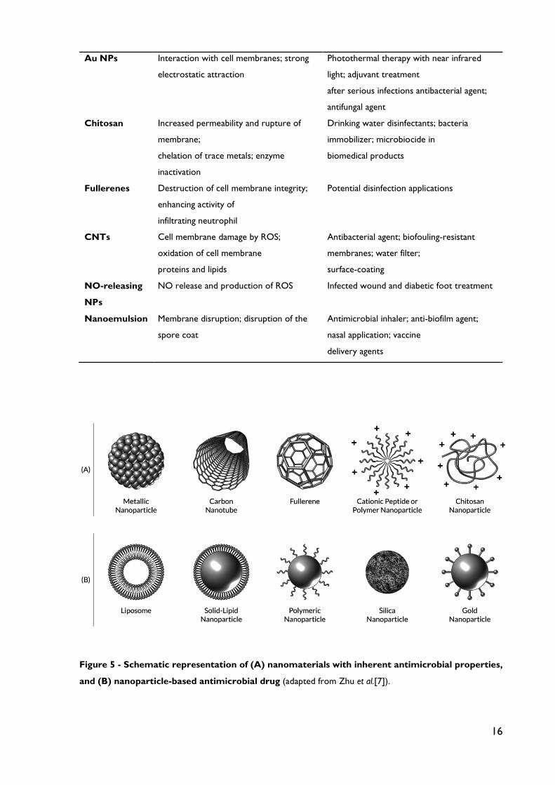
16
Au NPs Interaction with cell membranes; strong
electrostatic attraction
Photothermal therapy with near infrared
light; adjuvant treatment
after serious infections antibacterial agent;
antifungal agent
Chitosan Increased permeability and rupture of
membrane;
chelation of trace metals; enzyme
inactivation
Drinking water disinfectants; bacteria
immobilizer; microbiocide in
biomedical products
Fullerenes Destruction of cell membrane integrity;
enhancing activity of
infiltrating neutrophil
Potential disinfection applications
CNTs Cell membrane damage by ROS;
oxidation of cell membrane
proteins and lipids
Antibacterial agent; biofouling-resistant
membranes; water filter;
surface-coating
NO-releasing
NPs
NO release and production of ROS Infected wound and diabetic foot treatment
Nanoemulsion Membrane disruption; disruption of the
spore coat
Antimicrobial inhaler; anti-biofilm agent;
nasal application; vaccine
delivery agents
Figure 5 - Schematic representation of (A) nanomaterials with inherent antimicrobial properties,
and (B) nanoparticle-based antimicrobial drug (adapted from Zhu et al.[7]).
17
2.3. How can nanoantibiotics help to bypass bacterial drug resistance?
All over the world, researchers developed different nanotechnology-based approaches with
the aim to overcome the currently known bacterial resistance mechanisms to antibiotics. In
this subsection, evidence of how nanosystems can overcome bacterial mechanismas of
resistance is presented.
2.3.1. Alteration of bacteria’s efflux pump activity
In this regard, recently-reported advances could be mentioned. Khameneh et al. developed
piperine-containing nanoliposomes as a vector for gentamicin. The liposomal formulation was
specifically developed to fight MRSA, which is widely recognized as a nosocomial pathogen
[52]. The encapsulation of gentamicin in classical nanoliposomes or piperine-containing
nanoliposomes resulted in a dramatic decrease of minimum inhibitory concentration (MIC)
values of 16- and 32- folds, respectively. Similarly, minimum bactericidal concentration (MBC)
values were also reduced 4- and 8- folds for encapsulated gentamicin in classical nanoliposomes
or piperine-containing nanoliposomes, respectively. These hopeful results were attributed to
the piperine inhibiting effect on the bacterial efflux pump. This argument was confirmed using
ethidium bromide (EtBr) fluorescence assay. The fluorescence of this compound occurs only
when it is bound to nucleic acid. Accordingly, bacterial suspension was incubated with EtBr
for 30 min in the presence of: i) bare nanoliposomes (without piperine); ii) piperine-containing
nanoliposomes or iii) piperine in its free form. After centrifugation and washing of bacteria,
the loss of fluorescence was checked in order to investigate the efflux of EtBr outside bacterial
cells. Consistently, a gradual decrease of fluorescence during the assay period was observed
in the first case, i.e. in the absence of piperine. However, in the presence of piperine the
fluorescence was significantly enhanced indicating a significant inhibition of the efflux pump
[52]. Therefore, the enhanced antibacterial activity of gentamicin encapsulated in piperine-
containing nanoliposomes is likely to be the consequence of an increase in its intracellular
concentration. It is of note that piperine in its free form was less effective in inhibiting the
efflux pump than the liposomal one, as demonstrated by the EtBr fluorescence assay [17].
2.3.2. Antibiofilm activity
Nitric oxide (NO)-releasing NPs were found to prevent the formation of bacterial biofilms
and to eradicate already formed biofilms. Some examples of recent breakthroughs in this
domain are presented hereafter. Jardeleza et al. encapsulated isosorbide mononitrate (ISMN),
as NO donor into different liposomal formulations with the purpose toenhance the antibiofilm

18
activity against S. aureus’s biofilms [53]. NO-releasing multilamellar vesicles (MLV) efficiently
eliminated S. aureus’s biofilms in vitro. A five minutes’ exposure to 60 mg/mL ISMN-loaded MLV
induced an almost complete eradication of the biofilms. Paradoxically, the authors observed
that at low concentrations NO-releasing MLV enhanced the formation of biofilms, which is in
accordance with previously obtained results [54]
Duong et al. developed nanoparticulate NO-core cross-linked star polymers as new
therapeutics able to combating biofilms that are frequently formed during long exposure of
the body to medical devices and catheters [55]. These systems were found to release NO in
a controlled and slowed-down manner in bacterial cultures and showed great efficacy in
preventing both cell attachment and biofilm formation in P. aeruginosa over time. This study
unveiled, in part, the inherent mechanisms of NO’s antibiofilm activity. Accordingly, NO-
releasing NPs inhibits the switch of planktonic cells in contact with a surface to the biofilm
form by continuously stimulating phosphodiesterase activity. Thus, NO-releasing NP
maintained low intracellular concentrations of cyclic di-guanosine monophosphate (c-di-GMP)
in the growing bacterial population, thereby confining growth to an unattached free-swimming
mode [55].
The dual delivery of two antibiotics via their co-encapsulation in nanoliposomes is
another proposed strategy to bypass resistance mediated by biofilm formation. For instance,
Moghadas-Sharif proposed vancomycin/rifampin-co-loaded nanoliposomes as a new
therapeutic against S. epidermidis [56]. This strategy was based on two points. First,
combination therapy of vancomycin and rifampicin helps avoid the emergence of rifampin-
resistant strains. Indeed, numerous studies have already reported the antibiofilm activities of
rifampin in combinations with other antibiotics [57, 58]. Second, rifampicin fails alone to
eradicate bacterial biofilm [59]. Nevertheless, the developed liposomal combination was
ineffective to eradicate S. epidermidis’ biofilm. The authors attributed this result to the lack of
liposomal adsorption or low penetration into the bacterial biofilm [56]. A more adjusted
formulation with enhanced penetration behavior into the biofilm may lead to the initially
expected effect.
2.3.3. Enhanced penetration through biofilms
Several research papers reported the improved penetration across bacterial biofilms as a
plausible reason behind the enhanced antibacterial activity of encapsulated antibiotics against
resistant bacteria. For instance, liposomal encapsulation of polymyxin B was first described by
Alipour et al. as a strategy to enhance its antibacterial activity against P. aeruginosa resistant

19
strains [60]. As they expected, lower MIC values were observed for liposomal formulations
with respect to that of the free drug. In an attempt to elucidate the involved mechanisms, the
researchers focused on the drug uptake and more precisely on its penetration across the
biofilm formed by the polymyxin B-resistant P. aeruginosa strain. They used a coupled
immunocytochemistry-transmission electron microscopy (TEM) imaging technique.
Accordingly, a clinical strain of P. aeruginosa resistant to polymyxin B was incubated either with
free or liposomal polymyxin B at sub-MIC concentrations (i.e. 64 and 16 µg/mL, respectively).
Untreated bacteria were used as control. Penetration efficiency into biofilms was checked at
predetermined intervals of 0, 4, 8 and 16 h at 37°C. TEM studies showed that the uptake of
polymyxin B-loaded liposomes by the resistant strain was higher than that of the free drug
[29] [60]. It is important to mention that the treatment with both free drug and empty
liposomes did not display a superior effectiveness with regard to the free drug indicating that
the enhanced activity can only be attributed to the entrapped form. Furthermore, the
superiority of liposomal aminoglycosides was demonstrated on in vivo chronic Pseudomonas
infection model [61]. Consistently, mucoid P. aeruginosa-containing agar beads were instilled
intratracheally to Sprague-Dawley female rats. After the establishment of infection, animals
were treated by inhalation over 14 days. Two treatment regimens were used; tri-weekly
dosing schedule with free or liposomal amikacin at 6 mg/kg per dose and compared with the
classical aminoglycoside regimen, i.e. a twice daily dosing of free tobramycin at the same dose
(6 mg/kg/day). Finally, animals were killed and lungs were homogenized. Homogenates were
subsequently cultured on agar plates. Then, colony- forming units (CFU) were counted in
order to assess the effectiveness of the treatment. The researchers found that “free amikacin
was relatively ineffective in the reduction of CFU under these conditions, while bacteria were
undetectable in a large proportion of the group treated with liposomal amikacin” [61].
Interestingly, the thrice-weekly treatment with the liposomal amikacin was as effective as the
twice-daily treatment with free tobramycin. Although, tobramycin showed a lower MIC value
than amikacin against the planktonic form of P. aeruginosa [61]. The authors explained the
observed enhanced effectiveness of liposomal amikacin by the enhanced penetration through
biofilm and by the drug sustained release pattern. The researchers have demonstrated the
drug sustained- release profile from liposomes in CF-patients’ sputa [61]. They also checked
biofilm penetration on in vitro 4 days-grown biofilms produced by a mucoid form of PA01,
prepared using rat lung models with chronic infections. For this aim, fluorescently labeled
liposomal amikacin was used and biofilm penetration was imaged by confocal laser scanning
microscopy (CLSM) [61].

20
2.3.4. Protection against enzymatic degradation and inactivation
Nanoparticulate delivery systems provide a physical barrier shielding the entrapped antibiotic
from aggregation and inactivation with polyanionic compounds, such as bacterial endotoxins
e.g. LPS and LTA. Additionally, encapsulation may protect antibiotics against enzymatic
degradation by β-lactamases, macrolide esterases and other bacterial enzymes [62]
Two decades ago, Lagacé et al. demonstrated that liposomal encapsulation of
ticarcillin or tobramycin reverse the resistance of P. aeruginosa strains towards these both
antibiotics [63]. Growthinhibition of ticarcillin- and tobramycin- resistant strains was achieved
using ticarcillin and tobramycin liposomal formulations at 2 % and 20 % of their respective
MIC. Liposomal formulations were as effective against the β-lactamase -producing strains as
β- lactamase non-producing ones. Recently, Alipour et al. demonstrated the versatility of
liposomal encapsulation in protecting tobramycin or polymyxin B from inhibition by LPS, LTA,
neutrophil-derived DNA, actin filaments (F-actin) and glycoproteins e.g. mucin, common
components in the CF-patients‘ sputa [64]. Being polycationic, tobramycin and polymyxin B
can bind to these polyanionic compounds and thereby have their bioactivity reduced. The
authors postulated that “liposomes are able to reduce the antibiotic contact with polyanionic
factors in the sputum and to enhance bacteria-antibiotic interactions” [64]. In vitro stability
studies revealed that liposomal formulations were stable after an 18 h-incubation at 37°C with
i) a supernatant of biofilm-forming P. aeruginosa; ii) a combination of DNA, F-actin, LPS and
LTA or iii) an intact or an autoclaved patients’ sputum. No significant differences with respect
to control (before incubation) were observed. Furthermore, the antibacterial potency of
liposomal antibiotics was checked after both short (3 h) and prolonged (18 h) exposure to a
combination of DNA/F-actin or LPS/LTA at different concentrations. It was found that for
both free and liposomal drugs the antibioactivity was reduced in a concentration- dependent
manner. However, much higher concentrations (100 to 1000 mg/L) and (500 to 100 mg/L) of
LPS/LTA and DNA/Factin, respectively, were needed to inhibit liposomal forms in comparison
to free drugs. The authors explained this finding by the increased viscoelasticity induced by
the high concentrations of polyanionic elements that may hinder the interaction of liposomes
with bacteria. Indeed, the early leakage of antibiotics from liposomes cannot be used as a
plausible cause of the inactivation of liposomal antibiotic because in vitro stability studies
showed that liposomal vesicles were not disrupted [64]. To further confirm the superiority of
liposomal forms, the authors studied the bactericidal activity of liposomal formulations versus
free forms against P. aeruginosa found in CF-patients’ sputa. The antibacterial activities of

21
liposomal formulations were 4- fold higher when compared to the free drugs, despite the
presence of different bacterial strains in the patient’s sputum. It is of note that liposomal
tobramycin reduced growth at a high concentration (128 mg/L), whereas liposomal polymyxin
B did it at a markedly lower concentration (8 mg/L). The dissimilar activities of tobramycin
and polymyxin B was attributed to their different sites of action.
2.3.5. Intracellular bacterial killing
Obviously, the intracellular location reinforces bacterial resistance as it shields them from both
humoral and cellular host defenses and also from the action of therapeutic agents. Indeed,
intracellular bacteria, such as M. tuberculosis and L. monocytogenes, use cells of the innate
immune system, not only as reservoirs to launch recurrent infections but even more as vectors
enabling them to invade other sites of the body [65]. On the other hand, most of antibiotics,
e.g. aminoglycosides, β-lactams and glycopeptides, have restricted cellular penetration while
others can readily diffuse, e.g. fluoroquinolones and macrolides. Unfortunately, these latter
suffer from low intracellular retention [66]. Accordingly, a small number of available antibiotics
are effective against intracellular infections. To fight intracellular infections, NP are promising
vectors allowing antibiotics to target macrophages and to reach bacteria located in intracellular
compartments. In this field, a recent review article has already highlighted the role of NP for
targeting intracellular infections [67].
2.3.6. Specific targeting and sustained-release
Inherent toxicity of antibiotics is a crucial drawback that led to limit or even to stop the use
of some of them, such as aminoglycosides and lipopeptides known for their neuro- and
nephrotoxicity [68]. Therefore, specific targeting to bacteria would counteract drug toxicity,
since it enables to avoid non-selective and uncontrolled delivery to host cells. To date, few
works reported the design of NP with a specific targeting to bacteria for therapeutic purposes.
Some examples are presented hereafter. Qi et al. elaborated mesoporous silica NPs (MSN) as
nanocarriers of vancomycin (Van) in order to specifically target gram positive bacteria over
macrophage-like cells [69]. The specific recognition was based on hydrogen bonding
interactions of Van with the terminal D-alanyl-D-alanine moieties of gram positive bacteria.
Cell viability assay showed a good biocompatibility of Van-MSN with human embryonic kidney
and human hepatocytes. Tang et al. have recently described the design of a nanoparticulate
carrier loaded with a fluorescent dye, and called it “nanoprobe” for diagnostic purposes [70].
The surface of the nanoprobe was grafted with a bacterial ligand, i.e. concanavalin A, and

22
therefore displayed a high affinity to bacteria. The developed nanoprobe was shown to rapidly
detect and quantify the extent of bacterial colonization on wounds and catheters in real time.
Prolonged or sustained release of the loaded antibiotic is of great importance for
antibiotics with time-dependent action, such as lipoproteins, β-lactams, glycopeptides and
some fluoroquinolones. The importance of the sustained-release profile was highlighted by
Meers et al. [61]. Thanks to the prolonged release of amikacin from liposomes, this latter was
as effective, when administered triweekly, as free tobramycin administered twice-daily and
despite the fact that MIC of tobramycin is lower than that of amikacin. Additional examples of
antibiotic-loaded polymeric NPs were recently reviewed [71].
2.3.7. Downregulation of bacteria oxidative-stress resistance genes
Bacterial adaptation to oxidative and nitrosative stress could be considered as a resistance
mechanism to host defenses [72]. Indeed, innate immune cells generate reactive oxygen
species (ROS) and reactive nitrogen species (RNS) such as superoxide and peroxynitrite,
respectively, in order to kill phagocyted bacteria [73]. Consistently, pathogenic bacteria resist
to host-mediated oxidative stress by up-regulating the expression of their antioxidant
enzymes[74]. Importantly, it was claimed that many antibiotics exert their bactericidal effects
via the production of hydroxyl radicals, regardless of their molecular targets [75]. Recently, it
was found that metal NP, namely zinc oxide-NP (ZnONPs), exerts by themselves bactericidal
effects on gram positive bacteria and gram negative bacteria [76]. A synergistic killing effect on
acid fast bacteria (i.e. Mycobacterium bovis-BCG) was also observed for ZnONPs when used in
combination with rifampicin [76]. Moreover, ZnONPs effectively killed MRSA clinical strains
[76]. Several mechanisms were found to be involved in ZnO-NPs antibacterial activities. Most
importantly, ZnO-NPs were found to down-regulate the transcription of oxidative stress
resistance genes in S. aureus. Strictly speaking, the treatment with 300 µg/mL of ZnONPs
decreased the transcription of peroxide stress regulon kata and perR genes by 10- and 3.1-
folds, respectively, when compared to untreated bacteria [76]. These results highlight the
importance of ZnO-NP in fighting drug-resistant bacteria. It is of note that ZnO-NP induced
oxidative stress response on macrophages, as ROS and NO production was markedly
increased, thus reinforcing their bacterial killing capacity [76].
23
2.4. Nanoantibiotics delivery
2.4.1. Systemic versus local antibiotic delivery
Traditionally, systemic antibiotic administration has been the foundation of clinical therapies
to address the ever-present infectious onslaught. Unfortunately, poor penetration to ischemic
or post-operative tissue, inappropriate prescribing patterns, systemic toxicity, and poor
patient compliance, have predominated the conversation and limited the usefulness of certain
antibiotics. Furthermore, systemic administration is often not effective, as it does not provide
local tissue concentrations sufficient to kill bacteria prior to incurring serious side effects, such
as renal and liver damage. Sub-therapeutic or sub-inhibitory antibiotic concentrations are
known to inadvertently exacerbate infectious complication and promote antibiotic resistance
[2, 77]. Local delivery of current antibiotics and other antimicrobial biologics (e.g.,
antimicrobial peptides (AMPs), anti-quorum sensors, bacteriophage, etc.) may preserve and
extend their efficacy in the evolutionary race between antimicrobial development and bacterial
resistance. In fact, systemic toxicity, and to a lesser extent, antibiotic resistance is rarely seen
for local applications of the same drugs, achieving locally higher concentrations and
overcoming the reducing effects of lowered bacterial metabolism [78, 79]. Thus, device
integrated, local delivery strategies to mitigate the unacceptable consequences of systemic
antibiotic delivery (e.g., development of multi-drug resistant bacteria, systemic toxicity, and
rising healthcare costs, etc.) are urgently needed to keep pace with the rising demand for
medical devices. The concept of locally and sustainably delivering an anti-infective agent is not
new. Vancomycin, tobramycin, amoxicillin, gentamicin, cefamandol, caphalothin, and
carbenicillin have all been incorporated into commercially available local release systems [2,
80].
2.4.2. Nanoparticles against intracellular bacteria
NPs have also been used to combat intracellular bacteria. NPs, including liposomes, are small
enough to be phagocytosed by host phagocytes which contain intracellular microbes. Once
inside the host cell, these NPs can release drugs that then combat these intracellular microbes
[47, 67]. In addition, NPs can release high concentrations of antimicrobial drugs inside of
infected host cells while keeping the total dose of drug administered low [81]. The high local
dose at the site of infection kills the intracellular bacteria before they can develop resistance,
while the lower total dose decreases the probability that bacteria outside of the site of action
of the NPs will develop drug resistance [81].

24
NPs can combat intracellular microbes in alveolar macrophages. Intracellular microbes that
are phagocytosed and proliferate intracellularly in alveolar macrophages include L.
monocytogenes, M. tuberculosis, L. pneumophila, and Chlamydophila pneumoniae. As discussed
above, living inside of host cells protects these microbes from many antibiotics [1, 3].
Attachment of mannose to NPs containing antimicrobial drugs allows them to be targeted to
alveolar macrophages, which heavily express mannose surface receptors [1, 3]. Liposomes
containing ciprofloxacin and conjugated with mannose were administered by the pulmonary
route and shown to have high selectivity for alveolar macrophages [1, 3]. It was also shown in
vivo that mannose-conjugated liposomes lead to significantly higher concentrations of
antimicrobial drugs in alveolar macrophages relative to type II pneumocytes [1, 3].
2.4.3. Nanoparticles that target antimicrobial agents to the site of infection
NPs can target antimicrobial agents to the site of infection, so that higher doses of drug can
be given at the infected site, thereby overcoming existing resistance mechanisms with fewer
adverse effects upon the patient [82]. As with NPs targeting intracellular bacteria, NPs
targeting the site of infection can release high concentrations of antimicrobial drugs at the site
of infection, while keeping the total dose of drug administered low. Again, the high local dose
at the site of infection also kills the infecting bacteria before they can develop resistance, while
the lower total dose decreases the probability that bacteria outside of the site of action of the
NPs will develop drug resistance. NPs can be targeted to sites of infection passively or actively.
Passively targeted NPs selectively undergo extravasation at sites of infection, where
inflammation has led to increased blood vessel permeability. Actively targeted NPs contain
ligands (e.g. antibodies) that bind receptors (e.g. antigens) at sites of infection. The
antimicrobial action of NPs can also be activated by certain stimuli, such as reactive oxygen
species (ROS) or low pH at the site of infection. Drug release can also be regulated by magnetic
guidance or radio frequency, in order to target drug release by NPs to the site of infection
[47]. NPs can be conjugated with antibodies against a given antigen on the surface of the target
microbe. For example, AuNPs conjugated with antibodies against protein A have high
selectivity for killing S. aureus [47].
Aptamers can also target the site of infection. Aptamers are a type of nanoparticle
composed of DNA or RNA oligonucleotides which are folded into a 3-dimensional structure
and bind with high affinity to specific antigens, such as peptides and small molecules [1, 3].
Aptamers have been shown in vitro to have antibacterial activity against both Gram positive
and Gram negative bacteria expressing beta-lactamase [1, 3]. Aptamers also have antiviral

25
activity, including inhibition of HIV reverse transcriptase and inhibition of replication of
Vaccinia virus [1, 3]. Silver carbene complexes (SCCs) inside biodegradable NPs can also target
the lungs to treat pulmonary infections. SCCs contained in biodegradable NPs are small
enough to accumulate in the lung after administration using a nebulizer. Once inside the lung,
these NPs release and maintain therapeutically effective concentrations of SCCs at the site of
infection in the lung but not elsewhere, thereby limiting adverse effects [47]. L-Tyrosine
polyphosphate NPs (LTP-NPs) containing SCCs are one example of this type of biodegradable
nanoparticle [47]. In vitro, free SCCs and SCCs incorporated into LTP NPs have antimicrobial
activity against MRSA, multidrug resistant A. baumannii (MRAB), P. aeruginosa, B. cepacia, K.
pneumoniae, and C. albicans [47]. In a study by Hindi et al., two inhaled doses given over 72 h
of LTP-NPs containing SCCs decreased bacterial burden in the lung, decreased bacteremia,
and increased survival by 25% in mouse models with P. aeruginosa pneumonia [83]. The
mechanism of action of these NPs was thought to be slow sustained release of intact SCC
followed by release from the carbene of free Ag+, which then has antimicrobial effects at the
site of infection in the lung [83].
2.4.4. Synergistic effect of multidrug complexes
When chemically or physically combined with certain other chemical agents, some antibiotic
drug molecules form complexes that may be slowly soluble in body fluids. This slow dissolution
rate provides the extended release of the drug which accompanied with synergistic
pharmacological action. This phenomenon is of great interest for super-bugs treatment.
Among antibiotic based complexes the highest enhancing effect was observed for chitosan
(CS) and metal-based complexes (for metals such as: silver (Ag), zinc (Zn), cupper (Cu),
magnesium (Mg) and titanium (Ti)) due to their antibacterial nature. A better nanocarrier
platform is actually represented by the antibiotic-contained NPs. Instead of encapsulating the
antibiotic into nanocarriers, a core–shell nanoparticle structure made up of (i) antibiotic
nanoparticle as the core and (ii) lipid or polymer layer as the shell potentially represents a
more clinically effective nanoscale antibiotic delivery system. The antibiotic nanoparticle core
ensures high antibiotic loading, whereas the lipid or polymer shell can be functionalized for a
wide range of therapeutic functions. This combination was found to be active against several
resistant bacteria colonies. Recognizing that a majority of antibiotics are amphiphilic and
soluble in aqueous media, whether their acidic or basic solution, the most effective envelope
for antibiotic core was found to be an amphiphiles polyelectrolite. In particular polysaccharides
with antibacterial activity such as chitosan and dextran are well-established agents for antibiotic

26
complexation for drug delivery as well as for antibacterial resistance applications. While the
polyelectrolyte envelope provides positively charged surface, which enables complex
internalization into the bacteria body, the antibiotic core completely released from the
complex in the vicinity of the cells’ fluids and its antibacterial activity preserved against
bacteria’s resistant mechanisms [16].
2.4.5. Infection-activated delivery systems
An interesting approach for antibiotic delivery has been the development of systems that
differentially deliver therapeutic agents to infection sites. When bacterial infection happened,
bacteria will secrete many virulence factors, such as phospholipase, phosphotase, lipase, toxins,
protease, acidic pH and so on, to develop unique microenvironments [84]. Strategies utilizing
the unique microbial infection environment as a molecular cue to activate drug release or
facilitate their binding to the bacteria can achieve such an ambitious goal. These new strategies
can improve antibiotic targeting and activity with fewer side effects and can overwhelm drug
resistance mechanisms with high, sustained local drug concentrations.
Xiong et al. have developed a lipase-sensitive polymeric triple-layered nanogel (TLN)
for the differential delivery of antimicrobials to bacterial infection sites [85]. The TLN contains
a bacterial lipase-sensitive PCL interlayer between a cross-linked polyphosphoester core and
a shell of PEG. The PCL molecular fence protects the drug inside the polyphosphoester core
with minimal drug release prior to reaching sites of bacterial infection, thus eliminating
potential adverse side effects. However, once the TLN senses lipase-secreting bacteria, the
PCL fence of the TLN is degraded by lipase to release the drug. Using S. aureus as a model
bacterium and vancomycin as a model antimicrobial, it was demonstrated that the TLN
released almost all the encapsulated vancomycin within 24 h, but only in the presence of S.
aureus, significantly inhibiting bacterial growth. The TLN further delivered the drug into
bacteria-infected cells and efficiently released the drug to kill intracellular bacteria [71, 85].
2.5. Advantages and disadvantages of nanoantibiotics
The use of NPs as delivery vehicles for antimicrobial agents suggests a new and promising
paradigm in the design of effective therapeutics against many pathogenic bacteria [86].
Antimicrobial NPs propose several clinical advantages. First, nanocarriers can be engineered
to be activated by stimuli (e.g., chemical, magnetic field, heat, and pH) for targeted delivery as
well as biological sensors [87, 88]. For example, amoxicillin was freeze-dried in formulation
with CS and polyvinyl pyrrolidone (PVP) for acid-responsive release of antibiotics [89]. This
27
system could be particularly useful for treating abscess which is frequently acidic and reduces
the potency of conventional antimicrobial therapy. Generally, most molecules poorly cross
the blood brain barriers (BBB), a tight barrier to protect the brain from the penetration of
xenobiotics. However, it was also reported that antimicrobial NPs made of certain materials
and at varying particle sizes were capable of efficiently targeting infectious diseases by
overcoming anatomic barriers (e.g., BBB) [90]. Second, NPs can be molecularly tailored for
versatile physico-chemical properties in order to minimize side effects generated upon
systemic administration of traditional antimicrobial agents (e.g., hepatotoxicity of
cephalosporins, and ototoxicity and nephrotoxicity of aminoglycosides) [91]. Nanocarriers
seem to be able to reduce the side effects by improving the solubility and stability of
antimicrobial agents [92]. Third, NP-based antimicrobial drug delivery is promising in
overcoming resistance to traditional antibiotics developed by many pathogenic bacteria [86].
Fourth, administration of antimicrobial agents using NPs can improve therapeutic index,
prolong drug circulation (i.e., extended half-life), and achieve controlled drug release,
enhancing the overall pharmacokinetics [93]. Many studies demonstrated greater efficacy of
antimicrobial NPs than their constituent antibiotics alone. For example, vancomycin-capped
AuNPs exhibited 64-fold improved efficacy against VRE strains and Gram-negative bacteria
such as E. coli over vancomycin alone [94]. In addition, antimicrobial NPs can be prepared and
administered in convenient and cost-effective ways via various routes with lowered
administration frequency [51]. NP-based antimicrobial drug delivery can achieve improved
solubility and suspension of drugs, and concurrent delivery of multiple agents for synergistic
antimicrobial therapy [95]. Thus, antimicrobial NPs are of great interest as they provide a
number of benefits over free antimicrobial agents (Table 3).
Although nanoantibiotics promises significant benefits and advances in addressing the
key hurdles in treating infectious disease, there are foreseeable challenges in translating this
exciting technology for clinical use. These include thoroughly evaluating the interactions of
nanoantibiotics with cells, tissues, and organs, which consequently recalibrates doses and
identifies proper administration routs to obtain desired therapeutic effects [87]. Profound
knowledge about the potential toxicity of nanoantibiotics is also required to warrant successful
clinical translation [96]. It has been shown that intravenously injected NPs can be accumulated
in colon, lung, bone marrow, liver, spleen, and lymphatics [97]. Inhaled NPs also can enter the
systemic circulation and reach lung, liver, heart, spleen, and brain [96], which is particularly
facilitated for small size NPs because of efficient cellular uptake and transcytosis across
epithelial and endothelial cells into blood and lymph circulation [98]. Potential toxicity of

28
nanoantibiotics to human health is not known much at the moment although it likely shares
the nanotoxicity of various non-antibiotic nanomaterials [96, 99]. Many recent studies suggest
the possibility of multi-organ nanotoxicity that therapeutically administered antimicrobial NPs
may generate. For example, free radical-mediated oxidative stress generated by the interaction
of antimicrobial NPs with cells may result in hepatotoxicity and pulmonary toxicity [100].
Table 3 - Advantages and disadvantages of antimicrobial nanodelivery systems over free
antimicrobial agents (adaptedfrom Huh and Kwon [1]).
Antimicrobial NPs Free antimicrobial agents
Advantage Targeted drug delivery via specific accumulation
Lowered side effects of chemical antimicrobials
Low antimicrobial resistance
Extended therapeutic lifetime due to slow elimination
Controlled drug release
Broad therapeutic index
Improved solubility
Low immunosuppression
Low cost
Disadvantage No specific accumulation
High side effects of chemical antimicrobials
High antimicrobial resistance
Short half-life due to fast elimination
Usual pharmacokinetics of free drugs
Narrow therapeutic index
Sometimes poor solubility
Immunosuppression
High cost
Disadvantage Accumulation of intravenously injected nanomaterials in tissues and organs
High systemic exposure to locally administrated drugs
Nanotoxicity (lung, kidney, liver, brain, germ cell, metabolic, etc.)
Lack of characterization techniques that are not affected by NPs' properties
Advantage Absence of nanomaterials in the whole body
Low systemic exposure to locally administrated drugs
Absence of nanotoxicity
Well-established characterization techniques
Various metabolic changes suggest mitochondrial failure, and enhanced ketogenesis, fatty acid
β- oxidation, and glycolysis, contributing to hepatotoxicity and nephrotoxicity [100]. The toxic
effects of antimicrobial NPs on central nervous system (CNS) are still unknown, and the
interactions of NPs with the cells and tissues in CNS are poorly understood, and very small

29
steps are being taken towards that understanding [90, 101] Besides, some classes of NPs can
affect the circulatory system by altering heart rate [102] as well as reproductive system by
increased detachment of seminiferous epithelium [103] and possible spermatotoxicity [96,
104]. Table 5 presents potential multiorgan nanotoxicity that has been implicated to be
generated by therapeutically used antimicrobial NPs. NPs exhibit size-specific properties that
limit the use of currently available in vitro assays in a universal way, and there is no standardized
definition for NP dose in mass, number, surface area, and biological specimens (e.g., blood,
urine, and inside organs) [97, 105]. This means that there is a high demand to develop new
characterization techniques that are not affected by NP properties as well as biological media
[1].
Table 4 - Potential toxicity of therapeutically used NPs.(adapted from Huh and Kwon [1])
Toxicity type Mechanism for toxicity Pulmonary toxicity Acute inflammatory change; granuloma formation; oxidative stress
Renal toxicity Renal glomerulus swelling; proximal tubular necrosis; mitochondrial failure; enhanced ketogenesis, fatty acid beta-oxidation, and glycolysis
Hepatotoxicity ROS generation; mitochondrial dysfunction; GSH depletion; LDH leakage
Neurotoxicity Reduced neuro viabilities; exacerbation of cytoskeletal and blood-brain barrier (BBB) disruption; diminished ability to form neuritis in response to NGF; mild cognitive impairment, edema formation
Spermatotoxicity Sperm fragmentation; partial vacuolation of seminiferous tubules and cellular adhesion of seminiferous epithelium; suppressed proliferation of Leydig cell
NP-protein interactions
Abnormal protein functions generated by structural and conformational changes upon adsorption to NPs; raising protein potential for autoimmune effects
Others Embryo neurotoxicity; embryo death; metabolic alkalosis
Abbreviations GSH, glutathione; LDH, lactic dehydrogenase; NGF nerve growth factor.

30
CHAPTER III
Antimicrobial Peptides (AMPs) – new add on the
therapeutic arsenal
Antimicrobial peptides (AMPs) are natural small oligopeptides with a varying number of amino
acids, from 5 to 100. AMPs have a broad spectrum of action targeting microorganisms from
viruses to parasites. AMPs are also referred in literature as host-defense peptides, because
they are synthetized molecules that take action on the defense mechanisms against biological
threats of the living organism of origin. The discovery of AMPs dates back to the first half of
the 20th century, when in 1939 Dubos extracted an antimicrobial agent from a soil Bacillus
strain, proven to be effective in mice pneumococci infection. This extract was then fractioned
allowing the identification of gramicidin. Despite some systemic toxicity, gramicidin has shown
to be effective in the topical treatment of wounds and ulcers. The first animal-originated AMP
to be reported is defensin, isolated from rabbit leukocytes in 1956 [44]. Nowadays more than
2000 AMP have been described and current molecular developments can be consulted in a
series of databases available in the web, including natural identified molecules, as well as
peptidomimetic molecules and analogues, pharmacologically designed, thanks to the use of
bioinformatics [106]. Current FDA approved AMPs with well stablished use include bacitracin,
colistin and polymyxin B, only for topical administration [2].

31
Despite the lack of consensus on the influence of peptide sequence in biologic activity
of AMPs, some common characteristics seem to be important and fairly related to their
antimicrobial property. The main ones are primarily charge, with 90% of AMPs being cationic,
and secondly hydrophobicity or amphipathicity, influencing solubility profiles and consequently
bioavailability [107]. Associated with structural characteristics, these are also the main
physicochemical properties that also should be take into account on the design of new
synthetic AMPs [44].
Table 5- Currently available AMPs databases (adapted from da Costa et al.[106])
Database Description
Collection of antimicrobial peptides
(CAMP)
Holds experimentally validated and predicted AMP
sequences
AMPer Database and automated discovery tool for
gene-coded AMPs
Antimicrobial Peptide Database (APD) Contains mostly AMPs from natural sources
(~98 % of the entries)
Yet Another Database of Antimicrobial
Peptides (YADAMP)
Mostly focused on bacterial AMPs
BACTIBASE Data repository of bacteriocin AMPs
PhytAMP Database dedicated to antimicrobial plant peptides
RAPD Database containing recombinantly-produced AMPs
HIPdb Experimentally validated HIV inhibitory peptides
Bagel2 Bacteriocin mining tool
Peptaibol Database for peptaibols (unusual peptides)
PenBase Database devoted to penaeidins
Defensins KnowledgeBase Information and database dedicated to defensins
CyBase Database specialized in cyclotides
In addition, a comparison of AMPs pharmacological properties as antimicrobial agents
with conventional antibiotics is summarized in Table 2. AMPs demonstrate significant
advantages such as potency and board spectrum of activity, as well as an additional activity to
modulate the immune system responses, and low resistance rates. They also show some
limitations that impair their safe therapeutic use, such as sensitivity to proteolysis, influencing
stability, and undefined toxicological data for systemic use [108]. In order to overcome these
limitations some strategies have been developed, maximizing the proven therapeutic potential
of AMPs. There are numerous methods for obtaining AMP delivery systems. AMPs could be
immobilized into a variety of materials or onto a variety of surfaces and still retain their

32
antibacterial activity. Also, AMPs can be targeted through loading them in nanoparticulate
systems with selective delivery capacities, include polymers, liposomes, hydrogels,
nanospheres, nanocapsules, and carbon nanotubes [107]. As practical examples on of these
type a strategies are the studies of Mishra et al. on the development of an AMP-coated surface,
specifically immobilizing Lassioglossin-III onto silicone-based catheters and its activity against
urinary tract infections [109], and also of Water et al. on encapsulating plectasin, a cationic
AMP, into polymeric NPs and the evaluation of their efficacy on S. aureus strains [110].
Table 6- Properties of AMP's and conventional antibiotics (adapted from Mok and Li [108]).
Properties Conventional antibiotics Antimicrobial Peptides AMP’s Spectrum of activity
Usually selective Broad-spectrum (many with antibacterial, antifungal, antiviral, and anti-parasitic activities)
Uptake mechanism
Requires specific uptake mechanism
Non-specific, charge-dependent mechanism; self-promoted uptake
Cellular targets Usually has one or one class of targets
Multiple targets, including the membrane and intracellular targets
Rate of killing Dependent on growth rate (slow for cells in the lag phase)
Fast
Stability Dependent on the antibiotic (half-lives can range from hours to days)
Short half-life due to sensitivity to proteolysis
Additional activities
None Can neutralize endotoxins and modulate immune responses
Resistance rates
Resistance detected after a few passages at sub-MIC
Resistance is not readily induced and is only detected after multiple passages (~30) at sub-MIC; some species may be intrinsically resistant to the peptides, including those with impermeable outer membranes and those that produce proteases
Toxicology Relatively safe Safe for topical use; safety for systemic use is undefined
Manufacturing costs
Most are inexpensive (many range from $1-20 per gram)
Expensive ($50-400 per gram)
For better understanding these unique molecules, it is important to revise the main
classes in which they are subdivided and also their mechanism of action in microorganisms.
Also, potential targets and applications, as well as future perspectives on the implementation
and use of AMPs are discussed.

33
3.1. Classification of AMPs
The establishment of a consensual classification system for AMPs has received some
controversial discussion, mainly due to the large number of molecules identified and the lack
of data on their full biochemical and structural characterization. Classification may vary by
author or simply by complexity degree. A simple way to distinguish the main classes of AMPs
is taking into account their biological source. Several species, from prokaryotes to higher
eukaryotes, synthesize AMPs. Therefore, AMPs can be classified as either non-ribosomally
synthesized peptides (NRAMPs) or ribosomally synthesized peptides (RAMPs). NRAMP
synthesis catalyzed by peptide synthetases takes place in the cytosol of bacteria and fungi,
mainly possessing antibacterial properties, whereas RAMP synthesis occurs in the ribosomes
of the eukaryotic cells, also possessing antiviral, antiparasitic, antineoplastic and
immunomodulatory activity. Main examples of NRAMPs are polymyxin B, bacitracin,
vancomycin, gramicidin A, and cyclic peptides (daptomycin, dalfopristin, quinupristin and
cyclosporin A), whereas nisin is a gene-encoded RAMP [111]. Only some NRAMPs are
currently FDA approved and marketed.
Other two class division may be achieved considering molecule charge property at
neutral pH. Thus AMPs are subdivided in cationic antimicrobial peptides (CAMPs), and anionic
antimicrobial peptides (AAMPs). As referred, the majority of AMPs are of cationic nature,
despite the identification of AAMPs in invertebrates, vertebrates, and plants. AAMPs are part
of various vital organs of the body, including respiratory tract, the brain, the epidermis, the
epididymis, blood, and the gastrointestinal tract. As examples of AAMPs there are bovine
chromacins and kappacin, human dermcidin, and the amphibian temporin [107].
Nevertheless, authors have proposed more elaborated classifications of AMPs, based
on their biochemical characteristics, conformation and structure, or even taking account
biological activity and targets. Some clarification on this matter is discussed downward.
3.1.1. Biochemical classification
This classification is based mainly based on amino acid composition, dividing AMPs
into the following classes: (i) linear, (ii) cysteine-rich peptides, and (iii) peptides rich in specific
amino acids, like glycine, proline, arginine or histidine. Examples of AMPs are given in Table 3
[107].
Table 7- AMPs examples based on biochemical properties (adapted from Narayana and Chen [107]).
Class Example

34
Linear Cecropins, Clavanin, Piscidins, Styelin, Magainins, Dermaseptins, Buforins-II Pexiganan, LL-37
Cysteine-rich Single Esculentin, Bactenecin-1, Thanatin,
Double Tachyplesin, Androctonin, Protegrin-1 Three Defensins: α- (HNP3), β- (TAP), θ- (SapecinA) Four Drosomycin, Hepcidin
Specific amino acid-rich Proline Drosocin, Metchnikowins, Pyrrhocoricin, Metalnikowin Glycine Diptericins, Attacins Arginine Penetratin Histidine Histatin Tyrosine Indolicidin
3.1.2. Structural classification
Recently, a classification based on structure-function relationships studies with
Nuclear Magnetic Resonance (NMR) has been proposed, it classifies AMPs into three
structural groups: alpha-helical peptides, beta-sheet peptides and extended or loop peptides
[4].
Alpha-helical AMPs are characterized by the propensity towards the formation of
alpha-helix structures, and these peptides display highly cationic and amphipathic properties,
being mainly active against Gram-positive bacteria and fungi. However, helical content may be
also correlated to an increased hemolytic activity and cytotoxicity. Some examples of alpha-
helical AMPs are cecropins, magainins, temporins, buforins and clavanins [4, 10].
Beta-sheets AMPs are often stabilized by disulfide bridges between cysteine residues. This
group is mainly formed by beta-hairpin peptides and defensin mini-proteins. Examples include
protegrin-1, thanatin and lactoferricin B [4] .
Extended and loop AMPs are not folded into regular alpha-helix or beta-sheet
structures. The extended activity or cyclic character is due to the presence of some specific
amino acid residues, for instance, His, Pro, Cys, Arg and Trp. This type of AMPs examples are
histatins, proline rich isolates from insects, hepcidin, and indolicin [4].
3.1.3. Biological activity classification
Bahar et al., in their 2013 review have categorized AMPs based on their biological target and
mode of action. Hence the classes proposed are antibacterial peptides, antiviral peptides,
antifungal peptides, and antiparasitic peptides. Despite different target, AMPs tend to act in a
common manner, disrupting cell wall or viral envelope, causing disintegration and intracellular
components leakage. In case of antibacterial and antifungal peptides, there is also evidence of

35
intracellular targeting, inhibiting some important pathways inside the cell such as DNA
replication and protein synthesis [44].
3.2. Mechanisms of action
AMPs are unique molecules that can mimic endogenous host peptides, exerting their action in
several fronts and targets. Studies have shown that AMPs demonstrate different antimicrobial
action mechanisms. The most recognized and explored one is the disruption of cell integrity.
However, recently, intracellular active AMPs have been shown also to interact with targets
inside the cells, interfering in DNA synthesis and other critical biochemical pathways.
Furthermore, the immunomodulatory functions of some AMPs have significant contribution
to their efficacy as antimicrobial agents [4, 44, 108] (Figure 4).
Figure 6– Mechanisms of Action of AMPs. Potential targets (1) Bacterial cell wall (2) DNA Synthesis (3)
Protein Synthesis (4) Protein Folding (5) Cell wall Synthesis. (adapted from Cruz et al.[4]).
These combination of cellular and biological actions may offer increased security by decreasing
dosage, however, action in multiple targets and immunomodulatory response may raise some
toxicity or immunogenicity issues.
3.2.1. Membrane disruption and permeation

36
Membrane disruption and permeation is the first line of action of AMPs in microorganism
eradication. This type of mechanism is, primarily, strongly dependent on the interaction of
AMPs with the cell wall. Peptides usually bind to the target cell be ion exchange, suggesting a
non-specific membrane interaction. Subsequently, mechanisms of disruption and permeation
are strongly dependent on peptide physicochemical characteristics. Accordingly, several
models have been proposed (Figure 5), that differ mainly in the mode of attachment to bacteria
and cell membrane insertion. The models in discussion are: the barrel-stove model, the
toroidal pore model, both pore-inducing models, the aggregate channel model, and the carpet
model, the lipid charge clustering model, and the detergent model, described as non-pore
models [4].
The pore-inducing models describe an initial lateral peptide-membrane interaction.
Past this first contact, in the barrel-stove model, peptides oligomerize and the monomers are
internalized into the membrane. Then, within the lipid bilayer, these minor subunits expose
their hydrophobic regions aligning them perpendicularly with the lipid membrane content,
forming small pores from where intracellular content is expelled. In the toroidal pore model,
hydrophilic regions of peptides and lipid head groups interacts together forming a pore
structures, which is generally larger than barrel stave type pores [4, 108].
In the aggregate channel mode, may be seen as a variation of the pore induced models,
by the similar channel formation where intracellular content leakage occurs. The initial action
of the peptide involves competitive displacement of lipopolysaccharide (LPS) associated with
divalent cations (Mg2+ and Ca2+), where the peptides destabilize this supramolecular assembly
and gain access to both external and internal membranes. Then, aggregation of peptides forms
channels allowing diffusion of ions through the membrane [4].
The non-pore models assume that the peptide aggregation and accumulation onto
the membrane surface causes several membrane disruptions, which can ultimately lead to
disintegration and leakage. In the carpet model, the accumulation of peptides parallel to the
membrane past a threshold level disrupts membrane curvature and causes the formation of
transient holes. The presence of cationic peptides on membranes can also induce the clustering
of anionic lipids, as illustrated by the lipid clustering models. Ultimate consequences to the
AMP-membrane interaction can be more dramatic, as suggested by the detergent model. High
concentrations of peptide can cause total membrane dissolution, resulting in micelle formation
and complete bilayer breakdown [108].

37
Figure 7 – Membrane permeabilization models. As a consequence of AMP interaction with the membranes
of their target microorganisms, their integrity is disrupted by mechanisms that may or may not involve pore
formation. The figure illustrates the most recognized models. (adapted from Mok and Li [108]).
3.2.2. Intracellular activity
Beyond disrupting the integrity of the cellular membrane, AMPs can undergo cellular uptake,
mainly by endocytosis. The internalization of AMP is the first step to initiate a series of
intracellular activities that can compromise cell viability. For instance, APMs can interfere both
in DNA and RNA synthesis, inhibiting signaling pathways and enzymatic mechanisms. They can

38
also inhibit microbe proteases and may interact with certain specific metabolic pathways during
bacterial growth [44, 108].
3.2.3. Inducing immune response
In addition to the previous described mechanisms, some AMPs, as host-defensive peptides,
have also some intrinsic immunomodulatory properties. The use of immunosuppressive or
immunostimulatory agents alone for the treatment of infections may be counterproductive, as
immunosuppressive compounds may increase the risk of infections, while immunostimulatory
ones can trigger inflammation, autoimmunity, and even fatal cytokine storms. Thus,
combination with antimicrobial activity is an important advantage to consider to their
therapeutic potential. These peptides have been demonstrated to target various aspects of the
host immune response. They include the recruitment of monocytes, neutrophils, mast cells,
and other immune cells by chemoattraction, regulation of cytokines, chemokines, and
histamine release stimulation of fibroblast growth, vascular endothelium proliferation,
angiogenesis, and wound repair, promotion of apoptosis and clearance of infected cells
neutralization of bacterial LPS, LTA, and endotoxins and the expression of genes related to
cell proliferation and adhesion in cells in a macrophage cell line [108].
3.3. Future perspectives
AMPs are promising molecules with unique characteristics that show enormous potential as a
new add on the therapeutic arsenal for infectious disease treatment and for overcoming
antimicrobial resistance. However, studies on these same molecules are relatively recent and
their knowledge has been growing. The future perspectives on AMPs may be assured in two
fronts: the identification of new sources and new molecules, and the establishment of
therapeutic use by designing safe and effective formulations.
The study of human synthetized AMPs is very promising, with the intent to isolate
and identify specific tissue molecules in order to fight same tissue related infections. For
instance the oral cavity and saliva has been considered a very promising source of potent AMPs
[106], and they have shown good activity against oral cavity pathogens and infections [112].
Great advances have also been made on designing AMPs therapeutic formulations. These
AMPs may be considered to be a new class of antimicrobial therapeutics, which are particularly
promising because of unlikely peptide resistance [107]. In 2014 there were 20 AMP and AMP
derivatives formulations in clinical or pre-clinical trials, mainly in phase II [4]. Despite the
earlier stage, it seems to be just a matter of time in the establishment of therapeutic use and
commercialization of AMPs as new class of antimicrobial drugs.
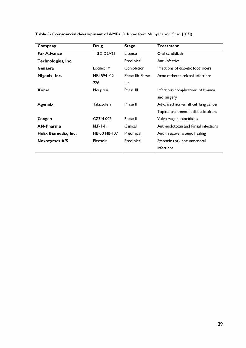
39
Table 8- Commercial development of AMPs. (adapted from Narayana and Chen [107]).
Company Drug Stage Treatment
Par Advance
Technologies, Inc.
113D D2A21 License
Preclinical
Oral candidiasis
Anti-infective
Genaera LocilexTM Completion Infections of diabetic foot ulcers
Migenix, Inc. MBI-594 MX-
226
Phase IIb Phase
IIIb
Acne catheter-related infections
Xoma Neuprex Phase III Infectious complications of trauma
and surgery
Agennix Talactoferrin Phase II Advanced non-small cell lung cancer
Topical treatment in diabetic ulcers
Zengen CZEN-002 Phase II Vulvo-vaginal candidiasis
AM-Pharma hLF-1-11 Clinical Anti-endotoxin and fungal infections
Helix Biomedix, Inc. HB-50 HB-107 Preclinical Anti-infective, wound healing
Novozymes A/S Plectasin Preclinical Systemic anti- pneumococcal
infections

40
CHAPTER IV
Polymeric nanostructures for AMPs delivery
Different biodegradable polymeric nanosystems have been explored as carriers for
antimicrobial agents that exhibit a high bactericidal activity. The efficacy of this strategy is well
proven, with reports that polymeric nanosystems can effectively improve the cellular
penetration, intracellular retention and specific subcellular distribution of antimicrobial agents,
and even evade intracellular inactivation of antimicrobial agents [113].
Controlled drug release using biocompatible and biodegradable polymers further
emerged in the 1980s [114]. after the first polymer-based delivery of macromolecules using
poly[ethylene vinyl acetate] polymer was demonstrated in 1976 [115, 116], Antimicrobial drug
delivery using polymeric NPs offers several advantages: i) structural stability in biological fluids
and under harsh and various conditions for preparation; ii) precisely tunable properties, such
as size, zeta-potentials, and drug release profiles, by manipulating polymer lengths, surfactants,
and organic solvents used for NP preparation [95], and iii) facile and versatile surface
functionalization for conjugating drugs and targeting ligands [117].
There are two major types of polymeric for antimicrobial drug delivery NPs have
been explored: linear polymers (e.g., polyalkyl acrylates and polymethyl methacrylate) and
amphiphilic block copolymers. The majority of polymeric NPs prepared with linear polymers
are nanocapsules or solid nanospheres [118]. In polymeric nanocapsules, a polymeric

41
membrane that controls the release rate surrounds the drugs that are solubilized in aqueous
or oily solvents. In solid nanospheres drugs are homogeneously distributed in the polymeric
matrices of variable porosities [119, 120]. Amphiphilic block copolymers form self-assemble
micellar NPs with the drug being encapsulated in the hydrophobic core and surrounded by a
hydrophilic shield. This shied allows the core to be protected from degradation [121]. Several
biodegradable polymers, including poly (lactic acid) (PLA), poly (glycolic acid) (PGA), poly
(lactide-co-glycolide) (PLGA), poly (carprolactone) (PCL), and poly (cyanoacrylate) (PCA),
have been used as the hydrophobic core of the amphilphilic copolymers, whereas PEG has
been the most commonly used hydrophilic segment [95]. Targeting ligands can also be
conjugated on the termini of PEG for targeted and selective delivery [94, 122].
Polymeric NPs have been explored to deliver various antimicrobial agents and greatly
enhanced therapeutic efficacy in treating many types of infectious diseases has been reported.
For instance, encapsulated ampicillin im polymeric NPs was effective forS. typhimurium infection
treatment [123] and intracellular L. monocytogenes infection in mouse peritoneal macrophages
[124]. The use of polymeric NPs can overcome the limited oral administration of unstable or
inadequately absorbed drugs, [125].and, in addition, PEGylation of NPs, can increase drug half-
life in serum, and improve mucoadhesive capabilities by reducing phagocytosis [126] Thus,
among nanoparticle platforms, polymer NPs may be the most suitable system that can be used
for antimicrobial drug delivery.
Biodegradable polymers ang bioorganic polymers are also promising materials in the
delivery of peptide based drugs due to their compatibility, degradation behavior, and nontoxic
nature of administration [127]. The development of polymeric therapeutic nanostructures for
AMP delivery may offer an excellent technological strategy to improve drug bioavailability and
safety. CS-based NPs (CSNPs) are particularly interesting as the broad spectrum of
antibacterial activity of CS is well known and documented, offering the possibility of sinegistic
effects with antimicrobial molecules. Moreover, due to its biocompatibility properties, CS
nanostructures have been extensively studied for drug delivery and that is no different for
AMPs delivery. In fact, tha majority of the studies revised in the present work involves CS
nanostructures. Therefore, in addition to the review of the recent advances in polymeric
nanodelivery of AMPs, section 4.1 of this chapter is especially dedicated to the recognized
properties of this biopolymer.
4.1. Chitosan: a highly recognized biopolymer

42
CS, a versatile biopolymer of the aminoglucopyran family is being extensively explored for
various biomedical and pharmaceutical applications such as drug delivery. CS is a natural,
cationic aminopolysaccharide (pKa 6.5) copolymer of glucosamine and N-acetylglucosamine
obtained by the alkaline, partial deacetylation of chitin. It is the second most abundant natural
polysaccharide and originates from shells of crustaceans. CS is a biodegradable, biocompatible,
positively charged nontoxic mucoadhesive biopolymer. The structure of CS is very similar to
that of cellulose [made up of b (1-4)-linked D-glucose units], in which there are hydroxyl
groups at C-2 positions of glucose rings. CS is a linear copolymer polysaccharide consisting of
b (1-4)-linked 2-amino-2-deoxy-D-glucose (D-glucosamine) and 2-acetamido-2-deoxy-D-
glucose (N-acetyl-D-glucosamine) units [127].
Figure 6 – Chemical structure of Chitosan (CS) polymeric unit.
CS can be considered mostly hydrophilic, but the percentage of acetylated monomers
and their distribution in the chains has a critical effect on its solubility and conformation in
aqueous media. Because of these molecular features, CS exhibits in solution a pH-dependent
behavior and interesting biopharmaceutical properties such as mucoadhesiveness and ability
to open epithelial tight junctions. As a result of the research undertaken over the last two
decades, there is nowadays an acceptable understanding of the biocompatibility and general
safety features of CS. For example, it is currently known that CS is biodegraded by a number
of enzymes, such as lysozyme, di-Nacetylchitobiase,N-acetyl-beta-D-glucosaminidase and
chitiotriosidase, which are present in human mucosas and other physiological fluids [128].
From a regulatory perspective, CS has a FDA GRAS [129] status and it is currently being used
as a common dietary supplement for preventing fat absorption and also in the form of wound
dressings. Examples of current marketed CS-based medical devices and oral nutraceuticals are
described in Table 4 [130].

43
Table 9 - Examples of commercial medical devices and oral nutraceuticals with CS (adapted from
Szymanska and Winnika [130]).
Product Material Usage/Application Manufacturer Wound-healing and hemostatic products
Chitodine® CS powder with adsorbed
elementary iodine Disinfection of wounded skin,
surgical dressing
International Medical Services
ChitoPack C® Cotton-like CS
Regeneration and reconstruction of body tissue, subcutaneous
tissue and skin
Eisai Co.
CeloxTM Gauze and granules with CS
Control of bleeding from non-cavitary grain wounds
MedTrade ChitoFlex® CS acetate sponge
HemCon Medical Technologies INC.
HemCon® Bandage Pro HemCon®
Strip First Aid
Freeze-dried CS acetate salt
PosiSep®
N,O-carboxymethyl CS sponge
Intranasal hemostatic splint for patients undergoing nasal/sinus
surgery
Hemostatis LLC.
Syvek Excel™ Lyophilized three-dimensional
CS fibers Rapid control of bleeding for
anticoagulated patients
Marine Polymer Technologies Inc.
Clo-Sur® PAD Non-woven seal with a
soluble CS
Control of moderate to severe bleeding
Scion Cardio-vascular
ChitoSeal® Soluble CS salt Abbott Vascular
Devices
TraumaStat® Porous polyethylene fibers
filled with silica, coated with CS (ChitoClear®)
Ore-Medix
Tegasorb® CS particles Tesla-Pharma
Vulnosorb®
Composition of microcrystalline CS with fibrinogenic tissue glue
3M
Nutraceutical products
Slim Med™ Non-animal CS Prevention and treatment of
overweight KitoZyme S.A.
KiOcardio™ Non-animal CS Maintenance of normal blood
cholesterol level KitoZyme S.A.
LipoSan Ultra® Composition of CS
(ChitoClear®) and succinic acid
Binding dietary fat and reducing its absorption in the intestine
Primex
Liposorb™ CS extracted from squid
Preventing irritable bowel syndrome;
Binding dietary fat and reducing its absorption in the intestine
Good Health
4.1.1. Production of Chitosan and Chitosan Derivatives

44
The starter material for the production of CS is chitin. The main sources of chitin are the
shells of crustaceans, mainly crabs and shrimps. Shells of the same size and species are grouped,
then cleaned, dried and ground into small shell pieces. There are no standard purification
methods as different chitin sources require different treatments due to the diversity in their
structures. Conventionally, the protocol is divided into demineralization, deproteinization and
decolorization steps which can be carried out using chemical or biological (enzymatic
treatment or fermentation) treatments. The end-products need to be highly purified if they
are to be used for biomedical or pharmaceutical purposes, as residual proteins, minerals or
pigments can cause serious side effects. Conversion of chitin to CS can be achieved by
enzymatic or chemical deacetylation. Chemical deacetylation is more commonly used for
commercial preparation because of economic issues and feasibility for mass production. The
degree of deacetylation influences directly the physicochemical properties of the final polymer
[131, 132].
Subsequently, CS molecules can be modified by chemical processes, in order to
engineer the optimal physicochemical properties for the purposes required. For instance,
chemical modification is a powerful tool in the design of CS based drug delivery systems,
controlling the interaction between polymer and drug and thus enhancing the loading capacity
and controlling drug release [127]. In the book chapter authored by Sonia et al. the following
types of modification on CS molecules are revised: hydrophobic modification, thiolation,
quaternized forms and chemical grafting. The hydrophobic character of CS can be increased
by the covalent attachment of hydrophobic excipients. Hydrophobic interactions are believed
to enhance the stability of substituted CS by reducing the hydration of the matrix and thereby
increasing resistance to degradation by gastric enzymes.
Thiolated polymers or thiomers are hydrophilic macromolecules exhibiting free thiol
groups on the polymeric backbone and represent new promise in the field of mucoadhesive
polymers. Quaternized derivatives of CS are obtained by introducing various alkyl groups to
the amino groups of CS molecule structure. These derivatives are drastically more soluble in
neutral and alkaline environments of the intestine and, hence, are more efficient than CS for
drug delivery and absorption across the gastrointestinal tract. Chemical grafting of CS is an
important field of study for the functionalization and practical use of CS is an attractive
technique for modifying the chemical and physical properties of CS. A graft copolymer is a
macromolecular chain with one or more species of block connected to the main chain as side
chain(s). The properties of the resulting graft copolymers are broadly controlled by the
characteristics of the side chains, including molecular structure, length, and number [127].

45
Luo et al. 2014 review, describes yet another type of CS derivatives by the formation
of polyelectrolyte complexes between CS molecules and other natural polysaccharides.
Polyelectrolyte complexes [133] are formed simultaneously by mixing oppositely charged
polyelectrolytes in solution without any chemical covalent cross-linker, thus biocompatibility
of CS is maintained in the final complex, overcoming toxicity issues associated with chemical
modification. The major interactions between two polyelectrolyte polymers include the strong
but reversible electrostatic and dipole–dipole association, as well as hydrogen and
hydrophobic bonds. Due to the protonation of amino groups on the backbone, CS becomes
a cationic polyelectrolyte in acidic medium, which could form PEC with negatively charged
polyelectrolytes, resulting in various applications based on different preparation methods. CS-
based PEC has been proved to possess various applications in biomedical and pharmaceutical
areas, such as drug delivery for nutrients with delayed digestibility and controlled release, non-
viral vector for gene delivery system, three-dimensional scaffold to mimic tumor
microenvironment and for bone tissue engineering [133].
4.1.2. Chitosan recognized properties
Bernkop-Schnürch et al. 2012 revised the main recognized physicochemical properties of CS
that offer advantage in the design of CS-based drug delivery systems. Considering chemical
structure, CS primary amino groups are responsible for properties such as controlled drug
release, mucoadhesion, permeation enhancement, and self-assembling. By chemical
modifications, most of these properties can even be further improved. When sustained drug
release cannot be provided by making use of a simple drug dissolution process, by diffusion,
by erosion, by membrane control, or by osmotic systems, retardation mediated by ionic
interactions is often the ultimate ratio. The cationic character of CS molecules influences
controlled release CS-based systems design and can be achieved by conjugation with anionic
polymeric excipients such as polyacrylates, sodium carboxymethylcellulose, or alginate. The
mucoadhesive properties are likely also based on its cationic character. The mucus gel layer
exhibits anionic substructures in the form of sialic acid and sulfonic acid substructures. Ionic
interactions between the cationic primary amino groups of CS and these anionic substructures
of the mucus, in addition, with minor hydrophobic interactions mucoadhesion can be achieved.
The mechanism being responsible for the permeation enhancing effect of CS is also based on
the positive charges of the polymer, which seems to interact with the cell membrane resulting
in a structural reorganization of tight junction-associated proteins, promoting membrane
opening and solute permeation [134].

46
Self-assembly is a spontaneous process by which organized structures with particular
functions and properties could be obtained without additional complicated processing or
modification steps, leading to easier and less onerous production methods for therapeutic
vehicles [135]. Self-assembly of CS is mainly possible through electrostatic interaction of ionic
polysaccharides of opposite charges. Ionic complexes are attractive for their simplicity and
because their self-assembly is responsive to pH and ionic strength. Polysaccharide drug
delivery systems can also be made responsive to other stimuli, including heat, magnetism, and
light. In many cases, this response to environmental change is often reversible, thus, a
“switching” effect can be built into the polymer nanostructure that will respond to stimulation
in vivo. Some CS derivatives have proven in situ gellification properties in response to biological
fluids pH alterations, and local stimulation by heat or light [136].
CS and its derivatives have also diverse biological activities, including antioxidant, anti-
hypertensive, anticoagulant, antidiabetic, anti-obesity, anti-allergic, anti-inflammatory,
antimicrobial, anticancer, neuroprotective and matrix metalloproteinases inhibitory effects.
[137].
4.2. Recent advances on the development of polymeric based nanostructures for
AMPs delivery
4.2.1. Recent advances in the development of chitosan-based nanostructures
In this subsection a review of current advances in the development of CS-based
nanostructures for AMPs deliveryis offered (Table 10). Research has been covering, the
technological development of new carrier systems and their full characterization, and the
evaluation of their efficacy as drug delivery improvers.
CS micro- and NPs are still the most studied types of CS-based drug delivery systems.
In fact, new structures or new production methods are recent study objects. Rivera et al.,
developed biodegradable nanocapsules, as carriers for two bioactive compounds, 5-
aminosalycilic acid and glycomacropeptide. Nanocapsules were produced through layer-by-
layer deposition of CS and alginate layers on polystyrene NPs. The bioactive compounds were
incorporated on the third layer of the nanocapsules being its encapsulation efficiency and
release behavior evaluated. Final results demonstrated that the synthesized nanocapsules
presented spherical morphology and a good capacity to encapsulate different bioactive
compounds [138]. Developments on the antimicrobial delivery by CS particles are still
emerging, with target delivery being one promising field. Cerchiara et al. prepared CS-based

47
particulate formulations for colon delivery of vancomycin. CS particles were prepared by ionic
gelation and freeze-drying or spray-drying as recovery methods. Antibacterial activity against
S. aureus, a Gram-positive model strain, was evaluated [139]. CS particles are also considered
for AMPs delivery. Considering peptide character, Piras et al., in 2014, developed a
nanoparticle model with commercially available CS loaded with lysozyme [140] as
antimicrobial protein drug model. Beyond the intrinsic antibacterial activity of CS and LZ, the
LZ-NPs evidenced a sustained antibacterial activity that resulted in about 2 log reduction of
the number of viable S. epidermidis compared to plain CS NPs. In addition to these potential
antimicrobial applications, LZ-NPs demonstrated good biocompatibility, indicating that this
study could serve as an optimal model for development of CS NPs carrying antimicrobial
peptides for biomedical applications [140]. In 2015, studies for specific encapsulation of AMPs
with CS-based particulate systems have been reported. Sun et al. have prepared and
characterized CS and poly-gamma-glutamic acid [141] composite micro particles as carriers
for antimicrobial peptides [141] and nitric oxide (NO) delivery systems using ionic
complexation method. The results indicated that both LL-37 and NO were co-loaded
successfully in micro particles, and the composite particles could sustain LL-37 and NO release
at physiological pH, in vitro [141]. Rishi et al. encapsulated cryptdin-2 [123] in CS
tripolyphosphate (CS-TPP) NPs by ionotropic gelation. The formulation was then
characterized on the basis of particle size, zeta potential and polydispersity, and antimicrobial
in vivo assays were performed. Infected mice when treated against Salmonella enterica infection,
with the encapsulated peptide showed 83% survivability and approximately 2 log unit
reductions in the bacterial load in the tissues versus 100% mortality observed with the free
peptide. The study is a first pre-clinical report on the oral effectiveness of cryptdin-2 by its
suitable encapsulation and has potential for future clinical applications [123]. Piras et al, the
same authors who developed the previous described model, tested their hypothesis by
encapsulating frog-skin derived AMP temporin B [142] into CS NPs (CS-NPs) and evaluated
possible increase in antibacterial activity, while reducing its toxic potential. Temporin B-loaded
CS-NPs were prepared based on the ionotropic gelation between CS and sodium
tripolyphosphate (NaTPP). The nanocarrier evidenced a sustained antibacterial action against

48
Tab
le 1
0 –
Rec
ent
stu
dies
on
CS
bas
ed n
ano
stru
cutu
res
for
AM
P d
eliv
ery
Sys
tem
A
MP
S
tudy
des
crip
tio
n A
utho
rs
NP
s CS-a
lgin
ate
nano
caps
ule
---
Dev
elop
men
t of
nan
ocap
sule
s ca
rrie
rs fo
r bi
oact
ive
com
poun
ds, p
rodu
ced
thro
ugh
LbL
tech
niqu
e us
ing,
5-a
min
osal
ycili
c ac
id a
nd
glyc
omac
rope
ptid
e m
odel
.
Riv
era
et a
l. [1
38]
CS-b
ased
nan
opar
ticle
V
anco
myc
in
CS
part
icle
s w
ere
prep
ared
by
ioni
c ge
latio
n an
d fr
eeze
-dry
ing
or s
pray
-dry
ing
as r
ecov
ery
met
hods
. Ant
ibac
teri
al a
ctiv
ity a
gain
st
S. a
ureu
s.
Cer
chia
ra e
t al.[
139]
CS-b
ased
nan
opar
ticle
Ly
sozy
me
as
mod
el
Dev
elop
men
t of
a n
anop
artic
le m
odel
with
com
mer
cial
ly a
vaila
ble
CS
load
ed w
ith ly
sozy
me
as a
ntim
icro
bial
pro
tein
dru
g m
odel
.
Pira
s et
al.
[140
]
CS a
nd p
oly-
gam
ma-
glut
amic
acid
co
mpo
site
LL-3
7 T
he r
esul
ts in
dica
ted
that
bot
h LL
-37
and
NO
wer
e co
-load
ed s
ucce
ssfu
lly in
mic
ro p
artic
les,
and
the
com
posi
te p
artic
les
coul
d su
stai
n LL
-37
and
NO
rel
ease
at
phys
iolo
gica
l pH
, in
vitro
.
Sun
et a
l. [1
41]
CS tr
ipol
ypho
spha
te (C
S-TP
P) N
Ps
Cry
ptdi
n-2
Prep
arat
ion
of C
S tr
ipol
ypho
spha
te (
CS-
TPP
) N
Ps b
y io
notr
opic
gel
atio
n. T
he fo
rmul
atio
n w
as t
hen
char
acte
rize
d on
the
bas
is o
f pa
rtic
le s
ize,
zet
a po
tent
ial a
nd p
olyd
ispe
rsity
, and
ant
imic
robi
al in
vivo
ass
ays
agai
nst
Salm
onel
la e
nter
ica w
ere
perf
orm
ed.
R
ishi
et a
l. [1
23]
CS-b
ased
nan
opar
ticle
T
empo
rin
B C
S-N
Ps w
ere
prep
ared
bas
ed o
n th
e io
notr
opic
gel
atio
n be
twee
n C
S an
d so
dium
tri
poly
phos
phat
e. T
he n
anoc
arri
er e
vide
nced
a
sust
aine
d an
tibac
teri
al a
ctio
n ag
ains
t va
riou
s st
rain
s of
S. e
pide
rmid
is.
Pira
s et
al.
[142
]
Nan
oge
ls
Nan
ogel
com
posit
e
---
Prep
atio
n of
AgN
Ps e
mbe
dded
in a
bio
com
patib
le n
anog
el c
ompr
isin
g de
grad
able
, nat
ural
pol
ymer
s. In
thi
s st
udy,
hyb
rid
nano
gels
w
ere
prep
ared
with
var
ying
pol
ymer
con
tent
and
the
ir p
oten
tial b
y de
term
inin
g th
eir
antib
acte
rial
pro
pert
ies
agai
nst
E. c
olia
nd S
. au
reus
str
ains
.
Col
l Fer
rer
et a
l., [
143]
Glyc
ol- C
S na
noge
l --
- St
udy
of t
he b
ioco
mpa
tibili
ty o
f a g
lyco
l CS
nano
gel b
y ev
alua
tion
of e
ffect
s on
met
abol
ic a
ctiv
ity, c
ell c
ycle
s bl
ood
com
patib
ility
. O
vera
ll, t
he r
esul
ts d
emon
stra
ted
the
safe
ty o
f the
use
of t
he G
C n
anog
el a
s dr
ug d
eliv
ery
syst
em.
Pere
ira
et a
l. [1
44]
Nan
ofib
ers
and
film
s
CS th
in fi
lms
hLF1
-11
Imm
obili
zatio
n pe
rfor
med
ont
o C
S th
in fi
lms
as a
mod
el fo
r an
impl
ant
coat
ing
due
to it
s re
port
ed o
steo
geni
c an
d an
tibac
teri
al
prop
ertie
s. C
S th
in fi
lms
wer
e pr
oduc
ed b
y sp
in-c
oatin
g on
Au
surf
aces
. Act
ivity
aga
inst
met
hici
llin-
resi
stan
t S.
aur
eus
(MR
SA).
C
osta
et a
l. [1
45]
Nan
ofib
ers
Def
ensi
n-1,
D
erm
asep
tin
LL-3
7 M
agai
nin
1
Alte
rnat
e de
posi
tion
of p
olyc
atio
n (C
S) a
nd p
olya
nion
ove
r co
tton
gau
zes.
Ant
imic
robi
al a
ssay
s w
ere
perf
orm
ed w
ith t
wo
stra
ins:
S. a
ureu
s an
d K.
pne
umon
ia.
Gom
es e
t al.
[145
]
Fo
od
pack
agin
g sy
stem
s
CS fi
lms
Nis
in
Stud
y of
the
effi
cien
cy a
s an
timic
robi
al c
arri
ers
of h
ydro
xypr
opyl
met
hylc
ellu
lose
[14
6], c
hito
san
(CS)
, sod
ium
cas
eina
te (
SC)
and
poly
lact
ic a
cid
[146
] fil
ms,
in t
he r
elea
se r
ates
of f
luor
esce
ntly
labe
led
nisi
n Z
, eva
luat
ing
thei
r po
tent
ial a
s fo
od p
acka
ging
po
lym
ers.
Im
ran
et a
l. [1
46]

49
various strains of Staphylococcus epidermidis for at least 4 days, with up to 4-log reduction in
the number of viable bacteria compared to plain CS-NPs. The antimicrobial evaluation tests
also demonstrated that while the intrinsic antimicrobial activity of CS ensured a "burst" effect,
the gradual release of TB further reduced the viable bacterial count, preventing the regrowth
of the residual cells and ensuring a long-lasting antibacterial effect [142].
Nanogel carriers are also promising nanobased antimicrobial delivery systems. Coll
Ferrer et al., in 2014, have developed a novel and simple synthesis route to create nanosized
Ag NPs [143] embedded in a biocompatible nanogel (NG) comprising degradable, natural
polymers. In this study, hybrid nanogels were prepared with varying polymer content and their
potential by determining their antibacterial properties against E. coli and S. aureus strains [143].
The research of CS-based nanogels for biomedical applications has also grown exponentially
in the last years, however its biocompatibility is still insufficiently reported. Therefore, Pereira
et al. studied the biocompatibility of a glycol CS [144] nanogel effects on metabolic activity,
cell cycles blood compatibility. Overall, the results demonstrated the safety of the use of the
GC nanogel as drug delivery system [144].
Studies of AMP functionalized surfaces and fibers have been characterized and
evaluated. Costa et al. studied the antimicrobial activity effect of hLF1-11, an antimicrobial
peptide with high activity against MRSA, immobilization onto a polymeric surface.
Immobilization was performed onto CS thin films as a model for an implant coating due to its
reported osteogenic and antibacterial properties. CS thin films were produced by spin-coating
on Au surfaces. hLF1-11 was immobilized onto these films by its C-terminal cysteine in an
orientation that exposes the antimicrobial activity-related arginine-rich portion of the peptide.
Surface antimicrobial activity was assessed through surface adhesion and viability assays using
an MRSA, and the incorporation of hLF1-11 decreased significantly bacterial adhesion to CS
films [147]. Gomes et al. developed a new strategy to obtain antimicrobial wound-dressings
based on the incorporation of four different antimicrobial peptides into polyelectrolyte
multilayer films built by the alternate deposition of polycation (CS) and polyanion over cotton
gauzes[145]. Antimicrobial assays were performed with two strains: S. aureus and K.
pneumonia. Results showed that all antimicrobial peptides used in this work have provided a
higher antimicrobial effect (in the range of 4 log - 6 log reduction) for both microorganisms,
in comparison with the controls, and are non-cytotoxic to normal human dermal fibroblasts
at the concentrations tested [145].
In a different concept of application, Imran et al. studied the efficiency as antimicrobial
carriers of hydroxypropyl methylcellulose (HPMC), chitosan (CS), sodium caseinate (SC) and
50
polylactic acidfilms, in the release rates of fluorescently labeled nisin Z, evaluating their
potential as food packaging polymers [146].Nisin is a small cationic peptide composed of 34
amino acid residues. It exhibits a wide spectrumantimicrobial activity against Gram-positive
bacteria and is suitable for food preservation [148]. HPMC, CS, SC packaging films showed
ability to progressively release nisin to sustain an anti-bacterial against food borne pathogens
effect and can be favorably used for prolonging shelf life of packed food [146].
4.2.2. Recent advances in other polymeric nanostructures
In addition to CS-based nanostructures, recent studies on other polymeric nanostructures for
AMPs delivery have been developed. These studies are presented in this subsection (Table
11).
D’Angelo et al. designed and developed a system of nano-embedded microparticles
(NEM) for sustained delivery of cationic AMPs (CAMPs) [149] in the lung, studying its effect
on P. aeruginosa, a known lung infection pathogen. To this purpose, PLGA NPs containing a
model CAMP, colistin (Col), were produced by the emulsion/solvent diffusion technique, and
then spray-dried in different carriers (lactose or mannitol), thus producing NEM. The most
promising NEM formulations were selected on the basis of bulk and flow properties,
distribution of NPs in the carrier and aerosolization performance upon delivery through a
breath-actuated dry powder inhaler. Col–loaded NEM were found to kill P. aeruginosa biofilms
and to display a prolonged efficacy compared to the free Col.[149]. Another CAMP, plectasin,
was encapsulated into PLGA NPs using the double emulsion solvent evaporation method, in
the work of Water et al. [110] The plectasin-loaded NPs displayed a high encapsulation
efficiency (71–90%) and mediated release of the peptide over 24 h. The antimicrobial efficacy
was investigated using bronchial epithelial Calu-3 cell monolayers infected with S. aureus, and
encapsulated plectasin displayed improved efficacy as compared to nonencapsulated plectasin.
The author also assessed the subcellular localization of the prepared NPs in different relevant
cell lines: Calu-3 epithelial cells, THP-1 macrophages and A549 epithelial cells. Here the results
have shown good patterns of penetration on Calu-3 epithelial cell lines, as well as in THP-1
macrophages [110].
Hydrogels and nanogels are an important class of biomaterials that have been widely utilized
for a variety of biomedical/medical applications. The biological performance of these systems,
particularly those used as wound dressing; can be complemented with antimicrobial activity
capable of preventing colonization of the wound site by opportunistic bacterial pathogens [143,
150, 151]. These types of structures have also been studied recentely for AMPs

51
delivery.Continously to their study of the antimicrobial activity of multi-domain CAMPs (MD-
CAMPs) in solution, Jiang et al. investigated the same effect of self-assembled 3-D hydrogels
supramolecular nanostructures and its rheological properties [152]. Among the studied MD-
CAMPs solutions, the bactericidal activity of peptide hydrogels was found to be improved. The
improved antimicrobial activity of the self-assembled peptide hydrogels was found to be
related to the combined effect of supramolecular surface chemistry and storage modulus of
the bulk materials, rather than the ability of individual peptides/peptide assemblies to penetrate
bacterial cell membrane as observed in solution. Thus, the structure–property–activity
relationship developed through this study may provide important knowldge for designing
biocompatible peptide hydrogels with built-in antimicrobial activity for various biomedical
applications [152]. The Water et al. group also designed novel nanogel-based novicidin delivery
system. The peptide novicidin was self-assembled with ananoctenyl succinic anhydride-
modified analogue of hyaluronic acid, and this formulation was optimized using a microfluidics-
based quality-by-design approach. The encapsulation efficiency of novicidin (15% to 71%) and
the zetapotential (−24 to −57 mV) of the nanogels could be tailored bychanging the
preparation process parameters, with a maximum peptideloading of 36±4%. The nanogels
exhibited good colloidal stabilityunder different ionic strength conditions and allowed
complete release of the peptide over 14 days. Furthermore, self-assembly ofnovicidin with
hyaluronic acid into nanogels significantly improved the safety profile at least five-fold and six-
fold when tested in HUVECsand NIH 3T3 cells, respectively, whilst showing no loss of
antimicrobial activity against E. coli and S. aureus[153].Li et al. explored the potential application
of AMPs in wound healing, by developing a biodegradable poly(L-lactic acid)-Pluronic L35-
poly(L-lactic acid) (PLLA-L35-PLLA) in situ gel-forming system [154]. An injectable formulation
composed of human AMPs 57 (AP-57) loaded NPs and thermosensitive hydrogel was
prepared. AP-57 peptides were enclosed with biocompatible NPs (AP-57-NPs) with high drug
loading and encapsulation efficiency. AP-57-NPs were further encapsulated in a
thermosensitive hydrogel (AP-57-NPs-H) to facilitate its application in cutaneous wound
repair. As a result, AP-57-NPs-H released AP-57 in an extended period and exhibited quite
low cytotoxicity and high anti-oxidant activity in vitro. The in vivo wound healing assay using
full-thickness dermal defect model of SD rats indicated that AP-57-NPs-H could significantly
promote wound healing. At day 14 after operation, the treated group showed nearly complete
wound closure of 96,78 ±3,12% [154].
Other studies of nisin nanoencapsulation were performed, with the purpose to protect
to ensure the stability of this AMP during food processing and storage period. Nisin-loaded
52
pectin NPs (NLP-NPs) were prepared and analysed by Krivirotova et al. by a simple
complexation method [155]. Three types of pectin biopolymer were tested and found that
the methoxylation degree of pectin influenced on the nisin loading efficiency and particle size.
For the complex formation, both electrostatic and hydrophobic interactions were important.
NLP-NPs exhibited antimicrobial activity also dependent on the type of biopolymer. Overall,
the results indicated that NLP-NPs may be a suitable antimicrobial system to be used in food
industry [155].

53
Tab
le 1
1 –
Rec
ent
stu
dies
on
oth
er p
oly
mer
ic n
ano
stru
cutu
res
for
AM
P d
eliv
ery
Sys
tem
A
MP
S
tudy
des
crip
tio
n A
utho
rs
NP
s
PLG
A na
nopa
rticl
e C
olis
tin
Dev
elop
men
t of
a s
yste
m o
f nan
o-em
bedd
ed m
icro
part
icle
s (N
EM)
for
sust
aine
d de
liver
y of
CA
MPs
in t
he lu
ng
D’A
ngel
o et
al.
[149
]
PLG
A na
nopa
rticl
e Pl
ecta
sin
Intr
acel
lulla
r an
tibac
teri
al a
ctiv
ity a
gain
st S
. aur
eus
in e
pith
eial
cel
ls.
Wat
er e
t al.
[110
]
Hyd
ro-
and
Nan
oge
ls
Self-
asse
mbl
ed 3
-D h
ydro
gels
M
D-C
AM
Ps
Stud
y of
bac
teri
cida
l act
ivity
com
pare
d to
MD
-CA
MPs
in s
olut
ion,
and
rhe
olog
ical
pro
pert
ies.
Jia
ng e
t al.
[152
]
Hya
luro
nic
acid
nan
ogel
N
ovic
din
Qua
lity-
by-d
esig
n no
vel n
anog
el-b
ased
nov
icid
in d
eliv
ery
syst
em.
Wat
er e
t al.
[153
]
PLLA
-L35
-PLL
Ain
situ
gel
- A
P-57
Pote
ntia
l app
licat
ion
of A
MPs
in w
ound
hea
ling,
by
deve
lopi
ng a
bio
degr
adab
le p
oly
(L-la
ctic
aci
d)-P
luro
nic
L35-
poly
(L-
lact
ic a
cid)
(PLL
A-L
35-P
LLA
) in
situ
gel
-form
ing
syst
em
Li e
t al.
[154
]
Fo
od
pres
erva
tio
n sy
stem
s
Pect
in n
anop
artic
le
Nis
in
Stud
y of
a s
afe
suita
ble
antim
icro
bial
sys
tem
to
be u
sed
in fo
od in
dust
ry. I
nflu
ence
of p
ectin
deg
ree
of a
cetil
atio
n on
NP
prop
ertie
s.
Kri
voro
tova
et a
l. [1
55]

54
CHAPTER V
Inorganic or metallic nanostructures for AMPs
delivery In recent years, an increasing number of papers reporting on a new generation of antimicrobial
metallic NPs has been published. Consequently, many of the information on the application of
nanotechnology in the infectious disease field regards the use of silver (Ag) and gold (Au) NPs.
Recently, derivatives of other metals have been studied for antimicrobial applications, and the
antibacterial effects of zero-valent bismuth NPs and uncoated Au, nickel (Ni) and silicon (Si)
NPs were reported [156, 157]
Despite the demonstrated intrinsic antimicrobial properties, dispersed metallic NPs
tend to aggregate and separate in solution, resulting in a decrease in their antimicrobial
efficiency. With the aim of improving antibacterial properties, functionalization of NPs has
been attempted with surfactants, polymers, or antibiotics resulting in more stable, less
aggregated NPs suspension and innovative synergistic antibacterial agents. For instance, silver
NPs stabilized by polymers (polyvinylpyrrolidone) and surfactants (SDS and Tween 80) exhibit
enhanced antibacterial activities [158]. NPs can also act as drug-carriers able to pass through
cell membranes [159, 160]. Widely used antibiotics such as ciprofloxacin may benefit from the
association with NPs, and conjugation may result in an antibacterial effect also against micro-
organisms resistant to the same molecule in the naturally occurring form [161]. When the
antimicrobial agents are covalently linked to, or contained within, NPs, a higher drug

55
concentration is attained in the area of interest, resulting in better efficacy at comparable
doses and/or in slower release over time that may be exploited for preventing bacterial
colonization [162, 163]. Moreover, specific biological sites can be attacked after modification
of NPs with target molecules [164, 165]. As the NPs themselves may have antibacterial
properties, the combination of NPs and loaded drugs exerts a synergistic action [166].
Current advances in the use of inorganic nanostructures for AMPs delivery, involve
essentially the development of Ag and AuNPs, as well as silicon derivates nanosystems. The
studies revised in this chapter are summarized in table 12.
5.1. Gold nanoparticles
Currently, two studies have been reported exploring the use of Au nanostructures for AMPs
delivery. Photoluminescent Au nanodots (AuNDs) were prepared by Chen et al. [167] These
AuNDs were functionalized with hybridized ligands, an antimicrobial peptide (surfactin; SFT),
and 1-dodecanethiol (DT), on AuNPs. Ultrasmall SFT/DT–Au NDs (size ≈2.5 nm) were
achieved and exhibited highly efficient antimicrobial activity. The photoluminescence
properties and stability as well as theantimicrobial activity of SFT/DT–Au NDs were also
studied, and it was shown that these characteristics are highly dependent on the density of
SFT on Au NDs. Relative to SFT, SFT/DT–AuNDs exhibited greater antimicrobial activity, not
only to non multidrug-resistant bacteria but also to the multidrug-resistant bacteria. The
minimal inhibitory concentration values of SFT/DT–AuNDs were much lower (>80-fold) than
that of SFT. The authors considered that the antimicrobial activity of SFT/DT–AuNDs was
mainly achieved by the synergistic effect of SFT and DT–AuNDs on the disruption of the
bacterial membrane. In vitro cytotoxicity and hemolysis analyses were also performed and have
revealed superior biocompatibility of SFT/DT–AuNDs than that of SFT. Moreover, in vivo
methicillin-resistant S. aureus infected wound healing studies in rats showed faster healing,
better epithelialization. This study suggested that the SFT/DT–AuNDs system may be a
promising antimicrobialcandidate for preclinical applications in treating wounds and skin
infections [167]. Rai et al. also reported a one-step methodology to generate AMP-conjugated
(AuNPs) [168]. The AMP-conjugated AuNPs prepared showed controlled size (14 nm) and
low polydispersity, and allowed the inclusion of high concentration of AMPs. Also these

56
Tab
le 1
2 –
Rec
ent
stu
dies
on
ino
rgan
ic n
ano
stru
cutu
res
for
AM
P d
eliv
ery
Sys
tem
A
MP
S
tudy
des
crip
tio
n A
utho
rs
Au
nano
stru
ctur
es
Phot
olum
ines
cent
gol
d na
nodo
ts
(AuN
Ds)
Su
rfac
tin
AuN
Ds
func
tiona
lized
with
hyb
ridi
zed
ligan
ds, a
n an
timic
robi
al p
eptid
e (s
urfa
ctin
; SFT
), an
d 1-
dode
cane
thio
l (D
T),
on A
u N
Ps, f
or
appl
icat
ion
in w
ound
infe
ctio
n tr
eatm
ent.
Che
n et
al.
[167
]
AuN
Ps
Cec
ropi
n
mel
ittin
Mag
aini
n-1
Tet
-20
Dev
elop
men
t of
a o
ne-s
tep
synt
hetic
rou
te fo
rm fu
nctio
naliz
atio
n of
AuN
Ps w
ith A
MPs
Rai
et a
l. [1
68]
Ag
nano
stru
ctur
es
AgN
Ps n
anof
iber
PA
s St
udy
of m
etal
lized
org
anic
nan
ofib
ers
for
appl
icat
ion
in w
ound
infe
ctio
n tr
eatm
ent
Pazo
s et
al.
[169
]
AgN
Ps
M
BP-1
St
udy
of t
he s
yner
gist
ic a
ntib
acte
rial
effe
ct o
f pla
nt p
eptid
e M
BP-1
and
AgN
Ps o
n in
fect
ed w
ound
s ca
used
by
S. a
ureu
s Sa
lout
i et a
l. [1
70]
Si n
ano
stru
ctur
es
Anio
nic
mes
opor
ous
SiN
Ps
LL-3
7 St
udy
of m
embr
ane
inte
ract
ion
with
AM
Ps in
diff
eren
t ty
pes
of m
esop
orou
s pa
rtic
les.
Br
aun
et a
l. [1
71]
Nan
osph
eres
Tri
chog
in G
A
IV
Am
pullo
spor
in
A
Stud
y of
AM
Ps p
rope
rtie
s ad
sorb
ed o
n si
lica
base
d su
rfac
es fo
r po
tent
ial a
pplic
atio
ns in
intr
acel
lula
r dr
ug d
eliv
ery
Syry
amin
a et
al.
[172
]
Nan
ocla
ys
Nis
in
Pedi
ocin
St
udy
of n
anoc
lays
as
nano
carr
iers
for
nisi
n an
d pe
dioc
in a
dsor
ptio
n, fo
r ap
plic
atio
ns in
food
indu
stry
. M
eira
et a
l. [1
73]
57
systems demonstrated higher antimicrobial activity and stability in serum and in the presence
of non-physiological concentrations of proteolytic enzymes than soluble AMP, as well as
lowcytotoxicity against human cells [168].
5.2. Silver nanoparticles
Recent advances in AMPs delivery by Ag nanostructures, Pazos et al. [169] reported on
supramolecular assemblies of novel peptide amphiphiles (PAs) containing aldehyde
functionality in order to reduce Ag ions and subsequently nucleate Ag metal NPs in water.
This proposed system spontaneously generates monodisperse Ag particles at fairly regular
distances along the length of the filamentous organic assemblies. The metal−organic hybrid
structures exhibited antimicrobial activity and significantly less toxicity toward eukaryotic cells.
Metallized organic nanofibers of the type described offer the possibility to create other
structures, for instance, hydrogels, that can be potentially applied in wound dressing
development [169]. Also addressing the wound infection problem, Salouti et al. investigated
the synergistic antibacterial effect of plant peptide MBP-1 and Ag NPs on infected wounds
caused by S. aureus [170].The MIC and MBC of MBP-1 and Ag NPs both on their own and in
combination form were determined against S. aureus via macrodilution and microdilution
methods. The MIC and MBC of MBP-1 were found to be 0.6 and 0.7 mg/mL, respectively. MIC
and MBC of AgNPs were determined to be 6.25 and 12.5 mg/L, respectively. MIC and MBC
of the AgNPs and MBP-1 combination were found to be 3,125 mg/mL, 0.5 mg/L; and 6.25
mg/mL, 0.6 mg/L, respectively. The synergistic antibacterial effect of Ag NPs and MBP-1 was
investigated on infected wounds caused by S. aureus in a mouse model and the infected wound
healed properly after the combined use of MBP-1 and AgNPs [170].
5.3. Silicon nanostructures
As delivery systems for AMPs, silicon and silicon derivates nanostructures have also been
investigated recently. Membrane interactions are critical for the successful use of mesoporous
SiNPs. In order to elucidate these, Braun et al. have studied the effects of NPcharge and
porosity on AMP loading and release, as well as consequences of this for membrane
interactions and antimicrobial effects [171]. Anionic mesoporous SiNPs were found to
incorporate considerable amounts of the CAMP LL-37, whereas loading was found to be much
lower for non-porous or positively charged SiNPs. The results also demonstrated that due to
preferential pore localization, anionic mesoporous particles, but not the other particles,
protects LL-37 from degradation byinfection-related proteases. For anionic SiNPs, membrane
disruption is mediated almost exclusively by peptide release. In contrast, non-porous SiNPs

58
builded up a resilient LL-37 surface coating due to their higher negative surface charge, and
display largely particle-mediated membrane interactions and antimicrobial effects. For
positively charged mesoporous SiNPs, LL-37 incorporationpromoted the membrane binding
and disruption displayed by the particles in the absence of peptide, but also caused toxicity
against human erythrocytes. Thus, the use of mesoporous SiNPsas AMPs delivery systems
requires consideration of membrane interactions and selectivity of both free peptide and the
peptide-loaded NPs [171]. The properties of AMPs adsorbed on inorganic or organic surfaces
are of interest for their potential applications in intracellular drug delivery. In the work of
Syryamina et al. continuous-wave (CW) electron paramagnetic resonance (EPR) and pulsed
electron-electron double resonance (PELDOR) techniques were applied to study adsorption
of the AMPs trichogin GA IV and ampullosporin A on monodisperse colloidal silica
nanospheres (SiNS) of 20 nm diameter [172]. The results obtained by CW EPR supported the
view that the adsorbed peptides form close-packed clusters. PELDOR data show that both
trichogin and ampullosporin adsorbed on the silica surface possess a more disordered
conformation as compared to that in solution. Forampullosporin, disordering is much more
pronounced than for trichogin. Afterdesorption, the peptides restored their conformations;
upon adsorption the peptidesi n some cases may lose partly their biradical character [172].
This results may be of interest as antimicrobial activity is often related to peptide
conformation.
Nanoclays or layered silicates are an interesting nanostructure that have been used
for remediation of environmental contaminants, delivery of drugs and various active molecules,
and to enhance polymer mechanical and barrier properties in packaging films. They typically
present a stacked arrangement of silicate layers with a nanometric thickness [174]. Meira et
al. studied three different nanoclays (bentonite, octadecylamine-modified montmorillonite and
halloysite) as potential carriers for the AMPs nisin and pediocin, known bacteriocins, the first
referred above as having application as a food preservative. Higher adsorption at room
temperature of nisin and pediocinwas obtained on bentonite. The antimicrobial activity of the
resultant bacteriocin-nanoclay systems was analyzed using skimmed milk agar as food simulant
and the largest inhibition zones were observed against Gram-positive bacteria for halloysite
samples. Bacteriocins were intercalated into the interlayerspace of montmorillonites as
deduced from the increase of the basal spacing measured by X-ray diffraction (XRD) assay.
These results indicate that nanoclays, especially halloysite, are suitable nanocarriers for nisin
and pediocin adsorption, and the results may be consider interesting for the food industry
[173].

59
CHAPTER VI
Conclusions and future prospects
Microbial infections are still one of the major public health concerns in humans, in particular,
in patients suffering from immunosuppressive therapies and/or diseases (e.g. chemotherapy,
AIDS). The classical therapeutic strategies that suppress the immune system of compromised
patients are costly and associated to severe adverse side effects. In addition, several yeast and
fungi organisms, and other pathogenic microorganisms, are responsible for continuous growth
of infections and drug resistance against antimicrobial molecules. The current antimicrobial
resistance rate is impelling in the urgency to research and develop new strategies to fight drug
resistant superbugs. Efforts in new medicines discover have not accompanied the development
of drug resistance, thus a technological approach on improving existent ones is gaining special
interest.
Therapeutic nanostructures may overcome several of the current therapeutic
limitations by a selective delivery of the drug in the site of action, reducing the adverse side
effects and microbial drug resistance by the reduced administered dose. Nanomedical research
provides easy access to innovative nanodevices and nanosytems, which ultimately enable us to
design and fabricate targeted delivery systems of most efficient drugs with increased efficacy
and reduced toxicity. Based on good performance, successful experiments, and considerable
market prospects, nanotechnology will undoubtedly lead a revolution in medical markets also

60
for infectious diseases, as it was proven in Chapter II that are several nanotechnological
approaches the exhibit important roles in the restoration of antibiotic activity for resistant
bacteria.
AMPs are promising antimicrobial compounds that constitute the most promising
drug candidates, in a foreseeable future, in the fight against infections and to overcome the
alarming rise in microbial drug resistance. However, they also display some limitations in terms
of bioavailability and safety, and may also possess additional biological activities and functions.
These biological activities and functions include signaling molecules, tissue regeneration,
biomarkers and even tumoricidal agents, as the current level of research on AMPs reveals
additional roles for these versatile molecules. Nonetheless, no matter the possible fields of
applications, AMPs constitute the paradigm of the current view on translational medicine: a
collaborative, two-way road from bench to bedside. Even so, after years of promising data,
the main question lingers: can we effectively use primitive molecules as the basis for new drug?
The development of AMPs delivery nanostructures seems to offer a very appealing
and effective manner to overcome these issues, and several systems have been designed and
studied with this purpose, as it was showed in Chapters IV and V. However, several questions
in how to regulate the distribution of nanocarriers in the body or specific organs are also
needed to be answered. Nano-drugs are foreign substances to the body and may produce
inflammation. Therefore, safety data for long-term therapy or repeated dosage are needed to
circumvent the potential risk. To date, few studies have investigated the toxicologicaland
environmental effects of direct and indirect exposure to nanomaterials and no clear guidelines
exist to quantify these effects. Therefore, there is an urgent need for developing guideline,
which can assure the safer use of nanomaterials. Moreover, more powerful ex vivo models or
animal models are needed to assess the safety issues and to comply with government
regulations. How to extend the shelf life of nano-drugs is also a problem due to their
agglomeration is also a problem. The production methods for nanostructures should also be
improved, and scalable studies for industrial production are also of great importance, in order
to promote cost effectiveness of these new formulations. The cost and production of
nanomaterials at large scale is one of the hurdles in effective implementation of these products.
Hence, scientific community should also pay attention to develop affordable methodologies so
that nanotechnology can reach to patients.
In conclusion, it seems that, although the promising research results in this area are
rising, it is also urgent to start directing efforts in making these new drug formulations a reality
as therapeutic agents.

61
References
[1] A.J. Huh, Y.J. Kwon, "Nanoantibiotics": a new paradigm for treating infectious diseases
using nanomaterials in the antibiotics resistant era, J Control Release, 156 (2011) 128-145.
[2] B.D. Brooks, A.E. Brooks, Therapeutic strategies to combat antibiotic resistance, Adv Drug
Deliv Rev, 78 (2014) 14-27.
[3] R.Y. Pelgrift, A.J. Friedman, Nanotechnology as a therapeutic tool to combat microbial
resistance, Advanced Drug Delivery Reviews, 65 (2013) 1803-1815.
[4] J. Cruz, C. Ortiz, F. Guzman, R. Fernandez-Lafuente, R. Torres, Antimicrobial peptides:
promising compounds against pathogenic microorganisms, Current medicinal chemistry, 21
(2014) 2299-2321.
[5] C.o.D. Control, ANTIBIOTIC RESISTANCE THREATS in the United States, in, 2013.
[6] F. Cavalieri, M. Tortora, A. Stringaro, M. Colone, L. Baldassarri, Nanomedicines for
antimicrobial interventions, J Hosp Infect, 88 (2014) 183-190.
[7] X. Zhu, A.F. Radovic-Moreno, J. Wu, R. Langer, J. Shi, Nanomedicine in the Management
of Microbial Infection - Overview and Perspectives, Nano Today, 9 (2014) 478-498.
[8] C.A. Arias, B.E. Murray, The rise of the Enterococcus: beyond vancomycin resistance,
Nature Reviews Microbiology, 10 (2012) 266-278.
[9] B. Spellberg, J.G. Bartlett, D.N. Gilbert, The Future of Antibiotics and Resistance, New
England Journal of Medicine, 368 (2013) 299-302.
[10] E. Gullberg, S. Cao, O.G. Berg, C. Ilback, L. Sandegren, D. Hughes, D.I. Andersson,
Selection of Resistant Bacteria at Very Low Antibiotic Concentrations, Plos Pathogens, 7
(2011).

62
[11] N.J. Cira, J.Y. Ho, M.E. Dueck, D.B. Weibel, A self-loading microfluidic device for
determining the minimum inhibitory concentration of antibiotics, Lab on a Chip, 12 (2012)
1052-1059.
[12] V.M. D'Costa, K.M. McGrann, D.W. Hughes, G.D. Wright, Sampling the antibiotic
resistome, Science, 311 (2006) 374-377.
[13] J. Davies, Bacteria on the rampage, Nature, 383 (1996) 219-220.
[14] B.G. Spratt, RESISTANCE TO ANTIBIOTICS MEDIATED BY TARGET ALTERATIONS,
Science, 264 (1994) 388-393.
[15] I. Chopra, M. Roberts, Tetracycline antibiotics: Mode of action, applications, molecular
biology, and epidemiology of bacterial resistance, Microbiology and Molecular Biology Reviews,
65 (2001) 232-+.
[16] U. Shimanovich, A. Gedanken, Nanotechnology solutions to restore antibiotic activity,
Journal of Materials Chemistry B, 4 (2016) 824-833.
[17] R. Diab, B. Khameneh, O. Joubert, R. Duval, Insights in Nanoparticle-Bacterium
Interactions: New Frontiers to Bypass Bacterial Resistance to Antibiotics, Current
Pharmaceutical Design, 21 (2015) 4095-4105.
[18] M. Bassetti, F. Ginocchio, M. Mikulska, New treatment options against gram-negative
organisms, Crit Care, 15 (2011) 215.
[19] M. Bassetti, E. Righi, A. Carnelutti, New therapeutic options for respiratory tract
infections, Curr Opin Infect Dis, 29 (2016) 178-186.
[20] R.R. Roberts, B. Hota, I. Ahmad, R.D. Scott, S.D. Foster, F. Abbasi, S. Schabowski, L.M.
Kampe, G.G. Ciavarella, M. Supino, J. Naples, R. Cordell, S.B. Levy, R.A. Weinstein, Hospital
and societal costs of antimicrobial-resistant infections in a Chicago teaching hospital:
implications for antibiotic stewardship, Clin Infect Dis, 49 (2009) 1175-1184.

63
[21] Y. Hu, A. Shamaei-Tousi, Y. Liu, A. Coates, A new approach for the discovery of
antibiotics by targeting non-multiplying bacteria: a novel topical antibiotic for staphylococcal
infections, PLoS One, 5 (2010) e11818.
[22] C. Walsh, Molecular mechanisms that confer antibacterial drug resistance, Nature, 406
(2000) 775-781.
[23] H.W. Boucher, G.H. Talbot, J.S. Bradley, J.E. Edwards, D. Gilbert, L.B. Rice, M. Scheld, B.
Spellberg, J. Bartlett, Bad bugs, no drugs: no ESKAPE! An update from the Infectious Diseases
Society of America, Clin Infect Dis, 48 (2009) 1-12.
[24] A.M. Bal, I.M. Gould, Antibiotic stewardship: overcoming implementation barriers, Curr
Opin Infect Dis, 24 (2011) 357-362.
[25] J. Cole, Antimicrobial resistance, infection control and planning for pandemics: the
importance of knowledge transfer in healthcare resilience and emergency planning, J Bus
Contin Emer Plan, 6 (2012) 122-135.
[26] B. Spellberg, M. Blaser, R.J. Guidos, H.W. Boucher, J.S. Bradley, B.I. Eisenstein, D. Gerding,
R. Lynfield, L.B. Reller, J. Rex, D. Schwartz, E. Septimus, F.C. Tenover, D.N. Gilbert, I.D.S.o.A.
(IDSA), Combating antimicrobial resistance: policy recommendations to save lives, Clin Infect
Dis, 52 Suppl 5 (2011) S397-428.
[27] D.A. Goldmann, W.C. Huskins, Control of nosocomial antimicrobial-resistant bacteria: a
strategic priority for hospitals worldwide, Clin Infect Dis, 24 Suppl 1 (1997) S139-145.
[28] R.H. Deurenberg, E.E. Stobberingh, The evolution of Staphylococcus aureus, Infect Genet
Evol, 8 (2008) 747-763.
[29] R. Colgan, J.H. Powers, Appropriate antimicrobial prescribing: approaches that limit
antibiotic resistance, Am Fam Physician, 64 (2001) 999-1004.
[30] M.J. Hajipour, K.M. Fromm, A.A. Ashkarran, D. Jimenez de Aberasturi, I. Ruiz de
Larramendi, T. Rojo, V. Serpooshan, W.J. Parak, M. Mahmoudi, Antibacterial properties of
NPs, Trends in Biotechnology, 30 (2012) 499-511.

64
[31] H. Ganjian, I. Nikokar, A. Tieshayar, A. Mostafaei, N. Amirmozafari, S. Kiani, Effects of
Salt Stress on the Antimicrobial Drug Resistance and Protein Profile of Staphylococcus aureus,
Jundishapur Journal of Microbiology, 5 (2012) 328-331.
[32] R. Jayaraman, Antibiotic resistance: an overview of mechanisms and a paradigm shift,
Current Science, 96 (2009) 1475-1484.
[33] H.H. Lara, N.V. Ayala-Nunez, L.d.C. Ixtepan Turrent, C. Rodriguez Padilla, Bactericidal
effect of Ag NPs against multidrug-resistant bacteria, World Journal of Microbiology &
Biotechnology, 26 (2010) 615-621.
[34] P. Gao, X. Nie, M. Zou, Y. Shi, G. Cheng, Recent advances in materials for extended-
release antibiotic delivery system, Journal of Antibiotics, 64 (2011) 625-634.
[35] K. Poole, Mechanisms of bacterial biocide and antibiotic resistance, Journal of Applied
Microbiology, 92 (2002) 55S-64S.
[36] H.F. Chambers, Penicillin-binding protein-mediated resistance in pneumococci and
staphylococci, Journal of Infectious Diseases, 179 (1999) S353-S359.
[37] T. Triglia, J.G.T. Menting, C. Wilson, A.F. Cowman, Mutations in dihydropteroate synthase
are responsible for sulfone and sulfonamide resistance in Plasmodium falciparum, Proceedings
of the National Academy of Sciences of the United States of America, 94 (1997) 13944-13949.
[38] P.G. Higgins, A.C. Fluit, F.J. Schmitz, Fluoroquinolones: Structure and target sites, Current
Drug Targets, 4 (2003) 181-190.
[39] J.M. Rodriguez-Martinez, A. Briales, C. Velasco, M.C. Conejo, L. Martinez-Martinez, A.
Pascual, Mutational analysis of quinolone resistance in the plasmid-encoded pentapeptide
repeat proteins QnrA, QnrB and QnrS, Journal of Antimicrobial Chemotherapy, 63 (2009)
1128-1134.

65
[40] A. Robicsek, J. Strahilevitz, G.A. Jacoby, M. Macielag, D. Abbanat, C.H. Park, K. Bush, D.C.
Hooper, Fluoroquinolone-modifying enzyme: a new adaptation of a common aminoglycoside
acetyltransferase, Nature Medicine, 12 (2006) 83-88.
[41] C.H. Park, A. Robicsek, G.A. Jacoby, D. Sahm, D.C. Hooper, Prevalence in the United
States of aac(6 ')-Ib-cr encoding a ciprofloxacin-modifying enzyme, Antimicrobial Agents and
Chemotherapy, 50 (2006) 3953-3955.
[42] S.B. Levy, B. Marshall, Antibacterial resistance worldwide: causes, challenges and
responses, Nature Medicine, 10 (2004) S122-S129.
[43] P. Iliades, S.R. Meshnick, I.G. Macreadie, Dihydropteroate synthase mutations in
Pneumocystis jiroveci can affect sulfamethoxazole resistance in a Saccharomyces cerevisiae
model, Antimicrobial Agents and Chemotherapy, 48 (2004) 2617-2623.
[44] A.A. Bahar, D. Ren, Antimicrobial peptides, Pharmaceuticals, 6 (2013) 1543-1575.
[45] N. Rabin, Y. Zheng, C. Opoku-Temeng, Y.X. Du, E. Bonsu, H.O. Sintim, Agents that inhibit
bacterial biofilm formation, Future Medicinal Chemistry, 7 (2015) 647-671.
[46] D. Alves, M.O. Pereira, Mini-review: Antimicrobial peptides and enzymes as promising
candidates to functionalize biomaterial surfaces, Biofouling, 30 (2014) 483-499.
[47] K. Blecher, A. Nasir, A. Friedman, The growing role of nanotechnology in combating
infectious disease, Virulence, 2 (2011) 395-401.
[48] N.A. Groathouse, S.E. Brown, D.L. Knudson, P.J. Brennan, R.A. Slayden, Isothermal
amplification and molecular typing of the obligate intracellular pathogen Mycobacterium leprae
isolated from tissues of unknown origins, Journal of Clinical Microbiology, 44 (2006) 1502-
1508.
[49] Q. Li, S. Mahendra, D.Y. Lyon, L. Brunet, M.V. Liga, D. Li, P.J.J. Alvarez, Antimicrobial
nanomaterials for water disinfection and microbial control: Potential applications and
implications, Water Research, 42 (2008) 4591-4602.

66
[50] M. Rai, A.P. Ingle, S. Gaikwad, I. Gupta, A. Gade, S.S. da Silva, Nanotechnology based anti-
infectives to fight microbial intrusions, Journal of Applied Microbiology, 120 (2016) 527-542.
[51] E. Weir, A. Lawlor, A. Whelan, F. Regan, The use of NPs in anti-microbial materials and
their characterization, Analyst, 133 (2008) 835-845.
[52] B. Khameneh, M. Iranshahy, M. Ghandadi, D.G. Atashbeyk, B.S.F. Bazzaz, M. Iranshahi,
Investigation of the antibacterial activity and efflux pump inhibitory effect of co-loaded piperine
and gentamicin nanoliposomes in methicillin-resistant Staphylococcus aureus, Drug
Development and Industrial Pharmacy, 41 (2015) 989-994.
[53] C. Jardeleza, S. Rao, B. Thierry, P. Gajjar, S. Vreugde, C.A. Prestidge, P.-J. Wormald,
Liposome-Encapsulated ISMN: A Novel Nitric Oxide-Based Therapeutic Agent against
Staphylococcus aureus Biofilms, Plos One, 9 (2014).
[54] C. Jardeleza, A. Foreman, L. Baker, S. Paramasivan, J. Field, L.W. Tan, P.-J. Wormald, The
effects of nitric oxide on Staphylococcus aureus biofilm growth and its implications in chronic
rhinosinusitis, International Forum of Allergy & Rhinology, 1 (2011) 438-444.
[55] H.T.T. Duong, K. Jung, S.K. Kutty, S. Agustina, N.N.M. Adnan, J.S. Basuki, N. Kumar, T.P.
Davis, N. Barraud, C. Boyer, Nanoparticle (Star Polymer) Delivery of Nitric Oxide Effectively
Negates Pseudomonas aeruginosa Biofilm Formation, Biomacromolecules, 15 (2014) 2583-
2589.
[56] N. Moghadas-Sharif, B.S.F. Bazzaz, B. Khameneh, B. Malaekeh-Nikouei, The effect of
nanoliposomal formulations on Staphylococcus epidermidis biofilm, Drug Development and
Industrial Pharmacy, 41 (2015) 445-450.
[57] R. Mihailescu, U.F. Tafin, S. Corvec, A. Oliva, B. Betrisey, O. Borens, A. Trampuz, High
Activity of Fosfomycin and Rifampin against MethicillinResistant Staphylococcus aureus Biofilm
In Vitro and in an Experimental Foreign-Body Infection Model, Antimicrobial Agents and
Chemotherapy, 58 (2014) 2547-2553.

67
[58] E. Szczuka, A. Kaznowski, Antimicrobial activity of tigecycline alone or in combination
with rifampin against Staphylococcus epidermidis in biofilm, Folia Microbiologica, 59 (2014)
283-288.
[59] K.C. Reiter, G.E. Sambrano, B. Villa, T.G. da Silva Paim, C.F. de Oliveira, P.A. d'Azevedo,
Rifampicin fails to eradicate mature biofilm formed by methicillin-resistant Staphylococcus
aureus, Revista Da Sociedade Brasileira De Medicina Tropical, 45 (2012) 471-474.
[60] M. Alipour, M. Halwani, A. Omri, Z.E. Suntres, Antimicrobial effectiveness of liposomal
polymyxin B against resistant Gram-negative bacterial strains, International Journal of
Pharmaceutics, 355 (2008) 293-298.
[61] P. Meers, M. Neville, V. Malinin, A.W. Scotto, G. Sardaryan, R. Kurumunda, C. Mackinson,
G. James, S. Fisher, W.R. Perkins, Biofilm penetration, triggered release and in vivo activity of
inhaled liposomal amikacin in chronic Pseudomonas aeruginosa lung infections, Journal of
Antimicrobial Chemotherapy, 61 (2008) 859-868.
[62] G.D. Wright, Bacterial resistance to antibiotics: Enzymatic degradation and modification,
Advanced Drug Delivery Reviews, 57 (2005) 1451-1470.
[63] J. Lagace, M. Dubreuil, S. Montplaisir, LIPOSOME-ENCAPSULATED ANTIBIOTICS -
PREPARATION, DRUG RELEASE AND ANTIMICROBIAL ACTIVITY AGAINST
PSEUDOMONAS-AERUGINOSA, Journal of Microencapsulation, 8 (1991) 53-61.
[64] M. Alipour, Z.E. Suntres, M. Halwani, A.O. Azghani, A. Omri, Activity and Interactions of
Liposomal Antibiotics in Presence of Polyanions and Sputum of Patients with Cystic Fibrosis,
Plos One, 4 (2009).
[65] C.M. Rosenberger, B.B. Finlay, Phagocyte sabotage: Disruption of macrophage signalling
by bacterial pathogens, Nature Reviews Molecular Cell Biology, 4 (2003) 385-396.
[66] D. Vazifeh, A. Bryskier, M.T. Labro, Mechanism underlying levofloxacin uptake by human
polymorphonuclear neutrophils, Antimicrobial Agents and Chemotherapy, 43 (1999) 246-252.

68
[67] N. Abed, P. Couvreur, Nanocarriers for antibiotics: A promising solution to treat
intracellular bacterial infections, International Journal of Antimicrobial Agents, 43 (2014) 485-
496.
[68] L. Sorli, S. Luque, S. Grau, N. Berenguer, C. Segura, M. Milagro Montero, F. Alvarez-
Lerma, H. Knobel, N. Benito, J.P. Horcajada, Trough colistin plasma level is an independent
risk factor for nephrotoxicity: a prospective observational cohort study, Bmc Infectious
Diseases, 13 (2013).
[69] G. Qi, L. Li, F. Yu, H. Wang, Vancomycin-Modified Mesoporous Silica NPs for Selective
Recognition and Killing of Pathogenic Gram-Positive Bacteria Over Macrophage-Like Cells,
Acs Applied Materials & Interfaces, 5 (2013) 10874-10881.
[70] E.N. Tang, A. Nair, D.W. Baker, W. Hu, J. Zhou, In Vivo Imaging of Infection Using a
Bacteria-Targeting Optical Nanoprobe, Journal of Biomedical Nanotechnology, 10 (2014) 856-
863.
[71] M.-H. Xiong, Y. Bao, X.-Z. Yang, Y.-H. Zhu, J. Wang, Delivery of antibiotics with polymeric
particles, Advanced Drug Delivery Reviews, 78 (2014) 63-76.
[72] D.J. Hassett, M.S. Cohen, BACTERIAL ADAPTATION TO OXIDATIVE STRESS -
IMPLICATIONS FOR PATHOGENESIS AND INTERACTION WITH PHAGOCYTIC-CELLS,
Faseb Journal, 3 (1989) 2574-2582.
[73] F.C. Fang, Antimicrobial reactive oxygen and nitrogen species: Concepts and
controversies, Nature Reviews Microbiology, 2 (2004) 820-832.
[74] W.Y. Tu, S. Pohl, P. Summpunn, S. Hering, S. Kerstan, C.R. Harwood, Comparative
analysis of the responses of related pathogenic and environmental bacteria to oxidative stress,
Microbiology-Sgm, 158 (2012) 636-647.
[75] D.J. Hassett, J.A. Imlay, Bactericidal antibiotics and oxidative stress: A radical proposal,
Acs Chemical Biology, 2 (2007) 708-710.

69
[76] R. Pati, R.K. Mehta, S. Mohanty, A. Padhi, M. Sengupta, B. Vaseeharan, C. Goswami, A.
Sonawane, Topical application of zinc oxide NPs reduces bacterial skin infection in mice and
exhibits antibacterial activity by inducing oxidative stress response and cell membrane
disintegration in macrophages, Nanomedicine-Nanotechnology Biology and Medicine, 10
(2014) 1195-1208.
[77] P. Wu, D.W. Grainger, Drug/device combinations for local drug therapies and infection
prophylaxis, Biomaterials, 27 (2006) 2450-2467.
[78] J.K. Smith, A.R. Moshref, J.A. Jennings, H.S. Courtney, W.O. Haggard, Chitosan sponges
for local synergistic infection therapy: a pilot study, Clin Orthop Relat Res, 471 (2013) 3158-
3164.
[79] D. Campoccia, L. Montanaro, P. Speziale, C.R. Arciola, Antibiotic-loaded biomaterials and
the risks for the spread of antibiotic resistance following their prophylactic and therapeutic
clinical use, Biomaterials, 31 (2010) 6363-6377.
[80] M. Zilberman, A. Kraitzer, O. Grinberg, J.J. Elsner, Drug-eluting medical implants, Handb
Exp Pharmacol, (2010) 299-341.
[81] C.-M. Huang, C.-H. Chen, D. Pornpattananangkul, L. Zhang, M. Chan, M.-F. Hsieh, L.
Zhang, Eradication of drug resistant Staphylococcus aureus by liposomal oleic acids,
Biomaterials, 32 (2011) 214-221.
[82] J.G. Leid, A.J. Ditto, A. Knapp, P.N. Shah, B.D. Wright, R. Blust, L. Christensen, C.B.
Clemons, J.P. Wilber, G.W. Young, A.G. Kang, M.J. Panzner, C.L. Cannon, Y.H. Yun, W.J.
Youngs, N.M. Seckinger, E.K. Cope, In vitro antimicrobial studies of Ag carbene complexes:
activity of free and nanoparticle carbene formulations against clinical isolates of pathogenic
bacteria, Journal of Antimicrobial Chemotherapy, 67 (2012) 138-148.
[83] K.M. Hindi, A.J. Ditto, M.J. Panzner, D.A. Medvetz, D.S. Han, C.E. Hovis, J.K. Hilliard, J.B.
Taylor, Y.H. Yun, C.L. Cannon, W.J. Youngs, The antimicrobial efficacy of sustained release
Ag-carbene complex-loaded L-tyrosine polyphosphate NPs: Characterization, in vitro and in
vivo studies, Biomaterials, 30 (2009) 3771-3779.

70
[84] L.G. Rahme, E.J. Stevens, S.F. Wolfort, J. Shao, R.G. Tompkins, F.M. Ausubel, Common
virulence factors for bacterial pathogenicity in plants and animals, Science, 268 (1995) 1899-
1902.
[85] M.H. Xiong, Y. Bao, X.Z. Yang, Y.C. Wang, B. Sun, J. Wang, Lipase-sensitive polymeric
triple-layered nanogel for "on-demand" drug delivery, J Am Chem Soc, 134 (2012) 4355-4362.
[86] G. Taubes, The bacteria fight back, Science, 321 (2008) 356-361.
[87] S. Sandhiya, S.A. Dkhar, A. Surendiran, Emerging trends of nanomedicine - an overview,
Fundamental & Clinical Pharmacology, 23 (2009) 263-269.
[88] M.H. Dufresne, D. Le Garrec, V. Sant, J.C. Leroux, M. Ranger, Preparation and
characterization of water-soluble pH-sensitive nanocarriers for drug delivery, International
Journal of Pharmaceutics, 277 (2004) 81-90.
[89] M.V. Risbud, A.A. Hardikar, S.V. Bhat, R.R. Bhonde, pH-sensitive freeze-dried chitosan-
polyvinyl pyrrolidone hydrogels as controlled release system for antibiotic delivery, Journal of
Controlled Release, 68 (2000) 23-30.
[90] Y.-L. Hu, J.-Q. Gao, Potential neurotoxicity of NPs, International Journal of Pharmaceutics,
394 (2010) 115-121.
[91] E.M. Hetrick, J.H. Shin, N.A. Stasko, C.B. Johnson, D.A. Wespe, E. Holmuhamedov, M.H.
Schoenfisch, Bactericidal efficacy of nitric oxide-releasing silica NPs, Acs Nano, 2 (2008) 235-
246.
[92] S.M. Moghimi, A.C. Hunter, J.C. Murray, Nanomedicine: current status and future
prospects, Faseb Journal, 19 (2005) 311-330.
[93] B.G. Trewyn, C.M. Whitman, V.S.Y. Lin, Morphological control of room-temperature
ionic liquid templated mesoporous silica NPs for controlled release of antibacterial agents,
Abstracts of Papers of the American Chemical Society, 229 (2005) U1082-U1082.

71
[94] H.W. Gu, P.L. Ho, E. Tong, L. Wang, B. Xu, Presenting vancomycin on NPs to enhance
antimicrobial activities, Nano Letters, 3 (2003) 1261-1263.
[95] L. Zhang, D. Pornpattananangkul, C.M.J. Hu, C.M. Huang, Development of NPs for
Antimicrobial Drug Delivery, Current Medicinal Chemistry, 17 (2010) 585-594.
[96] A. El-Ansary, S. Al-Daihan, On the toxicity of therapeutically used NPs: an overview,
Journal of toxicology, 2009 (2009) 754810-754810.
[97] W.I. Hagens, A.G. Oomen, W.H. de Jong, F.R. Cassee, A.J.A.M. Sips, What do we (need
to) know about the kinetic properties of NPs in the body?, Regulatory Toxicology and
Pharmacology, 49 (2007) 217-229.
[98] E.I. Rabea, M.E.T. Badawy, C.V. Stevens, G. Smagghe, W. Steurbaut, Chitosan as
antimicrobial agent: Applications and mode of action, Biomacromolecules, 4 (2003) 1457-1465.
[99] A. Poma, M.L. Di Giorgio, Toxicogenomics to Improve Comprehension of the
Mechanisms Underlying Responses of In Vitro and In Vivo Systems to Nanomaterials: A
Review, Current Genomics, 9 (2008) 571-585.
[100] R. Lei, C. Wu, B. Yang, H. Ma, C. Shi, Q. Wang, Q. Wang, Y. Yuan, M. Liao, Integrated
metabolomic analysis of the nano-sized copper particle-induced hepatotoxicity and
nephrotoxicity in rats: A rapid in vivo screening method for nanotoxicity, Toxicology and
Applied Pharmacology, 232 (2008) 292-301.
[101] V. Cotte, S. Bonnet, D. Le Rhun, E. Le Naour, A. Chauvin, H.-J. Boulouis, B. Lecuelle, T.
Lilin, M. Vayssier-Taussat, Transmission of Bartonella henselae by Ixodes ricinus, Emerging
Infectious Diseases, 14 (2008) 1074-1080.
[102] D.C. Chalupa, P.E. Morrow, G. Oberdorster, M.J. Utell, M.W. Frampton, Ultrafine
particle deposition in subjects with asthma, Environmental Health Perspectives, 112 (2004)
879-882.

72
[103] S. Yoshida, K. Hiyoshi, S. Oshio, H. Takano, K. Takeda, T. Ichinose, Effects of fetal
exposure to carbon NPs on reproductive function in male offspring, Fertility and Sterility, 93
(2010) 1695-1699.
[104] T. Komatsu, M. Tabata, M. Kubo-Irie, T. Shimizu, K.-i. Suzuki, Y. Nihei, K. Takeda, The
effects of NPs on mouse testis Leydig cells in vitro, Toxicology in Vitro, 22 (2008) 1825-1831.
[105] A. Kroll, M.H. Pillukat, D. Hahn, J. Schnekenburger, Current in vitro methods in
nanoparticle risk assessment: Limitations and challenges, European Journal of Pharmaceutics
and Biopharmaceutics, 72 (2009) 370-377.
[106] J.P. da Costa, M. Cova, R. Ferreira, R. Vitorino, Antimicrobial peptides: an alternative
for innovative medicines?, Appl Microbiol Biotechnol, 99 (2015) 2023-2040.
[107] J.L. Narayana, J.Y. Chen, Antimicrobial Peptides: Possible Anti-Infective agents, Peptides,
(2015).
[108] W.W. Mok, Y. Li, Therapeutic peptides: new arsenal against drug resistant pathogens,
Current pharmaceutical design, 20 (2014) 771-792.
[109] B. Mishra, A. Basu, R.R.Y. Chua, R. Saravanan, P.A. Tambyah, B. Ho, M.W. Chang, S.S.J.
Leong, Site specific immobilization of a potent antimicrobial peptide onto silicone catheters:
evaluation against urinary tract infection pathogens, J Mater Chem B, 2 (2014) 1706-1716.
[110] J.J. Water, S. Smart, H. Franzyk, C. Foged, H.M. Nielsen, Nanoparticle-mediated delivery
of the antimicrobial peptide plectasin against Staphylococcus aureus in infected epithelial cells,
Eur J Pharm Biopharm, 92 (2015) 65-73.
[111] A.M. Carmona-Ribeiro, L.D.D. Carrasco, Novel Formulations for Antimicrobial
Peptides, Int J Mol Sci, 15 (2014) 18040-18083.
[112] R.P. Allaker, C.W.I. Douglas, Non-conventional therapeutics for oral infections,
Virulence, 6 (2015) 196-207.

73
[113] S. Xie, Y. Tao, Y. Pan, W. Qu, G. Cheng, L. Huang, D. Chen, X. Wang, Z. Liu, Z. Yuan,
Biodegradable NPs for intracellular delivery of antimicrobial agents, J Control Release, 187
(2014) 101-117.
[114] K.S. Soppimath, T.M. Aminabhavi, A.R. Kulkarni, W.E. Rudzinski, Biodegradable
polymeric NPs as drug delivery devices, Journal of Controlled Release, 70 (2001) 1-20.
[115] R. Langer, J. Folkman, POLYMERS FOR SUSTAINED-RELEASE OF PROTEINS AND
OTHER MACROMOLECULES, Nature, 263 (1976) 797-800.
[116] J. Murray, L. Brown, R. Langer, CONTROLLED RELEASE OF MICROQUANTITIES OF
MACROMOLECULES, Cancer Drug Delivery, 1 (1984) 119-123.
[117] N.S. Santos-Magalhaes, V.C. Furtado Mosqueira, Nanotechnology applied to the
treatment of malaria, Advanced Drug Delivery Reviews, 62 (2010) 560-575.
[118] A. Sosnik, A.M. Carcaboso, R.J. Glisoni, M.A. Moretton, D.A. Chiappetta, New old
challenges in tuberculosis: Potentially effective nanotechnologies in drug delivery, Advanced
Drug Delivery Reviews, 62 (2010) 547-559.
[119] C. Vauthier, C. Dubernet, E. Fattal, H. Pinto-Alphandary, P. Couvreur,
Poly(alkylcyanoacrylates) as biodegradable materials for biomedical applications, Advanced
Drug Delivery Reviews, 55 (2003) 519-548.
[120] S.C. Abeylath, E. Turos, Drug delivery approaches to overcome bacterial resistance to
beta-lactam antibiotics, Expert Opinion on Drug Delivery, 5 (2008) 931-949.
[121] D.E. Owens, N.A. Peppas, Opsonization, biodistribution, and pharmacokinetics of
polymeric NPs, International Journal of Pharmaceutics, 307 (2006) 93-102.
[122] J. Cheng, B.A. Teply, I. Sherifi, J. Sung, G. Luther, F.X. Gu, E. Levy-Nissenbaum, A.F.
Radovic-Moreno, R. Langer, O.C. Farokhzad, Formulation of functionalized PLGA-PEG NPs
for in vivo targeted drug delivery, Biomaterials, 28 (2007) 869-876.

74
[123] P. Rishi, A. Bhogal, S. Arora, S.K. Pandey, I. Verma, I.P. Kaur, Improved oral therapeutic
potential of nanoencapsulated cryptdin formulation against Salmonella infection, European
Journal of Pharmaceutical Sciences, 72 (2015) 27-33.
[124] F. Forestier, P. Gerrier, C. Chaumard, A.M. Quero, P. Couvreur, C. Labarre, EFFECT
OF NANOPARTICLE-BOUND AMPICILLIN ON THE SURVIVAL OF LISTERIA-
MONOCYTOGENES IN MOUSE PERITONEAL-MACROPHAGES, Journal of Antimicrobial
Chemotherapy, 30 (1992) 173-179.
[125] G. Fontana, G. Pitarresi, V. Tomarchio, B. Carlisi, P.L. San Biagio, Preparation,
characterization and in vitro antimicrobial activity of ampicillin-loaded polyethylcyanoacrylate
NPs, Biomaterials, 19 (1998) 1009-1017.
[126] G. Fontana, M. Licciardi, S. Mansueto, D. Schillaci, G. Giammona, Amoxicillin-loaded
polyethylcyanoacrylate NPs: Influence of PEG coating on the particle size, drug release rate
and phagocytic uptake, Biomaterials, 22 (2001) 2857-2865.
[127] T.A. Sonia, C.P. Sharma, Chitosan and Its Derivatives for Drug Delivery Perspective,
Adv Polym Sci, 243 (2011) 23-53.
[128] M. Garcia-Fuentes, M.J. Alonso, Chitosan-based drug nanocarriers: where do we stand?,
J Control Release, 161 (2012) 496-504.
[129] L. Casettari, L. Illum, Chitosan in nasal delivery systems for therapeutic drugs, J Control
Release, 190 (2014) 189-200.
[130] E. Szymanska, K. Winnicka, Stability of chitosan-a challenge for pharmaceutical and
biomedical applications, Marine drugs, 13 (2015) 1819-1846.
[131] R.C. Cheung, T.B. Ng, J.H. Wong, W.Y. Chan, Chitosan: An Update on Potential
Biomedical and Pharmaceutical Applications, Mar Drugs, 13 (2015) 5156-5186.
[132] I. Younes, M. Rinaudo, Chitin and chitosan preparation from marine sources. Structure,
properties and applications, Mar Drugs, 13 (2015) 1133-1174.

75
[133] Y.C. Luo, Q. Wang, Recent development of chitosan-based polyelectrolyte complexes
with natural polysaccharides for drug delivery, International Journal of Biological
Macromolecules, 64 (2014) 353-367.
[134] A. Bernkop-Schnurch, S. Dunnhaupt, Chitosan-based drug delivery systems, European
journal of pharmaceutics and biopharmaceutics : official journal of Arbeitsgemeinschaft fur
Pharmazeutische Verfahrenstechnik e.V, 81 (2012) 463-469.
[135] J.M. Myrick, V.K. Vendra, S. Krishnan, Self-assembled polysaccharide nanostructures for
controlled-release applications, Nanotechnol Rev, 3 (2014) 319-346.
[136] Y. Yang, S. Wang, Y. Wang, X. Wang, Q. Wang, M. Chen, Advances in self-assembled
chitosan nanomaterials for drug delivery, Biotechnology advances, 32 (2014) 1301-1316.
[137] D.-H. Ngo, T.-S. Vo, D.-N. Ngo, K.-H. Kang, J.-Y. Je, H.N.-D. Pham, H.-G. Byun, S.-K.
Kim, Biological effects of chitosan and its derivatives, Food Hydrocolloids, 51 (2015) 200-216.
[138] M.C. Rivera, A.C. Pinheiro, A.I. Bourbon, M.A. Cerqueira, A.A. Vicente, Hollow
chitosan/alginate nanocapsules for bioactive compound delivery, Int J Biol Macromol, 79 (2015)
95-102.
[139] T. Cerchiara, A. Abruzzo, M. di Cagno, F. Bigucci, A. Bauer-Brandl, C. Parolin, B. Vitali,
M.C. Gallucci, B. Luppi, Chitosan based micro- and NPs for colon-targeted delivery of
vancomycin prepared by alternative processing methods, Eur J Pharm Biopharm, 92 (2015)
112-119.
[140] A.M. Piras, G. Maisetta, S. Sandreschi, S. Esin, M. Gazzarri, G. Batoni, F. Chiellini,
Preparation, physical-chemical and biological characterization of chitosan NPs loaded with
lysozyme, Int J Biol Macromol, 67 (2014) 124-131.
[141] Y. Sun, Y. Liu, W. Liu, C.J. Lu, L. Wang, Chitosan microparticles ionically cross-linked
with poly(gamma-glutamic acid) as antimicrobial peptides and nitric oxide delivery systems,
Biochemical Engineering Journal, 95 (2015) 78-85.

76
[142] A.M. Piras, G. Maisetta, S. Sandreschi, M. Gazzarri, C. Bartoli, L. Grassi, S. Esin, F.
Chiellini, G. Batoni, Chitosan NPs loaded with the antimicrobial peptide temporin B exert a
long-term antibacterial activity in vitro against clinical isolates of Staphylococcus epidermidis,
Front Microbiol, 6 (2015) 372.
[143] M.C. Coll Ferrer, S. Dastgheyb, N.J. Hickok, D.M. Eckmann, R.J. Composto, Designing
nanogel carriers for antibacterial applications, Acta Biomater, 10 (2014) 2105-2111.
[144] P. Pereira, S.S. Pedrosa, A. Correia, C.F. Lima, M.P. Olmedo, A. Gonzalez-Fernandez, M.
Vilanova, F.M. Gama, Biocompatibility of a self-assembled glycol chitosan nanogel, Toxicology
in Vitro, 29 (2015) 638-646.
[145] A.P. Gomes, J.F. Mano, J.A. Queiroz, I.C. Gouveia, Incorporation of antimicrobial
peptides on functionalized cotton gauzes for medical applications, Carbohydr Polym, 127
(2015) 451-461.
[146] M. Imran, A. Klouj, A.M. Revol-Junelles, S. Desobry, Controlled release of nisin from
HPMC, sodium caseinate, poly-lactic acid and chitosan for active packaging applications, Journal
of Food Engineering, 143 (2014) 178-185.
[147] F. Costa, S. Maia, J. Gomes, P. Gomes, M.C. Martins, Characterization of hLF1-11
immobilization onto chitosan ultrathin films, and its effects on antimicrobial activity, Acta
Biomater, 10 (2014) 3513-3521.
[148] J.M. Shin, J.W. Gwak, P. Kamarajan, J.C. Fenno, A.H. Rickard, Y.L. Kapila, Biomedical
applications of nisin, Journal of applied microbiology, 120 (2016) 1449-1465.
[149] I. d'Angelo, B. Casciaro, A. Miro, F. Quaglia, M.L. Mangoni, F. Ungaro, Overcoming
barriers in Pseudomonas aeruginosa lung infections: Engineered NPs for local delivery of a
cationic antimicrobial peptide, Colloids and Surfaces B-Biointerfaces, 135 (2015) 717-725.
[150] C. Ghobril, M.W. Grinstaff, The chemistry and engineering of polymeric hydrogel
adhesives for wound closure: a tutorial, Chem Soc Rev, 44 (2015) 1820-1835.

77
[151] J.D. Cabral, Antimicrobial Polymeric Hydrogels, in: S. Kalia (Ed.) Polymeric Hydrogels as
Smart Biomaterials, Springer Int Publishing Ag, Cham, 2016, pp. 153-170.
[152] L.H. Jiang, D.W. Xu, T.J. Sellati, H. Dong, Self-assembly of cationic multidomain peptide
hydrogels: supramolecular nanostructure and rheological properties dictate antimicrobial
activity, Nanoscale, 7 (2015) 19160-19169.
[153] J.J. Water, Y. Kim, M.J. Maltesen, H. Franzyk, C. Foged, H.M. Nielsen, Hyaluronic Acid-
Based Nano gels Produced by Microfluidics-Facilitated Self-Assembly Improves the Safety
Profile of the Cationic Host Defense Peptide Novicidin, Pharmaceutical Research, 32 (2015)
2727-2735.
[154] X. Li, R. Fan, A. Tong, M. Yang, J. Deng, L. Zhou, X. Zhang, G. Guo, In situ gel-forming
AP-57 peptide delivery system for cutaneous wound healing, Int J Pharm, 495 (2015) 560-571.
[155] T. Krivorotova, A. Cirkovas, S. Maciulyte, R. Staneviciene, S. Budriene, E. Serviene, J.
Sereikaite, Nisin-loaded pectin NPs for food preservation, Food Hydrocolloids, 54 (2016) 49-
56.
[156] R. Hernandez-Delgadillo, D. Velasco-Arias, D. Diaz, K. Arevalo-Niño, M. Garza-
Enriquez, M.A. De la Garza-Ramos, C. Cabral-Romero, Zerovalent bismuth NPs inhibit
Streptococcus mutans growth and formation of biofilm, Int J Nanomedicine, 7 (2012) 2109-
2113.
[157] W. Zhang, Y. Li, J. Niu, Y. Chen, Photogeneration of reactive oxygen species on uncoated
Ag, gold, nickel, and silicon NPs and their antibacterial effects, Langmuir, 29 (2013) 4647-4651.
[158] K.A. Huynh, K.L. Chen, Aggregation kinetics of citrate and polyvinylpyrrolidone coated
Ag NPs in monovalent and divalent electrolyte solutions, Environ Sci Technol, 45 (2011) 5564-
5571.
[159] N.L. Rosi, D.A. Giljohann, C.S. Thaxton, A.K. Lytton-Jean, M.S. Han, C.A. Mirkin,
Oligonucleotide-modified gold NPs for intracellular gene regulation, Science, 312 (2006) 1027-
1030.

78
[160] E.C. Cho, L. Au, Q. Zhang, Y. Xia, The effects of size, shape, and surface functional group
of gold nanostructures on their adsorption and internalization by cells, Small, 6 (2010) 517-
522.
[161] M.J. Rosemary, I. MacLaren, T. Pradeep, Investigations of the antibacterial properties of
ciprofloxacin@SiO2, Langmuir, 22 (2006) 10125-10129.
[162] P.I. Kitov, G.L. Mulvey, T.P. Griener, T. Lipinski, D. Solomon, E. Paszkiewicz, J.M.
Jacobson, J.M. Sadowska, M. Suzuki, K. Yamamura, G.D. Armstrong, D.R. Bundle, In vivo
supramolecular templating enhances the activity of multivalent ligands: a potential therapeutic
against the Escherichia coli O157 AB5 toxins, Proc Natl Acad Sci U S A, 105 (2008) 16837-
16842.
[163] M.S. Yavuz, Y. Cheng, J. Chen, C.M. Cobley, Q. Zhang, M. Rycenga, J. Xie, C. Kim, K.H.
Song, A.G. Schwartz, L.V. Wang, Y. Xia, Gold nanocages covered by smart polymers for
controlled release with near-infrared light, Nat Mater, 8 (2009) 935-939.
[164] C.C. Lin, Y.C. Yeh, C.Y. Yang, C.L. Chen, G.F. Chen, C.C. Chen, Y.C. Wu, Selective
binding of mannose-encapsulated gold NPs to type 1 pili in Escherichia coli, J Am Chem Soc,
124 (2002) 3508-3509.
[165] S. Nair, A. Sasidharan, V.V.D. Rani, D. Menon, S. Nair, K. Manzoor, S. Raina, Role of size
scale of ZnO NPs and microparticles on toxicity toward bacteria and osteoblast cancer cells,
Journal of Materials Science-Materials in Medicine, 20 (2009) 235-241.
[166] A. Kumar, P.K. Vemula, P.M. Ajayan, G. John, Ag-nanoparticle-embedded antimicrobial
paints based on vegetable oil, Nat Mater, 7 (2008) 236-241.
[167] W.Y. Chen, H.Y. Chang, J.K. Lu, Y.C. Huang, S.G. Harroun, Y.T. Tseng, Y.J. Li, C.C.
Huang, H.T. Chang, Self-Assembly of Antimicrobial Peptides on Gold Nanodots: Against
Multidrug-Resistant Bacteria and Wound-Healing Application, Advanced Functional Materials,
25 (2015) 7189-7199.

79
[168] A. Rai, S. Pinto, T.R. Velho, A.F. Ferreira, C. Moita, U. Trivedi, M. Evangelista, M.
Comune, K.P. Rumbaugh, P.N. Simoes, L. Moita, L. Ferreira, One-step synthesis of high-density
peptide-conjugated gold NPs with antimicrobial efficacy in a systemic infection model,
Biomaterials, 85 (2016) 99-110.
[169] E. Pazos, E. Sleep, C.M.R. Perez, S.S. Lee, F. Tantakitti, S.I. Stupp, Nucleation and Growth
of Ordered Arrays of Ag NPs on Peptide Nanofibers: Hybrid Nanostructures with
Antimicrobial Properties, Journal of the American Chemical Society, 138 (2016) 5507-5510.
[170] M. Salouti, F. Mirzaei, R. Shapouri, A. Ahangari, Synergistic Antibacterial Activity of Plant
Peptide MBP-1 and Ag NPs Combination on Healing of Infected Wound Due to
Staphylococcus aureus, Jundishapur Journal of Microbiology, 9 (2016) 5.
[171] K. Braun, A. Pochert, M. Linden, M. Davoudi, A. Schmidtchen, R. Nordstrom, M.
Malmsten, Membrane interactions of mesoporous silica NPs as carriers of antimicrobial
peptides, Journal of Colloid and Interface Science, 475 (2016) 161-170.
[172] V.N. Syryamina, R.I. Samoilova, Y.D. Tsvetkov, A.V. Ischenko, M. De Zotti, M. Gobbo,
C. Toniolo, F. Formaggio, S.A. Dzuba, Peptides on the Surface: Spin-Label EPR and PELDOR
Study of Adsorption of the Antimicrobial Peptides Trichogin GA IV and Ampullosporin A on
the Silica NPs, Applied Magnetic Resonance, 47 (2016) 309-320.
[173] S.M.M. Meira, A.I. Jardim, A. Brandelli, Adsorption of nisin and pediocin on nanoclays,
Food Chemistry, 188 (2015) 161-169.
[174] H.M.C. de Azeredo, Antimicrobial nanostructures in food packaging, Trends in Food
Science & Technology, 30 (2013) 56-69.