n rheumatology: current research...two cases of rheumatoid arthritis that exhibited bilateral hip...
TRANSCRIPT

Two Cases of Rheumatoid Arthritis that Exhibited Bilateral Hip JointDestruction since Early Stage of OnsetMasato Kamiya*, Kenji Yamazaki, Seishi Mori, Teppei Murakami and Satoshi Soen
Department of Orthopaedic Surgery and Rheumatology, Nara Hospital, Kindai University, Ikoma, Japan*Corresponding author: Masato Kamiya, Department of Orthopaedic Surgery and Rheumatology, Nara Hospital, Kindai University, Ikoma, Japan, Tel: +81-743-0880;Fax: +81-743-77-0890; E-mail: [email protected]
Received date: January 7, 2018; Accepted date: January 18, 2018; Published date: January 25, 2018
Copyright: ©2018 Kamiya M, et al. This is an open-access article distributed under the terms of the Creative Commons Attribution License, which permits unrestricteduse, distribution, and reproduction in any medium, provided the original author and source are credited.
Abstract
Here, we report two cases of rheumatoid arthritis (RA) that exhibited bilateral hip joint destruction since earlystage of onset. Both patients were postmenopausal women. Arthritis developed at the right shoulder joint while thepatient was on non-steroidal anti-inflammatory drugs and pain in the right shoulder joint was relieved. However, shesubsequently developed bilateral destruction of the hip joints with acetabular dysplasia, which progressed rapidlyduring the next 6-12 months, for which she had to undergo bilateral total hip arthroplasty. Our second case featuredbilateral hip joint destruction. Despite the single administration of methotrexate and the subsequent use of biologicalproducts, the efficacy of this treatment gradually weakened. However, the biological product used as the fifth agentwas effective, and the progression of joint destruction was suppressed for 3 years until the final examination. RArarely occurs in the hip joints in the early stage of the disease, but when it does, the process of joint destruction mayprogress rapidly in patients with a morphological abnormality such as acetabular dysplasia. Therefore, when hiparthralgia appears, it is necessary to perform morphological evaluation by X-ray photography and follow-up inparallel with active treatment using disease-modifying anti-rheumatic drugs.
Keywords: Rheumatoid arthritis; Hip joint; Rapidly progressivedestruction; Diagnosis of early RA; Acetabular dysplasia
IntroductionRheumatoid arthritis (RA) is a chronic inflammatory disease of
unknown etiology which may occur in any joint in the body. Smalljoints such as those of the hands and feet are the first to be affected,followed by major joints such as those of the hip and knee [1,2].Although rheumatoid arthritis develops in the hip joint in 5% to 15%of cases, the progression of joint destruction is gradual in most cases[3].
Here, we report on two cases of early stage RA wherein the patientsdeveloped bilateral hip joint destruction since early stage of onset butunderwent different clinical courses. The patient provided writteninformed consent for the publication of their data and accompanyingimages.
Case Presentation
Case 1The patient was a 64-year-old unemployed woman who had no
history of smoking. Her height and weight were 155 cm and 55 kg,respectively. Her elder sister had been diagnosed with polymyalgiarheumatica. The patient had undergone valvuloplasty for mitralregurgitation 11 years earlier, but did not notice any pain in any jointincluding the hip joint. Approximately 1 year prior to presentation, shevisited a nearby clinic for pain in the right shoulder joint. Bloodbiochemistry showed rheumatoid factor (RF) of 15I U/mL and C-reactive protein (CRP) of 0.07 mg/dL.
Figure 1: MRI axial image taken one month after onset of pain inthe right shoulder joint showing bone erosion and fluidaccumulation at the proximal extremity of the humerus.
Though magnetic resonance imaging (MRI) revealed fluidaccumulation accompanied by bone erosion in the proximal extremityof the humerus (Figure 1), she had a diagnosis of undifferentiatedarthritis without aspiration of joint fluid, and pain in the right shoulder
Rheu
mat
ology: Current Research
ISSN: 2161-1149Rheumatology: Current Research Kamiya et al., Rheumatology (Sunnyvale) 2018, 8:1
DOI: 10.4172/2161-1149.1000231
Case Report Open Access
Rheumatology (Sunnyvale), an open access journalISSN:2161-1149
Volume 8 • Issue 1 • 1000231
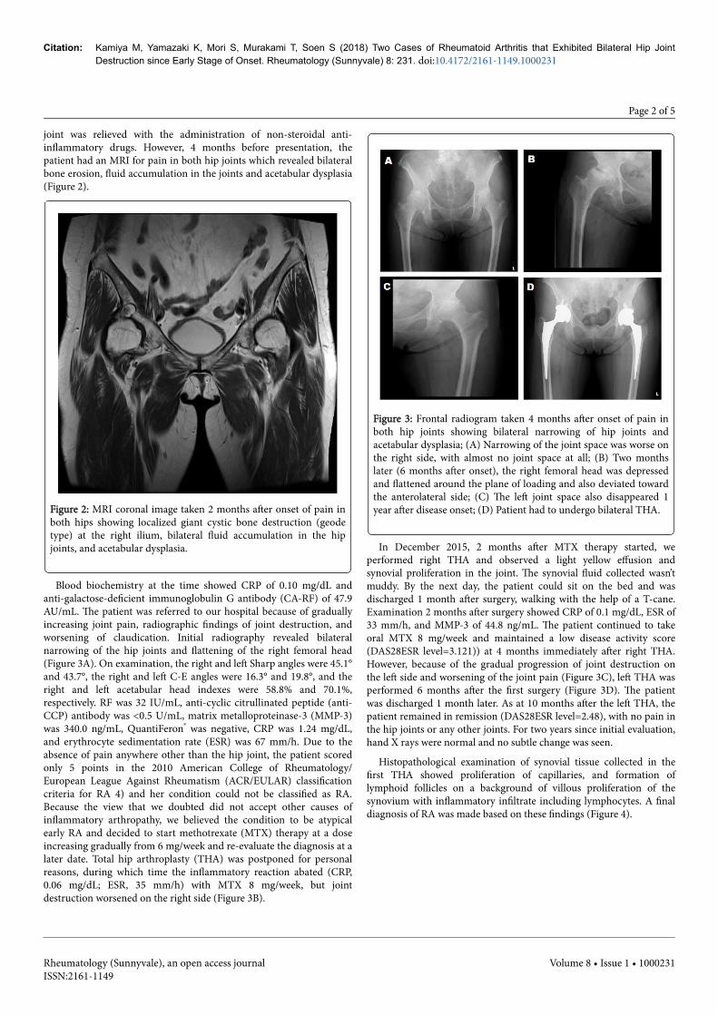
joint was relieved with the administration of non-steroidal anti-inflammatory drugs. However, 4 months before presentation, thepatient had an MRI for pain in both hip joints which revealed bilateralbone erosion, fluid accumulation in the joints and acetabular dysplasia(Figure 2).
Figure 2: MRI coronal image taken 2 months after onset of pain inboth hips showing localized giant cystic bone destruction (geodetype) at the right ilium, bilateral fluid accumulation in the hipjoints, and acetabular dysplasia.
Blood biochemistry at the time showed CRP of 0.10 mg/dL andanti-galactose-deficient immunoglobulin G antibody (CA-RF) of 47.9AU/mL. The patient was referred to our hospital because of graduallyincreasing joint pain, radiographic findings of joint destruction, andworsening of claudication. Initial radiography revealed bilateralnarrowing of the hip joints and flattening of the right femoral head(Figure 3A). On examination, the right and left Sharp angles were 45.1°and 43.7°, the right and left C-E angles were 16.3° and 19.8°, and theright and left acetabular head indexes were 58.8% and 70.1%,respectively. RF was 32 IU/mL, anti-cyclic citrullinated peptide (anti-CCP) antibody was <0.5 U/mL, matrix metalloproteinase-3 (MMP-3)was 340.0 ng/mL, QuantiFeron® was negative, CRP was 1.24 mg/dL,and erythrocyte sedimentation rate (ESR) was 67 mm/h. Due to theabsence of pain anywhere other than the hip joint, the patient scoredonly 5 points in the 2010 American College of Rheumatology/European League Against Rheumatism (ACR/EULAR) classificationcriteria for RA 4) and her condition could not be classified as RA.Because the view that we doubted did not accept other causes ofinflammatory arthropathy, we believed the condition to be atypicalearly RA and decided to start methotrexate (MTX) therapy at a doseincreasing gradually from 6 mg/week and re-evaluate the diagnosis at alater date. Total hip arthroplasty (THA) was postponed for personalreasons, during which time the inflammatory reaction abated (CRP,0.06 mg/dL; ESR, 35 mm/h) with MTX 8 mg/week, but jointdestruction worsened on the right side (Figure 3B).
Figure 3: Frontal radiogram taken 4 months after onset of pain inboth hip joints showing bilateral narrowing of hip joints andacetabular dysplasia; (A) Narrowing of the joint space was worse onthe right side, with almost no joint space at all; (B) Two monthslater (6 months after onset), the right femoral head was depressedand flattened around the plane of loading and also deviated towardthe anterolateral side; (C) The left joint space also disappeared 1year after disease onset; (D) Patient had to undergo bilateral THA.
In December 2015, 2 months after MTX therapy started, weperformed right THA and observed a light yellow effusion andsynovial proliferation in the joint. The synovial fluid collected wasn’tmuddy. By the next day, the patient could sit on the bed and wasdischarged 1 month after surgery, walking with the help of a T-cane.Examination 2 months after surgery showed CRP of 0.1 mg/dL, ESR of33 mm/h, and MMP-3 of 44.8 ng/mL. The patient continued to takeoral MTX 8 mg/week and maintained a low disease activity score(DAS28ESR level=3.121)) at 4 months immediately after right THA.However, because of the gradual progression of joint destruction onthe left side and worsening of the joint pain (Figure 3C), left THA wasperformed 6 months after the first surgery (Figure 3D). The patientwas discharged 1 month later. As at 10 months after the left THA, thepatient remained in remission (DAS28ESR level=2.48), with no pain inthe hip joints or any other joints. For two years since initial evaluation,hand X rays were normal and no subtle change was seen.
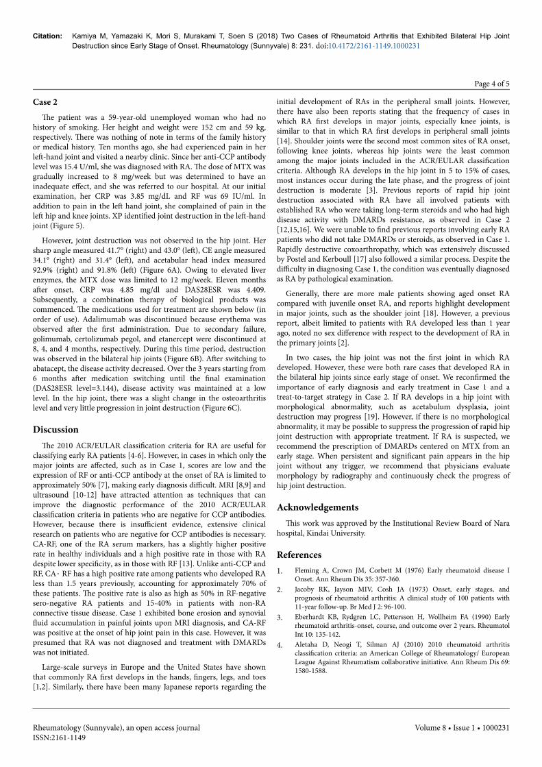
Histopathological examination of synovial tissue collected in thefirst THA showed proliferation of capillaries, and formation oflymphoid follicles on a background of villous proliferation of thesynovium with inflammatory infiltrate including lymphocytes. A finaldiagnosis of RA was made based on these findings (Figure 4).
Citation: Kamiya M, Yamazaki K, Mori S, Murakami T, Soen S (2018) Two Cases of Rheumatoid Arthritis that Exhibited Bilateral Hip JointDestruction since Early Stage of Onset. Rheumatology (Sunnyvale) 8: 231. doi:10.4172/2161-1149.1000231
Page 2 of 5
Rheumatology (Sunnyvale), an open access journalISSN:2161-1149
Volume 8 • Issue 1 • 1000231
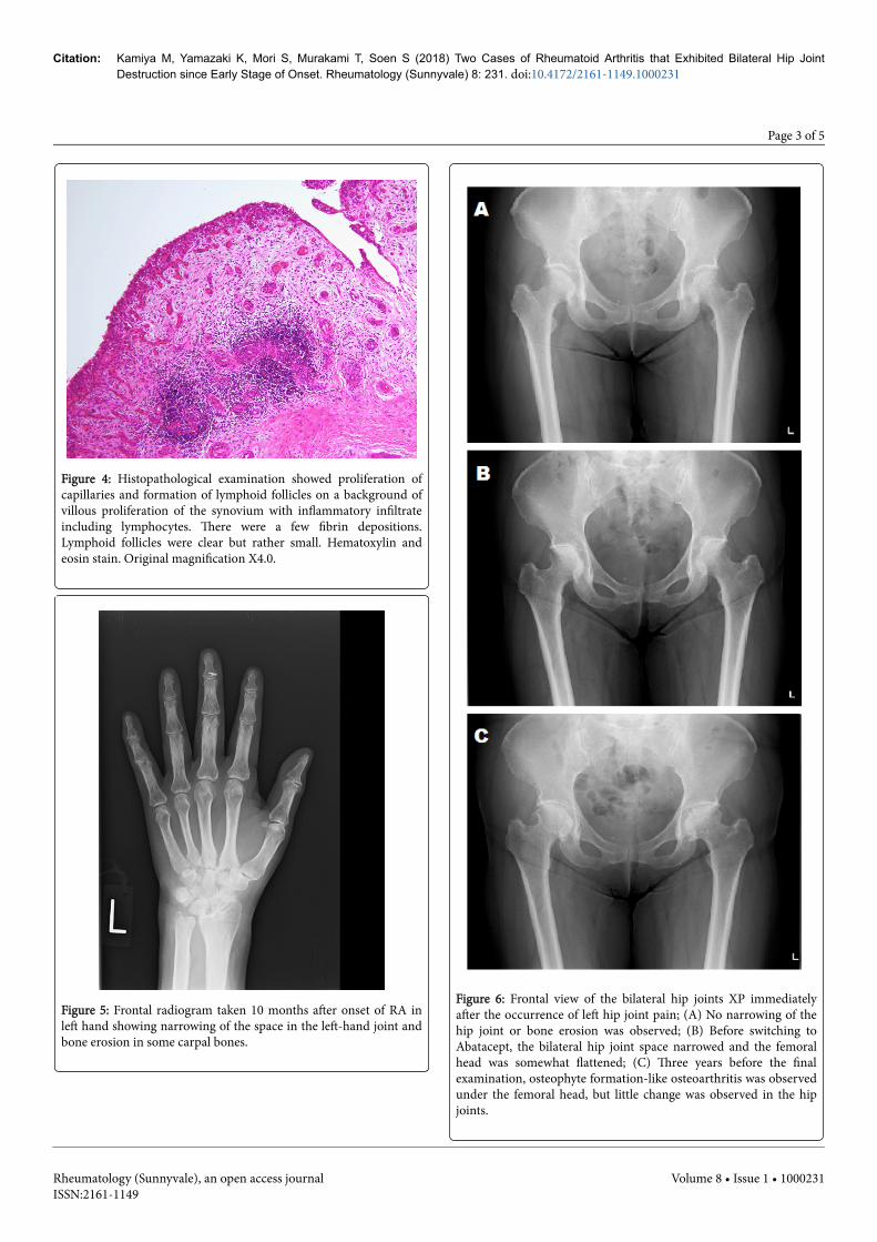
Figure 4: Histopathological examination showed proliferation ofcapillaries and formation of lymphoid follicles on a background ofvillous proliferation of the synovium with inflammatory infiltrateincluding lymphocytes. There were a few fibrin depositions.Lymphoid follicles were clear but rather small. Hematoxylin andeosin stain. Original magnification X4.0.
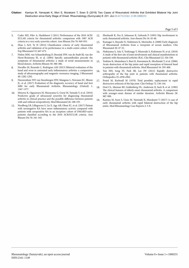
Figure 5: Frontal radiogram taken 10 months after onset of RA inleft hand showing narrowing of the space in the left-hand joint andbone erosion in some carpal bones.
Figure 6: Frontal view of the bilateral hip joints XP immediatelyafter the occurrence of left hip joint pain; (A) No narrowing of thehip joint or bone erosion was observed; (B) Before switching toAbatacept, the bilateral hip joint space narrowed and the femoralhead was somewhat flattened; (C) Three years before the finalexamination, osteophyte formation-like osteoarthritis was observedunder the femoral head, but little change was observed in the hipjoints.
Citation: Kamiya M, Yamazaki K, Mori S, Murakami T, Soen S (2018) Two Cases of Rheumatoid Arthritis that Exhibited Bilateral Hip JointDestruction since Early Stage of Onset. Rheumatology (Sunnyvale) 8: 231. doi:10.4172/2161-1149.1000231
Page 3 of 5
Rheumatology (Sunnyvale), an open access journalISSN:2161-1149
Volume 8 • Issue 1 • 1000231
Case 2The patient was a 59-year-old unemployed woman who had no
history of smoking. Her height and weight were 152 cm and 59 kg,respectively. There was nothing of note in terms of the family historyor medical history. Ten months ago, she had experienced pain in herleft-hand joint and visited a nearby clinic. Since her anti-CCP antibodylevel was 15.4 U/ml, she was diagnosed with RA. The dose of MTX wasgradually increased to 8 mg/week but was determined to have aninadequate effect, and she was referred to our hospital. At our initialexamination, her CRP was 3.85 mg/dL and RF was 69 IU/ml. Inaddition to pain in the left hand joint, she complained of pain in theleft hip and knee joints. XP identified joint destruction in the left-handjoint (Figure 5).
However, joint destruction was not observed in the hip joint. Hersharp angle measured 41.7° (right) and 43.0° (left), CE angle measured34.1° (right) and 31.4° (left), and acetabular head index measured92.9% (right) and 91.8% (left) (Figure 6A). Owing to elevated liverenzymes, the MTX dose was limited to 12 mg/week. Eleven monthsafter onset, CRP was 4.85 mg/dl and DAS28ESR was 4.409.Subsequently, a combination therapy of biological products wascommenced. The medications used for treatment are shown below (inorder of use). Adalimumab was discontinued because erythema wasobserved after the first administration. Due to secondary failure,golimumab, certolizumab pegol, and etanercept were discontinued at8, 4, and 4 months, respectively. During this time period, destructionwas observed in the bilateral hip joints (Figure 6B). After switching toabatacept, the disease activity decreased. Over the 3 years starting from6 months after medication switching until the final examination(DAS28ESR level=3.144), disease activity was maintained at a lowlevel. In the hip joint, there was a slight change in the osteoarthritislevel and very little progression in joint destruction (Figure 6C).
DiscussionThe 2010 ACR/EULAR classification criteria for RA are useful for
classifying early RA patients [4-6]. However, in cases in which only themajor joints are affected, such as in Case 1, scores are low and theexpression of RF or anti-CCP antibody at the onset of RA is limited toapproximately 50% [7], making early diagnosis difficult. MRI [8,9] andultrasound [10-12] have attracted attention as techniques that canimprove the diagnostic performance of the 2010 ACR/EULARclassification criteria in patients who are negative for CCP antibodies.However, because there is insufficient evidence, extensive clinicalresearch on patients who are negative for CCP antibodies is necessary.CA-RF, one of the RA serum markers, has a slightly higher positiverate in healthy individuals and a high positive rate in those with RAdespite lower specificity, as in those with RF [13]. Unlike anti-CCP andRF, CA⋅ RF has a high positive rate among patients who developed RAless than 1.5 years previously, accounting for approximately 70% ofthese patients. The positive rate is also as high as 50% in RF-negativesero-negative RA patients and 15-40% in patients with non-RAconnective tissue disease. Case 1 exhibited bone erosion and synovialfluid accumulation in painful joints upon MRI diagnosis, and CA-RFwas positive at the onset of hip joint pain in this case. However, it waspresumed that RA was not diagnosed and treatment with DMARDswas not initiated.
Large-scale surveys in Europe and the United States have shownthat commonly RA first develops in the hands, fingers, legs, and toes[1,2]. Similarly, there have been many Japanese reports regarding the
initial development of RAs in the peripheral small joints. However,there have also been reports stating that the frequency of cases inwhich RA first develops in major joints, especially knee joints, issimilar to that in which RA first develops in peripheral small joints[14]. Shoulder joints were the second most common sites of RA onset,following knee joints, whereas hip joints were the least commonamong the major joints included in the ACR/EULAR classificationcriteria. Although RA develops in the hip joint in 5 to 15% of cases,most instances occur during the late phase, and the progress of jointdestruction is moderate [3]. Previous reports of rapid hip jointdestruction associated with RA have all involved patients withestablished RA who were taking long-term steroids and who had highdisease activity with DMARDs resistance, as observed in Case 2[12,15,16]. We were unable to find previous reports involving early RApatients who did not take DMARDs or steroids, as observed in Case 1.Rapidly destructive coxoarthropathy, which was extensively discussedby Postel and Kerboull [17] also followed a similar process. Despite thedifficulty in diagnosing Case 1, the condition was eventually diagnosedas RA by pathological examination.
Generally, there are more male patients showing aged onset RAcompared with juvenile onset RA, and reports highlight developmentin major joints, such as the shoulder joint [18]. However, a previousreport, albeit limited to patients with RA developed less than 1 yearago, noted no sex difference with respect to the development of RA inthe primary joints [2].
In two cases, the hip joint was not the first joint in which RAdeveloped. However, these were both rare cases that developed RA inthe bilateral hip joints since early stage of onset. We reconfirmed theimportance of early diagnosis and early treatment in Case 1 and atreat-to-target strategy in Case 2. If RA develops in a hip joint withmorphological abnormality, such as acetabulum dysplasia, jointdestruction may progress [19]. However, if there is no morphologicalabnormality, it may be possible to suppress the progression of rapid hipjoint destruction with appropriate treatment. If RA is suspected, werecommend the prescription of DMARDs centered on MTX from anearly stage. When persistent and significant pain appears in the hipjoint without any trigger, we recommend that physicians evaluatemorphology by radiography and continuously check the progress ofhip joint destruction.
AcknowledgementsThis work was approved by the Institutional Review Board of Nara
hospital, Kindai University.
References1. Fleming A, Crown JM, Corbett M (1976) Early rheumatoid disease I
Onset. Ann Rheum Dis 35: 357-360.2. Jacoby RK, Jayson MIV, Cosh JA (1973) Onset, early stages, and
prognosis of rheumatoid arthritis: A clinical study of 100 patients with11-year follow-up. Br Med J 2: 96-100.
3. Eberhardt KB, Rydgren LC, Pettersson H, Wollheim FA (1990) Earlyrheumatoid arthritis-onset, course, and outcome over 2 years. RheumatolInt 10: 135-142.
4. Aletaha D, Neogi T, Silman AJ (2010) 2010 rheumatoid arthritisclassification criteria: an American College of Rheumatology/ EuropeanLeague Against Rheumatism collaborative initiative. Ann Rheum Dis 69:1580-1588.
Citation: Kamiya M, Yamazaki K, Mori S, Murakami T, Soen S (2018) Two Cases of Rheumatoid Arthritis that Exhibited Bilateral Hip JointDestruction since Early Stage of Onset. Rheumatology (Sunnyvale) 8: 231. doi:10.4172/2161-1149.1000231
Page 4 of 5
Rheumatology (Sunnyvale), an open access journalISSN:2161-1149
Volume 8 • Issue 1 • 1000231
5. Cader MZ, Filer A, Hazlehurst J (2011) Performance of the 2010 ACR/EULAR criteria for rheumatoid arthritis: comparison with 1987 ACRcriteria in a very early synovitis cohort. Ann Rheum Dis 70: 949-955.
6. Zhao J, SuY, Ye H (2014) Classification criteria of early rheumatoidarthritis and validation of its performance in a multi-centre cohort. ClinExp Rheumatol 32: 667-673.
7. Nielen MM, van Schaardenburg D, Reesink HW, van de Stadt RJ, van derHorst-Bruinsma IE, et al. (2004) Specific autoantibodies precede thesymptoms of rheumatoid arthritis: a study of serial measurements inblood donors. Arthritis Rheum 50: 380-386.
8. Navalho M, Resende C, Rodrigues AM (2013) Bilateral evaluation of thehand and wrist in untreated early inflammatory arthritis: a comparativestudy of ultrasonography and magnetic resonance imaging. J Rheumatol40: 1282-1292.
9. Nieuwenhuis WP, van Steenbergen HW, Mangnus L, Newsum EC, BloemJL, et al. (2017) Evaluation of the diagnostic accuracy of hand and footMRI for early Rheumatoid Arthritis. Rheumatology (Oxford) 1:1367-1377.
10. Minowa K, Ogasawara M, Murayama G, Gorai M, Yamada Y, et al. (2016)Predictive grade of ultrasound synovitis for diagnosing rheumatoidarthritis in clinical practice and the possible difference between patientswith and without seropositivety. Mod Rheumatol 26: 188-193.
11. Nordberg LB, Lillegraven S, Lie E, Aga AB, Olsen IC, et al. (2017) Patientwith seronegative RA have more inflammatory activity compared withpatients with seropositive RA in an inception cohort of DMARD-naïvepatients classified according to the 2010 ACR/EULAR criteria. AnnRheum Dis 76: 341-345.
12. Eberhardt K, Fex E, Johnsson K, Geborek P (1995) Hip involvement inearly rheumatoid arthritis. Ann rheum Dis 54: 45-48.
13. Kumagai S, Hayashi N, Nishimura K, Morinobu A (2008) Early diagnosisof Rheumatoid Arthritis from a viewpoint of serum markers. ClinRheumatol 20: 47-52.
14. Nishiyama S, Aita Y, Yoshinaga Y, Miyawaki S, Kishimoto H, et al. (2010)A study of the first site of joint involvement and clinical manifestations inpatients with rheumatoid arthritis (RA). Clin Rheumatol 22: 326-330.
15. Yoshino K, Momohara S, Ikari K, Kawamura K, Mochizuki T, et al. (2006)Acute destruction of the hip joints and rapid resorption of femoral headin patients with rheumatoid arthritis. Mod Rheumatol 16: 395-400.
16. Yun HH, Song SY, Park SB, Lee JW (2012) Rapidly destructivearthropathy of the hip joint in patients with rheumatoid arthritis.Orthopedics 35: e958-e962.
17. Postal M, Kerboull M (1970) Total prosthtic replacement in rapiddestructive arthrosis of the hip joint. Clin Orthop 72: 138-144.
18. Deal CL, Meenan RF, Goldenberg DL, Anderson JJ, Sack B, et al. (1985)The clinical features of elderly-onset rheumatoid arthritis. A comparisonwith younger-onset disease of similar duration. Arthritis Rheum 28:987-999.
19. Kamiya M, Soen S, Ueno M, Yamizaki K, Murakami T (2017) A case ofearly rheumatoid arthritis with rapid bilateral destruction of the hipjoints. Mod Rheumatology Case Reports 2: 5-8.
Citation: Kamiya M, Yamazaki K, Mori S, Murakami T, Soen S (2018) Two Cases of Rheumatoid Arthritis that Exhibited Bilateral Hip JointDestruction since Early Stage of Onset. Rheumatology (Sunnyvale) 8: 231. doi:10.4172/2161-1149.1000231
Page 5 of 5
Rheumatology (Sunnyvale), an open access journalISSN:2161-1149
Volume 8 • Issue 1 • 1000231