moringa oleifera lam: targeting...
TRANSCRIPT

Asian Pacific Journal of Cancer Prevention, Vol 17, 2016 3675
10.14456/apjcp.2016.155/APJCP.2016.17.8.3675Moringa Oleifera Lam: Targeting Chemoprevention
Asian Pac J Cancer Prev, 17 (8), 3675-3686
Origin and Distribution of M. Oleifera
Moringa oleifera Lam. (M. oleifera) comes from the family Moringaceae in phylogeny tree. It is widely distributed and known native in India, Afghanistan, Bangladesh, and Pakistan (Amaglo et al., 2010). M. oleifera is also called with various names such as horseradish tree, drumstick tree and locally named ‘murungai’ or ‘kelor’. The plant is known to survive the arid environment which makes it easily grown around the world such as in West, East, South Africa, Tropical Asia and Latin America. The plant is edible and consumed by most of the people in Asia including Thailand, Malaysia and India (Ghosh, 2013) M. oleifera can be described as a perennial plant, has spirally arranged leaves, whitish flower and low quality of timber (Figure 1) (Ghosh, 2013). Scientific classification (Figure 2) shows that the plant is in order of Brassicales and is one of the cruciferous plants (Ganesan et al., 2014). The characteristics of the plant in order of Brassicales is it contains glucosinolates as their exclusive compound (Gueyrard et al., 2000; Rollin and
1Laboratory of UPM-MAKNA Cancer Research, Institute of Bioscience, 2Faculty of Food Science and Technology, Universiti Putra Malaysia, UPM Serdang, Selangor, 3Faculty of Industrial Sciences & Technology, Universiti Malaysia Pahang, Lebuhraya Tun Razak, Gambang Kuantan, Pahang, Malaysia *For correspondence: [email protected]
Abstract
Moringa oleifera Lam, family Moringaceae, is a perennial plant which is called various names, but is locally known in Malaysia as ‘’murungai’’ or ‘’kelor’’. Glucomoringin, a glucosinolate with from M. oleifera is a major secondary metabolite compound. The seeds and leaves of the plant are reported to have the highest amount of glucosinolates. M. oleifera is well known for its many uses health and benefits. It is claimed to have nutritional, medicinal and chemopreventive potentials. Chemopreventive effects of M. oleifera are expected due to the existence of glucosinolate which it is reported to have the ability to induce apoptosis in anticancer studies. Furthermore, chemopreventive value of M. oleifera has been demonstrated in studies utilizing its leaf extract to inhibit the growth of human cancer cell lines. This review highlights the advantages of M. oleifera targeting chemoprevention where glucosinolates could help to slow the process of carcinogenesis through several molecular targets. It is also includes inhibition of carcinogen activation and induction of carcinogen detoxification, anti-inflammatory, anti-tumor cell proliferation, induction of apoptosis and inhibition of tumor angiogenesis. Finally, for synergistic effects of M. oleifera with other drugs and safety, essential for chemoprevention, it is important that it safe to be consumed by human body and works well. Although there is promising evidence about M. oleifera in chemoprevention, extensive research need to be done due to the expected rise of cancer in coming years and to gain more information about the mechanisms involved in M. oleifera influence, which could be a good source to inhibit several major mechanisms involved in cancer development. Keywords: Moringa oleifera - glucosinolate - glucomoringin - chemopreventive
REVIEW
Moringa oleifera Lam: Targeting Chemoprevention
Nurul Ashikin Abd Karim1, Muhammad Din Ibrahim1, Saie Brindha Kntayya1, Yaya Rukayadi2, Hazrulizawati Abd Hamid3, Ahmad Faizal Abdull Razis1,2*
Tatibouet, 2011; Galuppo et al., 2013; Forster et al., 2015). Nowadays, Malaysians grow the plant to get constant source for food or use in traditional medicine. There are companies that grow the plant in large production to be sold as ingredient for medicinal purposes or research studies. The awareness of most people to consume high nutritional food from natural sources caused high demand of the raw M. oleifera in the market.
Uses of M. oleifera
M. oleifera is usually used in traditional medicine as ingredients because of its pharmacological properties. Some of the claimed properties, which had been proven in research were anti-bacteria and anti-tumor (Anwar et al., 2007). In pharmaceuticals industry, it had been used with some other ingredients to produce medicine. The plant had also been used to purify water, especially the seed (Tahir et al., 2010). The seed had been used as coagulant, to treat water turbidity of any impurities (Santos et al., 2005; Katayon et al., 2006; Gupta et al., 2010). It is

Nurul Ashikin Abd Karim et al
Asian Pacific Journal of Cancer Prevention, Vol 17, 20163676
also used to eliminate microbial in flocculated water and able to remove metals in liquid solution (Popoola and Obembe, 2013). In biodiesel field, the oil from the seed had been developed for biofuel which is environmental friendly (Rashid et al., 2008). Therefore, there are many uses of M. oleifera including ornamental plant, pesticide, medicine, cleaning agent, fencing, and biogas, used in haircare products and as food for chicken (Anwar et al., 2007; Ghosh 2013).
Phytochemical composition of M. oleifera
Several phytochemicals present in Moringa oleifera Lam. which distributed in its leaf, branch, seed, pod and root. Several major phytochemicals of M. oleifera are glucosinolates, phenolics, flavonoids, crude fats, fatty
acid, major nutrient, mineral and total protein (Flora and Pachauri, 2011). In addition, glucosinolates are the secondary metabolites that indicates the characteristic of Brassicales plant including M. oleifera where it only can be found in the order of Brassicales (Garima et al., 2011). Glucosinolates are most abundant in most part of M. oleifera plant except the root (Ghosh, 2013). M. oleifera contains high amount of aromatic glucosinolates such as p-hydroxybenzyl glucosinolates (sinalbin), 2-phnylethyl glucosinolates (gluconasturtiin), benzyl glucosinolates (glucotropaeolin) (Forster et al., 2015). In addition, benzyl glucosinolates is the dominant compound presented in the
Figure 1. Leaves and flowers of M. oleifera (Source: Ghosh, 2013)
Figure 1. Leaves and flowers of M. oleifera (Source: Ghosh, 2013)
Figure 2. Taxonomic Classification of M. oleifera (Source: Garima et al., 2011)
Scientific classification
Kingdom: Plantae
Division: Magnoliophyta
Class: Magnoliopsida
Order: Brassicales
Family: Moringaceae
Genus: Moringa
Species: Moringa oleifera
Binomial name: Moringa oleifera Lam.
Figure 2. Taxonomic classification of M. oleifera (Source: Garima et al., 2011)
Figure 3. Structure of a Glucosinolate Molecule from M. oleifera. (Source: Förster et al., 2015)
O
R1R2
R3
H3CO
N
S
HO
OHO OH
OH
O
S
O
O
O
Fig. 3. Structure of a glucosinolate molecules from M. oleifera. (Source: Förster et al., 2015)
R1,R2,R3 = OH
Table 1. Amino Acid Content in Fresh and Dried Leaves of M. oleifera per Gram (g) Edible Portion (Source: Shruti et al., 2011)
Amino acid Fresh leaf (g) Dried leaf (g)Arginine 0.041 0.133Histidine 0.149 0.613Isoleucine 0.299 0.825Leucine 0.492 0.195Lysine 0.342 0.133Methionine 0.117 0.35Phenylalanine 0.31 0.139Threonine 0.117 0.119Tryptophan 0.107 0.425Valine 0.374 0.106
Figure 4. Proposed Molecular Targets of M. oleifera as a Chemopreventive Agent (Source: Kundu et al. 2014)
Figure 4. A diagram shows proposed molecular target of M. oleifera as chemopreventive agent. (Source: Kundu et al. 2014)
Src Jak 2
STAT3
MMP-9 IL-8
Figure 5. Slowing the Process of Carcinogenesis to Increase Cancer Free Life Period
Figure 5. Slowing the process of carcinogenesis to increase cancer free life period.

Asian Pacific Journal of Cancer Prevention, Vol 17, 2016 3677
10.14456/apjcp.2016.155/APJCP.2016.17.8.3675Moringa Oleifera Lam: Targeting Chemoprevention
leaf of M. oleifera (Forster et al., 2015). Seed and leaf of M. oleifera have the highest glucosinolates content compared to other parts of plant (Ghosh, 2013). The leaf usually has high flavonoid value whereas the seed has high crude protein value (Ghosh, 2013). In many reported journals, the leaves extract are the most studied for various uses (Kasolo et al., 2010; Asare et al., 2012; Lambole and Kumar, 2012; Moyo et al., 2012; Ratshilivha et al., 2014). It was claimed that the dried leaf extract showed higher amino acid, vitamin and minerals content than
the fresh leaf extracts itself (Ghosh, 2013). Therefore, it is concluded that the dried extract of the plant will give higher yield of purified compound. Table 1 shows amino acid content in fresh and dried leaves of M. oleifera in 1 g of edible portion.
Phytochemicals of M. oleifera were also reported to have bioactivities such as anti-bacterial, anti-fungus and anti-tumor (Kasolo et al., 2010). Figure 3 shows structure of glucosinolates molecule from M. oleifera plant. The functional groups presented in the compound usually
Table 2. Structure of Major Phytochemicals from seeds of M. oleifera (Source: Flora and Pachauri, 2011)
1)
N
H
H
1) Alkaloid: moringin
2)
NC
S
2) FlavonoidsGlycosides: Benzyl isothiocyanate and its derivatives
3)
CN 3) (α-L-rhamnosyloxy), phenylacetonitrile,
4-hydroxyl phenyl-acetonitrile, and 4-hydroxyphenyl-acetamide
4)
OCH3
OH
OHHO
OH
4) 4-(α-L-rhamnosyloxy), 4-(α-L-rhamnosyloxy), phenylacetonitrile (β-carotene), sterols and lecithin
5)
O
Y
CH3
O
OHHO
N
H
C
X
OC2H5
5) O-ethyl-4-(α-L-rhamnosyloxy), benzylcarbamide with seven derivatives
6)
CH3
O-R
CH3
H3C CH3
CH3
CH3 6) Nutrients: Vitamin B1, B6, riboflavin, folic acid, nicotinic acid, vitamin C and E
Nurul Ashikin Abd Karim et al
Asian Pacific Journal of Cancer Prevention, Vol 17, 20163678
responsible for the bioactivities triggered.Brunelli et al. (2010) reported that glucosinolates were
found higher in the seeds by 8 % compared to other plant parts. Glucosinolates were reported to induce apoptosis in tumour cell and the major studies had been done on leaf extracts (Forster et al., 2015). While studies on the anti-cancer of the seed extract is very scarce, thus it is important to carry out anti-cancer studies of the seed due to the abundance of glucosinolates presented in the seeds.
Table 2 shows major phytochemicals listed and reported from seeds of M. oleifera.
Nutritional and medicinal value of M. oleifera
M. oleifera is recognized as a nutritious plant which can be consumed to fight famine due to its highly nutritious content (Trees for life organization, 2005). It has been reported that in 100 gram of dry M. oleifera leaf contained 12 times vitamin C than oranges, 10 times vitamin A than carrots, 9 times protein than yoghurt, 15 times potassium than banana, 17 times calcium than milk and 25 times iron than spinach (Tahir et al., 2010). This nutritional content was also responsible in many important function in biochemical process of human and animals (Sauberlich,1984; Senba and Nusemblatt, 2002). The animal and human cannot produce certain types of important nutrition by themselves that are essential in the natural function, so it must be consumed from the outside which this plant can be one of the best nutrition sources (Wu, 2010). In certain countries such as India, Pakistan, Philippines, Hawaii and many parts of Africa, the immature pods, leaves and flowers of the plant had also consumed as a source of vegetable (D’souza and Kulkarni, 1993; Anwar and Bhanger, 2003; Anwar et al., 2005).
Table 3. Reports on Cancer Chemoprevention Through ion Vitro Studies of M. oleifera
Study Remarks ReferenceAnti-proliferation and induction of apoptosis by Moringa oleifera leaf extract on human cancer cells
Extracts induced apoptosis in human tumor cell line
Sreelatha et al., (2011)
Ethanolic extract of Moringa oleifera increased cytotoxic effect of doxorubicin on HeLa cancer cells
Combination of extract and doxorubicin increased doxorubicin effect through apoptotic induction
Hermawan et al., (2012)
Antioxidant and anticancer activities of Moringa oleifera leaves
Dichloromethane extract showed high antioxidant and potent anti-cancer proliferation
Charoensin (2014)
Moringa species (Moringaceae): phytochemistry, cancer chemoprevention potentials with advanced traditional medicinal practice
Skin tumor prevention and exhibit good hepatoprotective
Dibyajyoti (2013)
Anticancer effect of Moringa oleifera leaf extract on human breast cancer cell
Anti-proliferative effect on 2 breast cancer cell lines, MDA MB 231 and MCF 7, showed a dose and time dependent effect
Ghosh (2013)
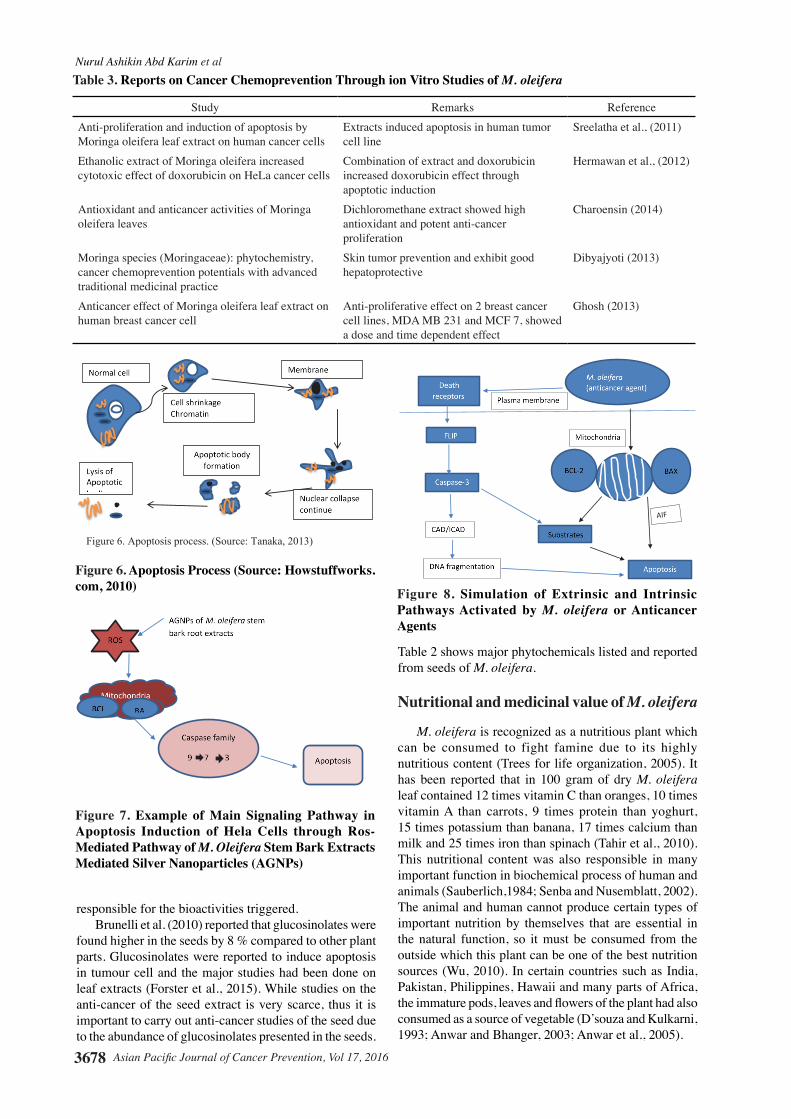
Figure 6. Apoptosis Process (Source: Howstuffworks.com, 2010)
Figure 6. Apoptosis process. (Source: Tanaka, 2013)
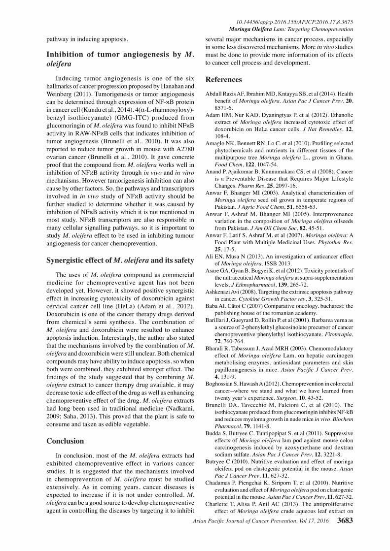
Figure 7. Example of Main Signaling Pathway in Apoptosis Induction of Hela Cells through Ros-Mediated Pathway of M. Oleifera Stem Bark Extracts Mediated Silver Nanoparticles (AGNPs)
Figure 7. Example of main signaling pathway in apoptosis induction of HeLa cells through ROS-mediated pathway of M. oleifera stem bark extracts mediated silver nanoparticles (AGNPs). Adapted from Vasanth et al. (2014).
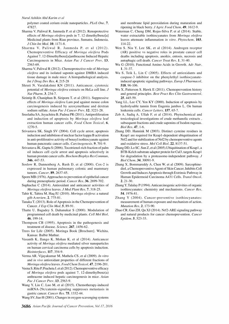
Figure 8. Simulation of Extrinsic and Intrinsic Pathways Activated by M. oleifera or Anticancer Agents
Figure 8. Simulation of Extrinsic and Intrinsic pathway activated by M. oleifera or anticancer
agent. Adapted from Fulda and Debatin (2006).

Asian Pacific Journal of Cancer Prevention, Vol 17, 2016 3679
10.14456/apjcp.2016.155/APJCP.2016.17.8.3675Moringa Oleifera Lam: Targeting Chemoprevention
M. oleifera extract had been utilized as an ingredient in many Indian traditional (Ghazali and Mohammed, 2011). The plant is said to have various beneficial effects such as anti-inflammatory, anti-fibrotic, anti-microbial, anti-oxidant, anti-hyperglycemic, anti-tumor and anti-cancer (Abdull et al., 2014). It is well known as a multipurpose crop because of the various properties from different part of the plant that contributed to the various beneficial effects (Amaglo et al., 2010). The boiled M. oleifera pod extract had exhibited anti-clastogenic activity when 1.5 %, 3.0 % and 6.0 % of the extracts were introduced to mouse by in vivo erythrocyte micronucleus assay (Chadamas et al., 2010). It was reported that the seed contains major phytochemicals with reported medicinal value (Fuglie et al., 2001; Chuang et al., 2007; Ghazali et al., 2011). In previous pharmacological studies, glucomoringin was reported to be able to relax bronchioles and exert liver protective effects (Mahajan and Mehta, 2010). The flavonoids and glycosides possess anti-oxidant, anti-inflammatory, anti-microbial, anti-cancer and anti-hypertensive activities (Brunelli et al., 2010; Sudha et al., 2010; Galuppo et al., 2014). The (α-L-rhamnosyloxy), phenylacetonitrile, 4-hydroxyl phenyl-acetonitrile, and 4-hydroxyphenyl-acetamide have mutagenic property (Galuppo et al., 2014). 4-(α-L-rhamnosyloxy), 4-(α-L-rhamnosyloxy), phenylacetonitrile (β-carotene), sterols and lecithin have anti-microbial and anti-biotic activities (Ghazali et al., 2011; Galuppo et al., 2014). O-ethyl-4-(α-L-rhamnosyloxy), benzylcarbamide with seven derivatives have antitumor and antihypertensive activities (Chumark et al., 2008). The last components are the nutrients which can provide nutrient and has anti-oxidant property (Garima et al., 2011). M. oleifera had also been used to produce commercial medicine and cosmetic products in India such as Rumalaya and Septilin (Himalaya Drug Company), Orthoherb (Walter Bushnell Ltd), Kupid Fort (Pharma Products) and Livospin (Herbals APS) (Mehta et al., 2003). Therefore, the root, leaf, stem bark, gum, flower and seed had been reported to have various medicinal uses (Anwar et al., 2007).
Chemopreventive value and targeting chemoprevention of M. oleifera
According to Dr. Michael Sporn (1976), who first defined the word chemoprevention, chemoprevention is ‘‘the use of specific agents to reverse, or prevent the carcinogenic process to invasive cancer’’. A compound that has the ability to induce apoptosis of cancer cell is called chemopreventive agent. Chemopreventive effect of the compound makes the plant as an alternative for anti-cancer. Chemoprevention studies had been an important research in order to combat cancer worldwide. The statistic death caused by cancer was increasing every year, which change of diet and lifestyle of people nowadays become the main influences (Anand et al., 2008). Various types of plant are reported to have phytochemicals that are able to slow down or fight cancer. Moringa oleifera possessed glucosinolates, an exclusive compound which is able to induce apoptosis. In fact, the hydrolysis product of
glucomoringin (GMG) with, called 4(α-L-rhamnosyloxy)-benzyl isothiocyanate (GMG-ITC) is working effectively better than the non-hydrolysis in inducing apoptosis. There are some studies showed that the crude extract of M. oleifera has the ability to inhibit carcinogenesis in cancer cells in vitro and in vivo (Foti Cuzzola et al., 2013; Galuppo et al., 2013). Vasanth et al. (2014), had reported that silver nanoparticles of M. oleifera extract, was discovered to induce apoptosis in human cervical carcinoma cells (HeLa). Furthermore, it was also found that M. oleifera pod had exhibited colitis-related colon carcinogenesis inhibition in mouse model induced by genotoxic colon carcinogen, azoxymethane and dextran sodium sulphate (Budda et al., 2011). In the strategy for M. oleifera to be a potential chemopreventive agent, the compound should involve in pathways or mechanisms in activating or inhibiting carcinogen detoxification, anti-oxidant effects, tumor cell proliferation, apoptosis, inflammation, tumor angiogenesis, migration, invasion and synergistic effect (Figure 4) (Neergheen et al., 2010; Boghossian and Hawash, 2012). Table 3 shows some reports regarding in vitro studies of M. oleifera for potential chemopreventive agent.
Cancer chemoprevention: evidence potential from in vitro studies
Mechanism of chemopreventionGlucosinolates showed an effect of inhibiting cancer
in different chemoprevention strategy (Wang et al., 2012). In general, the mechanisms that involved in hallmark of cancer are apoptosis inhibition, carcinogen activation and detoxification, tumor proliferation, enzyme modulation and tumor angiogenesis (Kundu et al., 2014). Cancer therapeutic agents will exhibit characteristics to block the mechanisms that involved in inducing cancer processes (Neerghen et al., 2010). Many studies targeting on apoptosis induction in order to determine the chemopreventive effect (Wang et al., 2012).This is because most pathways involved in apoptosis induction are recognized and been described in the previous studies (Kang et al., 1998; Lie-weber et al., 2010). Therefore, M. oleifera is expected to involve in several mechanisms of molecular target of cancer prevention as shown in figure 4. In addition, M. oleifera extracts were reported to involve in various anti-cancer activities such as apoptosis induction, tumor cell proliferation and anti-inflammatory (Guevara et al., 1999; Verma et al., 2009; Sreelatha et al., 2011).
Inhibition of carcinogen activation and induction of carcinogen detoxification
Carcinogenesis involved various biochemical process which it can be caused by a defect in apoptosis pathways and various xenobiotic-metabolizing enzymes. CYP or P450 is one of the xenobiotic-metabolizing enzyme that activates carcinogen process (Shumada, 2006). If expression of Cytochrome P (CYP) enzyme is inhibited, carcinogenesis will not occur. According to Bharali and colleague (2003), hydro-alcoholic extract of M.

Nurul Ashikin Abd Karim et al
Asian Pacific Journal of Cancer Prevention, Vol 17, 20163680
0
25.0
50.0
75.0
100.0
New
ly d
iagn
osed
with
out
trea
tmen
t
New
ly d
iagn
osed
with
tre
atm
ent
Pers
iste
nce
or r
ecur
renc
e
Rem
issi
on
Non
e
Chem
othe
rapy
Radi
othe
rapy
Conc
urre
nt c
hem
orad
iatio
n
10.3
0
12.8
30.025.0
20.310.16.3
51.7
75.051.1
30.031.354.2
46.856.3
27.625.033.130.031.3
23.738.0
31.3
0
25.0
50.0
75.0
100.0
New
ly d
iagn
osed
with
out
trea
tmen
t
New
ly d
iagn
osed
with
tre
atm
ent
Pers
iste
nce
or r
ecur
renc
e
Rem
issi
on
Non
e
Chem
othe
rapy
Radi
othe
rapy
Conc
urre
nt c
hem
orad
iatio
n
10.3
0
12.8
30.025.0
20.310.16.3
51.7
75.051.1
30.031.354.2
46.856.3
27.625.033.130.031.3
23.738.0
31.3
oleifera was found to inhibit chemical carcinogenesis of hepatic carcinogen metabolizing enzymes, elucidating that it balanced the xenobiotic metabolism towards detoxification, thus avoiding carcinogenesis. It was also reported to modulate both phase I and phase II system enzymes which are CYP (activate carcinogenesis) and glutathione-S- transferase (GST) (detoxify carcinogenesis) in swiss albino mouse (Banchini and Vanaio, 2001; Rupjyoti Bharali et al., 2003). Carcinogenesis of cells can be caused by various physical, chemical and biological factors (Baba and Catoi, 2007) The physical factors are such as UV rays or radiation, chemical factors usually involved toxic substances and biological factors are natural pollutants or compounds presented in human and animal food (Baba and Catoi, 2007). Preventing carcinogen in the first stage of cancer progression can avoid cancer to occur through targeting enzyme modulation (Banchini and Vanaion, 2001). Hydroethanolic extract of Moringa oleifera pod was observed to influence modulation of hepatic xenobiotic (phase I) drug metabolizing enzyme in induced hepatocellular damage in male mice (Sharma and Paliwal, 2012). Therefore, it indicated that the plant exhibited hepatoprotective activities through cytoprotective enzymes where the bioactive compounds presented were responsible for the action.
Alternatively, the opposite of carcinogenesis is carcinogen detoxification in which it can be activated through NRF2 (nuclear transcription factor erythroid 2p45 (NF-E2)-related factor 2) signalling pathway (Itoh et al., 1997). It could be a rational approach in preventing cancer by plant based product which contains phytochemicals that usually involved in chemoprevention (Jeong et al., 2011; Wang et al., 2012; Leonarduzzi et al., 2012). Plant compounds induced cytoprotective enzymes pathways (Figure 4) including phase II and anti-oxidative enzymes (through NRF2 pathway) to detoxify and execute dangerous intermediates to form in preventing carcinogenesis (Yang et al., 2001). Isothiocyanates from cruciferous vegetables were reported to induce phase II enzyme through Nrf2 that exhibited tumor inhibitory activity (Zhang et al., 2004). Cytoprotective enzymes which induced carcinogen detoxification were reported to be regulated by antioxidant responsive element (ARE) (Lee and Surh, 2005). When chemopreventive agent is activated, the antioxidant responsive elements (ARE) will be mediated through Nrf2 (Lee and Surh, 2005). So, it is concluded that chemoprevention through the NFR2-ARE pathway is one of the effective strategy to combat cancer activation. Studies suggested that NRF2 responses to inducers through two major mechanisms (Kalaany and Sabatini, 2008). First is down regulation of Nrf2 ubiquitination which disturb the Keap1–Cul3 and Keap1–Nrf2 complexes through modification of cysteine thiols of Keap1, phosphorylation of Nrf2, or both. The second mechanism involves alteration of the nuclear import or export of Nrf2 (Dinkova-Kostova, 2002). Various ARE inducers are usually natural plant compound such as epigallocatechin (EGCG), resveratrol, curcumin, and sulphur – containing compounds (Kou et al., 2013). Kempferol, phenolic compounds, quercetin and isothiocyanates were identified to target NRF2-Keap1-
ARE elements in chemoprevention strategy (Zhao et al., 2010). Sulphoraphane, is one of the isothiocyanates was reported to be responsible in increment of phase II enzymes expression at mRNA, protein and activity levels in cell lines. In human prostate cancer cells lines, when sulphoraphane was introduced, it helped to increase the cancer protective genes expression, quinone oxidoreductase 1 (NQO1) and glutathione S-transferase A1 (GSTA1), as well as the augmented activities of microsomal GSTA1 and NQO1 (Zhang et al., 1994). Therefore, it is important to study the potential chemopreventive activity of M. oleifera through modulation expression of a well-known cancer-activating gene, Cytochrome P450 1a1 (Cyp1a1), and cancer-protective genes as it will help in the strategy order to slow down the carcinogenesis process in chemoprevention strategy as shown in Figure 5.
Chemoprevention strategy as shown in figure 5 is a plan to increase cancer free life period through various research and studies. Normal progression of cancer is expected to take several periods to become chronic but when avenues of cancer studies and research had been done, the progression of cancer can be slowed down stage by stage until solutions to treat the cancer is finally found (Mukhtar, 2012).
Anti-inflammatory of M. oleifera
Induction of anti-inflammatory mediators by phytochemicals are important to avoid the trigger of inflammation by cell (Mueller et al., 2010). This is because inflammation can leads to carcinogenesis through activation of inflammatory mediators such as prostaglandins, cytokines, chemokines and nitric oxides (Kundu et al., 2014). Activation of MAP-kinase family also will activate NF-kB signalling which will induce inflammatory (Galuppo et al., 2014). Therefore, the plant compounds used for treatment must have the ability to inhibit the mediators and signals. Previous study stated that M. oleifera leaf extract was able to give anti-inflammatory effects in male albino rats against carrageenan induced hind paw oedema, and it seeds extracts was reported to reduce weight of distal colon which is a marker for inflammation (Rao et al., 1999; Minaiyan et al., 2014) In the studies of experimental autoimmune encephalomyelitis (EAEC) by mouse model, isothiocyanate (4(α-L-rhamnosyloxy)-benzyl isothiocyanate) (GMG-ITC)) reduced cytoplasmic level of protein when compared to control EAEC mouse (Galuppo et al., 2014). The author also stated that the ciplastin protein was reduced after treatment, it is indicated that the compound exhibited anti-inflammatory effect towards the cell, thus provide protective effects towards central nervous system (CNS) tissue. Targeting molecular target to inhibit cyclooxigenase (COX) and nitrogen oxidase (NO) production is a good way in order for inhibitors to exhibit anti-inflammatory effects (Surh and Kundu, 2005; Romagnolo et al., 2010).Various phytochemicals such as polyphenols were reported to have the ability to inhibit COX and NO production in many cancer studies (Wang et al., 2012). In a study of COX-2 as molecular target

Asian Pacific Journal of Cancer Prevention, Vol 17, 2016 3681
10.14456/apjcp.2016.155/APJCP.2016.17.8.3675Moringa Oleifera Lam: Targeting Chemoprevention
to combat colon cancer, nonsteroidal anti-inflammatory drug (NSAIDs) was used to inhibit COX enzyme that was able to exhibit chemopreventive effect in colon cancer (Kaur et al., 2012). When M. oleifera compounds or GMG from M. oleifera specifically inhibited production of cyclooxygenase and nitrogen oxigenase enzyme with minimal cytotoxicity, thus it can provide sources for chemopreventive agent for various cancer incidences. Various development of tumor can be prevented from the effects of inhibiting COX expression (Soslow et al., 2000). Crude extracts of Saeda fruticosa (S. fruticosa) was found in a study that able to inhibit NO expression in lipopolysaccharides (LPS)-stimulated RAW 264.7 macrophages by 66 % in 0.16g/mL concentration (Oueslati et al., 2012). The authors claimed that they measured the anti-inflammatory activity of S. fruticosa by nitrite quantification.
Anti-tumor cell proliferation of M. oleifera
The formation of tumor involved multistage process that happens in series of events and often take a long time (Gupta et al., 2010). The sign of tumor cell proliferation is when P1K1 in biochemical basis of cancer is activated. Thus, by inhibiting the mechanism, it will avoid activation of cell proliferation as well as inhibiting cancer development to occur. It was reported that both methanol and dichloromethane M. oleifera leaves extract were able to inhibit cell proliferation of HepG2, Caco-2, MCF-7 and human fibroblast cells (Supachai, 2014). Tumor cell proliferation can occur through cell cycle arrest or quinone oxidoreductase 1 (QR) gene expression (Hanahan and Weiberg, 2000; Lee et al., 2015). In the studies of human tumor cell line, M. oleifera leaves extracts were able to reduce cell proliferation by 15 % (Sreelatha et al., 2011).
Cell proliferation occurred in most sequence of carcinoma adenoma such as early and late adenoma. Activation of microtubule assembly will cause the tumor cell proliferation in molecular mechanism to occur. Therefore, if M. oleifera phytochemicals are able to inhibit protein signalling that caused the microtubule assembly through cell cycle arrest, thus it can inhibit cell proliferation. Glucomoringin derived-isothiocyanates (GMG-ITC) of M. oleifera was also reported to reduce tumor growth in Swiss Ncr nu/nu mice bearing A2780 ovarian cancer (Brunelli et al., 2010). It was suggested that GMG-ITC slowed down the progress of cells through all phases of cell cycle. In order for the compound to exert its effect, it is important for it to show the ability to induce drug metabolizing enzyme such as the phase-II drug metabolizing enzyme and Glutathione-S-Transferase (Li et al., 2009). Benzyl isothiocyanate (BITC) from cruciferous plant was also reported to induce cell cycle arrest at G2/M phase, thus inhibit cell proliferation (Srivastava and Singh, 2004). It indicates that isothiocyanate may able to suppress protein and pathway that involved in tumor cell proliferation such as Cdk4, cyclins, Bcl-2 and Bcl-x.
Induction of apoptosis by M. oleifera
Apoptosis is a programme cell’s death which plays
an important role in normal physiological functions (Levine et al., 2001; Wang et al., 2012). When tumor cells evade from cell death machinery, thus the cell will be transformed into cancer cell (Susan and Brad, 2005). Studies reported that the phytochemical compounds of M. oleifera were able to induce apoptosis in cancer cell (Sreelatha et al., 2011; Sharma et al., 2012; Vijay and Kumar, 2012; Waterman et al., 2014).
Apoptosis process as shown in Figure 6 is initiated in two pathways which are intrinsic and extrinsic pathway where the pathways occur with various different signallings and also correlated (Tanaka, 2013). M. oleifera leaf extract was reported to cause membrane blebbing and apoptotic bodies to occur in human tumor cell line (KB) when the extract was introduced to the cell line, thus induced apoptosis (Sreelatha et al., 2001). Morphological changes such as membrane blebbing and formation of apoptotic bodies are also one of the morphological alteration of apoptosis (Machuey et al., 2004). The author also stated that M. oleifera showed an anti-proliferative effect by morphology changes, causing loss of cell viability, and internucleosomal DNA fragmentation in KB cells due to chemical composition presented in the leaf extracts (Sreelatha et al., 2011). Various signalling and mechanisms involved in apoptosis induction which are also activated by caspase signalling. Extrinsic pathway can be called as caspase-dependent extrinsic apoptosis while intrinsic pathway is also known as caspase-independent intrinsic apoptosis (Wen et al., 2014). In the cytotoxicity study of M. oleifera plant extract using brine shrimp (Artemia salina Leach) lethality model, it was found that the extract gave positive result for anti-cancer agent (Ali and Musa, 2012). Brine shrimp lethality test had long been used as preliminary study to determine toxicity of compounds toward the brine shrimp. However, cell death mechanisms involved must be well understood. It is usually affected by type of cell’s death, apoptosis or necrosis pathway, which apoptosis is the most preferred way of cell’s death. The mechanisms in apoptosis induction to be targeted are involvement of reactive oxygen species (ROS), extrinsic and intrinsic mechanisms and role of P53. Therefore, if the mechanism of apoptosis induction by M. oleifera is well understood and discovered, it will provide potential medicine which is designed to inhibit or induce certain pathways that involved in cancer diseases.
i. ROS mediated signalling pathwayApoptosis induction is also related with mechanism
of reactive oxygen species (ROS) mediated signalling pathway. ROS mediators affect intracellular signalling, caused damage of DNA and indulged epigenetic alterations through the stages of cancer or tumour development, as well as activating apoptotic pathway when ROS is imbalance (Saravanan et al., 2003). In induction of apoptosis, ROS mediated signalling pathway provide mediators in cell cycle progression. If M. oleifera is used to induce apoptosis in cancerous cell through ROS mediated signalling pathway, it should has the ability to reduce the overexpression of ROS. This is because, a study was reported that 5-HMF (5-hydroxymethylfurfural, C6H6O3) reduced ROS expression in A375 cells in dose

Nurul Ashikin Abd Karim et al
Asian Pacific Journal of Cancer Prevention, Vol 17, 20163682
time dependent suggesting that it inhibits A375 cells growth by manipulating oxygen metabolism in the cell (Zhao et al., 2014).
A study was reported that M. oleifera stem bark extracts incorporated in silver nanoparticles (AGNPs) were found to induce apoptosis in human cervical carcinoma cells by increasing ROS generation and its subsequent action. It was suggested that AGNPs increase ROS generation by inhibiting cell’s replication (Vasanth et al., 2014). Figure 7 shows example of occurrence when AGNPs are utilized to activate ROS. Caspase family will be activated thus caused programmed cells death. Apoptosis of HeLa cells also occurred through extrinsic and intrinsic pathway when AGNPs were introduced to the cells. Expression of ROS was also responsible in inhibiting A375 cell proliferation through activation of cell cycle arrest (Zhao et al., 2014). Indeed, the study was corroborate with the fact that ROS played an important role to inhibit cancer or tumor progression. Therefore, the mechanism of ROS mediated pathway must be fully understood in broad aspects so that all the signalling that involved could be inhibited by potential chemopreventive agent.
ii. Targeting extrinsic and intrinsic pathway of apoptosis induction
When phytochemicals caused caspase activation, apoptosis can occur by two entry points, which are extrinsic pathway through receptor pathway in plasma membrane and the other entry point is intrinsic pathway through mitochondria pathway in mitochondria (Fulda and Debatin, 2006).
Extrinsic pathway is mediated by death receptors such as CD95, TRAIL and TNF that trigger death signal from plasma membrane through intracellular signalling to inhibit apoptosis (Wu et al., 2005). Some chemopreventive agent developed only to target extrinsic pathway to induce apoptosis such as sarcophine-diol (SD), a chemopreventive agent for skin cancer (Zhang et al., 2009). This is because extrinsic pathway will independently activated p-53 which had indicated good preclinical results to induce apoptosis in various cancer cell lines (Ahskenavi, 2008). Isothiocyanates derived from myrosinase-glucomoringin activation (GMG-ITC) of M. oleifera was found to induce caspase-3 dependent apoptosis in multiple myeloma cell (Brunelli et al., 2010). It provided clear proof that hydrolysed glucomoringin from M. oleifera can induce apoptosis through extrinsic pathway. But the author claimed that the mechanisms of actions involved are still unknown. However, intrinsic and extrinsic pathways are interconnected with caspase-3 activation where activation of caspase-3 definitely leads to apoptosis (Repnik and Turk, 2010). It was reported that intervention of hormones can contribute to extrinsic apoptosis, as example, testosterones hormone was reported to induce extrinsic apoptosis in prostate cancer when the hormone level is increased (Hongmei, 2012). It is concluded that in balance concentration of the hormone, apoptosis will be inhibited and the hormone also can be an inducer of apoptosis.
Intrinsic pathway is dependent on activation of p53, it is also a caspase independent pathway, because caspase
is, not involve literally in the process (Hongmei, 2012). It also can occur by in vivo and in vitro cell ligands. Studies have focussed on the intrinsic apoptosis to understand the pathways and mechanisms involved so designated drug can be discovered to target apoptosis in inhibiting cancer and other related diseases. Basically, in intrinsic pathway (Figure 8), when anti-cancer agent is introduced into the cell which it is permeable into mitochondria, Bcl-2 and Bax will be expressed. Bcl-2 and Bax are regulator protein in intrinsic pathway which release cytochrome c or apoptosis inducing factor (AIF) then apoptosis will occur.
However, both extrinsic and intrinsic pathways are interconnected. In the study of inducing apoptosis of cancer cells, both pathway gave important beneficiary values. In the study of M. oleifera leaf extracts on human tumour cell line, the extract was able to cause a series of morphological changes such as membrane blebbing of the cells, cytoplasmic membrane shrinkage, loss of contact with neighboring cells, and apoptotic body formation which are the features to indicate apoptotic cell’s death (Sreelatha et al., 2011). The morphological changes are caused by caspase substrate cleavage which is involved in apoptosis pathways. The author also reported that M. oleifera extracts caused the tumor cell to increase the permeability to propidium-iodide (PI) staining that also displayed nuclear shrinking, DNA condensation and fragmentation. It determined that apoptosis induced by M. oleifera leaf extracts were involving intrinsic and extrinsic pathways. Therefore, from the studies, it also indicated that M. oleifera can provide promising anti-cancer drug from natural sources that induced apoptotic cell body in cancer cells.
iii. Role of p53 in apoptosis inductionP53 is a tumor suppressor that plays an important role
in inducing apoptosis that acts as a major key player in cellular response and affects the mitochondrial intrinsic apoptosis pathway (Yee and Vousden, 2005; Zhang et al., 2013). It is found that, in more than half human cancer diseases occurred, exhibited inactivation or lost function of p53 (Kundu et al., 2014). Therefore, it is important to study the ability of compound to activate p53 in order to induce apoptosis. A report stated that phytochemicals can induce apoptosis by activating p53 in ovarian cancer cells study (Luo et al., 2011). The expression of p53 protein in the cell was analysed by western blotting where the result showed that the phytochemicals increased p53 protein expression. P53 pathway involved in inducing apoptotic in activating caspase 9 pathway (Ashkenazi 2008; Patel et al., 2014). Luo et al. (2011) claimed that the study provided understanding of mechanisms involved in p53 pathway. So, it can be applied by using other phytochemicals such as glucosinolates from M. oleifera. However, it was reported that the phytochemicals used in the study cannot differentiate between normal and cancerous cell as it increased p53 protein in both type of the cells. It is important for the compound to be only cytotoxic towards cancer cell not the normal cell. M. oleifera leaf extracts were found to maintain 70-90 % of cell’s viability in normal cell (Ghosh, 2013). This indicates that M. oleifera exhibit good potential in targeting p53
Asian Pacific Journal of Cancer Prevention, Vol 17, 2016 3683
10.14456/apjcp.2016.155/APJCP.2016.17.8.3675Moringa Oleifera Lam: Targeting Chemoprevention
pathway in inducing apoptosis.
Inhibition of tumor angiogenesis by M. oleifera
Inducing tumor angiogenesis is one of the six hallmarks of cancer progression proposed by Hanahan and Weinberg (2011). Tumorigenesis or tumor angiogenesis can be determined through expression of NF-κB protein in cancer cell (Kundu et al., 2014). 4(α-L-rhamnosyloxy)-benzyl isothiocyanate) (GMG-ITC) produced from glucomoringin of M. oleifera was found to inhibit NFκB activity in RAW-NFκB cells that indicates inhibition of tumor angiogenesis (Brunelli et al., 2010). It was also reported to reduce tumor growth in mouse with A2780 ovarian cancer (Brunelli et al., 2010). It gave concrete proof that the compound from M. oleifera works well in inhibition of NFκB activity through in vivo and in vitro mechanisms. However tumorigenesis inhibition can also cause by other factors. So, the pathways and transcriptors involved in in vivo study of NFκB activity should be further studied to determine whether it was caused by inhibition of NFκB activity which it is not mentioned in most study. NFκB transcriptors are also responsible in many cellular signalling pathways, so it is important to study M. oleifera effect to be used in inhibiting tumour angiogenesis for cancer chemoprevention.
Synergistic effect of M. oleifera and its safety
The uses of M. oleifera compound as commercial medicine for chemopreventive agent has not been developed yet. However, it showed positive synergistic effect in increasing cytotoxicity of doxorubicin against cervical cancer cell line (HeLa) (Adam et al., 2012). Doxorubicin is one of the cancer therapy drugs derived from chemical’s semi synthesis. The combination of M. oleifera and doxorubicin were resulted to enhance apoptosis induction. Interestingly, the author also stated that the mechanisms involved by the combination of M. oleifera and doxorubicin were still unclear. Both chemical compounds may have ability to induce apoptosis, so when both were combined, they exhibited stronger effect. The findings of the study suggested that by combining M. oleifera extract to cancer therapy drug available, it may decrease toxic side effect of the drug as well as enhancing chemopreventive effect of the drug. M. oleifera extracts had long been used in traditional medicine (Nadkarni, 2009; Saha, 2013). This proved that the plant is safe to consume and taken as edible vegetable.
Conclusion
In conclusion, most of the M. oleifera extracts had exhibited chemopreventive effect in various cancer studies. It is suggested that the mechanisms involved in chemoprevention of M. oleifera must be studied extensively. As in coming years, cancer diseases is expected to increase if it is not under controlled. M. oleifera can be a good source to develop chemopreventive agent in controlling the diseases by targeting it to inhibit
several major mechanisms in cancer process, especially in some less discovered mechanisms. More in vivo studies must be done to provide more information of its effects to cancer cell process and development.
References
Abdull Razis AF, Ibrahim MD, Kntayya SB, et al (2014). Health benefit of Moringa oleifera. Asian Pac J Cancer Prev, 20, 8571-6.
Adam HM, Nur KAD, Dyaningtyas P, et al (2012). Ethanolic extract of Moringa oleifera increased cytotoxic effect of doxorubicin on HeLa cancer cells. J Nat Remedies, 12, 108-4.
Amaglo NK, Bennett RN, Lo C, et al (2010). Profiling selected phytochemicals and nutrients in different tissues of the multipurpose tree Moringa oleifera L., grown in Ghana. Food Chem, 122, 1047-54.
Anand P, Ajaikumar B, Kunnumakara CS, et al (2008). Cancer is a Preventable Disease that Requires Major Lifestyle Changes. Pharm Res, 25, 2097-16.
Anwar F, Bhanger MI (2003). Analytical characterization of Moringa oleifera seed oil grown in temperate regions of Pakistan. J Agric Food Chem, 51, 6558-63.
Anwar F, Ashraf M, Bhanger MI (2005). Interprovenance variation in the composition of Moringa oleifera oilseeds from Pakistan. J Am Oil Chem Soc, 82, 45-51.
Anwar F, Latif S, Ashraf M, et al (2007). Moringa oleifera: A Food Plant with Multiple Medicinal Uses. Phytother Res, 25, 17-5.
Ali EN, Musa N (2013). An investigation of anticancer effect of Moringa oleifera. ISSB 2013.
Asare GA, Gyan B, Bugyei K, et al (2012). Toxicity potentials of the nutraceutical Moringa oleifera at supra-supplementation levels. J Ethnopharmacol, 139, 265-72.
Ashkenazi Avi (2008). Targeting the extrinsic apoptosis pathway in cancer. Cytokine Growth Factor rev, 3, 325-31.
Baba AI, Câtoi C (2007) Comparative oncology. bucharest: the publishing house of the romanian academy.
Barillari J, Gueyrard D, Rollin P, et al (2001). Barbarea verna as a source of 2-phenylethyl glucosinolate precursor of cancer chemopreventive phenylethyl isothiocyanate. Fitoterapia, 72, 760-764.
Bharali R, Tabassum J, Azad MRH (2003). Chemomodulatory effect of Moringa oleifera Lam, on hepatic carcinogen metabolising enzymes, antioxidant parameters and skin papillomagenesis in mice. Asian Pacific J Cancer Prev, 4, 131-9.
Boghossian S, Hawash A (2012). Chemoprevention in colorectal cancer--where we stand and what we have learned from twenty year’s experience. Surgeon, 10, 43-52.
Brunelli DA, Tavecchio M, Falcioni C, et al (2010). The isothiocyanate produced from glucomoringin inhibits NF-kB and reduces myeloma growth in nude mice in vivo. Biochem Pharmacol, 79, 1141-8.
Budda S, Butryee C, Tuntipopipat S, et al (2011). Suppressive effects of Moringa oleifera lam pod against mouse colon carcinogenesis induced by azoxymethane and dextran sodium sulfate. Asian Pac J Cancer Prev, 12, 3221-8.
Butryee C (2010). Nutritive evaluation and effect of moringa oleifera pod on clastogenic potential in the mouse. Asian Pac J Cancer Prev, 11, 627-32.
Chadamas P, Piengchai K, Siriporn T, et al (2010). Nutritive evaluation and effect of Moringa oleifera pod on clastogenic potential in the mouse. Asian Pac J Cancer Prev, 11, 627-32.
Charlette T, Alisa P, Anil AC (2013). The antiproliferative effect of Moringa oleifera crude aqueous leaf extract on

Nurul Ashikin Abd Karim et al
Asian Pacific Journal of Cancer Prevention, Vol 17, 20163684
cancerous human alveolar epithelial cells. Bmc Complem Altern M, 13, 226.
Circu ML, Aw TY (2010). Reactive oxygen species, cellular redox systems, and apoptosis. Free Rad Biol Med, 48, 749-62.
Chumark P, Khunawat P, Sanvarinda Y, et al (2008). The in vitro and ex vivo antioxidant properties, hypolipidaemic and antiatherosclerotic activities of water extract of Moringa oleifera Lam. leaves. J Ethnopharmacol, 3, 439-46.
Cory S, Adams JM (2002). The Bcl-2 family: Regulators of the cellular life-or-death switch. Nat Rev Cancer, 2, 647-56.
Denault JB, Salvesen GS (2003). Human caspase-7 activity and regulation by its N-terminal peptide. J Biol Chem, 278, 34042-50.
Dinkova-Kostova AT, Holtzclaw WD, Cole RN, et al (2002). Direct evidence that sulfhydryl groups of Keap1 are the sensors regulating induction of phase 2 enzymes that protect against carcinogens and oxidants. Proc Natl Acad Sci U.S.A, 18, 11908-13.
Ding X, Wang MY, Yao YX, et al (2010). Protective effect of 5-hydroxymethylfurfural derived from processed Fructus corni on human hepatocyte L02 injured by hydrogen peroxide and its mechanism. J Ethnopharmacol, 128, 373-6.
Donepudi M, Mac Sweeney A, Briand C, et al (2003). Insights into the regulatory mechanism for caspase-8 activation. Mol Cell, 11, 543-9.
D’Souza J, Kulkarni AR (1993). Comparative studies on nutritive values of tender foliage of seedlings and mature plants of Moringa oleifera Lam. J Econ Taxonomic Bot, 17, 479-85.
Durling LJK, Busk L, Hellman BE (2006). Evaluation of the DNA damaging effect of the heat-induced food toxicant 5-hydroxymethylfurfural (HMF) in various cell lines with different activities of sulfotransferases. Food Chem Toxicol, 47, 880-4.
Eilert UB, Wolters A, Nahrstedt, et al (1981).The antibiotic principle of seeds of Moringa oleifera and Moringa stenopetala. Planta Med, 42, 55-61
Faizi S, Siddiqui B, Saleem R, et al (1994). Isolation and structure elucidation of new nitrile and mustard oil glycosides from Moringa oleifera and their effect on blood pressure. J Nat Prod, 57, 1256-61.
Fan CD, Jiang J, Yin X, et al (2012). Purification of selenium-containing allophycocyanin from selenium-enriched Spirulina platensis and its hepatoprotective effect against t-BOOH-induced apoptosis. Food Chem, 34, 253-61.
Fischer U, Janicke RU, Schulze-Osthoff K (2003). Many cuts to ruin: a comprehensive update of caspase substrates. Cell Death Differ, 10, 76-100.
Flora SJS, Pachauri V (2011). Nuts and Seeds in Health and Disease Prevention. Divis Pharmacol Toxic, 775-85.
Forster N, Ulrichs C, Schreiner M, et al (2015) Development of a reliable extraction and quantification method for glucosinolates in Moringa oleifera. Food Chem, 166, 456-64.
Foti Cuzzola V, Galuppo M, Iori R, et al (2013). Beneficial effects of (RS)-glucoraphanin on the tight junction dysfunction in a mouse model of restraint stress. Life Sci, 93, 288-305
Fulda S, Debatin KM (2006). Extrinsic versus intrinsic apoptosis pathways in anticancer chemotherapy. Oncogene, 34, 4798-881.
Fuglie LJ (1999). The miracle tree: moringa oleifera. natural nutrition for the tropics. church world service, dakar. revised in 2001 and published as the miracle Tree. Multiple Attributes Moringa, 68, 172.
Galuppo M, Nicola GRD, Iori R, et al (2013). Antibacterial activity of glucomoringin bioactivated with myrosinase against two important pathogens affecting the health of
long-term patients in hospitals. Molecules, 18, 14340-8.Galuppo M, Nicola GRD, Iori R, et al (2014). Antiinflammatory
activity of glucomoringin isothiocyanate in a mouse model of experimental autoimmune encephalomyelitis. Fitoterapia, 95, 160-74.
Ganesan SK, Singh R, Roy Choudhury D, et al (2014). Genetic diversity and population structure study of drumstick (Moringa oleifera Lam.) using morphological and SSR markers. Ind Crops Prod, 60, 316-25.
Ghazali HM, Mohammed AS (2011). Nuts and seeds in health and disease prevention. Nuts Seeds Heal Dis Prev, Elsevier.
Ghosh N (2013). Anticancer effect of moringa oleifera leaf extract on human breast cancer cell, 1-57.
Guevara AP, Vargas C, Sakurai H, et al (1999). An antitumor promoter from Moringa oleifera Lam. Mutat Res Toxicol Environ Mutagen, 2,181-8.
Gueyrard D, Barillari J, Iori R, et al (2000). First synthesis of an O -glycosylated glucosinolate isolated from Moringa oleifera, 41, 8307-9.
Gupta SC, Kim JH, Prasad S, et al (2010). Regulation of survival, proliferation, invasion, angiogenesis, and metastasis of tumor cells through modulation of inflammatory pathways by nutraceuticals. Cancer Metastasis Rev, 29, 405-34
Hanahan D, Weinberg RA (2011). Hallmarks of cancer: The next generation. Cell, 5, 646-74.
Hongmei Z (2012). Extrinsic and intrinsic apoptosis signal pathway review scientist. Intech, 3-22.
Itoh K, Wakabayashi N, Katoh Y, et al (1999). Keap1 represses nuclear activation of antioxidant responsive elements by Nrf2 through binding to the amino-terminal Neh2 domain. Genes Dev, 1, 76-86.
Jaiswal D, Kumar RP, Kumar A, et al (2009). Effect of Moringa oleifera Lam. leaves aqueous extract therapy on hyperglycemic rats. J Ethnopharmacol, 123, 392-6.
Jaiswal D, Rai PK, Mehta S, et al (2013). Role of moringa oleifera in regulation of diabetes-induced oxidative stress. Asian Pac J Trop Med, 6, 426-32.
Jeong WS, Kim JW, Hu R, et al (2004). Modulatory properties of various natural chemopreventive agents in the activation of NF-kB signalling pathway. Pharm Res, 21, 661-70.
Kang W, Lee D, Park CR (2012). Nest distribution of magpies Pica pica sericea as related to habitat connectivity in an urban environment. Landsc Urban Plan, 104, 212-19.
Kasolo JN, Bimenya GS, Ojok L, et al (2010). Phytochemicals and uses of Moringa oleifera leaves in Ugandan rural communities. J Med Plants Res, 4, 753-7.
Katayon S, Noor MJMM, Asma M, et al (2006). Effects of storage conditions of Moringa oleifera seeds on its performance in coagulation. Bioresour Technol, 13, 1455-60.
Kaur J, Vaish V, Sanyal, et al (2012). COX-2 as a molecular target of colon cancer chemoprevention: Promise and reality. Biomed Aging Pat, 3, 67-72.
Khatik GL, Kaur J, Kumar V, et al (2012). 1,2,4-Oxadiazoles: A new class of anti-prostate cancer agents. Med Chem Lett, 22, 1912-16.
Kjaer AO, Malver B, El-Menshawi, et al (1979). Isothiocyanates in myrosinase-treated seed extracts of Moringa peregrina. Phytochem, 18, 1485-7
Kobayashi A, Kang MI, Watai Y, et al (2006). Oxidative and electrophilic stresses activate Nrf2 through inhibition of ubiquitination activity of Keap1. Molecules Cell Biol, 1, 221-9.
Kou X, Kirberger M, Yang Y, et al (2013). Natural products for cancer prevention associated with Nrf2–ARE pathway. Food Sci Hum Wellness, 2, 22-28.
Kundu J, Chun K, Aruoma OI, et al (2014). Mechanistic perspectives on cancer chemoprevention/chemotherapeutic

Asian Pacific Journal of Cancer Prevention, Vol 17, 2016 3685
10.14456/apjcp.2016.155/APJCP.2016.17.8.3675Moringa Oleifera Lam: Targeting Chemoprevention
effects of thymoquinone. Fundam Mol Mech Mutagen, 768, 22-34.
Lambole V, Kumar U (2012). Effect of moringa oleifera lam. on normal and dexamethasone suppressed wound healing. Asian Pac J Trop Biomed, 2, 219-23.
Lee H, Oh ET, Choi BH, et al (2015). NQO1-induced activation of AMPK contributes to cancer cell death by oxygen-glucose deprivation. Sci Rep, 5, 7769.
Leonarduzzi C, Leonardi S, Menozzi P, et al (2012).Towards an optimal sampling effort for paternity analysis in forest trees: what do the raw numbers tell us? iForest - Biogeosciences For, 1, 18-25.
Lettre DP, Mishra G, Singh P, et al (2011). Traditional uses, phytochemistry and pharmacological properties of moringa oleifera plant: An overview. Der Pharmacia Lettre, 2, 141-64.
Levine A, Belenghi B, Damari-Weisler H, et al (2001). Vesicle-associated membrane protein of Arabidopsis suppresses Bax induced apoptosis in yeast downstream of oxidative burst. J Biol Chem, 276, 46284-9.
Li-Weber M (2010). Targeting apoptosis pathways in cancer by Chinese medicine. Cancer Lett, 2, 304-12.
Lockshin RA, Zakeri Z (2007).Cell death in health and disease. J Cell Mol Med, 11, 1214-24.
Luo H, Rankin GO, Li Z, et al (2011). Kaempferol induces apoptosis in ovarian cancer cells through activating p53 in the intrinsic pathway. Food Chem, 2, 513-9.
Machuy N, Raja lingam K, Rudel T (2004). Requirement of caspase mediated cleavage of c-Abl during stress-induced apoptosis. Cell Death Differ, 11, 90-300.
Ma C, Song M, Zhang Y, et al (2014). Nickel nanowires induce cell cycle arrest and apoptosis by generation of reactive oxygen species in HeLa cells. Toxicol Reports,1, 114-21.
Mahajan SG, Mehta Aa (2010). Immunosuppressive activity of ethanolic extract of seeds of Moringa oleifera Lam. in experimental immune inflammation. J Ethnopharmacol, 130, 183-6.
Mahmood Z, Shukla Y (2010). Death receptors: targets for cancer therapy. Exp Cell Res, 316, 887-99.
Mehta LK, Balaraman R, Amin AH, et al (2003). Effect of fruits of moringa oleifera on the lipid profile of normal and hypercholesterolaemic rabbits. J Ethnopharmacol, 86, 191-5.
Morse DE, Pendrys DG, Katz RV, et al (2000). Food group intake and the risk of oral epithelial dysplasia in a United States population. Cancer Cause Contr, 11, 713-20.
Moyo B, Oyedemi S, Masika PJ, et al (2012). Polyphenolic content and antioxidant properties of Moringa oleifera leaf extracts and enzymatic activity of liver from goats supplemented with Moringa oleifera leaves/sunflower seed cake. Meat Sci. 91, 441-7.
Mueller M, Hobiger S, Jungbauer A (2010). Anti-inflammatory activity of extracts from fruits, herbs and spices. Food Chem. 122, 987-96.
Mukhtar H (2012). Chemoprevention: making it a success story for controlling human cancer. Cancer Lett. 326, 123-7.
Nada Y, Kalaany, Sabatini DM (2008). NIH Public Access, 2, 157-62.
Nadkarni KM (2009). Indian materia indica. Bombay popular prakashan, 1, 811-6.
Nakao S, Mabuchi M, Shimizu T, et al (2014). Design and synthesis of prostate cancer antigen-1 (PCA-1/ALKBH3) inhibitors as anti-prostate cancer drugs. Bioorganic Med Chem Lett. 24, 1071-1074.
Nathan SS,Venkataswera R, Gopalakrishnan V, et al (1999 ). Anti- inflammatory activity of Moringa oliefera. Lam Anc Sc Life, 18, 198-200.
Neergheen VS, Bahorun T, Will E, et al (2010). Targeting specific cell signaling transduction pathways by dietary and medicinal phytochemicals in cancer chemoprevention. Toxicol, 278, 229-41.
Nikolova M, Berkov S, Ivancheva S (2004). A rapid TLC method for analysis of external flavonoids aglycones in plant exudates. ACTA Chromol, 14, 10-114.
Oueslati S, Ksouri R, Falleh H, et al (2012). Phenolic content, antioxidant, anti-inflammatory and anticancer activities of the edible halophyte Suaeda fruticosa Forssk. Food Chem 2, 943-7.
Patel P, Patel N, Patel D, et al (2014).Phytochemical Analysis and Antifungal Activity of Moringa Oleifera. Int J Pharm Sci, 6, 144-7.
Popoola JO, Obembe OO (2013). Local knowledge, use pattern and geographical distribution of Moringa oleifera Lam. (Moringaceae) in Nigeria. J Ethnopharmacol, 150, 682-91.
Rashid U, Anwar F, Moser BR, et al (2008). Moringa oleifera oil: a possible source of biodiesel. Bioresour Technol, 99, 8175-9.
Ratshilivha N, Awouafack MD, du Toit ES, et al (2014). The variation in antimicrobial and antioxidant activities of acetone leaf extracts of 12 Moringa oleifera (Moringaceae) trees enables the selection of trees with additional uses. South African J Bot, 92, 59-64.
Ray K, Hazrai R, Guha D (2003). Central inhibitory effect of Moringa oleifera root extract: possible role of neurotransmitters. Indian J Exp Biol, 41, 1279–84.
Ray K, Hazra R, Debnath PK, et al (2004). Role of 5-hydroxytryptamine in Moringa oleifera induced potentiation of pentobarbitone hypnosis in albino rats. Indian J Exp Biol, 42, 632-5.
Repnik U, Stoka V, Turk V, et al (2012). Lysosomes and lysosomal cathepsins in cell death. Biochim Biophys Acta, 10, 22-33.
Rollin P, Tatibouet A (2011). Glucosinolates: The synthetic approach. Comptes Rendus Chim, 14, 194-210.
Rupjyoti B, Jawahira T, Mohammed RHA (2003). Chemomodulatory effect of Moringa oleifera on hepatic carcinogen metabolizing enzymes, antioxidant parameters and skin papillomagenesis in mice. Asian Pac J Prev, 4, 131-9.
Saha D (2013). Moringa species ( Moringaceae ): phytochemistry, cancer chemoprevention potentials with advanced traditional medicinal practice. J Natural App Sc (Tanzania), 1, 634-6.
Santos AFS, Argolo ACC, Coelho LCBB, et al (2005). Detection of water soluble lectin and antioxidant component from Moringa oleifera seeds. Water Res, 39, 975-80.
Sarker KP, Obara S, Nakata M, et al (2000). Anandamide induces apoptosis of PC-12 cells: involvement of superoxide and caspase-3. FEBS Letter, 472, 39-44.
Saravanan BC, Sreekumar C, Bansal GC, et al (2003). Arapid MTT colorimetric assay to assess the proliferation index of two Indian strains of Theileria annulata. Veterinary Parasit, 113, 211-6.
Sauberlich HE (1984). Implications of nutritional status on human biochemistry, physiology, and health. Clin Biochem, 17, 132–142.
Schliephacke T, Meinl A, Kratzmeier M, et al (2004). The telomeric region is excluded from nucleosomal fragmentation during apoptosis, but the bulk nuclear chromatin is randomly degraded. Cell Death Differ, 11, 693-700.
Severin I, Dumont C, Jondeau-Cabaton A, et al (2010). Genotoxic activities of the food contaminant 5-hydroxymethylfurfural using different in vitro bioassays. Toxicol Letters, 192, 189-94.
Shah V, Shah S, Shah H, et al (2012). Antibacterial activity of
Nurul Ashikin Abd Karim et al
Asian Pacific Journal of Cancer Prevention, Vol 17, 20163686
polymer coated cerium oxide nanoparticles. PLoS One, 7, 47827.
Sharma V, Paliwal R, Janmeda P, et al (2012). Renoprotective effects of Moringa oleifera pods in 7, 12 dimethylbenz[a] Medicinal plants from Riau province, Sumatra, Indonesia. J Chin Int Med, 10, 1171-8.
Sharma V, Pa l iwal R, Janmeda P, e t a l (2012) . Chemopreventive Efficacy of Moringa oleifera Pods Against 7, 12-Dimethylbenz[a]anthracene Induced Hepatic Carcinogenesis in Mice. Asian Pac J Cancer Prev, 13, 2563-69.
Sharma V, Paliwal R (2012). Chemoprotective role of Moringa oleifera and its isolated saponin against DMBA induced tissue damage in male mice: A histopathological analysis. Int J Drug Dev Res, 4, 215-28
Shruti N, Varalakshmi KN (2011). Anticancer, cytotoxic potential of Moringa oleifera extracts on HeLa cell line, J Nat Pharm, 3, 234-7.
Sirintip B, Chaniphun B, Siriporn T, et al (2011). Suppressive effects of Moringa oleifera Lam pod against mouse colon carcinogenesis induced by azoxymethane and dextran sodium sulfate. Asian Pac J Cancer Prev, 12, 3221-28.
Sreelatha SA, Jeyachitra B, Padma PR (2011). Antiproliferation and induction of apoptosis by Moringa oleifera leaf extraction human cancer cells. Food Chem Toxicol, 6, 1270-5.
Srivastava SK, Singh SV (2004). Cell cycle arrest, apoptosis induction and inhibition of nuclear factor kappa B activation in anti-proliferative activity of benzyl isothiocyanate against human pancreatic cancer cells. Carcinogenesis, 9, 701-9.
Srivastava JK, Gupta S (2006). Tocotrienol-rich fraction of palm oil induces cell cycle arrest and apoptosis selectively in human prostate cancer cells. Biochem Biophys Res Commun, 346, 447-53.
Soslow R, Dannenberg A, Rush D, et al (2000). Cox-2 is expressed in human pulmonary colonic and mammary tumors. Cancer, 89, 2637-45.
Sporn MB (1976). Approaches to prevention of epithelial cancer during preneoplastic period. Cancer Res, 36, 2699-702.
Suphachai C (2014). Antioxidant and anticancer activities of Moringa oleifera leaves. J Med Plant Res, 7, 318-25.
Tahir K, Tahira M, Haq IU (2010). Moringa oleifera: a natural gift-A review, 2, 775-81.
Tanaka T, (2013). Role of Apoptosis in the Chemoprevention of Cancer. J Exp Clin Med, 5, 89-91.
Thatte U, Bagadey S, Dahanukar S (2000). Modulation of programmed cell death by medicinal plants. Cell Mol Biol, 46, 199-14.
Thompson CB (1995). Apoptosis in the pathogenesis and treatment of disease. Science, 267, 1456-62.
Trees for Life (2005). Moringa Book [Brochure]. Wichita, Kansas: Balbir Mathur.
Vasanth K, Ilango K, Mohan K, et al (2014). Anticancer activity of Moringa oleifera mediated silver nanoparticles on human cervical carcinoma cells by apoptosis induction. Biointerfaces, 117, 354-9.
Verma AR, Vijayakumar M, Mathela CS, et al (2009). In vitro and in vivo antioxidant properties of different fractions of Moringa oleifera leaves. Food Chem Toxicol, 47, 2196-201.
Veena S, Ritu P, Pracheta J, et al (2012). Chemopreventive efficacy of Moringa oleifera pods against 7, 12-dimethylbenz(a)anthracene induced hepatic carcinogenesis in mice. Asian Pac J Cancer Prev, 13, 2563-9.
Wang Y, Liu C, Luo M, et al (2015). Chemotherapy-induced miRNA-29c/catenin-signaling suppresses metastasis in gastric cancer. Cancer Res. 75, 1332-44.
Wang SV, Jiao H (2001). Changes in oxygen-scavenging systems
and membrane lipid peroxidation during maturation and ripening in black berry. J Agric Food Chem, 49, 1612-9.
Waterman C, Cheng DM, Rojas-Silva P, et al (2014). Stable, water extractable isothiocyanates from Moringa oleifera leaves attenuate inflammation in vitro. Phytochem, 103, 114-22.
Wen S, Niu Y, Lee SK, et al (2014). Androgen receptor (AR) positive vs negative roles in prostate cancer cell deaths including apoptosis, anoikis, entosis, necrosis and autophagic cell death. Cancer Treat Rev, 1, 31-40.
Wu G (2010). Functional Amino Acids in Growth. Adv Nutr, 1, 31-37.
Wu S, Teik L, Lin C (2005). Effects of antioxidants and caspase-3 inhibitor on the phenylethyl isothiocyanate-induced apoptotic signaling pathways. Europ J Pharmacol, 518, 96-106.
Wu X, Patterson S, Hawk E (2011). Chemoprevention history and general principles. Best Pract Res Clin Gastroenterol, 25, 445-59.
Yang LL, Lee CY, Yen KY (2000). Induction of apoptosis by hydrolysable tannin from Eugenia jambos L. On human leukemia cells. Cancer Letters, 157, 65-7.
Zeb A, Sadiq A, Ullah F, et al (2014). Phytochemical and toxicological investigations of crude methanolic extracts , subsequent fractions and crude saponins of Isodon rugosus. Biol Res, 47, 1-6.
Zhang DD, Hannink M (2003). Distinct cysteine residues in Keap1 are required for Keap1-dependent ubiquitination of Nrf2 and for stabilization of Nrf2 by chemopreventive agents and oxidative stress. Mol Cell Biol, 22, 8137-51.
Zhang DD, Lo SC, Sun Z, et al (2005).Ubiquitination of Keap1, a BTB-Kelch substrate adaptor protein for Cul3, targets Keap1 for degradation by a proteasome-independent pathway. J Biol Chem, 34, 30091-9.
Zhang X, Bommareddy A, Chen W, et al (2009). Sarcophine-diol, a Chemopreventive Agent of Skin Cancer, Inhibits Cell Growth and Induces Apoptosis through Extrinsic Pathway in Human Epidermoid Carcinoma A431 Cells. Transl Oncol, 2, 21-30.
Zhang Y, Talalay P (1994). Anticarcinogenic activities of organic isothiocyanates: chemistry and mechanisms. Cancer Res, 54, 1976-81.
Zhang Y (2004). Cancer-preventive isothiocyanates: measurement of human exposure and mechanism of action. Mutation Res, 2, 173-90.
Zhao CR, Gao ZH, Qu XJ (2014). Nrf2-ARE signaling pathway and natural products for cancer chemoprevention. Cancer Epidem, 5, 523-33.