molecular characterization of a novel gammaretrovirus in killer whales
TRANSCRIPT
MOLECULAR CHARACTERIZATION OF A NOVEL
GAMMARETROVIRUS IN KILLER WHALES (ORCINUS ORCA)
Sarah A. LaMere1, Judy A. St. Leger
2, Mark D. Schrenzel
3, Simon J. Anthony
3,
Bruce A. Rideout3, and Daniel R. Salomon*
1
1The Scripps Research Institute, Department of Molecular and Experimental Medicine,
La Jolla, CA 92037, USA 2SeaWorld San Diego, San Diego, CA 92109, USA.
3San Diego Zoo’s Institute for Conservation Research, 15600 San Pasqual Valley Road,
Escondido, CA 92027, USA.
Corresponding Author: Daniel R. Salomon, M.D.
Department of Molecular and Experimental Medicine
The Scripps Research Institute
10550 N. Torrey Pines Road
La Jolla, CA 92037
Phone: (858) 784-9381
Fax: (858) 784-2121
TSRI Manuscript #: 20186
Word Count: 5683
Figures: 7
Tables: 1
Supplementary Data: None
Funding: NIH R01 AI52349 (DRS, SAL), unrestricted research support from the Busch
Entertainment Corporation (SAL), NIH Training Grant DK007022 (SAL), the Molly
Baber Research Fund (DRS, SAL) and the Verna Harrah Research Fund (DRS, SAL)
Key Words: Killer Whale Endogenous Retrovirus, KWERV, Endogenous Retrovirus,
Gammaretrovirus, Cetacean Virus, Delphinid Virus
Copyright © 2009, American Society for Microbiology and/or the Listed Authors/Institutions. All Rights Reserved.J. Virol. doi:10.1128/JVI.01354-09 JVI Accepts, published online ahead of print on 7 October 2009
on January 31, 2018 by guesthttp://jvi.asm
.org/D
ownloaded from
1
ABSTRACT
There are currently no published data documenting the presence of retroviruses in
cetaceans, though occurrences of cancers and immunodeficiency states suggest the
potential. We examined tissues from adult killer whales and detected a novel
gammaretrovirus by degenerate PCR. RT-PCR also demonstrated tissue and serum
expression of retroviral mRNA. The full-length sequence of the provirus was obtained
by PCR, and a TaqMan-based copy number assay did not demonstrate evidence of
productive infection. PCR on blood samples from 11 healthy captive killer whales and
tissues from three free-ranging animals detected the proviral DNA in all tissues examined
from all animals. A survey of multiple cetacean species by PCR for gag, pol and env
sequences showed homologs of this virus in the DNA of eight species of delphinids,
pygmy and dwarf sperm whales, and harbor porpoises, but not in beluga or fin whales.
Analysis of the bottlenose dolphin genome revealed two full-length proviral sequences
with 97.4% and 96.9% nucleotide identity to the killer whale gammaretrovirus. Single
cell PCR on killer whale sperm and Southern blotting are also consistent with the
conclusion that the provirus is endogenous. We suggest that this gammaretrovirus
entered the delphinid ancestors' genome before the divergence of modern dolphins, or
that an exogenous variant existed following divergence that was ultimately endogenized.
However, the transcriptional activity demonstrated in tissues and the near intact viral
genome suggest a more recent integration into the killer whale genome, favoring the
latter hypothesis. The proposed name for this retrovirus is Killer Whale Endogenous
Retrovirus (KWERV).
Word Count: 250
on January 31, 2018 by guesthttp://jvi.asm
.org/D
ownloaded from
2
Introduction
Retroviruses have been linked to a variety of diseases in a wide array of different
vertebrate species. Most commonly, they have been implicated in neoplastic and
immunodeficiency disorders, although they have also been demonstrated as etiologic
agents in neurologic and respiratory diseases (8, 15, 16, 18, 28-30, 35, 38, 49).
Exogenous retroviruses, which are not present in the germline and are most often
horizontally transmitted, are more frequently associated with disease. Endogenous
retroviruses, which are incorporated into germ line DNA and may be transmitted
vertically or horizontally, have also been linked to disease, including neoplastic disorders
of mice and koalas (15, 46, 49-51, 58). However, in most cases (particularly autoimmune
disorders of people), it has been difficult to associate them with specific diseases because
of the confounding presence of the virus in all individuals of a given host species (11, 12,
25, 36, 41, 57). In addition, while a correlation between endogenous retroviral
expression and disease may be demonstrated in some instances, genes are often
aberrantly expressed in disease states, and expression of an endogenous provirus might
be an effect of the disease rather than a cause (8, 56, 58).
Most endogenous retroviruses are not associated with disease, though in many instances
it is assumed that there was a disease association prior to entering the germline
(endogenization) (8, 56-58). Following endogenization, evolution tends to select for
defective retroviruses, since maintaining a pathogenic virus in the germline of a species
would be actively selected against. Thus, most genomic loci of endogenous retroviruses
accumulate mutations gradually rendering them replication-incompetent. In situations
on January 31, 2018 by guesthttp://jvi.asm
.org/D
ownloaded from
3
where the virus has remained replication-competent, the host species develops other
methods of adaptation that prevent productive infection or disease (8).
Unfortunately, the distinction between endogenous and exogenous retroviruses is not
always straightforward, and the presence of related endogenous retroviruses has been
suggested to affect the course of exogenous retroviral infection. One example of this
phenomenon is feline leukemia virus (FeLV), where full-length, functional genomes of
endogenous FeLV are found in domestic cats and the difference with exogenous FeLV is
only in the U3 sequence of their long terminal repeat (6, 39, 47, 48). It has been reported
that endogenous and exogenous FeLV variants will recombine to produce new exogenous
variants (44). In addition, it has been suggested that immune tolerance can develop to
exogenous retroviruses that look very similar to endogenous antigens recognized as
“self,” resulting in immune evasion. Conversely, endogenous retroviruses have also been
reported to block receptors, thus preventing cell entry by their exogenous counterparts (8,
17, 48, 58).
The possibility of retroviral infection has been raised in some cases of marine mammal
neoplasia, such as immunoblastic malignant lymphoma in dolphins, several neoplastic
diseases of beluga whales, and malignant leukemia/lymphoma found in harbor seals (3,
31, 60). In particular, neoplasia is frequently reported in delphinids, including the killer
whale (3, 31, 60). However, neoplastic diseases of marine mammals are generally
hypothesized to be related to pollution of waterways and estuaries. For example, beluga
whales in the St. Lawrence Estuary have a high incidence of neoplasia (37%), and large
on January 31, 2018 by guesthttp://jvi.asm
.org/D
ownloaded from
4
amounts of chemical carcinogens have been documented in belugas from St. Lawrence
versus those in the arctic (3, 31, 60).
While environmental exposures, genetic mutations, and retroviral infections are known
causes of neoplasia, the role of retroviral infection in neoplasia of marine mammals
cannot be determined until such retroviruses are identified and better understood. It is
also probable that the cause of neoplasia in marine mammals is multifactorial, even if a
retroviral component exists. For example, it was recently discovered that an exogenous
murine leukemia-related gammaretrovirus was associated with some cases of prostate
cancer in humans, but infection with this virus was correlated with a mutation in a
specific host gene linked to the cell-mediated immune response (55). As environmental
contamination and human encroachment become an increasingly controversial issue with
regard to the survival and health of marine mammal species, it is important to understand
potential infectious agents affecting marine mammals and their interplay with host
genetics and environment.
Only one retrovirus has ever been identified in a marine mammal: an exogenous
spumavirus that was isolated from a California sea lion (Zalophus californianus) (19).
No retroviruses have been established in any cetacean species, endogenous or exogenous,
although there is one report of a polymerase sequence in a Risso's dolphin (Grampus
griseus) that was presumably betaretrovirus-derived (14). In the present manuscript, we
provide the first documentation of a full-length endogenous retrovirus in cetaceans,
including molecular characterization and phylogenetic analysis. The proposed name for
on January 31, 2018 by guesthttp://jvi.asm
.org/D
ownloaded from
5
this retrovirus is Killer Whale Endogenous Retrovirus (KWERV), though analysis of the
different cetacean species related to the killer whale suggests the presence of this
retroviral sequence in all delphinids.
Materials and Methods:
Nucleic acid isolation
All samples used for this work were collected opportunistically. Blood was collected
from 11 captive killer whales. Tissues were collected from two captive and three
deceased free-ranging killer whales. All tissue samples collected were stored at -80° and
transported on dry ice. Blood samples were collected into EDTA and either processed
immediately or frozen at -80°. Genomic DNA was prepared using the Qiagen DNEasy
Blood and Tissue kit. Blood samples for RNA extraction were transported in PaxGene
tubes and processed per manufacturer protocol or EDTA tubes on ice and extracted using
Trizol (Life Technologies). Plasma RNA was extracted using the Qiagen viral RNA kit.
For each sample, 1 µg of RNA was DNase treated with Invitrogen RT grade DNase I for
15 minutes at room temperature. DNase was disabled after 15 minutes with EDTA (2.5
mM) at 65°C for 10 minutes. cDNA was prepared with Invitrogen Superscript III or with
Bio-Rad iScript according to the manufacturer’s instructions. For each cDNA sample, a
control without reverse transcriptase was performed, and a ribosomal RNA PCR was
done to rule out genomic DNA contamination.
Initial gammaretrovirus amplification and cloning
on January 31, 2018 by guesthttp://jvi.asm
.org/D
ownloaded from
6
Consensus PCR with previously described primers (4) was used to amplify a 5 kb product
from killer whale genomic DNA (animal 0162). The reaction mix included 200 nM each
of primers P-tRNA and Pol-Cm (Table 1), 200 µM each of dNTP’s, 1.8 mM of MgSO4
(2 uL of Buffer A and 8 uL of buffer B), 2 µL of Invitrogen Elongase, and 200 ng of
genomic DNA in a 50 µL reaction. PCR was performed on an ABI 200 thermocycler:
one cycle of 94°C for 30 seconds followed by 35 cycles of 94°C for 30 seconds, 65°C for
30 seconds, and 68°C for 4 minutes. PCR products were resolved on a 1% modified
TAE agarose gel stained with crystal violet, cut out, and purified (Millipore Ultra-free
kit). PCR products were cloned using Invitrogen Topo XL. Sequencing was performed
with the Beckman CEQ automated capillary sequencer and analyzed with the CEQ 8000
genetic analysis software.
Genewalking to obtain the 5’ LTR
The SeeGene Genewalking kit was used to obtain the region 5’ of the 5 kb amplicon from
the original consensus PCR (Figure 1). In the first round of PCR, the template-specific
primer KWERV 3 Walk1 (Table 1) was used with 100 ng of genomic DNA from animal
0162 with the kit’s ACP primers. The second PCR included 3 µL of product from the
first PCR, 250 nM KWERV 3 Walk2 (Table 1) and 2 µL of DW2-ACPN supplied in the
kit. The third reaction consisted of 1 µL of product from the second PCR, 250 nM
KWERV 3 Walk3 (Table 1) and 1 µL of the UniP2 supplied in the kit. PCR products
were resolved on a 1.5% modified TAE agarose gel stained with ethidium bromide. PCR
products were purified, cloned and sequenced.
on January 31, 2018 by guesthttp://jvi.asm
.org/D
ownloaded from
7
Amplification of env
A forward primer (3’ Pol) was designed at the 3’ end of the polymerase region and used
with LTR 3’ End, a specific primer designed to the U5 region of the LTR (Table 1), in
order to amplify the envelope gene (Figure 1). The reaction used Invitrogen Elongase
with the same conditions described for the original consensus PCR, but with a 56°C
annealing temperature. The PCR product (40 µL) was run on a 1% modified TAE
agarose gel stained with crystal violet, and an approximately 3.2 kb band was cut out and
cloned using the Invitrogen Topo XL cloning kit. Sequencing confirmed the presence of
the envelope gene.
Near Full-length Proviral Amplification and Cloning
Attempts to amplify the full-length provirus with both LTR’s were unsuccessful due to
preferential LTR amplification. A near full-length provirus without the 5’ LTR was
amplified initially from 200 ng of genomic DNA of brain tissue from animal 0162
(Figure 2). Reaction conditions included 200 nM each of primers P-tRNA KW and
KWERV 3’ LTR (Table 1), 1.5 U Invitrogen Accuprime Taq DNA Polymerase High
Fidelity, 1X Accuprime PCR Buffer II, and 3 mM MgSO4 (50 µL reaction). The PCR
was run on an ABI 200 thermocycler as follows: 94°C for 2 minutes; 35 cycles of 94°C
for 20 seconds, 53°C for 30 seconds, and 68°C for 10 minutes. Overlapping clones of the
5’ end (1.5 kb) were produced with primers 5’ LTR and KWERV gag R using the same
conditions, but with a 2 minute extension. Products were resolved on a 1% TAE agarose
gel stained with ethidium bromide (Figure 2), and the remaining 40 µL were resolved on
a 1% TAE agarose gel stained with crystal violet. The near full-length product from the
on January 31, 2018 by guesthttp://jvi.asm
.org/D
ownloaded from
8
200 ng reaction and the 5’ amplicon were cut out from the crystal violet gel and purified
using the Invitrogen Topo XL Purification System. Products were cloned using the
Invitrogen Topo XL cloning kit, and six clones were sequenced in their entirety using the
ABI 3100 prism automated sequencer. Near full-length products were also amplified
from all killer whale genomic DNA samples extracted using the same reaction conditions
and 200 ng of genomic DNA.
Sequence Analysis
The cloned 5’ end of KWERV and the near full-length amplicon were overlapped and
assembled using Assemblylign software to generate a full-length proviral sequence. The
overlapping region of the two cloned pieces was 1044 bp in length.
Original searches were performed on the NCBI BLASTX database
(http://blast.ncbi.nlm.nih.gov/Blast.cgi). Initially, MacVector version 10 (37) was used
for DNA and protein sequence alignments. Open reading frames (ORF’s) were predicted
using the NCBI ORF finder (http://www.ncbi.nlm.nih.gov/projects/gorf/) in conjunction
with comparative alignments to known retroviral ORF’s. Phylogenetic analysis was
performed comparing KWERV with several full-length gammaretroviruses: Moloney
MLV [MMLV; GenBank accession no. J02255 (45)], Friend MLV [FMLV; M93134
(30)], Xenotropic MLV-related virus [XMRV; DQ399707 (55)], Feline Leukemia Virus
[FeLV; M18247 (13)], porcine ERV A [PERV A; Aj293656 (2)], porcine ERV B [PERV
B; AY099324 (20)], porcine ERV C [PERV C; EF133960 (24)], Mus dunni ERV
[MDEV; AF053745 (59)], Koala Retrovirus [KoRV; AF151794 (15)], Gibbon Ape
on January 31, 2018 by guesthttp://jvi.asm
.org/D
ownloaded from
9
Leukemia Virus [GALV; NC_001885 (9)], and RD114 [RD114; EU030001 (K. Ghani,
M-C. Caron, and M. Caruso, unpublished data)]. Pairwise distances (P-distance model)
were computed for nucleotide and amino acid alignments using MEGA3.1 software (21),
and neighbor joining phylogenies were created for gag, pol, env, and the full-length
genome. Because the envelope sequence of the only natural reported full-length GALV
is truncated, we compared our results for the env gene with those obtained using the
GALV SEATO envelope [GALV SEATO; AF055060 (52)]. For analysis of the full-
length genome based on protein sequence, putative amino acid sequences of Gag, Pol,
and Env were concatenated for each virus. Distances were tested with 1000 bootstrap
replications. Nucleotide alignments for gag, pol, env, and the full-length genome were
performed using ClustalW 1.83 (7), and Bayesian phylogenies were computed using
MrBayes v3.1 (40). For each gene and also the full-length genome, the MCMC chain
was run for 100,000 generations, sampling every 100th
generation after a burn-in of 250
generations. Neighbor-joining trees of nucleotide sequences and amino acid sequences
were compared with Bayesian trees to determine most probable results (Figure 4).
Splice donor and acceptor sites were predicted using the splice predictor tool available
through the Berkeley Drosophila Genome Project (http://www.fruitfly.org).
RT-PCR
Total RNA from tissue and blood samples was used to amplify regions of gag, pol, and
env. Reaction conditions for the pol and env genes included 200 µM dNTP, Amplitaq
Gold Buffer, 250 µM MgCl2, 1.25 U Amplitaq Gold, and 500 nM each of primer pairs
pol2 F/pol2 R, env 2 F/env 2 R, and env 3 F/env 3 R (Table 1). Reactions were
on January 31, 2018 by guesthttp://jvi.asm
.org/D
ownloaded from

10
performed with 1 µL of cDNA on the ABI 200 thermocycler: one cycle at 94°C for 5
minutes; 35 cycles of 94°C for 1 minute, 55°C for 1 minute, and 72°C for 30 seconds;
and a final extension at 72°C for 7 minutes. For KWERV gag, PCR conditions were the
same but with an annealing temperature of 55°. For gag amplification, a subsequent
reaction was performed using 1 µL of PCR product with the same conditions. Controls
with no added RT were run in parallel. Primer sequences are listed in Table 1. All PCR
products were purified with the Millipore Ultra-free kit and cloned using Invitrogen Topo
TA cloning kit. Cloned products were sequenced to verify their origin was KWERV.
Copy Number Analysis
To determine KWERV gene copy numbers in the killer whale genome, quantitative PCR-
based TaqMan assays were developed for gag, pol, and env using an Orcinus orca
interleukin-10 sequence (GenBank accession U93260) as a single-copy standard in
duplex reactions. Primer/probe sets were designed using the ABI Primer Express
software (version 3.0; www.appliedbiosystems.com; Table 1). Each reaction consisted of
10 ng of genomic DNA, 900 nM each of the retroviral forward and reverse primers, 150
nM each of the IL-10 forward and reverse primers, and 250 nM each of the IL-10 and
retroviral probes. Reactions were in a volume of 20 µL, with 10 µL consisting of
Applied Biosystems TaqMan Universal master mix with AmpErase. Four replicates of
each sample were run, along with four replicates of the non-template controls. The
TaqMan program included one cycle at 50°C for 2 minutes, one cycle at 95°C for 10
minutes, and then 40 cycles of 95°C for 15 seconds and 60°C for 1 minute. Results were
on January 31, 2018 by guesthttp://jvi.asm
.org/D
ownloaded from
11
analyzed with the CT threshold set at 0.2 using the ABI TaqMan Copy number macro and
ABI CopyCaller software (version 1.0), which computed estimated copy numbers.
Southern Hybridization
Genomic DNA (10 µg) from brain and spleen tissue of two unrelated captive killer
whales and also liver tissue from a beluga whale was digested with 10 U of EcoRI and
PstI at 37° overnight. DNA was run on a 1% TAE agarose gel and stained with ethidium
bromide to visualize DNA digestion. DNA was denatured by soaking the gel in 1.5 M
NaCl with 0.5 N NaOH and neutralized in 1M Tris (pH 7.4) with 1.5 M NaCl, transferred
to a nylon membrane by capillary transfer in 10X SSC for 17 hours and fixed to the
membrane using UV irradiation. A 32
P-labeled probe was prepared with the Roche Nick
Translation kit using a 410 bp gag PCR fragment amplified from a KWERV plasmid
using the primers KWERV gag F and KWERV gag R (Table 1). Southern hybridization
was performed using previously described conditions and methods (42) and a
phosphorimager.
Analysis for KWERV in other species
All samples used for this work were collected opportunistically from captive animals.
Livers were collected from one each of a common dolphin (Delphinus delphis), a
Commerson dolphin (Cephalorhynchus commersonii), two bottlenose dolphins (Tursiops
truncatus), a false killer whale (Pseudorca crassidens), a Risso’s dolphin (Grampus
griseus), a rough-toothed dolphin (Steno bredanensis), a Pacific white-sided dolphin
(Lagenorhynchus obliquidens), two beluga whales (Delphinapterus leucas), a fin whale
on January 31, 2018 by guesthttp://jvi.asm
.org/D
ownloaded from
12
(Balaenoptera physalus), three harbor porpoises (Phocoena phocoena), a dwarf sperm
whale (Kogia sima), a pygmy sperm whale (Kogia breviceps), and a hippopotamus
(Hippopotamus amphibious). Blood samples were collected from two short-finned pilot
whales (Globicephala macrorhynchus) into EDTA tubes and frozen at -80°C until
extraction. Kidney tissues from a domestic pig (Sus scrofa) and a mouse (Mus musculus)
were used as controls. Genomic DNA was extracted as described previously using the
Qiagen DNeasy kit. PCR was performed with primer pairs KWERV gag F and R,
KWERV pol F and R, and KWERV env 1 QF and R 1 (Table 1). Reaction conditions for
each gene included 200 µM dNTP, Amplitaq Buffer with 150 µM MgCl2, 1.25 U of
Amplitaq, and 1 µM each of primers. Reactions were performed with 100-200 ng of
genomic DNA as follows: one cycle at 94°C for 10 minutes; 35 cycles at 94°C for 1
minute, 55°C for 1 minute, and 72°C for 1 minute; and a final extension at 72°C for 7
minutes. PCR products were resolved on a 1.5% TAE agarose gel and purified using
Qiagen MiniElute columns. Products were sequenced in both directions using the ABI
3100 prism capillary sequencer to confirm positive results. In addition, an Ensembl
database search (http://www.ensembl.org/index.html) for full-length KWERV in the
recently sequenced bottlenose dolphin (Tursiops truncatus) genome
(http://www.hgsc.bcm.tmc.edu/project-species-m-Dolphin.hgsc?pageLocation=Dolphin)
was performed, and results were compared to the original KWERV sequence.
Sperm single cell PCR
Sperm collected from a live captive killer whale was diluted in phosphate buffered saline
(pH 7.4) to one cell per 0.5 uL. One cell (0.5 uL) was transferred to each of 21 PCR
on January 31, 2018 by guesthttp://jvi.asm
.org/D
ownloaded from
13
tubes and visualized under a microscope to confirm transfer. DirectPCR lysis buffer (1.5
uL) with 0.1 mg/mL proteinase K was added to each tube, and the sperm were incubated
at 55°C for 3 hours, then lysed at 85°C for 45 minutes. Conventional PCR was
performed on four of the samples using two rounds of nested PCR. The first round
included 200 µM dNTP, Amplitaq Gold Buffer, 250 µM MgCl2, 1.25 U Amplitaq Gold,
and 100 nM each of primer pairs gag F SS1 and gag R SS1. Reactions were run at 94°C
for 5’ and at 45 cycles of 94°C for 30 seconds, 58°C for 30 seconds, and 72°C for 45
seconds, with a 7 minute final extension at 72°C. Nested PCR was performed using
primers gag F SS2 and gag R SS2 under similar conditions, except primer concentrations
per reaction were 500 nM each, and extension times were 45 seconds per cycle.
As an adjunct to conventional PCR, TaqMan PCR was performed as a sensitive means to
detect KWERV pol DNA on a non-quantitative basis. TaqMan conditions were the same
as described for the copy number assays, except no IL-10 primers or probe were
included. Positive controls included replicates of 10 pg of killer whale genomic DNA.
Nucleotide sequence accession number
The most intact assembled provirus sequence reported here is recorded in the GenBank
nucleotide sequence database under accession no. GQ222416.
Results
Molecular Characterization
on January 31, 2018 by guesthttp://jvi.asm
.org/D
ownloaded from
14
Initially, 5 kb of KWERV sequence was amplified from DNA from multiple tissues of
the index animal (0162) with retroviral consensus PCR primers (4) (Figure 1). The 436
bp 5' LTR was obtained through gene-walking. The remainder of the genome was
amplified and sequenced based on primers designed for the 3' end of the LTR.
Sequences of KWERV were detected in DNA from peripheral blood mononuclear cells
and tissue of all killer whale samples examined using a variety of specific primers listed
in Table 1. PCR conditions were optimized for near full-length proviral sequences (not
including the 5’ LTR) (Figure 2) using KWERV P-tRNA KW (coding) and KWERV
3’LTR (non-coding). Near full-length sequences were documented in all the animals. Six
near full-length clones from the same animal (0162) were sequenced. Four out of the six
clones had stop codons and frameshifts disrupting the open reading frames, while two
identical clones (i.e. the sequence reported here) had complete open reading frames with
the exception of a single stop codon in the polymerase gene, discussed below. A
predicted splice donor site is located at positions 502 to 503, and a predicted splice
acceptor is found at bases 5567 to 5568.
The assembled full-length sequence of the most genomically intact KWERV has been
deposited into GenBank (accession number GQ222416), and our subsequent molecular
analysis was focused upon this sequence. It is comprised of 8273 nucleotides (Figure 3).
GenBank indicated the closest alignment was with Porcine Endogenous Retrovirus, and
phylogenetic analysis revealed a 57% identity at the nucleotide level and 61% identity at
the amino acid level for all three open reading frames (i.e. Gag, Pol, Env) considered
on January 31, 2018 by guesthttp://jvi.asm
.org/D
ownloaded from
15
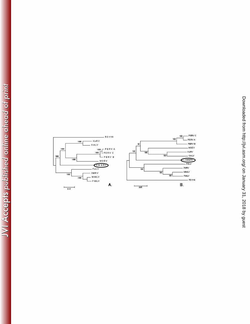
together. A comparison of the Bayesian phylogenetic tree of the full-length nucleotide
sequences with the neighbor-joining tree of the amino acid sequences shows KWERV to
cluster with MDEV, PERV, KoRV, and GALV (Figure 4) though it is clearly in a
distinct line. These results were also consistent for each gene individually using the same
analyses.
From the six proviruses sequenced, there were two separate sets of LTR’s identified for
this endogenous retrovirus family. One set is identical on each end of the viral genome
and is comprised of 436 base pairs, each of which is bounded by a 3 base pair inverted
repeat. The second LTR has a 2% divergence in sequence between its 3’ and 5’ ends,
suggesting it has been integrated into the killer whale genome longer than the first. It is
approximately 89% identical with the first LTR. The 3’ LTR is preceded by a 16 base
pair polypurine tract. The TATA box is located at position 263. The transcriptional start
site is yet to be determined, but is estimated to fall around position 294 based upon
alignment with the koala gammaretrovirus LTR (15). The area surrounding the putative
KoRV transcriptional start site is 63% identical over 20 base pairs to KWERV.
Therefore, the U3 region is estimated to be 293 base pairs. The R region is 76 bp
spanning from the estimated transcriptional start site, past the poly A signal at position
348 to a CA dinucleotide located 16 bp downstream of the PAS. The U5 region spans
positions 370 to 436, making it 67 bp in length. The CCAAT box (a common promoter
motif in eukaryotes) is located at position 163. The 3’ end of U5 in the 5’ LTR is defined
by the presence of an 18 bp proline tRNA primer-binding site, which primes the plus-
strand synthesis during reverse transcription.
on January 31, 2018 by guesthttp://jvi.asm
.org/D
ownloaded from
16
The predicted start codon for gag (1584 bp) is at position 849. The gag sequence aligns
most closely with Porcine Endogenous Retrovirus (PERV): 62.3% identity at the
nucleotide level and 62.1% at the amino acid level. A viable Cys-His box motif,
commonly found in the nucleocapsid region and important in viral encapsidization, is
located at the position 2325. Notably, both the PPPY and PSAP L domains are intact. In
contrast, the endogenous form of KoRV has a dysfunctional form of the PPXY motif
resulting in significantly lower levels of viral particle assembly. Thus, reported titers for
KoRV versus GALV were approximately1800-fold lower (10, 34).
The predicted start for pol (3504 bp) is at position 2433, immediately after the leaky stop
codon common to all gammaretroviruses (8). A stop codon is located between the RNase
H and integrase genes at position 4767, but another start is found at the beginning of the
integrase at position 4893. The significance of this feature is unknown, as
gammaretroviruses do not typically present with a stop codon in this location. However,
they are known to read through stop codons in other locations, such as the one commonly
found between the gag and pol genes (8). The polymerase region also aligns most
closely with PERV (64% nucleotide, 67% amino acid identity).
The predicted start codon for env (2145 bp) is at position 5651. The env nucleotide
sequence aligns most closely with the koala gammaretrovirus (KoRV; 54.2%). However,
the amino acid sequence aligns most closely with a murine endogenous retrovirus
(MDEV; 52.4%). A second KWERV envelope sequence (env B) was identified by
on January 31, 2018 by guesthttp://jvi.asm
.org/D
ownloaded from
17
amplification of the envelope gene alone, but was not found with any of the full length
sequences. This variant is disrupted by stop codons at positions 6131 and 6509 and is
89.7% identical to the first at the nucleotide level and 86.6% identical at the amino acid
level. It also aligns most closely with KoRV at the nucleotide level (54.5%) and with
MDEV at the amino acid level (52.2%). A possible limitation to this first analysis was
the use of a GALV env sequence that is truncated by several amino acids at the 3’ end.
Repetition of the analysis with the full-length GALV SEATO env sequence produced the
same results (accession no. AF055060).
RNA Expression
RT-PCR of multiple tissues (brain, axillary and cervical lymph nodes, adrenal, kidney,
serum, skin, spleen) from the original animal (0162) showed gag expression in brain,
axillary lymph node, and spleen. Pol RNA expression was demonstrated in brain,
axillary lymph node, spleen, and serum. Expression of the first env variant (env A) was
demonstrated in cervical and axillary lymph nodes, spleen, adrenal, serum, and brain,
while transcription of the second variant (env B) was present in the same tissues as well
as skin. Out of 11 blood samples tested, 7 were RT-PCR positive for env in peripheral
blood mononuclear cells (four of env A and all seven for env B), and 9 were RT-PCR
positive for pol, but no viral RNA was ever amplified from plasma (data not shown).
Quantitative Copy Number Assays and Southern Hybridization
Quantitative TaqMan PCR-based assays for proviral copy numbers were devised for gag,
pol and env and tested in multiple tissues in animal 0162. Subsequently, tissues from two
on January 31, 2018 by guesthttp://jvi.asm
.org/D
ownloaded from
18
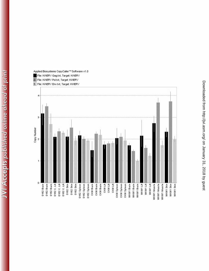
additional animals were tested using the same assay and compared (Figure 5). In all
cases, copy numbers were between 2 and 3, and did not exceed 4 copies per diploid
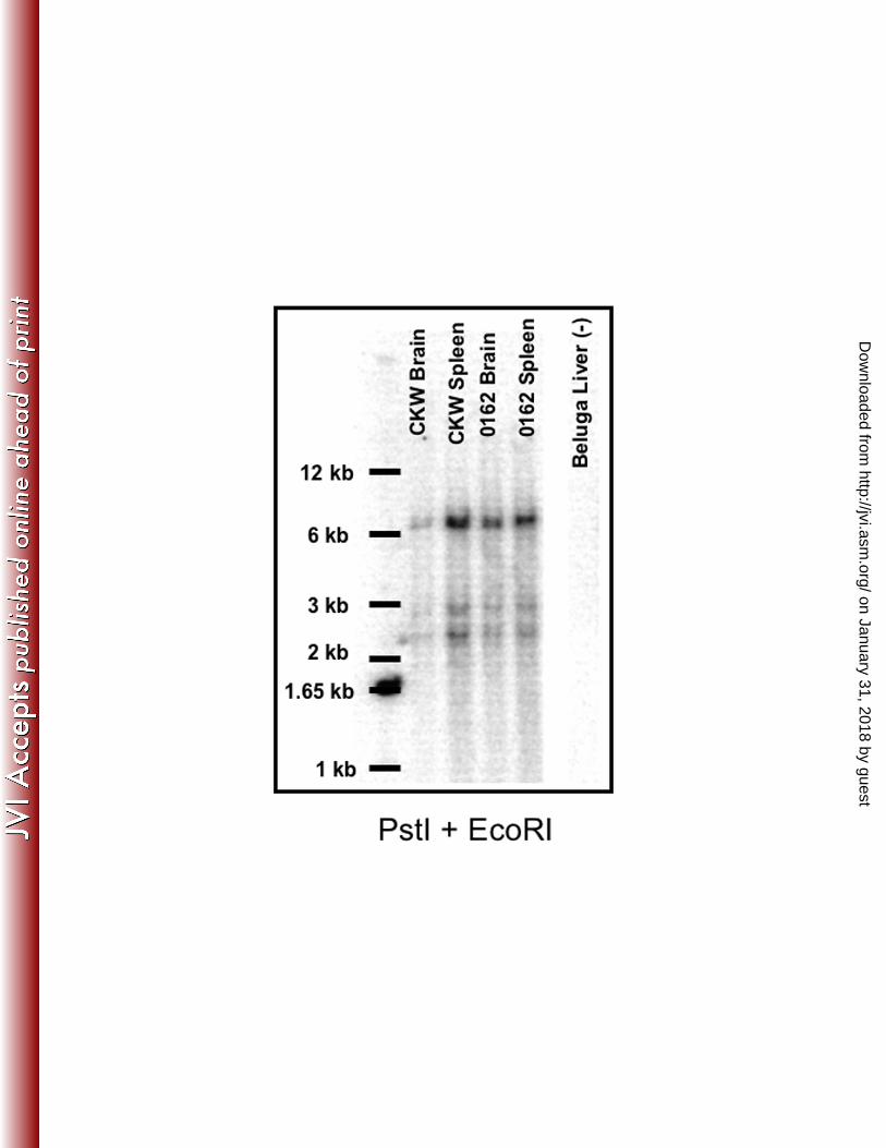
genome. Results for Southern blots are shown in Figure 6. Restriction enzyme PstI was
chosen for the digest due to its placement with respect to the gag probe, and EcoRI
(which has no sites in the reported sequence) was added for more complete digestion of
flanking sequences. Genomic DNA from tissues of two captive animals from different
facilities was compared. Banding patterns appeared the same for all samples,
demonstrating a common integration site and supporting the conclusion that KWERV is
an endogenous retrovirus present in all killer whales.
Analysis for KWERV in other species
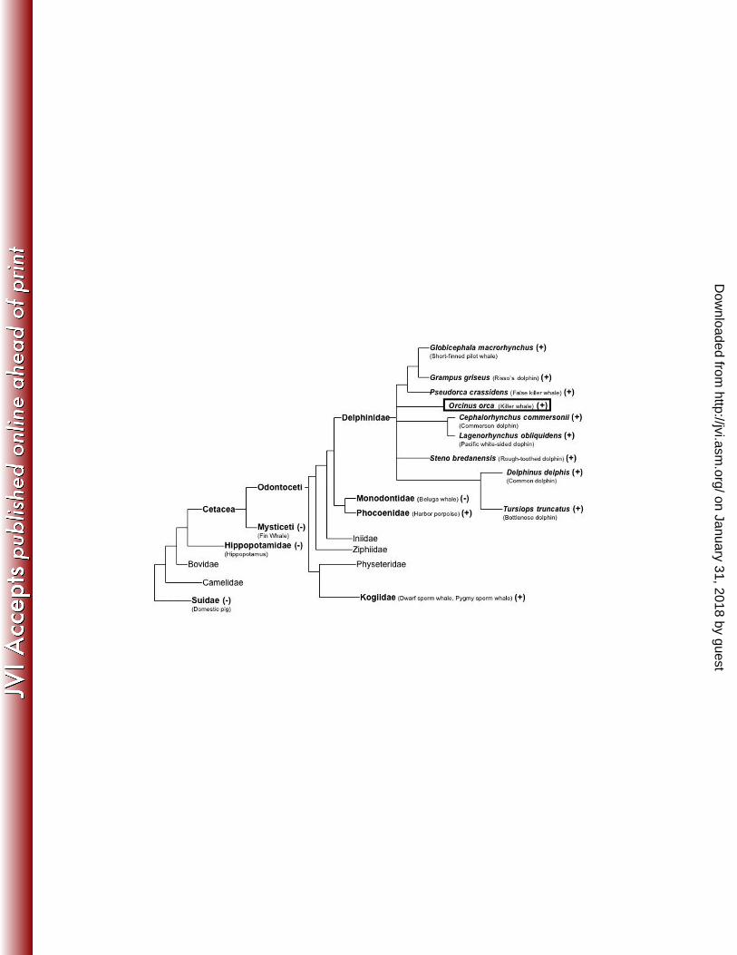
Results for PCR analysis in relation to the phylogeny of other cetacean and artiodactylid
species are shown in Figure 7. The domestic pig, hippopotamus, fin whale, and beluga
whales were all negative for KWERV gag, pol, and env. All delphinid species tested
were PCR positive for all three KWERV genes: gag, pol, and env. In addition, both
animals from the Kogia genus (dwarf and pygmy sperm whales) and Phocoena genus
(harbor porpoises) were positive for gag, but not pol and env. Sequencing of the gag
products from these species showed the amplicons were closely related, but distinct
retroviral sequences from KWERV (70% identical for Kogia and 81% identical for
Phocoena). In contrast, gag products from all the delphinids were nearly identical (95-
98%) to the original KWERV nucleotide sequence. The Ensembl database search for
full-length KWERV in the bottlenose dolphin (Tursiops truncatus) genome yielded two
full-length sequences (scaffold 2349 and scaffold 111015) that were 97.4% and 96.9%
on January 31, 2018 by guesthttp://jvi.asm
.org/D
ownloaded from
19
identical to the KWERV nucleotide sequence, respectively. However, both Tursiops
sequences were disrupted by numerous frameshifts and stop codons.
Single Cell PCR of sperm
Through conventional nested PCR, 2 of 4 single sperm were PCR positive for KWERV
gag. The TaqMan pol assay showed 10 of 17 single sperm as positive, with a median CT
of 36.3 (with a positive control of 10 pg of genomic DNA CT value of 29.3 and non-
template controls completely negative). As a frame of reference, human sperm have been
reported to have an average of 3 pg of DNA per cell (43).
Discussion
We conclude from the data reported here that the retrovirus initially amplified from killer
whales exists in multiple odontocete species (Figure 7), and that KWERV should be
classified among the gammaretroviruses. The KWERV genome exhibits all the classical
features of a gammaretrovirus, including a CCAAT box, a TATA box, a Cys-His box, a
polyadenylation signal, a proline-based tRNA primer binding site, splice donor and
acceptor sites, the polypurine tract and the PPXY/PSAP L domains (8). We also
conclude that the virus is endogenous in killer whales and likely other delphinids based
upon the ubiquitous presence of the provirus among individuals of the species, consistent
copy numbers between individuals and tissues, common integration sites, and presence in
single sperm cells of a killer whale. However, it is quite possible that exogenous variants
of the virus exist in nature. Based upon phylogenetic analysis of KWERV with other
gammaretroviruses, it groups most closely with the PERV/GALV clade (Figure 4).
on January 31, 2018 by guesthttp://jvi.asm
.org/D
ownloaded from
20
There are both endogenous and exogenous members of this retrovirus family. While
some have been directly linked to disease in their respective species (e.g. GALV), others
are in the process of being endogenized (e.g. KoRV) and many appear to be innocuous to
the host (e.g. PERV).
The closest documented relative of KWERV is PERV. However, the percent identity at
both the nucleotide and amino acid level is not substantially different than the average for
the computed pairwise distances of all gammaretroviruses included in the analysis (57%
nucleotide identity with an average of 59.2% and 61% amino acid identity versus an
average of 60.6%, respectively). It is apparent that the most recent viral ancestors from
which KWERV evolved are not presently known. Of note is the fact that pigs and
cetaceans diverged from a common ancestor over 60 million years ago (1) (Figure 7), but
PERV is only estimated to be about 7 million years old (53). Thus, although pigs and
cetaceans share a common ancestor, KWERV was not derived from an endogenous
precursor in that common ancestor. It follows this conclusion that not all cetaceans are
positive for the virus (e.g. fin and beluga whales). Thus, KWERV appears to have been
endogenized after the divergence between cetaceans and artiodactylids (i.e. hoofed
mammals).
Based upon our initial characterization of KWERV in killer whales, this retrovirus is not
a disease-causing agent for this species as there is currently no molecular evidence for
productive infection in vivo. Stable copy numbers of only 2-4 suggest the virus either
became replication defective quickly after integration into killer whales, or that the
on January 31, 2018 by guesthttp://jvi.asm
.org/D
ownloaded from
21
species quickly adapted to prevent further viral replication. While the virus is
transcriptionally active in multiple individuals and in multiple tissues, viral RNA was not
found in cell-free plasma of any individuals. Viral polymerase and envelope RNA was
amplified from serum of the original animal (0162), but TaqMan copy number results
from that animal were the same in all the tissues tested. It is possible that the positive
result from the serum was due to cellular RNA contamination. Alternatively, endogenous
retroviral RNA and proteins are produced routinely in vivo without the production of
replication-competent viral particles (56, 57, 61). In addition, attempts to visualize the
virus through electron microscopy or express the virus in vitro through activation of
PBMC’s have not been successful thus far (data not shown).
Based upon the near intact nature of the open reading frames, the transcriptional activity
of the provirus, and the identical LTR’s flanking one of the two variants, it appears that at
least one of the variants found in the killer whale was a relatively recent integration into
the genome. Endogenous retroviruses can be useful markers for reinforcing evolutionary
relationships (22, 32, 61). Delphinids are estimated to have diverged close to 12 million
years ago (1, 5), and it is tempting to hypothesize that this retrovirus was endogenized
before that time by a delphinid ancestor since all delphinid species examined were
positive for all three retroviral genes (Figure 7). However, while analysis of the
bottlenose dolphin genome showed a provirus with a 97.4% nucleotide identity to this
retrovirus, the open reading frames were severely disrupted by stop codons and
frameshifts, suggesting it was integrated into that species at an earlier point in time than
the most intact KWERV sequence we obtained. In addition, PCR positive results were
on January 31, 2018 by guesthttp://jvi.asm
.org/D
ownloaded from
22
obtained for a related gag gene in both harbor porpoises and in Kogia species, but not in
beluga whales. These results are inconsistent with the phylogenetic groupings reported
for cetaceans to date (1, 5, 23, 33), suggesting that related exogenous retroviruses have
infected several odontocete species and endogenized independently at different times.
It is unknown at this time whether an exogenous variant of this virus is still circulating in
cetacean populations. It is noteworthy that harbor porpoises, a dwarf sperm whale, and a
pygmy sperm whale were PCR positive for the gag, but not for pol or env, suggesting the
presence of either a severely disrupted provirus or an altogether different but related
virus. While the gag portion amplified from dwarf and pygmy sperm whales and harbor
porpoises was related to KWERV, the sequences were far more divergent from KWERV
than those amplified within the Delphinidae family. There are two possible explanations
for the presence of the gag gene in these species. Either a KWERV predecessor was
incorporated into the cetacean genomes before the divergence of those families (16-19
million years ago for harbor porpoises, 32 million years ago for Kogia (1, 5)) and has co-
evolved with its respective hosts, or a different (but related) retrovirus was integrated into
the genomes of those families independently. The failure to amplify any KWERV-
related sequences from beluga whales argues against the first hypothesis, based upon the
estimated divergences of those species (Figure 7).
Viral diseases in cetaceans have been largely unexplored, and it is imperative to gain
understanding of the infectious diseases affecting these species in order to guide
decisions for managing husbandry and veterinary medical care. It is also becoming
on January 31, 2018 by guesthttp://jvi.asm
.org/D
ownloaded from
23
increasingly important to further an understanding of the environmental and genetic
factors affecting the onset and course of disease in free-ranging populations of marine
mammals. Although the study of retroviruses is a unique challenge in these animals,
their proclivity for jumping species unpredictably (26, 27, 54, 55) makes them an
important area of focus. Moreover, the clear homology between this novel retrovirus of
cetaceans and the gammaretroviruses of terrestrial mammals suggests that the histories
and life cycles of these retroviruses have intersected and may continue to do so.
on January 31, 2018 by guesthttp://jvi.asm
.org/D
ownloaded from
24
ACKNOWLEDGMENTS
We thank Tammy Tucker, Erika Nilson and Frank Harrison for excellent technical
assistance and Drs. Pauline Lee and Stephanie Cherqui for invaluable technical advice.
We also thank Dr. Carolyn Wilson for her advice and helpful comments on the
manuscript. We thank Dr. James Casey, Dr. Davey Smith, Dr. Douglas Richman, Dr.
Ellen Sparger, Dr. Marcy Auerbach, and Dr. Katie Marcucci for all of their intellectual
input. We also thank Dr. Stephen Raverty, Dr. Ted Cranford, Megan Stolen, Wendy
Noke, and Todd Robeck for their assistance with the collection of samples.
This work was funded by the following: NIH R01 AI52349 (DRS, SAL), unrestricted
research support from the Busch Entertainment Corporation (SAL), NIH Training Grant
DK007022 (SAL), the Molly Baber Research Fund (DRS, SAL) and the Verna Harrah
Research Fund (DRS, SAL)
on January 31, 2018 by guesthttp://jvi.asm
.org/D
ownloaded from
25
1. Arnason, U., A. Gullberg, and A. Janke. 2004. Mitogenomic analyses provide new insights into cetacean
origin and evolution. Gene 333:27-34.
2. Bartosch, B., R. A. Weiss, and Y. Takeuchi. 2002. PCR-based cloning and immunocytological titration
of infectious porcine endogenous retrovirus subgroup A and B. J Gen Virol 83:2231-40.
3. Bossart, G. D., R. Ewing, A. J. Herron, C. Cray, B. Mase, S. J. Decker, J. W. Alexander, and N. H.
Altman. 1997. Immunoblastic malignant lymphoma in dolphins: histologic, ultrastructural, and
immunohistochemical features. J Vet Diagn Invest 9:454-8.
4. Burmeister, T., S. Schwartz, and E. Thiel. 2001. A PCR primer system for detecting oncoretroviruses
based on conserved DNA sequence motifs of animal retroviruses and its application to human leukaemias
and lymphomas. J Gen Virol 82:2205-13.
5. Caballero, S., J. Jackson, A. A. Mignucci-Giannoni, H. Barrios-Garrido, S. Beltran-Pedreros, M. A.
Montiel-Villalobos, K. M. Robertson, and C. S. Baker. 2008. Molecular systematics of South American
dolphins Sotalia: sister taxa determination and phylogenetic relationships, with insights into a multi-locus
phylogeny of the Delphinidae. Mol Phylogenet Evol 46:252-68.
6. Casey, J. W., A. Roach, J. I. Mullins, K. B. Burck, M. O. Nicolson, M. B. Gardner, and N. Davidson.
1981. The U3 portion of feline leukemia virus DNA identifies horizontally acquired proviruses in leukemic
cats. Proc Natl Acad Sci U S A 78:7778-82.
7. Chenna, R., H. Sugawara, T. Koike, R. Lopez, T. J. Gibson, D. G. Higgins, and J. D. Thompson.
2003. Multiple sequence alignment with the Clustal series of programs. Nucleic Acids Res 31:3497-500.
8. Coffin, J. M., S. H. Hughes, and H. E. Varmus. 1997. Retroviruses. Cold Spring Harbor Laboratory
Press, Woodbury, NY.
9. Delassus, S., P. Sonigo, and S. Wain-Hobson. 1989. Genetic organization of gibbon ape leukemia virus.
Virology 173:205-13.
10. Demirov, D. G., and E. O. Freed. 2004. Retrovirus budding. Virus Res 106:87-102.
11. Dickerson, F., E. Rubalcaba, R. Viscidi, S. Yang, C. Stallings, A. Sullens, A. Origoni, F. Leister, and
R. Yolken. 2008. Polymorphisms in human endogenous retrovirus K-18 and risk of type 2 diabetes in
individuals with schizophrenia. Schizophr Res 104:121-6.
12. Dolei, A., and H. Perron. 2009. The multiple sclerosis-associated retrovirus and its HERV-W endogenous
family: a biological interface between virology, genetics, and immunology in human physiology and
disease. J Neurovirol 15:4-13.
13. Donahue, P. R., E. A. Hoover, G. A. Beltz, N. Riedel, V. M. Hirsch, J. Overbaugh, and J. I. Mullins.
1988. Strong sequence conservation among horizontally transmissible, minimally pathogenic feline
leukemia viruses. J Virol 62:722-31.
14. Gifford, R., P. Kabat, J. Martin, C. Lynch, and M. Tristem. 2005. Evolution and distribution of class II-
related endogenous retroviruses. J Virol 79:6478-86.
15. Hanger, J. J., L. D. Bromham, J. J. McKee, T. M. O'Brien, and W. F. Robinson. 2000. The nucleotide
sequence of koala (Phascolarctos cinereus) retrovirus: a novel type C endogenous virus related to Gibbon
ape leukemia virus. J Virol 74:4264-72.
16. Holzschu, D. L., D. Martineau, S. K. Fodor, V. M. Vogt, P. R. Bowser, and J. W. Casey. 1995.
Nucleotide sequence and protein analysis of a complex piscine retrovirus, walleye dermal sarcoma virus. J
Virol 69:5320-31.
17. Ikeda, H., and H. Sugimura. 1989. Fv-4 resistance gene: a truncated endogenous murine leukemia virus
with ecotropic interference properties. J Virol 63:5405-12.
18. Katsoulos, P. D., G. Christodoulopoulos, G. Kontopidis, A. Minas, A. Tzivara, and S. K. Kritas. 2009.
Leukocyte counts in bronchoalveolar lavage fluid obtained from normal and Maedi-Visna-infected sheep.
Vet Clin Pathol 38:397-402.
19. Kennedy-Stoskopf, S., M. K. Stoskopf, M. A. Eckhaus, and J. D. Strandberg. 1986. Isolation of a
retrovirus and a herpesvirus from a captive California sea lion. J Wildl Dis 22:156-64.
20. Krach, U., N. Fischer, F. Czauderna, and R. R. Tonjes. 2001. Comparison of replication-competent
molecular clones of porcine endogenous retrovirus class A and class B derived from pig and human cells. J
Virol 75:5465-72.
on January 31, 2018 by guesthttp://jvi.asm
.org/D
ownloaded from
26
21. Kumar, S., K. Tamura, and M. Nei. 2004. MEGA3: Integrated software for Molecular Evolutionary
Genetics Analysis and sequence alignment. Brief Bioinform 5:150-63.
22. Lapuk, A. V., P. P. Khil, I. V. Lavrentieva, Y. B. Lebedev, and E. D. Sverdlov. 1999. A human
endogenous retrovirus-like (HERV) LTR formed more than 10 million years ago due to an insertion of
HERV-H LTR into the 5' LTR of HERV-K is situated on human chromosomes 10, 19 and Y. J Gen Virol
80 ( Pt 4):835-9.
23. LeDuc, R. G., W. F. Perrin, and A. E. Dizon. 1999. PHYLOGENETIC RELATIONSHIPS AMONG
THE DELPHINID CETACEANS BASED ON FULL CYTOCHROME <i>B</i> SEQUENCES. Marine
Mammal Science 15:619-648.
24. Ma, Y., M. Lv, S. Xu, J. Wu, K. Tian, and J. Zhang. 2008. Identification of full-length proviral DNA of
porcine endogenous retrovirus from Chinese Wuzhishan miniature pigs inbred. Comp Immunol Microbiol
Infect Dis.
25. Marguerat, S., W. Y. Wang, J. A. Todd, and B. Conrad. 2004. Association of human endogenous
retrovirus K-18 polymorphisms with type 1 diabetes. Diabetes 53:852-4.
26. Martin, J., E. Herniou, J. Cook, R. W. O'Neill, and M. Tristem. 1999. Interclass transmission and
phyletic host tracking in murine leukemia virus-related retroviruses. J Virol 73:2442-9.
27. Martin, J., E. Herniou, J. Cook, R. Waugh O'Neill, and M. Tristem. 1997. Human endogenous
retrovirus type I-related viruses have an apparently widespread distribution within vertebrates. J Virol
71:437-43.
28. Martineau, D., P. R. Bowser, R. R. Renshaw, and J. W. Casey. 1992. Molecular characterization of a
unique retrovirus associated with a fish tumor. J Virol 66:596-9.
29. Martineau, D., R. R., W. J.R., C. J.W., and B. P.R. 1991. A large unintegrated retrovirus DNA species
present in a dermal tumor of walleye Stizostedion vitreum. Diseases of Aquatic Organisms 10:153-8.
30. Masuda, M., M. P. Remington, P. M. Hoffman, and S. K. Ruscetti. 1992. Molecular characterization of
a neuropathogenic and nonerythroleukemogenic variant of Friend murine leukemia virus PVC-211. J Virol
66:2798-806.
31. Newman, S. J., and S. A. Smith. 2006. Marine mammal neoplasia: a review. Vet Pathol 43:865-80.
32. Niebert, M., and R. R. Tonjes. 2005. Evolutionary spread and recombination of porcine endogenous
retroviruses in the suiformes. J Virol 79:649-54.
33. Nikaido, M., F. Matsuno, H. Hamilton, R. L. Brownell, Jr., Y. Cao, W. Ding, Z. Zuoyan, A. M.
Shedlock, R. E. Fordyce, M. Hasegawa, and N. Okada. 2001. Retroposon analysis of major cetacean
lineages: the monophyly of toothed whales and the paraphyly of river dolphins. Proc Natl Acad Sci U S A
98:7384-9.
34. Oliveira, N. M., H. Satija, I. A. Kouwenhoven, and M. V. Eiden. 2007. Changes in viral protein function
that accompany retroviral endogenization. Proc Natl Acad Sci U S A 104:17506-11.
35. Paul, T. A., S. L. Quackenbush, C. Sutton, R. N. Casey, P. R. Bowser, and J. W. Casey. 2006.
Identification and characterization of an exogenous retrovirus from atlantic salmon swim bladder sarcomas.
J Virol 80:2941-8.
36. Perron, H., J. A. Garson, F. Bedin, F. Beseme, G. Paranhos-Baccala, F. Komurian-Pradel, F. Mallet,
P. W. Tuke, C. Voisset, J. L. Blond, B. Lalande, J. M. Seigneurin, and B. Mandrand. 1997. Molecular
identification of a novel retrovirus repeatedly isolated from patients with multiple sclerosis. The
Collaborative Research Group on Multiple Sclerosis. Proc Natl Acad Sci U S A 94:7583-8.
37. Rastogi, P. A. 1999. MacVector, p. 47-69, vol. 132.
38. Reyes, R. A., and G. L. Cockerell. 1996. Unintegrated bovine leukemia virus DNA: association with viral
expression and disease. J Virol 70:4961-5.
39. Roca, A. L., J. Pecon-Slattery, and S. J. O'Brien. 2004. Genomically intact endogenous feline leukemia
viruses of recent origin. J Virol 78:4370-5.
40. Ronquist, F., and J. P. Huelsenbeck. 2003. MrBayes 3: Bayesian phylogenetic inference under mixed
models. Bioinformatics 19:1572-4.
on January 31, 2018 by guesthttp://jvi.asm
.org/D
ownloaded from
27
41. Ruprecht, K., F. Gronen, M. Sauter, B. Best, P. Rieckmann, and N. Mueller-Lantzsch. 2008. Lack of
immune responses against multiple sclerosis-associated retrovirus/human endogenous retrovirus W in
patients with multiple sclerosis. J Neurovirol 14:143-51.
42. Sambrook, J., E. F. Fritsch, and T. Maniatis. 1989. Molecular cloning : a laboratory manual, 2nd ed.
Cold Spring Harbor Laboratory Press, Cold Spring Harbor, N.Y.
43. Sarkar, S., O. W. Jones, and N. Shioura. 1974. Constancy in human sperm DNA content. Proc Natl Acad
Sci U S A 71:3512-6.
44. Sheets, R. L., R. Pandey, W. C. Jen, and P. Roy-Burman. 1993. Recombinant feline leukemia virus
genes detected in naturally occurring feline lymphosarcomas. J Virol 67:3118-25.
45. Shinnick, T. M., R. A. Lerner, and J. G. Sutcliffe. 1981. Nucleotide sequence of Moloney murine
leukaemia virus. Nature 293:543-8.
46. Stoye, J. P. 2006. Koala retrovirus: a genome invasion in real time. Genome Biol 7:241.
47. Tandon, R., V. Cattori, A. C. Pepin, B. Riond, M. L. Meli, M. McDonald, M. G. Doherr, H. Lutz, and
R. Hofmann-Lehmann. 2008. Association between endogenous feline leukemia virus loads and
exogenous feline leukemia virus infection in domestic cats. Virus Res 135:136-43.
48. Tandon, R., V. Cattori, B. Willi, M. L. Meli, M. A. Gomes-Keller, H. Lutz, and R. Hofmann-
Lehmann. 2007. Copy number polymorphism of endogenous feline leukemia virus-like sequences. Mol
Cell Probes 21:257-66.
49. Tarlinton, R., J. Meers, J. Hanger, and P. Young. 2005. Real-time reverse transcriptase PCR for the
endogenous koala retrovirus reveals an association between plasma viral load and neoplastic disease in
koalas. J Gen Virol 86:783-7.
50. Tarlinton, R., J. Meers, and P. Young. 2008. Biology and evolution of the endogenous koala retrovirus.
Cell Mol Life Sci 65:3413-21.
51. Tarlinton, R. E., J. Meers, and P. R. Young. 2006. Retroviral invasion of the koala genome. Nature
442:79-81.
52. Ting, Y. T., C. A. Wilson, K. B. Farrell, G. J. Chaudry, and M. V. Eiden. 1998. Simian sarcoma-
associated virus fails to infect Chinese hamster cells despite the presence of functional gibbon ape leukemia
virus receptors. J Virol 72:9453-8.
53. Tonjes, R. R., and M. Niebert. 2003. Relative age of proviral porcine endogenous retrovirus sequences in
Sus scrofa based on the molecular clock hypothesis. J Virol 77:12363-8.
54. Tristem, M., P. Kabat, L. Lieberman, S. Linde, A. Karpas, and F. Hill. 1996. Characterization of a
novel murine leukemia virus-related subgroup within mammals. J Virol 70:8241-6.
55. Urisman, A., R. J. Molinaro, N. Fischer, S. J. Plummer, G. Casey, E. A. Klein, K. Malathi, C. Magi-
Galluzzi, R. R. Tubbs, D. Ganem, R. H. Silverman, and J. L. DeRisi. 2006. Identification of a novel
Gammaretrovirus in prostate tumors of patients homozygous for R462Q RNASEL variant. PLoS Pathog
2:e25.
56. Varmus, H. E. 1982. Form and function of retroviral proviruses. Science 216:812-20.
57. Voisset, C., R. A. Weiss, and D. J. Griffiths. 2008. Human RNA "rumor" viruses: the search for novel
human retroviruses in chronic disease. Microbiol Mol Biol Rev 72:157-96, table of contents.
58. Weiss, R. A. 2006. The discovery of endogenous retroviruses. Retrovirology 3:67.
59. Wolgamot, G., L. Bonham, and A. D. Miller. 1998. Sequence analysis of Mus dunni endogenous virus
reveals a hybrid VL30/gibbon ape leukemia virus-like structure and a distinct envelope. J Virol 72:7459-
66.
60. Yonezawa, M., H. Nakamine, T. Tanaka, and T. Miyaji. 1989. Hodgkin's disease in a killer whale
(Orcinus orca). J Comp Pathol 100:203-7.
61. Zsiros, J., M. F. Jebbink, V. V. Lukashov, P. A. Voute, and B. Berkhout. 1998. Evolutionary
relationships within a subgroup of HERV-K-related human endogenous retroviruses. J Gen Virol 79 ( Pt
1):61-70.
on January 31, 2018 by guesthttp://jvi.asm
.org/D
ownloaded from
28
Figure Legends
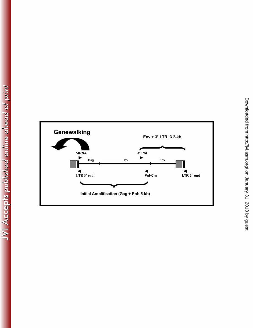
Figure 1. Strategy for obtaining sequence of the full KWERV proviral genome. Initial
amplification was with consensus P-tRNA and Pol-Cm primers. Genewalking was achieved
using three primers from the 5’ region of the gag gene to acquire the 5’ LTR sequence. A
reverse primer designed in the U5 region of the genome (LTR 3’ end) was used with a forward
pol primer (3’ pol) to amplify the remainder of the genome.
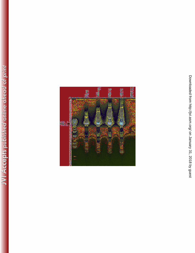
Figure 2. Optimized PCR at 35 cycles for amplifying the near full-length KWERV from
genomic DNA of a killer whale (0162). Expected product size for the near full-length genome
without the 5’ LTR is approximately 7.8 kb. The four lanes represent increasing concentrations
of starting total DNA.
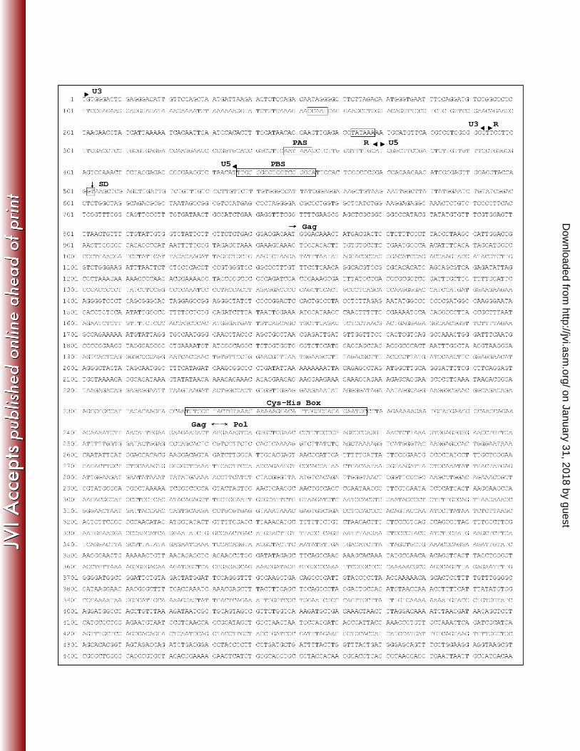
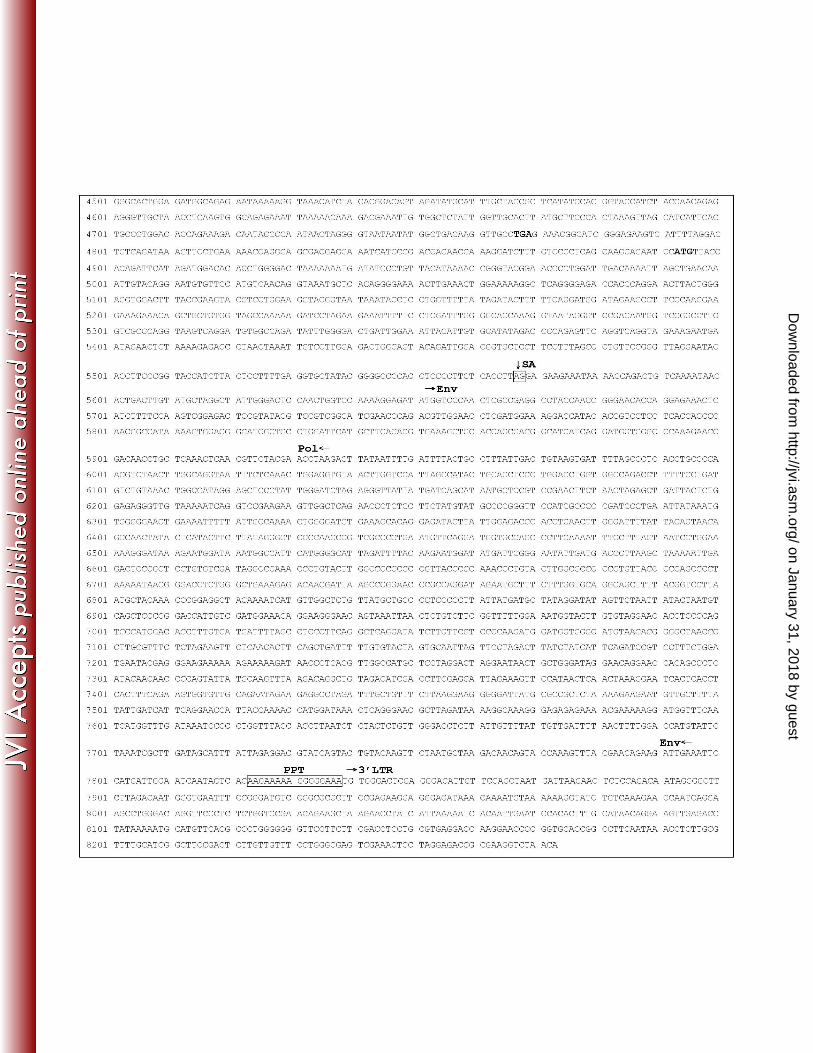
Figure 3. Full-length proviral sequence for Killer Whale Endogenous Retrovirus sequenced from
killer whale (KWERV). Predicted divisions of LTR’s (U3, R, and U5) are denoted by arrow
heads. Features of the sequence are contained in boxes, including the CCAAT box, the TATA
box, the polyadenylation site (PAS), the primer-binding site (PBS), the Cys-His Box, the
polypurine tract (PPT), the predicted splice donor site (SD), and the predicted splice acceptor site
(SA). Boundaries of the three genes, gag, pol, and env, are denoted by arrows. The stop and
start codons found in the polymerase gene between the RNase H and Integrase are denoted with
bold font. Note that the near full-length sequence beginning with the primer-binding site was
from one clone, and the reported 5’ LTR was from an overlapping clone of the 5’ end of the
genome extending approximately 1 kb into the leader and gag sequences.
on January 31, 2018 by guesthttp://jvi.asm
.org/D
ownloaded from

29
Figure 4. Phylogenetic trees of full-length KWERV with other gammaretroviruses. A Bayesian
tree was constructed based upon full-length retroviral nucleotide sequences with 100,000
generations (A) and compared to a neighbor-joining tree based upon amino acid sequences
designed from concatemerized Gag, Pol, and Env with 1,000 bootstraps (B). Clustering for
KWERV appeared similar for both, with KWERV grouping among PERV, MDEV, GALV, and
KoRV. PERV A (Porcine Endogenous Retrovirus A, accession Aj293656), PERV B (Porcine
Endogenous Retrovirus B, accession AY099324), PERV C (Porcine Endogenous Retrovirus C,
accession EF133960), MDEV (Mus dunni Endogenous Retrovirus, accession AF053745), KoRV
(Koala Retrovirus, accession AF151794), GALV (Gibbon Ape Leukemia Virus, accession
NC_001885), KWERV (Killer Whale Endogenous Retrovirus, accession GQ222416), FeLV
(Feline Leukemia Virus, accession M18247), XMRV (Xenotropic Murine Leukemia-Related
Virus, accession DQ399707), MMLV (Moloney Murine Leukemia Virus, accession J02255),
FMLV (Friend Murine Leukemia Virus, accession M93134), RD114 (RD114 Feline Endogenous
Retrovirus, accession EU030001). Distance scales shown for both trees represent number of
substitutions per site.
Figure 5. Calculated copy number results from tissues of three different killer whales. 0162
represents the original animal screened, while CKW represents an animal from a separate
institution. WKW1 represents a free-ranging killer whale. In all cases, copy numbers for gag,
pol, and env appear relatively stable between tissues and animals, in most cases varying between
2 and 4 copies per diploid genome.
Figure 6. Southern Hybridization of PstI and EcoRI digested genomic DNA using a 32
P
radiolabeled gag probe. Banding patterns for each digest were the same between lymphoid
tissues and brain of two animals from different facilities, suggesting sites of integration are the
same between samples.
on January 31, 2018 by guesthttp://jvi.asm
.org/D
ownloaded from
30
Figure 7. Phylogenetic distribution of KWERV. Families and species in bold represent
members tested for gag, pol, and env genes of KWERV using PCR. Those denoted “+” were
PCR positive for at least one KWERV gene. Sequencing of gag products from Kogiidae and
Phocoenidae showed a related gag gene with 70% and 81% nucleotide identity to KWERV,
respectively. Sequencing of gag products from Delphinidae showed nucleotide identities to
KWERV ranging from 95-98%.
on January 31, 2018 by guesthttp://jvi.asm
.org/D
ownloaded from
31
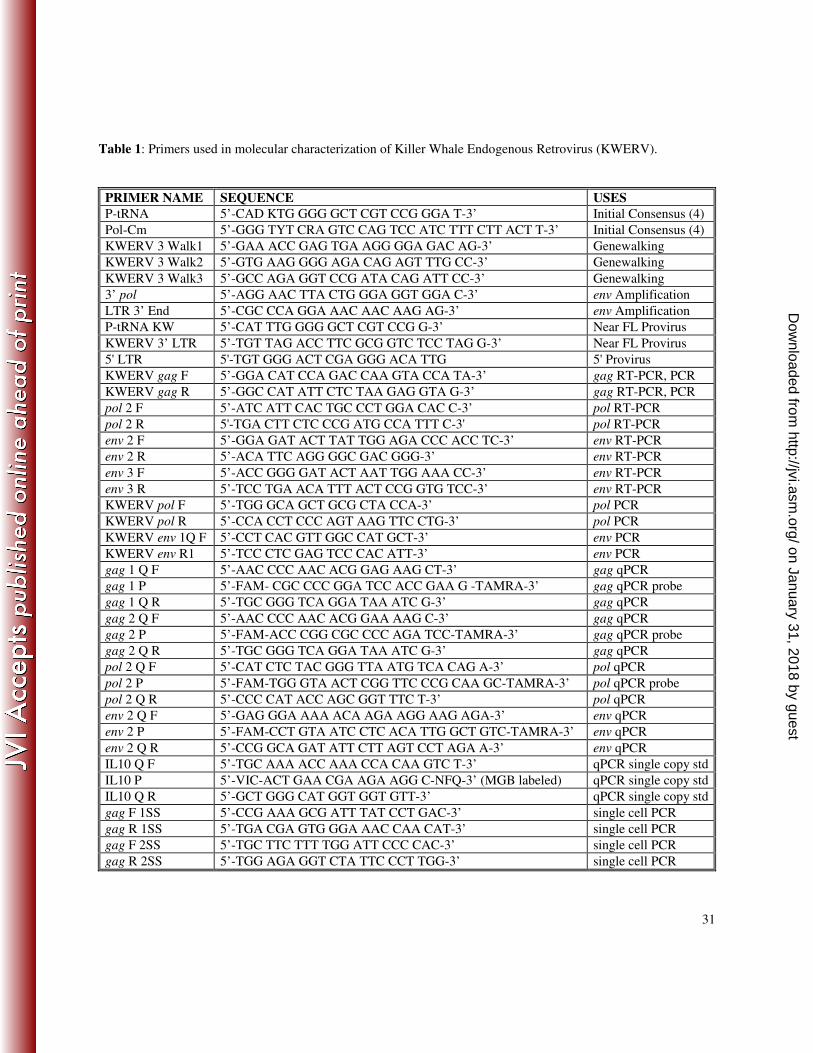
Table 1: Primers used in molecular characterization of Killer Whale Endogenous Retrovirus (KWERV).
PRIMER NAME SEQUENCE USES
P-tRNA 5’-CAD KTG GGG GCT CGT CCG GGA T-3’ Initial Consensus (4)
Pol-Cm 5’-GGG TYT CRA GTC CAG TCC ATC TTT CTT ACT T-3’ Initial Consensus (4)
KWERV 3 Walk1 5’-GAA ACC GAG TGA AGG GGA GAC AG-3’ Genewalking
KWERV 3 Walk2 5’-GTG AAG GGG AGA CAG AGT TTG CC-3’ Genewalking
KWERV 3 Walk3 5’-GCC AGA GGT CCG ATA CAG ATT CC-3’ Genewalking
3’ pol 5’-AGG AAC TTA CTG GGA GGT GGA C-3’ env Amplification
LTR 3’ End 5’-CGC CCA GGA AAC AAC AAG AG-3’ env Amplification
P-tRNA KW 5’-CAT TTG GGG GCT CGT CCG G-3’ Near FL Provirus
KWERV 3’ LTR 5’-TGT TAG ACC TTC GCG GTC TCC TAG G-3’ Near FL Provirus
5' LTR 5'-TGT GGG ACT CGA GGG ACA TTG 5' Provirus
KWERV gag F 5’-GGA CAT CCA GAC CAA GTA CCA TA-3’ gag RT-PCR, PCR
KWERV gag R 5’-GGC CAT ATT CTC TAA GAG GTA G-3’ gag RT-PCR, PCR
pol 2 F 5’-ATC ATT CAC TGC CCT GGA CAC C-3’ pol RT-PCR
pol 2 R 5'-TGA CTT CTC CCG ATG CCA TTT C-3' pol RT-PCR
env 2 F 5’-GGA GAT ACT TAT TGG AGA CCC ACC TC-3’ env RT-PCR
env 2 R 5’-ACA TTC AGG GGC GAC GGG-3’ env RT-PCR
env 3 F 5’-ACC GGG GAT ACT AAT TGG AAA CC-3’ env RT-PCR
env 3 R 5’-TCC TGA ACA TTT ACT CCG GTG TCC-3’ env RT-PCR
KWERV pol F 5’-TGG GCA GCT GCG CTA CCA-3’ pol PCR
KWERV pol R 5’-CCA CCT CCC AGT AAG TTC CTG-3’ pol PCR
KWERV env 1Q F 5’-CCT CAC GTT GGC CAT GCT-3’ env PCR
KWERV env R1 5’-TCC CTC GAG TCC CAC ATT-3’ env PCR
gag 1 Q F 5’-AAC CCC AAC ACG GAG AAG CT-3’ gag qPCR
gag 1 P 5’-FAM- CGC CCC GGA TCC ACC GAA G -TAMRA-3’ gag qPCR probe
gag 1 Q R 5’-TGC GGG TCA GGA TAA ATC G-3’ gag qPCR
gag 2 Q F 5’-AAC CCC AAC ACG GAA AAG C-3’ gag qPCR
gag 2 P 5’-FAM-ACC CGG CGC CCC AGA TCC-TAMRA-3’ gag qPCR probe
gag 2 Q R 5’-TGC GGG TCA GGA TAA ATC G-3’ gag qPCR
pol 2 Q F 5’-CAT CTC TAC GGG TTA ATG TCA CAG A-3’ pol qPCR
pol 2 P 5’-FAM-TGG GTA ACT CGG TTC CCG CAA GC-TAMRA-3’ pol qPCR probe
pol 2 Q R 5’-CCC CAT ACC AGC GGT TTC T-3’ pol qPCR
env 2 Q F 5’-GAG GGA AAA ACA AGA AGG AAG AGA-3’ env qPCR
env 2 P 5’-FAM-CCT GTA ATC CTC ACA TTG GCT GTC-TAMRA-3’ env qPCR
env 2 Q R 5’-CCG GCA GAT ATT CTT AGT CCT AGA A-3’ env qPCR
IL10 Q F 5’-TGC AAA ACC AAA CCA CAA GTC T-3’ qPCR single copy std
IL10 P 5’-VIC-ACT GAA CGA AGA AGG C-NFQ-3’ (MGB labeled) qPCR single copy std
IL10 Q R 5’-GCT GGG CAT GGT GGT GTT-3’ qPCR single copy std
gag F 1SS 5’-CCG AAA GCG ATT TAT CCT GAC-3’ single cell PCR
gag R 1SS 5’-TGA CGA GTG GGA AAC CAA CAT-3’ single cell PCR
gag F 2SS 5’-TGC TTC TTT TGG ATT CCC CAC-3’ single cell PCR
gag R 2SS 5’-TGG AGA GGT CTA TTC CCT TGG-3’ single cell PCR
on January 31, 2018 by guesthttp://jvi.asm
.org/D
ownloaded from