molecular cell article - flyrnai.org · absence of sumo, pc foci coagulate into larger ... in nine...
TRANSCRIPT

Molecular Cell
Article
Identification of Regulators of theThree-Dimensional Polycomb Organization by aMicroscopy-Based Genome-wide RNAi ScreenInma Gonzalez,1,2 Julio Mateos-Langerak,1,2 Aubin Thomas,1 Thierry Cheutin,1 and Giacomo Cavalli1,*1Institute of Human Genetics, UPR1142 CNRS, 141 Rue de la Cardonille, 34396 Montpellier Cedex 5, France2These authors contributed equally to this work
*Correspondence: [email protected]://dx.doi.org/10.1016/j.molcel.2014.03.004
SUMMARY
Polycomb group (PcG) proteins dynamically definecellular identities through epigenetic repression ofkey developmental genes. PcG target gene repres-sion can be stabilized through the interaction in thenucleus at PcG foci. Here, we report the results ofa high-resolution microscopy genome-wide RNAiscreen that identifies 129 genes that regulate thenuclear organization of Pc foci. Candidate genesinclude PcG components and chromatin factors, aswell as many protein-modifying enzymes, includingcomponents of the SUMOylation pathway. In theabsence of SUMO, Pc foci coagulate into largeraggregates. Conversely, loss of function of theSUMO peptidase Velo disperses Pc foci. Moreover,SUMO and Velo colocalize with PcG proteins atPREs, and Pc SUMOylation affects its chromatintargeting, suggesting that the dynamic regulation ofPc SUMOylation regulates PcG-mediated silencingbymodulating the kinetics of Pc binding to chromatinas well as its ability to form Polycomb foci.
INTRODUCTION
Polycomb group (PcG) proteins were initially identified as factors
maintaining repression of homeotic (HOX) genes during develop-
ment (Duncan, 1982). Later work demonstrated a more dynamic
role for PcG proteins in repressing many other developmental
regulators (reviewed in Schuettengruber and Cavalli, 2009;
Schuettengruber et al., 2007). In Drosophila, PcG proteins are
recruited to chromatin by specific DNA elements, called PcG
response elements (PREs). Interestingly, physical contact
between two PREs that can be located at Mb distances or
even on different chromosomes can enhance PcG-dependent
silencing (Bantignies and Cavalli, 2011; Rosa et al., 2013).
PcG proteins form three large multimeric complexes: the Plei-
ohomeotic repressive complex (PhoRC) (Klymenko et al., 2006),
the Polycomb repressive complex 1 (PRC1) (Shao et al., 1999),
and the Polycomb repressive complex 2 (PRC2) (Czermin
et al., 2002). Additional PcG-containing complexes, which
contribute to the diverse functions of PcG proteins, have also
been purified, suggesting that the composition of the known
PcG complexes might be dependent upon tissue and develop-
mental stage. Furthermore, there are PcG factors that are not
stable components of known PcG complexes; therefore, they
could not be identified by biochemical purification of the PcG
complexes (Grimaud et al., 2006). These proteins might interact
transiently with PcG complexes or might be part of yet-to-be-
identified PcG complexes (Otte and Kwaks, 2003).
PcG proteins were originally identified by genetic screenings
in Drosophila through the observation of body plan trans-
formations (so-called PcG phenotypes) due to misexpression
of homeotic (HOX) genes. This early genetic evidence led to an
estimated number of 30–40 PcG genes in Drosophila (Jurgens,
1985), far below the number of PcG genes known today, sug-
gesting that several PcG-related genes might have escaped ge-
netic screening and subsequent biochemical studies.
Pc localizes in the nucleus at foci (also called Pc bodies), and
their dynamic organization and intensity distribution changes
during development (Cheutin and Cavalli, 2012). PcG target
genes located at considerable linear distances along a chromo-
some can cluster within Pc foci (Bantignies et al., 2011; Sexton
et al., 2012), and PcG-mediated silencing of homeotic genes is
stabilized by long-distance interactions of HOX gene clusters
(Bantignies et al., 2011).
In addition to long-range interactions of PcG target genes,
many other contacts involve distant chromatin regions (Bantig-
nies and Cavalli, 2011; Hou et al., 2012; Sexton et al., 2012;
Tolhuis et al., 2011). This suggests that nuclear components
may regulate 3D genome architecture. Here, we present a
genome-wide RNAi screen to identify factors involved in the
regulation of the 3D distribution of PcG proteins.
RESULTS
Primary RNAi Screen for Genes that Regulate theNuclear Organization of PolycombImmunofluorescence (IF) and imaging of Pc-GFP fusion con-
structs show that Pc localizes in the nucleus in foci, which
have no apparent distribution preferences besides a clear ex-
clusion from pericentromeric heterochromatin (Figure 2B). To
look for molecular factors involved in PcG function, we per-
formed an RNAi screen in Drosophila S2 cells, looking for
changes in the Pc-IF pattern by high-resolution confocal
Molecular Cell 54, 485–499, May 8, 2014 ª2014 Elsevier Inc. 485

Molecular Cell
Genome-wide RNAi Screen for 3D Polycomb Regulators
microscopy (e.g., a loss of Pc staining, diffuse Pc staining,
sharper Pc foci, etc.). Although the Pc distribution in S2 cells
was characteristic, there was significant cell-to-cell variability.
Therefore, large numbers of cells were studied for each gene
knockdown. As a counterstain for the image analysis, as well
as to assist in the interpretation of the specificity of the results,
we simultaneously labeled DNAwith DAPI. The screening proce-
dure is summarized in Figure S1A, available online.
We screened two RNAi libraries provided by the Drosophila
RNAi Screening Center (DRSC; www.flyrnai.org) (Ramadan
et al., 2007); the first, a genome-wide library, was analyzed in
duplicate, and the second, a transcription factor library, was
analyzed in triplicate. The genome-wide library contains, in 66
384-well plates, more than 24,000 unique dsRNAs targeting
13,900genescoveringpractically theentire flygenecompendium.
The transcription factor library contains, in nine 384-well plates,
1,890 unique dsRNAs targeting 993 known transcription-related
proteins, such as DNA binding and nuclear proteins. In addition
to these two libraries, we custom prepared two plates containing
multiple replicas of dsRNAs targeting known PcG-related factors.
These control plateswere transfected and subsequently analyzed
under the same conditions (Figures S1B–S1E).
S2 cells were transfected with both RNAi libraries (Figure S1).
Pc was knocked down (KD) as a positive control in the genome-
wide screen. As further controls, we knocked down Pc, Su(z)12,
Sce, and Su(var)205, since KD affected Pc nuclear distribution
during preliminary studies (data not shown). As negative controls
we used dsRNA against either GFP or LacZ. Stained plates were
imaged using a high-content screening spinning disk confocal
microscope equippedwith a 603/NA1.2 water immersion objec-
tive to obtain the best possible resolution in high-throughput
mode. As a result of imaging all the plates, we obtained 1.5
million two-channel images. These images were automatically
treated and analyzed (see Supplemental Experimental Proce-
dures) to extract and characterize, in an unbiased way, the list
of genes whose downregulation had modified the Pc staining
pattern (Figures 1, 2, S2, and S3).
Secondary Validation ScreenIn order to minimize the rate of false positives, we performed a
secondary screen with a selection of candidate genes from the
primary screen. We chose genes clustering with known PcG-
related genes, and we selected (when available at the DRSC)
additional validation dsRNAs that were not present in the
genome-wide library. This resulted in a list of 214 candidate
genes and 288 dsRNAs, plus 96 controls.
The custom RNAi library was screened in quintuplicate
following similar labeling, imaging, and image analysis proce-
dures as used for the primary screen. Seventy-five genes were
confirmed as positive by this subsequent analysis (which gives
a total of 129 positives when added to the 54 hits from the tran-
scription factor screen; see Table S1). As expected, the majority
of genes that were not confirmed were those classified as weak
positives in the primary genome-wide screen.
To further characterize the phenotypes, we devised an auto-
mated selection of the most representative cells for each gene,
dsRNA, or well (see companion website: http://flyepigenome.
igh.cnrs.fr/PCscreen) and clustered their phenotypes in two
486 Molecular Cell 54, 485–499, May 8, 2014 ª2014 Elsevier Inc.
ways. First, we clustered hierarchically all the phenotypes pro-
duced by each dsRNA. This method produced a full, detailed,
and continuous classification tree (Figures S2 and S3A). As a
second method, we used k-means clustering and found that
the data could be robustly classified into four clusters (Figure 2).
The first cluster was comprised mainly of Pc itself, with KD
resulting in an obvious loss of Pc staining. KD of genes in the sec-
ond cluster produced more intense Pc foci. KD of genes in the
third cluster produced a loss of Pc foci and the appearance of
more diffuse nuclear staining. The final cluster, similar to cluster
three, was characterized by a phenotype of weaker intensity of
Pc foci; this phenotype was clearly defined by computer analysis
but barely detectable upon visual inspection, emphasizing the
high sensitivity of the automated image analysis. In rare cases,
different dsRNAs targeting the same gene produced slightly
different phenotypes and, as a consequence, some genes
were placed in two different clusters.
Analysis of the gene functions indicated that themost enriched
ontologies were presumably related to PcG function and to chro-
matin organization (Figure 1A). Among these, PcG proteins
themselves came out in clusters 3 and 4, as expected (Franke
et al., 1995). The brahma and cohesin complexes appeared
particularly enriched among the positives. Moreover, 23 positive
genes were not or were poorly characterized, and among them,
some contained protein-protein or protein-DNA interaction
domains, including chromo-, bromo-, and AT-rich interaction
domains, in addition to catalytic regions, such as peptidase,
phosphatase, and kinase domains. These genes constitute an
interesting set of candidates for future studies.
Components of the SUMOylation Pathway Affect theOrganization of Polycomb Foci in Drosophila
One of the most prominent phenotypes found in our screen was
produced by KD of smt3, which encodes the Drosophila small
ubiquitin-related modifier (SUMO) protein (Huang et al., 1998).
SUMO is covalently bound to other proteins and plays important
roles in regulating their activity, stability, protein-protein inter-
actions, and cellular localization. SUMOs are conjugated to
target proteins through a cascade of reactions that typically
involve three enzymes: an activating enzyme, E1, a conjugating
E2 enzyme, and usually a SUMO ligase (E3), which increases
the efficiency of SUMO conjugation. Before being conjugated
to its targets, SUMO has to undergo maturation by cleavage of
a short C-terminal extension from the precursor by a processing
protease. SUMO modification can be reversed by SENPs
(SUMO/sentrin-specific peptidases) (Ulrich, 2009).
In our screen, KD of SUMO (smt3) caused redistribution of
Pc to fewer and more intense Pc foci (Figure 2D). Among the
other positives, we found Su(var)2-10, a PIAS protein linked to
SUMOylation (Hari et al., 2001), and veloren (velo) (Berdnik
et al., 2012), a gene that encodes a polypeptide containing a
putative cysteine-type SUMO peptidase domain (Li and Hoch-
strasser, 2000). The human homologs of velo, SENP6 and
SENP7, have SUMO-deconjugating activity (Lima and Reverter,
2008). Consistent with a function opposed to SUMO conjuga-
tion, KD of velo produced a strong phenotype, namely diffuse
staining of Pc, the opposite phenotype to that observed after
KD of smt3 (Figure 2E).

Figure 1. Gene Ontology Analysis and Phenotypic Clustering
(A) Table showing the 14most-enriched Gene Ontology (GO) terms in the final list of genes found in the screen. Table shows the GO term category (MF, molecular
function; CC, cellular component; BP, biologic process), the GO term, the fold enrichment, and the p value corrected using the Benjamini-Hochberg procedure.
(B) Details of Figure S2A. As an example, five cases (boxes) are shownwith clusters of phenotypes. Biological replicas (different dsRNAs targeting the same gene)
and technical replicas (plate replicas) cluster together. In the two upper boxes, two clusters of genes with known functions are shown: ribosomal proteins (top left)
and tubulins (top right). The other boxes show genes grouping with PcG genes and other chromatin-related factors. Labels showDRSC code for the dsRNA, gene
targeted, Hits in Public Screens (HiPS) for how many times the dsRNA has been found as positive in a screen, and X19 denoting how many other genes have
sequence identity of at least 19 nucleotides as a measure for possible off-targets. See also Figures S1–S3.
Molecular Cell
Genome-wide RNAi Screen for 3D Polycomb Regulators
Molecular Cell 54, 485–499, May 8, 2014 ª2014 Elsevier Inc. 487

(legend on next page)
Molecular Cell
Genome-wide RNAi Screen for 3D Polycomb Regulators
488 Molecular Cell 54, 485–499, May 8, 2014 ª2014 Elsevier Inc.
Molecular Cell
Genome-wide RNAi Screen for 3D Polycomb Regulators
We expressed hairpin constructs to KD either smt3 or velo
using tissue-specific drivers. The KD efficiency was verified by
immunostaining experiments using anti-SUMO and anti-Velo
antibodies (Figures S4J–S4O). Depletion of SUMO in the wing
pouch of wing imaginal discs, using the nubbin GAL4 driver,
induced aPc redistribution similar to cultured cells (compare Fig-
ures 3E–3H0 with Figure 3D), whereas no effect was detected on
the staining of the control, RNA polymerase II (Figures S4D–S4I).
Since Pc is targeted to chromatin through the interaction of
its chromodomain with the trimethylated lysine 27 of histone
H3 (H3K27me3), we stained SUMO-depleted wing discs for
H3K27me3. Importantly, the large Pc foci observed after deple-
tion of SUMO frequently colocalize with H3K27me3 foci (Figures
3M–3P), suggesting their association with chromatin.
To KD velo in the posterior compartment of imaginal discs, we
used the engrailed GAL4 line. Similarly to velo KD cultured cells
(Figure 2E), there was a more diffuse Pc staining pattern (Figures
3A–3D0) in the posterior compartment (KD) compared to the
anterior (WT). Immunostaining of RNA polymerase II (data not
shown) and H3K27me3 (Figures S4A–S4C) was not affected,
suggesting that the effects of velo KD are specific for PRC1.
We then costained Pc and Polyhomeotic (Ph), another core
component of PRC1. Ph colocalized with Pc in smt3 (Figures
3F and 3F0) and velo (Figures 3B and 3B0) KDs, suggesting
that, in both cases, the relocalization affects the whole PRC1
complex.
Pc Is SUMOylated Both In Vivo and In VitroThe human Polycomb protein, Pc2, is SUMOylated and is itself
a SUMO E3 ligase (Kagey et al., 2003) that SUMOylates other
chromatin-associated factors including CTCF (MacPherson
et al., 2009), Dnmt3a (Li et al., 2007), or Bmi1 (Ismail et al.,
2012). However, SUMOylation of Pc in Drosophila has not
been studied. Recombinant GST-Pc purified from bacteria
produced a slower-migrating band in an in vitro SUMO conjuga-
tion reaction, which was not detected in the absence of ATP (Fig-
ure 4A), indicating that Pc can be covalently bound to SUMO
in vitro.
Next, we investigated whether Pc is a target for SUMOylation
in vivo. Only a small fraction of most SUMO substrates are
SUMOylated at any given time. This is due to the high rate of
enzymatic cleavage of the SUMO-protein isopeptide bond by
SUMO proteases or isopeptidases (Hay, 2005). To increase the
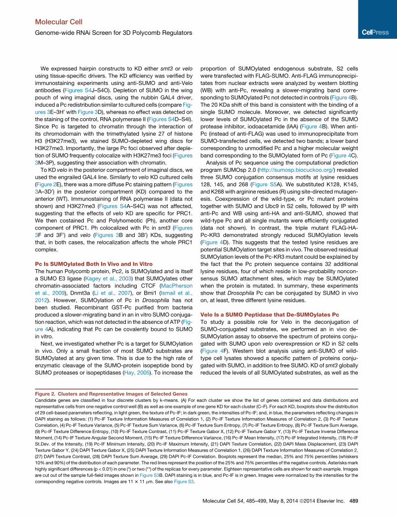
Figure 2. Clusters and Representative Images of Selected Genes
Candidate genes are classified in four discrete clusters by k-means. (A) For e
representative cells from one negative control well (B) as well as one example of o
of 29 cell-based parameters reflecting, in light green, the texture of Pc-IF; in dark g
DAPI staining as follows: (1) Pc-IF Texture Information Measures of Correlation
Correlation, (4) Pc-IF Texture Variance, (5) Pc-IF Texture Sum Variance, (6) Pc-IF T
(9) Pc-IF Texture Difference Entropy, (10) Pc-IF Texture Contrast, (11) Pc-IF Text
Moment, (14) Pc-IF Texture Angular Second Moment, (15) Pc-IF Texture Differenc
St.Dev. of the Intensity, (19) Pc-IF Minimum Intensity, (20) Pc-IF Maximum Inten
Texture Gabor Y, (24) DAPI Texture Gabor X, (25) DAPI Texture Information Meas
(27) DAPI Texture Contrast, (28) DAPI Texture Sum Average, (29) DAPI Pc-IF Co
10%and 90%) of the distribution of each parameter. The red lines represent the p
highly significant differences (p < 0.01) in one (*) or two (**) of the replicas for every
are cut out of the sample full-field images shown in Figure S3B. DAPI staining is i
corresponding negative controls. Images are 11 3 11 mm. See also Figure S3.
proportion of SUMOylated endogenous substrate, S2 cells
were transfected with FLAG-SUMO. Anti-FLAG immunoprecipi-
tates from nuclear extracts were analyzed by western blotting
(WB) with anti-Pc, revealing a slower-migrating band corre-
sponding to SUMOylated Pc not detected in controls (Figure 4B).
The 20 KDa shift of this band is consistent with the binding of a
single SUMO molecule. Moreover, we detected significantly
lower levels of SUMOylated Pc in the absence of the SUMO
protease inhibitor, iodoacetamide (IAA) (Figure 4B). When anti-
Pc (instead of anti-FLAG) was used to immunoprecipitate from
SUMO-transfected cells, we detected two bands; a lower band
corresponding to unmodified Pc and a higher molecular weight
band corresponding to the SUMOylated form of Pc (Figure 4C).
Analysis of Pc sequence using the computational prediction
program SUMOsp 2.0 (http://sumosp.biocuckoo.org/) revealed
three SUMO conjugation consensus motifs at lysine residues
128, 145, and 268 (Figure S5A). We substituted K128, K145,
and K268with arginine residues (R) using site-directedmutagen-
esis. Coexpression of the wild-type, or Pc mutant proteins
together with SUMO and Ubc9 in S2 cells, followed by IP with
anti-Pc and WB using anti-HA and anti-SUMO, showed that
wild-type Pc and all single mutants were efficiently conjugated
(data not shown). In contrast, the triple mutant FLAG-HA-
Pc-KR3 demonstrated strongly reduced SUMOylation levels
(Figure 4D). This suggests that the tested lysine residues are
potential SUMOylation target sites in vivo. The observed residual
SUMOylation levels of the Pc-KR3mutant could be explained by
the fact that the Pc protein sequence contains 32 additional
lysine residues, four of which reside in low-probability noncon-
sensus SUMO attachment sites, which may be SUMOylated
when the protein is mutated. In summary, these experiments
show that Drosophila Pc can be conjugated by SUMO in vivo
on, at least, three different lysine residues.
Velo Is a SUMO Peptidase that De-SUMOylates PcTo study a possible role for Velo in the deconjugation of
SUMO-conjugated substrates, we performed an in vivo de-
SUMOylation assay to observe the spectrum of proteins conju-
gated with SUMO upon velo overexpression or KD in S2 cells
(Figure 4F). Western blot analysis using anti-SUMO of wild-
type cell lysates showed a specific pattern of proteins conju-
gated with SUMO, in addition to free SUMO. KD of smt3 globally
reduced the levels of all SUMOylated substrates, as well as the
ach cluster we show the list of genes contained and data distributions and
ne gene KD for each cluster (C–F). For each KD, boxplots show the distribution
reen, the intensities of Pc-IF; and, in blue, the parameters reflecting changes in
1, (2) Pc-IF Texture Information Measures of Correlation 2, (3) Pc-IF Texture
exture Sum Entropy, (7) Pc-IF Texture Entropy, (8) Pc-IF Texture Sum Average,
ure Gabor X, (12) Pc-IF Texture Gabor Y, (13) Pc-IF Texture Inverse Difference
e Variance, (16) Pc-IF Mean Intensity, (17) Pc-IF Integrated Intensity, (18) Pc-IF
sity, (21) DAPI Texture Correlation, (22) DAPI Mass Displacement, (23) DAPI
ures of Correlation 1, (26) DAPI Texture Information Measures of Correlation 2,
rrelation. Boxplots represent the median, 25% and 75% percentiles (whiskers
osition of the 25% and 75%percentiles of the negative controls. Asterisksmark
parameter. Eighteen representative cells are shown for each example. Images
n blue, and Pc-IF is in green. Images were normalized by the intensities for the
Molecular Cell 54, 485–499, May 8, 2014 ª2014 Elsevier Inc. 489

(legend on next page)
Molecular Cell
Genome-wide RNAi Screen for 3D Polycomb Regulators
490 Molecular Cell 54, 485–499, May 8, 2014 ª2014 Elsevier Inc.

Molecular Cell
Genome-wide RNAi Screen for 3D Polycomb Regulators
free SUMO (Figure 4F). Most importantly, KD of velo changes
the equilibrium between SUMO-conjugated proteins and free
SUMO, resulting in an increase in the amount of SUMOylated
proteins and a decrease in the levels of free SUMO. In contrast,
Velo overexpression induced a strong decrease in the global
levels of SUMOylated proteins and an increase in free SUMO
when compared with the control.
Furthermore, cotransfection with Velo led to a significant
reduction in the levels of SUMO-conjugated Pc (Figure 4E,
line 2). To test whether Velo requires its catalytic domain to de-
SUMOylate Pc, we repeated the in vivo de-SUMOylation assay
after cotransfecting cells with VeloCS, a mutant (C502S) that
disrupts the catalytic domain. We did not observe changes in
the level of SUMOylated-Pc compared to the control conditions
(Figure 4E, line 3), indicating that the catalytic domain is
necessary for the de-SUMOylation of Pc. In summary, these
experiments show that Velo is a SUMO peptidase that can de-
SUMOylate Pc.
SUMO Conjugation of Pc Directly Modifies NuclearOrganization of Polycomb FociTo address if the lack of SUMOylation is directly responsible for
the observed redistribution of Pc in smt3 KD cells, we expressed
in S2 cells either the triple mutant fused to GFP (Pc-KR3:GFP), a
constitutively SUMOylated Pc protein by fusing SUMO to the C
terminus of Polycomb (Kang et al., 2010; Nayak et al., 2009;
Ross et al., 2002), or the wild-type Pc tagged with GFP (Pc:GFP),
which shows a similar nuclear distribution compared to endo-
genous Pc. Pc-KR3:GFP formed larger Pc foci when compared
to Pc:GFP, similar to those found after KD of smt3 (Figure 4G).
In contrast, Pc-SUMO:GFP showed a diffuse distribution
compared to Pc:GFP, similar to those found after KD of velo (Fig-
ure 4G). An unbiased quantification of the distribution of the
different GFP-fused versions of Pc among cells with similar
Figure 3. Distribution of Polycomb in Wing Imaginal Discs after Knock
(A) Immunostaining of Pc.
(A0) Magnification of (A). ‘‘a’’ indicates the anterior compartment containing wild-
expression of the RNAi transgene for velo. A clear decrease of the intensity of P
(B) Immunostaining of Ph.
(B0 ) Magnification of (B).
(C) Labeling with DAPI.
(C0) Magnification of (C).
(D) Merge of (A), (C), and GFP (in green) marking the expression domain of the e
(D0) Magnification of (D).
(E) Immunostaining of Pc.
(E0 ) Magnification of (E). An increase of the size of some Pc foci is observed in the
(F) Immunostaining of Ph.
(F0) Magnification of (F).
(G) Labeling with DAPI.
(G0 ) Magnification of (G).
(H) Merge of (I), (G), and GFP (in green) marking the expression domain of the Nu
(H0) Magnification of (H).
(I) Immunostaining with H3K27me3 in the wild-type region of wing imaginal discs
(J) Immunostaining with Ph.
(K) Labeling with DAPI.
(L) Merge of (I), (J), and (K).
(M) Immunostaining with H3K27me3 in the region of KD smt3 of wing imaginal d
(N) Immunostaining with Ph.
(O) Labeling with DAPI.
(P) Merge of (M), (N), and (O). See also Figure S4.
expression levels confirmed these observations (Figure S5D).
Therefore, SUMOylation of Pc regulates the nuclear distribution
of Pc directly.
Velo and SUMO Bind to Chromatin and Colocalizewith PcIn order to analyze whether Velo and SUMO exert their function
on chromatin, we raised antibodies against the two Drosophila
proteins (see Supplemental Experimental Procedures). Cellular
fractionation experiments (Figure S5B) revealed a 100 kDa
band corresponding to Velo in the cytoplasm and the nucleus,
with most of the nuclear protein fraction associated with
chromatin.
We then performed ChIP experiments followed by high-
throughput DNA sequencing (ChIP-seq) in S2 cells. Comparison
of the Pc binding profile with previously published Polycomb
response elements (PREs) in S2 cells (Schwartz et al., 2010)
showed a strong overlap (86% of Polycomb sites from Schwartz
et al. are boundbyPc). Strikingly, bothSUMOandVelo frequently
colocalized with Pc at known Pc-binding sites (Figure 4J). Most
Pc binding sites are associated with SUMO and most Velo sites
are bound by Pc (Figure 4I), implicating a prominent fraction of
the chromatin associated with the SUMO machinery in Pc-
related functions. Furthermore, the vast majority of Velo binding
sites (84.3%) are occupied by SUMO, showing that these pro-
teins act in concert at chromatin. Whereas most Pc binding sites
are also bound by SUMO, a significant number of SUMO binding
sites are not associated with Pc. Most of these sites correspond
to active promoter regions that are also bound by RNA Pol II and
H3K4me3 (Figure S5C), in agreement with SUMO 1 and SUMO 2
distribution in human cells (Neyret-Kahn et al., 2013). Despite the
strong overlap of Pc, Velo, andSUMOchromatin binding profiles,
double immunostaining experiments of Drosophila imaginal
discs of Velo or Sumo in conjunction with Ph revealed only a
down of velo and smt3
type cells. ‘‘p’’ indicates the posterior compartment where enGAL4 drives the
c foci is observed in this compartment after KD of velo.
nGAL4 line.
pouch where nubGAL4 drives the expression of the RNAi transgene for smt3.
bGAL4 line.
.
isc.
Molecular Cell 54, 485–499, May 8, 2014 ª2014 Elsevier Inc. 491

(legend on next page)
Molecular Cell
Genome-wide RNAi Screen for 3D Polycomb Regulators
492 Molecular Cell 54, 485–499, May 8, 2014 ª2014 Elsevier Inc.

Molecular Cell
Genome-wide RNAi Screen for 3D Polycomb Regulators
partial colocalization of Velo and SUMO with Pc foci (Figures
S5E–S5G and Figures S5H–S5J), suggesting that Pc foci mainly
contain Pc in a hypo-SUMOylated state in vivo.
Pc SUMOylation Levels Affect Its Chromatin BindingAffinity and Residence TimeOf note, even if the human homolog of Velo, SENP6, modulates
the cell cycle (Mukhopadhyay et al., 2010), FACS analysis of cells
depleted for Velo did not reveal a significant cell-cycle difference
(Figure 4H), indicating that the nuclear redistribution of PcG
proteins is not due to changes in cell-cycle regulation. Since it
was previously reported that SUMOylation can interfere with
the recruitment of transcription factors to chromatin (Chalkiadaki
and Talianidis, 2005), we then tested whether the changes in the
nuclear distribution of Pc upon perturbation of its SUMOylation
levels might reflect impaired recruitment of PcG proteins to their
target sites.
Overall Pc levels did not change after KD of components
belonging to the SUMOylation pathway (Figures S6K and S6L).
However, the staining of polytene chromosomes in salivary
glands depleted of Velo showed a significant decrease in the
number and intensity of binding sites for the PRC1 components
Pc and Ph (Figures 5A–5H). We further noted that Velo depletion
altered the structure of the polytene chromosomes, leading to
chromosome decondensation (Figures 5E–5H); however, the
localization and binding levels of polymerase II on polytene chro-
mosomes were unchanged after KD of velo (Figures S6E–S6H),
indicating that loss of velo function specifically affects binding
of PcG proteins. In contrast to the effect of Velo depletion, the
levels and number of binding sites for PcG proteins were not
significantly affected after KD of smt3 (Figures 5I–5L), suggesting
that SUMO modification of Pc is not required for chromatin
targeting in this tissue.
To extend these observations to diploid tissues, we performed
ChIP assays using anti-Pc in imaginal wing discs depleted of
Velo or SUMO. The occupancy of Pc on known PREs signifi-
cantly decreased inwing imaginal discs depleted of Velo (Figures
5M, 5O, and S6I). By contrast, we did not observe any significant
changes in Pcbinding, or in the levels of H3 andH3K27me3 at the
same PREs after KD of smt3 (Figures 5N, 5P, and S6J).
Figure 4. Smt3 and Velo Regulate the SUMOylation State of Polycomb
(A) Modification of Drosophila Pc using SUMO-3 Conjugation kit. Proteins were s
(B and C) Immunoprecipitation analysis of SUMOylated Pc. S2 cells were transi
noprecipitated with anti-FLAG (B) or anti-Pc (C). Western blots were analyzed w
(D) SUMOylation of Pc lysine mutants in vivo. S2 cells expressing FLAG-HA-tag
cipitated using anti-Pc and analyzed by western blotting with anti-HA (lanes 1 an
(E) Velo is a SUMO peptidase that requires a catalytically intact protease domai
plasmids. Immunoprecipitation was performed with anti-Pc (IP) from transfected
nonspecific bands. Overexpression levels of Velo were controlled by western blo
(F) The SUMO peptidase Velo regulates the equilibrium of SUMO-conjugated prot
with GFP dsRNA (lane 2), smt3 dsRNA (lane 3), velo dsRNA (lane 4), DNA empty ve
detected by western blotting using anti-SUMO. H3 is shown a loading control.
(G) Subcellular localization of GFP-Pc and GFP-Pc-KR3 or a constitutively SUMO
GFP-tagged Pc expression constructs and visualized by live-cell confocal fluore
(H) Cell-cycle profile of S2 cells comparing control cells (dsGFP) and cells deple
(I) Venn diagrams showing overlap between bound regions of indicated proteins
(J) Genomic distribution of PC, SUMO, and VELO in the Bithorax complex (BXC
regions are indicated by blue shadowed boxes (see Experimental Procedures for
our paper companion website (http://flyepigenome.igh.cnrs.fr/PCscreen) and are
In order to test whether the binding kinetics of Pc to chromatin
was affected upon KD of velo and smt3, we performed fluores-
cence recovery after photobleaching (FRAP) microscopy to
compare the recovery of PC-GFP in wing imaginal disc nuclei
after KD of velo and smt3. Interestingly, KD of smt3 strongly
slows down the recovery of PC-GFP, indicating that Pc binds
more stably to chromatin. In contrast, the recovery of PC-GFP
is much faster in nuclei KD for velo (Figure 5Q). These results
demonstrate that SUMOylation strongly affects the kinetics of
Pc binding to chromatin.
Velo Mutant Flies Show Homeotic TransformationsSimilar to PcG MutantsTo understand the relationship between the in vivo biological
roles of velo and PcG function, we examined the effect of velo
loss of function during fly development. Velo depletion at the
embryonic stage using a NanosGal4 driver induced segmenta-
tion defects that were detected in preparations of late embryonic
cuticles, with disruption or loss of denticle belts and frequent
denticle fusions (Figures 6A and 6B). These phenotypes
resemble those described for PcG mutants, reflecting a PcG-
dependent control of segmentation genes (McKeon et al.,
1994). Interestingly, similar cuticular phenotypes have been
described in a smt3 KD (Smith et al., 2011).
We next analyzed wing imaginal discs depleted of Velo using
the enGal4 driver. We observed derepression of the homeotic
gene Ultrabithorax (Ubx) in cells in the posterior compartment,
in which the velo gene is downregulated (Figures 6C and 6D).
This phenotype is similar to that of Pc mutants (Cabrera et al.,
1985). When KD of velo was performed at a later developmental
stage (using a NubGal4 driver), mutants displayed smaller wings
and halteres compared with the wild-type, consistent with dere-
pression of Ubx and consequent partial transformation of wings
to halteres (Figures 6E–6G).
DISCUSSION
In this study, we combined two high-resolution imaging-based
screens in order to discover factors involved in the 3D arrange-
ment of Pc in the Drosophila nucleus. We identified, in an
and Colocalize with Polycomb at Polycomb Response Elements
eparated by 4%–20% SDS-PAGE and visualized with Coomassie blue stain.
ently transfected with FLAG-tagged Smt3, and cell lysates were either immu-
ith anti-Pc. The positions of Pc and its SUMOylated forms are indicated.
ged Pc or Pc-KR3 mutants (Pc K128R, K145R, and K268R) were immunopre-
d 2) or with anti-SUMO (lanes 3 and 4).
n to specifically desumoylate Pc. S2 cells were transfected with the indicated
cell lysates, and western blot was detected with anti-HA. Asterisks indicate
tting using anti-Velo antibodies.
eins and free SUMO. S2 cells were either not transfected (lane 1) or transfected
ctor (lane 5), HA-Velo (lane 6), or Velo-GFP (lane 7). SUMOylated proteins were
ylated form of Pc (GFP-Pc-SUMO). S2 cells were transfected with the indicated
scence microscopy.
ted for velo (dsVelo).
.
) of chromosome 3R determined by ChIP-seq analysis. Significantly enriched
details). Asterisks indicate known PREs. The ChIP-Seq data can be browsed at
available in GEO under the accession number GSE55303. See also Figure S5.
Molecular Cell 54, 485–499, May 8, 2014 ª2014 Elsevier Inc. 493

(legend on next page)
Molecular Cell
Genome-wide RNAi Screen for 3D Polycomb Regulators
494 Molecular Cell 54, 485–499, May 8, 2014 ª2014 Elsevier Inc.

Figure 6. In Vivo Analysis of Loss of Func-
tion of velo during Drosophila Development
(A) Wild-type larval cuticle showing the pattern of
denticle belts.
(B) Larval cuticles of embryos depleted of velo
(NanosGal4/RNAivelo). KD of velomutant embryos
displays disruption and fusion of denticle belts. The
cuticles do not show homeotic phenotypes.
(C) Immunostaining analysis using anti-Ubx on
wing imaginal discs where Velo is depleted in
the posterior compartment (enGal4-UASGFP/
RNAivelo). Derepression of Ubx is detected in a
subset of cells in the posterior compartment.
(D) Merge of the different immunostainings of Ubx
(red), DAPI (blue), and GFP (green), which marks
the expression area of enGal4.
(E) Wing from a wild-type fly.
(F) Wing from Nub-Gal4/UAS-RNAivelo showing a
reduction and partial transformation of the wing.
Images are to scale.
(G) Adult fly Nub-Gal4/UAS-RNAivelo.
Molecular Cell
Genome-wide RNAi Screen for 3D Polycomb Regulators
unbiased way, more than 100 genes, and characterized two of
them, showing that the SUMO peptidase Velo is required for
Pc targeting to chromatin and that a tightly regulated balance
of Pc SUMOylation levels is required for the 3D organization of
Pc foci and the function of PcG proteins.
High-Resolution Microscopy RNAi Screen IdentifiesRegulators of the Nuclear Organization of Pc FociThe identification of all PcG proteins can be traced back to
genetic screens, which had a decisive role in gene discovery.
However, genetic screens also have limitations; it is difficult to
identify genes with substantial maternal components in screens
based on embryonic phenotypes; adult screens based on het-
erozygous mutations are affected by the fact that many genes
display haplo-sufficiency; and induced mutations demonstrate
irregular coverage of the genome. Moreover, mutations resulting
in pleiotropic effects might interfere with the normal develop-
ment of the organism, thereby preventing the display of the phe-
notypes of interest.
With the present screen, we identified two major classes of
modifiers. The first class contained genes whose KD effects
Figure 5. Velo Plays a Crucial Role in Chromatin Recruitment of Polyc
(A–L) Immunostaining of polytene chromosomes.
(A–D) Staining of control UAS-Dcr2; nubGal4 chromosomes. (A) Labeled with D
and (C).
(E–H) Same staining scheme as above of UAS-Dcr2; nubGal4-UASGFP/UAS-RN
(I–L) Same staining scheme as above of UAS-Dcr2; nubGal4-UASGFP/UAS-RNA
(M) qChIP analysis using anti-Pc antibodies of MS1096Gal4/UAS-RNAiGFP win
imaginal discs (velo KD, green bars). ChIP signal levels are represented as pe
independent experiments. *p < 0.05, **p < 0.01, unpaired, two-tailed Student’s t
(N) qChIP analysis using Pc antibodies of MS1096Gal4/UAS-RNAiGFP wing im
imaginal discs (smt3 KD, dark blue bars). Error bars represent SD.
(O) qChIP analysis using H3K27me3 antibodies of MS1096Gal4/UAS-RNAiGFP
imaginal discs (velo KD, green bars). Error bars represent SD.
(P) qChIP analysis using H3K27me3 antibodies of MS1096Gal4/UAS-RNAiGFP w
wing imaginal discs (smt3 KD, dark blue bars). Error bars represent SD.
(Q) FRAP experiments monitoring the recovery of fluorescence of PC-GFP in wild
(blue line). FRAP experiments were performed on wing imaginal disc expressing P
the recovery of PC-GFP after 20 s: WT versus KD of smt3 (p < 0.01); WT versus
were not restricted to Pc distribution but also induced changes
in the DAPI staining pattern, most likely reflecting large-scale
chromatin rearrangement. The second class (followed up in
this study) contained genes specifically affecting Pc distribution,
with little or no effect on general chromatin organization as
revealed by DAPI staining.
The current screen detected all positive controls, and no nega-
tive controls were classified as hits, indicating that the assay is
robust and efficient. Six core PcG genes out of the eleven known
to date were detected, including members of the PhoRC, PRC2,
and PRC1 complexes. The members that were not identified are
characterized by having redundant function with another PcG
protein (Phol for the PhoRC complex, Esc and Escl for the
PRC2 complex, and Psc and Su(z)2 for the PRC1 complex),
suggesting that, as for many other screening procedures, redun-
dancy hampers detection in RNAi-based screens.
Many of the hits were chromatin components, and several of
them have been recently related to PcG proteins. The genes
coding for cohesins and the cohesion associated proteins,
Smc1, Smc3/Cap, and Sa were pulled down in a biotinylation
tagging strategy (Strubbe et al., 2011). Another example is Hcf,
omb
API. (B) Staining with anti-Pc. (C) Staining with anti-Ph. (D) Merge of (A), (B),
Aivelo chromosomes.
ismt3 chromosomes.
g imaginal discs (control, black bars) and MS1096Gal4/UAS-RNAivelo wing
rcentage of input chromatin. Values represent means ± SD from four to six
test.
aginal discs (control, light blue bars) and MS1096Gal4/UAS-RNAismt3 wing
wing imaginal discs (control, gray bars) and MS1096Gal4/UAS-RNAivelo wing
ing imaginal discs (control, light blue bars) and MS1096Gal4/UAS-RNAismt3
-type imaginal disc (black line), upon KD of smt3 (green line) and upon KD velo
C-GFP by collecting 2D images every 2 s for 2 min. Student’s t test comparing
KD of velo (p < 0.001). See also Figure S6.
Molecular Cell 54, 485–499, May 8, 2014 ª2014 Elsevier Inc. 495
Molecular Cell
Genome-wide RNAi Screen for 3D Polycomb Regulators
a factor involved in both PcG and trxG functions (Rodriguez-Jato
et al., 2011). Other modifying factors are members of the
trithorax group (trxG) of genes, which counteract PcG function
during the regulation of Hox and other developmental genes.
Among these, trx, trr, and E(bx) are prominent, as they are linked
to the deposition and recognition of the histone mark H3K4me3,
which counteracts H3K27me3. Other prominent trxG genes
identified in the screen encode for multiple subunits of the
brahma complex (brm, Bap60, Bap170, mor, and Snr1).
Components of the dosage compensation machinery and
nucleoporins were also identified in the screen. Since these
factors are involved in the formation of active chromatin domains
(Mendjan et al., 2006; Vaquerizas et al., 2010), theymay compete
for PcG binding in a similar way to trxG components. However,
the possibility of indirect effects cannot be excluded at this point.
In particular, other nuclear import proteins were also scored as
positive (Kap-a3, Sec13); and another possible explanation for
some of these hits is that impaired nuclear transport of Pc or
other PcG components may affect the integrity of Pc foci.
Other hits are either uncharacterized genes or were not
previously related to PcG function. An interesting class among
these contains proteins with sequence-specific DNA binding
activity (C15, Chinmo, Crol, Fer1, HLH106, Kay, Lmpt, Luna,
Poxn, RunxB, and Ush), or proteins linked to sequence-specific
repression of transcription, such as Mip130 and Mip40, two
components of the repressive MMB complex that contains
Myb (Georlette et al., 2007; Korenjak et al., 2004). This is an
intriguing finding, since previous attempts to predict the
sequence-specific code for PcG targeting had limited success
(Schuettengruber and Cavalli, 2009; Schuettengruber et al.,
2009). An attractive possibility is that multiple transcription
factors may contribute to PcG recruitment at subsets of their
target genes (Okulski et al., 2011).
Other interesting Pc foci modifiers include factors involved in
RNA binding and metabolism (Ago1, Dek, Hel25E, Pea, SmD3),
and in signaling pathways (Axn) often linked to posttranslational
protein modification (for example, kinases, including Dsor1, Ial,
Stg, and Tlk; phosphatases, including, Pp0-19C, PPP4R2r; ubiq-uitylation factors, such as Gw, Roc1; and proteins involved in
SUMOylation, such as Su(var)2-10, Velo, and Smt3). These
genes may open new avenues for PcG regulation mediated by
posttranslational modification of Pc or of its partners.
Role of SUMOylation in Pc Recruitment to Chromatinand Nuclear ArchitectureSUMO is implicated in a plethora of cellular events (reviewed in
Hay, 2005). Increasing evidence suggests that SUMO may also
contribute to protein solubility (Marblestone et al., 2006; Panavas
et al., 2009). At a molecular level, SUMOylation affects protein
function by creating and abolishing binding interfaces, or by
inducing conformational changes that result in altered interac-
tions (for detailed examples, see Johnson, 2004).
SUMOylation of human Pc2 increases its binding affinity for
H3K27me3 (Kang et al., 2010). In C. elegans, SUMOylation of
SOP-2, a SAM-domain protein with homology to Drosophila
Scm and Ph, is thought to induce SOP-2 localization into Pc
foci and to repress the expression of Hox genes (Zhang et al.,
2004). In Drosophila, SUMOylation of the PRC1-interacting pro-
496 Molecular Cell 54, 485–499, May 8, 2014 ª2014 Elsevier Inc.
tein, Scm (Sex Comb on Midleg), reduces its association with
chromatin (Smith et al., 2011). Sfmbt, a subunit of the PhoRC
complex, has been identified in a screen for factors involved in
SUMO-dependent transcriptional repression (Stielow et al.,
2008). Comparison of genes identified in this screen with ours
revealed only a limited overlap of seven genes: Sfmbt, kay,
Su(var)2-10, sbb, Chd3, and CG7056. The fact that the screen
paradigms are largely different might explain why Pc was not
previously linked to SUMO by Stielow et al. and emphasizes
the importance of performing screens based on independent
readouts.
A well-characterized example of the importance of SUMO in
the regulation of nuclear architecture is the formation of promye-
locytic leukemia protein (PML) nuclear bodies (reviewed by
Seeler and Dejean, 2003). In PML bodies, a combination of
SUMO and SIMs (SUMO interaction motifs) enables the inter-
action of many proteins to nucleate and establish a PML nuclear
body scaffold (Shen et al., 2006). As another example, the
SENP7 SUMO peptidase was shown to maintain HP1 on
pericentric heterochromatin, whereas its depletion does not
affect H3K9me3 levels (Maison et al., 2012). In Drosophila,
SUMOylation affects the formation of insulator bodies. Both
the suppression and the induction of SUMOylation reduce the
coalescence of insulator proteins (Capelson and Corces, 2006;
Golovnin et al., 2012). This situation differs from the case of
Pc, showing that SUMO can affect different nuclear proteins in
specific ways that depend on how it affects protein-protein inter-
actions in multiprotein complexes.
How does SUMOylation affect the nuclear organization of Pc?
One possibility is that the effect of SUMO may be specifically
linked to the nuclear organization of Pc foci. Depletion of the
SUMO peptidase, Velo, induced loss of PcG targeting in diploid
cells and in polytene chromosomes (Figure 5). On the other hand,
SUMO depletion did not perturb Pc targeting, although it did
cause marked coalescence of PcG proteins to form massive
Pc foci. We have shown through FRAP experiments in a well-
characterized in vivo system (Cheutin and Cavalli, 2012) that
Pc suffers from significant changes in its residence time in Pc-
foci as a consequence of smt3 or velo KD (Figure 5Q). The
data thus suggest a scenario for the role of SUMO in the nuclear
organization of Pc.Without SUMO,more stable Pcmay promote
protein-protein interactions with other PRC1 complexes and
generate the formation of stable nuclear aggregates that are
observed upon smt3 KD. A limited degree of SUMOylation may
prevent the coalescence of PRC1 complexes within the
nucleus, whereas excess SUMO may solubilize Pc away from
its target chromatin.
SUMO modification has been shown to promote protein solu-
bility (Sabate et al., 2012), and this property prevents aggrega-
tion of a-synuclein, an aggregation-prone protein implicated in
Parkinson’s disease (Krumova et al., 2011). Our data suggest
that SUMOylation may similarly promote the solubility of PRC1
in vivo, thereby preventing aggregation of PRC1 in the nucleus.
Low, physiologic levels of Pc SUMOylationmay act as ‘‘fluidifier’’
of PRC1-mediated chromatin interactions, preventing excessive
in vivo aggregation at Pc foci while allowing robust chromatin
targeting and efficient silencing of its target genes. Future
studies should test whether other PcG or trxG components are
Molecular Cell
Genome-wide RNAi Screen for 3D Polycomb Regulators
SUMOylated, and to what extent the interplay of these modifica-
tions regulates PcG and trxG function in different cell types and
during development.
In summary, we presented here a screening approach that is
highly reproducible and sensitive, and relies on a cell biology
assay that is completely independent from previous screens.
Recently, a screen based on DNA FISH has allowed factors
involved in chromosome pairing to be identified (Joyce et al.,
2012). These approaches have the potential to be extended to
many other biological processes occurring within or beyond
the cell nucleus.
EXPERIMENTAL PROCEDURES
Screen Image Acquisition and Analysis
Plates were imaged in an Opera (Perkin Elmer) spinning-disk confocal
microscope equipped with a 603/NA 1.2 and providing a lateral resolution
of 0.25 mm. Images corresponding to three Z optical sections were maximum
intensity projected along the z axis into a single image using ImageJ (U.S.
National Institutes of Health, http://imagej.nih.gov/ij/). The rest of the image
analysis procedure was performed using CellProfiler (Carpenter et al., 2006)
(Broad Institute Imaging Platform, MIT/Harvard, http://www.cellprofiler.com).
Images were analyzed at the Orchestra computer cluster of Harvard Medical
School, and the resulting measurements were exported as CSV tables. Repre-
sentative images for the genes selected as positives after the secondary
screen can be consulted at the website http://flyepigenome.igh.cnrs.fr/
PCscreen.
Statistical Procedures
Data analysis was performed using the R software package (R Foundation for
Statistical Computing, http://www.R-project.org). We normalized all the data
in the genome-wide and Transcription Factors screens using the B-score
(Brideau et al., 2003). Secondary screen data were normalized using the
Normalized Percentage Inhibition method. Clustering was performed using
the gplots package in R. Euclidean n dimensional distance was computed in
combination with ward clustering.
Gene Ontology Terms Enrichment
Gene Ontology term enrichments were calculated using DAVID 6.7 (http://
david.abcc.ncifcrf.gov) (Huang et al., 2009). We introduced the list of positive
candidate genes from either the genome-wide screen and from the transcrip-
tion factors screen and used, respectively, the whole Drosophila genome or
the list of genes present in the transcriptions factors library as background
references.
Chromatin Immunoprecipitation
ChIP on wing imaginal discs was done as described in Schuettengruber et al.
(2009) with modifications as described in the Supplemental Experimental
Procedures.
Further details on the screening procedure, as well as information on
plasmids, primer sequences, antibodies, and other experimental procedures,
are provided in the Supplemental Information.
ACCESSION NUMBERS
The GEO accession number for the ChIP-Seq data reported in this paper is
GSE55303.
SUPPLEMENTAL INFORMATION
Supplemental Information includes six figures, one table, and Supplemental
Experimental Procedures and can be found with this article at http://dx.doi.
org/10.1016/j.molcel.2014.03.004.
ACKNOWLEDGMENTS
We are thankful to the DRSC for providing the RNAi libraries, the technical
means, and the expertise necessary to fulfill this study. We thank Bernd
Schuettengruber and Tom Sexton for critically reading the manuscript. We
thank theMRI-IGH imaging facility for critical guidance inmicroscopy analysis.
The Leica SP8 microscope used in part of this work was financed by the
Association pour la Recherche sur le Cancer (ARC) (EML20120904995).
Research at the G.C. lab was supported by grants from the European
Research Council (ERC-2008-AdG number 232947), the CNRS, the European
Network of Excellence EpiGeneSys, the Agence Nationale de la Recherche,
and the ARC. I.G. was supported by a fellowship from theMinisterio de Ciencia
e Innovacion/Fulbright and the ARC. J.M.-L. was supported by a fellowship
from the ARC, and by the Fondation pour la Recherche Medicale.
Received: January 14, 2013
Revised: February 11, 2014
Accepted: February 24, 2014
Published: April 3, 2014
REFERENCES
Bantignies, F., and Cavalli, G. (2011). Polycomb group proteins: repression in
3D. Trends Genet. 27, 454–464.
Bantignies, F., Roure, V., Comet, I., Leblanc, B., Schuettengruber, B., Bonnet,
J., Tixier, V., Mas, A., and Cavalli, G. (2011). Polycomb-dependent regulatory
contacts between distant Hox loci in Drosophila. Cell 144, 214–226.
Berdnik, D., Favaloro, V., and Luo, L. (2012). The SUMO protease Verloren
regulates dendrite and axon targeting in olfactory projection neurons.
J. Neurosci. 32, 8331–8340.
Brideau, C., Gunter, B., Pikounis, B., and Liaw, A. (2003). Improved statistical
methods for hit selection in high-throughput screening. J. Biomol. Screen 8,
634–647.
Cabrera, C.V., Botas, J., and Garcia-Bellido, A. (1985). Distribution of
Ultrabithorax proteins in mutants of Drosophila bithorax complex and its trans-
regulatory genes. Nature 318, 569–571.
Capelson, M., and Corces, V.G. (2006). SUMO conjugation attenuates the
activity of the gypsy chromatin insulator. EMBO J. 25, 1906–1914.
Carpenter, A.E., Jones, T.R., Lamprecht, M.R., Clarke, C., Kang, I.H., Friman,
O., Guertin, D.A., Chang, J.H., Lindquist, R.A., Moffat, J., et al. (2006).
CellProfiler: image analysis software for identifying and quantifying cell pheno-
types. Genome Biol. 7, R100.
Chalkiadaki, A., and Talianidis, I. (2005). SUMO-dependent compartmentali-
zation in promyelocytic leukemia protein nuclear bodies prevents the access
of LRH-1 to chromatin. Mol. Cell. Biol. 25, 5095–5105.
Cheutin, T., and Cavalli, G. (2012). Progressive polycomb assembly on
H3K27me3 compartments generates polycomb bodies with developmentally
regulated motion. PLoS Genet. 8, e1002465.
Czermin, B., Melfi, R., McCabe, D., Seitz, V., Imhof, A., and Pirrotta, V. (2002).
Drosophila enhancer of Zeste/ESC complexes have a histone H3methyltrans-
ferase activity that marks chromosomal Polycomb sites. Cell 111, 185–196.
Duncan, I.M. (1982). Polycomblike: a gene that appears to be required for the
normal expression of the bithorax and antennapedia gene complexes of
Drosophila melanogaster. Genetics 102, 49–70.
Franke, A., Messmer, S., and Paro, R. (1995). Mapping functional domains of
the polycomb protein of Drosophila melanogaster. Chromosome Res. 3,
351–360.
Georlette, D., Ahn, S., MacAlpine, D.M., Cheung, E., Lewis, P.W., Beall, E.L.,
Bell, S.P., Speed, T., Manak, J.R., and Botchan, M.R. (2007). Genomic
profiling and expression studies reveal both positive and negative activities
for the Drosophila Myb MuvB/dREAM complex in proliferating cells. Genes
Dev. 21, 2880–2896.
Molecular Cell 54, 485–499, May 8, 2014 ª2014 Elsevier Inc. 497

Molecular Cell
Genome-wide RNAi Screen for 3D Polycomb Regulators
Golovnin, A., Volkov, I., and Georgiev, P. (2012). SUMO conjugation is required
for the assembly of Drosophila Su(Hw) and Mod(mdg4) into insulator bodies
that facilitate insulator complex formation. J. Cell Sci. 125, 2064–2074.
Grimaud, C., Negre, N., and Cavalli, G. (2006). From genetics to epigenetics:
the tale of Polycomb group and trithorax group genes. Chromosome Res.
14, 363–375.
Hari, K.L., Cook, K.R., and Karpen, G.H. (2001). The Drosophila Su(var)2-10
locus regulates chromosome structure and function and encodes a member
of the PIAS protein family. Genes Dev. 15, 1334–1348.
Hay, R.T. (2005). SUMO: a history of modification. Mol. Cell 18, 1–12.
Hou, C., Li, L., Qin, Z.S., and Corces, V.G. (2012). Gene density, transcription,
and insulators contribute to the partition of the Drosophila genome into phys-
ical domains. Mol. Cell 48, 471–484.
Huang, H.W., Tsoi, S.C., Sun, Y.H., and Li, S.S. (1998). Identification and char-
acterization of the SMT3 cDNA and gene encoding ubiquitin-like protein from
Drosophila melanogaster. Biochem. Mol. Biol. Int. 46, 775–785.
Huang, W., Sherman, B.T., and Lempicki, R.A. (2009). Systematic and integra-
tive analysis of large gene lists using DAVID bioinformatics resources. Nat.
Protoc. 4, 44–57.
Ismail, I.H., Gagne, J.P., Caron, M.C., McDonald, D., Xu, Z., Masson, J.Y.,
Poirier, G.G., and Hendzel, M.J. (2012). CBX4-mediated SUMO modification
regulates BMI1 recruitment at sites of DNA damage. Nucleic Acids Res. 40,
5497–5510.
Johnson, E.S. (2004). Protein modification by SUMO. Annu. Rev. Biochem. 73,
355–382.
Joyce, E.F., Williams, B.R., Xie, T., andWu, C.T. (2012). Identification of genes
that promote or antagonize somatic homolog pairing using a high-throughput
FISH-based screen. PLoS Genet. 8, e1002667.
Jurgens, G. (1985). A group of genes controlling the spatial expression of the
bithorax complex in Drosophila. Nature 316, 153–155.
Kagey, M.H., Melhuish, T.A., andWotton, D. (2003). The polycomb protein Pc2
is a SUMO E3. Cell 113, 127–137.
Kang, X., Qi, Y., Zuo, Y., Wang, Q., Zou, Y., Schwartz, R.J., Cheng, J., and Yeh,
E.T. (2010). SUMO-specific protease 2 is essential for suppression of poly-
comb group protein-mediated gene silencing during embryonic development.
Mol. Cell 38, 191–201.
Klymenko, T., Papp, B., Fischle, W., Kocher, T., Schelder, M., Fritsch, C., Wild,
B., Wilm, M., and Muller, J. (2006). A Polycomb group protein complex with
sequence-specific DNA-binding and selective methyl-lysine-binding activ-
ities. Genes Dev. 20, 1110–1122.
Korenjak, M., Taylor-Harding, B., Binne, U.K., Satterlee, J.S., Stevaux, O.,
Aasland, R., White-Cooper, H., Dyson, N., and Brehm, A. (2004). Native E2F/
RBF complexes contain Myb-interacting proteins and repress transcription
of developmentally controlled E2F target genes. Cell 119, 181–193.
Krumova, P., Meulmeester, E., Garrido, M., Tirard, M., Hsiao, H.H., Bossis, G.,
Urlaub, H., Zweckstetter, M., Kugler, S., Melchior, F., et al. (2011). Sumoylation
inhibits alpha-synuclein aggregation and toxicity. J. Cell Biol. 194, 49–60.
Li, S.J., and Hochstrasser, M. (2000). The yeast ULP2 (SMT4) gene encodes a
novel protease specific for the ubiquitin-like Smt3 protein. Mol. Cell. Biol. 20,
2367–2377.
Li, B., Zhou, J., Liu, P., Hu, J., Jin, H., Shimono, Y., Takahashi, M., and Xu, G.
(2007). Polycomb protein Cbx4 promotes SUMOmodification of de novo DNA
methyltransferase Dnmt3a. Biochem. J. 405, 369–378.
Lima, C.D., and Reverter, D. (2008). Structure of the human SENP7 catalytic
domain and poly-SUMO deconjugation activities for SENP6 and SENP7.
J. Biol. Chem. 283, 32045–32055.
MacPherson, M.J., Beatty, L.G., Zhou, W., Du, M., and Sadowski, P.D. (2009).
The CTCF insulator protein is posttranslationally modified by SUMO.Mol. Cell.
Biol. 29, 714–725.
Maison, C., Romeo, K., Bailly, D., Dubarry, M., Quivy, J.P., and Almouzni, G.
(2012). The SUMO protease SENP7 is a critical component to ensure HP1
enrichment at pericentric heterochromatin. Nat. Struct. Mol. Biol. 19, 458–460.
498 Molecular Cell 54, 485–499, May 8, 2014 ª2014 Elsevier Inc.
Marblestone, J.G., Edavettal, S.C., Lim, Y., Lim, P., Zuo, X., and Butt, T.R.
(2006). Comparison of SUMO fusion technology with traditional gene fusion
systems: enhanced expression and solubility with SUMO. Protein Sci. 15,
182–189.
McKeon, J., Slade, E., Sinclair, D.A., Cheng, N., Couling, M., and Brock, H.W.
(1994). Mutations in some Polycomb group genes of Drosophila interfere with
regulation of segmentation genes. Mol. Gen. Genet. 244, 474–483.
Mendjan, S., Taipale, M., Kind, J., Holz, H., Gebhardt, P., Schelder, M.,
Vermeulen, M., Buscaino, A., Duncan, K., Mueller, J., et al. (2006). Nuclear
pore components are involved in the transcriptional regulation of dosage
compensation in Drosophila. Mol. Cell 21, 811–823.
Mukhopadhyay, D., Arnaoutov, A., and Dasso, M. (2010). The SUMO protease
SENP6 is essential for inner kinetochore assembly. J. Cell Biol. 188, 681–692.
Nayak, A., Glockner-Pagel, J., Vaeth, M., Schumann, J.E., Buttmann, M.,
Bopp, T., Schmitt, E., Serfling, E., and Berberich-Siebelt, F. (2009).
Sumoylation of the transcription factor NFATc1 leads to its subnuclear reloc-
alization and interleukin-2 repression by histone deacetylase. J. Biol. Chem.
284, 10935–10946.
Neyret-Kahn, H., Benhamed,M., Ye, T., LeGras, S., Cossec, J.C., Lapaquette,
P., Bischof, O., Ouspenskaia, M., Dasso, M., Seeler, J., et al. (2013).
Sumoylation at chromatin governs coordinated repression of a transcriptional
program essential for cell growth and proliferation. Genome Res. 23, 1563–
1579.
Okulski, H., Druck, B., Bhalerao, S., and Ringrose, L. (2011). Quantitative anal-
ysis of polycomb response elements (PREs) at identical genomic locations
distinguishes contributions of PRE sequence and genomic environment.
Epigenetics Chromatin 4, 4.
Otte, A.P., and Kwaks, T.H. (2003). Gene repression by Polycomb group
protein complexes: a distinct complex for every occasion? Curr. Opin.
Genet. Dev. 13, 448–454.
Panavas, T., Sanders, C., and Butt, T.R. (2009). SUMO fusion technology for
enhanced protein production in prokaryotic and eukaryotic expression
systems. Methods Mol. Biol. 497, 303–317.
Ramadan, N., Flockhart, I., Booker, M., Perrimon, N., and Mathey-Prevot, B.
(2007). Design and implementation of high-throughput RNAi screens in
cultured Drosophila cells. Nat. Protoc. 2, 2245–2264.
Rodriguez-Jato, S., Busturia, A., andHerr, W. (2011). Drosophila melanogaster
dHCF interacts with both PcG and TrxG epigenetic regulators. PLoS ONE 6,
e27479.
Rosa, S., De Lucia, F., Mylne, J.S., Zhu, D., Ohmido, N., Pendle, A., Kato, N.,
Shaw, P., and Dean, C. (2013). Physical clustering of FLC alleles during
Polycomb-mediated epigenetic silencing in vernalization. Genes Dev. 27,
1845–1850.
Ross, S., Best, J.L., Zon, L.I., and Gill, G. (2002). SUMO-1 modification
represses Sp3 transcriptional activation and modulates its subnuclear locali-
zation. Mol. Cell 10, 831–842.
Sabate, R., Espargaro, A., Grana-Montes, R., Reverter, D., and Ventura, S.
(2012). Native structure protects SUMO proteins from aggregation into
amyloid fibrils. Biomacromolecules 13, 1916–1926.
Schuettengruber, B., and Cavalli, G. (2009). Recruitment of polycomb group
complexes and their role in the dynamic regulation of cell fate choice.
Development 136, 3531–3542.
Schuettengruber, B., Chourrout, D., Vervoort, M., Leblanc, B., and Cavalli, G.
(2007). Genome regulation by polycomb and trithorax proteins. Cell 128,
735–745.
Schuettengruber, B., Ganapathi, M., Leblanc, B., Portoso, M., Jaschek, R.,
Tolhuis, B., van Lohuizen, M., Tanay, A., and Cavalli, G. (2009). Functional
anatomy of polycomb and trithorax chromatin landscapes in Drosophila
embryos. PLoS Biol. 7, e13.
Schwartz, Y.B., Kahn, T.G., Stenberg, P., Ohno, K., Bourgon, R., and Pirrotta,
V. (2010). Alternative epigenetic chromatin states of polycomb target genes.
PLoS Genet. 6, e1000805.

Molecular Cell
Genome-wide RNAi Screen for 3D Polycomb Regulators
Seeler, J.S., and Dejean, A. (2003). Nuclear and unclear functions of SUMO.
Nat. Rev. Mol. Cell Biol. 4, 690–699.
Sexton, T., Yaffe, E., Kenigsberg, E., Bantignies, F., Leblanc, B., Hoichman,
M., Parrinello, H., Tanay, A., and Cavalli, G. (2012). Three-dimensional folding
and functional organization principles of the Drosophila genome. Cell 148,
458–472.
Shao, Z., Raible, F., Mollaaghababa, R., Guyon, J.R., Wu, C.T., Bender, W.,
and Kingston, R.E. (1999). Stabilization of chromatin structure by PRC1, a
Polycomb complex. Cell 98, 37–46.
Shen, T.H., Lin, H.K., Scaglioni, P.P., Yung, T.M., and Pandolfi, P.P. (2006).
The mechanisms of PML-nuclear body formation. Mol. Cell 24, 331–339.
Smith, M., Mallin, D.R., Simon, J.A., and Courey, A.J. (2011). Small ubiquitin-
like modifier (SUMO) conjugation impedes transcriptional silencing by the
polycomb group repressor Sex Comb on Midleg. J. Biol. Chem. 286, 11391–
11400.
Stielow, B., Sapetschnig, A., Kruger, I., Kunert, N., Brehm, A., Boutros, M., and
Suske, G. (2008). Identification of SUMO-dependent chromatin-associated
transcriptional repression components by a genome-wide RNAi screen. Mol.
Cell 29, 742–754.
Strubbe, G., Popp, C., Schmidt, A., Pauli, A., Ringrose, L., Beisel, C., and Paro,
R. (2011). Polycomb purification by in vivo biotinylation tagging reveals cohe-
sin and Trithorax group proteins as interaction partners. Proc. Natl. Acad. Sci.
USA 108, 5572–5577.
Tolhuis, B., Blom, M., Kerkhoven, R.M., Pagie, L., Teunissen, H., Nieuwland,
M., Simonis, M., de Laat, W., van Lohuizen, M., and van Steensel, B. (2011).
Interactions among Polycomb domains are guided by chromosome architec-
ture. PLoS Genet. 7, e1001343.
Ulrich, H.D. (2009). SUMO protocols. Preface. Methods Mol. Biol. 497, v–vi.
Vaquerizas, J.M., Suyama, R., Kind, J., Miura, K., Luscombe, N.M., and
Akhtar, A. (2010). Nuclear pore proteins nup153 and megator define transcrip-
tionally active regions in the Drosophila genome. PLoS Genet. 6, e1000846.
Zhang, H., Smolen, G.A., Palmer, R., Christoforou, A., van den Heuvel, S., and
Haber, D.A. (2004). SUMO modification is required for in vivo Hox gene regu-
lation by the Caenorhabditis elegans Polycomb group protein SOP-2. Nat.
Genet. 36, 507–511.
Molecular Cell 54, 485–499, May 8, 2014 ª2014 Elsevier Inc. 499