molecular and cellular evidence of natural venezuelan ...veterinary world, eissn: 2231-0916 495...
TRANSCRIPT
Veterinary World, EISSN: 2231-0916 495
Veterinary World, EISSN: 2231-0916Available at www.veterinaryworld.org/Vol.13/March-2020/15.pdf
RESEARCH ARTICLEOpen Access
Molecular and cellular evidence of natural Venezuelan equine encephalitis virus infection in frugivorous bats in Colombia
Camilo Guzmán1, Alfonso Calderón2, Teresa Oviedo3, Salim Mattar4, José Castañeda5, Virginia Rodriguez6 and Luiz Tadeu Moraes Figueiredo7
1. Department of Pharmacy, Faculty of Health Sciences, Institute of Biological Research of the Tropics, University of Córdoba, Colombia; 2. Faculty of Veterinary Medicine and Animal, Institute for Biological Research in the Tropics,
University of Córdoba, Colombia; 3. University of Córdoba, Colombia; 4. Faculty of Veterinary Medicine and Animal, Institute of Biological Research of the Tropics, University of Córdoba, Colombia; 5. ICA Diagnostic Center - Córdoba,
Colombia; 6. Faculty of Health Sciences, University of Córdoba, Colombia; 7. Center for Virological Research, University of Sao Paulo, Riberao Preto, Brazil.
Corresponding author: Salim Mattar, e-mail: [email protected]: CG: [email protected], AC: [email protected],
TO: [email protected], JC: [email protected], VR: [email protected], LTMF: [email protected]
Received: 22-05-2019, Accepted: 22-01-2020, Published online: 16-03-2020
doi: www.doi.org/10.14202/vetworld.2020.495-501 How to cite this article: Guzmán C, Calderón A, Oviedo T, Mattar S, Castañeda J, Rodriguez V, Figueiredo LTM (2020) Molecular and cellular evidence of natural Venezuelan equine encephalitis virus infection in frugivorous bats in Colombia, Veterinary World, 13(3): 495-501.
Abstract Background and Aim: Venezuelan equine encephalitis virus (VEEV) is an alphavirus that causes encephalitis with a high impact on public health in Latin America. However, only in Guatemala, Trinidad and Tobago, and Mexico have found antibodies in VEEV in bats, using immunohistochemistry, the sensitivity and specificity are improved; thus, it is better for demonstrating natural infection in bats as potential hosts. This study aimed to determine the presence of VEEV in tissues of frugivorous bats.
Materials and Methods: A prospective descriptive cross-sectional study with a non-probabilistic sampling was carried out in 12 localities of Córdoba and Sucre area of the Colombian Caribbean. Two hundred and eighty-six bats were captured using fog nets, and the specimens according to taxonomic keys were classified. According to the Ethics Committee of the University of Córdoba, the bats were treated with analgesics and anesthetics. Blood samples were taken and then euthanized to obtain tissues and organs which were preserved in liquid N2 at −196°C. A portion of each organ was fixed in 10% buffered formalin for the detection of antigens by immunohistochemistry. Several pathological anatomy analyses were performed to determine the histological characteristics of tissue lesions of frugivorous bats naturally infected with the VEEV.
Results: Of the 286 bats captured, 23 species were identified. In samples of the brain, spleen, and lung of two frugivorous bats (2/286=0.70%) Artibeus planirostris and Sturnira lilium, the presence of VEEV was confirmed by immunohistochemistry.
Conclusion: A fragment of the nsP4 non-structural protein gene corresponding to the alphavirus was amplified. Two samples were positive (2/286=0.70%) in frugivorous bats; A. planirostris (code GenBank: MG820274) and S. lilium (code GenBank: MG820275). The present study showed the first molecular evidence and cellular evidence (histopathology and immunohistochemistry) of natural VEEV infection in frugivorous bats in Colombia; these bats could be a host of this zoonosis.
Keywords: Alphavirus infections, Chiroptera, pathology.Introduction
Venezuelan equine encephalitis virus (VEEV) is a positive-sense virus RNA of the family Togaviridae and the genus Alphavirus, within this same group are the equine encephalitis viruses (EEV) of the East and West, Mayaro, Madariaga, Mucambo, and Everglades [1,2]. Venezuelan equine encephalitis is an emerging infectious disease in Latin America [3,4]. There are several strains of
VEEV closely related that have been classified into two epidemiological groups: Enzootic and epizo-otic strains [3,5]. Enzootic strains (Subtypes I, DF varieties, and Subtypes II-VI) are regularly isolated in lowland and tropical forests in Florida, Mexico, Central, and South America, where the Culex vector mosquito (Melanoconiun) spp.[3,6,7].
On the other hand, strains of the VEEV epizo-otic cycle (Subtype I, varieties AB and C), which are responsible for the main outbreaks in humans and equines, use several species of mosquito vectors and equines, are the highly efficient amplification hosts [8]. The outbreaks have been registered for decades in countries with enzootic circulation. The implementa-tion of surveillance systems has allowed the detection of additional human cases in countries and areas with previously unknown VEEV activity. The enzootic
Copyright: Guzmán, et al. Open Access. This article is distributed under the terms of the Creative Commons Attribution 4.0 International License (http://creativecommons.org/licenses/by/4.0/), which permits unrestricted use, distribution, and reproduction in any medium, provided you give appropriate credit to the original author(s) and the source, provide a link to the Creative Commons license, and indicate if changes were made. The Creative Commons Public Domain Dedication waiver (http://creativecommons.org/publicdomain/zero/1.0/) applies to the data made available in this article, unless otherwise stated.
Veterinary World, EISSN: 2231-0916 496
Available at www.veterinaryworld.org/Vol.13/March-2020/15.pdf
subtypes of VEEV are frequently detected and isolated in habitats where they circulate in rodent reservoirs and mosquitoes. The main reservoirs are rodents of wild species of Oryzomys, Zigodontomys, Heteromys, Peromyscus, and Proechimys. These animals become infected and develop viremia that is sufficient to infect the vectors [9,10]. Different studies have reported evi-dence of VEEV infection, suggesting that bats (fru-givorous, sanguinivorous, and insectivorous) could be host to arboviruses of public health impact in America [11-15]. Clinically, VEEV is indistinguish-able from dengue and other arboviral diseases, often humans infected with VEEV symptoms appear within 2-5 days, and range from febrile, or flu-like symp-toms, such as malaise, fever, chills, and myalgia, to coma and death are registered in ~1% of cases [3]. The confirmatory diagnosis requires specialized labora-tory tests that are difficult to afford in regions with limited economic incomes.
Therefore, an endemic disease in developing countries remains largely unknown, and surveil-lance suggests that VEEV may represent up to 10% of the dengue burden in neotropical cities, or tens of thousands of cases per year throughout the Latin America [3,16].
The objective of this study was to detect antigens of the VEEV in tissues of bats.Materials and MethodsEthical approval
The ethics committee of the Faculty of Veterinary Medicine of the University of Córdoba, Colombia, approved the study. The committee took into account the rules of the National Environmental Authority of Colombia for researching with animal with non-com-mercial purposes. The permit for scientific research in biological diversity was also obtained, involving activities of collection, capture, hunting, fishing, and biological resource manipulation at the University of Córdoba through resolution 00914 of August 4, 2017. To avoid animal suffering, bats were initially premed-icated with atropine (0.005 mg/kg; Laboratories ZOO, Bogota, Colombia) and acepromazine (0.11 mg/Kg; Laboratories ZOO, Bogota, Colombia) by intramus-cularly route of administration and euthanized by overdose de 0.2 ml of sodium pentobarbital by intra-cardiac (Invet, Bogota, Colombia). Brain, heart, lung, liver, kidney, and spleen samples were extracted in the field. The capture site dissections were performed with the use of biosecurity equipment and materials necessary for this type of study [17,18]. Specimens in danger of extinction and pregnant or lactating females bats were released.Study period, type of study, sample size, and geo-graphical areas
Between 2015 and 2017, a prospective descrip-tive cross-sectional study was carried out. A non-prob-abilistic sampling was carried out, and 286 bats were captured. The geographical areas chosen were two
Córdoba and Sucre departments of the Colombian Caribbean area. In total, there were 12 capture sites, eight in the department of Córdoba, and four in the department of Sucre.Capture of bats
The fog nets (6 m×2 m) were placed in places near water sources, forests, wetlands, tree plantations, farmlands and pastures, livestock pens, and sites near rural residences. Between 18:00-4:00 h, the nets were placed, and they were reviewed every 15 min to col-lect the specimens, the taxonomic identification was carried out by standard morphometric data such as total length, tail length, leg, and length of the fore-arm [19]. Biological data of bats were collected, such as sex, reproductive status, relative age, weight, and presence of ectoparasites. The data were geo-refer-enced and a descriptive statistic for each of the qual-itative and quantitative variables was entered into an Excel database. Molecular methods
RNA extraction was performed with Trizol (Invitrogen™) following the manufacturer’s protocol. The cDNA synthesis was obtained with the reverse transcriptase (RT) enzyme Moloney Murine Leukemia Virus (M-MLV) (Invitrogen™) using random prim-ers. The reverse transcription reaction was performed in a single cycle at 42°C using the M-MLV RT. A frag-ment of the nsP4 gene encoding the polymerase of the alphaviruses was amplified by RT-polymerase chain reaction (PCR)-nested, using the primers Alpha1+, Alpha1− and Alpha2+, Alpha2− (Invitrogen™) pro-posed by Sánchez-Seco et al. [20]. As control of species and internal control, complementary prim-ers were used to a sequence of a mitochondrial gene mtDNA from bats [21], as a positive control of the lyophilized vaccine prepared with the attenuated virus of equine encephalomyelitis TC83 was used; molec-ular water grade as a negative control was used. The positive samples were reamplified, and the amplicons were sequenced using the Sanger method [22]. For the analysis of pathological anatomy and immuno-histochemistry, positive and negative samples were included using molecular biology.Pathological anatomy analysis
Samples of brain, heart, lung, spleen, liver, and kidney tissues, positive by RT-PCR, and confirmed by Sanger sequencing, were analyzed by histopathol-ogy and immunohistochemistry. The samples were processed by the conventional histological method, which consists of dehydration in scaling concentration alcohols and their inclusion in paraffin. Sections with 4 μm thickness were stained with hematoxylin/eosin and processed for optical microscopy studies.Immunohistochemistry
For the detection of VEEV, a Mouse Encephalitis, Equine, Venezuelan Monoclonal Antibody (Chemicon International, Inc. USA) was performed. Histological
Veterinary World, EISSN: 2231-0916 497
Available at www.veterinaryworld.org/Vol.13/March-2020/15.pdf
specimens of 4 μm were placed on positively charged slides, and these were placed at 60°C for 2 h. The anti-genic recovery was made under pressure (Cuisinart Pressure Cooker Model CPC-600). For antigenic recovery, the heat-induced epitope recovery technique was used using the Trilogy® reagent (Cell Marque, Rocklin, CA, USA) at a 1:100 dilution for 15 min at 125°C) that allowed dewaxing, rehydration, and antigenic recovery simultaneously. The slides were washed 3 times in phosphate-buffered saline (PBS), then introduced in 9% H2O2 diluted in methanol to block the peroxidase, washed with PBS, the cuts were delineated with Dako Pen. The tissues were covered with the monoclonal antibody at a dilution of 1:100, and the HiDefTM amplifier was applied and incubated for 10 min at room temperature. Later three washes with PBS were made, and the HiDefTM horseradish peroxidase polymer detector was applied and washed with PBS. The tissue was covered with the Dako Liquid DAB+Substrate Chromogen System, washed in PBS, and contrast stained with hematoxylin for 1 min, the sections were dehydrated with alcohols of increasing concentration. Finally, they were immersed in xylol and covered with coverslips using Entellan®. In all cases, as a negative control of the technique, the primary antibody was replaced by 1% bovine serum albumin in PBS. Mouse brain slices inoculated with VEEV were used as positive controls [23]. Bat tissues RT-PCR negative were used as negative controls.Results
Twenty-three species belong to six families were grouped; Table-1 shows the distribution of capture bats.
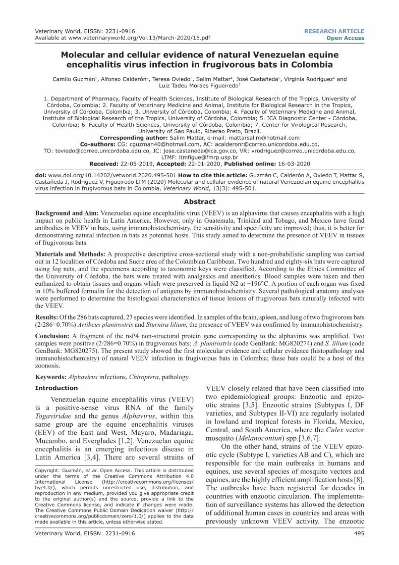
The phylogenetic analysis demonstrates that sequences were closely related to the VEEV and only showed 3% of sequence divergence with previous sequences registered in GenBank (Figure-1).
Figure-1 Shows phylogeny of the VEEV obtained with MEGA Version 7.0 (https://www.megasoft-ware.net/) detected in frugivorous bats, and compared with other alphaviruses reported in Genbank. The consensus sequence obtained in this study presented a similarity of 97%, and 100% coverage with sequences of the gene was encoding the nsp4 protein registered
in GenBank: KC344505.2 Guatemala/human [24] KC344485.2 (strain CoAn5384 equine isolated from Cali, Colombia) [25]. That is, the sequences found in frugivorous bats of the Colombian Caribbean are similar to sequences of the epizootic cycle of VEEV (Subtype I, varieties AB and C), which are the main responsible for outbreaks in humans and equines.Pathological anatomy analysis
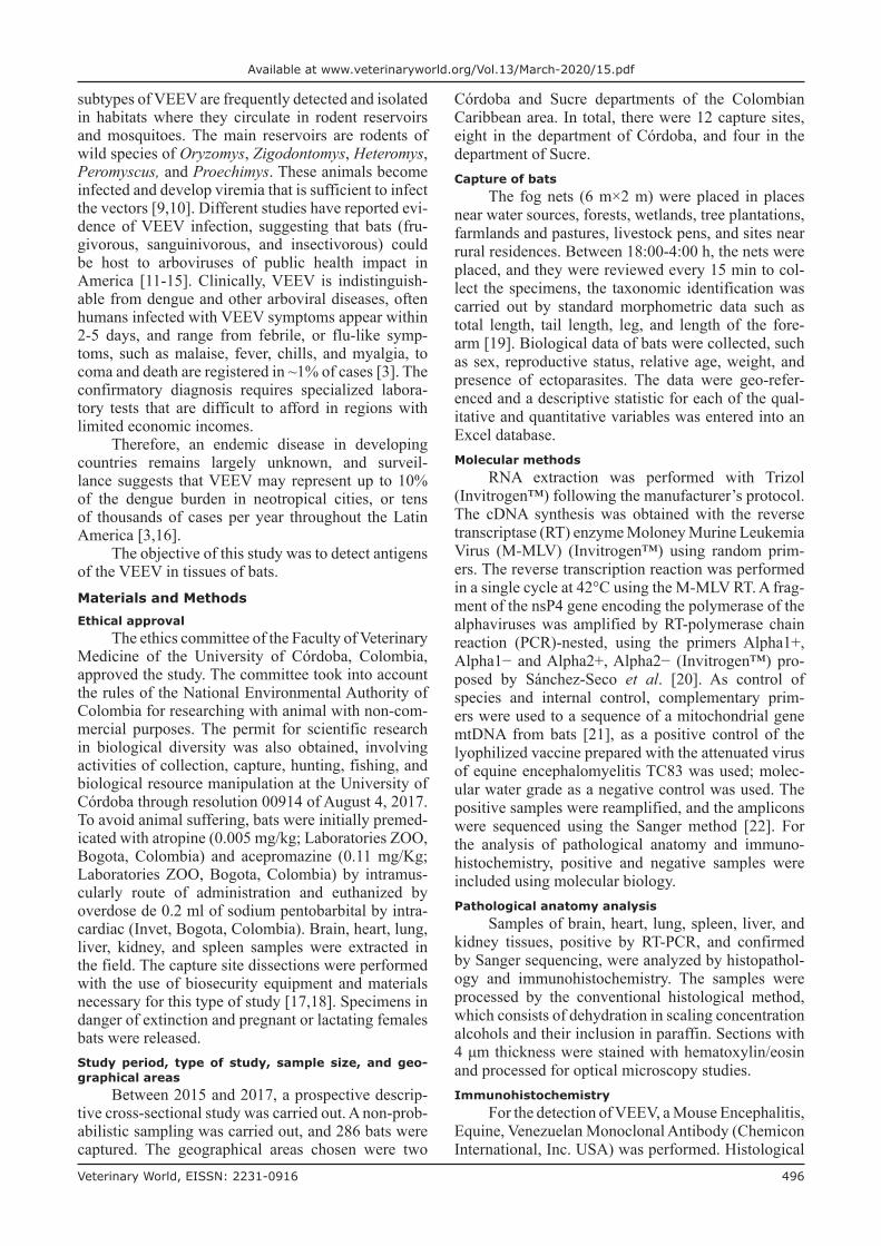
VEEV antigens and pathological lesions were detected in two specimens, and both were also posi-tive by RT-PCR. Figure-2 shows the findings of the pathological anatomy found in the tissues of Sturnira lilium in the central nervous system, severe meningi-tis, and non-suppurative vasculitis of the lymphocytic type, perivascular and perineuronal edema, diffuse gliosis, satellitosis, neuronophagia, neuronal death, and neuropil vacuolization. In the liver, there is a severe mononuclear infiltrate of lymphocytic type in the portal triad and multifocally in the periacinar and centrolubulillar area and vascular changes in hepato-cytes. The final diagnosis was a severe non-suppu-rative meningoencephalitis of viral type and severe non-suppurative hepatitis of infectious viral type. Concerning the other specimen Artibeus planirostris, in the lung, there was a hyperplasia of a lymphoid type associated with bronchi, thickening of alveolar septa, mononuclear lymphocytic alveolitis, and mixed infil-trate with mononuclear predominance. In the liver, mild non-suppurative hepatitis of the lymphocytic type was observed.Immunohistochemical analysis
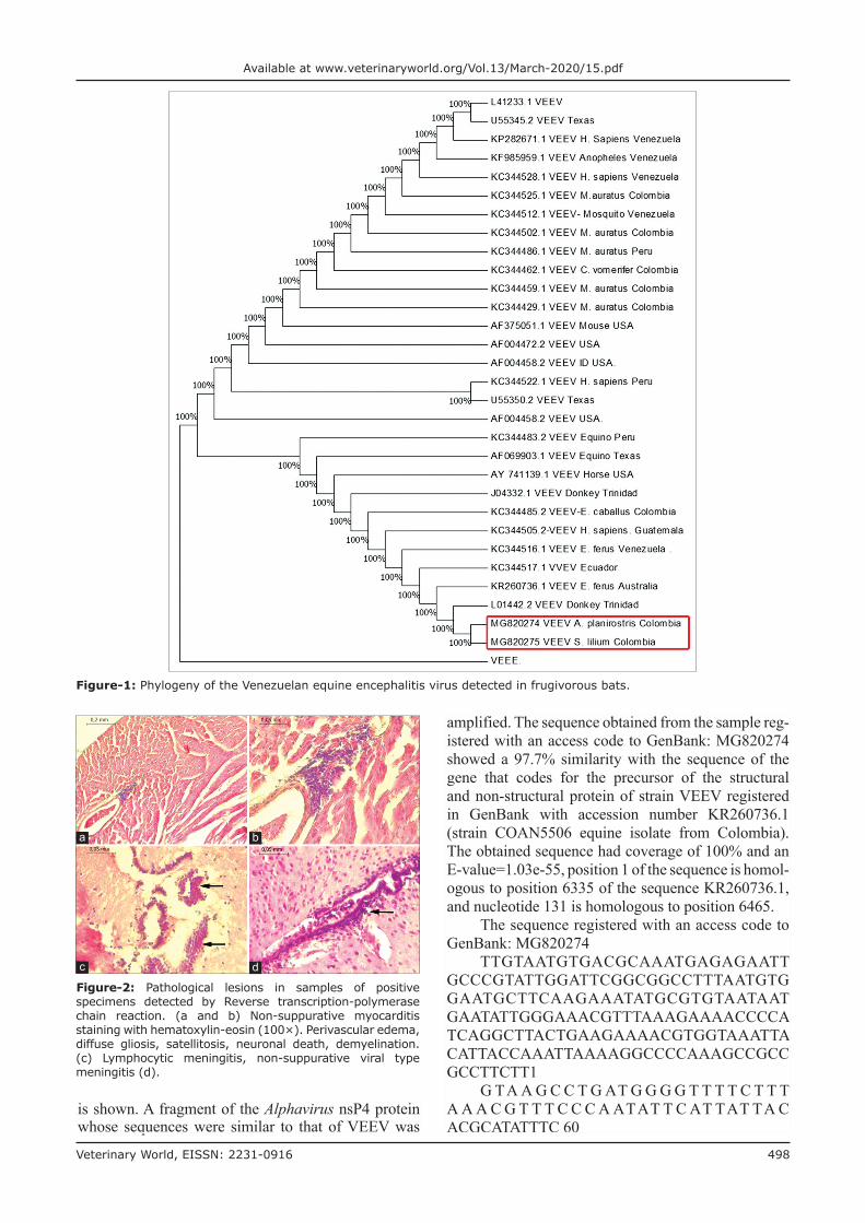
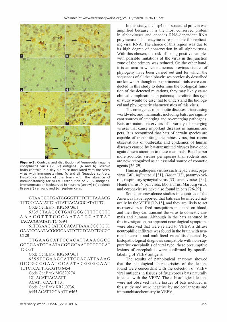
Tissues of brain, spleen, and lung of frugivo-rous bats that were previously positive by RT-PCR and sequenced by the Sanger method were tested with specific antibodies for the VEEV. In Figure-3: (a and b), positive controls (c and d) negative controls, (e, f, and g) positive samples. It is possible to observe VEEV antigens in the brain, spleen, and lung.Discussion
The role of bats in the cycle of transmission of VEEV has not yet been established; in this study, for the first time in the world, evidence of the presence of VEEV-specific antigens in frugivorous bats’ tissues
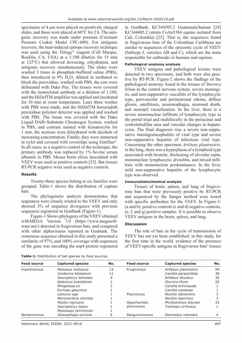
Table-1: Distribution of bat species by food sources.
Food source Captured species No. Food source Captured species No.
Insectivorous Molossus molossus 14 Frugivorous Artibeus planirostris 99Uroderma bilobatum 11 Carollia perspicillata 38Saccopteryx bilineata 4 Artibeus lituratus 30Eptesicus brasiliensis 1 Sturnira lilium 20Rhogeessa yo 2 Carollia brevicauda 1Eumops glaucinus 1 Carollia castanea 1Lasiurus ega 1 Piscivorous Noctilio albiventris 3Micronycteris microtis 1 Noctilio leporinus 3Myotis nigricans 1 Opportunistic
piscivorousPhyllostomus discolor 42
Saccopteryx leptur 1 Trachops cirrhosus 1Molossops temminckii 1
Nectarivorous Glossophaga soricina 6 Sanguinovorous Desmodus rotundus 4
Veterinary World, EISSN: 2231-0916 498
Available at www.veterinaryworld.org/Vol.13/March-2020/15.pdf
amplified. The sequence obtained from the sample reg-istered with an access code to GenBank: MG820274 showed a 97.7% similarity with the sequence of the gene that codes for the precursor of the structural and non-structural protein of strain VEEV registered in GenBank with accession number KR260736.1 (strain COAN5506 equine isolate from Colombia). The obtained sequence had coverage of 100% and an E-value=1.03e-55, position 1 of the sequence is homol-ogous to position 6335 of the sequence KR260736.1, and nucleotide 131 is homologous to position 6465.
The sequence registered with an access code to GenBank: MG820274
TTGTAATGTGACGCAAATGAGAGAATT GCCCGTATTGGATTCGGCGGCCTTTAATGTG GAATGCTTCAAGAAATATGCGTGTAATAAT GAATATTGGGAAACGTTTAAAGAAAACCCCA TCAGGCTTACTGAAGAAAACGTGGTAAATTA CATTACCAAATTAAAAGGCCCCAAAGCCGCC GCCTTCTT1
G TA A G C C T G AT G G G G T T T T C T T T A A A C G T T T C C C A ATAT T C AT TAT TA C ACGCATATTTC 60
Figure-1: Phylogeny of the Venezuelan equine encephalitis virus detected in frugivorous bats.
is shown. A fragment of the Alphavirus nsP4 protein whose sequences were similar to that of VEEV was
Figure-2: Pathological lesions in samples of positive specimens detected by Reverse transcription-polymerase chain reaction. (a and b) Non-suppurative myocarditis staining with hematoxylin-eosin (100×). Perivascular edema, diffuse gliosis, satellitosis, neuronal death, demyelination. (c) Lymphocytic meningitis, non-suppurative viral type meningitis (d).
a b
c d
Veterinary World, EISSN: 2231-0916 499
Available at www.veterinaryworld.org/Vol.13/March-2020/15.pdf
GTAAGCCTGATGGGGTTTTCTTTAAACG TTTCCCAATATTCATTATTACACGCATATTTC
Code GenBank: KR260736.16335GTAAGCCTGATGGGGTTTTCTTT
A A A C G T T T C C C A A T A T T C A T T A T TACACGCATATTTC 6394
61TTGAAGCATTCCACATTAAAGGCCGCC GAATCCAATACGGGCAATTCTCTCATCTGCGT C120
T T G A A G C AT T C C A C AT TA A A G G C C GCCGAATCCAATACGGGCAATTCTCTCAT TGCGT
Code GenBank: KR260736.16 3 9 5 T T G A A G C AT T C C A C AT TA A A G
G C C G C C G A AT C C A AT A C G G G C A AT TCTCTCATTTGCGTG 6454
Code GenBank MG820274121 ACATTACAATTACATT CAATT 131Code GenBank KR260736.16455 ACATTGCAATT 6465
In this study, the nsp4 non-structural protein was amplified because it is the most conserved protein in alphaviruses and encodes RNA-dependent RNA polymerase. This enzyme is responsible for replicat-ing viral RNA. The choice of this region was due to its high degree of conservation in all alphaviruses. With this chosen, the risk of losing positive samples with possible mutations of the virus in the junction zone of the primers was reduced. On the other hand, it is an area in which numerous previous studies of phylogeny have been carried out and for which the sequences of all the alphaviruses previously described are known. Although no experimental trials were con-ducted in this study to determine the biological func-tion of the detected mutations, they may likely cause clinical complications in patients; therefore, this type of study would be essential to understand the biologi-cal and phylogenetic characteristics of this virus.
The emergence of zoonotic diseases is increasing worldwide, and mammals, including bats, are signifi-cant sources of emerging and re-emerging pathogens. Bats are natural reservoirs of a variety of emerging viruses that cause important diseases in humans and pets. It is recognized that bats of certain species are capable of transmitting the rabies virus, but recent observations of outbreaks and epidemics of human diseases caused by bat-transmitted viruses have once again drawn attention to these mammals. Bats harbor more zoonotic viruses per species than rodents and are now recognized as an essential source of zoonotic agents [26-29].
Human pathogens viruses such hepacivirus, pegi-virus [30], Influenza A [31], Hanta [32], paramyxovi-rus, respiratory syncytial virus [33], arenaviruses [34], Hendra virus, Nipah virus, Ebola virus, Marburg virus, and coronaviruses have also found in bats [26-29].
Some seroprevalence studies in countries of the Americas have reported that bats can be infected nat-urally by the VEEV [12-15], and they are likely to act as reservoirs to feed mosquitoes that feed on blood, and then they can transmit the virus to domestic ani-mals and humans. Although in the bats captured in this investigation, no apparent neurological symptoms were observed that were related to VEEV, a diffuse neutrophilic infiltrate was found in the brain with neu-ronal necrosis and multifocal vasculitis detected by histopathological diagnosis compatible with non-sup-purative encephalitis of viral type, these presumptive lesions of encephalitis were confirmed by specific labeling of VEEV antigens.
The results of pathological anatomy showed that the histological characteristics of the lesions found were concordant with the detection of VEEV viral antigens in tissues of frugivorous bats naturally infected with the VEEV. These histological lesions were not observed in the tissues of bats included in this study and were negative by molecular tests and immunohistochemistry to VEEV.
Figure-3: Controls and distribution of Venezuelan equine encephalitis virus (VEEV) antigens. (a and b) Positive brain controls in 3-day-old mice inoculated with the VEEV virus with immunostaining. (c and d) Negative controls. Histological section of the brain with the absence of immunostaining for VEEV. Distribution of VEEV antigens. Immunoreaction is observed in neurons (arrow) (e); splenic tissue (f) (arrow); and (g) septum cells.
a b
c d
e f
g
Veterinary World, EISSN: 2231-0916 500
Available at www.veterinaryworld.org/Vol.13/March-2020/15.pdf
Several arboviruses that are maintained in more than one host/vector species are considered general-ists, such as St. Louis EEV, West Nile virus, Japanese encephalitis virus, EEV, western EEV (WEEV), and VEEV.
Immunohistochemical detection of VEEV anti-gens in paraffin-embedded tissues provides a rapid and reliable means of confirmation of histological diagnosis. The results of this study correlate well with the results of molecular virus detection and sequenc-ing. These results indicate that the IHC technique con-stitutes one of the useful tools of specific diagnosis due to its high sensitivity and specificity, the reason why it can also be used for the identification of other tropical etiologies. Unfortunately, this technique has a high cost to be transferred to regional reference labo-ratories in Colombia.
The analysis of the dynamic maintenance of these viruses appears to link to a transmission network instead of a transmission cycle [11]. The mechanisms for the maintenance of arboviruses are intrinsically complicated and must be taken into account and incorporated when designing and constructing math-ematical models that allow predicting their enzootic/epidemic activity in a given ecosystem (wild, urban, and agricultural, as well). Consequently, to better understand the activity pattern and the transmission networks of arboviruses, it is essential to understand the assembly of host species and vectors, their inter-actions, and fluctuations through time and space. Although the natural infection rate found in this study by molecular methods is low (0.2%), it poses the chal-lenge of being able to complement surveillance with serological detection to establish the presence of the virus in regions where the disease is absent in humans. The role of naturally infected frugivorous bats is chal-lenging due to the short period during which these infected animals “could be viremic,” and the serolog-ical detection of specific immunoglobulin offers the most effective means of confirming it.
On the other hand, the “viremic” animals in their wild cycle could reach the water sources and have contact with the mosquitoes, and these infect domes-tic animals that are in contact with humans. Likewise, the relative proximity of frugivorous bats and humans due to disruption of their natural habitat makes it pos-sible for the viral transmission to occur through the consumption of contaminated fruits. Therefore, the approach of these arboviruses requires designs that contemplate an integral and connectable perspective in a network that allows to improve the understand-ing of the dynamics of the viruses and thus improve the preventive measures in vector control and public health policies.
Finally, this study highlights the circulation of VEEV in frugivorous bats captured in rural areas of the departments of Córdoba and Sucre, Colombia. The finding is useful for public health authorities since they can establish permanent surveillance of
wild VEEV cycles that anticipate a possible outbreak to implement prevention and control measures.Conclusion
The presence of VEEV antigen was confirmed in the brain, lung, and spleen of frugivorous bats that were also positive by RT-PCR, sequencing, and histopathology.Authors’ Contributions
CG: Designed the study, writing of the manu-script and analyzed the data. AC: Helped in the prac-tical experiment, drafted the manuscript and analyzed the data. TO: Helped in the practical experiment, drafted the manuscript and analyzed the data. SM: Helped in design of the study, supervised and drafted the manuscript. JC: Helped in the practical experi-ment, drafted the manuscript and analyzed the data. VR: Helped in the practical experiment, drafted the manuscript and analyzed the data. LTMF: Helped in the practical experiment, drafted the manuscript and analyzed the data. All authors read and approved the final manuscript.Acknowledgments
The authors are thankful to Institute of Virology, Faculty of Medicine, University of Sao Paulo, Ribeirao Preto, for the donation of positive controls for the per-formance of immunohistochemistry. The authors also thankful to the University of Cordoba, a sustainability program for researching groups-CIUC-2018.Competing Interests
The authors declare that they have no competing interests.Publisher’s Note
Veterinary World remains neutral with regard to jurisdictional claims in published institutional affiliation.References1. Zacks, M.A. and Paessler, S. (2010) Encephalitic
Alphaviruses. Vet. Microbiol., 140(3-4): 281-286.2. Gardner, S.N., McLoughlin, K., Be, N.A., Allen, J.,
Weaver, S.C. and Forrester, N. (2016) Characterization of genetic variability of Venezuelan equine encephalitis viruses. PLoS One, 11(4): e0152604.
3. Aguilar, P.V., Estrada-Franco, J.G., Navarro-Lopez, R., Ferro, C., Haddow, A.D. and Weaver, S.C. (2011) Endemic Venezuelan equine encephalitis in the Americas: Hidden under the dengue umbrella. Future Virol., 6(6): 721-740.
4. Vittor, A.Y., Armien, B., Gonzalez, P., Carrera, J.P., Dominguez, C. and Valderrama, A. (2016) Epidemiology of emergent Madariaga encephalitis in a region with endemic Venezuelan equine encephalitis: Initial host studies and human cross-sectional study in Darien, Panama. PLoS Negl. Trop. Dis., 10(4): e0004554.
5. Weaver, S.C., Winegar, R., Manger, I.D. and Forrester, N.L. (2012) Alphaviruses: Population genetics and determinants of emergence. Antiviral Res., 94(3): 242-257.
6. Forrester, N.L., Auguste, A.J., Guerbois, M., Rossi, S.L., Lin, D. and Hari, K. (2016) Venezuelan equine encepha-litis virus strain COAN5506, complete genome. GenBank:
Veterinary World, EISSN: 2231-0916 501
Available at www.veterinaryworld.org/Vol.13/March-2020/15.pdf
KR260736.1.7. Medina, G., Garzaro, D.J., Barrios, M., Auguste, A.J.,
Weaver, S.C. and Pujol, F.H. (2015) Venezuelan equine encephalitis virus isolate AB66640 (VEEVIAB) nonstruc-tural polyprotein precursor and structural polyprotein genes, complete CDS. Am. J. Trop. Med. Hyg., 93(1): 7-10.
8. Carrara, A., Gonzales, M., Ferro, C., Tamayo, M., Aronson, J. and Paessler, S. (2005) Venezuelan equine encephalitis virus infection of spiny rats. Emerg. Infect. Dis., 11(5): 663-669.
9. Johnson, K.M. and Martin, D.H. (1974) Venezuelan equine encephalitis. Classic review providing excellent descrip-tions of the first documentation of endemic Venezuelan equine encephalitis (VEE), as well as studies of equine vir-ulence and amplification. Adv. Vet. Sci. Comp. Med., 18 : 79-116.
10. Deardorff, E.R. and Scott, C. (2010) Weaver vector compe-tence of culex (melanoconion) taeniopus for equine-virulent subtype IE strains of Venezuelan equine encephalitis virus. Am. J. Trop. Med. Hyg., 82(6): 1047-1052.
11. Diaz, L.A., Flores, F.S., Quaglia, A. and Contigiani, M.S. (2012) Intertwined arbovirus transmission activity: Reassessing the transmission cycle paradigm. Front. Physiol., 3: 493.
12. Correa, G.P., Calisher, C.H. and Baer, G.M. (1972) Epidemic strain of Venezuelan equine encephalomyelitis virus from a vampire bat captured in Oaxaca, Mexico, 1970. Science, 175(4021): 546-547.
13. Seymour, C., Dickeman, R.W. and Martin, M.S. (1978) Venezuelan encephalitis virus infection in neotropical bats. I. Natural infection in a Guatemala enzootic focus. Am. J. Trop. Med. Hyg., 27(2 Pt 1): 290-296.
14. Thompson, N.N., Auguste, A.J., Travassos da Rosa, A.P.A., Carrington, C.V.F., Blitvich, B.J. and Chadee, D.D. (2015) Seroepidemiology of selected alphaviruses and flaviviruses in bats in Trinidad. Zoonoses Public Health, 62(1): 53-60.
15. Sotomayor-Bonilla, J., Abella, C., Chaves, A., Álvarez, P., Rico-Chávez, O. and Ibáñez, S. (2017) Potential sympat-ric vectors and mammalian hosts of Venezuelan equine encephalitis virus in Southern Mexico. J. Wildl. Dis., 53(3): 657-661.
16. Forrester, N.L., Wertheim, J.O., Dugan, V.G., Auguste, A.J., Lin, D., Adams, A.P. and Weaver, S.C. (2017) Evolution and spread of Venezuelan equine encephalitis complex Alphavirus in the Americas. PLoS Negl. Trop. Dis., 11(8): e0005693.
17. Linares, O. (2002) Mamíferos de Venezuela. Venezuela. Sociedad Conservacionista Audubon, Venezuela.
18. Mills, J.N., Childs, J.E., Ksiazek, T.G. and Peters, C.J. (1995) Methods for Trapping and Sampling Small Mammals for Virologic Testing. US Department of Health and Human Services, Public Health Service/Centers for Disease Control and Prevention, United States.
19. Fernández, B., Guerrero, R., Lord, R., Ochoa, J. and Ulloa, G. (1988) Mamíferos de Venezuela, Lista y Claves Para su Identificación. Museo del Instituto de Zoología Agrícola, Universidad Central de Venezuela, Caracas.
20. Sánchez-Seco, M., Rosario, D., Quiroz, E., Guzmán, G. and Tenorio, A. (2001) A generic nested-RT-PCR followed by sequencing for detection and identification of members of the Alphavirus genus. J. Virol. Methods, 95(1-2): 153-161.
21. Ramírez, N.N., Alegre, E.A., Ruiz, R.M., De Biasio, M.B. and Bastiani, C.E. (2004) Detección de leptospiras patóge-nas en tejido renal de murciélagos de Corrientes, Argentina. Rev. Vet., 25(1): 16-20.
22. Cabral, B.G., De Paula, J.R., Pereira, D.S.R., Sequetin, C.M., Luch, A. and Figueiredo, C.A. (2018) M. simple protocol for population (Sanger) sequencing for Zika virus genomic regions. Mem. Inst. Oswaldo Cruz, 113(1): 38-44.
23. Charles, P.C., Walters, E., Margolis, F. and Johnston, R.E. (1995) Mechanism of neuroinvasion of Venezuelan equine encephalitis virus in the mouse. Virology, 208(2): 662-671.
24. Dugan, V.G., Halpinm, R., Ghedin, E., Wester, E., Bera, J. and Fedorova, N. (2016) Venezuelan equine encephalitis virus strain VEEV/homo sapiens/GTM/69Z1/1969/IAB, complete genome. GenBank: KC344505.2.
25. Dugan, V.G., Halpin, R., Ghedin, E., Wester, E., Bera, J. and Fedorova, N. (2012) Venezuelan equine encephalitis virus strain VEEV/Equus ferus caballus/COL/CoAn5384/1967/IAB, complete genome. GenBank: KC344485.2.
26. Plowright, R.K., Eby, P., Hudson, P.J., Smith, I.L., Westcott, D. and Bryden, W.L. (2015) Ecological dynamics of emerging bat virus spillover. Proc. Biol. Sci., 282(1798): 20142124.
27. Jones, K.E., Patel, N.G., Levy, M.A., Storeygard, A., Balk D. and Gittleman, J.L. (2008) Global trends in emerg-ing infectious diseases. Nature, 451(7181): 990-993.
28. Luis, A.D., Hayman, D.T.S., O’Shea, T.J., Cryan, P.M., Gilbert, A.T., Pulliam, J.R. and Webb, C.T. (2013) A com-parison of bats and rodents as reservoirs of zoonotic viruses: Are bats special? Proc. Biol. Sci., 280(1756): 20122753.
29. Olival, K.J., Hosseini, P.R., Zambrana-Torrelio, C., Ross, N., Bogich, T.L. and Daszak, P. (2017) Host and viral traits predict zoonotic spillover from mammals. Nature, 546(7660): 646-650.
30. Quan, P.L., Firth, C., Conte, J.M., Williams, S.H., Zambrana-Torrelio, C.M. and Anthony, S.J. (2013) Bats are a major natural reservoir for hepaciviruses and pegiviruses. Proc. Natl. Acad. Sci. U.S.A., 110(20): 8194-8199.
31. Tong, S.X., Li, Y., Rivailler, P., Conrardy, C., Castillo, D.A. and Chen, L.M. (2012) A distinct lineage of influenza A virus from bats. Proc. Natl. Acad. Sci. U.S.A., 109(11): 4269-4274.
32. Guo, W.P., Lin, X.D., Wang, W., Tian, J.H., Cong, M.L. and Zhang, H.L. (2013) Phylogeny and origins of hantaviruses harbored by bats, insectivores, and rodents. PLoS Pathog., 9(2): e1003159.
33. Drexler, J.F., Corman, V.M., Muller, M.A., Maganga, G.D., Vallo, P. and Binger, T. (2012) Bats host major mammalian paramyxoviruses. Nat. Commun., 796 (3): 796.
34. Malmlov, A., Seetahal, J., Carrington, C., Ramkisson, V., Foster, J. and Miazgowicz, K.L. (2017) Serological evi-dence of Arenavirus circulation among fruit bats in Trinidad. PLoS One, 12(9): e0185308.
********