prevalence and diversity of gastrointestinal … world, eissn: 2231-0916 198 veterinary world,...
TRANSCRIPT
Veterinary World, EISSN: 2231-0916 198
Veterinary World, EISSN: 2231-0916Available at www.veterinaryworld.org/Vol.12/February-2019/2.pdf
RESEARCH ARTICLEOpen Access
Prevalence and diversity of gastrointestinal protozoa in Madura cattle at Bangkalan Regency, East Java, Indonesia
Poedji Hastutiek1, Wiwik Misaco Yuniarti2, Mufasirin Djaeri1, Nunuk Dyah Retno Lastuti1, Endang Suprihati1 and Lucia Tri Suwanti1,3
1. Department of Parasitology, Faculty of Veterinary Medicine, Universitas Airlangga, Jl. Mulyorejo, Kampus C Unair, Surabaya, Indonesia; 2. Department of Clinical Science, Faculty of Veterinary Medicine, Universitas Airlangga, Jl. Mulyorejo, Kampus C Unair, Surabaya, Indonesia; 3. Department of Parasitology, Faculty of Veterinary Medicine, Universitas Airlangga,
Institute of Tropical Diseases, Universitas Airlangga, Jl. Mulyorejo, Kampus C Unair, Surabaya, Indonesia.Corresponding author: Poedji Hastutiek, e-mail: [email protected]
Co-authors: WMY: [email protected], MD: [email protected], NDRL: [email protected] ES: [email protected], LTS: [email protected]
Received: 21-09-2018, Accepted: 20-12-2018, Published online: 08-02-2019
doi: 10.14202/vetworld.2019.198-204 How to cite this article: Hastutiek P, Yuniarti WM, Djaeri M, Lastuti NDR, Suprihati E, Suwanti LT (2019) Prevalence and diversity of gastrointestinal protozoa in Madura cattle at Bangkalan Regency, East Java, Indonesia, Veterinary World, 12(2): 198-204.
AbstractAim: This study aimed to describe the gastrointestinal protozoa in Madura cattle at Bangkalan Regency, East Java, Indonesia.
Materials and Methods: A total of 500 samples of Madura cattle feces were collected from 10 districts at Bangkalan Regency. Those ten districts represent the lowland and upland areas, and each district was represented by one village. The collected feces were examined using native, sedimentation, and floating methods. The species identification was determined by their morphology.
Results: There were 357 (71.4%) samples positively infected with protozoan. The highest rate of sample with protozoan infection was at Kamal District (88.23%), and Bangkalan District (52.83%) was the lowest one. There were six species of protozoa that infected gastrointestinal tract; those are Eimeria spp., Balantidium spp., Isospora spp., Blastocystis spp., Entamoeba spp., and Cryptosporidium spp. The highest number of protozoa found in this research was Eimeria (53.42%) followed by Blastocystis (14.43%). In this study, we found that 295 samples (58.76%) infected by one kind of protozoa, 53 samples (10.56%) infected by two kinds of protozoa, and 11 samples (2.19%) infected by three kinds of protozoa. In addition, there were 65.54% of bulls infected with protozoa, considerably lower than cows (72.97%). Cattle aged 6 months-2 years old (73.39%) and >2 years old (71.25%) are known more prone to protozoan infections than cattle aged <6 months (66.15%).
Conclusion: The present study revealed that protozoan infection of cattle is common in Bangkalan Regency. Studies focused on determining that the prevalence of protozoan, risk factors for the parasitism, and the geographic distribution are needed and will be effective guide for prevention and control measures.
Keywords: Bangkalan, Madura cattle, mapping, protozoa.
Introduction
Madura cattle is one of the Indonesian local cat-tle that are widely developed in East Java, especially in Madura Island. Madura cattle have great poten-tials to be developed because they are genetically tolerant to hot climates and marginal environments, resistant to tick infestations, and highly adaptable to low feed quality, as well as require less food com-pared to imported cattle. Besides, Madura cattle are easy to maintain, easy to breed, and resistant to var-ious diseases [1]. The population of Madura cattle in Bangkalan Regency in 2016 was 200,279 and pre-dicted to increase every year parallel to the promotion
of crossbreed artificial insemination program between Madura cattle and Limousine cattle (Madrasin) [2].
Beef cattle as a potential commodity in the devel-opment of rural farming must be supported by an ade-quate maintenance system. The business of animal husbandry is faced with problems of reproductive disorders and chronic parasitic diseases, especially protozoan infections. Gastrointestinal disease needs special attention because it can be an obstacle that affects the acceleration of livestock development in the countryside so that it can cause economic losses due to decreased livestock productivity, decreased weight, quality of meat, skin, and internal organs, growth retardation in young animals, and danger of zoonoses. Delay weight gain in cattle with protozoan infection can reach >40% compared with healthy cows [3,4].
Based on the examination results of fecal samples gathered from Madura cattle butchered at slaughter-house at Surabaya, it was found that those cattle were infected by several kinds of protozoa species, such as Eimeria spp., Balantidium spp., and Entamoeba spp.
Copyright: Hastutiek, et al. Open Access. This article is distributed under the terms of the Creative Commons Attribution 4.0 International License (http://creativecommons.org/licenses/by/4.0/), which permits unrestricted use, distribution, and reproduction in any medium, provided you give appropriate credit to the original author(s) and the source, provide a link to the Creative Commons license, and indicate if changes were made. The Creative Commons Public Domain Dedication waiver (http://creativecommons.org/publicdomain/zero/1.0/) applies to the data made available in this article, unless otherwise stated.
Veterinary World, EISSN: 2231-0916 199
Available at www.veterinaryworld.org/Vol.12/February-2019/2.pdf
(unpublished data). However, there has been no research published about protozoan infections in the gastrointestinal tract of Madura cattle in Bangkalan Regency, Madura, Indonesia.Materials and MethodsEthical approval
The present study was based on the laboratory examination of cattle feces without treatment. The samples were collected as per standard sample col-lection procedure, directly from the rectum without disturbing the animals, and were accompanied by a responsible veterinarian.Study area
This research was conducted at 10 districts located in coastal areas (each district was represented by one village) in Bangkalan Regency with high live-stock population. Fecal sampling was conducted at sites with different altitudes. Bangkalan, Socah, and Kamal districts are located on lowland with altitude <25 m above sea level, while Tanjung Bumi, Sepuluh, Klampis, Arosbaya, Burneh, Tragah, and Labang districts are located on a plateau with an altitude of 25-200 m above sea level.Fecal sample collection and analysis
A total of 500 fecal samples were collected for this research. Fecal samples were taken randomly from bulls and cows and then divided based on age such as <6 months, 6 months-2 years, and >2 years. Fecal sam-pling was conducted from April to May 2017. During fecal sampling process, questionnaire and interview with the farmer were also conducted to obtain certain information about farmer (name, age, gender, education, and breeding experience) and cattle (breed and quantity, gender, age, type of maintenance, treatment that has ever given, the other reared cattle, type or material of the enclosure, environmental condition, or livestock care).
The samples were collected directly from the rectum and brought to the laboratory in mini zip locked polythene bags and added with 2.5% potas-sium bichromate for examination. Each plastic bag was labeled with registered sample number and then stored in a container with ice. Afterward, the fecal samples were examined using native methods, as well as sedimentation and modified Fulleborn’s floating methods [5]. To determine the existence of protozoa, identification key methods were carried out [6]. The positive result of protozoan-infected cattle could be determined when protozoa were found during exam-ination using one of those methods. The prevalence of the protozoan infections was expressed in percentage value using the following formula:
P = (Positive results: Number of samples) × 100%.
Statistical analysisThe data obtained will be analyzed descrip-
tively and presented in the form of the prevalence of
protozoan infections based on district, kind of proto-zoa, sex, and age.Results and Discussion
Based on this research, it was found that there were 357 from 500 samples (71.4%) positively infected by protozoa. The highest prevalence of proto-zoan infections was found in Kamal district, in which 88.23% (45/51) samples were positive, followed by Sepuluh district 86.8% (46/53), Klampis district 80% (32/40), Tanjung Bumi district 75% (36/48), Arosbaya district 74.51% (38/51), Burneh district 68% (34/50), Labang district 67.31% (35/52), Socah district 67.28% (37/55), Tragah district 57.14% (28/49), and Bangkalan district 52.83% (28/53) (Table-1).
The high prevalence in this study was in accor-dance with the study of Volpato et al. [7], which reported the prevalence of intestinal protozoan infec-tion in dairy calf in Brazil. Those ten districts are located in coastal areas and are well known as live-stock meeting points from several locations as well as temporary shelters for cattle that will be traded out of the island or for Idul Adha. On the other hand, the results of this research indicate that the prevalence of protozoan infections in Madura cattle from those 10 districts was high. Therefore, routine monitoring for protozoan infections should be preceded by per-forming fecal examination so that the infections can be completely controlled to improve the health and productivity of the cattle.
Research result showed that, from 359 positive samples, there were 295 (58.76%) samples infected by one kind of protozoan, 53 (10.56%) samples by two kinds of protozoan, and 11 (2.19%) samples infected with three kinds of protozoan.
The highest of single species protozoan infection was at Klampis district 80% (32/40), while the low-est was at Bangkalan district 49.06% (26/53). District with the highest infection of two species protozoan was Sepuluh district with 26.42% (14/53), whereas the lowest one at Bangkalan district with only 3.77% (2/53). At Kamal district, there were 9.8% (5/51) cat-tle infected with three kinds of protozoan species. At Labang district, Arosbaya district and Socah district
Table-1: The prevalence of protozoan infection in the gastrointestinal of Madura cattle in each district in Bangkalan Regency.
District Number of samples
Number of positive samples (%)
Mangkon Arosbaya 51 38 (74.51)Bator Ma’adan Klampis 40 32 (80)Keleyan Socah 53 35 (66.04)Gili Anyar Kamal 51 45 (88.24)Maneron Sepuluh 53 46 (86.79)Bumi Anyar Tanjung Bumi 48 36 (75)Bringen Labang 52 35 (67.31)Kemoneng Tragah 49 28 (57.14)Ketengan Burneh 50 34 (68)Kramin Bangkalan 53 28 (52.83)Total 500 357 (71.4)

Veterinary World, EISSN: 2231-0916 200
Available at www.veterinaryworld.org/Vol.12/February-2019/2.pdf
were 7.69% (4/52), 1.96% (1/51), and 1.82% (1/55) respectively (Table-2).
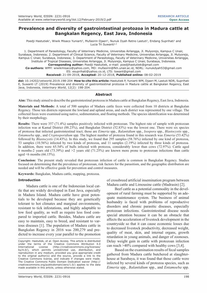
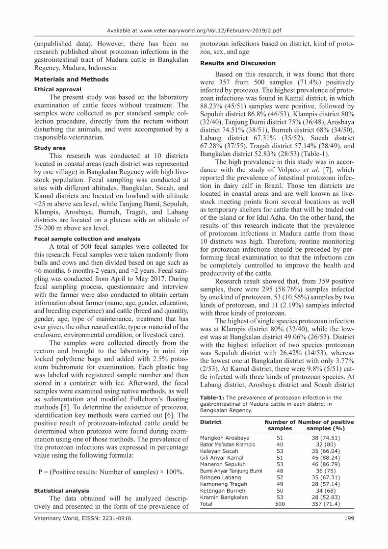
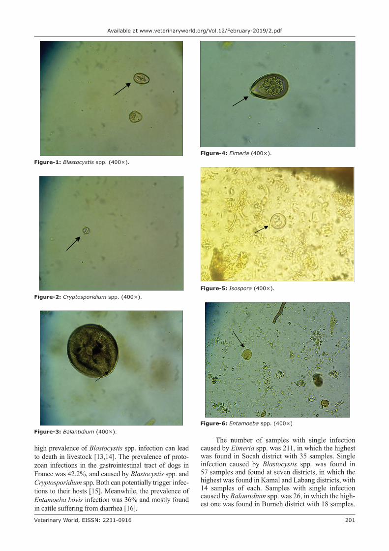
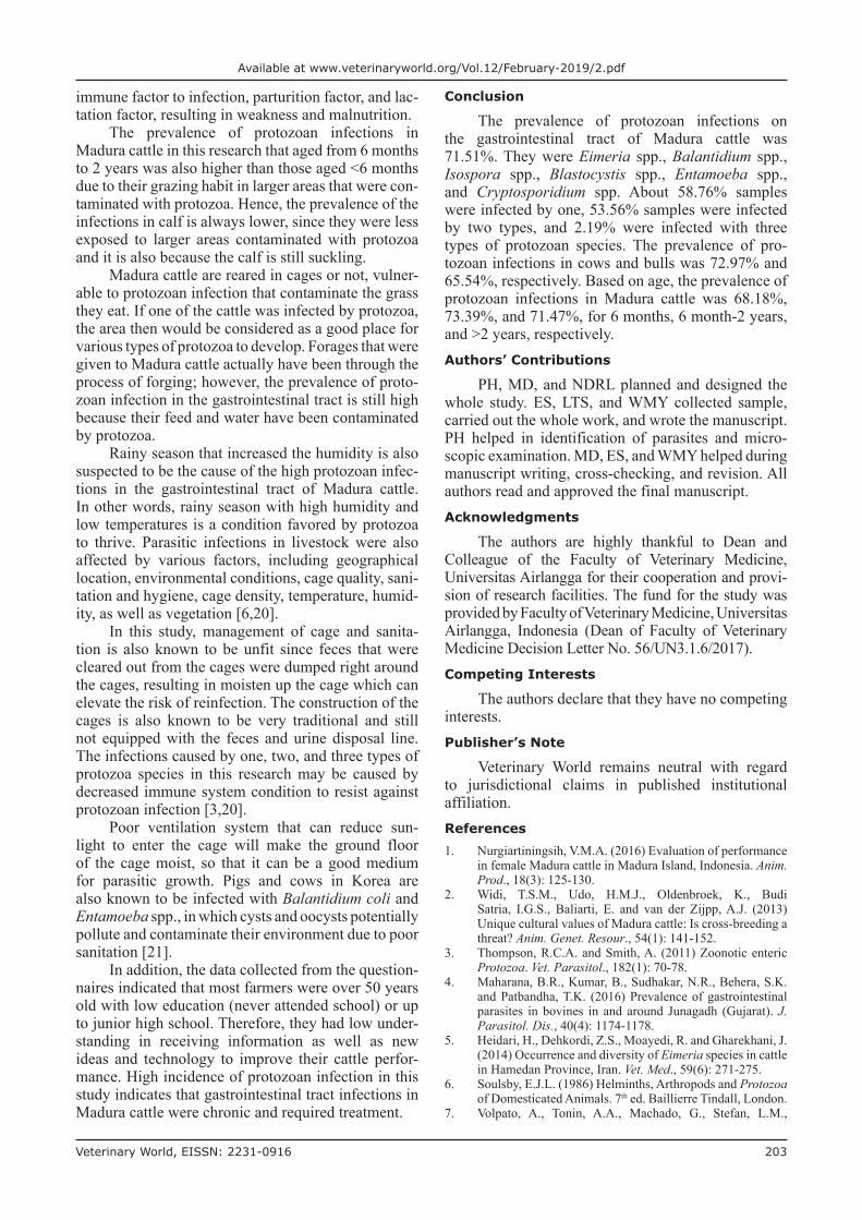
The results showed that there were six species of protozoa infecting the gastrointestinal tract of Madura cattle, i.e., Eimeria spp., Balantidium spp., Isospora spp., Blastocystis spp., Entamoeba spp., and Cryptosporidium spp. The result also indicated that one cattle could be infected by one, two, or even more three kinds of protozoa species at once (Table-2) (Figures-1-6).
Eimeria is the most common species of proto-zoan-infected Madura cattle in 10 districts, whether it was single or mixed infections. The second com-mon species of protozoan-infected Madura cattle in Bangkalan district, both single and mixed infec-tions, was Blastocystis. However, Blastocystis was
not found infecting cattle in Arosbaya, Klampis, and Burneh districts.
Some types of protozoa found in this research showed to have zoonotic potentials, such as Balantidium spp., Entamoeba spp., Blastocystis spp., and Cryptosporidium spp. Cryptosporidium parvum oocyte is usually found in exoskeleton and gastrointestinal tract of Musca domestica on dairy farms. Flies as mechani-cal vectors have the ability to spread diarrheal diseases caused by protozoa as they can travel up to 20 miles [8]. Cryptosporidium bovis oocyte can infect six species of livestock and poultry in Tunisia [9]. The prevalence of Cryptosporidium infection in cattle in Northern Nigeria reported to be 22.3% [10,11], whereas the prevalence of Blastocystis spp., Giardia spp., and Entamoeba spp. was 14.6%, 12.45%, and 7.45%, respectively [12]. The
Table-2: The prevalence of protozoan species in the gastrointestinal tract of Madura cattle in Bangkalan Regency.
Districts Samples positively infected with protozoa Number of positive samples (%)
One type of protozoan species
Total Two types of protozoan species
Total Three types of protozoan species
Total
MangkonArosbaya
Eimeria 31 EimeriaBalantidium
5 EimeriaBalantidium Isospora
1 38 (74.51)Balantidium 1
Bator Ma’adanKlampis
Eimeria 32 ‑ ‑ 32 (80)
KeleyanSocah
Eimeria 33 EimeriaBlastocystis
1 EimeriaBlastocystisBalantidium
1 35 (66.04)
Gili Anyar Kamal
Eimeria 12 EimeriaBalantidium
2 EimeriaBlastocystisBalantidium
3 45 (88.23)
Blastocystis 14 EimeriaBlastocystis
9 EimeriaBlastocystisEntamoeba
2
Balantidium 2 Blastocystis Balantidium
1
ManeronSepuluh
Eimeria 22 EimeriaBalantidium
2 - - 46 (86.8)
Blastocystis 10 EimeriaBlastocystis
11
BlastocystisBalantidium
1
Bumi AnyarTanjung Bumi
Eimeria 23 EimeriaBlastocystis
5 - - 36 (75)Blastocystis 7Balantidium 1
BringenLabang
Eimeria 10 EimeriaBlastocystis
3 EimeriaBlastocystisEntamoeba
1 35 (67.31)
Blastocystis 14 BlastocystisBalantidium
1 EimeriaBlastocystisBalantidium
1
Entamoeba 1 BlastocystisEntamoeba
1 EimeriaBlastocystisCryptosporidium
1
Blastocystis Cryptosporidium
1 EimeriaBlastocystisIsospora
1
KemonengTragah
Eimeria 12 EimeriaBlastocystis
5 - - 28 (57.14)Blastocystis 11
KetenganBurneh
Eimeria 13 EimeriaBalantidium
2 - - 34 (68)
Balantidium 18 EimeriaEntamoeba
1
KramatBangkalan
Eimeria 21 EimeriaBalantidium
2 - - 28 (52.83)Balantidium 4Blastocystis 1
Veterinary World, EISSN: 2231-0916 201
Available at www.veterinaryworld.org/Vol.12/February-2019/2.pdf
high prevalence of Blastocystis spp. infection can lead to death in livestock [13,14]. The prevalence of proto-zoan infections in the gastrointestinal tract of dogs in France was 42.2%, and caused by Blastocystis spp. and Cryptosporidium spp. Both can potentially trigger infec-tions to their hosts [15]. Meanwhile, the prevalence of Entamoeba bovis infection was 36% and mostly found in cattle suffering from diarrhea [16].
The number of samples with single infection caused by Eimeria spp. was 211, in which the highest was found in Socah district with 35 samples. Single infection caused by Blastocystis spp. was found in 57 samples and found at seven districts, in which the highest was found in Kamal and Labang districts, with 14 samples of each. Samples with single infection caused by Balantidium spp. was 26, in which the high-est one was found in Burneh district with 18 samples.
Figure-1: Blastocystis spp. (400×).
Figure-2: Cryptosporidium spp. (400×).
Figure-3: Balantidium (400×).
Figure-4: Eimeria (400×).
Figure-5: Isospora (400×).
Figure-6: Entamoeba spp. (400×)

Veterinary World, EISSN: 2231-0916 202
Available at www.veterinaryworld.org/Vol.12/February-2019/2.pdf
Single infection caused by Entamoeba spp. only found in one cattle in Labang district.
Samples infected by two species of protozoan, such as Eimeria and Blastocystis, were found in 34 at six districts. Sepuluh district with 11 samples was the highest. There were five samples infected with three species of protozoa (Eimeria spp., Blastocystis spp., and Balantidium spp.), which were found in Kamal district.
Based on sex, the prevalence of protozoan infec-tions in bulls 65.54% (116/169) was lower than cows 72.97% (243/333). The comparison of the prevalence of protozoan infections based on sex in each district is shown in Table-3.
The prevalence of protozoan infections in cat-tle <6 months of age was 68.18% (45/66), with the highest case found in Klampis, Tanjung Bumi, and Burneh districts (100%), and the lowest was found in Sepuluh district (25%). Furthermore, the prevalence of protozoan infections in cattle aged from 6 months
Table-3: The prevalence of protozoan infections in the gastrointestinal tract of Madura cattle in Bangkalan Regency based on sex.
Districts Bulls Cows Total of samples
Number of samples
Positive samples (%)
Number of samples
Positive samples (%)
Number of samples
Positive samples (%)
Mangkon Arosbaya 13 6 (46.15) 38 32 (84.21) 51 38 (74.51)Bator Ma’adan Klampis 10 8 (80) 30 24 (80) 40 32 (80)Keleyan Socah 14 6 (42.86) 39 29 (74.44) 53 37 (67.28)Gili Anyar Kamal 19 15 (78.95) 32 30 (93.75) 51 45 (88.23)Maneron Sepuluh 23 19 (82.61) 30 27 (90) 53 46 (86.8)Bumi Anyar Tanjung Bumi 22 17 (77.27) 26 19 (73.08) 48 36 (75)Bringen Labang 4 3 (75) 48 32 (66.67) 52 35 (67.31)Kemoneng Tragah 8 3 (37.5) 41 25 (60.98) 49 28 (57.14)Ketengan Burneh 50 34 (68) - - 50 34 (68)Kramin Bangkalan 4 3 (28) 49 25 (50.02) 53 28 (52.83)Total 167 114 (68.26) 333 243 (72.97) 500 357 (71.4)
Table-4: The prevalence of protozoan infections in the gastrointestinal tract of Madura cattle in Bangkalan Regency based on age.
Districts Age of samples Number of samples
Positive samples
<6 months
Positive samples (%)
>6 months- 2 years
Positive samples (%)
>2 years Positive samples (%)
Mangkon Arosbaya
2 1 (50) 31 27 (87.1) 19 10 (52.63) 51 38
Bator Ma’adan Klampis
6 6 (100) 9 7 (77.78) 25 19 (76) 40 32
Keleyan Socah 17 12 (70.59) 10 8 (80) 26 15 (57.69) 55 37Gili Anyar Kamal
2 1 (50) 18 15 (83.33) 31 29 (93.55) 51 45
Maneron Sepuluh
4 1 (25) 14 13 (86.67) 35 32 (94.12) 53 46
Bumi Anyar Tanjung Bumi
3 3 (100) 8 6 (75) 37 27 (72.97) 48 36
Bringen Labang
7 5 (71.43) 2 1 (50) 43 29 (67.44) 52 35
Kemoneng Tragah
10 8 (80) 2 - 37 20 (54.05) 49 28
Ketengan Burneh
1 1 (100) 16 11 (68.75) 33 22 (66.67) 50 34
Kramin Bangkalan
13 5 (38.46) 13 3 (23.08) 27 20 (74.07) 53 28
Total 65 43 (65.15) 122 91 (74.59) 313 223 (71.25) 500 357 (71.40)
to 2 years was 73.39% (91/124). The highest preva-lence was found in Arosbaya district (87.1%), and the lowest was found in Bangkalan district (23.08%). For the cattle >2 years, the prevalence of protozoan infec-tions was 71.47% (223/312), the highest was found in Sepuluh district (94.12%), and the lowest was in Arosbaya district (52.63%) (Table-4).
Research conducted in Iran revealed that there were four types of protozoa species found in pigs with a prevalence of 64% [17], while the prevalence of protozoan infections in the gastrointestinal tract of carnivores in Iran was 80.4% [18]. Another research in India showed that 83.08% of cattle were infected by endoparasites, with the higher risk factor found in cows (85.97%) than bulls (69.23%) and in the adult ones that aged >6 months (85.97%) compared to those that aged <6 months (61.17%) [19]. Similarly, the prevalence of protozoan infections in cows in this research was also higher than in bulls due to predis-posing genetic factor, hormonal factor, stress-reducing
Veterinary World, EISSN: 2231-0916 203
Available at www.veterinaryworld.org/Vol.12/February-2019/2.pdf
immune factor to infection, parturition factor, and lac-tation factor, resulting in weakness and malnutrition.
The prevalence of protozoan infections in Madura cattle in this research that aged from 6 months to 2 years was also higher than those aged <6 months due to their grazing habit in larger areas that were con-taminated with protozoa. Hence, the prevalence of the infections in calf is always lower, since they were less exposed to larger areas contaminated with protozoa and it is also because the calf is still suckling.
Madura cattle are reared in cages or not, vulner-able to protozoan infection that contaminate the grass they eat. If one of the cattle was infected by protozoa, the area then would be considered as a good place for various types of protozoa to develop. Forages that were given to Madura cattle actually have been through the process of forging; however, the prevalence of proto-zoan infection in the gastrointestinal tract is still high because their feed and water have been contaminated by protozoa.
Rainy season that increased the humidity is also suspected to be the cause of the high protozoan infec-tions in the gastrointestinal tract of Madura cattle. In other words, rainy season with high humidity and low temperatures is a condition favored by protozoa to thrive. Parasitic infections in livestock were also affected by various factors, including geographical location, environmental conditions, cage quality, sani-tation and hygiene, cage density, temperature, humid-ity, as well as vegetation [6,20].
In this study, management of cage and sanita-tion is also known to be unfit since feces that were cleared out from the cages were dumped right around the cages, resulting in moisten up the cage which can elevate the risk of reinfection. The construction of the cages is also known to be very traditional and still not equipped with the feces and urine disposal line. The infections caused by one, two, and three types of protozoa species in this research may be caused by decreased immune system condition to resist against protozoan infection [3,20].
Poor ventilation system that can reduce sun-light to enter the cage will make the ground floor of the cage moist, so that it can be a good medium for parasitic growth. Pigs and cows in Korea are also known to be infected with Balantidium coli and Entamoeba spp., in which cysts and oocysts potentially pollute and contaminate their environment due to poor sanitation [21].
In addition, the data collected from the question-naires indicated that most farmers were over 50 years old with low education (never attended school) or up to junior high school. Therefore, they had low under-standing in receiving information as well as new ideas and technology to improve their cattle perfor-mance. High incidence of protozoan infection in this study indicates that gastrointestinal tract infections in Madura cattle were chronic and required treatment.
Conclusion
The prevalence of protozoan infections on the gastrointestinal tract of Madura cattle was 71.51%. They were Eimeria spp., Balantidium spp., Isospora spp., Blastocystis spp., Entamoeba spp., and Cryptosporidium spp. About 58.76% samples were infected by one, 53.56% samples were infected by two types, and 2.19% were infected with three types of protozoan species. The prevalence of pro-tozoan infections in cows and bulls was 72.97% and 65.54%, respectively. Based on age, the prevalence of protozoan infections in Madura cattle was 68.18%, 73.39%, and 71.47%, for 6 months, 6 month-2 years, and >2 years, respectively.Authors’ Contributions
PH, MD, and NDRL planned and designed the whole study. ES, LTS, and WMY collected sample, carried out the whole work, and wrote the manuscript. PH helped in identification of parasites and micro-scopic examination. MD, ES, and WMY helped during manuscript writing, cross-checking, and revision. All authors read and approved the final manuscript.Acknowledgments
The authors are highly thankful to Dean and Colleague of the Faculty of Veterinary Medicine, Universitas Airlangga for their cooperation and provi-sion of research facilities. The fund for the study was provided by Faculty of Veterinary Medicine, Universitas Airlangga, Indonesia (Dean of Faculty of Veterinary Medicine Decision Letter No. 56/UN3.1.6/2017).Competing Interests
The authors declare that they have no competing interests.Publisher’s Note
Veterinary World remains neutral with regard to jurisdictional claims in published institutional affiliation.References1. Nurgiartiningsih, V.M.A. (2016) Evaluation of performance
in female Madura cattle in Madura Island, Indonesia. Anim. Prod., 18(3): 125-130.
2. Widi, T.S.M., Udo, H.M.J., Oldenbroek, K., Budi Satria, I.G.S., Baliarti, E. and van der Zijpp, A.J. (2013) Unique cultural values of Madura cattle: Is cross-breeding a threat? Anim. Genet. Resour., 54(1): 141-152.
3. Thompson, R.C.A. and Smith, A. (2011) Zoonotic enteric Protozoa. Vet. Parasitol., 182(1): 70-78.
4. Maharana, B.R., Kumar, B., Sudhakar, N.R., Behera, S.K. and Patbandha, T.K. (2016) Prevalence of gastrointestinal parasites in bovines in and around Junagadh (Gujarat). J. Parasitol. Dis., 40(4): 1174-1178.
5. Heidari, H., Dehkordi, Z.S., Moayedi, R. and Gharekhani, J. (2014) Occurrence and diversity of Eimeria species in cattle in Hamedan Province, Iran. Vet. Med., 59(6): 271-275.
6. Soulsby, E.J.L. (1986) Helminths, Arthropods and Protozoa of Domesticated Animals. 7th ed. Baillierre Tindall, London.
7. Volpato, A., Tonin, A.A., Machado, G., Stefan, L.M.,
Veterinary World, EISSN: 2231-0916 204
Available at www.veterinaryworld.org/Vol.12/February-2019/2.pdf
Campigotto, G., Glombowsky, P., Galli, G.M., Favero, J.F. and da Silva, A.S. (2017) Gastrointestinal Protozoa in dairy calves: Identification of risk factors for infection. Rev. MVZ Cordoba, 22(2): 5910-5924.
8. Szostakowska, B., Lozowska, W.K., Racewicz, M., Knight, R., Tamang, L., Myjak, P. and Graczyk, T.K. (2004) Cryptosporidium parvum and Giardia lamblia recovered from flies on a cattle farm and in a landfill. Appl. Environ. Microbiol., 70(6): 3742-3744.
9. Soltane, R., Guyot, K., Dei-Cas, E. and Ayadi, A. (2007) Prevalence of Cryptosporidium Spp. (Eucoccidiorida: Cryptosporidiidae) in seven species of farm animals in Tunisia. Parasite, 14(4): 335-338.
10. Adamu, S.G., Adamu, N.B., Aliyu, A.U., Atsanda, N.N., Mustapha, F.B., Muhammad, Y.A. and Umaru G.A. (2015) Prevalence of Cryptosporidium infection in cattle in Maiduguri, North Eastern Nigeria. Bangl. J. Vet. Med., 13(1): 25-28.
11. Ayinmode, A.B. and Fagbemi, B.O. (2010). Prevalence of Cryptosporidium infection in cattle from South Western infection in cattle from South Western Nigeria. Vet. Arch., 80(6): 723-731.
12. Badparva, E., Fallahi, S. and Aarab-Mazar, Z. (2015) Blastocystis: Emerging protozoan with high prevalence in Iran. Nov. Biomed., 3(4): 214-221.
13. Stensvold, C.R., Suresh, G.K., Tan, K.S., Thompson, R.C., Traub, R.J., Viscogliosi, E., Yoshikawa, H. and Clark, C.G. (2007) Terminology for Blastocystis of subtype a consen-sus. Trends Parasitol., 23(3): 93-96.
14. Yoshikawa H., Wu, Z., Pandey, K., Pandey, B.D., Sherchand, J.B. and Yanagi T. (2009) Molecular charac-teristic of Blastocystis isolates from children and rhesus
********
monkey in Kathmandu, Nepal. Vet. Parasitol., 160(3-4): 295-300.
15. Orman M., Bories, J., El Safadi, D., Poirel, M.T., Gantois, N., Benamrouz-Vanneste, S., Delhaes, L., Hugonnard, M., Certad, G., Zenner, L. and Viscogliosi, E. (2015) Prevalence and genetic diversity of the intestinal parasite Blastocystis spp. and Cryptosporidium Spp, in household dogs in France and evaluation of zoonotic trans-mission risk. Vet. Parasitol., 214(1-2): 167-170.
16. Al-shabbani, A.H. (2016) Direct detection of Entamoeba bovis in calves infected by diarrhea by using polymerase chain reaction technique. J. Vet. Med. Sci., 7(1): 132-137.
17. Yaghobii, K., Sarkari, B., Mansouri, M. and Motazedian, M.H. (2016) Zoonotic intestinal Protozoa of the wild boars, Sus scrofa, in Persian Gulf’s Coastal Area (Bushehr Province), Southwestern Iran. Vet. World, 9(2): 1047-1050.
18. Sarvi, S., Daryani, A., Syarif, M., Rahimi, M.T., Kohansal, M.H., Mirshafiee, S., Siyadatpanah, S., Hossein, A.A. and Gholami, S. (2018) Zoonotic intesti-nal parasites of Carnivora: A systemic review in Iran. Vet. World, 11(11): 58-65.
19. Singh, E., Kaur, P., Singla, L.D. and Bal, S. (2017) Prevalence of gastrointestinal parasitism in small ruminant in Western Punjab, India. Vet. World, 10(1): 61-66.
20. Marskole, P., Verma, Y., Dixit, A.K. and Swamy, M. (2016) Prevalence and burden of gastrointestinal parasites in cat-tle and buffaloes in Jabalpur, India. Vet. World, 9(11): 1214-1217.
21. Ismail, H.A., Jeon, H.K., Yu, Y.M., Do, C. and Lee, Y.H. (2010) Intestinal parasite infections in pigs and beef cattle in rural areas of Chungcheongnam-do, Korea. Korean J. Parasitol., 48(4): 347-349.