modulation of activation-associated host cell gene ... · modulation of activation-associated host...
TRANSCRIPT
Modulation of activation-associated host cell geneexpression by the apicomplexan parasiteTheileria annulata
Zeeshan Durrani, William Weir, Sreerekha Pillai,†
Jane Kinnaird and Brian Shiels*Institute of Infection, Immunity & Inflammation, Collegeof Medical, Veterinary and Life Sciences, University ofGlasgow, Bearsden Road, Glasgow G61 1QH,Scotland, UK.
Summary
Infection of bovine leucocytes by Theileria annulataresults in establishment of transformed, infectedcells. Infection of the host cell is known to promoteconstitutive activation of pro-inflammatory tran-scription factors that have the potential to bebeneficial or detrimental. In this study we havecompared the effect of LPS activation on uninfectedbovine leucocytes (BL20 cells) and their Theileria-infected counterpart (TBL20). Gene expressionprofiles representing activated uninfected BL20relative to TBL20 cells were also compared. Theresults show that while prolonged stimulation withLPS induces cell death and activation of NF-kB inBL20 cells, the viability of Theileria-infected cellswas unaffected. Analysis of gene expression net-works provided evidence that the parasite estab-lishes tight control over pathways associated withcellular activation by modulating reception ofextrinsic stimuli and by significantly altering theexpression outcome of genes targeted by infection-activated transcription factors. Pathway analysisof the data set identified novel candidate genesinvolved in manipulation of cellular functions asso-ciated with the infected transformed cell. The dataindicate that the T. annulata parasite can irrevers-ibly reconfigure host cell gene expression networksassociated with development of inflammatorydisease and cancer to generate an outcome that
is beneficial to survival and propagation of theinfected leucocyte.
Introduction
The protozoan parasites Theileria annulata and T. parvacause economically important lymphoproliferative disor-ders of cattle in developing countries of the Old World(Irvin and Morrison, 1987). In the laboratory they providea unique model to investigate how a simple eukaryotic cellcan direct gene expression and induce proliferation ofanother, more complex, eukaryotic host cell. Followinginvasion of bovine leucocytes by tick-inoculated sporo-zoites, rhoptries and microspheres discharge and theenveloping host cell membrane is disrupted. The interme-diate trophozoite stage is rapidly surrounded by an arrayof host microtubules and within 48 h differentiates into themacroschizont stage (Shaw and Tilney, 1992). This differ-entiation event is accompanied by parasite-dependenttransformation of the host cell (reviewed in Dobbelaereand Heussler, 1999), which in vivo manifests in exponen-tial proliferation of infected leucocytes over 4–5 days,followed by dissemination to a wide range of tissues. Inacute cases of disease, disorganization and destruction ofthe lymphoid system occurs and death is often caused bymassive pulmonary oedema associated with migration ofinfected cells to the lungs (Irvin and Morrison, 1987).
Transformation of T. annulata- and T. parva-infectedleucocytes is known to be dependent on constitutive acti-vation of bovine transcription factors and associatedsignal transduction pathways (reviewed in Dobbelaereand Heussler, 1999; Shiels et al., 2006). Activation ofAP-1, ATF-2, cMyc, NF-kB and STAT3 have all beendocumented and are likely to be responsible for many ofthe phenotypic alterations displayed by the infected cell.However, in addition to being beneficial, constitutive tran-scription factor activation has the potential to be detrimen-tal to survival of the infected cell or host. For example,constitutive activation of c-Jun (a dominant component ofAP-1 transcription factor), is thought to play an importantrole in metastasis of Theileria-infected cells (Adamsonet al., 2000), while activation of NF-kB confers protectionagainst apoptosis (Heussler et al., 1999). However, boththese factors can be pro-apoptotic (Dunn et al., 2002;
Received 25 January, 2012; revised 29 March, 2012; accepted19 April, 2012. *For correspondence. E-mail [email protected]; Tel. (+44)(0) 141 330 5756; Fax (+44)(0) 141 3305603.†Present address: Wellcome Trust Centre for Gene Regulationand Expression, College of Life Sciences, University of Dundee,Dundee, UK.
Cellular Microbiology (2012) 14(9), 1434–1454 doi:10.1111/j.1462-5822.2012.01809.xFirst published online 23 May 2012
© 2012 Blackwell Publishing Ltd
cellular microbiology

Dutta et al., 2006) by upregulating genes encoding death-inducing signals, including pro-inflammatory cytokines. Ithas been predicted that NF-kB-dependent regulation ofinflammatory genes contributes to the pathogenesis oftheileriosis (Heussler et al., 2002).
A recent study demonstrated that only a proportion ofleucocytes infected with T. parva sporozoites successfullyprogress to proliferation, while the rest undergo apoptosis(Rocchi et al., 2006). Thus, events that are detrimentalto the infected cell can occur following sporozoite inva-sion and transformation is not guaranteed. In addition,although basal expression of inflammatory/cellulardefence genes such as TNFa, iNOS and ISG15 can bedetected (Sager et al., 1997; Oura et al., 2006), Theileria-infected cells are refractory to LPS-mediated stimulationof high-level expression, despite constitutive activation oftranscription factors (e.g. AP-1 and NF-kB) associatedwith this event. Furthermore, parasite-mediated repres-sion of highly abundant mRNA levels has been indicatedfor the ISGylation pathway genes, UBP43 and ISG15(Oura et al., 2006) whose expression, like TNFa andiNOS, can be induced by activated NF-kB (Li et al., 2001;Liu et al., 2011). Taken together, these studies predict thatTheileria parasites must direct gene expression networksassociated with activation of host cell transcription factorsto promote an outcome that favours establishment of thetransformed cell.
In this study we have conducted a comprehensive analy-sis of expression profiles associated with LPS stimulationof the uninfected bovine lymphosarcoma cell line BL20(Olobo and Black, 1989) and the T. annulata-infectedcounterpart, TBL20 (Shiels et al., 1986). These lines werechosen as they provide uniform cellular populations toallow robust detection of differences in gene expressionassociated with altered cellular phenotypes. Moreover,TBL cells have been used to investigate infection-associated phenotypes (Shiels et al., 1986; Baylis et al.,1995) and are known to be dependent on a viable parasitefor survival, as they undergo apoptosis when the macro-schizont is killed by BW720c (buparvaquone) (Dobbelaereand Heussler, 1999). The results of our analysis indicatethat while the modulated gene expression profiles of LPS-stimulated, uninfected BL20 and infected TBL20 cells sig-nificantly overlap, a major reorganization of activated geneexpression is orchestrated by the parasite-infected cell topromote its establishment and alter receptiveness toextrinsic activation signals.
Results and discussion
Differential growth and activation response of BL20 andTBL20 cells to stimulation by LPS
From the results of previous studies we postulated thatuninfected cells could display a detrimental response to
prolonged stimulation with an inflammatory mediator,while Theileria-infected cells would be refractory to suchstimulation. To test this postulation, BL20 and TBL20 cul-tures were treated with LPS and the number of viable anddead cells estimated after 18 h. As shown by Fig. 1A, thenumber of viable BL20 cells following stimulation withLPS was significantly lower (2.3-fold) relative to theun-stimulated control. This loss of growth potential wasaccompanied by a 4-fold increase in the percentage ofnon-viable cells (Fig. 1B) and a 2.9-fold increase incaspase 3/7 activity (Fig. 1C). Infected TBL20 cells, incontrast, showed no significant difference between LPS-stimulated and control cultures. These results suggestthat TBL20 cells are resistant to a detrimental effect ofstimulation with LPS (18 h).
In addition to growth analysis, infected and uninfectedcells were assayed for NF-kB reporter activity. NF-kBreporter activity was significantly elevated in uninfectedBL20 cells following LPS stimulation and, while basalreporter activity was higher in TBL20, no significant eleva-tion in constitutive NF-kB activity following LPS treatmentwas detected (Fig. 1C). Confirmation of NF-kB activationby LPS in BL20 was obtained by detection of the p65subunit of NF-kB in the nucleus and IKKg signalosomecomplexes in the cytoplasm (Fig. S1). The finding thatNF-kB activation occurs in LPS-stimulated BL20 and isconstitutively activated in infected TBL20 suggests that forthis transcription factor, at least, TBL20 cells may avoida potentially detrimental response by modulating theoutcome of the activation event.
Activation of BL20 cells by LPS gives rise to geneexpression changes that show significant similarity tochanges associated with infected TBL20 cells
To compare bovine gene expression changes associatedwith activation of BL20 cells with LPS relative to changesassociated with infected TBL20 cells, microarray analysiswas performed. The oligonucleotide microarray platformrepresented all confirmed and putative bovine mRNA fea-tures available (31 481) and provided an increase in genenumber relative to the BoMP array utilized by Jensenet al. (2008). A subset of 19 777 genes was used forfurther analysis, as this represents sequences on thearray that were found to be present in the current bovinegenome assembly or RefSeq database. The array washybridized with cDNA representing RNA from cultures ofBL20 and TBL20 cells before and at 4 and 18 h post LPSstimulation. Differential gene expression on the normal-ized data set was assessed by pair-wise comparisonusing Rank Product Analysis (Breitling et al., 2004) anddifferences were considered to be significant at a falsediscovery rate (FDR) of less than 5% and a fold changegreater than 2.
Modulation of host gene expression by T. annulata 1435
© 2012 Blackwell Publishing Ltd, Cellular Microbiology, 14, 1434–1454

The results showed that TBL20 cells were refractory tomajor LPS-induced global expression changes, as only asmall subset of genes (214 genes) showed significantlyaltered expression following stimulation of TBL20 with
LPS, whereas a total of 2180 genes were identified asaltered by LPS stimulation of BL20 (BL20-LPS data set).Thus, as implied from previous studies, T. annulata-infected leucocytes are associated with major suppression
Fig. 1. A. Viability of BL20 and TBL20 cells following LPS stimulation. Cell viability of BL20 and TBL20 cell cultures was assessedusing Trypan Blue exclusion in the presence of either LPS (1 mg ml-1) or no drug DMSO (1 ml ml-1) vehicle control (ND) at 18 hpost treatment. Counts are expressed as group mean � SD, n = 3. The number of viable cells in BL20 LPS-treated cultures was significantlylower than BL20 no drug (ND) and TBL20 cultures (t-test, P < 0.01); TBL20 compared with TBL20-LPS cultures showed no significantdifference.B. Percentage dead cells in BL20 and TBL20 cultures following LPS stimulation. Cell viability of BL20 and TBL20 cell cultures was assessedusing Trypan Blue exclusion in the presence of either LPS (1 mg ml-1) or no drug (ND) at 18 h post treatment. The percentage dead cells ineach culture condition are expressed as group mean � SD, n = 3. The percentage dead cells in BL20 LPS-treated cultures was significantlygreater than BL20 no drug (ND) and TBL20 cultures (t-test, P < 0.01); TBL20 compared with TBL20-LPS cultures showed no significantdifference.C. Caspase 3/7 activity in BL20 and TBL20 cells following LPS stimulation. Caspase 3/7 activity was determined after incubation of cells inthe presence of either LPS (1 mg ml-1) or the no drug DMSO (1 ml ml-1) vehicle control (ND) for 18 h. Results are expressed as relativefluorescence units (mean � SD, n = 3). Caspase activity was significantly greater in BL20-LPS compared with BL20 no drug (t-test, P < 0.01);TBL20 compared with TBL20-LPS cultures showed no significant difference.D. NF-kB-dependent reporter activity in BL20 and TBL20 cells following LPS stimulation. Levels of NF-kB activation in Theileria annulata-infected TBL20 cells and uninfected BL20 cells following stimulation with LPS are shown relative to untreated controls. Cells were transientlyco-transfected with an NF-kB promoter/Firefly luciferase reporter construct and a control Renilla luciferase construct. Twenty-four hours aftertransfection, cells were stimulated either with LPS (1 mg ml-1) or no drug DMSO (1 ml ml-1) vehicle control (ND) for 4 h before extraction andassay for luciferase activity. NF-kB promoter activity is expressed as the ratio of NF-kB Firefly luciferase/Renilla activity (mean � SEM, n = 4).NF-kB activity was significantly higher in BL20-LPS compared with BL20 no drug (t-test, P < 0.005); TBL20 compared TBL20-LPS culturesshowed no significant difference.
1436 Z. Durrani et al.
© 2012 Blackwell Publishing Ltd, Cellular Microbiology, 14, 1434–1454

of lipopolysaccharide (LPS) inducible gene expression(Sager et al., 1997; Oura et al., 2006; Jensen et al., 2008).
A large number of genes (1811) showed changes inexpression that were associated with infection of BL20cells by T. annulata [the infection-associated data set,TBL20(IA)]. Comparison of the BL20-LPS data set andthe TBL20(IA) data set showed the common overlappinggene list (794 genes) was significantly larger than wouldbe expected to occur by chance (Fig. 2). This result dem-onstrates that even though it is largely unresponsive toLPS, the gene expression profile of the parasite-infectedcell is related to an activated state induced by a classicalinflammatory mediator. Pathway analysis of the BL20-LPS data set confirmed that enrichment for inflammatoryresponse genes was of greatest significance (data notshown). As infection of the host leucocyte by T. annulataresults in constitutive activation of transcription factorssuch as NF-kB (Oura et al., 2006) and AP1 (Baylis et al.,1995) that are important regulators of the inflammatoryresponse, it was of interest to investigate whether theoutcome of their activation is altered in the parasite-infected cell and results in a prediction of phenotype thatis favourable to the establishment of infection.
Gene expression changes in response to activationof BL20 cells by LPS are modulated inT. annulata-infected TBL20 cells
To investigate whether gene expression changes associ-ated with cellular activation of BL20 cells are modulated in
parasite-infected cells, gene expression in TBL20 cellswas contrasted with that of uninfected, activated cells. Toachieve this, the set of differentially expressed genes inthe BL20/BL20-LPS data set was compared against theexpression values from the entire TBL20 data set. For anLPS stimulated gene to be considered as a candidate formodulation by infected cells, the expression level in theinfected cell had to show a difference of > 2-fold com-pared with that of the LPS stimulated BL20 cell (BL20-LPS: TBL20 ratio > 2), or show a contrasting RankProducts result. This resulted in the generation of an LPSresponse, infected cell-modulated gene list of 1214 genes(hereafter known as the infected cell-modulated gene list).Of these, 198 were more highly expressed in TBL20 cellsthan in BL20 LPS-treated cells, while a considerablylarger number, 1016 genes, were repressed in TBL20cells. As expected (see above), the vast majority (> 95%)of the genes in the list of 1214 showed no significantchange in their expression level in TBL20 cells stimulatedwith LPS (FDR < 5%).
Further analysis of the data set was conducted byincorporating data obtained from an independent studyon the response of host cell gene expression followingtreatment of TBL20 with BW720c for 48 h (J. Kinnairdet al., in preparation). This enabled identification ofexpression changes that are more likely to be underdirect control of a viable parasite, i.e. if an infection-associated modulation was reversed upon BW720ctreatment (see Tables S1–S8). In addition, analysis ofexpression values for the set of 1214 genes across allsix cellular conditions partitioned the gene list into fourpairs of reciprocal profiles with evidence of modulatedexpression in TBL20 relative to BL20-LPS (Figs 3 and 4for selected genes and Tables S1–S8 for full gene listsof each profile). To provide insight into potential biologi-cal relevance of the infected cell-associated modulation,the data set for each profile was subjected to pathwayanalysis to investigate enrichment for predicted molecu-lar and cellular functions. Functional annotation derivedfrom the bovine RefSeq database was also utilized. Forvalidation of the microarray results, between three andnine genes were chosen from each profile for semi-quantitative reverse transcription PCR (RT-PCR). Thesewere selected based, primarily, on annotation that couldprovide possible insight into the infected cell modulationevent. Two validated genes of interest were thenselected for further validation by qRT-PCR.
Profiles 1 and 2 represent an enhancement of geneexpression in infected cells compared with the modulationdetected for LPS stimulated BL20 (elevation of an LPSupregulated gene, profile 1; stronger repression of an LPSdownregulated gene, profile 2). These patterns of modu-lation could reflect a quantitative difference in activationstatus in the infected cells. The level of NF-kB reporter
Fig. 2. Identification of a significant overlap between TBL20(IA)and BL20-LPS data sets. Analysis of the lists of differentiallyexpressed genes (FDR < 5%, FC > 2) generated from eachexperimental condition led to the identification of a set of 794genes common to data sets representing parasite-infected cellsand LPS treatment of uninfected cells. The overlap between twodata sets was found to be highly significant (P < 0.001).
Modulation of host gene expression by T. annulata 1437
© 2012 Blackwell Publishing Ltd, Cellular Microbiology, 14, 1434–1454
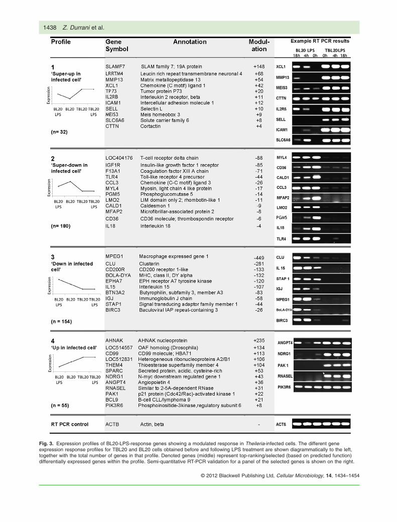
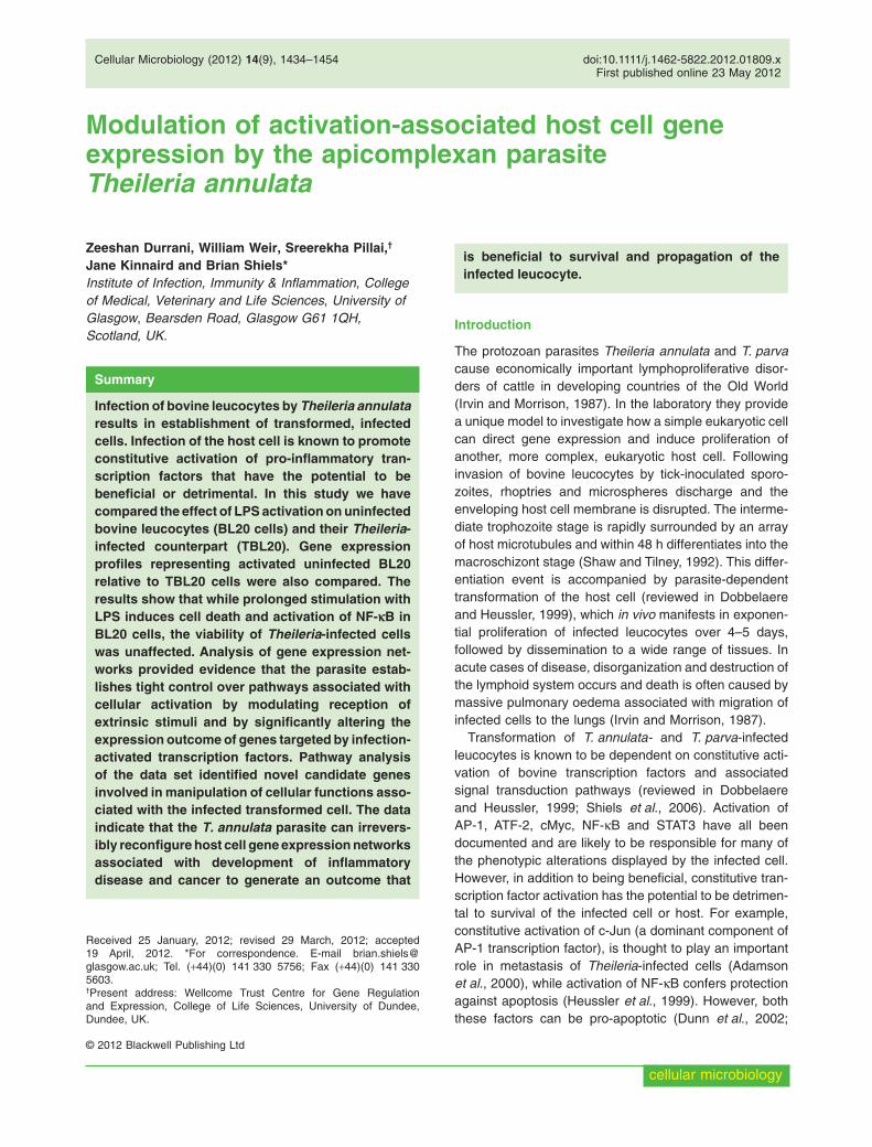
Fig. 3. Expression profiles of BL20-LPS-response genes showing a modulated response in Theileria-infected cells. The different geneexpression response profiles for TBL20 and BL20 cells obtained before and following LPS treatment are shown diagrammatically to the left,together with the total number of genes in that profile. Denoted genes (middle) represent top-ranking/selected (based on predicted function)differentially expressed genes within the profile. Semi-quantitative RT-PCR validation for a panel of the selected genes is shown on the right.
1438 Z. Durrani et al.
© 2012 Blackwell Publishing Ltd, Cellular Microbiology, 14, 1434–1454
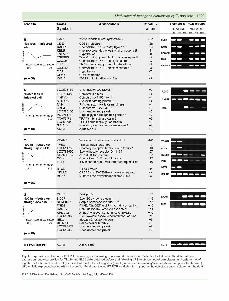
Fig. 4. Expression profiles of BL20-LPS-response genes showing a modulated response in Theileria-infected cells. The different geneexpression response profiles for TBL20 and BL20 cells obtained before and following LPS treatment are shown diagrammatically to the left,together with the total number of genes in that profile. Denoted genes (middle) represent top-ranking/selected (based on predicted function)differentially expressed genes within the profile. Semi-quantitative RT-PCR validation for a panel of the selected genes is shown on the right.
Modulation of host gene expression by T. annulata 1439
© 2012 Blackwell Publishing Ltd, Cellular Microbiology, 14, 1434–1454

activity, for example, was found to be significantly higherin TBL20 compared with BL20-LPS.
Profile 1 comprised 32 genes and for many of thesegenes the amplitude of enhanced, expression associatedwith infected cell was considerable (Fig. 3 and Table S1).Thus, the genes encoding chemokine (C motif) ligand 1,MMP13 and an IL2RB-like protein were elevated 42-, 54-and 11-fold respectively in TBL20 relative to the maximalresponse obtained for LPS stimulated BL20 cells. Of the14 BW720c-sensitive genes identified (Table S1), 10(71.4%) responded in an expected manner, i.e. expres-sion was significantly reduced by treatment over 48 h.Semi-quantitative RT-PCR was performed for eightgenes, each of which validated the microarray profile(Fig. 3); the gene encoding the MEIS3 transcription factor,for example, showed a clear elevation in PCR product inLPS-stimulated uninfected BL20 cells but a considerablyhigher level in infected TBL20, with no obvious detectablealteration following LPS stimulation of TBL20. Further vali-dation of the profile was obtained by qRT-PCR for cortac-tin and MEIS3 (see Fig. S2, panels 1A and 1B) where theresults agreed with the microarray data. qRT-PCR forMEIS3 showed a 5.98-fold difference (log2) in TBL20 rela-tive to BL20-LPS, compared with a difference of 3.1-fold(log2) using the array data, while qRT-PCR for cortactincalculated the difference to be 1.31-fold (log2) comparedwith 1.18-fold (log2) from the array data. The two func-tional categories that showed the most significant enrich-ment for profile 1 genes (Fig. S3) were ‘control of cellularmovement’ and ‘cell-to-cell signalling’, and there was alsoan enrichment of genes associated with inflammation inthis profile. In addition, a number of genes in profile 1have predicted functions that can be linked to the pheno-type of Theileria-transformed leucocytes. Thus, elevatedexpression was detected for a matrix metallopeptidasegene (Baylis et al., 1995), the IL2 receptor B-like gene(Herrmann et al., 1989), genes encoding cellular adhe-sion molecules, ICAM1 (Jensen et al., 2008) and selectinL (SELL), and genes that regulate the cytoskeleton, cort-actin (CTTN) (Baumgartner, 2011). It can be postulatedthat genes captured in profile 1 are expressed at anelevated level in TBL20 because they are advantageousfor establishment and or survival of the infected cell.
Profile 2 is the reciprocal of profile 1 and represents 180genes whose expression is repressed in BL20-LPS butsuper-repressed in TBL20. Twenty-four of these genesshowed major repression of expression associated withthe infected cell (> 10-fold). Of the 83 genes sensitive toBW720c, 99.8% responded in the expected manner, indi-cating repression requires a viable parasite. Nine genesof this profile were validated by RT-PCR and agreed withthe array profile (Fig. 3). qRT-PCR validation was per-formed for TLR4 and MYL4 and showed good correlationwith the microarray results (Fig. S2, panels 5A and 5B). A
number of genes encoding transcription factors werepresent in the data set including LEF-1 (down 15-fold) andLMO2 (down 11-fold), both of which are strongly associ-ated with leukaemia (Curtis and McCormack, 2010; Guti-errez et al., 2010). Moreover, genes encoding membraneproteins/receptors were particularly prominent in the dataset. Among 13 receptor genes that showed strongerdownregulation in TBL20 compared with BL20-LPS werethose encoding the IGFR1 receptor (down 85-fold inTBL20 relative to BL20-LPS), the Toll-like receptor 4(TLR4: down 44-fold), the G-protein-coupled receptor(CCR6: down 23-fold), S1PR1 (down 6-fold) and thethrombospondin receptor (down 6-fold). Thus, data fromthis profile strongly suggests that genes encoding mol-ecules located to the surface of the infected cell arerepressed and could influence interaction of the infectedcell with a range of ligands, inflammatory mediators andcells of the bovine immune system. It can be predictedfrom the super-repression profile that high-level expres-sion of the identified genes is most likely to be detrimentalto the infected cell.
TLR4 is the major cellular receptor that transduces theLPS activation signal (Chow et al., 1999) and significantreduction in expression (61.4-fold between BL20 versusTBL20) could play a role in the unresponsiveness ofTheileria-infected cells to LPS identified in this and previ-ous studies. The gene encoding the LPS ligand CD14 wasalso expressed at a very low level in both uninfected BL20and TBL20 cells. It has been demonstrated, however, thata lack of CD14 expression in bovine cells does not pre-clude recognition of LPS stimulation, possibly via thepresence of soluble CD14 present in fetal calf serum(Sauter et al., 2007).
Super-repression of TLR4 was validated at the proteinlevel: immunoblotting with a specific anti-TLR4 antibodyclearly demonstrated lower levels of TLR4 protein inTBL20 extracts relative to uninfected BL20 extracts fromcontrol or LPS stimulated cells (see Fig. 5A). In addition,both RT-PCR and immunoblotting showed that killing theparasite with BW720c considerably elevated the level ofTLR4 mRNA and protein (Fig. 5B and C). Thus, repres-sion of TLR4 by the infected cell is dependent on a viableparasite. The TLR4 signal transduction pathway plays amajor role in activation of NF-kB following recognition ofpathogen-associated molecular patterns (PAMPs). Sim-plistically, repression of this pathway may not be in theinterest of the Theileria-infected cell, since it has beendemonstrated that constitutive activation of NF-kB isrequired for its survival (Heussler et al., 1999). Parasite-dependent constitutive activation of NF-kB, however, isconsidered to be independent of extrinsic signalling, asthe parasite promotes aggregation of active IKK com-plexes at the macroschizont surface (Heussler et al.,2002). Whether the repression of TLR4 gene expression
1440 Z. Durrani et al.
© 2012 Blackwell Publishing Ltd, Cellular Microbiology, 14, 1434–1454

provides an advantage to the infected cell is not clear butit could reduce the level of activation of NF-kB that occursvia extrinsic inflammatory stimuli. This would allow thelevel of NF-kB activation to be primarily set by theparasite-dependent IKK signalosome pathway. A block inTLR signalling has been reported for Toxoplasma gondii.It has been proposed that this allows prevention of pro-inflammatory responses triggered by T. gondii-derivedTLR ligands, or gut flora, or reflects a general non-responsiveness to pro-inflammatory stimulation (reviewedby Leng et al., 2009). In addition, mammalian p38 MAPKswhich are activated by LPS and play a major role inapoptosis (Guha and Mackman, 2001; Zhou et al., 2007)are known to have low or undetectable activity inTheileria-infected cells (Botteron and Dobbelaere, 1998).
While modulation of expression associated with profiles1 and 2 may relate to the level of transcription factoractivation, this is unlikely to be the case for profiles 3 and4. In these profiles TBL20 was associated with a geneexpression change opposite to that observed for BL20-LPS, and it can be postulated that it is of benefit to theinfected cell to strongly repress (profile 3) or elevate(profile 4) expression of these genes. Profile 3 represents154 genes elevated by LPS stimulation of uninfected cellsthat are significantly repressed in infected TBL20 (Fig. 3).Almost all of the 63 genes that were sensitive to BW720ctreatment in this profile responded in a reversible manner(i.e. 98.4% showed elevated expression with BW720ctreatment), indicating active repression by a viable para-site. Many genes (n = 84) provided evidence of a repres-sion corresponding to a fold change of greater thanten. Seven genes from profile 3 were validated bysemi-quantitative RT-PCR (Fig. 3). The gene encodingMPEG1, for example, showed an elevation in PCRproduct in LPS-stimulated uninfected BL20 cells while thelevel was clearly lower in infected TBL20 relative to BL20cells. qRT-PCR performed for the BOLA-DYA and BIRC3genes (Fig. S2, panels 2A and 2B) validated the microar-ray profile. Profile 3 showed enrichment for genes pre-dicted to encode proteins involved in cell-to-cell signalling,cell morphology, cellular development and cellular growthand proliferation. A significant number of genes wereassociated with cancer and apoptosis. Genes in profile 3included those encoding macrophage expressed MPEG1,the inflammatory cytokine IL15, and the major histocom-patibility complex class II DY alpha. Repression of thesegenes may be advantageous to the parasite-infected cellby altering the differentiation status of the host cell ormanipulating the immune response.
Profile 4 is the reciprocal of profile 3 and represents 55genes that were repressed by LPS treatment of unin-fected BL20 cells but were elevated in infected TBL20cells. The level of modulation associated with this profilewas considerable. For example, the expression of genesencoding AHNAK, CMRF-35 and SLA were amplified 235-fold, 99-fold and 45-fold respectively. A further 33 geneswere elevated more than 10-fold. Interestingly, a largeproportion (61.5%) of the 13 BW720c-sensitive genesresponded in an unexpected manner, i.e. they wereupregulated by BW720c. Microarray expression data forprofile 4 was validated by RT-PCR using five genes. qRT-PCR validation was performed for ANGPT4 and NDRG1and showed good correlation with the microarray (seeFig. S2, panels 6A and 6B). Of the 51 genes that couldbe mapped onto the pathway analysis framework, 21showed an association with cancer. The profile 4 data setshowed enrichment for genes encoding proteins thatfunction in ‘cell movement’, ‘cell death’ and ‘cell growthand proliferation’. Gene ontology indicated that a number
Fig. 5. A. Immunoblot analysis of TLR4 protein expressionfollowing stimulation of BL20 and TBL20 cells with LPS. Expressionof TLR4 protein was detected by immunoblot analysis of totalprotein extracts derived from uninfected and Theileria-infected cellcultures stimulated with either LPS for 4 and 18 h or no drugDMSO (1 ml ml-1) vehicle control (ND). Detection of b-tubulin wasused as a constitutive control to ensure equal loading of extract.B. RT-PCR analysis of TLR4 expression following treatment ofTBL20 cells with BW720c. Expression of TLR4 at the mRNA levelwas assessed by semi-quantitative RT-PCR using RNA isolatedfrom BL20 and TBL20 cells cultured with no drug (ND) or TBL20cells cultured with BW720c at 50 ng ml-1 for 48 h. Primers specificfor the b-actin gene (ACT b) were used as a control for constitutiveexpression.C. Immunoblot analysis of TLR4 protein expression followingtreatment of TBL20 cells with BW720c. Expression of TLR4 proteinwas assessed by immunoblot analysis of total protein extracts ofBL20 and TBL20 cells cultured with no drug (ND) or TBL20 cellscultured with BW720c at 50 ng ml-1 for 48 h. Detection of b-tubulinwas used as a constitutive control to ensure equal loading ofextract.
Modulation of host gene expression by T. annulata 1441
© 2012 Blackwell Publishing Ltd, Cellular Microbiology, 14, 1434–1454

of genes (ANGPT4, NDRG1, PAK1, PI3KR6, SLA,SPARC) operate in signal transduction events. Overall,this profile suggests that the infected cell is strongly asso-ciated with upregulation of genes linked to neoplasia.
Profiles 5 and 6 both provide evidence that infectedTBL20 cells attenuate the response of uninfected BL20cells to LPS. Thus, profile 5 represents 38 genes withexpression levels elevated in Theileria-infected cells, butto a lesser extent than in LPS stimulated BL20 cells. Anexample of this profile is the gene for interferon-inducedprotein 15, ISG15, elevated 121-fold in BL20-LPS but only26-fold by infection TBL20 (relative to BL20) and nochange in TBL20-LPS. These kinetics mirror reducedexpression associated with Theileria-infected cells previ-ously observed by Northern blotting (Oura et al., 2006).Five genes of the profile were validated by semi-quantitative RT-PCR (Fig. 4). qRT-PCR performed for theCD69 and ISG15 genes confirmed the microarray data(Fig. S2, panels 3A and 3B). Functional analysis showedenrichment for genes encoding proteins involved inprotein modification, the antimicrobial response, theinflammatory response, cellular defence and the innateimmune response (e.g. ISG15). Genes encoding tran-scription factors activated by Theileria infection such asNF-kB2, NF-kBIZ and Jun were also identified. Alteredexpression of genes in profile 5 could indicate a limitedupregulation of a beneficial gene that does not occur tothe same degree as in BL20-LPS (e.g. the Jun proto-oncogene). However, it may be more likely that high-levelexpression of cellular defence genes, such as ISG15(Oura et al., 2006) that occur via activation of the cell, arerepressed by infection as they are potentially detrimental.The enrichment in genes associated with the interferon/innate immune/cellular defence response, e.g. OAS2,ISG15, MX2, IRF5 and IL7 (Moller et al., 1996; Jin et al.,1999; Urosevic, 2003; Pitha-Rowe and Pitha, 2007; Binet al., 2011), supports this possibility. Furthermore, 72% ofthe 18 BW720c-sensitive genes in profile 5 showedelevated expression upon drug treatment, indicatingactive repression by the viable parasite.
Profile 6 was the reciprocal of profile 5 and repre-sented genes where expression was repressed with LPStreatment of BL20 but repressed to a lesser extent inparasite infected TBL20 cells. This profile representedthe smallest number of genes (n = 13), and four out ofthe five BW720c-sensitive genes were elevated upontreatment. The array profile was validated for threegenes by RT-PCR and by qRT-PCR for CYP4F3 andRYK (Fig. S2, panels 7A and 7B). Pathway analysis ofthe data set for this profile showed enrichment for genespredicted to function in metabolism or small moleculebiochemistry.
Profiles 7 and 8 represent no change in infected TBL20cells compared with an altered response identified for
BL20-LPS (up or down). Profile 7 represents 650 geneselevated by LPS stimulation of BL20 that showed nochange on comparison of BL20 with TBL20 and was thelargest obtained by the study. Only a small number ofthese genes (n = 6) were sensitive to BW720c, supportinga mechanism of non-responsiveness, rather than repres-sion of an activation event associated with the infectedcell. In some cases, however, postulation of a lack ofresponse mechanism may be over-simplistic. VCAM1, forexample, is a classical NF-kB-dependent gene (Luo et al.,2010) and it is conceivable that infection of the host cell isassociated with a more direct repression of VCAM1expression. The microarray profile was successfully vali-dated by RT-PCR on six candidate genes (see Fig. 4) withthe exception of Runx2 where an increase in Runx2 PCRproduct was detected at the TBL20 LPS 18 h time point.qRT-PCR validation indicated a 13.9-fold (log2) lowerexpression level for VCAM1 in TBL20 versus BL20-LPS(Fig. S2, panels 4A), compared with a difference of 7.4-fold (log2) generated by the array data; while for Runx2,qRT-PCR showed a 5.1-fold (log2) reduction in expressionvalues between TBL20 and BL20-LPS (Fig. S2, panels4B), compared with a difference of 2.5-fold (log2) from thearray data. Pathway analysis of this profile identifiedenrichment for genes within the categories ‘inflammatoryresponse’ and ‘inflammatory disease’, and it was of inter-est that ‘activation of IRF by cytosolic pattern recognitionreceptors’ and ‘interferon signalling’ were identified as themost significantly enriched canonical pathways.
Profile 8 is the reciprocal of profile 7 and represents 98genes where expression was downregulated by LPStreatment of BL20 but showed no change associated withinfection of BL20 (BL20 versus TBL20). Like profile 7,genes sensitive to BW720c treatment were under-represented in this profile with only a single one present.The microarray data were validated by RT-PCR for threegenes and by qRT-PCR for BCOR and RGS1 (Fig. 4 andFig. S2, panels 8A and 8B). Pathway analysis showed thisprofile is enriched for genes predicted to function in cellsignalling, small molecule biochemistry and metabolism.This suggests that the infected cell maintains metabolicprocesses required for cellular proliferation (as also indi-cated for profile 6).
Together, the eight profiles identified above provide evi-dence that the parasite-infected cell is associated withsignificant modulation of the response of uninfected BL20cells to activation with LPS. A sizeable proportion of thesealterations to host cell gene expression are under controlof a viable parasite, as they are altered by treatment withBW720c. However, the majority of the identified infectedcell modulations to gene expression did not respond todrug treatment. While this may be influenced by theshorter time periods used for drug treatment relative toprevious studies (Oura et al., 2006; von Schubert et al.,
1442 Z. Durrani et al.
© 2012 Blackwell Publishing Ltd, Cellular Microbiology, 14, 1434–1454
2010), it seems likely that a large number of the altera-tions to host cell gene expression are genuinely non-responsive. Such alterations could be derived fromepigenetic events that occurred during establishment orpassage of the BL20 and TBL20 cell lines. These couldbe disparate events that are independent of infection,although it is also possible that epigenetic events areinfluenced by the parasite to generate infected cell-associated alterations that cannot be altered by drugtreatment over 48 h.
Pathway analysis of the infected cell-modulateddata set can predict phenotypes displayed byT. annulata-infected leucocytes
Derivation of the eight profiles of genes displaying alteredexpression levels in infected TBL20 cells relative to LPS-stimulated uninfected BL20, provides evidence thatT. annulata-infected cells alter the gene expressionoutcome associated with an activation event, i.e. theresponse to LPS. To gain information on the potentialimpact of these alterations, the entire list of 1214 genes
was analysed for enrichment in specific molecular func-tions and canonical pathways. A large number of catego-ries were identified for the 976 genes mapped onto thepathway analysis framework but it was clear that the dataset was very significantly enriched for genes predicted tofunction in the inflammatory response, cell death, cancerand the antimicrobial response (Fig. 6).
To make the output of the pathway analysis more pre-dictive, further enrichment analysis was undertaken,noting whether each molecule promoted (pro) or inhibited(anti) a particular molecular function. The proportion ofgenes designated as ‘pro’ or ‘anti’ for each functionalcategory was then examined in relation to whether theirexpression is repressed or enhanced by infected TBL20, inorder to infer how the infection-associated modulationcould influence a functional outcome. Interestingly, func-tional categories highlighted by this analysis (Table 1) pre-dominantly show an association with a phenotype that isknown or could be reasonably predicted for the infectedcell. For example, in the ‘metastasis of cells’ subcategory,the observed trend indicated repression of anti-metastaticgenes and elevated expression of pro-metastatic genes,
Fig. 6. Enrichment analysis of the infected cell-modulated gene list. The infected cell-modulated gene list (n = 1214) was analysed forevidence of enrichment with respect to two areas within the pathway analysis database, i.e. molecular function and canonical pathways.The gene list was also analysed for enrichment with respect to a number of denoted custom pathways. Top-ranking categories are presentedtogether with the significance of the enrichment [-log(P-value)].
Modulation of host gene expression by T. annulata 1443
© 2012 Blackwell Publishing Ltd, Cellular Microbiology, 14, 1434–1454

implying that the parasite-infected leucocyte promotesmetastasis. Genes classified within ‘cancer’ and ‘tumori-genesis’ were associated with a bias towards pro functionin genes showing enhanced expression associated withthe infected cell, whereas a bias towards anti-function wasassociated with repression by infected TBL20 within thecategories ‘disease of tumour’ and ‘growth of tumour’.Additional categories include the inflammatory responseand cell movement. In the inflammatory response cat-egory, the data indicated that genes promoting an‘anti-microbial/anti-viral’ response or ‘inflammation’ areassociated with repression by parasite-infected cells, whilefor ‘activation of leucocytes’ and ‘activation of lymphocytes’a trend towards elevated expression of positive effectorgenes was obtained. Similar results were observed for cellmovement, one subcategory ‘invasion of eukaryotic cells’indicating elevated expression of genes pro this function,whereas the next three subcategories all indicated a trendtowards repression of genes pro movement, includingthose categorized in ‘influx of leucocytes’ and ‘chemoat-traction of leucocytes’. One possible explanation for theseresults is that they reflect a requirement for activation anddissemination of the infected leucocyte together withdysregulation/evasion of the immune response.
The ability of the pathway analysis to predict pheno-types displayed by the Theileria-infected cell suggeststhat the infected cell-modulated gene list could be minedto highlight candidate novel pathways and genes thatoperate in its establishment and maintenance. Examplesof several candidate genes and categories of interest aregiven below.
Sixty genes (24%) were placed in the cancer category(P-value, 1.56 ¥ 10-5), including pro-cancer genesMMP13, SPARC, NDRG1, PAK1, ICAM1, ANGPT4,IL2RB-like, RGS1, SELL, SERPINE2, CCR7, CTTN,LTB4R, SLC2A1 and TP73 which are expressed at sig-nificantly higher levels in parasite-infected TBL20 relativeto BL20-LPS. Intercellular adhesion molecule-1 (ICAM1)is elevated sixfold in TBL20 cells relative to BL20-LPS(profile 1), and elevated 23-fold relative to uninfectedBL20. ICAM1 is a trans-membrane glycoprotein and isencoded by a classical NF-kB target gene (Huang et al.,2004; Lin et al., 2006). It plays an important role in theimmune response, but has also been implicated in cancermetastasis and linked to tumour prognosis and progres-sion (Huang et al., 2004; Lin et al., 2006). Interestingly,the level of ICAM1 mRNA was found to be significantlyhigher in infected monocytes derived from disease-
Table 1. Functional classification of the infected cell-modulated gene set.
Functional classification
Repressed in parasite-infected cell Enhanced in parasite-infected cell
No. genes (pro/anti) P-value No. genes (pro/anti) P-value
1 Inflammatory responseActivation of leucocytes 46 (27/9) 2.7 ¥ 10-6 13 (9/1) 3.8 ¥ 10-3
Antimicrobial response 19 (3/1) 4.9 ¥ 10-5 – –Activation of lymphocytes 31 (19/7) 9.5 ¥ 10-5 9 (5/1) 1.2 ¥ 10-2
Antiviral response 13 (3/1) 9.6 ¥ 10-5 2 (0/0) 1.2 ¥ 10-2
Inflammation 30 (12/3) 7.2 ¥ 10-4 12 (3/2) 6.5 ¥ 10-4
2 Cell deathApoptosis of tumour cell line 72 (34/22) 2.3 ¥ 10-6 20 (12/7) 3.1 ¥ 10-3
Apoptosis 134 (70/53) 2.1 ¥ 10-5 42 (24/19) 1.1 ¥ 10-4
Cell death of tumour cell line 78 (38/28) 2.1 ¥ 10-5 24 (13/9) 9.4 ¥ 10-4
Cell death 151 (75/62) 6.6 ¥ 10-5 49 (26/22) 3.2 ¥ 10-5
3 Cellular movementInfiltration of cells 29 (10/6) 4.2 ¥ 10-5 – –Invasion of eukaryotic cells – – 14 (10/3) 5.9 ¥ 10-4
Migration of antigen presenting cells 14 (7/1) 1.6 ¥ 10-4 – –Influx of leucocytes 7 (4/1) 2.0 ¥ 10-3 – –Chemoattraction of leucocytes 6 (6/0) 4.2 ¥ 10-3 – –
4 CancerMetastasis of cells 10 (2/6) 3.2 ¥ 10-3 13 (3/1) 2.3 ¥ 10-3
Tumorigenesis 179 (26/25) 1.7 ¥ 10-3 60 (6/2) 1.7 ¥ 10-5
Cancer 173 (16/13) 7.6 ¥ 10-5 56 (3/1) 1.6 ¥ 10-5
Disease of tumour 18 (2/4) 7.3 ¥ 10-4 – –Growth of tumour 20 (2/9) 1.2 ¥ 10-3 – –Tumour – – 43 (4/2) 1.1 ¥ 10-4
5 Cell assembly and organizationFormation of actin filaments – – 8 (3/1) 1.3 ¥ 10-3
Formation of actin cytoskeleton 4 (0/0) 1.3 ¥ 10-3 4 (2/0) 1.1 ¥ 10-2
Formation of filaments – – 9 (5/1) 4.1 ¥ 10-3
Formation of cellular protrusions – – 10 (4/0) 2.7 ¥ 10-3
1444 Z. Durrani et al.
© 2012 Blackwell Publishing Ltd, Cellular Microbiology, 14, 1434–1454

susceptible Holstein cattle relative to resistant Sahiwal(Jensen et al., 2008). Such a difference could be gener-ated by variability in the level of activated NF-kB ininfected cells from different breeds, or a difference in aparasite-dependent mechanism that controls NF-kBtarget gene expression. Further investigations arerequired to characterize the role of ICAM1 expression indetermining the virulence of the T. annulata-infected cell.
Secreted protein acidic and rich in cysteine (SPARC) isa matricellular protein that influences cell–matrix interac-tions and has been reported to have multiple roles inmacrophage function, inflammatory processes, cell mor-phology, inhibition of cell cycle progression and synthesisof the extracellular matrix (Rempel et al., 2001). Manycancer types exhibit increased SPARC levels upon inva-sion or metastasis (Rempel et al., 2001; Podhajcer et al.,2008) and upregulation of SPARC expression (53-fold) ininfected in TBL20 cells (profile 4) may play an importantrole in establishing the infected cell phenotype. Forexample, recent studies have described a connectionbetween SPARC and the p53 tumour suppressor(Fenouille et al., 2011). Essentially, SPARC acts as ananti-stress factor by mediating degradation of p53 throughAKT-mediated MDM2 phosphorylation to promotemelanoma cell survival. Array data indicate that the levelof p53 mRNA is high in Theileria-infected leucocytes, butprotein levels are virtually undetectable (B. Shiels and S.McKellar, unpublished). The PI3K/AKT pathway is knownto be constitutively activated in the Theileria-infected leu-cocyte (Baumgartner et al., 2000; Heussler et al., 2001).Investigation of a mechanism that may operate topromote constitutive degradation of p53 is required.
The category ‘cellular assembly and organization’ waspredicted to show an association with the infected cellphenotype, as a bias towards enhanced expression ofpro genes within a number of subcategories was identi-fied. Subcategories included ‘formation of actin filaments’and ‘formation of actin cytoskeleton’ and predict thatinfection of the host cell is associated with manipulationof cell shape and the actin cytoskeleton. Upregulatedgenes included CTTN, SDC4, ICAM1, PAK1, ELN,SPARC, SORBS1, TNS1 and F2R. Theileria infectionis known to alter the morphology of the host cell(Schmuckli-Maurer et al., 2010; Baumgartner, 2011) andit has been postulated that organization of the actincytoskeleton has an impact on parasite-dependent acti-vation of NF-kB via the IKK signalosome (Schmuckli-Maurer et al., 2010). Recent studies have shown thatparasite-dependent polarization of actin-based structuressuch as lamellipodia and podosomes are critical for bothattachment to substrates and motility (Baumgartner,2011). Molecules thought to function in regulation ofthese structures include the Src kinase, Hck, the Rhokinase, ROCK and cortactin. Elevated expression of the
cortactin (CTTN) and Hck (haemopoietic cell kinase)genes in infected TBL20 cells support a role in regulationof infected cell shape.
The gene, PAK1, encoding the p21 (Cdc42/Rac)-activated kinase 1 (member of a family of serine/threonineprotein kinases) is significantly upregulated (> 22-fold) inTheileria-infected TBL20 compared with BL20-LPS(profile 4). PAK1 has been reported to have a number offunctions (e.g. cell survival, motility, proliferation) and isimplicated in transformation and tumour progression(Huynh et al., 2010). PAK1 is a key molecule in the regu-lation of the actin and microtubule cytoskeleton (reviewedby Bokoch, 2003), plays a crucial role in formation ofpodosomes and membrane ruffles, can phosphorylatecortactin to regulate the dynamics of branched actin fila-ments (Webb et al., 2006) and enhances cell migrationand proliferation via AKT (Huynh et al., 2010). Intriguingly,PAK1 has been reported to operate in pathogen-dependent activation of NF-kB via NIK (Neumann et al.,2006) and inhibition of NIK reduces activation of NF-kB inTheileria-infected cells by 25% (Heussler et al., 2002).Further studies investigating the role PAK1 may play inestablishing the transformed phenotype of the Theileria-infected cell are indicated.
In contrast to the categories and genes highlightedabove, an expected bias towards upregulation of anti-apoptotic genes together with downregulation of pro-apoptotic genes could not be identified, although thepresent analysis framework may not have had the powerto resolve such a phenomenon. This may be due tounequal numbers of ‘pro’ and ‘anti’ apoptosis genesdenoted in the database together with the fact thatgenes functioning in apoptosis commonly switch from a‘pro’ to ‘anti’ function depending on cellular context (Fan-njiang et al., 2003; Hess et al., 2004) and such subtle-ties will not be reflected in the database. Moreover, theprobability of progressing towards apoptosis may beassociated with qualitative differences, such as phos-phorylation status, or the stoichiometry of a number ofkey factors and this would not have been revealed bythe analysis.
Theileria-infected TBL20 cells modulate expressionoutcomes associated with activation of NF-kB andother infection-activated transcription factors
Stimulation of cells with inflammatory mediators such asLPS, and infection by T. annulata, is known to result inactivation of transcription factors via a number of signal-ling pathways. Therefore, the infected cell-modulated dataset of 1214 genes was analysed with respect to canonicalpathways, including those involved in signalling. Unsur-prisingly, the results highlighted modulation of signallingevents associated with the inflammatory response. Sig-
Modulation of host gene expression by T. annulata 1445
© 2012 Blackwell Publishing Ltd, Cellular Microbiology, 14, 1434–1454

nificant pathways include TNFR2, pattern recognitionreceptors, CD40, NF-kB, PI3K, LPS-stimulated MAPKand interferon signalling (Fig. 6). An event commonamong many of these pathways is the activation of NF-kB.As predicted from the findings of Heussler et al. (2002),the microarray data generated little evidence for majorrepression of the NF-kB signalling pathway below thelevel of the activated signalosome (Fig. 7); repressedexpression of the genes encoding three IkBs (relative to
LPS) would promote NF-kB activation but there is noevidence of a difference in expression of these IkB genesbetween non-stimulated BL20 and TBL20 cells. Upstreamof the signalosome, repression of genes encoding recep-tors appears to be in operation and it can be concludedthat, in general, infected TBL20 downregulate the ability torecognize extrinsic stimuli that induce activation of NF-kB.Exceptions to this observation are upregulation of thegenes encoding the TNFR2 and TGFBR3 receptors. Both
Fig. 7. NF-kB signalling pathway. The genes which are coloured are differentially expressed in TBL20 compared with BL20; red = up in TBL,green = down in TBL. The arrows are beside genes present in the infected cell-modulated list (1214). For example, means super-up inBL20-LPS | up in TBL20 compared with BL20. The data for IL-1R/TLR and growth factor receptor are exploded in two boxes.
1446 Z. Durrani et al.
© 2012 Blackwell Publishing Ltd, Cellular Microbiology, 14, 1434–1454
TNF and TGFb signalling pathways have been associatedwith the Theileria-infected leucocyte (Guergnon et al.,2003; Chaussepied et al., 2010) and could potentiallyactivate NF-kB via TRAF2 or PI3K. However, evidence fora role of the PIK3 pathway in activating NF-kB is conflict-ing (Baumgartner et al., 2000; Heussler et al., 2001), andthe importance of a TNF autocrine loop is unclear (Guer-gnon et al., 2003). Furthermore, microarray data indicatethat the gene encoding tumour necrosis factor alphainduced protein 3 (TNFAIP3/A20) that acts as an inhibitorof TNFR2-dependent NF-kB signalling (Charan et al.,2011) is upregulated in TBL20 relative to BL20. Theimportance of the PI3K pathway for proliferation of theinfected leucocyte (Baumgartner et al., 2000; Heussleret al., 2001) is supported in the present study by evidencefor upregulation of the genes encoding PI3K regulatorysubunits 5 and 6 relative to repression by the LPSresponse.
Using a series of custom pathways, the infection-modulated data set was also screened for enrichment oftarget genes of transcription factors activated by theparasite-infected cell and associated with the inflamma-tory response. As shown in Fig. 6, significant enrichmentof NF-kB targets and cMyc repression targets wasobserved, together with a particular enrichment of genesregulated in response to stimulation by TGFb. This pro-vides evidence that the Theileria-infected leucocyte sig-nificantly alters the outcome of cellular activation bymodulating the expression of target genes regulated byconstitutively activated transcription factors.
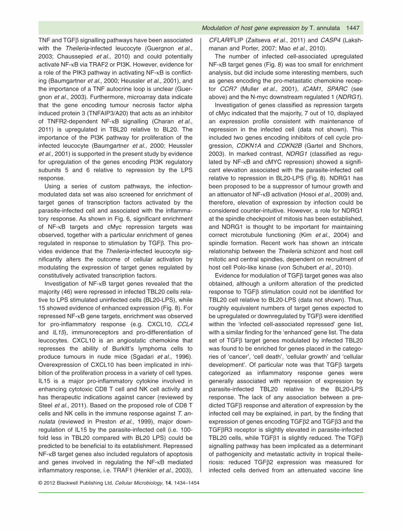
Investigation of NF-kB target genes revealed that themajority (46) were repressed in infected TBL20 cells rela-tive to LPS stimulated uninfected cells (BL20-LPS), while15 showed evidence of enhanced expression (Fig. 8). Forrepressed NF-kB gene targets, enrichment was observedfor pro-inflammatory response (e.g. CXCL10, CCL4and IL15), immunoreceptors and pro-differentiation ofleucocytes. CXCL10 is an angiostatic chemokine thatrepresses the ability of Burkitt’s lymphoma cells toproduce tumours in nude mice (Sgadari et al., 1996).Overexpression of CXCL10 has been implicated in inhi-bition of the proliferation process in a variety of cell types.IL15 is a major pro-inflammatory cytokine involved inenhancing cytotoxic CD8 T cell and NK cell activity andhas therapeutic indications against cancer (reviewed bySteel et al., 2011). Based on the proposed role of CD8 Tcells and NK cells in the immune response against T. an-nulata (reviewed in Preston et al., 1999), major down-regulation of IL15 by the parasite-infected cell (i.e. 100-fold less in TBL20 compared with BL20 LPS) could bepredicted to be beneficial to its establishment. RepressedNF-kB target genes also included regulators of apoptosisand genes involved in regulating the NF-kB mediatedinflammatory response, i.e. TRAF1 (Henkler et al., 2003),
CFLAR/FLIP (Zaitseva et al., 2011) and CASP4 (Laksh-manan and Porter, 2007; Mao et al., 2010).
The number of infected cell-associated upregulatedNF-kB target genes (Fig. 8) was too small for enrichmentanalysis, but did include some interesting members, suchas genes encoding the pro-metastatic chemokine recep-tor CCR7 (Muller et al., 2001), ICAM1, SPARC (seeabove) and the N-myc downstream regulated 1 (NDRG1).
Investigation of genes classified as repression targetsof cMyc indicated that the majority, 7 out of 10, displayedan expression profile consistent with maintenance ofrepression in the infected cell (data not shown). Thisincluded two genes encoding inhibitors of cell cycle pro-gression, CDKN1A and CDKN2B (Gartel and Shchors,2003). In marked contrast, NDRG1 (classified as regu-lated by NF-kB and cMYC repression) showed a signifi-cant elevation associated with the parasite-infected cellrelative to repression in BL20-LPS (Fig. 8). NDRG1 hasbeen proposed to be a suppressor of tumour growth andan attenuator of NF-kB activation (Hosoi et al., 2009) and,therefore, elevation of expression by infection could beconsidered counter-intuitive. However, a role for NDRG1at the spindle checkpoint of mitosis has been established,and NDRG1 is thought to be important for maintainingcorrect microtubule functioning (Kim et al., 2004) andspindle formation. Recent work has shown an intricaterelationship between the Theileria schizont and host cellmitotic and central spindles, dependent on recruitment ofhost cell Polo-like kinase (von Schubert et al., 2010).
Evidence for modulation of TGFb target genes was alsoobtained, although a uniform alteration of the predictedresponse to TGFb stimulation could not be identified forTBL20 cell relative to BL20-LPS (data not shown). Thus,roughly equivalent numbers of target genes expected tobe upregulated or downregulated by TGFb were identifiedwithin the ‘infected cell-associated repressed’ gene list,with a similar finding for the ‘enhanced’ gene list. The dataset of TGFb target genes modulated by infected TBL20was found to be enriched for genes placed in the catego-ries of ‘cancer’, ‘cell death’, ‘cellular growth’ and ‘cellulardevelopment’. Of particular note was that TGFb targetscategorized as inflammatory response genes weregenerally associated with repression of expression byparasite-infected TBL20 relative to the BL20-LPSresponse. The lack of any association between a pre-dicted TGFb response and alteration of expression by theinfected cell may be explained, in part, by the finding thatexpression of genes encoding TGFb2 and TGFb3 and theTGFbR3 receptor is slightly elevated in parasite-infectedTBL20 cells, while TGFb1 is slightly reduced. The TGFbsignalling pathway has been implicated as a determinantof pathogenicity and metastatic activity in tropical theile-riosis: reduced TGFb2 expression was measured forinfected cells derived from an attenuated vaccine line
Modulation of host gene expression by T. annulata 1447
© 2012 Blackwell Publishing Ltd, Cellular Microbiology, 14, 1434–1454

Fig. 8. Hierarchical clustering of NF-kB response genes. The NF-kB targets genes identified in the infected cell-modulated gene list wereselected for hierarchical clustering analysis and the results are presented as a heat map. Genes highlighted in bold show altered arrayexpression following BW720c treatment for 48 h to kill the parasite. Underlined genes show altered RT-PCR expression following BW720ctreatment for 72 h.
1448 Z. Durrani et al.
© 2012 Blackwell Publishing Ltd, Cellular Microbiology, 14, 1434–1454
and disease-resistant Sahiwal (Bos indicus) cattle(Chaussepied et al., 2010). Differential expression ofTGFb target genes was also identified, with elevated aswell as repressed expression reported for the attenuatedvaccine cell line. Thus, modulation of TGFb signalling andthe respective target genes appears to be a commonevent associated with T. annulata-infected leucocytes.
The results of this study provide evidence that theT. annulata-infected cell tightly controls the outcome ofNF-kB activation. This modulation, in general, can bepredicted to be beneficial to establishment of the infectedcell. The data also indicates that the outcome of activatingadditional pathways/transcription factors known to beassociated with Theileria-transformed leucocytes aremodulated, e.g. TNF, TGFb and cMyc. Thus, a majorre-organization of networks that regulate gene expressionchanges associated with the inflammatory response andcancer is likely to be concomitant with transformation ofthe infected leucocyte.
Recent studies on the related apicomplexan parasite,T. gondii have identified parasite proteins that regulateinflammatory gene networks by activating transcriptionfactors, including NF-kB (Saeij et al., 2007; Rosowskiet al., 2011). Evidence has also been generated thatparasite-dependent modification of histone H3 canprevent transcription factors binding to host-cell inflamma-tory gene promoters (Leng and Denkers, 2009). It hasbeen proposed that such a mechanism could account forthe large-scale downregulation of pro-inflammatory genesby T. gondii (Leng et al., 2009). While it is possible thatTheileria parasites deploy similar mechanisms to manipu-late gene expression on a wide scale, our results suggestadditional alterations are involved. This is indicated by thefour pairs of reciprocal modulated expression profiles(Figs 3 and 4), the ability of infected cells to reverse geneexpression changes induced by LPS and the non-responsive and non-reversible nature of a large number ofthe host cell gene expression changes when the parasiteis killed by drug. Such changes could be generated byan irreversible reconfiguration of transcription factor/expression networks associated with differentiationevents, as increasingly proposed for stem cell systems(Chickarmane et al., 2009; Wang et al., 2010). In addition,deployment by T. annulata of DNA-binding proteins to thehost cell nucleus that show similarity to mammalianHMGA proteins could be involved (Swan et al., 1999).HMGAs act as hubs of nuclear function and play a majorrole in organization of gene expression networks associ-ated with inflammation and cancer (reviewed in Cleynenand Van de Ven, 2008). Given the propensity of Theileriaparasites to control gene expression networks associatedwith inflammatory disease and cancer, further study of themechanisms deployed and the outcome they promote iswarranted.
Experimental procedures
Cell culture and LPS stimulation
Low-passage BL20, an uninfected bovine lymphosarcoma cellline (Olobo and Black, 1989), and TBL20, a T. annulata (Ankarastrain) infected BL20 cell line (Shiels et al., 1986), were culturedin standard complete medium (Swan et al., 1999) except thatheat-inactivated fetal bovine serum (FBS; Sigma) was used.Cultures were seeded with 2 ¥ 105 cells ml-1 and maintained byfeeding with fresh medium every 2–3 days (Schmuckli-Maureret al., 2010). Stimulation of cells was carried out for up to 48 h insix-well plates with 5 ¥ 106 cells well-1 in a total of 5 ml ofmedium, set up immediately prior to addition of 1 mg ml-1 LPS(Sigma: L2630). Optimum LPS concentration was determined bypreliminary titration experiments which measured the minimumconcentration of drug required to stimulate cells, based on NF-kBreporter activity, without causing rapid cytotoxicity (data notshown). LPS was dissolved in dimethyl-sulfoxide (DMSO) andcontrol cells were incubated with DMSO alone. Treatment ofTBL20 with BW720c was performed as described previously(Schmuckli-Maurer et al., 2010).
Cell viability counts and apoptosis assay
Assessment of viable and non-viable cell numbers was per-formed using Trypan Blue exclusion counted using a haemocy-tometer; viable cells exclude the dye. Three replicate cultures ofBL20 and TBL20 were seeded with 2.5 ¥ 105 cells ml-1 and incu-bated at 37°C for 18 h in the presence of LPS (1 mg ml-1) orDMSO (1 ml ml-1) as no drug control. Control experiments com-paring cells culture in the presence or absence of DMSO showedno evidence of an effect on cell viability or activation of NF-kB(data not shown). Caspase-3/7 activities were measured using aCaspase-3/7 Assay kit (Promega) according to the manufactur-er’s protocol. Each assay was performed using 2.0 ¥ 104 cellsfrom three replicate cultures per experimental condition at 18 h.Luminescence was measured using a TD -20/20 luminometer(Turner Designs). A no-cell control was carried out to account forbaseline signal generated from fresh complete medium.
NF-kB-dependent reporter assay (dual luciferase assay)
Estimation of NF-kB activity was performed by transientco-transfection of a reporter plasmid containing a promoter withan NF-kB3 recognition sequence linked to Firefly luciferase (NF-kB-LUC) and a Renilla luciferase plasmid (phRG-TK renilla),followed by sequential measurement of Firefly and Renilla luci-ferase activity, as previously described (Schmuckli-Maurer et al.,2010). Two micrograms of NF-kB3-Luc and 0.5 mg of phRG-TKRenilla plasmids were mixed with 4 ¥ 106 BL20 or TBL20 cells in300 ml of RPMI-1640-2% FBS without antibiotics and electropo-ration performed using a Gene Pulser (Bio-Rad) set at 220V,950 mF. After electroporation, cells were immediately added to5 ml of pre-warmed complete medium in six-well plates. After24 h recovery, LPS (1 mg ml-1) or DMSO were added for 4 h.Samples were harvested and the cell pellet washed with PBS(phosphate buffer saline).
A Dual Luciferase® Reporter Assay System (Promega) wasused to assay activity, using the manufacturer’s protocol. NF-kBpromoter activity was calculated by normalizing firefly luciferase
Modulation of host gene expression by T. annulata 1449
© 2012 Blackwell Publishing Ltd, Cellular Microbiology, 14, 1434–1454
activity relative to the activity of constitutive Renilla luciferase.Each electroporation and luciferase assay was performed inquadruplicate, 20 ml of cell extract was used per assay.
Statistical analysis
Statistical analysis and generation of graphs were performedusing GraphPad Prism Software version 5.0 (La Jolla, CA). Datawere expressed as mean � standard error of the mean (SEM).Differences and significance between mean values were calcu-lated using a two-tailed Student’s t-test with P-values � 0.05considered to be statistically significant.
Indirect immunofluorescence analysis
Indirect immunofluorescence analysis (IFA) was performed onuninfected BL20 and infected TBL20 cells cultured with LPS or inDMSO (1 ml ml-1), as no drug control, for 4 h. Cytospin slideswere generated using poly L-lysine glass slides and IFA per-formed, as described previously (Schmuckli-Maurer et al., 2010).Primary antibodies, anti-IKKg/NEMO (BD Biosciences; 611306),anti-NF-kB p65 (Santa Cruz; sc-372) and anti-NF-kB p65 (SantaCruz; sc-8008), were used at 1:50 dilution; anti-rabbit IgG andanti-mouse IgG secondary antibodies, conjugated to Alexa 488(Invitrogen; A-11034; A-11029), were used at 1:200 dilution.Images were acquired using an Olympus BX60 microscope,SPOT camera and SPOTTM Advanced image software VersionMac: 4.6.1.26 (Diagnostic Instruments).
Western blotting
Total cell extracts were prepared from approximately 5 ¥ 106 cellsgrown to a density of 1 ¥ 106 cells ml-1 in a six-well plate, resus-pended in 100 ml of PBS and lysed with an equal volume ofSDS sample buffer (2¥ concentrate, Sigma; S3401). Extracts,adjusted for equal protein loading, were resolved by SDS-PAGE(Laemmli, 1970) with subsequent immunoblotting and detectionas described previously (Schmuckli-Maurer et al., 2010). Theanti-TLR4 antibody (Santa Cruz-Biotechnology, sc-10741) wasused at 1:1000 dilution and anti-tubulin antibody (Sigma, T9028)at 1:5000.
RNA isolation and processing
Total RNA for analysis by microarray was isolated in triplicatefrom 30 ml of cultures of infected and uninfected cells and cellstreated with LPS for 4 and 18 h. Cell pellets of 1.5–2.0 ¥ 107 cellswere resuspended immediately in TRI Reagent (Sigma; T9424),and extracted according to the manufacturer’s instructions. Toeliminate possible contamination with genomic DNA, RNAsamples used for RT-PCR and QRT-PCR were treated withDNase I (Qiagen; 79254) either before purification on a silica-gelmembrane-based RNeasy® column (Qiagen; 74104) or by anon-column DNase I treatment. A260/A280 ratios obtained for allsamples fell in the acceptable range of 1.78–2.1 and quality andintegrity of the total RNA preparations was confirmed by gelelectrophoresis.
Custom bovine microarray and analysis
The oligonucleotide microarray used in this study included allbovine RNA RefSeq sequences available at NCBI (26 751) cor-
responding to the Btau 4.0 assembly of the bovine genome(http://www.ncbi.nlm.nih.gov/RefSeq/) and a number of putativebovine RNA Sequences and ESTs (4730) kindly provided byKirsty Jensen, Roslin Institute, University of Edinburgh. Onlyextant sequences in the RefSeq mRNA database (December2011) or UMD3.1 genome assembly were used in the analysisand this represents 19 777 sequences. Design of oligonucle-otides, array synthesis, hybridization to the array and normaliza-tion of array data were carried out by Roche NimbleGen,Madison, USA. Each gene was represented by two identical setsof five 60-mer oligonucleotide probes which were isothermal withrespect to melting temperature. cDNA was generated from 10 mgof total RNA using oligo(dT) primer and tagged with 3′-Cy3 dyeafter which 13 mg of labelled cDNA was hybridized to the array.Gene expression values were calculated from a RMA-normalizeddata set (Irizarry et al., 2003) and differentially expressed geneswere identified using Rank Product Analysis (Breitling et al.,2004). Genes were defined as differentially expressed using thecriteria of a false discovery rate of less than 5% and a fold changeof greater than two. In addition to the bovine sequences, thearray contained oligonucleotide probes representing the T. annu-lata genome, although these data were not analysed in thecourse of the present study. The data discussed in this publica-tion have been deposited in NCBI’s Gene Expression Omnibus(Edgar et al., 2002) and are accessible through GEO SeriesAccession No. GSE36428 (http://www.ncbi.nlm.nih.gov/geo/query/acc.cgi?acc=GSE36428).
To assess the relationship between the differentially expressedgene sets identified by RPA in TBL20 and LPS stimulated BL20cultures, a Monte Carlo simulation was used to estimate theoverlap expected to occur by chance (Metropolis and Ulam,1949; Kabakchiev et al., 2010). A chi-squared test was used tostatistically test whether the observed overlap was greater thanthat expected by chance.
In order to identify gene expression networks that wereenriched for genes highlighted in this study, Ingenuity PathwayAnalysis V9 (Ingenuity® Systems, http://www.ingenuity.com) wasutilized when possible. Each bovine RNA sequence representedon the array was mapped to its corresponding gene in the Inge-nuity Pathways Knowledge Base. The list of canonical pathwaysin the system was augmented with a number of specific path-ways, namely a list of 453 validated and predicted NF-kB targetgenes (Sharif et al., 2007), the NF-kB pathway, the cMycpathway, validated targets of cMyc transcriptional repression,validated targets of cMyc transcriptional activation, AP-1 targetgenes, ATF2 network genes and the WNT signalling pathway,largely derived from information in the NCBI BioSystems data-base. The significance of the association between novel genelists and each pathway was measured in two ways. First, a ratiowas calculated for the number of genes in a given list that map tothe pathway divided by the total number of genes on the arraythat map to the same pathway. Second, Fischer’s exact test wasused to calculate a P-value to determine the probability that anassociation between a given gene list and a particular canonicalpathway occurred by chance alone.
Reverse transcription PCR (RT-PCR)
Relative expression levels of selected candidate genes of inter-est were analysed by semi-quantitative PCR using SuperscriptIII-One Step RT-PCR (Invitrogen, 12574-026) and the standard
1450 Z. Durrani et al.
© 2012 Blackwell Publishing Ltd, Cellular Microbiology, 14, 1434–1454

manufacturer’s protocol. Primers were designed and tested forspecificity using NCBI’s online program (http://www.ncbi.nlm.nih.gov/tools/primer-blast/index.cgi) and oligonucleotide pro-perties calculator (http://www.basic.northwestern.edu/biotools/oligocalc.html). Primer sequences, GenBank accession num-bers, annealing temperatures and product lengths for all gene-specific primers used in this study are given in Table S9. Primerswere synthesized by Eurofins MWG Operon (Ebersberg,Germany) and whenever possible one of the primer pair spannedan intron/exon junction so that only cDNA was amplified or theprimer pair amplified across at least one intron to give differen-tially sized genomic and cDNA products. Initial PCR optimizationreactions were performed to ascertain the best annealing tem-perature (Tm), magnesium concentration and overall PCR effi-ciency. RT-PCR was performed in a final reaction volume of 25 mlwith gene-specific primers at 200 nM and 12.5 ng of RNA tem-plate. Thermal cycling conditions were: an initial cycle of 50°C for30 min, 94°C for 2 min, followed by 36 cycles of 94°C 15 s, X°Cfor 30 s (where X°C indicates the experimentally determinedannealing temperature) and 72°C for 1 min; a final extension stepwas performed at 72°C for 10 min.
RT-PCR products were resolved relative to a 100 bp DNAladder by electrophoresis through ethidium bromide-stained 1%TAE agarose gels. For all semi-quantitative RT-PCR assays, theexpression levels of candidate genes were normalized to thelevels obtained with primers for the genes encoding Betaactin (ACTb) or glyceraldehyde-3-phosphate dehydrogenase(GAPDH). These constitutively expressed genes did not showsignificant changes on the present array experiment and havebeen used previously as normalizing genes for bovine geneexpression studies in T. annulata-infected cells (Sager et al.,1997).
SYBR QRT-PCR methodology was utilized to quantify moreaccurately the relative fold change in mRNA expression forselected differentially expressed genes. cDNA synthesis wascarried out using 2 mg of DNase I-treated total RNA and anAffinityscript qPCR cDNA synthesis kit (Stratagene, USA;600559) with oligo(dT) primers, according to the manufacturer’sinstructions. Control reactions without reverse transcriptase werealways included. Real-time florescence detection was performedusing 1 ml of cDNA with Brilliant SYBR Green qPCR Master Mixbuffer (Stratagene, 600548) according to the manufacturer’s pro-tocol. The final concentration of each primer was 200 nM. AStratagene Mx3005P Real-Time PCR System was used with thefollowing thermal cycling parameters: 10 min at 95°C; 40 cyclesof 30 s at 95°C (denaturation), 30 s annealing at X°C (Tm ofprimer pairs minus 3°C) and 30 s at 72°C (extension). After 40cycles of amplification, a melting curve analysis was carried outto verify the correct product by its specific melting temperature(Tm). All qPCR data were captured and analysed by StratageneMxPro v4.10 software. Quantitative values were normalized rela-tive to those obtained for housekeeping genes, either b-actin orGAPDH, and fold changes calculated relative to the calibrator(DMSO-treated uninfected BL20 cells) using -2-DDCt equation(Livak and Schmittgen, 2001). Statistical analysis and generationof graphs were performed using a two-tailed Student’s t-test withGraphPad Prism Software version 5.0 (La Jolla, CA).
Acknowledgements
We thank Professor Liz Glass and Dr Kirsty Jensen (RoslinInstitute, Edinburgh) for providing sequence data which were
used in the array design. This work was supported by a WellcomeTrust grant (083488/Z/07/Z). Zeeshan Durrani was funded by theHigher Education Commission of Pakistan.
References
Adamson, R., Logan, M., Kinnaird, J., Langsley, G., and Hall,R. (2000) Loss of matrix metalloproteinase 9 activity inTheileria annulata-attenuated cells is at the transcriptionallevel and is associated with differentially expressed AP-1species. Mol Biochem Parasitol 106: 51–61.
Baumgartner, M. (2011) Theileria annulata promotes Srckinase-dependent host cell polarization by manipulatingactin dynamics in podosomes and lamellipodia. Cell Micro-biol 13: 538–553.
Baumgartner, M., Chaussepied, M., Moreau, M.F., Werling,D., Davis, W.C., Garcia, A., and Langsley, G. (2000) Con-stitutive PI3-K activity is essential for proliferation, but notsurvival, of Theileria parva-transformed B cells. Cell Micro-biol 2: 329–339.
Baylis, H.A., Megson, A., and Hall, R. (1995) Infection withTheileria annulata induces expression of matrix metallo-proteinase 9 and transcription factor AP-1 in bovine leuco-cytes. Mol Biochem Parasitol 69: 211–222.
Bin, L., Howell, M.D., Kim, B.E., Streib, J.E., Hall, C.F., andLeung, D.Y. (2011) Specificity protein 1 is pivotal in the skin’santiviral response. J Allergy Clin Immunol 127: 430–438.
Bokoch, G.M. (2003) Biology of the p21-activated kinases.Annu Rev Biochem 72: 743–781.
Botteron, C., and Dobbelaere, D. (1998) AP-1 and ATF-2 areconstitutively activated via the JNK pathway in Theileriaparva-transformed T-cells. Biochem Biophys Res Commun246: 418–421.
Breitling, R., Armengaud, P., Amtmann, A., and Herzyk, P.(2004) Rank products: a simple, yet powerful, new methodto detect differentially regulated genes in replicated micro-array experiments. FEBS Lett 573: 83–92.
Charan, R.A., Hanson, R., and Clemens, P.R. (2011) Deubiq-uitinating enzyme A20 negatively regulates NF-{kappa}Bsignaling in skeletal muscle in mdx mice. FASEB J 26:587–595.
Chaussepied, M., Janski, N., Baumgartner, M., Lizundia, R.,Jensen, K., Weir, W., et al. (2010) TGF-b2 induction regu-lates invasiveness of Theileria-transformed leukocytes anddisease susceptibility. PLoS Pathog 6: e1001197.
Chickarmane, V., Enver, T., and Peterson, C. (2009) Compu-tational modeling of the hematopoietic erythroid-myeloidswitch reveals insights into cooperativity, priming, and irre-versibility. PLoS Comput Biol 5: e1000268.
Chow, J.C., Young, D.W., Golenbock, D.T., Christ, W.J., andGusovsky, F. (1999) Toll-like receptor-4 mediateslipopolysaccharide-induced signal transduction. J BiolChem 274: 10689–10692.
Cleynen, I., and Van de Ven, W.J. (2008) The HMGA proteins:a myriad of functions (Review). Int J Oncol 32: 289–305.
Curtis, D.J., and McCormack, M.P. (2010) The molecularbasis of Lmo2-induced T-cell acute lymphoblastic leuke-mia. Clin Cancer Res 16: 5618–5623.
Dobbelaere, D., and Heussler, V. (1999) Transformation ofleukocytes by Theileria parva and T. annulata. Annu RevMicrobiol 53: 1–42.
Modulation of host gene expression by T. annulata 1451
© 2012 Blackwell Publishing Ltd, Cellular Microbiology, 14, 1434–1454

Dunn, C., Wiltshire, C., MacLaren, A., and Gillespie, D.A.(2002) Molecular mechanism and biological functions ofc-Jun N-terminal kinase signalling via the c-Jun transcrip-tion factor. Cell Signal 14: 585–593.
Dutta, J., Fan, Y., Gupta, N., Fan, G., and Gelinas, C. (2006)Current insights into the regulation of programmed celldeath by NF-kappaB. Oncogene 25: 6800–6816.
Edgar, R., Domrachev, M., and Lash, A.E. (2002) GeneExpression Omnibus: NCBI gene expression and hybridiza-tion array data repository. Nucleic Acids Res 30: 207–210.
Fannjiang, Y., Kim, C.H., Huganir, R.L., Zou, S., Lindsten, T.,Thompson, C.B., et al. (2003) BAK alters neuronal excit-ability and can switch from anti- to pro-death functionduring postnatal development. Dev Cell 4: 575–585.
Fenouille, N., Puissant, A., Tichet, M., Zimniak, G., Abbe, P.,Mallavialle, A., et al. (2011) SPARC functions as an anti-stress factor by inactivating p53 through Akt-mediatedMDM2 phosphorylation to promote melanoma cell survival.Oncogene 30: 4887–4900.
Gartel, A.L., and Shchors, K. (2003) Mechanisms of c-myc-mediated transcriptional repression of growth arrest genes.Exp Cell Res 283: 17–21.
Guergnon, J., Chaussepied, M., Sopp, P., Lizundia, R.,Moreau, M.F., Blumen, B., et al. (2003) A tumour necrosisfactor alpha autocrine loop contributes to proliferation andnuclear factor-kappaB activation of Theileria parva-transformed B cells. Cell Microbiol 5: 709–716.
Guha, M., and Mackman, N. (2001) LPS induction of geneexpression in human monocytes. Cell Signal 13: 85–94.
Gutierrez, A., Sanda, T., Ma, W., Zhang, J., Grebliunaite, R.,Dahlberg, S., et al. (2010) Inactivation of LEF1 in T-cellacute lymphoblastic leukemia. Blood 115: 2845–2851.
Henkler, F., Baumann, B., Fotin-Mleczek, M., Weingartner,M., Schwenzer, R., Peters, N., et al. (2003) Caspase-mediated cleavage converts the tumor necrosis factor(TNF) receptor-associated factor (TRAF)-1 from a selec-tive modulator of TNF receptor signaling to a general inhibi-tor of NF-kappaB activation. J Biol Chem 278: 29216–29230.
Herrmann, T., Ahmed, J.S., and Diamantstein, T. (1989) Theintermediate-affinity interleukin (IL)2 receptor expressedon Theileria annulata-infected cells comprises a single IL2-binding protein. Partial characterization of bovine IL2receptors. Eur J Immunol 19: 1339–1342.
Hess, J., Angel, P., and Schorpp-Kistner, M. (2004) AP-1subunits: quarrel and harmony among siblings. J Cell Sci117: 5965–5973.
Heussler, V.T., Machado, J., Jr, Fernandez, P.C., Botteron,C., Chen, C.G., Pearse, M.J., and Dobbelaere, D.A. (1999)The intracellular parasite Theileria parva protects infectedT cells from apoptosis. Proc Natl Acad Sci USA 96: 7312–7317.
Heussler, V.T., Kuenzi, P., Fraga, F., Schwab, R.A., Hem-mings, B.A., and Dobbelaere, D.A. (2001) The Akt/PKBpathway is constitutively activated in Theileria-transformedleucocytes, but does not directly control constitutiveNF-kappaB activation. Cell Microbiol 3: 537–550.
Heussler, V.T., Rottenberg, S., Schwab, R., Kuenzi, P., Fern-andez, P.C., McKellar, S., et al. (2002) Hijacking of host cellIKK signalosomes by the transforming parasite Theileria.Science 298: 1033–1036.
Hosoi, F., Izumi, H., Kawahara, A., Murakami, Y., Kinoshita,H., Kage, M., et al. (2009) N-myc downstream regulatedgene 1/Cap43 suppresses tumor growth and angiogenesisof pancreatic cancer through attenuation of inhibitor ofkappaB kinase beta expression. Cancer Res 69: 4983–4991.
Huang, W.C., Chan, S.T., Yang, T.L., Tzeng, C.C., and Chen,C.C. (2004) Inhibition of ICAM-1 gene expression, mono-cyte adhesion and cancer cell invasion by targeting IKKcomplex: molecular and functional study of novel alpha-methylene-gamma-butyrolactone derivatives. Carcinogen-esis 25: 1925–1934.
Huynh, N., Liu, K.H., Baldwin, G.S., and He, H. (2010) P21-activated kinase 1 stimulates colon cancer cell growth andmigration/invasion via ERK- and AKT-dependent path-ways. Biochim Biophys Acta 1803: 1106–1113.
Irizarry, R.A., Hobbs, B., Collin, F., Beazer-Barclay, Y.D.,Antonellis, K.J., Scherf, U., and Speed, T.P. (2003) Explo-ration, normalization, and summaries of high densityoligonucleotide array probe level data. Biostatistics 4: 249–264.
Irvin, A.D., and Morrison, I.W. (1987) Immunopathology,immunology, and immunoprophylaxis of Theileria infec-tions. In Immune Responses in Parasitic Infections: Immu-nology, Immunopathology and Immunoprophylaxis ofTheileria Infections. Soulsby, E.J.L. (ed.). Baton Rouge,FL: CRC Press, pp. 223–274.
Jensen, K., Paxton, E., Waddington, D., Talbot, R.,Darghouth, M.A., and Glass, E.J. (2008) Differences in thetranscriptional responses induced by Theileria annulatainfection in bovine monocytes derived from resistant andsusceptible cattle breeds. Int J Parasitol 38: 313–325.
Jin, H.K., Takada, A., Kon, Y., Haller, O., and Watanabe, T.(1999) Identification of the murine Mx2 gene: interferon-induced expression of the Mx2 protein from the feralmouse gene confers resistance to vesicular stomatitisvirus. J Virol 73: 4925–4930.
Kabakchiev, B., Turner, D., Hyams, J., Mack, D., Leleiko, N.,Crandall, W., et al. (2010) Gene expression changes asso-ciated with resistance to intravenous corticosteroid therapyin children with severe ulcerative colitis. PLoS ONE 5:e13085.
Kim, K.T., Ongusaha, P.P., Hong, Y.K., Kurdistani, S.K.,Nakamura, M., Lu, K.P., and Lee, S.W. (2004) Function ofDrg1/Rit42 in p53-dependent mitotic spindle checkpoint.J Biol Chem 279: 38597–38602.
Laemmli, U.K. (1970) Cleavage of structural proteins duringthe assembly of the head of bacteriophage T4. Nature 227:680–685.
Lakshmanan, U., and Porter, A.G. (2007) Caspase-4 inter-acts with TNF receptor-associated factor 6 and mediateslipopolysaccharide-induced NF-kappaB-dependent pro-duction of IL-8 and CC chemokine ligand 4 (macrophage-inflammatory protein-1). J Immunol 179: 8480–8490.
Leng, J., and Denkers, E.Y. (2009) Toxoplasma gondii inhibitscovalent modification of histone H3 at the IL-10 promoter ininfected macrophages. PLoS ONE 4: e7589.
Leng, J., Butcher, B.A., and Denkers, E.Y. (2009) Dysregu-lation of macrophage signal transduction by Toxoplasmagondii: past progress and recent advances. ParasiteImmunol 31: 717–728.
1452 Z. Durrani et al.
© 2012 Blackwell Publishing Ltd, Cellular Microbiology, 14, 1434–1454
Li, J., Peet, G.W., Balzarano, D., Li, X., Massa, P., Barton,R.W., and Marcu, K.B. (2001) Novel NEMO/IkappaBkinase and NF-kappa B target genes at the pre-B to imma-ture B cell transition. J Biol Chem 276: 18579–18590.
Lin, Y.C., Shun, C.T., Wu, M.S., and Chen, C.C. (2006) Anovel anticancer effect of thalidomide: inhibition of intercel-lular adhesion molecule-1-mediated cell invasion andmetastasis through suppression of nuclear factor-kappaB.Clin Cancer Res 12: 7165–7173.
Liu, M.C., Lin, T.H., Wu, T.S., Yu, F.Y., Lu, C.C., and Liu, B.H.(2011) Aristolochic acid I suppressed iNOS gene expres-sion and NF-kappaB activation in stimulated macrophagecells. Toxicol Lett 202: 93–99.
Livak, K.J., and Schmittgen, T.D. (2001) Analysis of relativegene expression data using real-time quantitative PCR andthe 2(-Delta Delta C(T)) Method. Methods 25: 402–408.
Luo, S.F., Fang, R.Y., Hsieh, H.L., Chi, P.L., Lin, C.C., Hsiao,L.D., et al. (2010) Involvement of MAPKs and NF-kappaBin tumor necrosis factor alpha-induced vascular cell adhe-sion molecule 1 expression in human rheumatoid arthritissynovial fibroblasts. Arthritis Rheum 62: 105–116.
Mao, Z.G., Jiang, C.C., Yang, F., Thorne, R.F., Hersey, P.,and Zhang, X.D. (2010) TRAIL-induced apoptosis ofhuman melanoma cells involves activation of caspase-4.Apoptosis 15: 1211–1222.
Metropolis, N., and Ulam, S. (1949) The Monte Carlo method.J Am Stat Assoc 44: 335–341.
Moller, P., Bohm, M., Czarnetszki, B.M., and Schadendorf, D.(1996) Interleukin-7. Biology and implications for dermatol-ogy. Exp Dermatol 5: 129–137.
Muller, A., Homey, B., Soto, H., Ge, N., Catron, D., Bucha-nan, M.E., et al. (2001) Involvement of chemokine recep-tors in breast cancer metastasis. Nature 410: 50–56.
Neumann, M., Foryst-Ludwig, A., Klar, S., Schweitzer, K.,and Naumann, M. (2006) The PAK1 autoregulatory domainis required for interaction with NIK in Helicobacter pylori-induced NF-kappaB activation. Biol Chem 387: 79–86.
Olobo, J.O., and Black, S.J. (1989) Selected phenotypic andcloning properties of a bovine lymphoblastoid cell line,BL20. Vet Immunol Immunopathol 20: 165–172.
Oura, C.A., McKellar, S., Swan, D.G., Okan, E., and Shiels,B.R. (2006) Infection of bovine cells by the protozoan para-site Theileria annulata modulates expression of the ISGyla-tion system. Cell Microbiol 8: 276–288.
Pitha-Rowe, I.F., and Pitha, P.M. (2007) Viral defense, car-cinogenesis and ISG15: novel roles for an old ISG.Cytokine Growth Factor Rev 18: 409–417.
Podhajcer, O.L., Benedetti, L., Girotti, M.R., Prada, F., Salva-tierra, E., and Llera, A.S. (2008) The role of the matricellularprotein SPARC in the dynamic interaction between thetumor and the host. Cancer Metastasis Rev 27: 523–537.
Preston, P.M., Hall, F.R., Glass, E.J., Campbell, J.D.,Darghouth, M.A., Ahmed, J.S., et al. (1999) Innate andadaptive immune responses co-operate to protect cattleagainst Theileria annulata. Parasitol Today 15: 268–274.
Rempel, S.A., Golembieski, W.A., Fisher, J.L., Maile, M., andNakeff, A. (2001) SPARC modulates cell growth, attach-ment and migration of U87 glioma cells on brain extracel-lular matrix proteins. J Neurooncol 53: 149–160.
Rocchi, M.S., Ballingall, K.T., MacHugh, N.D., and McKeever,D.J. (2006) The kinetics of Theileria parva infection and
lymphocyte transformation in vitro. Int J Parasitol 36: 771–778.
Rosowski, E.E., Lu, D., Julien, L., Rodda, L., Gaiser, R.A.,Jensen, K.D., and Saeij, J.P. (2011) Strain-specific activa-tion of the NF-kappaB pathway by GRA15, a novel Toxo-plasma gondii dense granule protein. J Exp Med 208:195–212.
Saeij, J.P., Coller, S., Boyle, J.P., Jerome, M.E., White, M.W.,and Boothroyd, J.C. (2007) Toxoplasma co-opts host geneexpression by injection of a polymorphic kinase homo-logue. Nature 445: 324–327.
Sager, H., Davis, W.C., Dobbelaere, D.A., and Jungi, T.W.(1997) Macrophage-parasite relationship in theileriosis.Reversible phenotypic and functional dedifferentiation ofmacrophages infected with Theileria annulata. J LeukocBiol 61: 459–468.
Sauter, K.S., Brcic, M., Franchini, M., and Jungi, T.W. (2007)Stable transduction of bovine TLR4 and bovine MD-2 intoLPS-nonresponsive cells and soluble CD14 promote theability to respond to LPS. Vet Immunol Immunopathol 118:92–104.
Schmuckli-Maurer, J., Kinnaird, J., Pillai, S., Hermann, P.,McKellar, S., Weir, W., et al. (2010) Modulation ofNF-kappaB activation in Theileria annulata-infected clonedcell lines is associated with detection of parasite-dependent IKK signalosomes and disruption of the actincytoskeleton. Cell Microbiol 12: 158–173.
von Schubert, C., Xue, G., Schmuckli-Maurer, J., Woods,K.L., Nigg, E.A., and Dobbelaere, D.A. (2010) The trans-forming parasite Theileria co-opts host cell mitotic andcentral spindles to persist in continuously dividing cells.PLoS Biol 8: e1000499.
Sgadari, C., Angiolillo, A.L., Cherney, B.W., Pike, S.E.,Farber, J.M., Koniaris, L.G., et al. (1996) Interferon-inducible protein-10 identified as a mediator of tumornecrosis in vivo. Proc Natl Acad Sci USA 93: 13791–13796.
Sharif, O., Bolshakov, V.N., Raines, S., Newham, P., andPerkins, N.D. (2007) Transcriptional profiling of the LPSinduced NF-kappaB response in macrophages. BMCImmunol 8: 1.
Shaw, M.K., and Tilney, L.G. (1992) How individual cellsdevelop from a syncytium: merogony in Theileria parva(Apicomplexa). J Cell Sci 101 (Part 1): 109–123.
Shiels, B., Langsley, G., Weir, W., Pain, A., McKellar, S., andDobbelaere, D. (2006) Alteration of host cell phenotypeby Theileria annulata and Theileria parva: mining formanipulators in the parasite genomes. Int J Parasitol 36:9–21.
Shiels, B.R., McDougall, C., Tait, A., and Brown, C.G. (1986)Identification of infection-associated antigens in Theileriaannulata transformed cells. Parasite Immunol 8: 69–77.
Steel, J.C., Waldmann, T.A., and Morris, J.C. (2011)Interleukin-15 biology and its therapeutic implications incancer. Trends Pharmacol Sci 33: 35–41.
Swan, D.G., Phillips, K., Tait, A., and Shiels, B.R. (1999)Evidence for localisation of a Theileria parasite AT hookDNA-binding protein to the nucleus of immortalised bovinehost cells. Mol Biochem Parasitol 101: 117–129.
Urosevic, N. (2003) Is flavivirus resistance interferon typeI-independent? Immunol Cell Biol 81: 224–229.
Modulation of host gene expression by T. annulata 1453
© 2012 Blackwell Publishing Ltd, Cellular Microbiology, 14, 1434–1454
Wang, J., Xu, L., Wang, E., and Huang, S. (2010) The poten-tial landscape of genetic circuits imposes the arrow of timein stem cell differentiation. Biophys J 99: 29–39.
Webb, B.A., Zhou, S., Eves, R., Shen, L., Jia, L., and Mak,A.S. (2006) Phosphorylation of cortactin by p21-activatedkinase. Arch Biochem Biophys 456: 183–193.
Zaitseva, L., Rushworth, S.A., and MacEwan, D.J. (2011)Silencing FLIP(L) modifies TNF-induced apoptotic proteinexpression. Cell Cycle 10: 1067–1072.
Zhou, L., Opalinska, J., and Verma, A. (2007) p38 MAPkinase regulates stem cell apoptosis in human hematopoi-etic failure. Cell Cycle 6: 534–537.
Supporting information
Additional Supporting Information may be found in the onlineversion of this article:
Fig. S1. Immunofluorescence analysis of activated BL20 cells inresponse to LPS stimulation. Panels 1–3: IKK signalosomeswere detected in BL20 and LPS stimulated BL20 using a mono-clonal anti-IKKg antibody. IKK signalosomes (green dots) wereobserved only in BL20 LPS. DAPI nuclear staining is shown in
panel 3. The arrow denotes a cluster of IKK signalosomes.Panels 4–6: NF-kB was detected in BL20 and LPS stimulatedBL20 using anti-p65 antibody. Elevated expression and nuclearlocalization of NF-kB was observed in BL20 LPS. DAPI nuclearstaining is shown in panel 6. Bar = 15 mm.Fig. S2. Panel of qRT-PCR results.Fig. S3. Top ranking cellular and molecular functions frompathway analysis.Table S1. Full list of genes in profile 1 (n = 32).Table S2. Full list of genes in profile 2 (n = 180).Table S3. Full list of genes in profile 3 (n = 154).Table S4. Full list of genes in profile 4 (n = 55).Table S5. Full list of genes in profile 5 (n = 38).Table S6. Full list of genes in profile 6 (n = 13).Table S7. Full list of genes in profile 7 (n = 650).Table S8. Full list of genes in profile 8 (n = 98).Table S9. Details of primers used for semi-quantitative RT-PCRand real-time quantitative RT-PCR.
Please note: Wiley-Blackwell are not responsible for the contentor functionality of any supporting materials supplied by theauthors. Any queries (other than missing material) should bedirected to the corresponding author for the article.
1454 Z. Durrani et al.
© 2012 Blackwell Publishing Ltd, Cellular Microbiology, 14, 1434–1454