mode and cellular evolution viral-eukaryotic gene exchange
TRANSCRIPT
Page 1/22
Viral-eukaryotic gene exchange drives infectionmode and cellular evolutionNicholas Irwin ( [email protected] )
University of Oxford https://orcid.org/0000-0002-2904-8214Alexandros Pittis
European Molecular Laboratory (EMBL) https://orcid.org/0000-0003-4116-9972Thomas Richards
University of Oxford https://orcid.org/0000-0002-9692-0973Patrick Keeling
University of British Columbia https://orcid.org/0000-0002-7644-0745
Article
Keywords: Horizontal Gene Transfer, Infection Strategies, Viral Host-manipulation Strategies, ViralGlycosyltransferases
Posted Date: April 29th, 2021
DOI: https://doi.org/10.21203/rs.3.rs-380297/v1
License: This work is licensed under a Creative Commons Attribution 4.0 International License. Read Full License
Version of Record: A version of this preprint was published at Nature Microbiology on December 31st,2021. See the published version at https://doi.org/10.1038/s41564-021-01026-3.

Page 2/22
AbstractGene exchange between viruses and their hosts acts as a key facilitator of horizontal gene transfer and isthought to be a major driver of evolutionary change 1–3. Our understanding of this process comesprimarily from bacteria and phage co-evolution4, but the mode and functional signi�cance of genetransfers between eukaryotes and their viruses remains more anecdotal. Here we show that viral-eukaryotic gene exchange can de�ne infection strategies and has recurrently in�uenced eukaryoticevolution. Using a systematic, phylogenetically-informed approach, we characterized viral-eukaryoticgene exchange across diverse taxa, identifying thousands of transfers, and revealing their frequency,taxonomic distribution, and projected functions, across the eukaryotic tree of life. Eukaryote-derived viralgenes revealed common viral host-manipulation strategies, including the key cellular pathways andcompartments targeted during infection, identifying potential targets for broad-spectrum host-targetedantiviral therapeutics. Furthermore, viral-derived eukaryotic genes exposed a recurring role for viralglycosyltransferases in the diversi�cation of eukaryotic morphology, as viral-derived genes haveimpacted the evolution of structures as diverse as algal cell walls, trypanosome mitochondria, andanimal tissues. These �ndings illuminate the nature of viral-eukaryotic gene exchange and its impact onthe biology of viruses and their eukaryotic hosts, providing novel perspectives for understanding viralinfection mechanisms and revealing the in�uence of viruses on eukaryotic evolution.
Main TextThe exchange of genes between viruses and eukaryotes through horizontal gene transfer (HGT) is a keyevolutionary driver capable of facilitating host manipulation and viral resistance 2,3,5. Host-derived genesare known to be employed by viruses for replication and cellular control 5,6. This is observed across adiversity of viral lineages which encode cellular-derived informational genes like tRNA synthetases andpolymerases, as well as operational genes, such as immune effectors and metabolic enzymes 6–15.These genes counter host immunity, hijack cellular machinery, and circumvent nutritional bottlenecks,making them key resources for adaptation 5,16.
Conversely, viral-derived genes in eukaryotic genomes are frequently perceived as inconsequentialremnants of viral interactions, or even discarded as contamination in genomic analyses. However, thesegenes can be co-opted and supplement or supplant existing cellular components and functions. Forexample, core proteins such as histones and E2F transcription factors have been replaced by viralproteins in dino�agellates and fungi, respectively 17,18, while viral structural proteins, fusogens, andproviruses are utilized for communication, cellular fusion, and antiviral defense, in mammals and othereukaryotes 2,3,19–22. The co-option of such viral proteins has been found to coincide with cellularinnovation and the radiation of major eukaryotic lineages where these genes serve key functions 23,24.
Accordingly, these transfers have important evolutionary, ecological, and health implications, but wenonetheless lack a general understanding of the mode, tempo, and functional patterns of viral-eukaryoticgene exchange due to a lack of systematic analyses across diverse taxa. To reconcile this, we

Page 3/22
comprehensively characterized viral-eukaryotic gene transfer in 201 eukaryotic and 108,842 viral taxa bydeveloping a phylogenetic pipeline capable of screening thousands of evolutionary trees for HGT-indicative topologies while accounting for phylogenetic statistics and contamination (Extended Data Fig.1, 2). These analyses identify 1,333 candidate virus-to-eukaryote and 4,807 eukaryote-to-virus transfers,along with 600 transfers with unknown directionality, affecting 2,841 distinct protein families (Fig. 1a,Supplementary Table 1). Phylogenetically ambiguous or long branching HGTs were considered weaklysupported and were excluded in downstream analyses (Fig. 1a, Supplementary Table 1), which, alongwith limitations in taxon sampling, make these �gures a conservative estimate of HGT events.
The resulting HGTs revealed trends regarding the nature of viral-eukaryotic gene exchange. Transfersfrom eukaryotes to viruses were observed approximately twice as frequently as transfers in the reversedirection (Fig. 1a, b). This imbalance is explained by the higher number of viral recipients compared todonors per eukaryotic taxa (Fig. 1c) and the greater number of genes transferred to each viral recipientrelative those received per viral donor (Fig. 1d, e). These data also demonstrate a correlation betweengene acquisition and donation (rPearson = 0.50, p < 1x10-18, Fig. 1b), suggesting that viral-eukaryotic genetransfer is reciprocal, likely instigated through speci�c host-virus interactions as opposed to non-speci�c(e.g., environmental) uptake, and is biased towards viral acquisition. This may re�ect the expandedrepertoire of eukaryotic genes compared to their viral counterparts, which would generate greateropportunity for viral gene acquisition during host-interaction.
Identifying the taxonomy of donors and recipients revealed the propensity of certain lineages toparticipate in HGT. Nucleocytoplasmic large DNA viruses (NCLDV or Nucleocytoviricota, includingPhycodnaviridae, Mimiviridae, Iridoviridae, Pithoviridae, Asfarviridae, and Poxviridae) contributed to themajority of genetic exchanges (78%), although lineage-speci�c associations, such as the acquisition ofanimal genes by herpes- and poxviruses, were also noted, and highlight the variable host breadth of viralgroups (Fig. 1f, g, Extended Data Figure 1c). Amongst eukaryotes, gene exchange was more prevalent inunicellular compared to multicellular organisms, and particularly abundant in unicellular opisthokonts(the protist relatives of animals and fungi), the diverse protist clade known as SAR (Stramenopila,Alveolata, and Rhizaria), and other ecologically important algal groups such as chlorophytes andhaptophytes. This included numerous HGTs coinciding with the diversi�cation of SAR and the largestin�ux of viral genes was detected around the origin of the dino�agellates (Fig. 1f, g). Elevated genetransfer amongst unicellular eukaryotes may result from more frequent encounters with NCLDV, whichare hyper-diverse and abundant in aquatic environments 10, as well as a lack of germline segregation,which likely contributes to the reduced frequency of HGTs observed in animals and plants (Fig. 1g) 25.However, gene exchange was more common amongst invertebrates compared to vertebrate animals, andour methodology likely under-represents viral gene transfer in animals due to the under-estimation ofretroviral acquisitions, which are commonly observed throughout animal lineages but whose detection islimited by the lack of host-free retroviral genome assemblies 26.
Page 4/22
We also noted eukaryotic species harboring particularly large numbers of viral genes (Fig. 1e, g). Theseincluded species previously described to contain substantial viral genomic insertions fromphycodnaviruses (Ectocarpus siliculosus and Tetrabaena socialis), phycodnaviruses and asfarviruses(Hyphochytrium catenoides), or multiple poorly classi�ed viruses (Acanthamoeba castellanii), indicatingsingle or few sources (Fig. 1g, Supplementary Table 1) 27–30. Other species also exhibited elevatednumbers of viral genes derived from multiple NCLDV sources (Fig. 1e, g). Whether these large multigeneacquisitions retain functional roles, such as in anti-viral virophage production 31, or re�ect remnants ofpast infections, is unclear. However, large insertions were not detected at ancestral nodes (Fig. 1g),suggesting that viral integrations are recurrent, affect diverse eukaryotic lineages, and are generally onlytransiently retained, but provide an opportunity for the longer-term retention and co-option of individualviral genes given adaptive signi�cance and selection for �xation.
To investigate the functional relevance of these HGTs, we examined the transfer direction and functionalenrichments of exchanged protein families. Of the 1,859 families exhibiting HGT with knowndirectionality, the majority (93%) underwent unidirectional transfer (Fig. 2a). Dividing this dataset bydirection, genes involved in viral acquisitions were generally transferred unidirectionally (92%), whereas alarger proportion of families undergoing virus-to-eukaryote transfer participated in bidirectional exchange(29%) (Fig. 2a), suggesting that some of these exchanges may involve transduction (cell-virus-cell HGT).By moving across the phylogenies of all families exhibiting eukaryotic acquisitions, from viral donorstowards the root, we estimated that 30.5% (n = 259) of viral genes acquired by eukaryotes were originallyeukaryotic, whereas fewer (8.2%, n = 70) originated in prokaryotes (Extended Data Fig. 3, SupplementaryTable 1). The remainder had unclear origins (24.2%, n = 205) or were not attributable to a cellular lineage(37.1%, n = 315), suggesting that these genes are either viral innovations or ancient viral acquisitionssharing deep cellular homology undetectable in our dataset (Extended Data Fig. 3a). These datademonstrate that over evolutionary time, viruses have a capacity to mediate intra-eukaryotic and inter-domain HGT through transduction. This suggests that viruses act as a gene conduit between eukaryoticlineages, as in prokaryotes, where viral transduction is key in ecological adaptation and genome evolution1,4,32.
Direction of transfer was also associated with distinct functional biases. Eukaryote-to-virus transferswere enriched in functions associated with cellular activity and house-keeping, such as metabolicproteins, E3-ligases, and tRNA synthetases (Fig. 2b, Supplementary Table 1, Supplementary Table 3). Theenrichment of metabolic proteins highlights the role of cellular-derived genes in reprogramming hostmetabolism during infection, which appears to be achieved through both de novo metabolite synthesispathways and uptake (e.g., metabolic enzymes and/or nutrient transporters), as well as cellular recyclingvia proteolysis (e.g., proteasomal degradation and autophagy) (Fig. 2a, b, Supplementary Table 1,Supplementary Table 3). Additionally, signalling and stress response proteins are frequently acquired andlikely also contribute to regulating host physiology, gene expression, immune responses, and viralprocessing. The functions of viral-derived genes in eukaryotes are less obvious and have fewer functionalassociations, but are strongly enriched for proteins functioning in glycosylation and, to a lesser extent,

Page 5/22
nuclear proteins (Fig. 2a, c, Supplementary Table 1, Supplementary Table 3). Bidirectionally transferredgenes are also enriched in metabolic processes, protein modi�cation, and stress response proteins, whichrepresent a subset of functions most often acquired by viruses (Fig. 2d, Supplementary Table 1,Supplementary Table 3). These data show that eukaryote-to-virus and virus-to-eukaryote HGTs bothinvolve functional tendencies which are not equivalent, but re�ect the different adaptive contexts ofviruses and eukaryotes.
To understand how these genes are used in viral and eukaryotic systems, we �rst examined thesubcellular targets of eukaryote-derived viral proteins to understand where the proteins may operate inhost cells. Cellular localizations were predicted using a neural network-based approach (DeepLoc) 33,revealing that most eukaryote-to-virus HGTs likely function in the cytoplasm (n = 909), nucleus (n = 482),mitochondrion (n = 284), and extracellular space (n = 214) (Fig. 3a, Supplementary Table 1). However,relative to all eukaryotic protein families, viral-acquisitions were enriched in cytoplasmic, endoplasmicreticulum (ER), extracellular, and peroxisomal proteins, the last of which suggests functions involvinglipid catabolism and oxidation (Fig 3b). Moreover, predicted localizations were generally equivalentbetween donor and recipient proteins, with variation likely resulting from prediction inconsistencies andviral sequence divergence (Fig 3c, 71% consistent), indicating that genes acquired by viruses tend tofunction in their original subcellular contexts.
To corroborate the predicted localizations and better understand the impact of these genes on cellularcompartments, we conducted localization-based functional enrichments revealing additional cellularprocesses targeted during infection. Cytoplasmic proteins were largely involved in translation,metabolism, proteolysis, and signaling, whereas nuclear proteins mainly functioned in DNA processing,chromatin organization, cell cycle regulation, and protein modi�cation (Fig 3d, e, Supplementary Table 1,Supplementary Table 4). Endoplasmic reticulum proteins were predominantly associated with lipidmetabolism and membrane remodeling (Fig. 3f, Supplementary Table 4). Proteins such as sphingolipidsynthesis enzymes contribute to the localization bias, since many function in the ER, were frequentlytransferred (Supplementary Table 1), and are known to be used by diverse viruses for cellular regulation16,34,35. Additionally, ER remodeling is important for generating membrane-enclosed viral factories 36.Extracellular proteins acquired by viruses were enriched for functions including carbohydrate metabolism,protein maturation, and proteolysis, implying a tendency for cell-surface modulation (Fig. 3g,Supplementary Table 4). These results highlight the key cellular systems targeted by eukaryote-derivedgenes during infection. However, these processes are also known to be manipulated by viruses that lackeukaryotic genes (e.g., many non-NCLDV viruses), which instead often rely on small, functionally crypticeffectors. This suggests that cellular manipulation strategies are ubiquitous, but that the mode throughwhich modi�cation is accomplished may depend on viral coding capacity (e.g., reduced codinglimitations in the NCLDV could permit the use of more and larger eukaryotic genes).
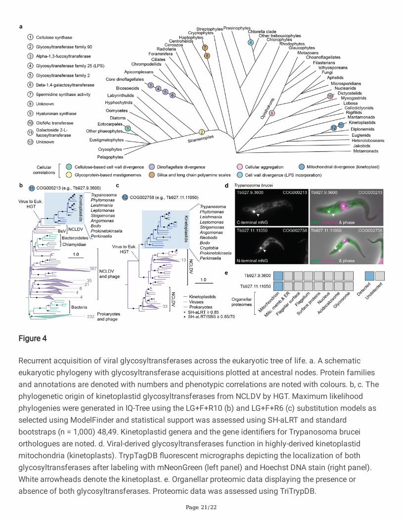
Lastly, to gain insights into the role viral genes play in eukaryotic systems, we inspected the distributionsand functions of viral-derived glycosyltransferases, which were strongly enriched in virus-to-eukaryoteHGTs (Fig. 2c). We identi�ed 63 instances of eukaryotes acquiring viral glycosyltransferases, of which 13

Page 6/22
mapped to ancestral nodes, implying functional relevance under long term selection (SupplementaryTable 5). Plotting transfer events and annotations over a eukaryotic phylogeny revealed the functionaldiversity and recurrent acquisitions of these enzymes across eukaryotic lineages (Fig. 4a, Extended DataFig. 4). These HGTs were often correlated with morphological and structural synapomorphies includingalgal cell wall elaboration (e.g., lipopolysaccharide (LPS) and cellulose synthesis enzymes) 37, long-chainpolyamine-containing scale formation in haptophytes (spermidine synthase) 38, cellular aggregation inthe opisthokonts and dictyostelid slime molds (hyaluronan synthase and GlcNAc transferase), andmitochondrial divergence in the kinetoplastids (fucosyltransferase), a group primarily comprised ofanimal parasites such as trypanosomes (Fig 4a). Experimental data supported a number of thesecorrelations, including the unusual identi�cation of LPS in the cell walls of Chlorella 39, the importance ofhyaluronan in vertebrate tissues 40, and the role of the dictyostelid N-acetylglucosamine transferase,Gnt2, in calcium-independent cellular aggregation 41,42, demonstrating that virally sourced genes are co-opted during the evolution of cellular traits (Fig. 4a). We further examined two glycosyltransferaseacquisitions in kinetoplastids, hypothesizing that, given the correlation between the HGT acquisitions andthe origin of the highly derived kinetoplastid mitochondria (called kinetoplasts), they should function inthat compartment. Phylogenetic analyses revealed that both genes were derived from the NCLDV,highlighted the prokaryotic origin of the fucosyltransferase (COG000231), and con�rmed that both geneswere conserved throughout kinetoplastids (Fig. 4b, c). Moreover, both proteins localized to the kinetoplastin Trypanosoma brucei (identi�able as a non-nuclear DNA-stained foci) both when tagged withmNeonGreen (Fig. 4d) and by organellar proteomics (Fig. 4e). A recent report also suggests an essentialrole for the fucosyltransferase in kinetoplast function in T. brucei 43, altogether indicating that these viral-derived glycosyltransferases were co-opted for use in the kinetoplast at the same time as it underwentmassive evolutionary change. These data, along with the tendency for viruses to modify cell surfaces,suggest that viral-derived genes may have played various roles in the evolution of cellular morphologyacross the eukaryotic tree of life, possibly affecting the diversi�cation of eukaryotic forms.
Horizontal gene transfer between viruses and eukaryotes has been observed and assumed to impactgenome evolution in both participants, but until now we lacked the systematic characterization of thesegene exchanges necessary to generalize their mode and functional signi�cance in both viral andeukaryotic contexts. As with all computational surveys, our dataset is limited by speci�city and sensitivity,but nonetheless it provides an extensive resource from which phylogenetic patterns can be observed andtheir genomic and functional importance may be predicted. From a viral perspective, the apparentubiquity of host-manipulation strategies suggests that the cellular processes outlined above mayrepresent targets for the development of broad-spectrum, host-targeting, antiviral therapeutics. Indeed,many important emerging human pathogens, such as Ebola virus, Zika virus, and coronaviruses, dependon the manipulation of the same cellular processes outlined above, such as autophagy, proteolysis, ERmodi�cation, and sphingolipid metabolism 35,44–46. Functional investigations of eukaryote-derived viralgenes, particularly using heterologous expression 7, may also provide insights into how virusesmanipulate these cellular pathways while circumventing the need for tractable host-virus model systems.From a eukaryotic perspective, our analyses suggest that viruses can not only mediate intra-eukaryotic
Page 7/22
gene exchange but that the evolution of cellular morphology and structure has been in�uenced by viralgenes, particularly glycosyltransferases. These have recurrently impacted transitions as fundamental asthe evolution of tissues or divergent mitochondria, reminiscent of how retroviral fusogens have repeatedlydriven placental evolution in mammals and lizards 22. Our survey also identi�es protein candidates forwhich experimental characterizations would help reveal the full impact of these genes on cellularsystems and their role in driving the evolution of eukaryotic complexity.
Materials And MethodsDataset assembly
To systematically identify instances of viral-eukaryotic gene exchange, groups of homologous eukaryotic,viral, and prokaryotic proteins were clustered into protein families and phylogenetic analyses wereperformed (Extended Data Fig. 1a). The eukaryotic dataset was generated from 196 genome-predictedeukaryotic proteomes, primarily from UniProt (release 2018_11, see Data and Material Availability) 50, thatrepresented diverse species from all available major eukaryotic lineages, were individually clustered at99% percent-identity with CD-Hit v4.8.1 51, and combined. The eukaryotic dataset was furthersupplemented with �ve high quality transcriptomes to �ll taxonomic gaps in lineages with poor genomicsampling (four dino�agellates and a cercozoan) (Extended Data Fig. 1a, b) 52,53. Viral proteins predictedfrom viral genomes were obtained from UniProt and �ltered to exclude those derived from humanimmunode�ciency virus-1, which were over-represented, and additional viral proteins were acquired fromlow-contamination nucleocytoplasmic large DNA virus (NCLDV) metagenomes from diverseenvironments (Extended Data Fig. 1a) 10. Viral taxonomic annotations were assigned to metagenomesbased on previously conducted phylogenomic analyses 10.
Eukaryotic and viral proteins were then clustered into protein families using a similarity-based approachand the Markov clustering (MCL) algorithm (in�ation = 2) after comparing sequences to one anotherusing Diamond v2.0.2 BLASTp (sensitive mode, e-value < 10-5, query coverage > 50%) 54–56. Proteinfamilies containing both viral and eukaryotic representatives were retained, aligned with MAFFT v.7.39757, and used to generate pro�le hidden Markov models (HMMs) which were used to search 9,035prokaryotic proteomes from UniProt with HMMER v.3.2.1 (e-value < 10-5, incE < 10-5, domE < 10-5)
58
(Extended Data Fig. 1b). Due to the large number of prokaryotic sequences, the resulting hits werereduced by taking the most signi�cant hit (based on e-value) per genus or per strain, to a maximum of150 sequences (on average 39% of the total hits). Sequences assigned to viral-eukaryotic protein familieswere then combined with the prokaryotic proteins and re-clustered, as above (Extended Data Fig. 1a).
Phylogenetic analysis
Phylogenetic trees were generated from clustered protein families to infer the evolutionary relationshipsbetween viral and eukaryotic homologues. Protein families were �ltered to retain only those with virusesand eukaryotes, aligned with MAFFT, trimmed using a gap-threshold of 30% in trimAl v1.2, and sequences

Page 8/22
with less than 50 amino acid positions were removed 59. Maximum likelihood phylogenies wereconducted in IQ-Tree v1.6 using the LG+F+R5 substitution model, and statistical support was calculatedusing SH-aLRT (Shimodaira-Hasegawa approximate likelihood ratio test, n = 1,000), which was chosendue to its speed, insensitivity to model violations and taxon sampling, and its comparableconservativeness to standard bootstrapping 48,60,61. Phylogenies for large protein families with over 1,500sequences (n = 103) were generated using the fast search mode in IQ-Tree. Phylogenetic rooting wasdone using minimal ancestral deviation (MAD), which is a rooting method that is more robust toheterotachy then midpoint rooting 62.
For individual phylogenies of particular interest, such as those shown in Fig. 4, Extended Data Fig. 3, andExtended Data Fig. 4, analyses were repeated as above but after alignment with the more accurate L-INS-ialgorithm in MAFFT and limited curation (e.g., the removal of long-branching taxa as de�ned below, seeHorizontal gene transfer detection). Additionally, substitution models were selected using ModelFinder inIQ-Tree 48,49 and phylogenies were visualized and annotated using iTOL 63. Notably, the topologies ofthese trees were consistent with their initial iterations and ModelFinder consistently selected the LGsubstitution model similar to that used in the other phylogenies, corroborating the use of theaforementioned methods (see Extended Data Fig. 4).
Horizontal gene transfer detection
After generating phylogenetic trees for each protein family, we developed an automated pipeline using thepython package, ETE 3 64, to identify HGT-indicative topologies. Speci�cally, we aimed to identifyeukaryotic species nested within viral clades (viral-to-eukaryote HGT) or viral taxa within eukaryoticclades (eukaryote-to-virus HGT) (Extended Data Fig. 1a). To this end, phylogenies were initially processedto account for statistical support and directionality (i.e., rooting), and to assign taxonomic annotations.Firstly, phylogenetic nodes with SH-aLRT values below 0.8 were collapsed, a threshold with a similar falsepositive rate to standard bootstrapping that balances speci�city and sensitivity 60. Collapsed phylogenieswere then rooted using MAD (96.4%) 62 or midpoint rooting and taxa were annotated as eukaryotic, viral,or prokaryotic based on National Centre for Biotechnology Information (NCBI) taxonomy (Extended DataFig. 2a).
Following tree processing and annotation, but before identifying HGT events, the phylogenies wereanalyzed to assess rooting ambiguity. In particular, we checked whether viral and eukaryotic sequencescould be separated into two monophyletic groups using alternative root placements. In this case rootingbecomes unclear unless the phylogeny is strongly biased toward viral or eukaryotic speciesrepresentation (e.g., it is unlikely that a gene conserved throughout a eukaryotic supergroup was derivedfrom a single virus). To evaluate this, if a phylogeny could be split into two discrete taxonomic clades, theratio of eukaryotic to viral species was determined. If the ratio was heavily skewed towards eukaryotes orviruses (eukaryote:viral species ratio > 49 or < 0.15, re�ecting the top and bottom 20% of all proteinfamilies), the tree was rooted normally. Otherwise, the topology would be classi�ed as an HGT withunknown directionality. Lastly, single prokaryotic taxa and HGTs between prokaryotes and viruses or

Page 9/22
eukaryotes (identi�ed as below) were removed for simplicity but did not increase the false positive rateamongst viral-eukaryotic HGTs.
After processing, phylogenies were screened for HGT topologies. To achieve this, viral and eukaryoticclades were identi�ed and the taxonomy of their sister group (i.e., the most closely related phylogeneticgroup) and ‘cousin’ group (i.e., the second most closely related phylogenetic group) were determined. Aeukaryote-to-virus HGT topology was de�ned as a viral clade with a eukaryotic sister and cousin whereasa virus-to-eukaryote HGT required a eukaryotic clade with a viral sister and cousin (Extended Data Fig.2a). Initially, viral and eukaryotic clades were identi�ed and the taxonomy of their sister and cousingroups were assessed. To classify the taxonomy of these groups, the number of viral, eukaryotic, andprokaryotic sequences in each group was counted. Sister and cousin groups were then classi�ed as viral,eukaryotic, or prokaryotic if the taxonomies were consistent across the members of the group. If thetaxonomies of a group were mixed (e.g., if both viral and eukaryotic sequences were present), but viral oreukaryotic taxa dominated at least 80% of the sequences, the group was described as ‘probably’ viral oreukaryotic, or else the group received an ambiguous designation. In the event of a polytomy, multiplesister and cousin groups could be present. To account for this, the taxonomy of the polytomy-wide groupwould be summarized by determining the taxonomy of each group within the polytomy (as above). If allcandidate sisters or cousins within the polytomy were classi�ed consistently, the group would beidenti�ed as viral, eukaryotic, or prokaryotic. Likewise, if a majority (>66%) of the groups were consistentlyclassi�ed, the sister or cousin would be denoted as ‘probably’ viral or eukaryotic, otherwise it would belabeled as ambiguous. After classifying both sister and cousin groups, if the topology was consistentwith one of the aforementioned scenarios, an HGT event would be noted. Each phylogeny was screenedfor eukaryote-to-virus and virus-to-eukaryote HGTs three times iteratively, given that once a viral oreukaryotic clade had been classi�ed as an HGT, it would be interpreted as eukaryotic or viral, respectively,in subsequent iterations. Finally, after three cycles of HGT identi�cation, if there were remaining viral andeukaryotic clades sister to one another with ambiguous or prokaryotic cousins, they were labeled as HGTswith unknown transfer directionality.
Once an HGT was identi�ed characteristics including the recipient, donor, phylogenetic statistics, andtopology notes were recorded (Supplementary Table 1). Recipient and donor taxa were assessed bydetermining the last common ancestor of the recipient and donor (sister), respectively, based on NCBItaxonomy. Moreover, node statistical support values were recorded along with the branch length of therecipient. If the branch length of the recipient or donor represented an extreme outlier (de�ned as themedian branch length plus three times the interquartile range), the HGT was highlighted as a potentiallong branch attraction (LBA) artifact. Additionally, if the donor only had a ‘probable’ taxonomicclassi�cation, ambiguity would also be noted. In both of these cases, HGTs were labeled as weaklysupported and excluded in downstream analyses. Lastly, the approximate origin of viral-derivedeukaryotic genes was determined by moving up through phylogenetic nodes from the donor cladetowards the root until a cellular lineage was reached, if possible (Extended Data Fig. 3a).
Contamination scoring

Page 10/22
After identifying the HGTs, individual transfer events were assessed for possible alternative sources ofphylogenetic incongruence, speci�cally contamination. This is important given that eukaryotic and viralgenes can be artifactually present in viral and eukaryotic genomes, respectively, which may give theimpression that HGT has occurred. To address this, only complete viral genomes and metagenomes withlow contamination scores were included in the analysis 10 and individual viral-derived eukaryotic geneswere assessed based on a series of criteria and a contamination scoring scheme (Extended Data Fig. 2b-e). Contamination was assessed based on two main attributes: 1) the presence of related taxa in the HGT,and 2) the characteristics of the genomic contig upon which the gene was encoded. Firstly, the taxonomiccomposition of the HGT-recipients was assessed based on the assumption that the same contaminationis unlikely to occur in multiple independently sampled genomic datasets, particularly if the species fromwhich they are derived are closely related. Therefore, points were given if the HGT-recipients includedmultiple members of the same (+3) or different (+1) phyla as the species encoding the gene of interest(Extended Data Fig. 2b).
Secondly, the characteristics of the genomic contigs encoding each viral-derived gene were inspectedbased upon the notion that they should share attributes with the host genome, such as consistent GC-content, reasonable contig size, and that the gene should be �anked by eukaryotic regions. To this end,contigs were identi�ed by mapping proteins to the genome using tBLASTn (e-value < 10-5) and pointswere given if the contig was within one standard deviation of the median genomic GC-content (+1) and ifthe contig was a reasonable size (greater than half of the L50) (+1). Lastly, the genomic context wasinspected by extracting DNA regions (5 kbp) upstream (-2.5 kbp) and downstream (+2.5 kbp) of eachgene. Extracted regions were then taxonomically classi�ed by comparing them to the SWISS-PROTdatabase (release 2019_11) using BLASTx 65 and assessing the taxonomy of the resulting hits. Amaximum of 20 hits (e-value < 10-3) were evaluated and normalized scores for viral, eukaryotic, andprokaryotic classi�cations were calculated to account for database bias. These scores were calculatedfor each taxonomic group as:
where tprop is the proportion of a taxonomic group in the database, and nt and St are the number andmedian bit-score of hits to that taxonomic group, respectively. The taxonomy was assigned to the groupwith the maximum score and points were given for eukaryotic classi�cations (+1 per region) andsubtracted for prokaryotic ones (-1 per region), which are more indicative of contamination. Viral anduncertain classi�cations were neutral (+0) to account for the possibility of large viral insertions.
After assigning contamination scores to each putative eukaryotic sequence involved in a virus-to-eukaryote HGT or an HGT with unknown directionality, sequences with scores less than two wereexcluded and HGTs and recipient taxonomies were reassessed (Extended Data Fig. 2c, d). Due to thestrict criteria applied during �ltering and HGT identi�cation, false positive rates should be low.
Functional analyses

Page 11/22
To examine HGT function, eukaryotic and viral proteins were annotated with eggNOG, Pfam andPANTHER (Protein analysis through evolutionary relationships) using a combination of eggNOG-Mapperv2 and InterProScan v.5.48 with the default parameters 66–70. For clarity, the resulting gene ontology (GO)terms were simpli�ed by mapping the terms to the yeast GO-slim subset using Map2Slim (seehttps://github.com/owlcollab/owltools/wiki/Map2Slim). Protein families were given functionalannotations based on a majority rule (Supplementary Table 1) and labeled with gene ontology (GO) termsif a given term was assigned to at least 20% of annotated proteins within a family. To conduct GO-enrichment analyses, protein families exhibiting HGT were compared against a eukaryotic backgroundcomprising all eukaryotic protein families containing either a virus or at least ten eukaryotic species. Thefrequencies of individual GO-terms in the HGT families were compared to the eukaryotic backgroundusing permutation tests which involved randomly sampling equally sized sets of annotated proteinfamilies without replacement (n = 107), with the null hypothesis being that GO-terms associated with theHGTs re�ect a random sampling of eukaryotic protein families, as has been done previously 71.Signi�cantly enriched GO terms (p < 0.01) were summarized and visualized using REVIGO 47.
To investigate the predicted subcellular localizations of eukaryote-derived viral genes, all eukaryoticproteins were annotated using DeepLoc v1.0 and the BLOSSUM62 matrix 33. Localization predictionswith likelihoods less than 0.5 were re-classi�ed as unknown and cellular targets were assigned toindividual eukaryote-to-virus HGTs based on the majority localization of the donor (i.e., eukaryotic)sequences. Enrichments were assessed by comparing the frequency of individual localizations in theHGTs to an equally sized random sampling of annotated eukaryotic proteins (p < 0.05, n = 106). The nullhypothesis was that viruses randomly acquire eukaryotic genes irrespective of their predicted subcellularlocalizations.
Data and code availability
All data, including proteomes, protein families, annotations, alignments, phylogenies, and Python scriptsfor phylogenetic analyses, contamination scoring, and functional enrichments are available from Dryad(https://datadryad.org/stash/share/jT_8Q2Yh3197gDLiAFh4JBiTs0-WbKYg_DYD-3Zqml4) (Revieweraccessible link).
DeclarationsAcknowledgements
We thank Richard Wheeler for providing �uorescent micrographs of Trypanosoma brucei, as part ofTrypTagDB. This work was supported by grants from the Natural Sciences and Engineering ResearchCouncil of Canada (NSERC, RGPIN-2014-03994) and from the Gordon and Betty Moore Foundation(https://doi.org/10.37807/GBMF9201) to P.J.K. N.A.T.I. was supported by an NSERC Canadian GraduateScholarship and a Junior Research Fellowship from Merton College, Oxford. A.A.P. was supported by

Page 12/22
European Molecular Biology Organization (EMBO) long-term fellowship. T.A.R. was supported by a RoyalSociety University Research Fellowship (UF130382).
Author Contributions
Conceptualization, N.A.T.I. and A.A.P.; Funding acquisition, P.J.K. and T.A.R.; Investigation N.A.T.I. andA.A.P.; Resources, P.J.K. and T.A.R.; Supervision, P.J.K. and T.A.R.; Writing N.A.T.I with input from allauthors.
Competing Interests
The authors declare no competing interests.Materials and Correspondence
Material requests and correspondence should be addressed to N.A.T.I.
References1. Chen, J. et al. Genome hypermobility by lateral transduction. Science 362, 207–212 (2018).
2. Koonin, E. V. & Krupovic, M. The depths of virus exaptation. Curr. Opin. Virol. 31, 1–8 (2018).
3. Frank, J. A. & Feschotte, C. Co-option of endogenous viral sequences for host cell function. Curr.Opin. Virol. 25, 81–89 (2017).
4. Touchon, M., Moura de Sousa, J. A. & Rocha, E. P. Embracing the enemy: The diversi�cation ofmicrobial gene repertoires by phage-mediated horizontal gene transfer. Curr. Opin. Microbiol. 38, 66–73 (2017).
5. Zimmerman, A. E. et al. Metabolic and biogeochemical consequences of viral infection in aquaticecosystems. Nat. Rev. Microbiol. 18, (2019).
�. Filée, J., Pouget, N. & Chandler, M. Phylogenetic evidence for extensive lateral acquisition of cellulargenes by Nucleocytoplasmic large DNA viruses. BMC Evol. Biol. 8, 1–13 (2008).
7. Monier, A. et al. Host-derived viral transporter protein for nitrogen uptake in infected marinephytoplankton. Proc. Natl. Acad. Sci. 114, E7489–E7498 (2017).
�. Monier, A. et al. Phosphate transporters in marine phytoplankton and their viruses: Cross-domaincommonalities in viral-host gene exchanges. Environ. Microbiol. 14, 162–176 (2012).
9. Monier, A. et al. Horizontal gene transfer of an entire metabolic pathway between a eukaryotic algaand its DNA virus. Genome Res. 1441–1449 (2009) doi:10.1101/gr.091686.109.
10. Schulz, F. et al. Giant virus diversity and host interactions through global metagenomics. Nature 578,432–436 (2020).
11. Aswad, A. & Katzourakis, A. Cell-derived viral genes evolve under stronger purifying selection inrhadinoviruses. J. Virol. 92, e00539-18 (2018).

Page 13/22
12. Schulz, F. et al. Giant viruses with an expanded complement of translation system components.Science 356, 82–85 (2017).
13. Guglielmini, J., Woo, A. C., Krupovic, M., Forterre, P. & Gaia, M. Diversi�cation of giant and largeeukaryotic dsDNA viruses predated the origin of modern eukaryotes. Proc. Natl. Acad. Sci. U. S. A.116, 19585–19592 (2019).
14. Enav, H., Mandel-Gutfreund, Y. & Béjà, O. Comparative metagenomic analyses reveal viral-inducedshifts of host metabolism towards nucleotide biosynthesis. Microbiome 2, 1–11 (2014).
15. Rozenberg, A. et al. Lateral gene transfer of anion-conducting channel rhodopsins between greenalgae and giant viruses. Curr. Biol. 4910-4920.e5 (2020) doi:10.1016/j.cub.2020.09.056.
1�. Vardi, A. et al. Host-virus dynamics and subcellular controls of cell fate in a natural coccolithophorepopulation. Proc. Natl. Acad. Sci. U. S. A. 109, 19327–19332 (2012).
17. Gornik, S. G. et al. Loss of nucleosomal DNA condensation coincides with appearance of a novelnuclear protein in dino�agellates. Curr. Biol. 22, 2303–2312 (2012).
1�. Medina, E. M., Turner, J. J., Gordân, R., Skotheim, J. M. & Buchler, N. E. Punctuated evolution andtransitional hybrid network in an ancestral cell cycle of fungi. Elife 5, e09492 (2016).
19. Pastuzyn, E. D. et al. The neuronal gene Arc encodes a repurposed retrotransposon Gag protein thatmediates intercellular RNA transfer. Cell 172, 275–288 (2018).
20. Mi, S. et al. Syncytin is a captive retroviral envelope protein involved in human placentalmorphogenesis. Nature 403, 785–789 (2002).
21. Fédry, J. et al. The Ancient Gamete Fusogen HAP2 Is a Eukaryotic Class II Fusion Protein. Cell 168,904–915 (2017).
22. Cornelis, G. et al. An endogenous retroviral envelope syncytin and its cognate receptor identi�ed inthe viviparous placental Mabuya lizard. Proc. Natl. Acad. Sci. U. S. A. 114, E10991–E11000 (2017).
23. Irwin, N. A. T. et al. Viral proteins as a potential driver of histone depletion in dino�agellates. Nat.Commun. 9, 1535 (2018).
24. Forterre, P. & Prangishvili, D. The major role of viruses in cellular evolution: Facts and hypotheses.Curr. Opin. Virol. 3, 558–565 (2013).
25. Richards, T. A., Hirt, R. P., Williams, B. A. P. & Embley, T. M. Horizontal gene transfer and the evolutionof parasitic protozoa. Protist 154, 17–32 (2003).
2�. Hayward, A., Cornwallis, C. K. & Jern, P. Pan-vertebrate comparative genomics unmasks retrovirusmacroevolution. Proc. Natl. Acad. Sci. U. S. A. 112, 464–469 (2015).
27. Cock, J. M. et al. The Ectocarpus genome and the independent evolution of multicellularity in brownalgae. Nature 465, 617–621 (2010).
2�. Moniruzzaman, M., Weinheimer, A. R., Martinez-Gutierrez, C. A. & Aylward, F. O. Widespreadendogenization of giant viruses shapes genomes of green algae. Nature 588, (2020).
29. Leonard, G. et al. Comparative genomic analysis of the ‘pseudofungus’ Hyphochytrium catenoides.Open Biol. 8, (2018).

Page 14/22
30. Maumus, F. & Blanc, G. Study of gene tra�cking between acanthamoeba and giant viruses suggestsan undiscovered family of amoeba-infecting viruses. Genome Biol. Evol. 8, 3351–3363 (2016).
31. Blanc, G., Gallot-Lavallée, L. & Maumus, F. Provirophages in the Bigelowiella genome bear testimonyto past encounters with giant viruses. Proc. Natl. Acad. Sci. U. S. A. 112, E5318–E5326 (2015).
32. Davidson, A. R. A common trick for transferring bacterial DNA. Science 362, 152–153 (2018).
33. Almagro Armenteros, J. J., Sønderby, C. K., Sønderby, S. K., Nielsen, H. & Winther, O. DeepLoc:Prediction of protein subcellular localization using deep learning. Bioinformatics 33, 3387–3395(2017).
34. Pagarete, A., Allen, M. J., Wilson, W. H., Kimmance, S. A. & De Vargas, C. Host-virus shift of thesphingolipid pathway along an Emiliania huxleyi bloom: Survival of the fattest. Environ. Microbiol.11, 2840–2848 (2009).
35. Schneider-Schaulies, J. & Schneider-Schaulies, S. Sphingolipids in viral infection. Biol. Chem. 396,585–595 (2015).
3�. Fernández De Castro, I., Tenorio, R. & Risco, C. Virus assembly factories in a lipid world. Curr. Opin.Virol. 18, 20–26 (2016).
37. Michel, G., Tonon, T., Scornet, D., Cock, J. M. & Kloareg, B. Central and storage carbon metabolism ofthe brown alga Ectocarpus siliculosus: Insights into the origin and evolution of storagecarbohydrates in eukaryotes. New Phytol. 188, 67–81 (2010).
3�. Durak, G. M. et al. A role for diatom-like silicon transporters in calcifying coccolithophores. Nat.Commun. 7, 10543 (2016).
39. Armstrong, P. B., Armstrong, M. T., Pardy, R. L., Child, A. & Wainwright, N. Immunohistochemicaldemonstration of a lipopolysaccharide in the cell wall of a eukaryote, the green alga, Chlorella. Biol.Bull. 203, 203–204 (2002).
40. Laurent, T. C. & Fraser, J. R. E. Hyaluronan. FASEB 6, 2397–2404 (1992).
41. Loomis, W. F., Wheeler, S. A., Springer, W. R. & Barondes, S. H. Adhesion mutants of Dictyosteliumdiscoideum lacking the saccharide determinant recognized by two adhesion-blocking monoclonalantibodies. Dev. Biol. 109, 111–117 (1985).
42. Chisholm, R. L. et al. dictyBase, the model organism database for Dictyostelium discoideum. NucleicAcids Res. 34, 423–427 (2006).
43. Bandini, G. et al. An essential GDP-Fuc: β-D-Gal α-1,2-fucosyltransferase is located in themitochondrion of Trypanosoma brucei. bioRxiv 726117 (2019) doi:10.1101/726117.
44. Fung, T. S. & Liu, D. X. Coronavirus infection, ER stress, apoptosis and innate immunity. Front.Microbiol. 5, 296 (2014).
45. Raaben, M. et al. The ubiquitin-proteasome system plays an important role during various stages ofthe coronavirus infection cycle. J. Virol. 84, 7869–7879 (2010).
4�. Leier, H. C. et al. A global lipid map de�nes a network essential for Zika virus replication. Nat.Commun. 11, 3652 (2020).

Page 15/22
47. Supek, F., Bošnjak, M., Škunca, N. & Šmuc, T. Revigo summarizes and visualizes long lists of geneontology terms. PLoS One 6, e21800 (2011).
4�. Nguyen, L. T., Schmidt, H. A., Von Haeseler, A. & Minh, B. Q. IQ-TREE: A fast and effective stochasticalgorithm for estimating maximum-likelihood phylogenies. Mol. Biol. Evol. 32, 268–274 (2015).
49. Kalyaanamoorthy, S., Minh, B. Q., Wong, T. K. F., Von Haeseler, A. & Jermiin, L. S. ModelFinder: Fastmodel selection for accurate phylogenetic estimates. Nat. Methods 14, 587–589 (2017).
50. Uniprot Consortium. UniProt: A hub for protein information. Nucleic Acids Res. 43, D204–D212(2015).
51. Li, W. & Godzik, A. Cd-hit: A fast program for clustering and comparing large sets of protein ornucleotide sequences. Bioinformatics 22, 1658–1659 (2006).
52. Keeling, P. J. et al. The Marine Microbial Eukaryote Transcriptome Sequencing Project (MMETSP):Illuminating the functional diversity of eukaryotic life in the oceans through transcriptomesequencing. PLoS Biol. 12, e1001889 (2014).
53. Nowack, E. C. M. et al. Gene transfers from diverse bacteria compensate for reductive genomeevolution in the chromatophore of Paulinella chromatophora. Proc. Natl. Acad. Sci. U. S. A. 113,12214–12219 (2016).
54. Enright, A. J., Van Dongen, S. & Ouzounis, C. A. An e�cient algorithm for large-scale detection ofprotein families. Nucleic Acids Res. 30, 1575–1584 (2002).
55. Altschul, S. F., Gish, W., Miller, W., Myers, E. W. & Lipman, D. J. Basic local alignment search tool. J.Mol. Biol. 215, 403–10 (1990).
5�. Buch�nk, B., Xie, C. & Huson, D. H. Fast and sensitive protein alignment using DIAMOND. Nat.Methods 12, 59–60 (2014).
57. Katoh, K. & Standley, D. M. MAFFT multiple sequence alignment software version 7: Improvements inperformance and usability. Mol. Biol. Evol. 30, 772–780 (2013).
5�. Mistry, J., Finn, R. D., Eddy, S. R., Bateman, A. & Punta, M. Challenges in homology search: HMMER3and convergent evolution of coiled-coil regions. Nucleic Acids Res. 41, e121 (2013).
59. Capella-Gutiérrez, S., Silla-Martínez, J. M. & Gabaldón, T. trimAl: A tool for automated alignmenttrimming in large-scale phylogenetic analyses. Bioinformatics 25, 1972–1973 (2009).
�0. Anisimova, M., Gil, M., Dufayard, J. F., Dessimoz, C. & Gascuel, O. Survey of branch support methodsdemonstrates accuracy, power, and robustness of fast likelihood-based approximation schemes.Syst. Biol. 60, 685–699 (2011).
�1. Shimodaira, H. & Hasegawa, M. Multiple comparisons of log-likelihoods with applications tophylogenetic inference. Mol. Biol. Evol 16, 1114–1116 (1999).
�2. Tria, F. D. K., Landan, G. & Dagan, T. Phylogenetic rooting using minimal ancestor deviation. Nat.Ecol. Evol. 1, 0193 (2017).
�3. Letunic, I. & Bork, P. Interactive Tree of Life (iTOL) v4: Recent updates and new developments. NucleicAcids Res. 47, 256–259 (2019).
Page 16/22
�4. Huerta-Cepas, J., Serra, F. & Bork, P. ETE 3: Reconstruction, analysis, and visualization ofphylogenomic data. Mol. Biol. Evol. 33, 1635–1638 (2016).
�5. Boeckmann, B. et al. The SWISS-PROT protein knowledgebase and its supplement TrEMBL in 2003.Nucleic Acids Res. 31, 365–370 (2003).
��. Huerta-Cepas, J. et al. Fast genome-wide functional annotation through orthology assignment byeggNOG-Mapper. Mol. Biol. Evol. 34, 2115–2122 (2017).
�7. El-Gebali, S. et al. The Pfam protein families database in 2019. Nucleic Acids Res. 47, D427–D432(2019).
��. Jones, P. et al. InterProScan 5: Genome-scale protein function classi�cation. Bioinformatics 30,1236–1240 (2014).
�9. Thomas, P. D. et al. PANTHER: A library of protein families and subfamilies indexed by function.Genome Res. 13, 2129–2141 (2003).
70. Huerta-Cepas, J. et al. eggNOG 4.5: A hierarchical orthology framework with improved functionalannotations for eukaryotic, prokaryotic and viral sequences. Nucleic Acids Res. 44, D286–D293(2016).
71. Irwin, N. A. T. et al. The function and evolution of motile DNA replication systems in ciliates. Curr.Biol. 31, 66-76.e6 (2021).
Figures

Page 17/22
Figure 1
The mode and taxonomic distribution of viral-eukaryotic gene exchange. a. Transfers from eukaryotes toviruses (E-to-V), viruses to eukaryotes (V-to-E), and with unknown directionality (Unkn.). Weakly-supportedtransfers had long branching participants or ambiguous donors (see Materials and Methods). Error barsrepresent 95% con�dence intervals from 1,000 bootstrap pseudoreplicates (random sampling ofphylogenies with replacement). b-e. Scatter plots comparing gene exchange statistics. Points represent

Page 18/22
eukaryotes (both species and higher-level classi�cations) and dashed lines represent lines of equality. f,g. Gene transfers from E-to-V (f) and V-to-E (g) across a eukaryotic phylogeny. Bar charts represent HGTspresent in a given genome, whereas pie charts present inferred ancestral HGTs. Bar height and piediameter re�ect transfer frequency and colours re�ect viral taxonomy. Viral taxa were mapped to theirrespective families, phyla, or genera. Taxonomic information and phylogenies are based on NCBI(National Center for Biotechnology Information) taxonomy. Transfers assigned to the last eukaryoticcommon ancestor are excluded but listed in Supplementary Table 1. Abbreviations: SAR, Stramenopila-Alveolata-Rhizaria; H. s., Homo sapiens; E. s., Ectocarpus siliculosus; T. s., Tetrabaena socialis; H. c.,Hyphochytrium catenoides; A. c., Acanthamoeba castellanii; E. h., Emiliania huxleyi; G. t., Guillardia theta;B. n., Bigelowiella natans; N. g., Naegleria gruberi.

Page 19/22
Figure 2
Gene function is related to transfer direction. a. A scatter plot relating the frequency of transfer events toprotein families, normalized for family size (number of sequences). Functional annotations for exemplaryfamilies are highlighted. b-d. Scatter plots displaying enriched gene ontology (GO) biological processterms from protein families participating in unidirectional (b, c) and bidirectional transfer (d) relative to alleukaryotic protein families. Labeling has been summarized for clarity, but complete terms are available in

Page 20/22
Supplementary Table 3. Semantic similarity was determined using REVIGO 47 and statistical signi�cancewas assessed using permutation tests (p < 0.01, n = 107).
Figure 3
Predicted subcellular localizations and functions of eukaryote-derived viral genes. a. Proportions of sub-cellular localizations for eukaryote-to-virus HGTs based on the predicted targeting of eukaryotic donorsequences. Asterisks denote statistically signi�cant enrichments (p < 0.05, see b). Error bars represent95% con�dence intervals determined from 1,000 bootstrap pseudoreplicates. b. The enrichment ofsubcellular compartments relative to total eukaryotic proteomes. Signi�cance was assessed usingpermutation tests (n = 106). c. A comparison between the predicted localization of eukaryotic donors andtheir viral recipients. The relative frequencies and proportions are indicated by edge thickness and colour,respectively. d-g. Scatter plots displaying enriched GO biological process terms for families with a givendonor localization relative to all eukaryotic protein families for localizations to (from left to right, colourcoded as in a) the cytoplasm, nucleus, endoplasmic reticulum, and extracellular space. Labeling has beensummarized for clarity but complete terms are available in Supplementary Table 4. Semantic similaritywas determined using REVIGO and statistical signi�cance was assessed using permutation tests (p <0.01, n = 107).
Page 21/22
Figure 4
Recurrent acquisition of viral glycosyltransferases across the eukaryotic tree of life. a. A schematiceukaryotic phylogeny with glycosyltransferase acquisitions plotted at ancestral nodes. Protein familiesand annotations are denoted with numbers and phenotypic correlations are noted with colours. b, c. Thephylogenetic origin of kinetoplastid glycosyltransferases from NCLDV by HGT. Maximum likelihoodphylogenies were generated in IQ-Tree using the LG+F+R10 (b) and LG+F+R6 (c) substitution models asselected using ModelFinder and statistical support was assessed using SH-aLRT and standardbootstraps (n = 1,000) 48,49. Kinetoplastid genera and the gene identi�ers for Trypanosoma bruceiorthologues are noted. d. Viral-derived glycosyltransferases function in highly-derived kinetoplastidmitochondria (kinetoplasts). TrypTagDB �uorescent micrographs depicting the localization of bothglycosyltransferases after labeling with mNeonGreen (left panel) and Hoechst DNA stain (right panel).White arrowheads denote the kinetoplast. e. Organellar proteomic data displaying the presence orabsence of both glycosyltransferases. Proteomic data was assessed using TriTrypDB.

Page 22/22
Supplementary Files
This is a list of supplementary �les associated with this preprint. Click to download.
SupplementaryTable1.xlsx
SupplementaryTable2.xlsx
SupplementaryTable3.xlsx
SupplementaryTable4.xlsx
SupplementaryTable5.xlsx
ExtendedData.docx