microstructural characterization of batio 3 ceramic ... · pdf filemicrostructural...
TRANSCRIPT

Microstructural Characterization of BaTiO3 Ceramic Nanoparticles
Synthesized by the Hydrothermal Technique
Xin Hua Zhu1,2, Jian Min Zhu 1, Shun Hua Zhou 1, Zhi Guo Liu 1,
Nai Ben Ming 1 and Dietrich Hesse 2
1National Laboratory of Solid State Microstructures, Department of Physics,
Nanjing University, Nanjing 210093, P.R.China, 2 Max-Planck-Institut für Mikrostrukturphysik, Weinberg 2, D-06120 Halle, Germany
Keywords: BaTiO3 Nanoparticles, Microstructures, Hydrothermal Technique, TEM, HRTEM
Abstract. BaTiO3 (BT) nanoparticles were prepared by the hydrothermal technique using different
starting materials and the microstructure examined by XRD, SEM, TEM and HRTEM. X-ray
diffraction and electron diffraction patterns showed that the nanoparticles were the cubic BaTiO3
phase. The BT nanoparticles prepared from the starting materials of as-prepared titanium hydroxide
and barium hydroxide have spherical grain morphology, an average size of 65 nm and a fairly narrow
size distribution. A uniform diffraction contrast across each single grain is observed in the TEM
images, and the clear lattice fringes (with d110 = 0.28 nm) observed in HRTEM images reveal that
well-crystallized BT nanoparticles are synthesized by the hydrothermal method. The edges of the
particles are very smooth, with no surface steps. BT nanoparticles with average grain size of 90 nm,
synthesized using barium hydroxide and titanium dioxide as the starting materials, show surface
facets. In this case a bimodal size distribution of large faceted and smaller particles is observed.
Diffraction contrast variation across the particles caused by high strains within the particles is clearly
observed. The high strains obviously stem from structural defects formed during hydrothermal
synthesis, presumable in the form of lattice OH− ions and their compensation by cation vacancies.
HRTEM images demonstrate that surface facets parallel to the (100) and (110) planes and small
islands with 3 ~ 4 atomic layer thickness are frequently observed around the edge of the particles.
Introduction
Barium titanate (BT) has good dielectric and ferroelectric properties, and is widely used in
thermistors, multilayer ceramic capacitors (MLCs), and electro-optic devices. Recent developments
in microelectronic and communication technology involve the miniaturization of MLCs. To achieve
this and to make the next advance, high dielectric constant ceramic particles of better quality and
small, uniform size are needed [1]. High permittivities and miniaturization can be achieved by
controlling the microstructure, which depends on the homogeneity, composition, surface area and
particle size of the starting powders. To manufacture reliable MLCs, high purity, agglomerate-free,
highly crystalline and superfine ceramic are required [2]. Although the bulk properties of BT
ceramics have been widely investigated, more recently there has been renewed interest in nano-scale
particles of the material because the electrical properties are strongly dependent on the grain size and
crystalline structure. Because tetragonal BaTiO3 is used in ferroelectrics and cubic BT is used in
capacitors a better understanding of the nanostructure of BT ultrafine particles of both phases is of
interest as well as the correlation of properties with particle size.
Traditionally BT powders are produced by the mixed oxide route, which involves repeated
Solid State Phenomena Vol. 106 (2005) pp. 41-46online at http://www.scientific.net© 2005 Trans Tech Publications, Switzerland
Licensed to MPI of Microstructure Physics - Halle/Saale - GermanyAll rights reserved. No part of the contents of this paper may be reproduced or transmitted in any form or by any means without thewritten permission of the publisher: Trans Tech Publications Ltd, Switzerland, www.ttp.net. (ID: 195.37.184.165-07/06/05,14:01:56)
calcination and regrinding of BaCO3 and TiO2 powders at temperatures above 1000°C. However, this
method produces BT particles with uncontrolled and irregular morphologies, which affects the
electrical properties of the resulting sintered ceramics. Therefore, wet and novel chemical routes
have been developed to produce high-quality BT nanoparticles, possessing great advantages over
micrometer-sized ceramic powders, suitable for use in MLCs. Recently nanocrystalline BaTiO3
particles have been prepared by wet chemical methods [3-6] such as sol-gel, coprecipitation, and
hydrothermal methods. However, the products obtained by (co)precipitation or the sol-gel method are
either amorphous or precursor compounds. Calcination at 800-1000°C, followed by milling, is
usually required to form crystalline BT powders. Thus, powder quality is not significantly improved
because the production process is similar to the solid-state reaction method. The hydrothermal
method provides an alternative method to produce fine, high purity, highly crystalline oxide powders
having a well-defined composition and narrow range of grain size with controlled characteristics,
directly from aqueous solutions at relatively low temperatures (<300°C ). In the past there have been
many investigations concerning the hydrothermal synthesis of nanocrystalline BaTiO3 particles,
generally focusing on the following aspects [6-10]: (1) optimization of preparation parameters (e.g.
type of precursors, Ba/Ti ratio, reaction temperature and time, pH value, and so on), (2) understanding
reaction kinetics and nanocrystal formation mechanisms of BT, (3) doping with other elements during
the hydrothermal synthesis process, morphology control of BT powders and the sintering behavior of
green bodies made from hydrothermally produced BT, (4) structural, microstructural, and chemical
characterization. Hydrothermally produced BT nanopowders show a number of structural
characteristics not seen in powders prepared by conventional solid-state reaction at high temperature.
X-ray diffraction of hydrothermal BT powders, particularly those synthesized at lower temperatures,
reveals a cubic structure that is normally only observed above the ferroelectric Curie temperature of
125∼130°C. The reasons for the appearance of the cubic structure and the non-ferroelectric properties
of fine BT nanocrystals are not well understood, although some possible causes are discussed by Frey
et al [11]. However, few detailed nanostructure analyses, at the atomic level, of BT nanopaticles
prepared by the hydrothermal method have been reported. It is well known that the physical
properties of BT nanoparticles are dependent on the microstructure, e.g., grain boundaries, point and
extended defects, as well as surface morphology. It is considered important to investigate the
microstructure of BT nanoparticles to obtain a better understanding of size effects on the physical
properties. In this work, BT nanoparticles were prepared by the hydrothermal technique using
different starting materials. Their microstructure, crystal structure, grain size and distribution, grain
morphology, and microstructural defects, were studied by X-ray diffraction (XRD), scanning electron
microscopy (SEM), and (high-resolution) transmission electron microscopy (HRTEM), and the
results are presented and discussed.
Experimental Procedure
Two kinds of BaTiO3 nanoparticles prepared by the hydrothermal technique using different starting
materials were studied. Sample A consisted of BT nanoparticles was synthesized by a modified
hydrothermal technique using the as-prepared titanium hydroxide and barium hydroxide as starting
materials. These were mixed in the ratio Ba:Ti = 1:1 by stirring and reacted in an autoclave at 100°C
for 5 h. After the reaction, the product was washed several times with organic acids and deionized
water, and finally dried in an oven for 24 h at 85°C. Sample B was synthesized using barium
hydroxide and titanium dioxide (TiO2, anatase) as starting materials under moderate conditions.
From Nanopowders to Functional Materials42
44.0 44.5 45.0 45.5 46.0
0
10
20
30
40
50
60
2 θ (degree)
Inte
nsi
ty (
Counts
)
BTa
20 30 40 50 60 70 80
0
40
80
120
160
200
(a)
(301
)
(221)
(211)
(22
0)
(201)(2
00)
(111)
(110)
*
(10
0)
2 θ (degree)
Inte
nsi
ty (
Counts
)
44.0 44.5 45.0 45.5 46.0
0
10
20
30
40
50
60
2 θ (degree)
Inte
nsi
ty (
Coun
ts)
20 30 40 50 60 70 80
0
40
80
120
160(b)
(211)
(301)
(221)(220)
(201)
(200)
(111)
(110)
*
(100)
2 θ (degree)
Inte
nsi
ty (
Coun
ts)
KOH was used as an alkaline mineralizer, and the hydrothermal reaction was carried out in an oven at
220°C for three days. After cooling to room temperature, BT powders were obtained by filtration and
washed with organic acids and water several times to remove the absorbed impurities, and finally
oven dried at 80°C for 24 h.
The phase purity of the BT powders was studied by X-ray diffraction in a Philips X’Pert MRD
four-circle diffractometer using CuKα radiation collected over a 2θ range of 20-80° with a scan step
of 0.04°. Morphology and grain size were investigated by SEM and TEM. The TEM specimens were
prepared by dispersing small amounts of BT powders in pure alcohol, mixing it in an ultrasonic
generator, and placing a drop of the dispersion on a copper mesh covered with a ‘holey’ carbon film.
Conventional TEM images were obtained from a Philips CM20 TEM operated at 200 kV, and
HRTEM images from a JEOL 4010 high-resolution electron microscope operated at 400 kV.
Results and Discussion
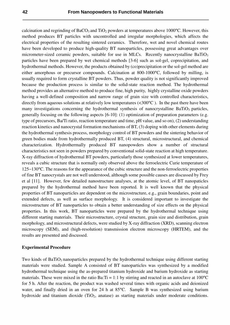
Figure 1(a) shows the XRD pattern of sample A. The inset represents the enlarged pattern between
2θ = 44.0 and 46.0°. The XRD pattern fits well with the peak positions of the standard cubic phase
BT. Furthermore, only a single diffraction peak at 2θ = 45.25° can be observed in the inset, i.e. no
split of the {200} peaks around 2θ = 45° can be seen. This demonstrates that the BT nanocrystals
prepared by the hydrothermal method at 100°C exhibit the characteristics of the cubic phase. This
was confirmed by the following selected area electron diffraction patterns of the same sample.
Similar X-ray diffraction patterns were also obtained from sample B, as shown in Fig.1 (b). The inset
shows that no peak separation of the (200) and (002) peaks around 2θ ≈ 45° can be observed. This
indicates that the BT nanocrystals are of the cubic phase, which is confirmed by selected area
electron diffraction patterns taken from the sample.
Fig.1. XRD patterns of the BT nanoparticles: (a) sample A synthesized by a modified hydrothermal
technique using as-prepared titanium hydroxide and barium hydroxide as starting materials, and (b)
sample B synthesized using barium hydroxide and titanium dioxide as starting materials. Insets are
the enlarged patterns between 2θ = 44.0 and 46.0°. Peaks marked * are from the powder holder.
The grain sizes and morphologies of samples A and B are shown in Figs.2 (a) and (b) respectively.
Fig.2 (a) shows that sample A has a fairly narrow size distribution and spherical grain morphology. In
sample B shown in Fig 2(b), coarser faceted particles and the bimodal size distribution of larger and
Solid State Phenomena Vol. 106 43

400 nm
(a)
400 nm
(b)
smaller particles can be clearly seen.
Fig.2. SEM images of (a) sample A, and (b) sample B.
Bright-field TEM images of samples A and B are shown in Fig.3 (a) and (b), respectively. The
particles are found to be single crystals, which was additionally proven by high-resolution lattice
images of individual particles. The electron diffraction patterns inserted in Fig.3 also show that the
particles are cubic BaTiO3, the diffraction rings corresponding well to the cubic perovskite structure,
which agrees with the XRD results. The average particle sizes, based on the SEM and TEM images,
were 65 nm for sample A, and 90 nm for sample B.
Fig.3. Bright-field TEM images of (a) sample A, and (b) sample B. In Fig.3 (a), the insets are a higher
magnified TEM image and a selected area electron pattern, respectively.
A uniform diffraction contrast across the single grains in sample A is clearly observed in Fig.3 (a),
whereas the diffraction contrast across a single grain varies in sample B, as shown in Fig.3 (b). This
indicates that the grains in the sample B have a higher strains than those in sample A. (In a TEM
image, large strains are indicated by contrast variation across a particle. If a particle is a single crystal
and is strain free, it should be uniform in contrast. However, if the TEM image of a single crystal
500 nm
200 nm
(a) (b)
100 nm
From Nanopowders to Functional Materials44
shows dark-bright variation in contrast, it is likely that the grain is highly strained). In these cubic BT
nanocrystals, the distortion of the TiO6 structure, resulting in a cubic-to-tetragonal phase transition
when cooling the sample through the Curie temperature, has obviously not taken place. A possible
reason is the size of the BT nanocrystals which are so small that the structural defects in the particles
prevent the completion of the structural transition. This has created high strains in the nanocrystals
which has caused some distortion of the cubic structure but it is obviously not sufficient to result in
the formation of the tetragonal phase. It is well known that structural defects of BT nanoparticles
form during hydrothermal synthesis primarily in the form of lattice OH− ions and their compensation
by cation vacancies [6,12]. Clark et al. observed that as-prepared BT powders contain many defects,
primarily in the form of lattice OH−
ions [6]. Shi et al. reported that stabilization of the cubic phase of
BT prepared by the hydrothermal method is caused by surface defects including OH−
defects and
barium vacancies [12].
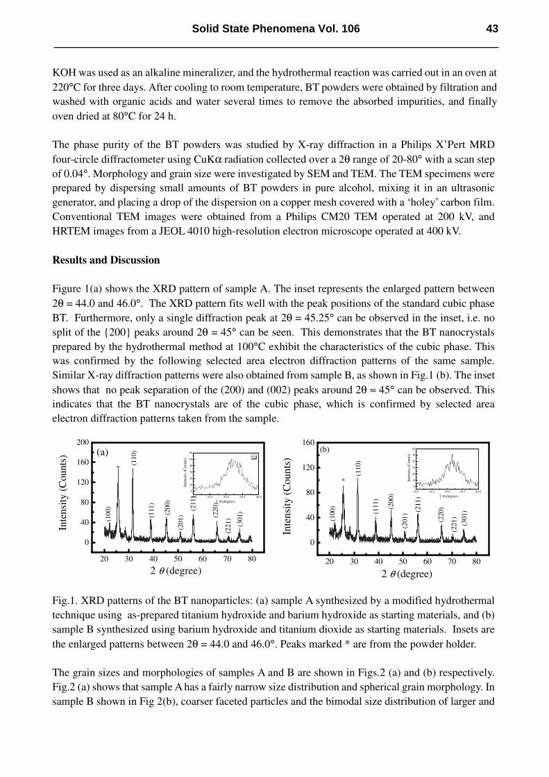
Fig.4. HRTEM images (a) a typical lattice image of nanocrystalline BT grain of size of 75 nm in
sample A, (b) a surface profile HRTEM image of part of a BT grain with size of 80 nm in sample B.
Fig 4(a) shows a typical lattice image of a 75 nm nanocrystalline grain in sample A. Clear lattice
fringes with d110 = 0.28 nm reveal that well-crystallized BT nanoparticles are formed. The
10 nm
(a)
4 nm
(b)
(110)
(100)
Solid State Phenomena Vol. 106 45

surrounding edges of the particle are very smooth and no surface steps were oberved. Fig.4 (b) shows
a surface profile HRTEM image of part of a 80nm BT grain in sample B. It is noticed that the surface
facets are parallel to the (100) and (110) planes. Small islands of 3 ~ 4 atom layer thickness were
frequently observed around the edge of the particle. The surface roughness of the grains in sample B
is much higher than that in sample A. This may be caused by the high strains in the grains. The
contrast variations across Fig.4 (b) are due to the thickness variations associated with the fine-scale
surface facets and surface roughness.
Conclusions
Microstructure of BaTiO3 nanoparticles prepared by the hydrothermal technique have been examined
by XRD, SEM, TEM and HRTEM. XRD results indicated that the BT nanoparticles were of cubic
phase, which was confirmed by electron diffraction. SEM and TEM images show that the BT
nanoparticles prepared using the as-prepared titanium hydroxide and barium hydroxide as starting
materials have a fairly narrow size distribution and a spherical grain morphology, with an average
grain size of 65 nm. A uniform diffraction contrast across single grains was observed. Preliminary
results show that well-crystallized BT nanoparticles are synthesized by the hydrothermal method.
The surrounding edges of the particles are very smooth, no surface steps were observed. The BT
nanoparticles synthesized using barium hydroxide and titanium dioxide as the starting materials have
surface facets and a bimodal size distribution. The average grain size was measured to be 90 nm, and
contrast variations across the particles were observed, indicating high strain caused by lattice defects.
HRTEM images show that the surface facets were parallel to the (100) and (110) planes and small
islands with 3 ~ 4 atomic layer thickness were frequently observed around the edge of the particle.
Acknowledgements
This work is financially supported by the opening project of National Laboratory of Solid State
Microstructures, Nanjing University and a grant for State Key Program for Basic Research of China.
One author, (X.H.Zhu), acknowledges financial support by the Alexander von Humboldt Foundation.
References:
[1] S.Venigalla: Am. Ceram. Soc. Bull. 6 (2001) 63
[2] J.M.Wilson: Am. Ceram. Soc. Bull. 74 (1975) 106.
[3] H.Shimooka, M.Kuwabara: J. Am. Ceram. Soc. 79 (1996) 2983.
[4] H.S.Potdar, P.Singh, S.B.Deshpande, P.D.Godbole, S.K.Date: Mater. Lett. 10 (1990)112.
[5] S.Kumar, G.L.Messing, W.B.White: J. Am. Ceram. Soc. 76 (1993) 617.
[6] I.J.Clark, T.Takeuchi, N.Ohtori, D.C.Sinclair: J. Mater. Chem. 9 (1999) 83.
[7] E.Ciftci, M.N.Rahaman, M.Shumsky: J. Mater. Sci. 36 (2001) 4875.
[8] X.Y.Wang, B.I.Lee, M.Z.Hu, E.A.Payzant, D.A.Blom: J. Mater. Sci: Mater. Electr. 14 (2003)
495.
[9] R.K.Dutta, J.R.Gregg: Chem. Mater. 4 (1992) 843.
[10] S.W.Lu, B.I.Lee, Z.L.Wang, W.D.Samuels: J. Cryst. Growth 219 (2000) 269.
[11] M.H.Frey, D.A.Payne: Phys. Rev. B 54 (1996) 3158.
[12] E.W.Shi, C.T.Xia, W.E.Zhang, B.G.Wang, C.D.Feng: J. Am. Ceram. Soc. 80 (1997) 1567.
From Nanopowders to Functional Materials46