micromechanical control of cell–cell interactions - … control of cell–cell interactions elliot...
TRANSCRIPT
Micromechanical control of cell–cell interactionsElliot E. Hui*† and Sangeeta N. Bhatia*†‡§
*Department of Bioengineering, University of California at San Diego, La Jolla, CA 92093; †Harvard–Massachusetts Institute of Technology Divisionof Health Sciences and Technology/Electrical Engineering and Computer Science, Massachusetts Institute of Technology, Cambridge, MA 02139;and ‡Division of Medicine, Brigham and Women’s Hospital, Boston, MA 02115
Edited by L. B. Freund, Brown University, Providence, RI, and approved February 9, 2007 (received for review September 29, 2006)
The development and function of living tissues depends largelyon interactions between cells that can vary in both time andspace; however, temporal control of cell–cell interaction is exper-imentally challenging. By using a micromachined silicon substratewith moving parts, we demonstrate the dynamic regulation ofcell–cell interactions via direct manipulation of adherent cells withmicrometer-scale precision. We thereby achieve mechanical controlof both tissue composition and spatial organization. As a casestudy, we demonstrate the utility of this tool in deconstructing thedynamics of intercellular communication between hepatocytesand supportive stromal cells in coculture. Our findings indicate thatthe maintenance of the hepatocellular phenotype by stroma re-quires direct contact for a limited time (�hours) followed by asustained soluble signal that has an effective range of <400 �m.This platform enables investigation of dynamic cell–cell interactionin a multitude of applications, spanning embryogenesis, ho-meostasis, and pathogenic processes.
dynamic substrate � intercellular communication � microelectromechanicalsystems � microenvironment � microfabrication
Mammalian cells in vivo integrate and respond to cues intheir microenvironment that vary in both time and space.
In particular, interactions between neighboring cells can regulateboth the fate and function of individual cells and govern theemergent properties of the resultant tissue. Because such cell–cell interactions occur primarily through direct contact or ex-change of soluble factors, understanding the temporal andspatial aspects of these signals is of fundamental importance totissue biology. Recent advances in cell ‘‘micropatterning’’ havealready proven invaluable in increasing our understanding of thestructure–function relationships of such multicellular commu-nities (1–4); however, dynamic manipulation of tissue structurein vitro has remained largely out of reach.
Previous efforts toward spatiotemporal control of tissue or-ganization at the cellular scale have focused on modulation ofthe adhesive properties of the culture substrate (5–7). Throughthe micropatterning of surface chemistries that can be dynam-ically altered, localized attachment and release of cells has beendemonstrated (8, 9). Nonetheless, these manipulations are typ-ically not reversible (i.e., nonadhesive surfaces are renderedadhesive just once), they do not allow the decoupling of pro-cesses associated with adhesion from those correlated withcell–cell interaction (i.e., attachment, spreading, and contactwith neighboring cells have overlapping time scales), nor canthese platforms accommodate serial manipulations to mimic keybiological events (i.e., sequential exposure of a target cellpopulation to different inducer populations). Manipulations ofsurface chemistry are also limited by the inability to preciselycontrol tissue composition: (i) sequential seeding of differentcell types can result in contamination of pure populations and (ii)maintaining �m-scale proximity of two different cell populationsin the absence of contact over many days, important for decou-pling the relative role of contact and paracrine signals (4), hasnot been achieved.
We introduce a different approach to this problem by leveragingtools from the field of microelectromechanical systems, whichoffers precise physical manipulation at a length scale comparable to
that of many biological processes. In our approach, cells are grownon an array of micromachined plates that are physically rearrangedto change the spatial organization of the culture, which will bereferred to as micromechanical reconfigurable culture (�RC). Cellsremain attached to the substrate throughout the repositioningprocess (10, 11). Using �RC, we are able to demonstrate dynamicregulation of cell–cell interactions via direct manipulation of cellpositioning. Specifically, cell–cell contact between different cellpopulations is regulated by positioning plates together or apart. Byimposing a small �m-scale separation between the plates, cell–cellcontact can be abrogated while soluble signaling is maintained. Byusing larger separation distances, the extent of soluble signaling canalso be modulated. In addition, by removing a plate and replacingit, one population of cells can be exchanged for another in amodular fashion. Thus, this micromechanical approach providesdynamic control of both tissue organization and composition.
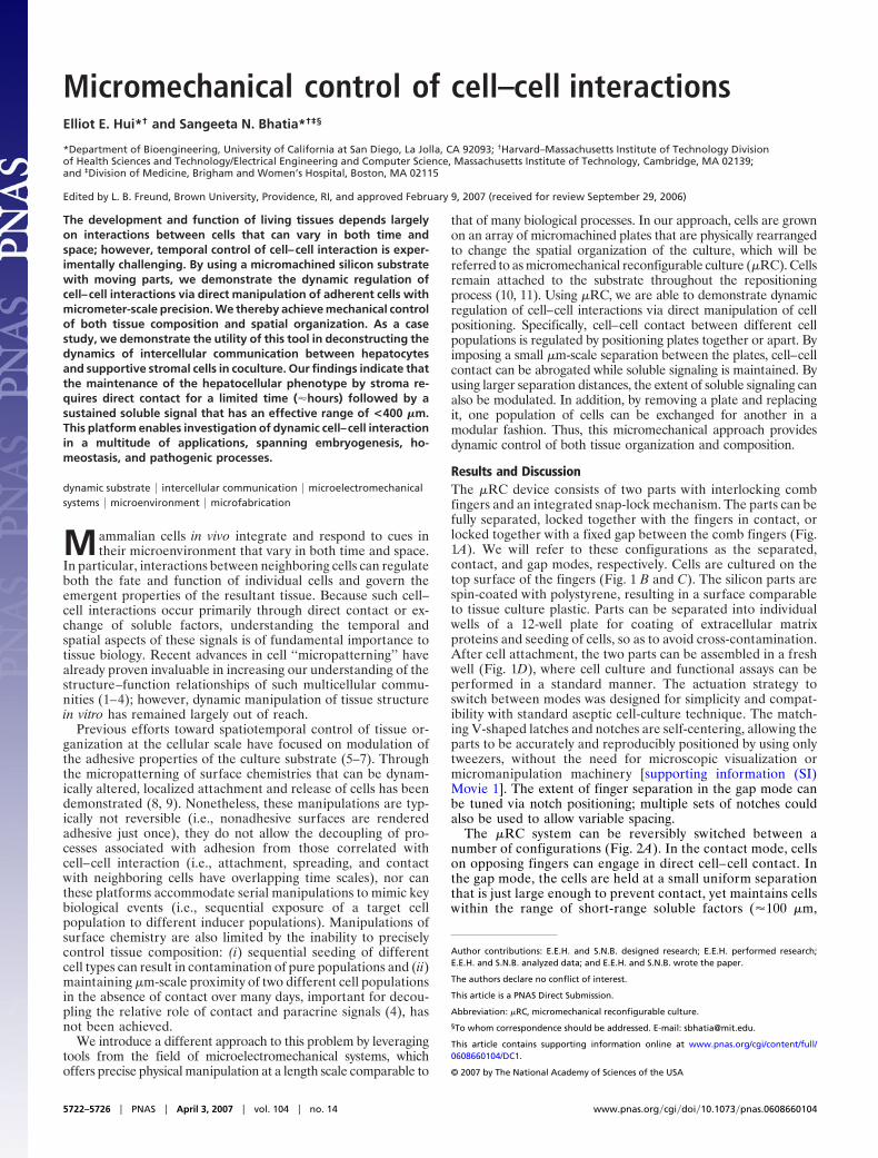
Results and DiscussionThe �RC device consists of two parts with interlocking combfingers and an integrated snap-lock mechanism. The parts can befully separated, locked together with the fingers in contact, orlocked together with a fixed gap between the comb fingers (Fig.1A). We will refer to these configurations as the separated,contact, and gap modes, respectively. Cells are cultured on thetop surface of the fingers (Fig. 1 B and C). The silicon parts arespin-coated with polystyrene, resulting in a surface comparableto tissue culture plastic. Parts can be separated into individualwells of a 12-well plate for coating of extracellular matrixproteins and seeding of cells, so as to avoid cross-contamination.After cell attachment, the two parts can be assembled in a freshwell (Fig. 1D), where cell culture and functional assays can beperformed in a standard manner. The actuation strategy toswitch between modes was designed for simplicity and compat-ibility with standard aseptic cell-culture technique. The match-ing V-shaped latches and notches are self-centering, allowing theparts to be accurately and reproducibly positioned by using onlytweezers, without the need for microscopic visualization ormicromanipulation machinery [supporting information (SI)Movie 1]. The extent of finger separation in the gap mode canbe tuned via notch positioning; multiple sets of notches couldalso be used to allow variable spacing.
The �RC system can be reversibly switched between anumber of configurations (Fig. 2A). In the contact mode, cellson opposing fingers can engage in direct cell–cell contact. Inthe gap mode, the cells are held at a small uniform separationthat is just large enough to prevent contact, yet maintains cellswithin the range of short-range soluble factors (�100 �m,
Author contributions: E.E.H. and S.N.B. designed research; E.E.H. performed research;E.E.H. and S.N.B. analyzed data; and E.E.H. and S.N.B. wrote the paper.
The authors declare no conflict of interest.
This article is a PNAS Direct Submission.
Abbreviation: �RC, micromechanical reconfigurable culture.
§To whom correspondence should be addressed. E-mail: [email protected].
This article contains supporting information online at www.pnas.org/cgi/content/full/0608660104/DC1.
© 2007 by The National Academy of Sciences of the USA
5722–5726 � PNAS � April 3, 2007 � vol. 104 � no. 14 www.pnas.org�cgi�doi�10.1073�pnas.0608660104

demonstrated below). Finally, cell populations can be ex-changed in a modular fashion by removing one set of fingersand snapping in a replacement seeded with another cell type.Thus, contact-mediated signaling can be dynamically regulatedby switching between the contact and gap modes. Likewise,soluble signaling can be dynamically regulated by swappingbetween various cell types in the gap mode. To provide the
necessary mechanical precision, silicon parts were fabricatedin a single-mask, through-wafer, deep reactive ion etchingprocess (12, 13). A separation of 6 �m or less was measured inthe contact mode, and a separation of 79 � 1 �m was measuredin the gap mode. Fluorescence microscopy with membranedyes showed that cells on opposing fingers form intimatecontacts in contact mode (Fig. 2B). In addition, contamination
Fig. 1. Micromechanical substrates enable micrometer-resolution cell positioning. (A) Microfabricated silicon parts can be fully separated (Left), lockedtogether with comb fingers in contact (Center), or slightly separated (Right). Cells are cultured on the top surfaces; manual scraping can be used to restrict cellsto the comb fingers only (Inset). The slope of the tapered comb fingers results in a 20:1 mechanical transmission ratio; that is, sliding the parts 1.6 mm changesthe gap between the fingers by only 80 �m. Together with the integrated snap-lock mechanism, it is thereby possible to control separation with repeatablemicrometer-scale precision by using unassisted manual actuation. (B and C) Bright-field images of hepatocytes (darker cells) and 3T3 fibroblasts cultured on thecomb fingers. The silicon is first functionalized by spin-coating with polystyrene followed by plasma treatment, resulting in a surface comparable to tissue cultureplastic. Devices can be reused multiple times (�20). (D) Devices in a standard 12-well plate. Cell culture and functional assays were performed with standardmethods. Actuation is also performed directly on the plate with sterile tweezers.
Fig. 2. Reconfigurable cell culture. Cultures can be reversibly switched to initiate or to eliminate contact between two cell populations; individual populationscan also be removed and replaced. (A) Fluorescent images illustrating possible device manipulations. Each cell type was prelabeled with an individual dye color.(B) Fluorescent image showing intimate contact between hepatocytes (green) and stroma (red, 3T3 fibroblasts) at the interface between neighboring combfingers. The image was taken 18 h after initiation of contact. Cell nuclei are counterstained in blue. (C) Cross-migration of cells is minimal for moderate durationsof contact. Representative fluorescent image showing small numbers of stromal cells (red, arrows indicate selected cells) remaining behind on a hepatocyte finger(green) after combs were separated after 18 h of contact. In this work, contact was limited to 18 h to minimize cross-migration, but longer durations are possiblewith other cell types (data not shown).
Hui and Bhatia PNAS � April 3, 2007 � vol. 104 � no. 14 � 5723
APP
LIED
PHYS
ICA
LSC
IEN
CES

of cells between adjacent fingers after 18 h of contact wasminimal (Fig. 2C).
As a case study, we applied our dynamic platform to the studyof cell–cell interactions between hepatocytes and stromal cells incoculture. As with many other cell types, interaction of epitheliawith supportive stroma or ‘‘feeder layers’’ promotes tissue-specific gene expression in vitro. In the case of primary hepato-cytes, cocultivation of hepatocytes with many different mesen-chymal cell types (endothelia, fibroblasts, etc.) promotesretention of hepatocyte viability and liver-specific functions thatare otherwise rapidly lost in vitro (2). This robust ‘‘coculture’’phenomenon, although poorly understood, has wide-rangingapplications in both therapeutic and diagnostic applications ofengineered liver tissue (14–16). Using both conventional tech-niques and micropatterning approaches, we and others havepreviously found that the degree of interaction between the twocell types (‘‘heterotypic interaction’’) modulated the amount ofliver-specific function retained in vitro (2, 17). These findingssuggested an important role for proximity between the two celltypes in the rescue of hepatocyte phenotype; however, therelative role of contact-mediated versus soluble signals, thedynamics of interaction, and the potential for reciprocal signal-ing had not been established.
Hence, we set out to explore this system by using the �RCsubstrates, with primary rat hepatocytes and Swiss 3T3 murinefibroblasts cultured on opposing combs. Hepatocyte morphologyand viability were assessed microscopically, and albumin pro-duction was measured as a quantitative marker of liver-specificfunction. Comparison of cultures in the contact, gap, andseparated modes demonstrated that contact was necessary formaintenance of liver-specific function (Fig. 3A). Even in the gapmode, which corresponded to only an 80-�m separation betweenthe two cell populations, hepatocyte function declined at a ratesimilar to that of hepatocytes cultured alone. Next, we conducteddynamic experiments in which cells were repositioned after 18 hof contact. Here, transient contact alone proved insufficient torescue the hepatocyte phenotype, and liver-specific functionsrapidly declined. In contrast, transient contact followed bysustained culture in the gap mode provided complete rescue ofliver-specific function (Fig. 3B). These observations thus implya necessary role for both heterotypic contact and soluble factorsthat diffuse across the gap.
Notably, it would appear that contact was required onlyinitially, whereas soluble interactions were required for theduration of the experiment. This finding raised the possibilitythat reciprocal interactions, i.e., sustained alterations in fibro-
blast function as a result of hepatocyte contact, might play a role.To test this possibility, we exploited the ‘‘modular’’ nature of the�RC platform. Cocultures were conducted in contact mode for18 h as before; however, the fibroblasts were then replaced withnaı̈ve fibroblasts (no exposure to heterotypic contact) in gapmode. Under these conditions, paracrine signals provided bynaı̈ve fibroblasts were still sufficient to sustain hepatic functions(Fig. 3C). Conversely, if naı̈ve hepatocytes were substituted,hepatic function deteriorated. Hence, the data are consistentwith constitutive expression of critical soluble factors by fibro-blasts independent of hepatocyte interaction rather than sup-porting a role for reciprocal cell–cell interaction.
To investigate the importance of cell proximity, hepatocytes andfibroblasts were separated into different wells after 18 h of initialcontact. Conditioned medium was then transferred from the fibro-blast well to the hepatocyte well every 2 days. However, hepaticfunction was not maintained (Fig. 4A), underscoring the impor-tance of close positioning in the gap configuration. Further, mi-croscopic examination of cocultures yielded a striking observation:in cultures stabilized via transient contact followed by gap mode,hepatocytes toward the rear of each comb finger lost viability overthe course of 2 weeks (Fig. 4B). Hepatocyte–fibroblast distance isgreater in this region compared with the rest of the comb fingerbecause of the geometry of the device in the gap configuration (Fig.1A Inset). Quantifying viability with a fluorescent membraneintegrity dye yielded a characteristic length of decay in viability of�325 �m (Fig. 4C). It was demonstrated through finite elementmodeling that diffusion of a rapidly decaying (�hours) or rapidlyconsumed (comparable to rate of production) soluble factor couldproduce concentration profiles similar to the survival pattern of Fig.4B (SI Appendix). These data suggest that the fibroblast-derivedsoluble signals critical for rescue of the hepatocyte phenotype andviability are effective over a very limited range, on the order of only10 cell diameters.
In summary, we hypothesize that preservation of hepatocyteviability and liver-specific functions in coculture depends on aninitial contact-mediated signal followed by a sustained short-range soluble signal, from fibroblasts to hepatocytes (Fig. 5). Interms of the contact-mediated signal, it is not clear whether thisis junctional in nature (hepatocytes and 3T3 fibroblasts do notexpress similar cadherin or connexin subtypes) or caused bycell-associated matrix molecules. It is also unknown why onlytransient contact is required. One possibility is that transientcontact triggers an irreversible signaling pathway. Alternatively,the contaminating cells that remain after separation (Fig. 2C)may play a role in the response. But that seems unlikely because
Fig. 3. Dynamic regulation of hepatocyte–stromal interactions reveals temporal dependencies in intercellular communication. (A) Contact between hepatocyteand fibroblast combs was required to maintain albumin secretion over a 2-wk period (red). In the gap mode (blue), function dropped almost as rapidly as withhepatocytes alone (green). (B) An 18-h period of transient initial contact followed by long-term culture in the gap mode (which allows diffusion of paracrinesignals) resulted in sustained liver-specific function (blue) similar to that obtained with sustained contact (red). However, 18 h of initial contact followed byremoval of adjacent stroma resulted in deterioration of function (green). (C) After 18 h of initial contact, stroma were removed and replaced by naı̈ve stroma(in gap mode). Liver-specific function was maintained at similar levels (blue) to that obtained with no cell swapping (red). In a parallel experiment in which naı̈vehepatocytes were substituted, liver-specific function was not maintained (green).
5724 � www.pnas.org�cgi�doi�10.1073�pnas.0608660104 Hui and Bhatia

hepatic function could not be maintained in gap mode withoutinitial contact, even when low numbers of fibroblasts were dopedonto the hepatocyte fingers (data not shown). A third possibilityis that fibroblasts secrete critical extracellular matrix compo-nents onto the hepatocyte fingers during the transient contactperiod that help to sustain function thereafter. Regardless, thesedata point to the possibility that hepatocytes could be precon-ditioned and subsequently sustained without supportive stromalcells, a finding with significant practical implications for thetherapeutic and diagnostic applications of hepatocytes. Notably,only the peripheral hepatocytes can directly contact fibroblasts,yet the entire population is affected. This finding is consistentwith previous reports (2) but the precise mechanism has not beenestablished. Finally, the possible reasons that soluble signals areeffective over very limited distances include: that the criticalfactors are highly labile, are active at relatively high localconcentration, or are rapidly sequestered extracellularly viabinding to extracellular matrix proteins.
Through this case study, we have used �RC to execute a numberof previously inaccessible experiments. It was possible to decouplecontact-mediated and soluble signals, dynamically modulate bothcontact-mediated and soluble cell–cell signaling, examine the re-versibility of a pathway upon removal of the triggering signal, testfor the presence of reciprocal cell–cell signaling, and measure theeffective range of soluble signals. We propose that micromechani-
cal culture substrates are a robust and generalizable tool. Becauseour device surface is comparable to tissue culture plastic, it shouldbe readily adapted to a variety of cell types and molecular tech-niques. For example, we have demonstrated compatibility with liverprogenitors, sinusoidal endothelial cells, and bone marrow stromalcells, as well as transfection of siRNA into individual cell popula-tions (SI Fig. 6). We expect this methodology to find utility in theinvestigation of cellular niches (18), the dissection of developmentalprocesses (19), and the study of disease progression, in particular intissues where stromal interactions are thought to play a role (e.g.,tumorigenesis) (20). Future directions in device engineering couldinclude embedded microfluidics and sensors for local delivery ofsoluble factors and in situ monitoring (21) and integrated actuationfor heterogeneous mechanical control of array elements.
Materials and MethodsMaterials. Collagen-I was purified from rat tails as described (22).Briefly, rat-tail tendons were denatured in acetic acid, salt-precipitated, dialyzed against HCl, and sterilized with chloro-form. Because the silicon substrates are opaque, a reflectingnoninverted microscope is required to inspect cells duringculture. To examine cultures without compromising sterility, amicroscopy system was required with an optical working distancegreater than the thickness of a covered culture plate. We useda �5 objective with a 36-mm working distance and a �10objective with a 38-mm working distance (Optical ProductDevelopment, Lexington, MA) mounted on a Meiji MA655/05head (Microscope World, Encinitas, CA).
Device Fabrication. Fabrication was performed at the Universityof California at Berkeley Microfabrication Laboratory and theMassachusetts Institute of Technology Microsystems Technol-ogy Laboratories, using a similar process at both locations.Device parts were fabricated by using well established micro-electromechanical systems fabrication methods. Good refer-ences include papers by Ayon et al. (12) and Knobloch et al. (13).Briefly, a double-side-polished silicon wafer (4 inches, 500 �m;University Wafer, South Boston, MA) was oxidized (1,000°C,O2/H2O) to grow a 1-�m layer of silicon dioxide. A layer of thickphotoresist (Megaposit SPR220; Rohm and Hass, Philadelphia,PA) was spin-coated, patterned by using a chrome mask andcontact alignment (Karl Suss MA6; SUSS MicroTec, WaterburyCenter, VT), and developed (LDD-26W; Shipley, Marlborough,MA). The patterned wafer, or device wafer, was then attachedto a handle wafer by using a photoresist bond. After etchingthrough the oxide layer (He/CHF3/CF4 plasma), deep reactive
Fig. 4. Spatial reconfiguration reveals short-range soluble signaling. (A) After 18 h of initial contact, hepatocytes and stroma were separated into individualwells. Stromal conditioned medium was transferred every 2 days to the hepatocytes, but liver-specific function declined (blue). In contrast, transient contactfollowed by microscale separation (using the gap mode) resulted in sustained function (red). (B) Loss in liver-specific function progresses to loss in hepatocyteviability. Hepatocyte viability was probed by using a membrane integrity dye (calcein AM, green) with a nuclear counterstain for both cell types (blue). Afterinitial contact, cultures were maintained for 2 weeks in the gap mode, resulting in a sharp gradient in hepatocyte viability dependent on proximity to stroma(n � 3; representative image shown). Selected comb fingers are outlined in white for clarity. (C) Quantified calcein fluorescence along the length of a comb finger(n � 9). L, the characteristic decay length of viability, is measured to be 325 �m by using an exponential fit over x � 0.
Fig. 5. Proposed model for intercellular communication. Maintenance ofliver-specific function in hepatocytes requires an initial short-term (� � 18 h)contact-mediated signal from stromal cells (Upper), followed by sustainedshort-range (L � 325 �m) soluble signaling from the stroma (Lower).
Hui and Bhatia PNAS � April 3, 2007 � vol. 104 � no. 14 � 5725
APP
LIED
PHYS
ICA
LSC
IEN
CES

ion etching (ICP-ASE; Surface Technology Systems, Newport,UK) was used to etch through the entire device wafer asdescribed (13). The parts were then released in acetone andcleaned in Piranha solution (4:1 H2SO4/H2O2, 120°C, 10 min).Finally, the silicon surface was functionalized for cell culture byspin-coating with polystyrene (100 mg/ml in toluene, 2,400 rpm,1 min) (1-EC101D-R485; Headway Research, Garland, TX)followed by plasma treatment (O2, 200 mT, 200 W, 1 min),resulting in a surface comparable to tissue culture plastic.Devices were reused multiple times (�20). Between experi-ments, the parts were cleaned in toluene followed by Piranhasolution, and polystyrene was reapplied.
Cell Culture. Primary hepatocytes were isolated from 2- to3-month-old adult female Lewis rats (Charles River Laborato-ries, Wilmington, MA) weighing 180–200 g, following a modifiedprocedure of Seglen (23). Detailed procedures for hepatocyteisolation and purification have been described (22). Hepatocyteculture medium consisted of DMEM with high glucose, 10%(vol/vol) FBS, 0.5 units/ml insulin, 7 ng/ml glucagon, 7.5 g/mlhydrocortisone, and 1% (vol/vol) penicillin-streptomycin. Swiss3T3 fibroblasts were purchased from ATCC (Manassas, VA).J2–3T3 fibroblasts were a gift from Howard Green (HarvardMedical School, Cambridge, MA) (24). Fibroblast culture me-dium consisted of DMEM with high glucose, 10% bovine calfserum, and 1% penicillin-streptomycin.
Device Actuation. Actuation was performed within a biosafetycabinet by using stainless-steel tweezers (2-mm round tips),sterilized in 70% ethanol before use. Substrates were pushed orpicked up by using the round hole at the rear of each part. It ispossible for the parts to be misaligned vertically when they arelocked together. Therefore, after configuring substrates in theintended state, plates were covered and examined under thereflecting microscope to verify that interlocked fingers werein-plane. Typically, �5% of interlocked parts were misaligned.To fix alignment, parts were simply separated and locked backtogether.
Seeding of Cells onto Micromechanical Substrates. Polystyrene-coated silicon substrates were placed into individual wells onstandard 12-well culture plates. Substrates intended to supporthepatocytes were incubated in collagen solution (400 �g/ml inwater) at 37°C for at least 45 min. To provide a flat, uniform surfacefor seeding, substrates were each locked together with a comple-mentary part, in the contact mode. These complementary partswere used only during cell seeding and were set aside afterward.Substrates were sterilized by soaking in 70% ethanol for 1 h andthen washed twice in distilled water. Primary hepatocytes weretypically seeded onto the male parts (no arms), whereas fibroblasts(Swiss 3T3 or J2–3T3) were seeded onto the female parts (with
arms) (Fig. 1A). Cells were seeded at 500,000 cells per ml, with 1ml per well, in the appropriate culture medium and incubated for60 min at 37°C. Plates were shaken every 20 min to resuspendunattached cells. After 60 min, unattached cells were aspirated, thesubstrate was washed with culture medium, and seeding wasrepeated with a fresh cell suspension. This process was repeateduntil the substrate surface was fully coated, usually requiring two tofour seeding cycles for hepatocytes and two seeding cycles forfibroblasts. Within 6 h of completing cell seeding, the complemen-tary parts were removed from each substrate. Cell-coated sub-strates were then transferred to fresh wells and incubated overnightin the appropriate medium. The next day, a cell scraper (FisherScientific, Pittsburgh, PA) was used to remove hepatocytes from therear half of the substrates, to leave only the cells attached directlyon the comb fingers (plus a border of �1 mm caused by imprecisemanual scraping) (Fig. 1A Inset). Hepatocyte- and fibroblast-coated substrates were then assembled into their initial configura-tions for a particular experiment.
Fluorescent Labels. Hepatocytes were labeled with calcein AM(Molecular Probes, Eugene, OR) at 5 �g/ml in hepatoctye medium.Swiss 3T3 fibroblasts were labeled with CellTracker OrangeCMTMR (Molecular Probes) at 0.5 �M in serum-free fibroblastmedium. J2–3T3 fibroblasts were labeled with CellTracker BlueCMAC (Molecular Probes) at 2.5 �M in serum-free fibroblastmedium. For high-magnification images, hepatocyte membraneswere labeled with PHK67 (Sigma–Aldrich, St. Louis, MO) at1:1,000 in Diluent C (Sigma). Fibroblast membranes were labeledwith Vybrant DiI (Molecular Probes) at 5 �l/ml in serum-freefibroblast medium. Cell nuclei were labeled with Hoechst 33258(Molecular Probes) at 0.001% in hepatocyte medium.
Functional Assays. Albumin content was measured by using enzyme-linked immunosorbent assays (MP Biomedicals, Irvine, CA) withhorseradish peroxidase detection and 3,3�,5,5� tetramethylbenzi-dine (Pierce Biotechnology, Rockford, IL) as a substrate (22). Allexperiments were performed at least twice, with triplicate samplesfor each condition. One representative outcome is presented foreach experiment, with similar trends observed in multiple trials.Fluorescence quantification was performed with MetaVue 6.2r0software (Universal Imaging, Downingtown, PA).
We thank Salman Khetani, Jared Allen, Chris Flaim, and Austin Derfusfor insightful discussions and helpful suggestions; Alice Chen, GregoryUnderhill, and Sandra March for technical assistance; and Mary Weissand Helene Strick-Marchand (Institute Pasteur, Paris, France) for theBMEL 9A1 cell line. This work was supported by the National ScienceFoundation Faculty Early Career Development Program, NationalInstitutes of Health/National Institute of Diabetes and Digestive andKidney Diseases, and the David and Lucile Packard Foundation. E.E.H.was supported by a Ruth L. Kirschstein National Research ServiceAward.
1. El-Ali J, Sorger PK, Jensen KF (2006) Nature 442:403–411.2. Bhatia SN, Balis UJ, Yarmush ML, Toner M (1999) FASEB J 13:1883–1900.3. Nelson CM, Jean RP, Tan JL, Liu WF, Sniadecki NJ, Spector AA, Chen CS
(2005) Proc Natl Acad Sci USA 102:11594–11599.4. Liu WF, Nelson CM, Pirone DM, Chen CS (2006) J Cell Biol 173:431–441.5. Okano T, Yamada N, Okuhara M, Sakai H, Sakurai Y (1995) Biomaterials
16:297–303.6. Lahann J, Mitragotri S, Tran TN, Kaido H, Sundaram J, Choi IS, Hoffer S,
Somorjai GA, Langer R (2003) Science 299:371–374.7. Jiang X, Ferrigno R, Mrksich M, Whitesides GM (2003) J Am Chem Soc
125:2366–2367.8. Cheng XH, Wang YB, Hanein Y, Bohringer KF, Ratner BD (2004) J Biomed
Mater Res A 70:159–168.9. Yeo WS, Yousaf MN, Mrksich M (2003) J Am Chem Soc 125:14994–14995.
10. Chen CS, Mrksich M, Huang S, Whitesides GM, Ingber DE (1997) Science 276:1425–1428.
11. McBeath R, Pirone DM, Nelson CM, Bhadriraju K, Chen CS (2004) Dev Cell6:483–495.
12. Ayon AA, Braff R, Lin CC, Sawin HH, Schmidt MA (1999) J Electrochem Soc146:339–349.
13. Knobloch AJ, Wasilik M, Fernandez-Pello C, Pisano AP (2003) in 2003 ASMEInternational Mechanical Engineering Congress ed Turner KL (Am Soc Me-chanical Engineers, New York), Vol 5, pp 115–123.
14. Tilles AW, Baskaran H, Roy P, Yarmush ML, Toner M (2001) BiotechnolBioeng 73:379–389.
15. Allen JW, Khetani SR, Bhatia SN (2005) Toxicol Sci 84:110–119.16. Guillouzo A (1998) Environ Health Perspect 106(Suppl 2):511–532.17. Guguen-Guillouzo C, Guillouzo A (1983) Mol Cell Biochem 53/54:35–56.18. Moore KA, Lemischka IR (2006) Science 311:1880–1885.19. Lemaigre F, Zaret KS (2004) Curr Opin Genet Dev 14:582–590.20. Zigrino P, Loffek S, Mauch C (2005) Biochimie 87:321–328.21. Papageorgiou DP, Shore SE, Bledsoe SC, Jr, Wise KD (2006) J Microelectro-
mech Syst 15:1025–1033.22. Dunn JC, Tompkins RG, Yarmush ML (1991) Biotechnol Prog 7:237–245.23. Seglen PO (1976) Methods Cell Biol 13:29–83.24. Rheinwald JG, Green H (1975) Cell 6:331–343.
5726 � www.pnas.org�cgi�doi�10.1073�pnas.0608660104 Hui and Bhatia