microbial relatives of the seed storage proteins of higher ... · also includes many of the storage...
TRANSCRIPT

MICROBIOLOGY AND MOLECULAR BIOLOGY REVIEWS,1092-2172/00/$04.0010
Mar. 2000, p. 153–179 Vol. 64, No. 1
Copyright © 2000, American Society for Microbiology. All Rights Reserved.
Microbial Relatives of the Seed Storage Proteins of HigherPlants: Conservation of Structure and Diversification of
Function during Evolution of the Cupin SuperfamilyJIM M. DUNWELL,1* SAWSAN KHURI,1 AND PAUL J. GANE2
School of Plant Sciences, The University of Reading, Reading,1 and Drug Design Group,Department of Biochemistry, University of Cambridge, Cambridge,2 United Kingdom
INTRODUCTION .......................................................................................................................................................154DEFINITION OF THE CUPIN SUPERFAMILY ..................................................................................................154ANALYTICAL METHODS USED TO IDENTIFY CUPIN SEQUENCES.........................................................154MEMBERS OF THE CUPIN SUPERFAMILY......................................................................................................155SINGLE-DOMAIN CUPINS .....................................................................................................................................155
Phosphomannose Isomerases................................................................................................................................155Polyketide Synthases (Putative Cyclases)............................................................................................................157Dioxygenases............................................................................................................................................................157Spherulins ................................................................................................................................................................157Germin and Germin-Like Proteins from Higher Plants ...................................................................................158
Germin-like proteins are expressed at specific developmental stages in plants .......................................158(i) Floral induction.........................................................................................................................................158(ii) Fruit ripening ...........................................................................................................................................158(iii) Somatic and zygotic embryogenesis......................................................................................................158(iv) Seed development ....................................................................................................................................158(v) Wood development....................................................................................................................................158
Germin-like proteins are linked to specific plant-microbe responses.........................................................159(i) Nodulation in legumes..............................................................................................................................159(ii) Pathogen responses in plants.................................................................................................................159
Germin-like proteins are induced by abiotic stress in plants ......................................................................159Auxin-Binding Proteins..........................................................................................................................................160Epimerases...............................................................................................................................................................160
MULTIDOMAIN PROTEINS WITH A SINGLE CUPIN DOMAIN ..................................................................160AraC-Type Transcription Factors.........................................................................................................................160
TWO-DOMAIN BICUPINS.......................................................................................................................................160Gentisate 1,2-Dioxygenase and 1-Hydroxy-2-Naphthoate Dioxygenase...........................................................161Oxalate Decarboxylases .........................................................................................................................................162Sucrose-Binding Proteins ......................................................................................................................................162Seed Storage Proteins ............................................................................................................................................163Bicupins of Unknown Function ............................................................................................................................163
CRYPTIC SEQUENCES ENCODING CUPIN PROTEINS.................................................................................163ANALYSIS OF CUPIN SEQUENCES IN B. SUBTILIS .......................................................................................164
Overall Conservation of Cupin Motifs in Proteins Encoded by the B. subtilis Genome ..............................164Closest Neighbors and Possible Functions .........................................................................................................164Domain Structure ...................................................................................................................................................165Physical Location of Cupin Genes within the B. subtilis Chromosome...........................................................167
SUMMARY OF GENOME ANALYSES OF B. SUBTILIS AND OTHER ORGANISMS................................167EVOLUTIONARY ASPECTS OF CUPIN COMPOSITION IN MICROBIAL GENOMES ............................167
Size of Cupin Gene Families in Prokaryotes and Eukaryotes .........................................................................167Do Cupin Families Arise from Gene Duplication or Genome Fusion? ..........................................................167Physical Location of Cupin Genes in the Bacterial Genome ...........................................................................168Comparison of Single-Domain and Two-Domain Cupins.................................................................................168Cupins and the Comparative Structure of Microbial Cell Walls ....................................................................168
STRUCTURAL ASPECTS OF CUPINS..................................................................................................................169SUMMARY OF CUPIN FUNCTIONS ....................................................................................................................169BIOLOGICAL SIGNIFICANCE OF CUPINS IN OXALATE METABOLISM.................................................169
Microbiological Significance of Oxalic Acid and Oxalate-Degrading Enzymes.............................................169
* Corresponding author. Mailing address: School of Plant Sciences,The University of Reading, Whiteknights P.O. Box 221, Reading RG66AS, United Kingdom. Phone: 44-118-931-6313. Fax: 44-118-931-6577.E-mail: [email protected].
153
on March 15, 2020 by guest
http://mm
br.asm.org/
Dow
nloaded from

Role of Oxalate in Plant Pathogenesis ................................................................................................................170COMMERCIAL SIGNIFICANCE OF OXALATE-DEGRADING ENZYMES ..................................................170
Medical Diagnosis and Treatment .......................................................................................................................170Human Gene Therapy ............................................................................................................................................170Transgenic Plants ...................................................................................................................................................170
Resistance to plant pathogens ..........................................................................................................................170Improvements in digestibility............................................................................................................................171
Bioremediation and Industrial Uses ....................................................................................................................171OXALATE AND THE ORIGIN OF LIFE...............................................................................................................171ORIGINAL FUNCTION OF THE ANCESTRAL “PROTOCUPIN”...................................................................171CONCLUDING REMARKS AND FUTURE DIRECTIONS ................................................................................172ACKNOWLEDGMENTS ...........................................................................................................................................172REFERENCES ............................................................................................................................................................172
INTRODUCTION
The recent publication of the sequences of several completegenomes of archaea and bacteria has stimulated a range of newanalyses of gene and protein evolution. These studies haveincluded many which have considered the distribution of spe-cific families of paralogs (families of related proteins from thesame species) and orthologs (families of related proteins fromdifferent species). The power of these analyses (mostly depen-dent on algorithms designed to detect similarities in gene orprotein sequences) lies in their ability to identify similarity inthe many million sequences now held in the major databases.However, despite the undoubted efficiency of these compara-tive studies, there remain several constraints, which limit thevalue of any new information that can be generated. First, eachalgorithm depends upon a certain level of similarity (usuallyabove 30% identity) to detect a statistically valid relationshipbetween two or more sequences. It is much more difficult,though not impossible, to confirm similarity where the degreeof identity between sequences is 20% or lower. Second, simpleanalysis of primary sequence provides no information aboutthe secondary or tertiary structure of the protein(s) underinvestigation, and it is the structure of a protein that deter-mines its function. There is therefore a growing interest ex-panding from genome and transcriptome analysis (299) intostructural genomics (14) and studies of the proteome andmetabolome present in any specific cell or tissue (89, 159, 271).
This present review is designed to show how a detailedanalysis of protein sequence has been combined with informa-tion on tertiary structure and biochemical function to uncovera new superfamily of functionally diverse proteins, the cupins,and to trace their evolution from bacteria and archaea toeukaryotes including animals and higher plants. Specifically,this path leads from small enzymes found in primitive thermo-philic microbes to plant enzymes of great medical value andthence to the multimeric seed storage proteins that comprisethe major part of the human diet.
DEFINITION OF THE CUPIN SUPERFAMILYThe term cupin (from the Latin term “cupa,” for a small
barrel or cask) has been given (64) to a b-barrel structuraldomain identified in a superfamily of prokaryotic and eukary-otic proteins that include several enzymes, as well as factorsthat bind sugars and other compounds (69). This superfamilyalso includes many of the storage proteins from higher plants(20), and it was the knowledge of the three-dimensional struc-tures of these proteins (155, 177) that allowed the molecularmodelling of the wheat protein germin (90), an unusual pro-tease-resistant protein with oxalate oxidase (OXO) (EC1.2.3.4) activity (173). The main characteristic of the cupindomain is a two-motif sequence (69) in which motif 1 corre-
sponds to the C and D strands and motif 2 corresponds to theG and H strands of the unit structure of the bean storageprotein phaseolin (177). Between these two motifs (usually Hiscontaining) is a region, containing strands E and F, that variesin length from 15 residues in many of the bacterial enzymes tomore than 50 residues in some of the storage proteins (see Fig.1); the exact number of residues is one diagnostic feature ofeach subclass of protein. The other main diagnostic feature isthe overall organization of the protein, which can compriseeither a single domain, as in the germin and germin-like pro-teins (46, 200), or a duplicated, two-domain structure. Thislatter structure was identified first in the storage proteins andwas considered to be part of a presumed evolutionary progres-sion from a single-domain, eukaryotic precursor (20). It nowseems possible that the critical duplication event actually oc-curred in a prokaryote, with subsequent evolution leading tothe two-domain proteins in higher plants. For example, twosuch duplicated proteins, one from the cyanobacterium Syn-echocystis and one from the gram-positive bacterium Bacillussubtilis, were identified in 1998 by Dunwell and Gane (69), whoalso described a similar two-domain composition in an oxalatedecarboxylase (OXDC) (EC 4.1.1.2) from the wood-rottingfungus Collybia velutipes (now termed Flammulina velutipes).On the basis of these discoveries, Dunwell and Gane proposedthe hypothesis that all the higher-plant storage proteins, themajor component of the human diet, evolved from such dupli-cated, microbial sequences. It now seems much more likely(260) that the particular duplication event leading to the stor-age proteins in higher plants occurred independently of thatproducing the fungal OXDC enzymes.
In this review, the individual members of the cupin super-family are described in terms of their primary amino acidsequence, in addition to their structure and function (wherethese are known). Particular attention is given to a detailedanalysis of the cupin gene family in B. subtilis, the prokaryotewith the most complete range of relevant sequences describedto date. Finally, an assessment will be made of the biologicalsignificance of various cupins, the present practical value ofsome cupin microbial enzymes used in medicine, agriculture,and industry, and some possible future research directions.
ANALYTICAL METHODS USED TO IDENTIFYCUPIN SEQUENCES
The original starting point for this analysis was the identifi-cation of the so-called germin box (171), a nonapeptide se-quence (HI/THPRATEI) found in both the two wheat germins(GF-2.8 and GF-3.8) and the spherulins, a group of proteinsproduced during encystment of the slime mold Physarumpolycephalum (29). Previous analysis at PROSITE had desig-nated at PDOC00597 a germin family signature that included
154 DUNWELL ET AL. MICROBIOL. MOL. BIOL. REV.
on March 15, 2020 by guest
http://mm
br.asm.org/
Dow
nloaded from

the germin box; in addition, there is a three-element PRINTSfingerprint GERMIN based on the alignment of 12 proteins. Itshould be stressed that these prior analyses are now outdatedsince they used only a small proportion of the currently avail-able sequences. In particular, many of them use data only fromthe SwissProt database, a source that contains less than one-third of the available data.
The starting point for the secondary stage of this study wasthe conserved two-motif structure of cupins [conserved motif1, PG(X)5HXH(X)4E(X)7G; conserved motif 2, G(X)5PXG(X)2H(X)3N] with a variable intermotif spacing of 15 toca. 50 amino acids (aa) (69). This two-motif signature is lo-cated within several conserved sequences, including ProDom(49) (release 34.1) domains 2426 (this includes germin andgermin-like proteins from higher plants), 1428 (derived frombacterial phosphomannose isomerases, GDP mannose-1-phos-phate pyrophosphorylases, and polyketide synthases), 45821(bacterial regulatory proteins), and 6286 (bacterial AraC-typetranscription factors). Presumably, the conserved cupin motifswithin these domains had not been identified previously be-cause of the varied intermotif spacing.
The present systematic analysis was initiated using the indi-vidual cupin motifs referred to above, together with sequencesspanning the two motifs. A series of iterative database searcheswas conducted using the gapped Blast (7) and BLOCKS (114,225) programmes. The principal database used for this analysiswas the nonredundant GenBank site maintained at theNational Institute for Biotechnology Information, National In-stitutes of Health, Bethesda, Md., but analyses were also con-ducted on the genome of B. subtilis at SubtiList (http://www.pasteur.fr/Bio/SubtiList.html) and the genome of Syn-echocystis (145, 146), at CyanoBase (http://www.kazusa.or.jp/cyano/search/html) (211). Other microbial genomes, eithercomplete or unfinished, were accessible via GenBank andother sites, including those at NIH (http://www.ncbi.nlm.nih.gov/BLAST/unfinishedgenome.html), the Institute of Ge-nome Research (Rockville, Md.) (http://www.tigr.org/tigr_home/tdb/mdb.html), and the Sanger Centre (Cambridge,United Kingdom) (http://sanger.ac.uk/).
To identify previously unknown cryptic coding regions andtheir protein products (see below), particular attention waspaid to TBlastN searches. In many cases, these searches re-vealed significant matches in more than one reading frame(ORF) from a single gene (or expressed sequence tag [EST])sequence. This suggested the likelihood of insertions or dele-tions in the DNA sequence as a consequence of cloning orsequencing errors. Manual editing was therefore conducted onsuch sequences to generate amended polypeptide sequences,which were then tested in further searches. Alignments ofproteins and DNA sequences were conducted using a varietyof programmes including Clustal, MAP, Pima, and GeneQuiz(http://columba.ebi.ac.uk:8765/ext-genequiz/).
One specific aim of the present study was to conduct adetailed analysis of information from whole-genome sequenc-ing projects (42, 48, 53, 79, 80, 147, 153, 268), particularly thatof the gram-positive bacterium B. subtilis (166), in order toassess more completely the range of cupin sequences in thisbacterium and to confirm its identity as the most likely pro-genitor of the spectrum of cupins found in higher plants. Theresults of this analysis are reported below.
MEMBERS OF THE CUPIN SUPERFAMILY
The following sections provide details of each subclass of thecupin superfamily in turn, first categorized according to theprimary protein sequence (i.e., simple structure with a single
cupin domain, complex structure with a single domain, or du-plicated structure with two cupin domains) and then catego-rized according to the number of residues between the twoconserved motifs present within each domain. Figure 1 pro-vides an alignment of a selection of putative cupin sequencesarranged to show the two conserved motifs together with theincrease in intermotif spacing from the basic value of 15 inmany microbial enzymes up to 54, as found in a representativestorage protein. It is acknowledged that absolute confirmationthat all these sequences belong to the cupin family must awaitresolution of their tertiary structure, but in the meantime it isreasonable to propose this as a working hypothesis—an ap-proach supported by an independent study (12) using PSI-BLAST (7).
SINGLE-DOMAIN CUPINS
The great majority of cupin proteins contain only a singleconserved domain at the core of the protein. Within this largegrouping, the various subclasses considered below can be cat-egorized not only on the basis of the variable intermotif spac-ing within this domain but also on the basis of the specificconserved residues within each motif and, to a lesser extent,within the intermotif region. In the great majority of examples,the first motif comprises 20 or 21 residues and the second motifhas 16 residues (Fig. 1). The minimum intermotif spacingfound in cupins is 15 residues; this includes strands E and Ftogether with the interstrand loop. Presumably, there are stericconstraints in the tertiary structure that do not permit a shorterloop. Analysis from the various genome-sequencing projects(J. M. Dunwell, unpublished data) has now revealed a total ofmore than 200 microbial sequences with this 15-residue spac-ing.
Phosphomannose Isomerases
Phosphomannose isomerases (PMI) (EC 5.3.1.8) are en-zymes that catalyze the interconversion of mannose-6-phos-phate and fructose-6-phosphate. The subclass most relevant tothis review is that of the type II enzymes (139, 227), known tobe involved in a variety of microbial pathways including cap-sular polysaccharide biosynthesis and D-mannose metabolism.Such enzymes, which contain the two-motif cupin signatureseparated by 15 aa, exist either as a single-function protein ofabout 120 to 150 aa or as the C-terminal domain of a bifunc-tional enzyme (ca. 480 aa) with both PMI and GDP-man-nose pyrophosphorylase (GMP) (EC 2.7.7.22) activity. An ex-ample of the latter type of protein, and one of particularpractical importance, is the 56-kDa bifunctional enzyme en-coded by algA (179, 196, 259), which catalyzes the first andthird steps in the biosynthesis of alginate, PMI catalyzing thefirst step (152). This compound is composed of 1,4-linked a-L-guluronic acid and b-D-mannuronic acid and is of great eco-nomic importance, although for commercial production it isusually extracted from marine seaweeds rather than from bac-teria (237). Alginate also has medical significance because ofits production by Pseudomonas aeruginosa during the conver-sion of this bacterium to a mucoid form (256). This conversionis induced by several conditions: starvation, the presence ofmetabolic inhibitors, or, most importantly, growth of the bac-teria in the lungs of cystic fibrosis patients. Indeed, mortality insuch patients is usually associated with the inability of antibi-otics to penetrate the bacterial biofilm and to the fact that thealginate protects the bacteria from the host immune responses(136). Similarly, alginate is a major component of metaboli-cally dormant cysts in the aerobic nonsymbiotic soil bacterium
VOL. 64, 2000 EVOLUTION OF THE CUPIN SUPERFAMILY 155
on March 15, 2020 by guest
http://mm
br.asm.org/
Dow
nloaded from
Azotobacter vinelandii, where it may account for up to 70% ofthe intine (inner layer of wall) and 40% of the exine (outerlayer of wall) carbohydrates. This coating is believed to protectthe cell from desiccation and other stresses, and indeed itsproduction in the lungs of cystic fibrosis patients may be linked
to the need for the bacterial cells to protect themselves fromthe dehydrating environment.
The equivalent bifunctional enzyme in Escherichia coli isManC (gi3435180), part of the biosynthetic pathway forGDP-L-fucose and GDP-perosamine, components of the O-
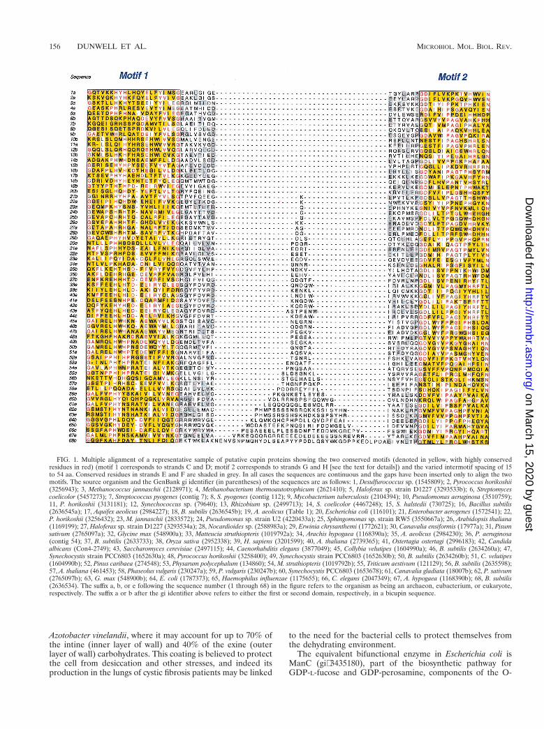
FIG. 1. Multiple alignment of a representative sample of putative cupin proteins showing the two conserved motifs (denoted in yellow, with highly conservedresidues in red) (motif 1 corresponds to strands C and D; motif 2 corresponds to strands G and H [see the text for details]) and the varied intermotif spacing of 15to 54 aa. Conserved residues in strands E and F are shaded in grey. In all cases the sequences are continuous and the gaps have been inserted only to align the twomotifs. The source organism and the GenBank gi identifier (in parentheses) of the sequences are as follows: 1, Desulfurococcus sp. (1545809); 2, Pyrococcus horikoshii(3256943); 3, Methanococcus jannaschii (2128971); 4, Methanobacterium thermoautotrophicum (2621410); 5, Haloferax sp. strain D1227 (3293533b); 6, Streptomycescoelicolor (5457273); 7, Streptococcus pyogenes (contig 7); 8, S. pyogenes (contig 112); 9, Mycobacterium tuberculosis (2104394); 10, Pseudomonas aeruginosa (3510759);11, P. horikoshii (3131181); 12, Synechococcus sp. (79640); 13, Rhizobium sp. (2499713); 14, S. coelicolor (4467248); 15, S. halstedii (730725); 16, Bacillus subtilis(2636545a); 17, Aquifex aeolicus (2984227); 18, B. subtilis (2636545b); 19, A. aeolicus (Table 1); 20, Escherichia coli (116101); 21, Enterobacter aerogenes (1572541); 22,P. horikoshii (3256432); 23, M. jannaschii (2833572); 24, Pseudomonas sp. strain U2 (4220433a); 25, Sphingomonas sp. strain RW5 (3550667a); 26, Arabidopsis thaliana(1169199); 27, Haloferax sp. strain D1227 (3293534a); 28, Nocardioides sp. (2588983a); 29, Erwinia chrysanthemi (1772621); 30, Canavalia ensiformis (17977a); 31, Pisumsativum (2765097a); 32, Glycine max (548900a); 33, Matteucia struthiopteris (1019792a); 34, Arachis hypogaea (1168390a); 35, A. aeolicus (2984230); 36, P. aeruginosa(contig 54); 37, B. subtilis (2633733); 38, Oryza sativa (2952338); 39, H. sapiens (3201599); 40, A. thaliana (2739365); 41, Ostertagia ostertagi (2996183); 42, Candidaalbicans (Con4-2749); 43, Saccharomyces cerevisiae (2497115); 44, Caenorhabditis elegans (3877049); 45, Collybia velutipes (1604990a); 46, B. subtilis (2634260a); 47,Synechocystis strain PCC6803 (1652630a); 48, Pyrococcus horikoshii (3258400); 49, Synechocystis strain PCC6803 (1652630b); 50, B. subtilis (2634260b); 51, C. velutipes(1604990b); 52, Pinus caribaea (274548); 53, Physarum polycephalum (134860); 54, M. struthiopteris (1019792b); 55, Triticum aestivum (121129); 56, B. subtilis (2635598);57, A. thaliana (461453); 58, Phaseolus vulgaris (230247a); 59, P. vulgaris (230247b); 60, Synechocystis PCC6803 (1653678); 61, Canavalia gladiata (18007b); 62, P. sativum(2765097b); 63, G. max (548900b); 64, E. coli (1787373); 65, Haemophilus influenzae (1175655); 66, C. elegans (2047349); 67, A. hypogaea (1168390b); 68, B. subtilis(2636534). The suffix a, b, or e following the sequence number (1 through 68) in the figure refers to the organism as being an archaeon, eubacterium, or eukaryote,respectively. The suffix a or b after the gi identifier above refers to either the first or second domain, respectively, in a bicupin sequence.
156 DUNWELL ET AL. MICROBIOL. MOL. BIOL. REV.
on March 15, 2020 by guest
http://mm
br.asm.org/
Dow
nloaded from

antigen gene cluster (140, 280, 283, 301). Other related bacte-rial enzymes include those encoded by noeJ from Rhizobium(81) and aceF, which is part of the acetan biosynthetic pathwayin Acetobacter xylinus (102). There are also related genes in thearchaeal species Pyrococcus horikoshii (gi3257338), Meth-anobacterium thermoautotrophicum (gi2622642), and Ar-chaeoglobus fulgidus (gi2649495).
Because of its importance in the synthesis of bacterial andfungal cell walls, PMI inhibition is a target for drug discovery(32). Although there is limited information on the structure ofthe active site, in the context of the conserved histidines in thetwo cupin motifs it is pertinent to note recent evidence (220)for the existence of a His residue in this site in a PMI fromXanthomonas campestris; this particular PMI is considered tobe a metalloenzyme and is activated by zinc.
Polyketide Synthases (Putative Cyclases)The polyketide pathway (115, 125, 194, 235) accounts for the
biosynthesis of many of the thousands of known secondarymetabolites, including antibiotics and pigments. Among theseproducts is curamycin (26), an antibiotic produced by Strepto-myces curacoi and based on a polyketide skeleton consisting ofa modified orsellinic acid—an unreduced version of 6-methyl-salicylic acid and the simplest of all aromatic polyketides. Itwas found (25) that the gene cluster responsible for the syn-thesis of this antibiotic was very similar to the S. coelicolor whiEgene cluster responsible for the synthesis of a grey spore pig-ment produced shortly before sporulation in the aerial myce-lium (51), and subsequent studies (36) demonstrated the wide-spread occurrence of gene clusters very similar to whiE amongother Streptomyces spp. Of specific interest to this review is thesequence of the homologous group of genes represented bycurC (S. curacoi), whiE ORFII (S. coelicolor) (8), sch ORFB(S. halstedii), and tcmJ (S. glaucescens) (33). The exact bio-chemical function of these gene products remains unknown,although it is suggested to be a cyclase (148, 318). Sequenceanalysis reveals the two conserved cupin motifs, separated by adistance of 15 residues, within a total protein size of approxi-mately 150 aa. It has been suggested recently (12) that use ofthe CurC sequence is the most efficient means of identifyingother members of the cupin family in a PSI-BLAST search (7).
Recent analysis (69) has extended the number of membersin this particular cupin subfamily to include several other closerelatives, such as the sequence gi2635101 (YrkC) from B.subtilis and the 140-aa Pep1 sequence (gi1572541) encoded bygene tnpA of the cryptic transposon Tn4321 within the broad-host-range IncPb plasmid R751 of Enterobacter aerogenes (267,291). The notes accompanying the Pep1 database submissionrecognized a “possible polyketide cyclase on basis of weaksimilarity to TcmJ of S. glaucescens” (E value 8.5). However, itis most similar (E value 8e-08) to a 97-aa sequence encoded bynucleotides 180 to 467 of a contig (gnlStanford_382smelil_423025B02.xl) from Sinorhizobium meliloti.
It was assumed previously that the smallest of all cupins isthe 77-aa “membrane-spanning protein” gi1017816 fromStreptomyces coelicolor (181, 182). However, the start codon forthis sequence has been reassigned, and it is now considered toencode a 115-aa protein (gi5457273) that is most similar to a79-aa polypeptide encoded by nucleotides 243111 to 243347from contig 7 of Streptococcus pyogenes.
DioxygenasesSeveral types of dioxygenase enzymes are probable members
of the cupin superfamily. They can be divided into two cate-gories, those with a single domain and those with two domains
(bicupins); within each subcategory the individual memberscan be recognized on the basis of a characteristic inter-motifspacing.
3-Hydroxyanthranilate 3,4-dioxygenase (3-HAO) (EC 1.13.11.6), with an intermotif spacing of 19 or 23 aa, is a eukaryoticenzyme that cleaves the aromatic ring of 3-hydroxyanthranilicacid to produce 2-amino-3-carboxymuconic semialdehyde, anintermediate in the synthesis of the excitotoxin quinolinic acid(21); this compound kills neurons by activation of N-methyl-D-aspartate receptors, and inhibition of 3-HAO is therefore apharmaceutical target (35). The enzyme is well characterizedin mammals (210) and is part of the kynurenine pathway forthe catabolism of tryptophan. Recently, the yeast geneYJR025c has been shown (164) to encode a 3-HOA(gi1353060) homologous to the human equivalent (190) andhas been renamed BNA1 (biosynthesis of nicotinic acid). Avery similar polypeptide (E value 5e-64) is encoded by part ofa contig (gnlStanford_5476C.albicans_Con4-2428) fromCandida albicans. Alignment of these 3-HOA sequences showsa notable difference between the Saccharomyces sequence andthe other sequences, in that the former protein has an inter-motif spacing of 23 residues compared with 19 for the othersequences. This insertion of 4 aa occurs in the loop betweenthe E and F strands of the barrel.
In common with most other dioxygenase enzymes, 3-HAOrequires nonheme iron as a cofactor. However, in contrast tothe multimeric composition of the related, two-domain dioxy-genases described below, this enzyme seems to be monomeric.
Cysteine dioxygenase (CDO) (EC 1.13.11.20), with an inter-motif spacing of 28 residues, is a key enzyme of cysteine me-tabolism and catalyzes the production of cysteine sulfinate.The rat (296), human (232), and Caenorhabditis elegans geneshave been well characterized, with the closest bacterial rela-tives of these eukaryotic sequences being those from B. subtilis(gi2635598), Streptomyces coelicolor (gi2687337), and Myco-bacterium tuberculosis (gi2896702). This enzyme is known tobe monomeric, with one atom of iron per molecule (312); itsactivity is strongly reduced by chelators of Cu1 and Fe21 (247).
Spherulins
The life cycle of the simple slime mold Physarum polyceph-alum involves a transition between two vegetative states, theamoeba and the plasmodium. Amoebae are the uninucleatehaploid cells, which under some conditions will fuse and dif-ferentiate into a giant multinucleate diploid plasmodium.When these latter cells are grown in liquid medium, they frag-ment into microplasmodia, which are capable of withstandingadverse conditions by encystment. This transition into hard-walled oligonucleated spherules is termed spherulation, and itis induced by starvation (or high concentrations of some car-bohydrates), cooling, dehydration, acidic pH, and/or sublethalconcentrations of heavy metals (47, 144).
As part of a molecular study of this phase transition, it wasshown first that the major changes in protein synthesis takeplace 24 h after the beginning of starvation-induced spherula-tion (31) and that the four most abundant spherulation-specificRNAs accounted for more than 10% of all mRNAs presentafter this period (30); these mRNAs were not present in en-cysting amoebae or in sporulating plasmodia. Differential hy-bridization of a cDNA library was used subsequently to isolatefull-length clones (29), of which two were found to be 76%similar and encoded proteins named spherulins 1a and 1b(81% identical). These proteins possess a potential signal pep-tide and an N-glycosylation site and were therefore presumedto be cell-wall glycoproteins. It was discovered subsequently
VOL. 64, 2000 EVOLUTION OF THE CUPIN SUPERFAMILY 157
on March 15, 2020 by guest
http://mm
br.asm.org/
Dow
nloaded from

(171) that there is 44% similarity at the amino acid level be-tween spherulin 1b and the wheat germin GF-2.8; this valueincreases to 60% for the central core sequence, the region thatcontains the conserved PH(I/T)HPRATEI decapeptide desig-nated the germin box. They can thus be considered cupins, withan intermotif spacing of 21 aa.
An interesting addition to the discussion on the evolutionaryorigin of the spherulin genes is provided by an analysis ofintron position (20) in a series of related cupins. The discoverythat the C-terminal domain of several seed storage proteins(e.g., those of Welwitschia mirabilis and Gingko biloba) sharedan intron position with the spherulins (although shifted by 2 bpin P. polycephalum) provided strong support for the conceptthat these proteins have a common ancestor.
To date, no biochemical function has been assigned to thesespherulins, although they do not seem to have any OXO ac-tivity (173). However, it is relevant to consider their possiblefunction(s) in the specific context of what is known about theconditions pertaining during spherulation and also in the gen-eral context of the link between cupins and stress responses inprokaryotes and eukaryotes. In particular, it is interesting tonote the link between oxidative stress and spherulation. Theinitial circumstantial evidence for such a link came from theobservation (2) that the herbicide paraquat, a compound thatgenerates free radicals, accelerated spherulation and also in-creased the specific activity of the manganese isoform of su-peroxide dismutase (3). It was also found that during thespherulation process in salts-only starvation medium, superox-ide dismutase activity increased 46-fold, along with an increasein the concentrations of H2O2 and organic peroxide (2); noneof these changes occurred in nondifferentiating cultures.
Germin and Germin-Like Proteins from Higher Plants
Wheat germin (which is an OXO), is the best characterizedof all the cupin proteins in terms of its biochemistry, function,and patterns of expression (45); it is therefore particularlyrelevant to consider these various features in some detail. Thefirst evidence for such an enzyme that converts oxalic acid anddioxygen to carbon dioxide and hydrogen peroxide came fromstudies of powdered wheat grains in 1912 (320), although it wasmore than 80 years later that the identity and sequence of thisenzyme were confirmed (173). In the meantime, there hadbeen two parallel and unrelated types of research concerningthis particular protein. The first of these concerned an impor-tant medical application of considerable commercial signifi-cance, namely, the use of barley OXO (98% identical to wheatgermin) in kits to assay levels of oxalate in blood plasma andurine. Some of these kits (e.g., the Sigma kit) utilize an enzymeisolated from barley roots, and although they are quick andeasy to use, there is a continuous effort to improve the accuracyand efficiency of the assay (175, 191, 228). Such efforts willbenefit from recently obtained data regarding fundamentalbiochemical and structural analysis of the barley enzyme itself(161, 162, 238, 310) and from the finding (173) that the ex-tremely well characterized wheat germin is also an OXO. Thisdiscovery was the culmination of the second important re-search track, one which started in the early 1980s, during whichthe GF-2.8 germin (gi121129) was found to be an apoplastic,multimeric (310), glycosylated (135) enzyme with extreme re-sistance to heat and to chemical degradation by protease orhydrogen peroxide. These unusual properties have recentlybeen explained by the realization that wheat germin and itsrelatives from barley and other cereals (206) are members ofthe cupin family and that their resistance to extremes of envi-ronment is likely to be a function of their structural similarity
to other desiccation-tolerant proteins including 7S and 11Sseed storage proteins; the resistance of the protein to H2O2 isof course linked to its enzymatic generation of this compound.
Germin-like proteins (GLPs) have a maximum ca. 90% se-quence identity (e.g., gi1772596) to wheat germin, althoughthe average level of identity is closer to 50%. There is almostcomplete identity in the conserved cupin core, in which theintermotif spacing is 20 to 23 aa. Since the discovery of the firstGLP in a higher plant (127), there has been a rapid expansionin the number of gene sequences identified, such that the latestestimates give a total of 21 sequences in Arabidopsis thaliana,the best-characterized plant genome to date (46; J. M. Dun-well, unpublished data). However, no function has yet beenassigned to any of these sequences, with the single exception ofa Pinus caribaea GLP, which does have OXO activity (212). Inaddition to the identification of GLP genes in analyses ofvarious plant genomes, expression of certain GLPs in plants,including liverworts (gi4718551) and mosses (gi6042701,gi6102532), is associated with a range of specific developmen-tal states but more particularly with specific biotic and abioticstresses, as detailed below.
Germin-like proteins are expressed at specific developmen-tal stages in plants. Various studies have identified GLPsduring specific stages of plant development.
(i) Floral induction. Interesting evidence for the develop-mental induction of GLPs in higher plants has come fromstudies of floral induction; for example, a specific GLP tran-script was found to show a circadian pattern of expression inthe long-day plant Sinapis alba (113) and its relative A. thaliana(273). Similar results were obtained in the short-day plantPharbitis nil (218), where a GLP mRNA was detected specifi-cally in the cotyledon and leaf. In a related study, the level ofa GLP in Raphanus sativus was found to be lower in youngflower buds than in leaf and root material (207), and a similarGLP (gi6090829) has recently been isolated from nectar ofNicotiana plumbaginifolia (46a).
(ii) Fruit ripening. Studies of ripening fruit of mandarin(118) (gi1669031), strawberry (Dunwell, unpublished), andapple (gi3088119) have all reported finding GLP sequences.
(iii) Somatic and zygotic embryogenesis. Following initialstudies which identified several GLPs in embryogenic culturesof Caribbean pine (Pinus caribaea Morelet var. honduriensis)(59), a full-length GLP (gi2745848) expressed in both somaticand zygotic embryos was reported recently (212). Similarly,GLP sequences have been found to be associated with somaticembryos of Monterey pine (Pinus radiata) (gi2935521), a sus-pension culture of potato (gi3171251), and a cell culture oflupin (309).
(iv) Seed development. In a study (180) of proteins known toprovoke severe allergic reactions (part of the celery-birch-mug-wort-spice syndrome), it was shown that the N-terminal se-quence of the 28-kDa allergenic protein extracted from pep-percorns of Piper nigrum has a high level of similarity (E value4e-05) to a GLP (gi2801803) from rice. This observation maybe linked to the fact that the well-characterized major peanutallergen Ara h1 is a vicilin-like protein (258).
(v) Wood development. Recent studies (6) on a cDNA li-brary produced from immature xylem from differentiatingwood in loblolly pine (Pinus taeda L.) identified a sequence(gi3365535) encoding a GLP similar (E value 3e-15) to anArabidopsis GLP (gi1755152) and the Physarum spherulin(gi1052776). It is relevant that the largest group of sequenceswith known function from this study were those associated withcell wall formation and the lignin biosynthetic pathway, anunsurprising conclusion in view of the fact that pine xylem ischaracterized by massive cell walls. Similar studies (275) on
158 DUNWELL ET AL. MICROBIOL. MOL. BIOL. REV.
on March 15, 2020 by guest
http://mm
br.asm.org/
Dow
nloaded from

developing xylem elements of poplar (Populus balsamiferasubsp. trichocarpa) also revealed two GLP sequences(gi3857819 and gi3858018).
Germin-like proteins are linked to specific plant-microberesponses. Evidence of a role for GLPs in the relationshipbetween plants and microbes has come from studies of nodu-lation in legumes, as well as from investigations of specificpathogen responses in cereals.
(i) Nodulation in legumes. The first evidence for the occur-rence of a GLP in a legume species came from a study of themechanism of attachment of Rhizobium (and probablyAgrobacterium) bacteria to the walls of plant cells, althoughthis was not recognized as being so in the publication in ques-tion (284). The initial step in this non-host-specific attachmentprocess involves rhicadhesin, a calcium-dependent (265) bac-terial surface protein of about 14 kDa (264, 266, 285). Using anassay based on the suppression of rhicadhesin activity, a puta-tive plant receptor molecule for this protein was purified fromcell walls of pea roots (284). The N-terminal 29 aa of thisprotein were determined to be ADADALQDLC(?)VADYASVILVNGFASK(Q)(P/Q)LI. Although the authors of thisstudy found no homology to known proteins, this sequence isvery similar (69% identity; E value 0.006) to an ArabidopsisGLP (gi1934730). Of particular relevance to the discussionelsewhere in this review is the observation that the receptormolecule was most easily removed from the cell wall with anaqueous solution of oxalate and oxalic acid. This finding sug-gests that the protein requires calcium for its anchoring, func-tion, or stability and adds to the circumstantial evidence linkingoxalate to the level of calcium in the cell wall and the conse-quent functional control of other proteins in that environment.
In addition to this evidence for the existence of a GLPrelated to bacterial attachment to the wall of legume root tips,it is known that oxalate itself is found at the very high level of70 mM in faba bean (Vicia faba) nodules (294). Application ofwater stress to such nodules increases the level of bacteroidOXO fourfold and reduces the level of oxalic acid by 55%(295). It is suggested that the oxalate found in this locationcould act as a complementary substrate for bacteroids and as ameans of slowing the decline in nitrogen fixation induced bywater-restricted conditions.
(ii) Pathogen responses in plants. Plants defend themselvesagainst pathogen attack by utilizing a variety of mechanismsthat include the production of specific antimicrobial com-pounds, the cross-linking of lignin and proteins in the cell wall,the synthesis of cell wall-strengthening carbohydrate polymers,and hypersensitive cell death. Although a role in pathogenresponse was among the earliest of functions suggested forgermin (170, 174), such a connection was not established untilthe identification of germin as an OXO, together with otherstudies on the interactions of powdery mildew, Blumeria (syn.Erisyphe) graminis, with leaves of barley (62, 63, 303, 322) andwheat (129). Subsequently, it has been shown that a specific-pathogen-response OXO transcript is found in the wall ofbarley mesophyll cells 6 h after inoculation with mildew; theenzyme accumulates after 15 to 24 h (324). Additionally, arelated sequence has been isolated from barley which showspapilla-mediated resistance to this disease (303). This partic-ular transcript peaks at about 18 to 24 h after infection, spe-cifically in the epidermal cells. Analysis shows that this tempo-ral and spatial pattern of expression closely follows theformation of papillae, appositions formed on the inner surfaceof the epidermal wall and thought to be composed of proteins,polyphenols, callose, silicon, and guanidine-containing com-pounds. Such a composition is reminiscent of the complexspherule and capsule walls referred to above. It has been sug-
gested that the H2O2 produced by the OXO members of thisfamily may act as a messenger for activation of other defensegenes in the same cell or in neighboring epidermal or meso-phyll cells. It is also relevant to note the tenacious associationbetween wheat germin and the arabinose-rich hemicelluloses(arabinoxylans or arabinogalactans) of cereal walls (135).
There is increasing evidence that there are common linksbetween the transduction pathways for the detection of andresponse to biotic and abiotic stresses and that active oxygenspecies are involved in the plant-environment interaction (290,308). In particular, the role of H2O2 in the generation ofhydroxyl radicals (OH) has been proposed (84). In this context,it may also be relevant to consider the potential role of thecrystal idioblasts, specialized cells that contain crystals of cal-cium oxalate and occur throughout the leaves of many plants.It has been demonstrated (58) that certain pathogenesis-re-lated proteins accumulate within these cells, and of course thesupply of oxalate in these cells would provide a source of H2O2
if adequate levels of OXO were present.Recently, the first circumstantial evidence linking a GLP to
a pathogen response in a dicotyledonous species was reported(B. Fristensky, unpublished data); the EST sequence gi4090021, found during a study of gene expression in leaves ofBrassica napus infiltrated with pycnidiospores of Leptosphaeriamaculans PG2, encodes a protein identical (with one frame-shift) to the GLP1 gi914911.
Germin-like proteins are induced by abiotic stress in plants.The first evidence for induction of GLP expression by abioticstress was provided by a study of salt stress in barley roots (126,128). Related results were subsequently obtained from thecommon ice plant Mesembryanthemum crystallinum, a faculta-tive halophyte and a model (37) for the induction of Crassu-lacean acid metabolism during water stress and treatment withhigh levels of salt. It was found (1) that the oxalate content ofthe leaf bladder cells increased from ,1 mM to 106 mM as saltlevels were increased from 1 to 5 mM. These results may berelated to the modulation of a GLP mRNA found duringtranscript analysis in this species (10, 204) and to the morerecent identification of other similar ESTs (e.g., gi3325551and gi4996622) in salt-treated plants. The link between ox-alate metabolism and GLP induction is considered in detailbelow.
Among the most interesting of the cupin proteins related toabiotic stress is BspA (for “boiling-stable protein”), a 66-kDaprotein highly expressed in cultured shoots of aspen (Populustremula) exposed to water stress (222). This protein is alsoinduced by abscisic acid application and by osmotic and coldstresses. In a recent study of greenhouse-grown plants (223) alower level of expression of BspA was found in Populus tomen-tosa than in Populus popularis, a species more tolerant of waterstress. It has been suggested that BspA contributes to mem-brane stability, a feature of considerable significance in rela-tion to stress responses. Other abiotic stresses which recentlyhave been shown to induce GLPs include manganese defi-ciency in tomato roots (gi2979494; gene Mdip1), aluminumtreatment in wheat (gene war13.2) (108), heat treatment inbarley (298), and submergence in rice (gi2952338, gi3201969; see also tomato EST gi28973890 and gi5827572from Botrytis). The most comprehensive of these studies is thatutilizing a promoter-glucuronidase (GUS) fusion (27, 28) andshowing induction of the wheat germin promoter in transgenictobacco treated with salt, heavy metals, aluminum and plantgrowth regulators, specifically auxin and gibberellin.
VOL. 64, 2000 EVOLUTION OF THE CUPIN SUPERFAMILY 159
on March 15, 2020 by guest
http://mm
br.asm.org/
Dow
nloaded from

Auxin-Binding Proteins
Auxin-binding proteins (ABPs) (intermotif spacing of 24 aa)are dimeric, glycosylated plant proteins encoded by a smallgene family in each species. They are thought to act as areceptor for the auxin indole-3-acetic acid (141, 142, 300) andthereby to mediate a wide range of physiological responsesincluding a reduction in cytoplasmic pH in certain cells (93).Analysis of the gene structure reveals a four-intron/five-exonarrangement, with the central, third exon encoding the regionwhich includes the peptide responsible for binding the carbox-ylic acid group of indole-3-acetic acid. This motif, known asbox A (41) or D16 (300), is now thought to be equivalent to theconserved motif 1 in the cupin notation (69), a finding sup-ported by observations on two similar proteins isolated fromshoot apices of peach (Prunus persica L. cv. Akatsuki) (217).These latter proteins have been designated ABP 19(gi1916807) and ABP 20 (gi1916809) on the basis of theirability to bind auxin, albeit at low affinity (217). Recent analysisof their sequences shows a greater level of similarity to theGLPs (the closest neighbor [E value 3e-78] is GLP3[gi1755164] from A. thaliana) than to any of the functionallybetter characterized ABPs.
Epimerases
Another group of cupin enzymes involved in the synthesis ofbacterial and archaeal cell wall components are the epime-rases, such as dTDP-4-dehydrorhamnose 3,5-epimerase (alsoknown as dTDP-L-rhamnose synthase) (EC 5.1.3.13), whichconverts dTDP-4-keto-6-deoxy-D-glucose into dTDP-4-keto-6-deoxy-L-mannose. These enzymes are about 185 aa in lengthand contain the two-motif cupin signature usually separated bya distance of 28 residues; both motifs contain a single globallyconserved histidine residue. They are encoded by rfbC (orequivalent), part of the rfb gene cluster (160, 189, 193, 205,276). Most rfb operons start with an rfbABCD cluster, which isresponsible for the synthesis of TDP-rhamnose (184); this clus-ter is followed by rfbIFGH in organisms that produce 3,6-dideoxyhexoses.
These epimerases are located in the periplasm, and it isrelevant to the theme of this review to note that periplasmicproteins are, as a rule, folded into stable, protease-resistantconformations, consistent with the digestive nature of thiscompartment (70).
Many of these capsular polysaccharides have potential eco-nomic importance as aqueous rheological control agents fordiverse industrial and food applications. Such compounds in-clude xanthan gum (Xanthomonas campestris) (22), and thesphingans (e.g., gellan, welan, and rhamsan) produced by spe-cies of Sphingomonas (314). It has been proposed that thevarious sphingans be thought of as defensive in nature, similarto the protective capsules (224, 249, 277, 297) of many invasivepathogenic bacteria (e.g. alginate).
MULTIDOMAIN PROTEINS WITH A SINGLECUPIN DOMAIN
In the multidomain proteins with a single cupin domain, theconserved cupin element does not lie at the core of the proteinbut instead represents a single domain in a complex multido-main organisation. The most notable group of proteins in thiscategory consists of a subset of the AraC bacterial transcriptionfactors.
AraC-Type Transcription Factors
Of all the bacterial transcriptional regulators, possibly thebest characterized are the members of the AraC/XylS family(88). This family, named after its first member, AraC (a regu-lator of the arabinose pathway in E. coli), contains more than100 members, which can be subdivided into various classes ona functional basis. These functions are associated primarilywith carbon metabolism, stress responses, and pathogenesis,with the former category including factors that control thedegradation of arabinose (AraC), cellobiose (CelD/ChbR),melibiose (MelR), raffinose (RafR), rhamnose (RhaR), andxylose (XylR).
Sequence analysis shows most members of this family to be250 to 300 residues in length, comprising a conserved C-ter-minal of about 100 aa which binds DNA, and a nonconservedN-terminal domain which binds the effector molecule (44).There is much more information available on the DNA bind-ing component, although the specific details of the N-terminalsection (particularly of the AraC protein) are more relevant tothe present review. This regulator has been subject to detailedstructural (269, 270) and molecular (250, 255) analysis overseveral years. In summary, the N-terminal section comprises anarabinose-binding, eight-stranded b-barrel, which is joined tothe DNA-binding domain via a linker region; the barrel-shaped section is also responsible for the dimerization of themolecule, a factor which determines its 3D shape and there-fore its ability to bend the associated DNA strand. Close anal-ysis of the sequence (90) and structure (Dunwell, unpublished)of this barrel-shaped element reveals a previously undetectedsimilarity to the conserved b-barrel core of the cupin proteins(Fig. 2). Of this related subgroup of regulators involved insugar degradation, that showing the closest sequence similarityto GLPs and other cupins is CelD (221). This protein wasnamed on the basis of its presumed involvement in the utili-zation of cellobiose, although recent studies (150) have shownthat the real function is as a regulator in the catabolism of thedisaccharide chitobiose; on that basis, its gene has been re-named chbR, part of the chb (N,N-diacetylchitobiose) operon.The significance of this reassignment is that it further supportsa functional link both to the other bacterial enzymes concernedwith sugar metabolism (e.g., PMIs and epimerases) and to thehigher-plant cupins, particularly the sucrose-binding proteins(detailed below). In this context, there is an additional circum-stantial link between chitobiose and cupins, in that vicilinsfrom cowpea (Vigna unguiculata) are known to bind chitin(248), and it has been suggested that the vicilin-induced inhi-bition of yeast cell growth is due to binding of the protein tothe chitin component of the cell walls (96, 97).
TWO-DOMAIN BICUPINS
The first two-domain proteins recognized to be members ofthe cupin superfamily were the seed storage proteins (20);these are discussed below, particularly with reference to thestructural analysis of cupins. More recently, several microbialproteins from archaea, bacteria, and fungi have been shown tohave a two-domain cupin composition (64, 69), and this infor-mation has provided a new insight into the possible ancestralorigin of the seed proteins. To distinguish the various sub-classes of two-domain cupin, sequences are described in termsof their intermotif spacing and in terms of whether this spacingis the same (homo-bicupins) or different (hetero-bicupins) inthe two domains.
160 DUNWELL ET AL. MICROBIOL. MOL. BIOL. REV.
on March 15, 2020 by guest
http://mm
br.asm.org/
Dow
nloaded from

Gentisate 1,2-Dioxygenase and1-Hydroxy-2-Naphthoate Dioxygenase
Identification of the two-domain composition of gentisate1,2-dioxygenase (GDO) and 1-hydroxy-2-naphthoate dioxyge-nase (HNDO) is a novel finding made during the preparationof this review. The two enzymes are involved in the degrada-tion of a range of related aromatic compounds, with the formerenzyme, GDO (EC 1.13.11.4), catalyzing the oxygenolyticcleavage (between carbons 1 and 2) of gentisate (2,5-dihy-droxybenzoate) to form maleylpyruvate, a compound that canbe converted to central metabolites of the Krebs cycle either bycleavage to pyruvate and maleate or by isomerization to fu-marylpyruvate and subsequent cleavage to fumarate and pyru-vate. GDOs have been purified and characterized in manygram-positive and gram-negative bacteria (Klebsiella pneu-moniae [143, 281], Moraxella osloensis [50], Sphingomonas[305], and Actinomycetales [109]), with possibly the best char-acterized such genes being those from species of Pseudomonas(110). For example, a GDO encoded by nagI (gi3406827) hasrecently been identified in P. aeruginosa strain U2 (86) and avery similar polypeptide (E value 3e-45) is encoded by nucle-otides 5549669 to 5548674 of a contig (gnlPAGP_287Paeruginosa_Contig54) from Pseudomonas strain PAO1. An-
other very similar sequence (gi3293534) (Fig. 1) has alsorecently been found in Haloferax sp. strain D1227, an extremehalophile isolated from soil contaminated with highly saline oilbrine and the only known aerobic archaeon able to utilizearomatic compounds as its sole carbon sources (85).
The only previous comment on the sequence similarity ofthese two types of dioxygenase was that made by Werwath etal. (305), who cloned the GDO gene gtdA (gi3550667) fromSphingomonas sp. strain RW5 and showed that its product hada low similarity to the HNDO (EC 1.13.11.38) (gi3288681)encoded by the phdI gene of the phenathrene-degrading No-cardioides sp. strain Kp7 (134). This latter enzyme catalyzes thecleavage of 1-hydroxy-2-naphthoate to trans-29-caboxybenzal-pyruvate, a ring cleavage between the carboxylated and hy-droxylated carbons analogous to that effected by GDO.
Both classes of enzyme described in this section have amultimeric structure; GDO has an apparent subunit molec-ular mass of 38 to 39 kDa and is claimed to have either atetrameric (85, 281, 305) or hexameric (151) composition,whereas HNDO has a molecular mass of 45 kDa and isconsidered to be hexameric (134). Like most other dioxyge-nases of the extradiol class (those that cleave an aromaticring adjacent to two vicinal hydroxyl groups), both GDO and
FIG. 2. Comparative structures of two orientations of the arabinose-binding domain of the AraC protein (above) and the two-domain phaseolin storage protein(below), showing the similar b-barrel element in the center of each domain, with associated a-helixes. The apparent gap in the E/F loop in phaseolin is due to the lackof resolution of the 3D structure at that point (177).
VOL. 64, 2000 EVOLUTION OF THE CUPIN SUPERFAMILY 161
on March 15, 2020 by guest
http://mm
br.asm.org/
Dow
nloaded from

HNDO contain 1 mol of Fe21 per mol of subunit (thosefrom Arthrobacter globiformis and Bacillus brevis containmanganese, although they utilize the same coordinating res-idues). These features, namely, a tetrameric or hexamericcomposition and the presence of a transition metal in theactive site, are shared with other cupin proteins described inthis review, such as barley OXO, which is now known tocontain manganese (238, 239; S. Bornemann, personal com-munication).
Oxalate Decarboxylases
Among the many oxalate-degrading enzymes isolatedfrom fungi, possibly the best characterized is that from thewood-rotting fungus Collybia velutipes. This particular ho-mo-bicupin enzyme (intermotif spacing of 20 aa in eachdomain) degrades oxalate to formate and carbon dioxideand appears not to have any requirement for cofactors. Itwas therefore selected for use in strategies to reduce thelevels of endogenous oxalate in plants (198, 199). The en-zyme itself has an acidic pI, is stable over a wide pH range,is moderately thermostable, and has a molecular mass of 560kDa as estimated by gel filtration and a subunit mass of 64kDa before and 55 kDa after treatment with endo-b-N-acetylglucosaminidase, thus suggesting a glycosylated status(198). The sequence of the C. velutipes enzyme has beenpublished as gi1604990 (52), and recently the sequence of
a similar enzyme from Aspergillus phoenices was reported(C. J. Scelonge and D. L. Bidney, 1 October 1998, PCTpatent application WO 98/42827). Presumed homologues ofthese sequences have also been identified (Dunwell, unpub-lished) (see below) in the bacterial species B. subtilis andStreptococcus mutans (encoded by nucleotides 555 to 1676from contig 1009) (Fig. 3).
Sucrose-Binding Proteins
Among the two-domain relatives of the seed storage pro-teins is a sucrose-binding protein (SBP) (gi548900 andgi2765097) found at low abundance in the plasma membraneof cotyledons, leaves, and mature phloem of legumes (103); asimilar sequence (gi2148163) from the cycad Zamia furfura-cea is known (40). Recent comparison (219) of the soybeanSBP sequence with that of vicilin has shown that the N-termi-nal domain of SBP contains 12 of the 13 residues conservedacross the whole vicilin family, with the C-terminal domainhaving 10 of the 12 conserved residues.
Although the overall tertiary structure of SBP can be pre-dicted by comparison to phaseolin, it is also possible that anal-ysis of the disaccharide-binding domain of CelD/ChbR (see“AraC-type transcription factors” above) would provide fur-ther information on the specific ligands in the binding site.
FIG. 3. Alignment of the six 20120 bicupin proteins (presumed OXDCs) from Streptococcus mutans (S. mut), Bacillus subtilis (B. sub1, YvrK; B. sub2, YoaN),Collybia velutipes (C. vel), Aspergillus phoenices (A. pho), and Synechocystis (Syn.) (see the text for details), showing the positions of the two conserved motifs (boxed)within each of the two domains. Residues conserved in all sequences are indicated with asterisks below the alignment; residues also conserved between the two domainsare indicated with asterisks above the alignment. The A. phoenices sequence (Scelonge and Bidney, PCT patent application WO 98/42827) has been amended byinsertion of an additional nucleotide at residue 344 to correct a presumed frameshift error introduced during the sequencing of this gene.
162 DUNWELL ET AL. MICROBIOL. MOL. BIOL. REV.
on March 15, 2020 by guest
http://mm
br.asm.org/
Dow
nloaded from

Seed Storage Proteins
During the development of plant seeds there is a massiveaccumulation of nitrogen and carbon reserves in the form ofproteins that can withstand desiccation and be used as a sourceof energy for the germinating embryo. In legumes, the globulintype of storage proteins can be divided into two forms, thelegumins and the vicilins. The former are usually found ashexameric complexes (sedimentation coefficient, 11S), witheach subunit derived from a precursor complex consisting oftwo domains, an N-terminal acidic a chain and a C-terminalbasic b chain, which remain associated following proteolyticprocessing. The latter proteins occur as 7S trimers, with eachsubunit being a 50- to 70-kDa polypeptide that is subject tovariable levels of processing. Examination of Fig. 1 shows thatmost of the storage proteins either lack any of the conservedHis residues or contain a single conserved His in motif 1. It ispresumed that, as a consequence, they have no metal-bindingligands and therefore no enzymatic activity. There is, however,a massive accumulation of oxalate (maximum 24% [dryweight]) during early seed development in soybean (131) andpresumably in other legumes, and it is tempting to speculate onthe possibility that this compound acts as a substrate for aresidual oxalate-degrading capacity provided by the storageproteins being produced at that period. Knowledge of thetertiary structure of the two storage proteins phaseolin (177)and canavalin (155) and the finding of certain globally con-served residues (20) provided the basis for the generation of ahomology model of wheat germin (90) and all subsequentpredictions of cupin structures (Fig. 2).
In addition to the well-known major storage proteins foundin seeds and spores (261), other, less abundant proteins of thistype have been the subject of detailed analysis. Among the bestcharacterized is the major peanut allergen Ara h1, a memberof the vicilin family (43, 54, 258) and the protein responsiblefor the majority of cases of fatal food-induced anaphylaxis. Ina recent study (258), it has been shown using molecular mod-elling that the 23 linear immunoglobulin E-binding epitopes
cluster into two main regions, thus providing a rational targetfor transgenic approaches (66, 67) to modify the allergenicresidues. Like many other members of the cupin family de-scribed in this review, the Ara h1 protein has a very high levelof stability; it survives intact in most food-processing methodsand also resists digestion by the gastrointestinal tract or its invitro equivalent (23). It has been suggested (258) that thisstability may be due to its compact structure, which limits thepossibility for protease digestion and also facilitates its passageacross the small intestine. It is presumed that these biophysicalcharacteristics are shared by the allergenic single-domain GLPrecently identified in ground black pepper (180).
Bicupins of Unknown Function
As described above, there is now good evidence for a widevariety of bicupins from archaeal species (e.g., the GDO fromHaloferax [85]), many bacteria including B. subtilis and Strep-tococcus pyogenes (the 15115 bicupin encoded by contig 272)and several eukaryotes (e.g., seed storage proteins). With theexception of the two classes of dioxygenase and the OXDCsfrom Collybia velutipes and Aspergillus phoenices, no biochem-ical function has yet been assigned to the microbial bicupins. Itwould be of particular interest to investigate the activities ofthe four examples from B. subtilis, which now probably repre-sents the best organism for the study of prokaryotic cupindiversity. Within the higher plants, there is also evidence foranother previously unidentified class of bicupins (e.g., the Ara-bidopsis thaliana hypothetical gene gi2244827).
CRYPTIC SEQUENCES ENCODING CUPIN PROTEINS
In addition to the cupins described in the above section,there is a group of other related coding sequences (Table 1)(Dunwell, unpublished) not previously identified in the data-bases. These are either complete or partial ORFs, often foundin apparently noncoding regions of other genes. These crypticORFs can be divided into various types, according to the rea-
TABLE 1. Summary of cryptic sequences encoding cupin proteins
SpeciesGenBank gi
identifier(reference)
Nucleotide position Closest neighbor, possible function
ArchaeaDesulfurococcus strain SY 1545808 (316) 104–448 giP3256943 from Pyrococcus horikoshii. probable PMIThermococcus strain KS-8 4001717 1–349 As above
EubacteriaAquifex aeolicus 2983162 (53) 7591–7914 15–164 gn1PTIGRPBTMBX89F Thermotoga maritimaAeromonas caviae 4204207 232–23 (negative strand) 2632508 from B. subtilis AraC?Mycobacterium leprae 1377767 19060–19155 giP2984230 from A. aeolicusAlicyclobacillus acidocaldarius 39300 (157) 2196–1755 (negative strand) giP1907078 human pirin (304)Streptomyces lividans 48953 (234) 171–1 (negative strand) As aboveBacillus stearothermophilus 560029 (216) 338–1 (negative strand) 146366–146533 contig 230 from P. aeruginosaDesulfovibrio desulfuricans 49285 (278) 238–726 16083–115762 contig 272 from Streptococcus pyogenesPseudomonas lemoignei 531465 1618–1286 (negative strand) 82–405 contig 854 from Bordetella pertussisMorganella morganii 508518 (60) 1666–1184 (negative strand) giP2632720 ydaE from Bacillus subtilis; sugar alcohol
metabolism?Corynebacterium glutamicum 2342561 (137) 3686–3357 (negative strand) Pep1 protein giP1572541 from E. aerogenesSynechocystis 287460 (178) 3–208 (add G at 104) C terminus of giP1652486, a PMI, same speciesAzorhizobium caulinodans 763059 (92) 1–201 giP1772621 from Erwinia chrysanthemiMycobacterium genavense 2558999 947–1519 (ATG at 960) As above
EukaryotaDictyostelium discoideum 1177288 (241) 302–3 (negative strand) 60364–60077 contig 229 from P. aeruginosaArabidopsis thaliana 987518 1195–1873 (add G at 1288) giP1755168 and giP16847 from Arabidopsis
VOL. 64, 2000 EVOLUTION OF THE CUPIN SUPERFAMILY 163
on March 15, 2020 by guest
http://mm
br.asm.org/
Dow
nloaded from

son for the previous lack of identification. In one case, thatfrom Mycobacterium genavense, it seems obvious that the in-correct start codon was selected and thus a protein with noknown similarity was generated. In contrast, the nonannotatedORF (NORF) in Aquifex aeolicus was simply not identified bythe algorithms used to find ORFs in such bacterial genomes(53). The occurrence of NORFs is well known from othercomplete genome or transcriptome studies such as that con-ducted on yeast, where serial analysis of gene expression tech-niques identified 160 NORFs (299). Presumably, the otherexamples identified in the present study were overlooked pre-viously simply because the ORF is in a reading frame differentfrom that used by the gene which was the main subject of thespecific study. In most cases, however, the analysis is alsocomplicated by the inclusion of one or more frameshift errorsin the sequence.
ANALYSIS OF CUPIN SEQUENCES IN B. SUBTILIS
Although the broad-ranging surveys described above are ofconsiderable value in determining the overall occurrence ofmembers of the cupin superfamily across various taxa, it wasconsidered particularly important to conduct a detailed surveyof a single prokaryotic genome in order to assess more accu-rately the spectrum of cupins encoded by such a genome. It wasalready known (64, 69) that archaeal genomes contain only afew (2 to 7) cupin genes, whereas the cyanobacterium Synecho-cystis has a complement of 18 cupins genes including oneencoding a bicupin (65). Preliminary studies (Dunwell, unpub-
lished) had suggested that B. subtilis was probably the mostappropriate organism for this analysis since its genome en-coded a greater variety of plant-related cupins.
Overall Conservation of Cupin Motifs in Proteins Encodedby the B. subtilis Genome
Analysis of the genome of B. subtilis, using the methodsdescribed above, identified a total of 20 sequences that fulfil, atleast in part, the characteristic two-motif cupin signature. Thealignment of this conserved section is given in Fig. 4, which alsoshows the range of intermotif spacing (15 to 54 aa) as well asthe overall protein size (113 to 432 aa). It can be seen that thesequences fall into several subgroups on the basis of theirdetailed similarity, with the great majority having the charac-teristic signature of three histidines (two in the first motif andone in the second), along with conserved proline and glycineresidues in the second motif.
Particular reference must be made to YdaE (gi2632720),which is most unusual in having an additional six residuesbetween strands C and D within motif 1. It also has a compar-atively long intermotif distance.
Closest Neighbors and Possible FunctionsOnly two of the cupins in B. subtilis have designated names
(PMI [phosphomannose isomerase] and SpsK [spore capsulesynthesis K protein]); most of the sequences are so-called ygenes (166), i.e., genes of unknown function that make up 70%of the total gene complement. The closest neighbor for each
FIG. 4. Alignment of the conserved two-motif signature in the cupin proteins from B. subtilis, showing the GenBank identifier, the gene name, site in the genome(in kilobases from origin; sequences likely to be included in the section of the chromosome trapped in the prespore during septation are denoted by asterisks) (311),coding strand, details of the two motifs, total size of the protein, and its calculated pI. The sequences are subdivided on the basis of similarity. In the four two-domainproteins (YxaG, YwfC, YoaN, and YvrK), the first and second domains are designated a and b, respectively.
164 DUNWELL ET AL. MICROBIOL. MOL. BIOL. REV.
on March 15, 2020 by guest
http://mm
br.asm.org/
Dow
nloaded from

protein sequence, as estimated by a BlastP analysis, is given inTable 2. In terms of function, it can be seen that the sequencescan be divided into various subgroups that include five AraC-type transcription factors, three PMIs, and a cysteine dioxyge-nase. However, an obvious problem inherent in this type ofcomparison based on the total sequence is that it takes noaccount of the occurrence of multidomain proteins. For exam-ple, analysis of sections of the SpsK protein suggests that itprobably represents a bifunctional enzyme similar to one fromActinobacillus actinomycetemcomitans, with an N-terminal do-main presumed to have dDTP–4-dehydrorhamnose reductaseactivity (cf gi2650312 from Archaeglobus fulgidus) and a C-terminal domain (containing the cupin element) with dTDP–4-dehydrorhamnose 3,5-epimerase activity (c.f. gi2622921from Methanobacterium thermoautotrophicum).
The unusual protein YdaE is most closely related to a pre-viously unidentified protein from Morganella morganii.
Additional confirmation of the different functional sub-groups can be obtained by examination of the pI values givenin Fig. 4. This shows that all the transcription factors havevalues between 6.10 to 8.48 whereas the other proteins (withthe exception of YjlB and YrkC) are more acidic, with valuesbetween 4.41 and 5.90.
Domain Structure
There are 16 single-domain and 4 two-domain (bicupin)proteins encoded by the B. subtilis genome (Fig. 4). Bicupinsare referred to below on the basis of their intermotif spacing(e.g., 15115, 20120). Of the former group of one-domainsequences, particular note should be made of the two examplesthat have a spacing of 20 residues, namely, YkrZ and YrkC.The former is most similar to a recently described sequencefrom the hyperthermophilic bacterium Aquifex aeolicus (53),whereas the second sequence is closer to a sequence fromPrunus persica.
Probably the most interesting of the latter group of bicupins
are the two sequences YoaN and YvrK, which have a very highlevel of similarity (E value 1e-130) to a sequence from Strep-tococcus mutans (contig 1009) and to the oxalate decarboxy-lases encoded by gi1604990 from Collybia velutipes, a wood-rotting basidiomycete (198), and the related sequence fromAspergillus phoenices (Scelonge and Bidney, patent applica-tion). These fungal enzymes are related to the Synechocystisprotein gi1652630 (69), the only other 20120 microbial bicu-pin identified to date. Detailed inspection of the six-sequencealignment provided in Fig. 3 reveals two main features. First,there are 64 (c. 16% of the total) globally conserved residues,mostly clustered within the two cupin motifs, which have thecomposition GX2RX2HWHX3/4EWX5G, and GX10HX4. Ofthese 64 residues, only 11 (ca. 3%), including the 3 histidines(90), also show conservation between the first and second do-mains. Second, the fungal OXDCs are more similar to thesequences from B. subtilis and S. mutans than they are to theSynechocystis protein.
Additional alignments of protein sequence (data not shown)suggest that the most likely single-domain progenitor of thetwo-domain 20120 proteins is YkrZ and that this protein isslightly more similar to YvrK than to YoaN. The evolutionarytime course of events is thus indicated to be (YkrZ) 3 2 3YvrK 3 YoaN. Similarly, it is likely that YjlB (18 spacing) isthe progenitor of its closest neighbor, the two-domain YxaG(15115) sequence (Table 2), although this would imply thatthe increase in intermotif spacing from 15 to 18 residues inYjlB occurred after the duplication event. It is also noticeablefrom the alignments of single cupins with their putative bicupinderivatives that the single-domain sequences (e.g., YjlB) al-ways show a higher degree of similarity to the C-terminaldomain than to the N-terminal domain of the respective bicu-pin (e.g., YxaG).
If alignments are based on the DNA rather than the proteinsequence, additional features can be observed (Fig. 5). Forexample, the doublet of bicupin genes (yvrK and yoaN) are very
TABLE 2. Analysis of the closest neighbors for each of the cupin sequences from B. subtilisa
Sequence (gi)Closest neighbor Length
(aa)Identity
(%)Similarity
(%)Gaps(%) P Function
Species (gi)
2633149 B. subtilis 2633014 96 31 58 5e-11 AraC2632815 L. monocytogenes 2745844 262 24 47 7 5e-17 AraC2633418 B. megaterium 2764541 265 25 44 12 9e-14 AraC2632508 E. coli 132526 248 23 41 8 8e-13 AraC2634298 P. leiognathi 2495367 243 27 44 10 5e-17 AraC2632900 B. subtilis 2636105 311 55 67 e-102 PMI2633556 B. subtilis 2636105 316 56 70 ,1 e-106 PMI2636105 B. subtilis 2632900 311 57 69 e-107 PMI2636545 A. aeolicus 2984227 61c 32 61 2e-042632720 M. morganii 508518b 155 41 55 14 e-182632741 S. meliloti Unfin.c 88d 42 61 7 2e-12 ?PKS2636309 E. faecalis Unfin.c 86d 26 44 2 0.362633581 D. radiodurans Unfin.c 136 37 57 2 5e-212633733 A. aeolicus 2984230 175 33 57 8 3e-232635101 P. persica 1916809 128 22 38 14 0.11 GLP2634260 B. subtilis 2635821 379 58 75 e-1342635821 B. subtilis 2634260 379 58 75 e-1342635598 C. albicans Unfin.c 101 32 50 5 2e-07 ?CDO2636317 A. actinomyc. Unfin.c 270 41 62 4 9e-66 dTDP-DRe
2636534 P. aeruginosa Unfin.c 236 23 40 0 2e-10
a Estimated by use of the gapped BlastP program.b This sequence was identified by a TBlastN search and encodes a previously unidentified polypeptide (see the text for details).c These sequences were identified by use of a TblastN search of the unfinished microbial genomes database, see text for details.d These relatively short regions span the conserved two-motif section of the protein.e dTDP-4-dehydrorhamnose reductase.
VOL. 64, 2000 EVOLUTION OF THE CUPIN SUPERFAMILY 165
on March 15, 2020 by guest
http://mm
br.asm.org/
Dow
nloaded from

similar to each other (65% identity; E value 2.2e-72), althougheach gene has a different pattern of insertions and deletions(indels). However, these differences in nucleotide sequence donot disrupt the conserved two-motif regions; where there are
indels within these motifs, they are equivalent in the two genesand do not alter the globally conserved residues.
In an earlier study (69) it was suggested that the two-domainOXDC proteins may represent direct progenitors of the two-
FIG. 5. Alignment of the two 20120 bicupin genes yoaN (giP2634260, denoted 4260) and yvrK (giP2635821, denoted 5821) from B. subtilis, showing the positionsof the two conserved motifs within each of the two domains. Similar nucleotides are shown as dots, and deletions are shown as dashes. The deletions marked withasterisks in motif 1 of the first domain (1-bp deletion) and motif 2 of the second domain (2-bp deletion) denote two examples of the compensatory system of deletionsand insertions that maintain the same reading frame for the two genes throughout the majority of the sequence. The deletions which produce a respective differencein the presence of a Gly residue (Fig. 3) are marked with vertical arrowheads. The statistical analysis is as follows: score 5 1,706 (256.0 bits), expect 5 2.2e-72, P 52.2e-72, identities 5 756/1,151 (65%).
166 DUNWELL ET AL. MICROBIOL. MOL. BIOL. REV.
on March 15, 2020 by guest
http://mm
br.asm.org/
Dow
nloaded from

domain storage proteins. Recent phylogenetic evidence (260)now shows that the two duplication events occurred indepen-dently.
Physical Location of Cupin Genes within theB. subtilis Chromosome
The cupin sequences are arranged on both DNA strands,and although they are distributed throughout the chromosome(Fig. 4), there is a possible increase in the kilobase value as thecomplexity of the protein increases. It is also noticeable thatthe two members of the doublet (yvrK and yoaN) are on op-posite strands and that all four of the two-domain sequencesare located in the second half of the chromosome (i.e., abovekb 2000).
SUMMARY OF GENOME ANALYSES OF B. SUBTILISAND OTHER ORGANISMS
There are several important conclusions to be drawn fromthis study on B. subtilis. Most importantly, it has identified apreviously unrecorded grouping of 20 cupin genes (0.5% of thetotal of 4,100) in the archetypal gram-positive species. Thisgroup of sequences provides evidence for two types of geneduplication having occurred during the evolution of the B.subtilis genome and/or the genome(s) of its progenitor(s).First, there has been duplication to increase the number ofcupin genes. It is estimated (166) that B. subtilis has 568 (14%)of its 4,100 genes in the form of doublets and 273 (7%) in theform of triplets. In the present study, the most obvious exampleof a doublet is yoaN and yvrK, the genes encoding two-domainproteins closely related to the fungal OXDCs. Similarly, pmiand its two related sequences are members of a triplet, and thefive genes encoding AraC-type transcription factors with iden-tifiable cupin motifs are representatives of an even larger genefamily (it is estimated that B. subtilis has a total of 11 membersof this class of transcription factor).
Second, there is evidence of duplication or fusion to producethe genes encoding the two-domain bicupin proteins. More-over, this process must have occurred on at least three occa-sions to produce YxaG (15115), YwfC (16116), and YvrK/YoaN (20120). One would assume that the chronologicalorder for these duplications was in the order 2 3 15, 2 3 16,and 2 3 20, although there is no certainty for this assumption.It is also unclear whether there is an extant progenitor for eachof the bicupins or whether the immediate progenitor was re-placed by its two-domain successor.
The other major conclusion to be drawn from this study isthat in comparison with other microbial genomes sequenced todate, the B. subtilis genome is the closest to the eukaryotes interms of both the overall number of cupins encoded and theirspecific domain composition. Of particular interest is the find-ing that one of the single-domain B. subtilis sequences(gi2635101) has a GLP sequence from an angiosperm (Table2) as its closest relative. It should be noted that this bacterialsequence, together with the similar sequence (gi3258400)from the archaeon Pyrococcus horikoshii (Fig. 1), were notavailable in the previous study (69). They are thus the first20-aa-spacing cupin sequences to have been identified in pro-karyotes. Together with the very similar 19-aa proteins(gi2984230 from Aquifex aeolicus and the polypeptide en-coded by nucleotides 46806 to 47348 in contig 235 fromPseudomonas aeruginosa), they are of great significance to ourunderstanding of the evolutionary origin of the increasinglyimportant eukaryotic GLPs, which in plants are encoded by agene family comprising many members. Such observationsconfirm that there has been large-scale multiplication of this
type of cupin sequence during the course of evolution. It isknown that GLPs occur in several species of gymnosperm,including Pinus caribaea (59, 212), P. radiata, and P. taeda (6).It is not known, however, whether this expansion in numbers,from two members in B. subtilis to a large family of genes inhigher plants, occurred before or after the divergence of theangiosperms from the gymnosperms.
EVOLUTIONARY ASPECTS OF CUPIN COMPOSITIONIN MICROBIAL GENOMES
Size of Cupin Gene Families in Prokaryotes and Eukaryotes
In general terms, it can be seen that the number of cupingenes varies from 2–7 in the archaeal genomes sequenced todate and in Aquifex aeolicus (often considered to be the mostdeeply branching bacterium) to 15–20 in B. subtilis and Syn-echocystis (65) to .40 in angiosperms and animals (Dunwell,unpublished). For example, in Arabidopsis thaliana there areabout 20 GLPs, which themselves can be further subdivided onthe basis of sequence into five to seven subclasses. Apart fromthe identification of OXO activity in some of the cereal ger-mins and in a related protein in Pinus caribaea (212), there isno functional information about these particular eukaryoticproteins.
Interestingly, with the exception of the epimerases (e.g.,gi1666505 from Leptospira interrogans), cupins appear to beabsent from spirochetes, as estimated by the analysis of therelatively small genomes of Borrelia burgorferi (genome size,1.44 Mb) (79), Treponema pallidum (1.14 Mb) (80), Rickettsiaprowazekii (the closest extant relative of the ancestor to mito-chondria, with a genome size of 1.10 Mb) (9), and Chlamydiatrachomatis (1.05 Mb) (274); for reference, the genome size ofB. subtilis is 4.20 Mb. Presumably, this specific absence ofcupins is part of the reductive evolution that leads to thegeneral absence of any genes for cellular biosynthetic func-tions, including most aspects of cell wall synthesis, in theseintracellular parasites.
It should also be noted that eukaryotic cupins are not re-stricted to fungi, yeasts, and plants. The latest estimate of theirabundance in animals (68) suggests a widespread occurrence,with Caenorhabditis elegans having a total of at least 15 se-quences (all single domain), including some (e.g., gi3877049)which have a 20-aa spacing and are very similar to sequencesfrom A. thaliana (gi2739365), rice (gi3201969), and H. sapi-ens (gi2822126). In terms of function, some of these animalsequences, such as PMIs, cysteine dioxygenases, and epime-rases, are related to microbial sequences described above,while others, such as the CENP-C centromeric proteins (282)and various zinc finger transcription factors, are specific toeukaryotes.
Do Cupin Families Arise from Gene Duplicationor Genome Fusion?
In the discussion above, it has been assumed that the geneduplication events to generate doublets and higher-order mul-tiples all took place in a single bacterial genome. However,there is a possibility that the present genome of B. subtilis is theresult of one or more fusions of ancestral genomes (either inwhole or in part) and that the doublets of genes represent theconsequence of this fusion process (130). Large-scale analysesof protein sequences have already led to the suggestion thatthe evolution of the archaea included at least one majormerger between ancestral cells from the bacterial lineage andthe lineage leading to the eukaryotic nucleocytoplasm (158,
VOL. 64, 2000 EVOLUTION OF THE CUPIN SUPERFAMILY 167
on March 15, 2020 by guest
http://mm
br.asm.org/
Dow
nloaded from
159, 240, 242). In the same context, it has also been proposed(105) that the eukaryotic nuclear genome is a chimera that hasreceived major contributions from both an archaeal speciesand a gram-negative species. Such a network of fusion eventswould clearly lead to a complicated structure for many pro-karyotic genomes, with gene doublets resulting from eitherfusion or internal duplication.
Physical Location of Cupin Genes in the Bacterial Genome
An important aspect of cupin evolution concerns the factthat cupin genes are distributed on both DNA strands of the B.subtilis chromosome. This suggests that at some stage duringevolution, there was a duplication of genes from one strand tothe other. If the specific example of the 20120 bicupins isexamined, for example, it can be seen that in Synechocystisthere is one such gene on the left strand whereas in B. subtilisthe two closely similar genes are transcribed by differentstrands (Fig. 4).
Analysis of the location of the cupin sequences within the B.subtilis genome (Fig. 4) shows a general progression, with thegenes encoding the shorter proteins being located closer to theorigin of replication than are the genes encoding the longerproteins. Similarly, all the two-domain sequences are locatedin the second half of the genome (from kb 2038 to 4106). Thereis little information on any factor(s) that might determine thehigher-order location of genes in this species, although it hasbeen suggested (166) that the “grey hole” located at kb ;600might be related to the temporary chromosome partition ob-served during the first stages of the sporulation, when a seg-ment of about one-third of the chromosome enters the pres-pore and remains the sole part of the chromosome in theprespore for a significant transition period (311). In light of thegeneral link between cupin proteins and sporulation/desicca-tion (20, 69), it is possible that there is a functional reason forthe clustering of genes required during this stage of the lifecycle.
There are two additional pieces of evidence for physicallinkage between related cupin genes and between such genesand other functionally related genes. Most interestingly, itseems likely that the recently sequenced genome of A. aeolicus(53) represents the earliest known stage of bacterial cupinevolution, a suggestion supported by the fact that the familyAquificaceae is the most deeply branching family within thebacterial domain on the basis of phylogenetic analysis of 16SrRNA sequences. Specifically, A. aeolicus genome containsthree cupin genes. Between aq_528 and aq_529 there is acryptic cupin gene encoding a protein with a 15-aa spacingsimilar to gi2128971 from Methanococcus jannaschii, and inaddition there are two other closely linked cupin genes(aq_1975 and aq_1978). The first encodes a protein with a19-aa spacing (gi2984230), and the second encodes a proteinwith a 15-aa spacing (gi2984227); both have close relatives inB. subtilis (Table 2). It seems most likely, therefore, thataq_1975 was the product of duplication of aq_1978 and that itthen diverged in sequence by the addition of 12 bp (encoding4 aa) to the intermotif region while retaining its close physicalposition in the chromosome. In contrast, their two paralogs inB. subtilis (yjlB and ykrZ) are located more than 200 kb apart,presumably as a consequence of recombination.
In this context, there is also an interesting relationship be-tween the ydbB gene and its adjacent sequences in the B.subtilis genome. A TBlastN analysis of this gene shows that itsclosest relative from plants is the wheat protein germin, sonamed because of its high level of expression in germinatingembryos (172). Adjacent to ydbB is gsiB (for “glucose starva-
tion-inducible protein B”) (gi2632740), a sequence that has asits closest neighbours (E values 2e-18, and 2e-15, respectively)the plant Em protein (gi1169515) from A. thaliana (91) andthe Lea (late embryogenesis abundant) protein (gi547819)from barley (75, 243). It is possible, therefore, that the twoadjacent bacterial sequences have a common developmentallink that began with a role in stress response, was retainedthroughout evolution, and is now associated with embryo de-velopment in higher plants.
Comparison of Single-Domain and Two-Domain CupinsDetailed analysis of various pairwise alignments of the sin-
gle-domain and two-domain cupins show a number of inter-esting features relating to the possible origin and evolution ofthese proteins. First, as described above, single-domain pro-teins are more similar to the C-terminal domain than to theN-terminal domain of their respective two-domain relatives.The reason for this disparity is unknown, but it is possible thatthere is less selection pressure to conserve the sequence of theN-terminal domain in a two-domain cupin. Another possibleexplanation for such variation is suggested by similar discov-eries in the family of extradiol dioxygenases, where there arealso single- and double-domain members (74). Among some ofthe latter enzymes, there is evidence that the two domainsexpress different phylogenies, suggesting the possibility thatthese particular enzymes arose from recombination or evenfusion of genes encoding different dioxygenases.
Cupins and the Comparative Structure ofMicrobial Cell Walls
The very close similarity between the two-domain 20120bicupin (gi1652630) from the gram-negative cyanobacteriumSynechocystis (65) and the related sequences from B. subtilisand Streptococcus mutans (Fig. 3) supports the observation(105) that the cyanobacteria constitute one of the deepest-branching clades within the gram-negative species and have aclose affinity to the gram-positive species. Indeed, analysisquoted by Gupta (105) and based on sequences from gluta-mate dehydrogenase, phosphoribosyl formyl glycinamidinesynthase, and some 16S rRNAs suggests a clade comprising thegram-positive bacteria, the cyanobacteria, and the archaea.Additional circumstantial evidence for a close link betweensome gram-positive bacteria and some archaea comes from ananalysis of the archaeal cupins. Whereas only two such se-quences have been identified in Methanococcus jannaschii (42),seven have been found in Methanobacterium thermoautotrophi-cum (Dunwell, unpublished). It may be that the additionalcupins in M. thermoautotrophicum are linked to its particularcell wall characteristics, since it was noted by Gupta (105) thata number of archaea, including Methanobacterium, exhibited athick and homogeneous cell wall that shows positive staining inthe Gram reaction. Perhaps the M. thermoautotrophicum se-quences with greatest relevance in the context of wall complex-ity are the two hypothetical proteins gi2621649 (89 aa) andgi2621650 (89 aa) that contain sequences with similarity tothe first and second motifs of the spsK (for “spore capsulesynthesis K” [gi2636317]) protein from B. subtilis. Indeed,close analysis of these two adjacent archaeal ORFs suggeststhat they are more likely to be contiguous sections of a singlecoding region, particularly since addition of a single nucleotide(to correct a frameshift) produces a modified, longer ORFencoding a polypeptide most similar (E value 3e-19) to theC-terminal 151 aa of this B. subtilis protein, a probable dTDP-4-dehydrorhamnose 3,5-epimerase (cf gi2622921). Such en-zymes are involved in the rhamnose biosynthetic pathway
168 DUNWELL ET AL. MICROBIOL. MOL. BIOL. REV.
on March 15, 2020 by guest
http://mm
br.asm.org/
Dow
nloaded from

linked to the production of complex exopolysaccharides in cellwalls.
In the latest discussion of prokaryotic classification, Gupta(106) has used differences in cell wall architecture to argueagainst the concept of considering the Archaea (307) to be aseparate kingdom and in favour of linking the gram-positivebacteria and the archaea in a grouping of monoderm pro-karyotes (surrounded by a single membrane) distinct from thediderm prokaryotes (i.e., all true gram-negative bacteria con-taining both an inner cytoplasmic membrane and an outermembrane). It is possible that the functional role of manycupins in cell wall synthesis will help in the resolution of thisdebate.
STRUCTURAL ASPECTS OF CUPINS
Despite the wealth of information on the primary sequenceof cupins and their conserved core motifs, relatively little isknown about their secondary, tertiary, and quaternary struc-ture. As described above, the major advance in this area camefrom the discovery (20) of the global conservation of a smallnumber of residues in the plant storage proteins and germinsand the slime mold spherulins (29). This discovery enabled the3D structure of the bean storage protein phaseolin (177) to beused to generate a homology model (90) of the wheat GF-2.8germin, an OXO, and to predict various quaternary structuresfor the arrangement of subunits. Comprehensive physicochem-ical studies (197) of the monomer and oligomer had suggesteda homopentameric assembly of subunits in native wheat ger-min, but subsequently X-ray diffraction studies of barley ger-min in the same laboratory (E. F. Pai and B. G. Lane, unpub-lished [but cited in reference 90]) excluded a pentamer anddictated a hexameric or tetrameric structure for the cerealgermins. The Mr (;25) of the glycosylated germin monomer,based on its mobility in sodium dodecyl sulfate-polyacrylamidegel electrophoresis gels, had been incorrect owing to a glycan-induced anomaly. The correct Mr of the germin monomer,based on the sizes of its polypeptide and N-glycan constituents(20 1 2522) (135), conforms with sedimentation-equilibriummeasurements of the Mr (;130) only if germin is homohex-americ, not homotetrameric. This conclusion was confirmed,definitively, by a more comprehensive X-ray diffraction studyof barley OXO crystals in our own laboratory (310); this en-zyme contains a hexameric arrangement of subunits of the typefound in the storage proteins, but of course these latter pro-teins are composed of a trimer of two-domain subunits ratherthan being a trimer of single-domain dimers. The homologymodel of OXO (90) also confirmed the potential catalyticsignificance and metal-binding capacity of the three conservedHis residues located within motifs 1 and 2 at the center of theb-barrel of many cupins. It is now considered likely that such aHis cluster, together with an adjacent conserved Glu, may bethe binding site for an Mn21 ion recently found to be the metalpresent in OXO, at least in those isolated from cereals (39,238, 239). A similar combination of modelling and experimen-tal approaches could be made using the structural data fromthe sugar-binding domain of the bacterial AraC transcriptionfactor (270) and the sequence data from SBP (bicupins) fromhigher plants (40, 219) in order to identify ligands specificallyinvolved in the binding of either mono- or disaccharides inthese subgroups.
SUMMARY OF CUPIN FUNCTIONS
In conclusion, therefore, cupins are found in a wide range ofcell types and have a wide range of biochemical functions,
including several enzymes related to cell wall synthesis, partic-ularly in reactions involving sugar modification. There seems tobe a consistent association with stress responses in both pro-karyotes and eukaryotes. In higher plants, this association withdesiccation tolerance (20) is exemplified by the seed storageproteins, a specialized and well-characterized group of nonen-zymatic proteins; they contain at most a single conserved His,and no enzyme activity is known. One additional recurringtheme in this analysis of cupin function is the link to oxalatemetabolism, an area of biochemistry that has received littleattention recently. The section below will attempt to remedythis omission by emphasizing the significance of oxalate (andoxalate degradation) in several fields of microbiology, plantscience, food science, and medicine.
BIOLOGICAL SIGNIFICANCE OF CUPINS INOXALATE METABOLISM
There are two pieces of important evidence that suggest alink between some of the cupin proteins and oxalate metabo-lism. First, the archetypal member of the cupin family is wheatgermin, a cereal protein with OXO activity (173) (see above).Second, and most significantly, the fungal OXDCs have beenfound previously to be bicupin proteins (69). It is very likely,therefore, that other cupins have similar enzymatic activities,and in particular one would predict that the two 20120 bicu-pins from B. subtilis and the related sequence from Streptococ-cus mutans identified in this review (Fig. 3) are also OXDCs.Unfortunately, there is scant information about the role ofoxalate metabolism in these bacterial species. It is known,however, that oxalic acid is among the range of organic acidsproduced by strains of B. subtilis isolated from certain Indiansoils (17).
Microbiological Significance of Oxalic Acid andOxalate-Degrading Enzymes
Oxalic acid has been implicated in a wide range of environ-mental effects including several biological and geochemicalprocesses in soils (71, 101). For example, oxalate is the majororganic anion in many forest soils throughout the world, and assuch it has a large effect on the availability of phosphorus,aluminum, and calcium (154). This is because oxalate willchelate aluminum and iron, thereby making more phosphorusavailable to plant roots. Interestingly, aluminum has recentlybeen found to stimulate the production and secretion of ox-alate from roots of buckwheat (188, 323) in addition to induc-ing the synthesis of a defence related GLP in wheat. As well asthis role in plant nutrition, oxalic acid is linked to generalweathering of soil minerals and the subsequent precipitation ofinsoluble metal oxalates (87). This latter process is associatedwith the survival of fungi growing in the presence of potentiallytoxic metal compounds (e.g., copper-containing wood preser-vatives). It has also been exploited in the solubilization ofheavy metals from bauxite, clay, sand, sewage sludge, and othermetal bearing materials. In many of these applications, As-pergillus niger is favored as the best species for oxalic acidproduction (251, 252, 279), in contrast to the medical dangersof such production by this organism.
A related environmental role for oxalic acid (and oxalate-degrading enzymes) comes from its key importance in thecarbon cycle and the release of CO2 from rotting wood (98,99), a process largely mediated by basidiomycetous white rotfungi. Such fungi, along with their brown rot equivalents (203),are known to produce oxalic acid (72, 257) and oxalate-degrad-ing enzymes (73, 202) under some conditions. The biochemical
VOL. 64, 2000 EVOLUTION OF THE CUPIN SUPERFAMILY 169
on March 15, 2020 by guest
http://mm
br.asm.org/
Dow
nloaded from

basis for the role of oxalate include its capacity to chelatemanganese and thus to stimulate the activity of Mn peroxidase,an extracellular, glycosylated enzyme involved in lignin degra-dation (16, 163, 292). This enzyme is also implicated in thecommercial use of the fungus Bjerkandera in the biobleachingof delignified craft pulp during paper making (201, 208, 236).In all applications of this type, the level of oxalate, and thus thepractical value of the particular technology, depends upon thelevel of the OXDC (or equivalent) enzyme, and therefore thediscovery of the putative structure of OXDC, albeit indirectlyby virtue of its relationship to OXO and seed storage proteins(69), provides the first opportunity to consider directed mod-ification of the enzyme.
It is also relevant to emphasize the key role of manganese inthe chemical reactions utilized in both lignin synthesis (Mn21
is the metal ion present in the active site of OXO, whichprovides H2O2 required for cell wall cross-linking) (239) andlignin degradation (see the comments above on lignin peroxi-dase). Oxalate and manganese together therefore control alarge segment of the carbon cycle which involves both thefixation of carbon in woody biomass and its later release fromthat material.
Role of Oxalate in Plant Pathogenesis
As described below in the context of transgenic plants, oxalicacid is a toxin associated with many plant pathogens, particu-larly Sclerotinia sclerotiorum (185) and related species. It issecreted into the plant during the infection process, is impli-cated in the degradation of the plant cells, and is then oftenreused by the pathogen or precipitated in the form of calciumoxalate crystals (78, 315).
COMMERCIAL SIGNIFICANCE OF OXALATE-DEGRADING ENZYMES
Several of the oxalate-degrading enzymes of bacterial, fun-gal (52; Scelonge and Bidney, PCT patent application WO98/42827), or plant (G. Freyssinet and A. Sailand, 17 Septem-ber 1992, PCT patent application WO 92/15685) origin, manyof them cupins, have either actual or potential commercialsignificance, with applications in many areas of medicine, ag-riculture, and industry. This section reviews these uses anddemonstrate how the increasing understanding of the cupinsuperfamily will have practical benefits.
Medical Diagnosis and Treatment
As described above, the OXO and OXDC enzymes havegreat commercial importance in assays for oxalate in blood andurine (116, 123). Such assays are vital in the control of hyper-oxaluria, particularly in patients suffering from urolithiasis, i.e.,the formation of stones (crystals of calcium oxalate) in theurinary tract (245); this complaint afflicts more than 10% of theU.S. population. Excess oxalate levels in humans can havemany causes, including hereditary intolerance to the normallevels of oxalate in the diet (100); consumption of ascorbate(13), ethylene glycol, diethylene glycol, or xylitol; inhalation ofthe anaesthetic methoxyflurane; and infection with Aspergillus(known as aspergillosis) (169, 226). Such levels are also foundin cystic fibrosis patients as a consequence of antibiotic treat-ment that eliminates Oxalobacter formigenes from the gut flora(262) and in premature babies fed with infant formula ratherthan breast milk (124). In addition to dietary or pharmacolog-ical treatments to reduce metabolic oxalate levels (120, 121),other suggested means of reducing oxalate intake include theuse of immobilized or encapsulated (18, 19) oxalate-degrading
enzymes in the digestive tract or in the peritoneal cavity. Forexample, OXO which had been immobilized by adsorptiononto chitosan and cross-linking with glutaraldehyde was shownto have enhanced resistance to proteolytic digestion and heavy-metal inactivation and was considered suitable for oral admin-istration (231). In a related study (230), rats implanted in theirperitoneal cavity with dialysis membranes containing bananaOXO were able to metabolize intraperitoneally injected[14C]oxalate, as well as its precursor, [14C]glyoxalate. The lead-ing therapeutic compound in preclinical development is prob-ably IxC2-62/47 (from Ixion Biotechnology Inc.), an orally ad-ministered product consisting of two recombinant enzymes ofbacterial origin (M. J. Allison and H. Sidhu, 26 November1998, PCT patent application WO 98/52586).
As an alternative to treatment of patients with these condi-tions, it is possible to reduce the oxalate content of food priorto its being eaten (176; R. Yoshida, T. Tanaka, and Y. Hotta,5 March 1994, European patent application 0639329 A1). Inone such method, it was shown (132) that the oxalate-degrad-ing bacterium Eubacterium lentum WYH-1, isolated from hu-man feces (133), could completely degrade 1 mg of oxalate perml in artificial intestinal juice or the oxalate in an infusion ofblack tea—one of the major sources of oxalate in the humandiet (321). Similarly, barley roots in a stirred-tank reactor havebeen used to reduce the level of oxalate in ginger juice (313).The use of microorganisms or isolated enzymes for this pur-pose is the subject of several patent applications (G. Kohl-becker, F. Heinz, and R. Schildbach, 8 April 1982, Germanpatent application). Transgenic approaches to reducing ox-alate levels in plants will be considered below.
In addition to this relatively common metabolic disorder,there is a more infrequent but much more serious geneticcondition (primary hyperoxaluria), which leads to depositionof calcium oxalate throughout the body, often with fatal con-sequences. Treatment of this condition is also discussed below.
Human Gene TherapyAs mentioned in the previous section, primary hyperoxaluria
(55) is a potentially fatal condition. It exists in two forms, type1, which is due to a defect in the liver-specific peroxisomalenzyme alanine:glyoxylate aminotransferase/serine pyruvateaminotransferase, and type 2, which involves glyoxalate reduc-tase/D-glycerate dehydrogenase, a cytosolic enzyme found inhepatocytes and leukocytes. At present, treatments includepharmacological approaches (121) or preemptive liver trans-plantation (149, 254). Aside from the implantation of immo-bilized OXO or the oral administration of recombinant en-zymes, there has been a longstanding interest in the potentialof gene therapy for treatment of these life-threatening condi-tions. Although their eventual aim was to clone and utilize thefungal oxalate decarboxylase gene (263; B. Finlayson and A.Peck, 3 November 1988, PCT patent application), success wasfirst achieved in the cloning of the bacterial equivalent (186,187), the oxalyl coenzyme A decarboxylase from Oxalobacterformigenes, an anaerobic oxalate-degrading bacterium found inthe mammalian intestine (4, 15, 165). It is hoped that theincreased understanding of the oxalate-degrading enzymes de-scribed in this review will soon lead to further advances in thisimportant area of human therapy.
Transgenic PlantsResistance to plant pathogens. Possibly the most extensive
application of oxalate-degrading enzymes concerns their use inimproving the tolerance of plants to fungal pathogens. Thebasis of this strategy is the toxic effect of the oxalic acid se-
170 DUNWELL ET AL. MICROBIOL. MOL. BIOL. REV.
on March 15, 2020 by guest
http://mm
br.asm.org/
Dow
nloaded from

creted by many such pathogens, particularly Sclerotinia sclero-tiorum (229), a fungus with a wide host range including sun-flower (195) and oilseed rape. During severe outbreaks, croplosses can reach 60%. At present, control of the disease byfungicide application to the plant is expensive and not alwaysreliable. The overwintering sclerotia can remain dormant inthe soil for several years, before germinating and either pro-ducing apothecia (fruiting bodies which generate large num-bers of the infective ascospores) or growing directly into my-celia, which can invade the plant stem at ground level. Fewgenetic sources of resistance to the pathogen are available tothe plant breeder, and despite the potential of mycoparasitesas a means of biological control (306) there is a continuingdemand for new approaches to combat this disease (246).
It has been known for several years that the mode ofaction of this pathogen involves an important role for oxalicacid (215, 244, 325). This acid is secreted by the fungalmycelium, which either develops on the petal and leaf sur-faces (138) after germination of the ascospore or grows outdirectly from the perennating sclerotium after its germina-tion in the soil. As the pathogen-derived acid enters theplant, it chelates the calcium from the middle lamellae ofthe plant cell walls, thus inducing embolisms in the xylemvessels and causing wilting (272). The acid also inhibits theactivity of o-diphenol oxidase activity in host cells (76),thereby suppressing defense responses. In addition, it re-duces the internal pH of the plant and consequently stimu-lates the activity of the cell wall-degrading cellulases andpectinases produced by the pathogen (192). Genetic evi-dence for the role of oxalic acid comes from studies onmutant strains of the fungus which are deficient in oxalateproduction and also avirulent; revertants regain their viru-lence (95).
A strategy was therefore developed to introduce into sus-ceptible plants a gene encoding an oxalate-degrading enzymewhich could reduce the level of the pathogen-derived toxin andthereby reduce the growth of the infective mycelium andspread of the disease. The best results achieved to date (287,288; D. L. Bidney, D. G. Charne, S. L. Coughlan, I. Falak,M. K. Mancl, K. A. Nazarian, C. J. Scelonge, and N. Yalpani,28 January 1999, PCT patent application; C. Thompson, G.Nisbet, H. Jones, and J. M. Dunwell, unpublished observa-tions). refer to investigations with oilseed rape and sunflowertransformed with the barley or wheat OXO. Such results haveconsiderable commercial potential (82, 111). Additionally, ithas been proposed that oxalate-degrading genes could be usedas selectable markers in transformation experiments (82, 253).
Improvements in digestibility. Oxalic acid and its salts arewell known to be toxic to humans at high doses (104, 183) andcan cause medical problems even when present at low concen-trations in the diet. Certain leafy vegetables such as spinachand Amaranthus have particularly high concentrations, andthere are many breeding efforts to improve the palatability ofthese and other crops for humans, and also for farm ruminantssuch as sheep and goats where aversion to fodder has beenassociated with oxalate content (83, 167).
In addition to a general reduction in levels of soluble orinsoluble oxalate, it is possible that introduction of oxalate-degrading genes (199) may be useful as a means of reducingspecific oxalate-related toxins such as the well-known neuro-toxin b-N-oxalyl-1-a,b-diaminopropanoic acid (ODAP), acompound that is found in the legume species Lathyrus sativusand is associated with the development of lathyrism (233, 283),a severe neurological disease.
Bioremediation and Industrial Uses
Oxalate, particularly sodium oxalate, is a significant environ-mental contaminant in industrial processes such as the smelt-ing of alumina by the Bayer process. At present, the disposalmethods comprise either burning or burial in landfill sites. It ishoped that these methods may be replaced in future by use ofvarious means of bacterial degradation involving either an al-kalophilic Bacillus species (209) or Pseudomonas oxalaticus(11), an organism isolated from rhubarb patches (a rich sourceof oxalate). It is possible that the recently described new spe-cies of obligately oxalotrophic Ammonophilus oxalaticus and A.oxalivorans (319) isolated from the rhizosphere of sorrel(Rumex acetosa, another species with a high oxalate content)could be used for these purposes.
A recent application in this area is the claimed use (N. O.Nilvebrandt, A. Reiman, and F. De Sousa, 26 February 1998,PCT patent application) of oxalate-degrading enzymes (OXOand/or OXDC) to reduce the levels of oxalate in the processliquids during the production of pulp and paper. Wood con-tains oxalic acid at concentrations of 0.1 to 0.4 kg/ton (barkcontains up to 15 kg/ton), and during processing this com-pound can easily precipitate in the form of calcium oxalatecrystals, which cause problems in pipework, washing filters, andheat exchangers. A similar previous application (117) involvedthe use of these enzymes to inhibit the deposition of beerstones during the brewing process. Levels of oxalate also mustbe kept low to prevent the problem of “gushing” (see alsoreference 107) during this process.
Another potential industrial use of OXDC and related en-zymes concerns the use of fungal exopolysaccharides. One suchcompound is scleroglucan, a neutral glucan produced by Scle-rotinia glucanicum and composed of a linear chain of b-D(1,3)-linked D-glucopyranosyl residues with single D-glycopyranosylresidues linked b(1,6) to every third residue of the main chain.Because of its structure and high molecular weight, it has greatvalue as a viscosifier in enhanced oil recovery, a role it which itsclosest rival is xanthan gum (122). Unfortunately, productionof scleroglucan in reactors is accompanied by the concurrentsynthesis of oxalate (302), an unwanted by-product that makespurification more costly. Such synthesis is stimulated at a pHabove 3.5, partly because of inhibition of OXDC. It wouldtherefore be most valuable to be able to modify the character-istics of this enzyme in order to reduce the level of oxalateformed during the production process.
OXALATE AND THE ORIGIN OF LIFE
In a recent review (112) of photosynthesis and the origin oflife, it was suggested that the first stage in the development ofphotosynthesis took place in iron-rich clays and involved thephotoreduction of carbon dioxide to form oxalate, which wasthen reduced to glyoxalate with the aid of manganese (see thecomments on Mn21 as the active-site metal in barley OXOabove). This phase was then followed by the entry of sulfur intothe evolving clay systems, the subsequent ability to fix nitrogen,and, finally, the involvement of phosphate. Oxalate is thereforeimplicated in the primary production of all organic chemicalson Earth.
ORIGINAL FUNCTION OF THE ANCESTRAL“PROTOCUPIN”
It is possible that the ancestral “protocupin” was a smallprotein (ca. 100 residues with an intermotif spacing of 15 aa)that was very heat stable (cf. proteins gi2984227 from Aquifex
VOL. 64, 2000 EVOLUTION OF THE CUPIN SUPERFAMILY 171
on March 15, 2020 by guest
http://mm
br.asm.org/
Dow
nloaded from

aeolicus and gi2128971 from Methanococcus jannaschii, withoptimum growth temperatures of 90 and 95°C, respectively),was probably metal containing, and had a relatively nonspecificability to bind a range of molecules which included sugar res-idues. This progenitor protein then assumed other functions,for example as a PMI able to bind mannose-6-phosphate, andit was also involved in a fusion event whereby a sugar-bindingform of the cupin domain combined with a DNA-binding do-main to generate the multidomain AraC type of transcriptionfactor, in which the sugar-binding element is located within theN-terminal effector-binding domain (270). (In this context it isrelevant to note the close association of wheat OXO with thearabinose-rich hemicelluloses [135].) Further diversification offunction of the original cupin domain then followed duringsubsequent evolution, by insertion of residues in the intermotifregion (Fig. 1) (168), addition of residues at both ends of theprotein (probably associated with the generation of a multi-meric structure), and duplication or fusion of the entire se-quence to produce bicupins. In addition, many of the eukary-otic cupin genes have introns, and this may be associated withthe occurrence of exon shuffling during the evolutionary pro-cess. Such variation means that the underlying cupin structureis now found in proteins that range in size from the extremo-phile proteins of about 100 aa to the heat-stable, hexameric,manganese-containing OXO from cereals (201 aa in each sub-unit [310]) and the desiccation-tolerant, trimeric seed storageproteins (400 to 500 aa in each two-domain unit) from higherplants. It is hoped that an improved understanding of thestructure-function relationships in this diverse superfamily ofproteins will soon help to elucidate the factors that allowedsuch a diversity to develop during the evolutionary process andled eventually to the proteins that are now the major part ofthe human diet.
In the context of this evolutionary summary, it should benoted that the sizes of the two conserved motifs within thecupin signature (20 or 21 aa in motif 1, and 16 aa in motif 2)correspond quite closely to the sizes of two of the conservedunits (15, 22, and 30 aa) presumed to make up all ancestralproteins in the progenote (56, 57, 94) and to be encoded by theancestral exons.
CONCLUDING REMARKS AND FUTURE DIRECTIONS
The discovery of the cupin superfamily epitomizes the valueof an integrated approach to the study of protein structure andfunction (24, 38, 119, 286). Only by such integration of infor-mation available from the previous X-ray analysis of storageproteins (155, 177) with data from the prokaryotic genomeprograms was it possible to detect the evolutionary connectionbetween this wide spectrum of enzymes and binding proteinsand thereby to reinforce the concept that there is only a rela-tively small number of structurally conserved elements fromwhich all proteins are constructed (271). This new form ofbiological research, which perhaps could be designated proteinmolecular biology, is a direct consequence of the increasingpower of analytical algorithms designed to compare primarysequence information with 3D structural data. In this particu-lar example of multifunctional cupins (64, 69), a discoveryrecently validated by Aravind and Koonin (12), we have un-covered an evolutionary sequence in which a particularly stableb-sheet structure has been coopted for a variety of purposes,many or all of which require a thermostable (222, 223), pepsin-resistant (170) framework, usually containing metal-bindingligands (90, 239). It is very likely that similar conserved ligandsin different members of the cupin family will be found to bind
different metals, a situation reminiscent of that found in thehomoprotocatachuate 2,3-dioxygenases.
Future studies will take a number of directions. First, theywill certainly add to this preliminary survey of superfamilymembers as more genomes are completed. More importantly,the functional information obtained from prokaryotic studieswill allow rational predictions of function for the variety ofrelated proteins in eukaryotes. Undoubtedly, this process willbe aided by the increasingly efficient microarray/chip technol-ogy (61) that is able to illustrate the pattern of expression of allgenes within a single organism grown in one or more environ-ments. In particular, it is hoped that eventually the role of thelarge number of GLPs in higher plants can be exemplified. Asdescribed above, many of these proteins are apoplastic and areassociated with responses to biotic and abiotic stress. They aretherefore of prime interest to plant breeders interested inimproving the growth of crops under limiting environments. Ina more fundamental context, resolution of additional cupinstructures at the atomic level (177, 270, 310) (Fig. 2) willconfirm the exact significance of the key active-site residuesand those that confer molecular integrity under extremes oftemperature and in the presence of normally destructive chem-icals. Such information will prove of great potential value, forexample in applied agricultural, medical, or environmentalprojects utilizing oxalate-degrading enzymes and in nutritionalprojects that aim to improve the quality of seed proteins.
ACKNOWLEDGMENTS
J.M.D. thanks the Biotechnology and Biological Sciences ResearchCouncil (BBSRC) and Zeneca Ltd. for financial support during thecourse of this review. S.K. is the recipient of a BBSRC Resource andStress Allocation in Plants grant.
Thanks are due to the two reviewers, who provided most detailedand helpful recommendations.
REFERENCES
1. Adams, P., J. C. Thomas, D. M. Vernon, H. J. Bohnert, and R. G. Jensen.1992. Distinct cellular and organismic responses to salt stress. Plant CellPhysiol. 33:1215–1223.
2. Allen, R. G., R. K. Newton, K. J. Farmer, and C. Nations. 1985. Effects ofthe free radical generator paraquat on differentiation, superoxide dis-mutase, glutathione and inorganic peroxides in microplasmodia of Physa-rum polycephalum. Cell Tissue Kinet. 18:623–630.
3. Allen, R. G., R. K. Newton, R. S. Sohal, G. L. Shipley, and C. Nations. 1985.Alterations in superoxide dismutase, glutathione, and peroxides in theplasmodial slime mold Physarum polycephalum during differentiation.J. Cell. Physiol. 125:413–419.
4. Allison, M. J., S. L. Daniel, and N. A. Cornick. 1995. Oxalate degradingbacteria, p. 131–168. In S. R. Khan (ed.), Calcium oxalate in biologicalsystems. CRC Press, Inc., Boca Raton, Fla.
5. Reference deleted.6. Allona, I., M. Quinn, E. Shoop, K. Swope, S. S. Cyr, J. Carlis, J. Riedl, E.
Retzel, M. M. Campbell, R. Sederoff, and R. W. Whetten. 1998. Analysis ofxylem formation in pine by cDNA sequencing. Proc. Natl. Acad. Sci. USA95:9693–9698.
7. Altschul, S. F., T. L. Madden, A. A. Schaffer, J. Zhang, Z. Zhang, W. Miller,and D. J. Lipman. 1997. Gapped BLAST and PSI-BLAST: a new genera-tion of protein database search programmes. Nucleic Acids Res. 25:3389–3402.
8. Alvarez, M. A., H. Fu, C. Khosla, D. A. Hopwood, and J. E. Bailey. 1996.Engineered biosynthesis of novel polyketides: properties of the whiE aro-matase/cyclase. Nat. Biotechnol. 14:335–338.
9. Andersson, S. G. E., A. Zomorodipour, J. O. Andersson, T. Sicheritz-Ponten, U. C. M. Alsmark, R. M. Podowski, A. K. Naslund, A-S. Eriksson,H. H. Winkler, and C. G. Kurland. 1998. The genome sequence of Rick-ettsia prowazekii and the origin of mitochondria. Nature 396:133–140.
10. Andolfatto, P., A. Bornhauser, H. J. Bohnert, and R. G. Jensen. 1994.Transformed hairy roots of Mesembryanthemum crystallinum; gene expres-sion patterns upon salt stress. Physiol. Plantarum 90:708–714.
11. Anonymous. 1992. These rhubarb-feeding bugs like sodium oxalate too.Chem. Eng. Oct:17.
12. Aravind, L., and E. V. Koonin. 1999. Gleaning non-trivial structural, func-tional and evolutionary information about proteins by iterative database
172 DUNWELL ET AL. MICROBIOL. MOL. BIOL. REV.
on March 15, 2020 by guest
http://mm
br.asm.org/
Dow
nloaded from

searches. J. Mol. Biol. 287:1023–1040.13. Auer, B. L., D. Auer, and A. L. Rodgers. 1998. Relative hyperoxaluria,
crystalluria and haematuria after megadose ingestion of vitamin C. Eur.J. Clin. Investig. 28:695–700.
14. Aurora, R., and G. D. Rose. 1998. Seeking an ancient enzyme in Methano-coccus jannaschii using ORF, a program based on predicted secondarystructure comparisons. Proc. Natl. Acad. Sci. USA 95:2818–2823.
15. Baetz, A. L., and M. J. Allison. 1989. Purification and characterisation ofoxalyl-coenzyme A decarboxylase from Oxalobacter formigenes. J. Bacteriol.171:2605–2608.
16. Banci, L., I. Bertini, L. Dal Pozzo, R. Del Conte, and M. Tien. 1998.Monitoring the role of oxalate in manganese peroxidase. Biochemistry37:9009–9015.
17. Banik, S., and B. K. Dey. 1983. Phosphate-solubilizing potentiality of themicroorganisms capable of utilizing aluminium phosphate as a sole phos-phate source. Zentbl. Mikrobiol. 138:17–23.
18. Batich, C., and F. Vaghefi. February 1994. Process for microencapsulatingcells. U.S. patent 5,286,495.
19. Batich, C., and F. Vaghefi. July 1997. Process for microencapsulating cells.U.S. patent 5,648,099.
20. Baumlein, H., H. Braun, I. A. Kakhovskaya, and A. D. Shutov. 1995. Seedstorage proteins of spermatophytes share a common ancestor with desic-cation proteins of fungi. J. Mol. Evol. 41:1070–1075.
21. Beagles, K. E., P. F. Morrison, and M. P. Heyes. 1998. Quinolinic acid invivo synthesis rates, extracellular concentrations, and intercompartmentaldistributions in normal and immune-activated brain as determined bymulti-isotope microdialysis. J. Neurochem. 70:281–291.
22. Becker, A., F. Katzen, A. Puhler, and L. Ielpi. 1998. Xanthan gum biosyn-thesis and application: a biochemical/genetic perspective. Appl. Microbiol.Biotechnol. 50:145–152.
23. Becker, W. M. 1997. Characterization of Ara h1 by two-dimensional elec-trophoresis immunoblot and recombinant techniques: new digestion exper-iments with peanuts imitating the gastrointestinal tract. Int. Arch. AllergyImmunol. 113:118–121.
24. Bergdoll, M., L. D. Eltis, A. D. Cameron, P. Dumas, and J. T. Bolin. 1998.All in the family: structural and evolutionary relationships among threemodular proteins with diverse functions and variable assembly. Protein Sci.7:1661–1670.
25. Bergh, S., and M. Uhlen. 1992. Analysis of a polyketide synthesis-encodinggene cluster of Streptomyces curacoi. Gene 117:131–136.
26. Bergh, S., and M. Uhlen. 1992. Cloning, analysis, and heterologous expres-sion of a polyketide synthase gene cluster of Streptomyces curacoi. Biotech-nol. Appl. Biochem. 15:80–89.
27. Berna, A., and F. Bernier. 1997. Regulated expression of a wheat germingene in tobacco: oxalate oxidase activity and apoplastic localization of theheterologous protein. Plant Mol. Biol. 33:417–429.
28. Berna, A., and F. Bernier. 1999. Regulation by biotic and abiotic stress ofa wheat germin gene encoding oxalate oxidase, a H2O2-producing enzyme.Plant Mol. Biol. 39:539–549.
29. Bernier, F., G. Lemieux, and A. Palotta. 1987. Gene families encode themajor encystment proteins of Physarum polycephalum plasmodia. Gene59:265–277.
30. Bernier, F., A. Palotta, and G. Lemieux. 1986. Molecular cloning of mRNAsexpressed specifically during spherulation in Physarum polycephalum. Bio-chim. Biophys. Acta 867:234–243.
31. Bernier, F., V. L. Seligy, D. Palotta, and G. Lemieux. 1986. Changes in geneexpression during spherulation in Physarum polycephalum. Biochem. CellBiol. 64:337–343.
32. Bhandari, A., D. G. Jones, J. R. Schullek, K. Vo, C. A. Schunk, L. L.Tamanaha, D. Chen, Z. Yuan, M. C. Needels, and M. A. Gallop. 1998.Exploring structure-activity relationships around the phosphomannoseisomerase inhibitor AF14049 via combinatorial synthesis. Bioorgan. Med.Chem. Lett. 8:2303–2308.
33. Bibb, M. J., S. Biro, H. Motamedi, J. F. Collins, and C. R. Hutchinson.1989. Analysis of the nucleotide sequence of the Streptomyces glaucescenstcm1 genes provides key information about the enzymology of polyketideantibiotic synthesis. EMBO J. 8:2727–2736.
34. Reference deleted.35. Bjork, S. K. M., B. K. Gotthammar, M. T. Linderberg, J. P. Luthman,
K. M. I. Persson, and R. Schwarcz. August 1997. 3-hydroxy anthanilic acidderivatives. U.S. patent 5,661,183.
36. Blanco, G., P. Brian, A. Pereda, C. Mendez, J. A. Salas, and K. F. Chater.1993. Hybridization and DNA sequence analyses suggest an early evolu-tionary divergence of related biosynthetic gene sets encoding polyketideantibiotics and spore pigments in Streptomyces spp. Gene 130:107–116.
37. Bohnert, H. J., J. A. Ostrem, J. C. Cushman, C. B. Michalowski, J. Rickers,G. Meyer, E. J. Derocher, D. M. Vernon, M. Krueger, L. Vasquez-Martin,J. Velten, R. Hoefner, and J. M. Schmitt. 1988. Mesembryanthemum crys-tallinum, a higher plant model for the study of environmentally inducedchanges in gene expression. Plant Mol. Biol. Rep. 6:10–28.
38. Bork, P., and E. V. Koonin. 1998. Predicting function from protein sequenc-
es—where are the bottlenecks? Nature Genet. 18:313–318.39. Reference deleted.40. Braun, H., A. Czihal, A. D. Shutov, and H. Baumlein. 1996. A vicilin-like
seed protein of cycads: similarity to sucrose-binding proteins. Plant Mol.Biol. 31:35–44.
41. Brown, J. C., and A. M. Jones. 1994. Mapping the auxin-binding site ofauxin-binding protein 1. J. Biol. Chem. 269:21136–21140.
42. Bult, C. J., O. White, G. J. Olsen, L. Zhou, R. D. Fleischmann, G. G. Sutton,J. A. Blake, L. M. Fitzgerald, R. A. Clayton, J. D. Gocayne, A. R. Kerlavage,B. A. Dougherty, J-F. Tomb, M. D. Adams, C. I. Reich, R. Overbeek, E. F.Kirkness, K. G. Weinstock, J. M. Merrick, A. Glodek, J. L. Scott, N. S. M.Geoghagen, J. F. Weidman, J. L. Fuhrman, E. A. Presley, D. Nguyen, T. R.Utterbeck, J. M. Kelley, J. D. Petersen, P. W. Sadow, M. C. Hanna, M. D.Cotton, M. A. Hurst, K. M. Roberts, B. P. Kaine, M. Borodovsky, H-P.Klenk, C. M. Fraser, H. O. Smith, C. R. Woese, and J. C. Venter. 1996.Complete genome sequence of the methanogenic archaeon Methanococcusjannaschii. Science 273:1058–1073.
43. Burks, A. W., D. Shin, G. Cockrell, J. S. Stanley, R. M. Helm, and G. A.Bannon. 1997. Mapping and mutational analysis of the IgE-bindingepitopes on Ara h 1, a legume vicilin protein and a major allergen in peanuthypersensitivity. Eur. J. Biochem. 15:334–339.
44. Bustos, S. A., and R. F. Schleif. 1993. Functional domains of the AraCprotein. Proc. Natl. Acad. Sci. USA 90:5638–5642.
45. Caliskan, M., and A. C. Cuming. 1998. Spatial specificity of H2O2-gener-ating oxalate oxidase gene expression during wheat embryo germination.Plant J. 15:165–171.
46. Carter, C., R. A. Graham, and R. W. Thornburg. 1998. Arabidopsis thalianacontains a large family of germin-like proteins: characterization of cDNAand genomic sequences encoding 12 unique family members. Plant Mol.Biol. 38:929–943.
46a.Carter, C., R. A. Graham, and R. W. Thornburg. 1999. Nectarin I is a novel,soluble germin-like protein expressed in the nectar of Nicotiana sp. PlantMol. Biol. 41:207–216.
47. Chet, I., and H. P. Rusch. 1969. Induction of spherule formation in Physa-rum polycephalum by polyols. J. Bacteriol. 100:674–678.
48. Cole, S. T., R. Brosch, J. Parkhill, T. Garnier, C. Churcher, D. Harris, S. V.Gordon, K. Eiglmeier, S. Gas, C. E. Barry, F. Tekaia, K. Badcock, D.Basham, D. Brown, T. Chillingworth, R. Connor, R. Davies, K. Devlin, T.Feltwell, S. Gentles, N. Hamlin, S. Holroyd, T. Hornsby, K. Jagels, A.Krogh, J. McLean, S. Moule, L. Murphy, S. Oliver, J. Osborne, M. A.Quail, M. A. Rajandream, J. Rogers, S. Rutter, K. Seeger, S. Skelton, R.Squares, S. Squares, J. E. Sulston, K. Taylor, S. Whitehead, and B. G.Barrell. 1998. Deciphering the biology of Mycobacterium tuberculosis fromthe complete genome sequence. Nature 393:537–544.
49. Corpet, F., J. Gouzy, and D. Kahn. 1998. The ProDom database of proteindomain families. Nucleic Acids Res. 26:323–326.
50. Crawford, R. L., S. W. Hutton, and P. J. Chapman. 1975. Purification andproperties of gentisate 1,2-dioxygenase from Moraxella osloensis. J. Bacte-riol. 121:794–799.
51. Davis, N. K., and K. F. Chater. 1990. Spore colour in Streptomyces coelicolorA3(2) involves the developmentally regulated synthesis of a compoundbiosynthetically related to polyketide antibiotics. Mol. Microbiol. 4:1679–1691.
52. Datta, A., A. Mehta, and K. Natarajan. August 1996. Oxalate decarboxyl-ate. U.S. patent 5,547,870.
53. Deckert, G., P. V. Warren, T. Gaasterland, W. G. Young, A. L. Lenox, D. E.Graham, R. Overbeek, M. A. Snead, M. Keller, M. Aujay, R. Huber, R. A.Feldman, J. M. Short, G. J. Olson, and R. V. Swanson. 1998. The completegenome of the hyperthermophilic bacterium Aquifex aeolicus. Nature 392:353–358.
54. De Jong, E. C., M. Van Zijverden, S. Spanhaak, S. J. Koppelman, H.Pellegrom, and A. H. Penninks. 1998. Identification and partial character-ization of multiple major allergens in peanut proteins. Clin. Exp. Allergy28:743–751.
55. De Pauw, L., and C. Toussinet. 1996. Primary hyperoxaluria. Rev. Med.Brux. 17:67–74.
56. De Souza, S. J., M. Long, L. Schoenbach, S. W. Roy, and W. Gilbert. 1997.The correlation between introns and the three-dimensional structure ofproteins. Gene 31:141–144.
57. De Souza, S. J., M. Long, R. J. Klein, S. Roy, S. Lin, and W. Gilbert. 1998.Towards a resolution of the introns early/late debate: only phase zerointrons are correlated with the structure of ancient proteins. Proc. Natl.Acad. Sci. USA 95:5094–5099.
58. Dixon, D. C., J. R. Cutt, and D. F. Klessig. 1991. Differential targeting ofthe tobacco PR-1 pathogenesis-related proteins to the extracellular spaceand vacuoles of crystal idioblasts. EMBO J. 10:1317–1324.
59. Domon, J.-M., B. Dumas, E. Laine, Y. Meyer, A. David, and H. David. 1995.Three glycosylated polypeptides secreted by several embryogenic cell linesof pine show highly specific serological affinity to antibodies directedagainst the wheat germin apoprotein monomer. Plant Physiol. 108:141–148.
60. Doten, R. C., and R. P. Mortlock. 1985. Inducible xylitol dehydrogenases inenteric bacteria. J. Bacteriol. 162:845–848.
VOL. 64, 2000 EVOLUTION OF THE CUPIN SUPERFAMILY 173
on March 15, 2020 by guest
http://mm
br.asm.org/
Dow
nloaded from

61. Duggan, D. J., M. Bittner, Y. Chen, P. Meltzer, and J. M. Trent. 1999.Expression profiling using cDNA microarrays. Nat. Genet. 21:10–14.
62. Dumas, B., G. Freysinnet, and K. E. Pallett. 1995. Tissue specific expressionof germin-like oxalate oxidase during development and fungal infection ofbarley seedlings. Plant Physiol. 107:1091–1096.
63. Dumas, B., A. Sailland, J-P. Cheviet, G. Freysinnet, and K. Pallett. 1993.Identification of barley oxalate oxidase as a germin-like protein. C. R. Acad.Sci. Paris 316:793–798.
64. Dunwell, J. M. 1998. Cupins: a new superfamily of functionally diverseproteins that include germins and plant storage proteins. Biotechnol.Genet. Eng. Rev. 15:1–32.
65. Dunwell, J. M. 1998. Sequence analysis of the cupin gene family in Syn-echocystis PCC6803. Microb. Comp. Genom. 3:141–148.
66. Dunwell, J. M. 1998. How to engineer a crop plant. Pestic. Outlook 9:29–33.67. Dunwell, J. M. 1998. Novel food products from genetically modified crop
plants: methods and future prospects. Int. J. Food Sci. Technol. 33:205–213.68. Reference deleted.69. Dunwell, J. M., and P. J. Gane. 1998. Microbial relatives of seed storage
proteins: conservation of motifs in a functionally diverse superfamily ofenzymes. J. Mol. Evol. 46:147–154.
70. Duong, F., J. Eichler, A. Price, M. R. Leonard, and W. Wickner. 1997.Biogenesis of the gram-negative bacterial envelope. Cell 91:567–573.
71. Dutton, M. V., and C. S. Evans. 1996. Oxalate production by fungi: its rolein pathogenicity and ecology in the soil environment. Can. J. Microbiol.42:881–895.
72. Dutton, M. V., C. S. Evans, P. T. Atkey, and D. A. Wood. 1993. Oxalateproduction by Basidiomycetes, including the white-rot species Coriolus ver-sicolor and Phanerochaete chrysosporium. Appl. Microbiol. Biotechnol. 39:5–10.
73. Dutton, M. V., M. Kathiara, I. M. Gallagher, and C. S. Evans. 1994.Purification and characterization of oxalate decarboxylase from Coriolusversicolor. FEMS Microbiol. Lett. 116:321–326.
74. Eltis, L. D., and J. T. Bolin. 1996. Evolutionary relationships among extra-diol dioxygenases. J. Bacteriol. 178:5930–5937.
75. Espelund, M., S. Saeboe-Larsson, D. W. Hughes, G. A. Galau, F. Larsen,and K. S. Jakobsen. 1992. Late embryogenesis-abundant genes encodingproteins with different numbers of hydrophilic repeats are regulated differ-entially by abscisic acid and osmotic stress. Plant J. 2:241–252.
76. Ferrar, P. H., and J. R. L. Walker. 1993. O-Diphenol oxidase inhibition—anadditional role for oxalic acid in the phytopathogenic arsenal of Sclerotiniasclerotiorum and Sclerotium rolfsii. Physiol. Mol. Plant Pathol. 43:415–422.
77. Reference deleted.78. Franceschi, V. R., and F. A. Loewus. 1995. Oxalate biosynthesis and func-
tion in plants and fungi, p. 113–130. In S. R. Khan (ed.), Calcium oxalate inbiological systems. CRC Press, Boca Raton, Fla.
79. Fraser, C. M., S. Casjens, W. M. Huang, G. G. Sutton, R. Clayton, R.Lathigra, O. White, K. A. Ketchum, R. Dodson, E. K. Hickey, M. Gwinn, B.Dougherty, J-F. Tomb, R. D. Fleischmann, D. Richardson, J. Peterson,A. R. Kerlavage, J. Quackenbush, S. Salzburg, M. Hansen, R. van Vugt, N.Palmer, M. D. Adams, G. Gocayne, J. Weidman, T. Utterback, L. Watthey,L. McDonald, P. Artiach, C. Bowman, S. Garland, C. Fujii, M. D. Cotton,K. Horst, K. Roberts, B. Hatch, H. O. Smith, and J. C. Venter. 1997.Genomic sequence of a Lyme disease spirochaete, Borrelia burgdorferi.Nature 390:580–586.
80. Fraser, C. M., S. J. Norris, G. M. Weinstock, O. White, G. G. Sutton, R.Dodson, M. Gwinn, E. K. Hickey, R. Clayton, K. A. Ketchum, E. Sodergren,J. M. Hardham, M. P. McLeod, S. Salzberg, J. Peterson, H. Khalak, D.Richardson, J. K. Howell, M. Chidambaram, T. Utterback, L. McDonald,P. Artiach, C. Bowman, M. D. Cotton, and J. C. Venter. 1998. Completegenome sequence of Treponema pallidum, the syphilis spirochete. Science281:375–388.
81. Freiberg, C., R. Fellay, A. Bairoch, W. J. Broughton, A. Rosenthal, and X.Perret. 1997. Molecular basis of symbiosis between Rhizobium and legumes.Nature 387:394–401.
82. Reference deleted.83. Frutos, P., A. J. Duncan, I. Kyriazakis, and I. J. Gordon. 1998. Learned
aversion towards oxalic acid-containing foods by goats: does rumen adap-tation to oxalic acid influence diet choice? J. Chem. Ecol. 24:383–397.
84. Fry, S. C. 1998. Oxidative scission of plant cell wall polysaccharides byascorbate-induced hydroxyl radicals. Eur. J. Biochem. 332:507–515.
85. Fu, W., and P. Oriel. 1998. Gentisate 1,2-dioxygenase from Haloferax sp.D1227. Extremophiles 2:439–446.
86. Fuenmayor, S. L., M. Wild, A. L. Boyes, and P. A. Williams. 1998. A genecluster encoding steps in conversion of naphthalene to gentisate in Pseudo-monas sp. strain U2. J. Bacteriol. 180:2522–2530.
87. Gadd, G. M. 1999. Fungal production of citric and oxalic acid: importancein metal speciation, physiology and biogeochemical processes. Adv. Microb.Physiol. 41:47–92.
88. Gallegos, M-T., R. Schleif, A. Bairoch, K. Hofman, and J. L. Ramos. 1997.AraC/XylS family of transcription regulators. Microbiol. Mol. Biol. Rev.61:393–410.
89. Galperin, M. Y., and S. E. Brenner. 1998. Using metabolic pathway data-
bases for functional annotation. Trends Genet. 14:332–333.90. Gane, P. J., J. M. Dunwell, and J. Warwicker. 1998. Modelling based on the
structure of vicilins predicts a histidine cluster in the active site of oxalateoxidase. J. Mol. Evol. 46:488–493.
91. Gaubier, P., M. Raynal, G. Hull, G. M. Huestis, F. Grellet, C. Arenas, M.Pages, and M. Delseny. 1993. Two different Em-like genes are expressed inArabidopsis thaliana seeds during maturation. Mol. Gen. Genet. 238:409–418.
92. Geelen, D., K. Goethals, M. Van Montagu, and M. Holsters. 1995. ThenodD locus from Azorhizobium caulinodans is flanked by two repetitiveelements. Gene 164:107–111.
93. Gehring, C. A., R. M. McConchie, M. A. Venis, and R. W. Parish. 1998.Auxin-binding-protein antibodies and peptides influence stomatal openingand alter cytoplasmic pH. Planta 205:581–586.
94. Gilbert, W., S. J. De Souza, and M. Long. 1997. Origin of genes. Proc. Natl.Acad. Sci. USA 94:7698–7703.
95. Godoy, G., J. R. Steadman, M. B. Dickman, and R. Dam. 1990. Use ofmutants to demonstrate the role of oxalic acid in pathogenicity of Sclero-tinia sclerotiorum on Phaseolus vulgaris. Physiol. Mol. Plant Pathol. 37:179–191.
96. Gomes, V. M., M-I. Mosqueda, A. Blanco-Labra, M. P. Sales, K. V. S.Fernandes, R. A. Cordeiro, and J. Xavier-Filho. 1997. Vicilin storage pro-teins from Vigna unguiculata (legume) seeds inhibit fungal growth. J. Agric.Food Chem. 45:4110–4115.
97. Gomes, V. M., L. A. Okorokov, T. L. Rose, K. V. S. Fernandez, and J.Xavier-Filho. 1998. Legume vicilins (7S storage globulins) inhibit yeastgrowth and glucose stimulated acidification of the medium by yeast cells.Biochim. Biophys. Acta 1379:207–216.
98. Goodell, B., and J. Jellison. 1998. The role of biological metal chelators inwood degradation and in xenobiotic degradation, p. 235–249. In A. Bruce,and J. W. Palfreyman (ed.), Forest products biotechnology. Taylor andFrancis, London, United Kingdom.
99. Goodell, B., J. Jellison, J. Liu, G. Daniel, A. Paszczynski, F. Fekete, S.Krishnamurthy, L. Jun, and G. Xu. 1997. Low molecular weight chelatorsand phenolic compounds isolated from wood decay fungi and their role inthe fungal biodegradation of wood. J. Biotechnol. 53:133–162.
100. Goodman, H. O., R. Brommage, D. G. Assimos, and R. P. Holmes. 1997.Genes in idiopathic calcium oxalate stone disease. World J. Urol. 15:186–194.
101. Graustein, W. C., K. Cromack, and P. Sollins. 1997. Calcium oxalate:occurrence in soils and effect on nutrient and geochemical cycles. Science198:1252–1254.
102. Griffin, A. M., E. S. Poelwijk, V. J. Morris, and M. J. Gasson. 1997. Cloningof the aceF gene encoding the phosphomannose isomerase and GDP-pyrophosphorylase activities involved in acetan biosynthesis in Acetobacterxylinum. FEBS Microbiol. Lett. 154:389–396.
103. Grimes, H. D., P. J. Overvoorde, K. Ripp, V. R. Franceschi, and W. D. Hitz.1992. A 62-kD sucrose binding protein is expressed and localized in tissuesactively engaged in sucrose transport. Plant Cell 4:1561–1574.
104. Guil, J. L., I. Rodriguez-Garcia, and E. Torija. 1997. Nutritional and toxicfactors in selected wild edible plants. Plant Foods Hum. Nutr. 51:99–107.
105. Gupta, R. S. 1997. Protein phylogenies and signature sequences: evolution-ary relationships within prokaryotes and between prokaryotes and eu-karyotes. Antonie Leeuwenhoek 72:49–61.
106. Gupta, R. S. 1998. What are archaebacteria: life’s third domain or mono-derm prokaryotes related to Gram-positive bacteria? A new proposal forthe classification of prokaryotic organisms. Mol. Microbiol. 29:695–707.
107. Haas, G. J., and A. I. Fleischman. 1961. The rapid enzymatic determinationof oxalate in wort and beer. J. Agric. Food Chem. 9:451–452.
108. Hamel, F., C. Breton, and M. Houde. 1998. Isolation and characterizationof wheat aluminum-regulated genes: possible involvement of aluminum asa pathogenesis response elicitor. Planta 205:531–538.
109. Hammann, R., and H. J. Kutzner. 1998. Key enzymes for the degradationof benzoate, m- and p-hydroxybenzoate by some members of the orderActinomycetales. J. Basic Microbiol. 38:207–220.
110. Harpel, M. R., and J. D. Lipscombe. 1990. Gentisate 1,2-dioxygenase fromPseudomonas acidovorans. Methods Enzymol. 188:101–107.
111. Hartman, C. L., S. S. Johal, and M. R. Schmitt. February 1999. Newlycharacterized oxalate and uses thereof. U.S. patent 5,866,778.
112. Hartman, H. 1998. Photosynthesis and the origin of life. Origins Life Evol.Biosphere 28:515–521.
113. Heintzen, C., R. Fischer, S. Melzer, S. Kappeler, K. Apel, and D. Staiger.1994. Circadian oscillations of a transcript encoding a germin-like proteinthat is associated with cell walls in young leaves of the long-day plantSinapis alba L. Plant Physiol. 106:905–915.
114. Henikoff, S., and J. G. Henikoff. 1991. Automated assembly of proteinblocks for database searching. Nucleic Acids Res. 19:6565–6572.
115. Hershberger, C. L. 1996. Metabolic engineering of polyketide biosynthesis.Curr. Opin. Biotechnol. 7:560–562.
116. Hesse, A., D. Bongartz, H. Heynck, and W. Berg. 1996. Measurement ofurinary oxalic acid: a comparison of five methods. Clin. Biochem. 29:467–472.
174 DUNWELL ET AL. MICROBIOL. MOL. BIOL. REV.
on March 15, 2020 by guest
http://mm
br.asm.org/
Dow
nloaded from
117. Hiatt, W. R., and J. L. Owades. March 1987. Oxalic acid removal in beerproduction. U.S. patent 4,652,452.
118. Hisada, S., T. Akihama, T. Endo, T. Moriguchi, and M. Omura. 1997.Expressed sequence tags of Citrus fruit during rapid cell developmentphase. J. Am. Soc. Hortic. Sci. 122:808–812.
119. Holm, L. 1998. Unification of protein families. Curr. Opin. Struct. Biol.8:372–379.
120. Holmes, R. P., D. G. Assimos, C. D. Leaf, and J. J. Whalen. 1997. Theeffects of (L)-2-oxothiazolidine-4-carboxylate on urinary oxalate excretion.J. Urol. 158:34–37.
121. Holmes, R. P., and D. G. Assimos. 1998. Glyoxylate synthesis, and itsmodulation and influence on oxalate synthesis. J. Urol. 160:1617–1624.
122. Holzwarth, G. 1985. Xanthan and scleroglucan: structure and use in en-hanced oil recovery. Dev. Ind. Microbiol. 26:271–280.
123. Honow, R., D. Bongartz, and A. Hesse. 1997. An improved HPLC-enzyme-reactor method for the determination of oxalic acid in complex matrices.Clin. Chim. Acta 261:131–139.
124. Hoppe, B., B. Roth, C. Bauerfeld, and C. B. Langman. 1998. Oxalate,citrate, and sulfate concentrations in human milk compared with formulapreparations: influence on urinary anion excretion. J. Pediatr. Gastroen-terol. Nutr. 27:383–386.
125. Hopwood, D. A., and D. H. Sherman. 1990. Molecular genetics ofpolyketides and its comparison to fatty acid biosynthesis. Annu. Rev.Genet. 24:37–66.
126. Hurkman, W. J., B. G. Lane, and C. K. Tanaka. 1994. Nucleotide sequenceof a transcript encoding a germin-like protein that is present in salt-stressedbarley (Hordeum vulgare L.) roots. Plant Physiol. 104:803–804.
127. Hurkman, W. J., H. P. Tao, and C. K. Tanaka. 1991. Germin-like polypep-tides increase in barley roots during salt stress. Plant Physiol. 110:971–977.
128. Hurkman, W. J., and C. K. Tanaka. 1996. Effect of salt stress on germingene expression in barley roots. Plant Physiol. 110:971–977.
129. Hurkman, W. J., and C. K. Tanaka. 1996. Germin gene expression isinduced in wheat leaves by powdery mildew infection. Plant Physiol. 111:735–739.
130. Huynen, M. A., and P. Bork. 1998. Measuring genome evolution. Proc. Natl.Acad. Sci. USA 95:5849–5856.
131. Harslan, H., R. G. Palmer, J. Imsande, and H. T. Horner. 1997. Quantita-tive determination of calcium oxalate and oxalate in developing seeds ofsoybean (Leguminosae). Am. J. Bot. 84:1042–1046.
132. Ito, H., T. Kotake, and M. Masai. 1996. In vitro degradation of oxalic acidby human faeces. Int. J. Urol. 3:207–211.
133. Ito, H., N. Miura, M. Masai, K. Yamamoto, and T. Hara. 1996. Reductionof oxalate content of foods by the oxalate degrading bacterium, Eubacte-rium lentum WYH-1. Int. J. Urol. 3:31–34.
134. Iwabuchi, T., and S. Harayama. 1998. Biochemical and molecular charac-terization of 1-hydroxy-2-naphthoate dioxygenase from Nocardioides sp.KP7. J. Biol. Chem. 273:8332–8336.
135. Jaikaran, A. S. I., T. D. Kennedy, E. Dratewka-Kos, and B. G. Lane. 1990.Covalently bonded and adventitious glycans in germin. J. Biol. Chem. 265:12503–12512.
136. Jain, S., and D. E. Ohman. 1998. Deletion of algK in mucoid Pseudomonasaeruginosa blocks alginate polymer formation and results in uronic acidsecretion. J. Bacteriol. 180:634–641.
137. Jakoby, M., M. Tesch, H. Sahm, R. Kraemer, and A. Burkovski. 1997.Isolation of the Corynebacterium glutamicum glnA gene encoding glutaminesynthase I. FEBS Microbiol. Lett. 154:81–88.
138. Jamaux, I., B. Gelie, and C. Lamarque. 1995. Early stages of infection ofrapeseed petals and leaves by Sclerotinia sclerotiorum revealed by scanningelectron microscope. Plant Pathol. 44:22–30.
139. Jensen, S. O., and P. R. Reeves. 1998. Domain organisation in phosphom-annose isomerases (types I and II). Biochim. Biophys. Acta 1382:5–7.
140. Jiang, X. M., B. Neal, F. Santiago, S. J. Lee, L. K. Romana, and P. R.Reeves. 1991. Structure and sequence of the rfb (O antigen) gene cluster ofSalmonella serovar typhimurium (strain LT2). Mol. Microbiol. 5:695–713.
141. Jones, A. M. 1994. Auxin-binding proteins. Annu. Rev. Plant Physiol. PlantMol. Biol. 45:393–420.
142. Jones, A. M., K. H. Im, M. A. Savka, M. J. Wu, N. G. DeWitt, R. Shillito,and A. N. Binns. 1998. Auxin-dependent cell expansion mediated by over-expressed auxin-binding protein. Science 282:1114–1117.
143. Jones, D. C., and R. A. Cooper. 1990. Catabolism of 3-hydroxybenzoate bythe gentisate pathway in Klebsiella pneumoniae M5a1. Arch. Microbiol.154:489–495.
144. Jump, J. A. 1954. Studies on sclerotization in Physarum polycephalum.Am. J. Bot. 41:561–567.
145. Kaneko, T., and S. Tabata. 1997. Complete genome structure of the uni-cellular cyanobacterium Synechocystis sp. PCC6803. Plant Cell Physiol. 38:1171–1176.
146. Kaneko, T., S. Sato, H. Kotani, A. Tanaka, E. Asamizu, Y. Nakamura, N.Miyajima, M. Hirosawa, M. Sigiura, S. Sasamoto, T. Kimura, T. Hosouchi,A. Matsuno, A. Muraki, N. Nakasaki, K. Naruo, S. Okumura, S. Shimpo,C. Takeuchi, T. Wada, A. Watanabe, M. Yamada, M. Yasuda, and S.Tabata. 1996. Sequence analysis of the genome of the unicellular cyanobac-
terium Synechocystis sp. strain PCC6803. II. Sequence determination of theentire genome and assignment of potential protein-coding regions. DNARes. 3:109–136.
147. Kawarabayasi, Y., M. Sawada, H. Horikawa, Y. Haikawa, Y. Hino, S.Yamamoto, M. Sekine, S. Baba, H. Kosugi, A. Hosoyama, Y. Nagai, M.Sakai, K. Ogura, R. Otuka, H. Nakazawa, M. Takamiya, Y. Ohfuku, T.Funahashi, T. Tanaka, Y. Kudoh, J. Yamazaki, N. Kushida, A. Oguchi, K.Aoki, Y. Nakamura, T. F. Robb, K. Horikoshi, Y. Masuchi, H. Shizuya, andH. Kikuchi. 1998. Complete sequence and gene organization of the genomeof a hyper-thermophilic archaebacterium, Pyrococcus horikoshii OT3. DNARes. 5:55–76.
148. Kelemen, G. H., P. Brian, K. Flardh, L. Chamberlin, K. F. Chater, andM. J. Buttner. 1998. Developmental regulation of transcription of whiE, alocus specifying the polyketide spore pigment in Streptomyces coelicolorA3(2). J. Bacteriol. 180:2515–2521.
149. Kemper, M. J., D. Nolkemper, X. Rogiers, K. Timmermann, E. Sturm, M.Malago, C. E. Broelsch, M. Burdelski, and D. E. Muller-Wiefel. 1998.Preemptive liver transplantation in primary hyperoxaluria type 1: timingand preliminary results. J. Nephrol. 11:46–48.
150. Keyhani, N. O., and S. Roseman. 1997. Wild-type Escherichia coli grows onthe chitin disaccharide, N,N9-diacetylchitobiose, by expressing the celoperon. Proc. Natl. Acad. Sci. USA 94:14367–14371.
151. Kiemer, P., B. Tshisuaka, S. Fetzner, and F. Lingens. 1996. Degradation ofbenzoate via benzoyl-coenzyme A and gentisate by Bacillus stearother-mophilus PK1, and purification of gentisate 1,2-dioxygenase. Biol. Fertil.Soils 23:307–313.
152. Kim, H-Y., D. Schlictman, S. Shankar, Z. Xie, A. M. Chakrabarty, and A.Kornberg. 1998. Alginate, inorganic polyphosphate, GTP and ppGpp syn-thesis co-regulated in Pseudomonas aeruginosa: implications for stationaryphase survival and synthesis of RNA/DNA precursors. Mol. Microbiol.27:717–725.
153. Klenk, H. P., R. R. Clayton, J. F. Tomb, O. White, K. E. Nelson, K. A.Ketchum, R. J. Dodson, M. Gwinn, E. K. Hickey, J. D. Peterson, D. L.Richardson, A. R. Kerlavage, D. E. Graham, N. C. Kyrpides, R. D. Fleisch-mann, J. Quackenbush, N. H. Lee, G. G. Sutton, S. Gill, E. F. Kirkness,B. A. Dougherty, K. McKenney, M. D. Adams, B. Loftus, S. Peterson, C. I.Reich, L. K. McNeil, J. H. Badger, A. Glodek, L. Zhou, R. Overbeek, J. D.Gocayne, J. F. Weidman, L. McDonald, T. Utterback, M. D. Cotton, T.Spriggs, P. Artiach, B. P. Kaine, S. M. Sykes, P. W. Sadow, K. P. D’Andrea,C. Bowman, C. Fijii, S. A. Garland, T. M. Mason, G. J. Olsen, C. M. Fraser,H. O. Smith, C. R. Woese, and J. C. Venter. 1997. The complete genomesequence of the hyperthermophilic, sulphate-reducing archaeon Archaeo-globus fulgidus. Nature 390:364–370.
154. Knutson, D. M., A. S. Hutchins, and K. Cromack. 1980. The association ofcalcium oxalate-utilizing Streptomyces with conifer ectomycorrhizae. An-tonie Leeuwenhoek 46:611–619.
155. Ko, T-P., J. D. Ng, and A. McPherson. 1993. The three-dimensional struc-ture of canavalin from jack bean (Canavalia ensiformis). Plant Physiol.101:729–744.
156. Reference deleted.157. Koivula, T. T., H. Hemila, R. Pakkanen, M. Sibakov, and I. Palva. 1993.
Cloning and sequencing of a gene encoding acidophilic amylase from Ba-cillus acidocaldarius. J. Gen. Microbiol. 139:2399–2407.
158. Koonin, E. V., A. R. Mushegian, M. Y. Galperin, and D. R. Walker. 1997.Comparison of archaeal and bacterial genomes: computer analysis of pro-tein sequences predicts novel functions and suggests a chimeric origin forthe archaea. Mol. Microbiol. 25:619–637.
159. Koonin, E. V., R. L. Tatusov, and M. Y. Galperin. 1998. Beyond completegenomes: from sequence to structure and function. Curr. Opin. Struct. Biol.8:355–363.
160. Koplin, R., G. Wang, B. Hotte, U. B. Priefer, and A. Puhler. 1993. A 3.9-kbDNA region of Xanthomonas campestris pv. campestris that is necessary forlipopolysaccharide production encodes a set of enzymes involved in thesynthesis of dTDP-rhamnose. J. Bacteriol. 175:7786–7792.
161. Kotsira, V. P., and Y. D. Clonis. 1997. Oxalate oxidase from barley roots:purification to homogeneity and study of some molecular, catalytic, andbinding properties. Arch. Biochem. Biophys. 340:239–249.
162. Kotsira, V. P., and Y. D. Clonis. 1998. Chemical modification of barley rootoxalate oxidase shows the presence of a lysine, a carboxylate, and disul-phides, essential for enzyme activity. Arch. Biochem. Biophys. 356:117–126.
163. Kuan, I.-C., and M. Tien. 1993. Stimulation of Mn peroxidase activity: Apossible role for oxalate in lignin biodegradation. Proc. Natl. Acad. Sci.USA 90:1242–1246.
164. Kucharczyk, R., M. Zagulski, J. Rytka, and C. J. Herbert. 1998. The yeastgene YJR025c encodes a 3-hydroxyanthranilic acid dioxygenase and is in-volved in nicotinic acid biosynthesis. FEBS Lett. 424:127–130.
165. Kuhner, C. H., P. A. Hartman, and M. J. Allison. 1996. Generation of aproton motive force by the anaerobic oxalate-degrading bacterium Ox-alobacter formigenes. Appl. Environ. Microbiol. 62:2494–2500.
166. Kunst, F., N. Ogasawara, I. Moszer, A. M. Albertini, G. Alloni, V. Azevedo,M. G. Bertero, P. Bessieres, A. Bolotin, S. Borchert, R. Borriss, L. Boursier,A. Brans, M. Braun, S. C. Brignell, S. Bron, S. Brouillet, C. V. Bruschi, B.
VOL. 64, 2000 EVOLUTION OF THE CUPIN SUPERFAMILY 175
on March 15, 2020 by guest
http://mm
br.asm.org/
Dow
nloaded from

Caldwell, V. Capuano, N. M. Carter, S. K. Choi, J. J. Codani, I. F. Con-nerton, N. J. Cummings, R. A. Daniel, F. Denizot, K. M. Devine, A. Dust-erhoft, S. D. Ehrlich, P. T. Emmerson, K. D. Entian, J. Errington, C.Fabret, E. Ferrari, D. Foulger, C. Fritz, M. Fujita, Y. Fujita, S. Fuma, A.Galizzi, N. Galleron, S. Y. Ghim, P. Glaser, A. Goffeau, E. J. Golightly, G.Grandi, G. Guiseppi, B. J. Guy, K. Haga, J. Haiech, C. R. Harwood, A.Henaut, H. Hilbert, S. Holsappel, S. Hosono, M. F. Hullo, M. Itaya, L.Jones, B. Joris, D. Karamata, Y. Kasahara, M. Klaerr-Blanchard, C. Klein,Y. Kobayashi, P. Koetter, G. Koningstein, S. Krogh, M. Kumano, K. Ku-rita, A. Lapidus, S. Lardinois, J. Lauber, V. Lazarevic, S. M. Lee, A. Levine,H. Liu, S. Masuda, C. Mauel, C. Medigue, N. Medina, R. P. Mellado, M.Mizuno, D. Moestl, S. Nakai, M. Noback, D. Noone, M. O’Reilly, K. Ogawa,A. Ogiwara, B. Oudega, S. H. Park, V. Parro, T. M. Pohl, D. Portetelle, S.Porwollik, A. M. Prescott, E. Presecan, P. Pujic, B. Purnelle, G. Rapoport,M. Rey, S. Reynolds, M. Rieger, C. Rivolta, E. Rocha, B. Roche, M. Rose,Y. Sadaie, T. Sato, E. Scanlan, S. Schleich, R. Schroeter, F. Scoffone, J.Sekiguchi, A. Sekowska, S. J. Seror, P. Serror, B. S. Shin, B. Soldo, A.Sorokin, E. Tacconi, T. Takagi, H. Takahashi, K. Takemaru, M. Takeuchi,A. Tamakoshi, T. Tanaka, P. Terpstra, A. Tognoni, V. Tosato, S. Uchiyama,M. Vandenbol, F. Vannier, A. Vassarotti, A. Viari, R. Wambutt, E. Wedler,H. Wedler, T. Weitzenegger, P. Winters, A. Wipat, H. Yamamoto, K. Ya-mane, K. Yasumoto, K. Yata, K. Yoshida, H. F. Yoshikawa, E. Zumstein, H.Yoshikawa, and A. Danchin. 1997. The complete genome sequence of theGram-positive bacterium Bacillus subtilis. Nature 390:249–256.
167. Kyriazakis, I., D. H. Anderson, and A. J. Duncan. 1998. Conditioned fla-vour aversions in sheep: the relationship between the dose rate of a sec-ondary plant compound and the acquisition and persistence of aversions.Br. J. Nutr. 79:55–62.
168. Ladurner, A. G., and A. R. Fersht. 1997. Glutamine, alanine or glycinerepeats inserted into the loop of a protein have minimal effects on stabilityand folding rates. J. Mol. Biol. 273:330–337.
169. Landry, M. M., and C. W. Parkins. 1993. Calcium oxalate crystal depositionin necrotizing otomycosis caused by Aspergillus niger. Mod. Pathol. 6:493–496.
170. Lane, B. G. 1994. Oxalate, germin and the extracellular matrix. FASEB J.8:294–301.
171. Lane, B. G., F. Bernier, E. Dratewka-Kos, R. Shafai, T. D. Kennedy, C.Pyne, J. R. Munro, T. Vaughan, D. Walters, and F. Altomare. 1991. Ho-mologies between members of the germin gene family in hexaploid wheatand similarities between these wheat germins and certain Physarum spheru-lins. J. Biol. Chem. 266:10461–10469.
172. Lane, B. G., A. C. Cuming, J. Fregeau, N. C. Carpita, W. J. Hurkman, F.Bernier, E. Dratewka-Kos, and T. D. Kennedy. 1992. Germin isoforms arediscrete temporal markers of wheat development. Eur. J. Biochem. 209:961–969.
173. Lane, B. G., J. M. Dunwell, J. Ray, M. R. Schmitt, and A. C. Cuming. 1993.Germin, a marker of early plant development, is an oxalate oxidase. J. Biol.Chem. 268:12239–12242.
174. Lane, B. G., Z. F. Grzelczak, T. D. Kennedy, R. Kajioka, J. Orr, S.D’Agostino, and A. Jaikaran. 1986. Germin. Compartmentation of twoforms of the protein by washing growing wheat embryos. Biochem. CellBiol. 64:1025–1037.
175. Langman, L. J., and L. C. Allen. 1998. An enzymatic method for oxalateautomated using the Hitachi 911 analyzer. Clin. Biochem. 31:429–432.
176. Lathika, K. M., S. Sharma, K. V. Inamdar, and K. G. Raghavan. 1995.Oxalate depletion from leafy vegetables using alginate entrapped bananaoxalate oxidase. Biotechnol. Lett. 17:407–410.
177. Lawrence, M. C., T. Izard, M. Beuchat, R. J. Blagrove, and P. M. Colman.1994. Structure of phaseolin at 2.2 Å resolution. Implications for a commonvicilin/legumin structure and the genetic engineering of seed storage pro-teins. J. Mol. Biol. 238:748–776.
178. Lehel, C., D. A. Los, H. Wada, J. Gyorgyei, I. Horvath, E. Kovacs, N.Murata, and L. Vigh. 1993. A second groEL-like gene, organised in agroESL operon is present in the genome of Synechocystis sp. PCC 6803.J. Biol. Chem. 268:1799–1804.
179. Leitao, J. H., and I. Sa-Correia. 1997. Oxygen-dependent upregulation oftranscription of alginate genes algA, algC and algD in Pseudomonas aerugi-nosa. Res. Microbiol. 148:37–43.
180. Leitner, A., E. Jensen-Jarolim, R. Grimm, B. Wuthrich, H. Ebner, O.Scheiner, D. Kraft, and C. Ebner. 1998. Allergens in pepper and paprika.Immunologic investigation of the celery-birch-mugwort-spice syndrome.Allergy 53:36–41.
181. Li, Y. 1995. Structure and function analysis of the asaA, ptpA, and cysA lociof Streptomyces coelicolor. Ph.D. thesis. Ohio State University, Columbus.
182. Li, Y., and W. R. Strohl. 1996. Cloning, purification, and properties of aphosphotyrosine protein from Streptomyces coelicolor A3(2). J. Bacteriol.178:136–142.
183. Lin, T. J., D. Z. Hung, W. H. Hu, D. Y. Yang, T. C. Wu, and J. F. Deng. 1998.Calcium oxalate is the main toxic component in clinical presentations ofAlocasis macrorrhiza (L) Schott and Endl poisonings. Vet. Hum. Toxicol.40:93–95.
184. Liu, H. W., and J. S. Thorson. 1994. Pathways and mechanisms in the
biogenesis of novel deoxysugars by bacteria. Annu. Rev. Microbiol. 48:223–256.
185. Loewus, F. A., K. Saito, R. K. Suto, and E. Maring. 1995. Conversion ofD-arabinose to D-erythroascorbic acid and oxalic acid in Sclerotinia sclero-tiorum. Biochem. Biophys. Res. Commun. 212:196–203.
186. Lung, H.-Y., A. L. Baetz, and A. B. Peck. 1994. Molecular cloning, DNAsequence, and gene expression of the oxalyl-coenzyme A decarboxylasegene, oxc, from the bacterium Oxalobacter formigenes. J. Bacteriol. 176:2468–2472.
187. Lung, H.-Y., J. G. Cornelius, and A. B. Peck. 1991. Cloning and expressionof the oxalyl-CoA decarboxylase gene from the bacterium, Oxalobacterformigenes: prospects for gene therapy to control Ca-oxalate kidney stoneformation. Am. J. Kidney Dis. 17:381–385.
188. Ma, J. F., S. Hiradate, and H. Matsumoto. 1998. High aluminum resistancein buckwheat. II. Oxalic acid detoxifies aluminum internally. Plant Physiol.117:753–759.
189. Macpherson, D. F., P. A. Manning, and R. Morona. 1994. Characterisationof the Dtdp-rhamnose biosynthetic genes encoded in the rfb locus of Shi-gella flexneri. Mol. Microbiol. 11:281–292.
190. Malherbe, P., C. Kohler, M. Da Prada, G. Lang, V. Kiefer, R. Schwarcz,H. W. Lahm, and A. M. Cesura. 1994. Molecular cloning and functionalexpression of human 3-hydroxyanthranilic-acid dioxygenase. J. Biol. Chem.269:13792–13797.
191. Mans, R. J., C. D. Batich, and I. McFetridge. July 1998. Materials andmethods for detecting oxalate. U.S. patent 5,776,701.
192. Marciano, P., P. Di Lenna, and P. Margo. 1983. Oxalic acid, cell walldegrading enzymes and their significance in the virulence of two Sclerotiniasclerotiorum isolates on sunflower. Plant Pathol. 22:339–345.
193. Marolda, C. L., and M. A. Valvano. 1995. Genetic analysis of the dTDP-rhamnose biosynthesis region of the Escherichia coli VW187 (O7:K1) rfbgene cluster: identification of functional homologs of rfbB and rfbA in therff cluster and correct location of the rffE gene. J. Bacteriol. 177:5539–5546.
194. Marsden, A. F., B. Wilkinson, J. Cortes, N. J. Dunster, J. Staunton, andP. F. Leadlay. 1998. Engineering broader specificity into an antibiotic-producing polyketide synthase. Science 279:199–202.
195. Masirevic, S., and T. J. Gulya. 1992. Sclerotinia and Phomopsis, two dev-astating sunflower pathogens. Field Crop. Res. 30:271–300.
196. May, T. B., D. Shinbarger, A. Boyd, and A. M. Chakrabarty. 1994. Identi-fication of amino acid residues involved in the activity of phosphomannoseisomerase-guanosine 59-diphospho-D-mannose pyrophosphorylase. J. Biol.Chem. 269:4872–4877.
197. McCubbin, W. C., C. M. Kay, T. D. Kennedy, and B. G. Lane. 1987.Germin: physicochemical properties of the glycoprotein which signals theonset of growth in the germinating wheat embryo. Biochem. Cell Biol.65:1039–1048.
198. Mehta, A., and A. Datta. 1991. Oxalate decarboxylase from Collybia veluti-pes. Purification, characterisation and cloning. J. Biol. Chem. 266:23548–23553.
199. Mehta, A., K. Natarajan, A. Raina, and A. Datta. 1994. A step towardsdeveloping transgenic plants with high nutritional quality. Proc. IndianNatl. Sci. Acad. B60:375–380.
200. Membre, N., A. Berna, G. Neutelings, A. David, H. David, D. Staiger, J. S.Vasquez, M. Raynal, M. Delseny, and F. Bernier. 1997. cDNA sequence,genome organization and differential expression of three Arabidopsis genesfor germin/oxalate oxidase-like proteins. Plant Mol. Biol. 35:459–469.
201. Mester, T., and J. A. Field. 1998. Characterization of a novel manganeseperoxidase-lignin peroxidase hybrid isozyme produced by Bjerkandera spe-cies strain BOS55 in the absence of manganese. J. Biol. Chem. 19:15412–15417.
202. Micales, J. A. 1995. In vitro oxalic acid production by the brown-rot fungusPostia placenta. Mater. Org. 29:159–176.
203. Micales, J. A. 1997. Localization and induction of oxalate decarboxylase inthe brown-rot wood decay fungus Postia placenta. Int. Biodeterior. Biode-grad. 39:125–132.
204. Michalowski, C. B., and H. J. Bohnert. 1992. Nucleotide sequence of aroot-specific transcript encoding a germin-like protein from the halophyteMesembryanthemum crystallinum. Plant Physiol. 100:537–538.
205. Mitchison, M., D. M. Bulach, T. Vinh, K. Rajakumar, S. Faine, and B.Adler. 1997. Identification and characterisation of the dTDP-rhamnosebiosynthesis and transfer genes of the lipopolysaccharide-related rfb locusin Leptospira interrogans serovar Copenhageni. J. Bacteriol. 179:1262–1267.
206. Monte, J. V., C. Casanova, and C. Soler. 1998. Presence and organizationof an osmotic stress response domain in wild Triticeae species. Theor. Appl.Genet. 96:76–79.
207. Moon, Y. H., S. Chae, J. Y. Jung, and G. An. 1998. Expressed sequence tagsof radish flower buds and characterization of a CONSTANS LIKE 1 gene.Mol. Cells 8:452–458.
208. Moreira, M. T., G. Feijoo, T. Mester, P. Mayorga, R. Sierra-Alvarez, andJ. A. Field. 1998. Role of organic acids in the manganese-independentbiobleaching system of Bjerkandera sp. strain BOS55. Appl. Environ. Mi-crobiol. 64:2409–2417.
209. Morton, R. A., M. J. Dilworth, and B. Wienecke. May 1994. Biological
176 DUNWELL ET AL. MICROBIOL. MOL. BIOL. REV.
on March 15, 2020 by guest
http://mm
br.asm.org/
Dow
nloaded from

degradation of sodium oxalate with Bacillus agal N91/005579. U.S. patent5,314,806.
210. Nakagawa, Y., H. Asai, T. Miura, J. Kitoh, H. Mori, and K. Nakano. 1998.Increased expression of 3-hydroxyanthranilate 3,4-dioxygenase gene inbrain of epilepsy-prone E1 mice. Brain Res. Mol. Brain Res. 58:132–137.
211. Nakamura, Y., T. Kaneko, M. Hirosawa, N. Miyajima, and S. Tabata. 1998.Cyanobase, a www database containing the complete nucleotide sequenceof the genome of Synechocystis sp. strain PCC6803. Nucleic Acids Res.26:63–67.
212. Neutelings, G., J. M. Domon, N. Membre, F. Bernier, Y. Meyer, A. David,and H. David. 1998. Characterization of a germin-like protein gene ex-pressed in somatic and zygotic embryos of pine. Plant Mol. Biol. 38:1179–1190.
213. Reference deleted.214. Reference deleted.215. Noyes, R. D., and J. G. Hancock. 1981. Role of oxalate in the Sclerotinia wilt
of sunflower. Physiol. Plant Pathol. 18:123–132.216. Oh, H., and Y. Choi. 1994. Sequence analysis of b-xylosidase gene from
Bacillus stearothermophilus. Kor. J. Appl. Microbiol. Biotechnol. 22:134–142.
217. Ohmiya, A., Y. Tanaka, K.-O. Kadowaki, and T. Hayashi. 1998. Cloning ofgenes encoding auxin-binding proteins (ABP19/20) from peach: significantpeptide sequence similarity with germin-like proteins. Plant Cell Physiol.39:492–499.
218. Ono, M., K. Sage-Ono, M. Inoue, H. Kamada, and H. Harada. 1996.Transient increase in the level of mRNA for a germin-like protein in theleaves of the short-day plant Pharbitis nil during the photoperiodic induc-tion of flowering. Plant Cell Physiol. 37:855–861.
219. Overvoorde, P. J., W. S. Chao, and H. D. Grimes. 1997. A plasma mem-brane sucrose-binding protein that mediates sucrose uptake shares struc-tural and sequence similarity with seed storage proteins but remains func-tionally distinct. J. Biol. Chem. 272:15898–15904.
220. Papoutsopoulou, S. V., and D. A. Kyriakidis. 1997. Phosphomannoseisomerase of Xanthomonas campestris: a zinc activated enzyme. Mol. Cell.Biochem. 177:183–191.
221. Parker, L. L., and B. G. Hall. 1990. Characterization and nucleotide se-quence of the cryptic cel operon of Escherichia coli K12. Genetics 124:455–471.
222. Pelah, D., O. Shoseyov, and M. Altman. 1995. Characterization of BspA, amajor boiling-stable, water-stress-responsive protein in aspen (Populustremula). Tree Physiol. 15:673–678.
223. Pelah, D., W. X. Wang, A. Altman, O. Shoseyov, and D. Bartels. 1997.Differential accumulation of water stress-related proteins, sucrose synthaseand soluble sugars in Populus species that differ in their water stress re-sponse. Physiol. Plant. 99:153–159.
224. Petering, H., S. Hammerschmidt, M. Frosch, J. P. Van Putten, C. A. Ison,and B. D. Robertson. 1996. Genes associated meningococcal capsule com-plex are also found in Neisseria gonorrhoeae. J. Bacteriol. 178:3342–3345.
225. Pietrokovski, S., J. G. Henikoff, and S. Henikoff. 1998. Exploring proteinhomology with the Blocks server. Trends Genet. 14:162–163.
226. Procop, G. W., and W. W. Johnston. 1997. Diagnostic value of conidiaassociated with pulmonary oxalosis: evidence of an Aspergillus niger infec-tion. Diagn. Cytopathol. 17:292–294.
227. Proudfoot, A. E. I., G. Turcatti, T. N. C. Wells, M. A. Payton, and D. J.Smith. 1994. Purification, cDNA cloning and heterologous expression ofhuman phosphomannose isomerase. Eur. J. Biochem. 219:415–423.
228. Pundir, C. S., N. K. Kuchhal, and A. K. Bhargava. 1998. Determination ofurinary oxalate with oxalate oxidase and peroxidase immobilized on to glassbeads. Biotechnol. Appl. Biochem. 27:103–107.
229. Purdy, L. H. 1979. Sclerotinia sclerotiorum: History, diseases and sympto-mology, host range, geographic distribution, and impact. Phytopathology69:875–880.
230. Raghavan, K. G., and U. Tarachand. 1986. Degradation of oxalate in ratsimplanted with immobilised oxalate oxidase. FEBS Lett. 195:101–105.
231. Ramakrishnan, V., K. M. Lathika, S. J. D’Souza, B. B. Singh, and K. G.Raghavan. 1997. Investigation with chitosan-oxalate oxidase-catalase con-jugate for degrading oxalate from hyperoxaluric rat chyme. Indian J. Bio-chem. Biophys. 34:373–378.
232. Ramsden, D. B., A. Kapadi, N. J. Fitch, M. J. Farmer, P. Bennett, and A. C.Williams. 1997. Human cysteine dioxygenase type 1(CDO-1; EC1.13.11.20): 59 flanking region and intron-exon structure of the gene. Mol.Pathol. 50:269–271.
233. Rao, S. L. N., K. Malathi, and P. S. Sarma. 1969. Lathyrism. World Rev.Nutr. Dietet. 10:214–238.
234. Rausch, H., and M. Lehman. 1991. Structural analysis of the actinophage fC31 attachment site. Nucleic Acids Res. 19:5187–5189.
235. Rawlings, B. J. 1997. Biosynthesis of polyketides. Nat. Prod. Rep. 14:523–556.
236. Reid, I. A., and M. G. Paice. 1998. Effects of manganese peroxidase onresidual lignin of softwood kraft pulp. Appl. Environ. Microbiol. 64:2273–2274.
237. Renn, D. 1997. Biotechnology and the red seaweed polysaccharide industry:
status, needs and prospects. Trends Biotechnol. 15:9–14.238. Requena, L., and S. Bornemann. 1999. Barley (Hordeum vulgare) oxalate
oxidase is a manganese-containing enzyme. Biochem. J. 343:185–190.239. Requena, L., and S. Bornemann. 1999. Plant oxalate oxidase is a novel
manganese-containing hydrogen peroxide producing enzyme. J. Inorg. Bio-chem. 74:275.
240. Ribeiro, S., and G. B. Golding. 1998. The mosaic nature of the eukaryoticnucleus. Mol. Biol. Evol. 15:779–788.
241. Richardson, D. L., and W. F. Loomis. 1992. Disruption of the sporulation-specific gene spiA in Dictyostelium discoideum leads to spore instability.Genes Dev. 6:1058–1070.
242. Rivera, M. C., R. Jain, J. E. Moore, and J. A. Lake. 1998. Genomic evidencefor two functionally distinct gene classes. Proc. Natl. Acad. Sci. USA 95:6239–6244.
243. Robin, A. P. S., and R. B. Aalen. 1998. Identification of sequence homologybetween the internal hydrophilic repeated motifs of group 1 late-embryo-genesis-abundant proteins in plants and hydrophilic repeats of the generalstress protein GsiB of Bacillus subtilis. Planta 206:476–478.
244. Rowe, D. E. 1993. Oxalic acid effects in exudates of Sclerotinia trifolium andS. sclerotiorum and potential uses in selection. Crop Sci. 33:1146–1149.
245. Ruml, L. A., M. S. Pearle, and C. Y. C. Pak. 1997. Medical therapy: calciumoxalate urolithiasis. Urol. Clin. North Am. 24:117–133.
246. Sackston, W. E. 1992. On the treadmill: breeding sunflowers for resistanceto disease. Annu. Rev. Phytopathol. 30:529–551.
247. Sakakibara, S., K. Yamaguchi, Y. Hosokawa, N. Kohashi, and I. Ueda.1976. Purification and some properties of rat liver cysteine oxidase (cysteinedioxygenase). Biochim. Biophys. Acta 422:273–279.
248. Sales, M. P., V. M. Gomes, K. V. Fernandez, and J. Xavier-Filho. 1996.Chitin-binding proteins from cowpea (Vigna unguiculata) seeds. Braz.J. Med. Biol. Res. 29:319–326.
249. Sau, S., and C. Y. Lee. 1996. Cloning of type 8 capsule genes and analysisof gene clusters of different capsular polysaccharides in Staphylococcusaureus. J. Bacteriol. 178:2118–2126.
250. Saviola, B., R. Seabold, and R. F. Schleif. 1998. Arm-domain interactions inAraC. J. Mol. Biol. 278:539–548.
251. Sayer, J. A., J. D. Cotter-Howells, C. Watson, S. Hillier, and G. M. Gadd.1999. Lead mineral transformation by fungi. Curr. Biol. 9:691–694.
252. Sayer, J. A., M. Kierans, and G. M. Gadd. 1997. Solubilisation of somenaturally occurring metal-bearing minerals, limescale and lead phosphateby Aspergillus niger. FEMS Microbiol. Lett. 154:29–35.
253. Reference deleted.254. Scheinman, J. I. 1998. Recent data on results of isolated kidney or com-
bined kidney/liver transplantation in the USA for primary hyperoxaluria. J.Nephrol. 11:42–45.
255. Seabold, R. R., and R. F. Schleif. 1998. Apo-AraC actively seeks to loop. J.Mol. Biol. 278:529–538.
256. Shankar, S., R. W. Ye, D. Schlictman, and A. M. Chakrabarty. 1995.Exopolysaccharide alginate synthesis in Pseudomonas aeruginosa: enzymol-ogy and regulation of gene expression. Adv. Enzymol. Relat. Areas Mol.Biol. 70:221–255.
257. Shimada, M., Y. Akamatsu, T. Tokimatsu, K. Mii, and T. Hattori. 1997.Possible biochemical roles of oxalic acid as a low molecular weight com-pound involved in brown-rot and white-rot decays. J. Biotechnol. 53:103–113.
258. Shin, D. S., C. M. Compadre, S. J. Maleki, R. A. Kopper, H. Sampson, S. K.Huang, A. W. Burk, and G. A. Bannon. 1998. Biochemical and structuralanalysis of the IgE binding sites on Ara h1, an abundant and highly aller-genic peanut protein. J. Biol. Chem. 273:13753–13759.
259. Shinabarger, D., A. Berry, T. B. May, R. Rothmel, A. S. Fialho, and A. M.Chakrabarty. 1991. Purification and characterization of phosphomannoseisomerase-guanosine diphospho-D-mannose pyrophosphorylase. A bifunc-tional enzyme in the alginate biosynthetic pathway of Pseudomonas aerugi-nosa. J. Biol. Chem. 266:2080–2088.
260. Shutov, A. D., F. R. Blattner, and H. Baumlein. 1999. Evolution of aconserved protein module from Archaea to plants. Trends Genet. 15:348–349.
261. Shutov, A. D., H. Braun, Y. V. Chesnokov, and H. Baumlein. 1998. A geneencoding a vicilin-like protein is specifically expressed in fern spores. Evo-lutionary pathway of seed storage globulins. Eur. J. Biochem. 252:79–89.
262. Sidhu, H., B. Hoppe, A. Hesse, K. Tenbrock, S. Bromme, E. Rietschel, andA. B. Peck. 1998. Absence of Oxalobacter formigenes in cystic fibrosis pa-tients: a risk factor for hyperoxaluria. Lancet 352:1026–1029.
263. Sidhu, H., S. D. Ogden, Y. Y. Lung, B. G. Luttge, A. L. Baetz, and A. B.Peck. 1997. DNA sequencing and expression of the formylcoenzyme Atransferase gene, frc from Oxalobacter formigenes. J. Microbiol. 179:3378–3381.
264. Smit, G., T. J. Logman, M. E. Boerrigter, J. W. Kijne, and B. J. Lugtenberg.1989. Purification and partial characterization of the Rhizobium legumino-sarum biovar viciae Ca21 dependent adhesin, which mediates the first stepin attachment of cells of the family Rhizobiaceae to plant root hair tips. J.Bacteriol. 171:4054–4062.
265. Smit, G., D. M. J. Tubbing, J. W. Kijne, and B. J. J. Lugtenberg. 1991. Role
VOL. 64, 2000 EVOLUTION OF THE CUPIN SUPERFAMILY 177
on March 15, 2020 by guest
http://mm
br.asm.org/
Dow
nloaded from

of Ca21 in the activity of rhicadhesin from Rhizobium leguminosarum bv.viciae which mediates the first step in attachment of Rhizobiaceae cells toplant root hair tips. Arch. Microbiol. 155:278–283.
266. Smit, G., S. Swart, B. J. J. Lugtenberg, and J. W. Kijne. 1992. Molecularmechanisms of attachment of Rhizobium bacteria to plant roots. Mol. Mi-crobiol. 6:2897–2903.
267. Smith, C. A., M. Pinkney, D. G. Guiney, and C. M. Thomas. 1993. Theancestral IncP replication system consisted of contiguous oriV and trfAsegments as deduced from a comparison of the nucleotide sequences ofdiverse IncP plasmids. J. Gen. Microbiol. 139:1761–1766.
268. Smith, D. R., L. A. Doucette-Stamm, C. Deloughery, H. Lee, J-A. Dubois, T.Aldredge, R. Bashirzadeh, D. Blakely, R. Cook, K. Gilbert, D. Harrison, L.Hoang, P. Keagle, W. Lumm, B. Pothier, D. Qiu, R. Spadafora, R. Vicaire,Y. Wang, J. Wierbowski, R. Gibson, N. Jiwani, A. Caruso, D. Bush, H.Safer, D. Patwell, S. Prahaker, S. McDougall, G. Shimer, A. Goyal, S.Pietrokovski, G. M. Church, C. J. Daniels, J. Mao, P. Rice, J. Nolling, andJ. N. Reeve. 1997. Complete genome sequence of Methanobacterium ther-moautotrophicum DH: functional analysis and comparative genomics. J.Bacteriol. 179:7135–7155.
269. Soisson, S. M., B. MacDougall-Shackleton, R. Schleif, and C. Wolberger.1997. Structural basis for ligand-regulated oligomerisation of AraC. Science276:421–425.
270. Soisson, S. M., B. MacDougall-Shackleton, R. Schleif, and C. Wolberger.1997. The 1.6 Å crystal structure of the AraC sugar-binding and dimerisa-tion domain complexed with D-fucose. J. Mol. Biol. 273:226–237.
271. Sowdhamini, R., D. F. Burke, J-F. Huang, K. Mizuguchi, H. A. Nagara-jaram, N. Srinavasan, R. E. Steward, and T. L. Blundell. 1998. CAMPASS:a database of structurally aligned proteins. Structure 6:1087–1094.
272. Sperry, J. S., and M. T. Tyree. 1988. Mechanism of water stress-inducedxylem embolism. Plant Physiol. 88:581–587.
273. Staiger, D., K. Apel, and G. Trepp. 1999. The Atger3 promoter conferscircadian clock-regulated transcription with peak expression at the begin-ning of the night. Plant Mol. Biol. 40:873–882.
274. Stephens, R. S., S. Kalman, C. Lammel, J. Fan, R. Marathe, L. Aravind, W.Mitchell, L. Olinger, R. L. Tatusov, Q. Zhao, E. V. Koonin, and R. W.Davis. 1998. Genome sequence of an obligate intracellular pathogen ofhumans: Chlamydia trachomatis. Science 282:754–759.
275. Sterky, F., S. Regan, J. Karlsson, M. Hertzberg, A. Rohde, A. Holmberg, B.Amini, R. Bhalerao, M. Larsson, R. Villarroel, M. Van Montagu, G. Sand-berg, O. Olsson, T. T. Teeri, W. Boerjan, P. Gustafsson, M. Uhlen, B.Sundberg, and J. Lundeberg. 1998. Gene discovery in the wood-formingtissues of poplar: analysis of 5,692 expressed sequence tags. Proc. Natl.Acad. Sci. USA 95:13330–13335.
276. Stern, R. J., T.-Y. Lee, T.-J. Lee, W. Yan, M. S. Scherman, V. D. Vissa, S.-K.Kim, B. L. Wanner, and M. R. McNeil. 1999. Conversion of dTDP-4-keto-6-deoxyglucose to free dTDP-4-keto-rhamnose by the rmlC gene productsof Escherichia coli and Mycobacterium tuberculosis. Microbiology 145:663–671.
277. Stevenson, G., K. Andrianopoulos, M. Hobbs, and P. R. Reeves. 1996.Organization of the Escherichia coli K-12 gene cluster responsible for pro-duction of the extracellular polysaccharide colanic acid. J. Bacteriol. 178:4885–4893.
278. Stokkermans, J. P. W. G., A. J. Pierik, R. B. G. Wolbert, W. R. Hagen,W. M. A. M. Van Dongen, and C. Veeger. 1992. The primary structure of aprotein containing a putative [6Fe-6S] prismane cluster from Desulfovibriovulgaris (Hildenborough). Eur. J. Biochem. 208:435–442.
279. Strasser, H., W. Burgstaller, and F. Schinner. 1994. High-yield productionof oxalic acid for metal leaching processes by Aspergillus niger. FEMSMicrobiol. Lett. 119:365–370.
280. Stroeher, U. H., L. E. Karageorgos, M. H. Brown, R. Morona, and P. A.Manning. 1995. A putative pathway for perosamine biosynthesis is the firstfunction encoded within the rfb region of Vibrio cholerae O1. Gene 166:33–42.
281. Suarez, M., E. Ferrer, and M. Martin. 1996. Purification and biochemicalcharacterization of gentisate 1,2-dioxygenase from Klebsiella pneumoniaeM5a1. FEMS Microbiol. Lett. 143:89–95.
282. Sugimoto, K., K. Kuriyama, A. Shibata, and M. Himeno. 1997. Characteri-sation of internal DNA-binding and C-terminal dimerisation domains ofhuman centromere/kinetochore autoantigen CENP-C in vitro: role ofDNA-binding and self-associating activities in kinetochore organisation.Chromosome Res. 5:132–141.
283. Sugiyama, T., N. Kido, Y. Kato, N. Koide, T. Yoshida, and T. Yokochi. 1998.Generation of Escherichia coli O9a serotype, a subtype of E. coli O9, bytransfer of the wb* gene cluster of Klebsiella O3 into E. coli via recombi-nation. J. Bacteriol. 180:2775–2778.
284. Swart, S., T. J. J. Logman, G. Smit, B. J. J. Lugtenberg, and J. W. Kijne.1994. Purification and characterization of a glycoprotein from pea (Pisumsativum) with receptor activity for rhicadhesin, an attachment protein forRhizobiaceae. Plant Mol. Biol. 24:171–183.
285. Swart, S., B. J. Lugtenberg, G. Smit, and J. W. Kijne. 1994. Rhicadhesin-mediated attachment and virulence of an Agrobacterium tumefaciens chvB
mutant can be restored by growth in a highly osmotic medium. J. Bacteriol.176:3816–3819.
286. Tatusov, R. L., E. V. Koonin, and D. J. Lipman. 1997. A genomic perspec-tive on protein families. Science 278:631–637.
287. Thompson, C., J. M. Dunwell, C. E. Johnstone, V. Lay, J. Ray, H. Watson,and G. Nisbet. 1995. Introduction of genes for oxalate degrading enzymesinto plants as a potential means of providing tolerance to Sclerotinia. IOBCwprs Bull. 18:34–38.
288. Thompson, C., J. M. Dunwell, C. E. Johnstone, V. Lay, J. Ray, M. Schmitt,H. Watson, and G. Nisbet. 1995. Degradation of oxalic acid by transgenicoilseed rape plants expressing oxalate oxidase. Euphytica 85:169–172.
289. Reference deleted.290. Thordahl-Christensen, H., Z. Zhang, Y. Wei, and D. B. Collinge. 1997.
Subcellular localization of H2O2 in plants. H2O2 accumulation in papillaeand hypersensitive response during powdery-mildew interaction. Plant J.11:1187–1194.
291. Thorsted, P. B., D. P. Macartney, P. Akhtar, A. S. Haines, N. Ali, P.Davidson, T. Stafford, M. J. Pocklington, W. Pansegrau, B. M. Wilkins, E.Lanka, and C. M. Thomas. 1998. Complete sequence of the IncPb plasmidR751: implications for evolution and organisation of the IncP backbone. J.Mol. Biol. 282:969–990.
292. Timofeevski, S. L., N. S. Reading, and S. D. Aust. 1998. Mechanisms for theprotection against inactivation of manganese peroxidase by hydrogen per-oxide. Arch. Biochem. Biophys. 356:287–295.
293. Tiwari, K. R., and C. G. Campbell. 1996. Inheritance of neurotoxin(ODAP) content, flower and seed coat colour in the grass pea (Lathyrussativus L.). Euphytica 91:195–203.
294. Trinchant, J.-C., V. Guerin, and J. Rigaud. 1994. Acetylene reduction bysymbiosomes and free bacteroids from broad bean (Vicia faba L.) nodules:role of oxalate. Plant Physiol. 105:555–561.
295. Trinchant, J.-C., and J. Rigaud. 1996. Bacteroid oxalate oxidase and solu-ble oxalate in nodules of faba beans (Vicia faba L.) submitted to waterrestricted conditions: possible involvement in nitrogen fixation. J. Exp. Bot.47:1865–1870.
296. Tsuboyama, N., Y. Hosokawa, M. Totani, J. Oka, A. Matsumoto, T. Koide,and H. Kodama. 1996. Structural organisation and tissue-specific expres-sion of the gene encoding rat cysteine dioxygenase. Gene 181:161–165.
297. Tsukioka, Y., Y. Yamashita, T. Oho, Y. Nakano, and T. Koga. 1997. Bio-logical function of the dTDP-rhamnose synthesis pathway in Streptococcusmutans. J. Bacteriol. 179:1126–1134.
298. Vallelian-Bindschedler, L., E. Mosinger, J-P. Metraux, and P. Schweizer.1998. Structure, expression and localisation of a germin-like protein inbarley (Hordeum vulgare L.) that is insolubilized in stressed leaves. PlantMol. Biol. 37:297–308.
299. Velculescu, V. E., L. Zhang, W. Zhou, J. Vogelstein, M. A. Basrei, D. E.Bassett, P. Hieter, B. Vogelstein, and K. W. Kinzler. 1997. Characterisationof the yeast transcriptome. Cell 88:243–251.
300. Venis, M. A., and R. Napier. 1995. Auxin receptors and auxin bindingproteins. Crit. Rev. Plant Sci. 14:27–47.
301. Wang, L., and P. R. Reeves. 1998. Organization of Escherichia coli O157 Oantigen gene cluster and identification of its specific genes. Infect. Immun.66:3545–3551.
302. Wang, Y., and B. McNeil. 1995. pH effects on exopolysaccharide and oxalicacid production in cultures of Sclerotium glucanicum. Enzyme Microb.Technol. 17:124–130.
303. Wei, Y., Z. Zhang, C. H. Andersen, E. Schmelzer, P. L. Gregersen, D. B.Collinge, V. Smedegaard-Petersen, and H. Thordahl-Christensen. 1998. Anepidermis/papilla-specific oxalate oxidase-like protein in the defence re-sponse of barley attacked by the powdery mildew fungus. Plant Mol. Biol.36:101–112.
304. Wendler, W. M. F., E. Kremmer, R. Forster, and E-L. Winnacker. 1997.Identification of pirin, a novel highly conserved nuclear protein. J. Biol.Chem. 272:8482–8489.
305. Werwath, J., H.-A. Arfmann, D. H. Pieper, K. N. Timmis, and R-M. Wittich.1998. Biochemical and genetic characterization of a gentisate 1,2-dioxyge-nase from Sphingomonas sp strain RW5. J. Bacteriol. 180:4171–4176.
306. Whipps, J. M., M. P. McQuilken, and S. P. Budge. 1993. Use of fungalantagonists for biocontrol of damping-off and Sclerotinia diseases. Pestic.Sci. 37:309–313.
307. Woese, C. 1998. The universal ancestor. Proc. Natl. Acad. Sci. USA 95:6854–6859.
308. Wojtaszek, P. 1997. Oxidative burst: an early plant response to pathogeninfection. Biochem. J. 322:681–692.
309. Wojtaszek, P., M. Pislewska, G. P. Bolwell, and M. Stobiecki. 1998. Secre-tion of stress-related proteins by suspension-cultured Lupinus albus cells.Acta Biochim. Pol. 45:281–285.
310. Woo, E.-J., J. M. Dunwell, P. W. Goodenough, and R. W. Pickersgill. 1998.Barley oxalate oxidase is a hexameric protein related to seed storage pro-teins: evidence from X-ray crystallography. FEBS Lett. 437:87–90.
311. Wu, L. J., and J. Errington. 1998. Use of asymmetric cell division andspoIIIE mutants to probe chromosome orientation and organization inBacillus subtilis. Mol. Microbiol. 27:777–786.
178 DUNWELL ET AL. MICROBIOL. MOL. BIOL. REV.
on March 15, 2020 by guest
http://mm
br.asm.org/
Dow
nloaded from
312. Yamaguchi, K., Y. Hosokawa, N. Kohashi, Y. Kori, S. Sakakibara, and I.Ueda. 1978. Rat liver cysteine dioxygenase (cysteine oxidase). Further pu-rification, characterisation, and analysis of the activation and inactivation.J. Biochem. (Tokyo) 83:479–491.
313. Yamashita, S., A. Kubota, and N. Fukahori. 1999. Decomposition of oxalicacid in ginger juice by adding barley seedlings containing oxalate oxidase. J.Jpn. Soc. Food Sci. Technol. 46:346–351.
314. Yamazaki, M., L. Thorne, M. Mikolajczak, R. W. Armentrout, and T. J.Pollock. 1996. Linkage of genes essential for synthesis of a polysaccharidecapsule in Sphingomonas S88. J. Bacteriol. 178:2676–2687.
315. Yang, J., J. P. Tewari, and P. R. Verma. 1993. Calcium oxalate crystalformation in Rhizoctonia solani AG 2-1 culture and infected crucifer tissue:relationship between host calcium and resistance. Mycol. Res. 97:1516–1522.
316. Yohda, M., I. Endo, Y. Abe, T. Ohta, T. Iida, T. Maruyama, and Y. Kagawa.1996. Gene for aspartate racemase from the sulfur-dependent hyperther-mophilic archaeum Desulfurococcus strain SY. J. Biol. Chem. 271:22017–22021.
317. Reference deleted.318. Yu, T. W., and D. A. Hopwood. 1995. Ectopic expression of the Streptomyces
coelicolor whiE genes for polyketide spore pigment synthesis and theirinteraction with the act genes for actinorhodin biosynthesis. Microbiology141:2779–2791.
319. Zaitsev, G. M., I. V. Tsitko, F. A. Rainey, Y. A. Trotsenko, J. S. Uotila, E.Stackebrandt, and M. S. Salkinoja-Salonen. 1998. New aerobic ammoni-um-dependent obligately oxalotrophic bacteria: description of Ammoniphi-lus oxalaticus gen. nov., sp. nov. and Ammoniphilus oxalivorans gen. nov., sp.nov. Int. J. Syst. Bacteriol. 48:151–163.
320. Zaleski, W., and A. Reinhard. 1912. Uber die fermentative Oxydation derOxalsaure. Biochem. Zeitung 33:449–455.
321. Zarembski, P. M., and A. Hodgkinson. 1962. The oxalic acid content ofEnglish diets. Brit. J. Nutr. 16:627–634.
322. Zhang, Z., D. B. Collinge, and H. Thordahl-Christensen. 1995. Germin-likeoxalate oxidase, an H2O2-producing enzyme accumulates in barley attackedby the powdery mildew fungus. Plant J. 8:139–145.
323. Zheng, S. J., J. F. Ma, and H. Matsumoto. 1998. High aluminum resistancein buckwheat. I. Al-induced specific secretion of oxalic acid from root tips.Plant Physiol. 117:745–751.
324. Zhou, F., Z. Zhang, P. L. Gregersen, J. D. Mikkelsen, E. de Neergaard,D. B. Collinge, and H. Thordahl-Christensen. 1998. Molecular character-ization of the oxalate oxidase involved in the response of barley to thepowdery mildew fungus. Plant Physiol. 117:33–41.
325. Zhou, T., and G. J. Boland. 1999. Mycelial growth and production of oxalicacid by virulent and hypovirulent isolates of Sclerotinia sclerotiorum. Can. J.Plant Pathol. 21:93–99.
VOL. 64, 2000 EVOLUTION OF THE CUPIN SUPERFAMILY 179
on March 15, 2020 by guest
http://mm
br.asm.org/
Dow
nloaded from