measuring the functional residual capacity in...
TRANSCRIPT
Measuring the Functional Residual Capacity in Ventilated Neonates
Group 21 Douglas Anderson David Lammlein
Janine A. McKinnon Advisor: Dr. Bill Walsh
April 26, 2005

Group 21 2
Table of Contents Abstract....................................................................................... 3 Introduction................................................................................. 4 Methodology ............................................................................... 8 Innovation Workbench .................................................................... 8 Design............................................................................................. 8 Prototype....................................................................................... 10 Marketing Potential ....................................................................... 11 Testing and Results.................................................................. 12 Prototype Testing.......................................................................... 12 Simulations.................................................................................... 13 Safety Issues................................................................................. 17 Conclusions .............................................................................. 18 Recommendations.................................................................... 19 References ............................................................................... 19 Appendices............................................................................... 21 Appendix A: Simulation Implementation........................................ 21 Appendix B: DesignSafe Report.................................................... 26 Appendix C: Innovation Workbench Questionnaire....................... 34

Group 21 3
Abstract
Dr. William Walsh, M.D. and the Division of Neonatology at Vanderbilt
Children’s Hospital have an interest in determining the functional residual
capacity (FRC) in neonates who are mechanically ventilated. Such information
will allow doctors and researchers to optimize ventilator settings so as to prevent
shunting or to prevent over oxygenation of neonates coming in at 100% oxygen
with air in lungs. Additionally, knowing the FRC will allow physicians to utilize
appropriate methods to facilitate breathing in neonates suffering from lung
pathologies, and specifically allow physicians to assess the need for
extracorporeal membrane oxygenation (ECMO). Due to the critical nature of
mechanically ventilated neonates, methods must be simple, non-invasive, and
allow free, non-obstructive access to neonates. The proposed device, compatible
with both continuous positive airway pressure (CPAP) and mechanical
ventilation, consists of a helium sensor, air pump, three-way valve, anesthesia
bag in an airtight container, and standard medical tubing. The device, which uses
a modified version of the standard helium dilution method, allows for FRC
measurements and accounts for the effect of leaks within the system by
expressing them as an exponential function of time and measuring the helium
concentration at two time points—45 s and 60 s. This estimate of the final helium
concentration can then be used in the standard helium dilution equation to
calculate the FRC. Testing of the prototype device did not yield reliable results
due to what we believe was inadequate gas mixing within the sensing circuit.
Thus, the device was instead simulated using a simple, two compartment model.
For a circuit volume of 185 ml and a neonate with a FRC of 26.5 ml, and using
normal parameters for ventilated neonates, both the concentration of helium in
the measuring circuit and oxygen content of the lungs were simulated for multiple
leak states. For a neonate with normal ventilation parameters, the oxygen
content of the lungs remains at safe levels over the measurement period. To
ensure adequate gas mixing, the total volume of the sensing circuit must remain
as low as possible without compromising the neonate’s oxygen levels.

Group 21 4
Introduction
Functional residual capacity (FRC) of the human lung is the volume
remaining in the lungs at resting expiratory level, meaning the volume at the end
of normal, not forced exhalation. It is equivalent to the alveolar volume which
contains 60-70% of the total lung volume, or more specifically, it is the sum of the
residual volume and the expiratory reserve volume. Normal FRC in adults ranges
between 1.8 to 3.4 L [1], while estimated FRC in healthy neonates is
approximately 25 ml, and is known to be a function of the baby’s total weight.
Determination of the functional residual capacity is most commonly
measured by helium dilution method, nitrogen washout technique, or body
plethysmography (body box). In
healthy subjects all of the above
methods show good agreement;
however, in obstructive lung
disease, helium dilution method
and nitrogen washout technique
underestimate FRC because they
only measure areas of lung that
are in communication with the
system and do not measure
trapped gas. Nevertheless, they are the two most common methods used to
measure FRC because they are effectively simpler to perform and much more
cost efficient than the body-box.
Helium dilution requires the patient to breathe from a known volume of gas
with a known helium concentration. The gas in the lungs dilutes the helium taken
in causing the helium concentration to drop. After the gas has had time to
equilibrate with the patient’s lungs, the concentration is measured again. The
FRC can be calculated using,
!!
"
#
$$
%
&'= 1
f
i
C
CVFRC
Figure 1: Lung Volumes

Group 21 5
where Ci and Cf are the initial and final helium concentrations, respectively, and V
is the initial volume of the closed system. While potentially any gas can be used,
helium has some distinct advantages in that it is physiologically inert, and is not
absorbed by the blood in any appreciable amount. Additionally, it is relatively
common and inexpensive. Standard helium dilution does have a drawback as
well, as it requires a closed system. Any leaks, such as those from an uncuffed
endotracheal tube, which is used to intubate the neonate, will make the
measurement invalid.
The nitrogen washout technique is based on the underlying fact that the
unknown functional residual capacity contains about 78% nitrogen and an
unknown amount of oxygen and carbon dioxide. Nitrogen in the lungs is
consequently “washed out” by breathing in 100% oxygen beginning at the end of
exhalation for several minutes. The exhaled volume is then collected until the
expired nitrogen concentration falls between 1 and 1.5% by volume. By
measuring the volume of nitrogen in the FRC and applying a concentration
dilution formula, the FRC volume can be determined [2].
Although neonatal functional residual capacity has successfully been
measured using the helium dilution method and the nitrogen washout technique,
the methods have seldom been applied to mechanically ventilated neonates who
suffer from various lung pathologies and complications. Furthermore, extensive
patent searches on the U.S. Patent & Trademark Office database indicate that
neither methodologies nor devices that accomplish such have been successfully
patented.
The most common admitting diagnosis to the Neonatal Intensive Care Unit
(NICU) is respiratory distress, and as such, the two most common methods of
rehabilitation from such pathologies are continuous positive airway pressure
(CPAP) and mechanical ventilation. CPAP is the application of positive pressure
to the airways of the spontaneously breathing patient throughout the respiratory
cycle. CPAP maintains inspiratory and expiratory pressures above ambient
pressure, which results in an increase in functional residual capacity (FRC),
improvement in static lung compliance, and decreased airway resistance in the
Group 21 6
infant with unstable lung mechanics. This allows a greater volume change per
unit of pressure change (i.e., greater tidal volume for a given pressure change)
with subsequent reduction in the work of breathing and stabilization of minute
ventilation. CPAP increases mean airway pressure, and the associated increase
in FRC should improve ventilation-perfusion relationships and potentially reduce
oxygen requirements. Additionally, CPAP may expand or stint upper airway
structures preventing collapse and upper airway obstruction. Indications for
CPAP include respiratory distress syndrome, pulmonary edema, atelectasis,
apnea of prematurity, recent extubation, tracheal malacia or other similar
abnormality of the lower airways, and transient tachypnea of the newborn [3].
Mechanical ventilation, the other method commonly employed in the
NICU, is at base the removal of carbon dioxide from the blood, and is a function
of minute ventilation (respiratory rate x tidal volume). Such ventilation can
improve arterial oxygenation when either the fraction of inspired oxygen
concentration (FiO2) and/or the mean airway pressure are increased [4].
Indications for mechanical ventilation are hypoxemia/cyanosis from lung disease,
which are inadequately treated with supplemental oxygen alone or with CPAP;
hypoventilation or frank apnea, increased work of breathing, severe systemic
disease especially with circulatory failure requiring airway control [5].
The first step in managing a patient on a ventilator is to choose
appropriate goals for ventilation and oxygenation which essentially depend on
the patient's pathological state. Furthermore, the appropriateness of initial
ventilator support needs to be rapidly confirmed by checking the blood-gas ration
within 15-20 minutes if possible, and making adjustments accordingly. Initial
settings on mechanical ventilators are usually chosen based on typical minute
ventilation requirements. Today, the most common method physicians can
employ to optimize ventilator settings involves the usage of X-rays to help
determine positive end expiratory pressure (PEEP) and O2 saturation levels.
However, there are no current well-defined parameters used to guide physicians
in making the proper ventilation settings after such initial settings are in place.
Instead, physicians are left to their own utility of years of experience in the field.

Group 21 7
Unfortunately, problems arise when too small a FRC can result in the
inability to oxygenate blood and possibly death if blood entering the lung actually
exits the lung without coming into contact with an exchangeable gas surface; this
is called shunting. Thus, current trial and error methods used to adjust ventilator
settings to prevent this sort of shunting without knowing the FRC, and can cause
too much PEEP or CPAP which in turn can cause barotraumas, preventing the
blood from going into the lung. Furthermore, in knowing the FRC physicians can
not only optimize ventilator settings to achieve maximum oxygenation of the
blood, but can asses the need for extracorporeal membrane oxygenation
(ECMO).
ECMO is used when a ventilator does not provide sufficient oxygen or
remove enough carbon dioxide. It is a form
of long-term heart-lung bypass used in
infants, children, and adults in cardiac
and/or respiratory failure despite maximal
medical treatment. Typical respiratory
failures include Acute Respiratory Distress
Syndrome (ARDS), Pneumonia, Sepsis,
Congenital Diaphragmatic Hernia (CDH),
Pulmonary Hypertension, and inborn
errors of metabolism. The process of
ECMO provides that all the blood is
pumped out of the body and run through an artificial heart-lung machine to
oxygenate and remove carbon dioxide from the blood before it returns to the
body. In some cases (about 20% of the time), babies do not improve even with
the use of ECMO, and in other cases a complex problem cannot be diagnosed
until after ECMO has begun. Thus, due to the invasive nature of ECMO, babies
are at a greater risk for death. Nevertheless, successful ECMO takes over the
work for the lungs so they can rest and heal.
Thus, the Dr. William Walsh, M.D. and the Division of Neonatology at
Vanderbilt Children’s Hospital have an interest in determining the functional
Figure 2: Extracorporeal Membrane Oxygenation Circuit
Group 21 8
residual capacity (FRC) in neonates who are mechanically ventilated. Such
information will allow doctors and researchers to optimize ventilator settings so
as to prevent shunting or to prevent over oxygenation of neonates coming in at
100% oxygen with air in lungs. Additionally, knowing the FRC will allow
physicians to utilize appropriate methods to facilitate breathing in neonates
suffering from lung pathologies, and specifically allow physicians to assess the
need for ECMO. Due to the critical nature of mechanically ventilated neonates,
methods must be simple, non-invasive, and allow free, non-obstructive access to
neonates. Furthermore, the method must provide portability as it will be
employed in the Neonatal Intensive Care Unit (NICU) which includes 60 intensive
and intermediate beds, a 3 bed ECMO unit, and 10 bed intensive care nursery.
Methodology Innovation Workbench
The Innovation Workbench software package was used to help
develop ideas of how to measure the FRC within the constraints of maintaining
ventilator support and working in an open system. Two possible solutions
suggested by the software was to find a way to maintain ventilator support with a
closed system or to devise a way to measure the FRC without using a closed
system. The entirety of the Innovation Workbench process is documented in
Appendix C.
Design
An extensive literature review has provided us with a unique solution
meeting all design criterions. In “A Method for Measuring Functional Residual
Capacity in Neonates with Endotracheal Tubes.” by Schwartz, Fox, and Shaffer
[8], it was show that a closed circuit helium dilution technique could be used with
high effectiveness in the determination of functional residual capacity and in the
estimation of endotracheal tube leakage. The helium dilution technique has been
proven capable of measuring the FRC without suspending neonate ventilation.

Group 21 9
In this technique, a closed system consisting of a helium sensor, an air
pump, medical grade tubing and a respirating enclosure is separated initially from
the neonate and the CPAP ventilator by a three-way valve. A diagram of the
closed system is shown in Figure 3. The respirating enclosure consists of an
anesthesia bag and a plexiglass box with three orifices. Two of these orifices
connect either end of the anesthesia bag with medical tubing from the closed
helium dilution system. The third orifice opens to tubing connecting to the
ventilator and allows the ventilator to apply a pressure differential between the
bag and the inside of the plexiglass enclosure, thus ventilating the baby. The
neonate is only ventilated via the respirating enclosure during the 60-second
period of the helium dilution procedure, which will be described later. Otherwise,
the neonate is ventilated via direct ventilation. The method of ventilation is
controlled by the three-way
valve and the baby remains
ventilated in some way
regardless of this valve's
position.
The initial state of
the baby before the
procedure is standard,
direct CPAP ventilation.
Before the procedure
begins the air pump within the closed system must be set and the helium sensor
must be recording readings. The helium dilution procedure is initiated by
charging the closed system with a known concentration of helium. The helium
concentration is known simply from the indication of the helium sensor. Once the
system has been charged, the solenoid valve is switched allowing helium and
atmospheric air from the closed system to flow into the baby’s lungs. The
solenoid valve should be switched when the neonate has completed his exhale
and before he begins to inhale. After the solenoid has been switched, the baby
is being ventilated via indirect ventilation and the respirating enclosure
Figure 3: Device Diagram

Group 21 10
apparatus. Helium concentration values are now taken at 0, 45 and 60 seconds.
After one minute, the solenoid valve is switched and the baby is returned to
standard ventilation. At this point the entire procedure is over and the physician
is ready for data analysis and the determination of the FRC.
The determination of FRC using this system is straightforward and follows
the same general scheme as the standard helium dilution techniques described
in the introduction. However, the problem now encountered is that of probable
endotracheal tube leakage. The Cf actually measured is taken from a real world
system where a leak may exist and the Cf needed in the standard dilution
method to accurately determine FRC assumes a leakless system. The expected
equilibrium value of Cf as if no leak had occurred must be calculated. This is
done by taking two readings of helium concentration at 45 and 60 seconds,
rather than a single reading, and applying the following equation.
Where Cf-hat is the helium concentration estimator, CHe(t1) is the helium
concentration at 45 seconds, CHe(t2) is the helium concentration at 60 seconds,
t1 is 45 seconds, and t2 is 60 seconds. This Cf-hat estimator is then plugged
back into the standard helium dilution equation to account for the leakage and
obtain an accurate estimate of the FRC.
Prototype
We constructed a prototype of our proposed device for testing. The
prototype was constructed using a Collins Helium Analyzer (Collins Medical,
Braintree, MA), Millipore peristaltic pump (Millipore, Billerica, MA), a simple three-
way valve, a respirating chamber constructed from an 500 mL anesthesia bag
and a custom plexiglass container, and a length of standard medical tubing and
adapters. The Collins Helium Analyzer measures helium gas concentrations
from 0-15% and displays to an analog display with half-percent demarcations or
to a 0.25-inch output jack. The Millipore pump was chosen to provide a variable
!
C"
f = CHe
'(t2)CHe
'(t1)
CHe
'(t2)
#
$ %
&
' (
t2 t2) t1( )( )

Group 21 11
flow rate to aid in gas mixing and
maintain flow through the helium
sensor. Additionally, we plan to use
the helium sensor’s output jack to
measure the helium concentration
with a computer. The ventilator was
omitted for early testing stages. The
final volume of the system was 785
mL. A photo of the constructed
prototype is shown in Figure 4.
Marketing Potential
The closed system helium
dilution apparatus has a promising
market potential. A production unit of
the device tested would be a valuable tool for any neonatologist. Knowing the
FRC of his patient would enable a physician to better evaluate the condition of
his patient, more accurately diagnose his patient, and ultimately, more
appropriately treat his patient. In addition, the device can assess the need for
ECMO, and thus has the potential to save a hospital thousands of dollars. The
ECMO procedure costs hospitals $5000 per day compared with $2000 a day with
standard ventilation. Currently, no patents exist on this exact device, however,
more costly alternatives to the device such as computerized tomography, CT, do
exist. Currently, insurers and health care planners are expressing alarm over the
extraordinary costs of modern imaging. Though the cost varies widely, a typical
CT scan costs around $2000 (San Francisco Chronicle). Beyond the initial price
of the unit, costs associated with the helium dilution procedure differ negligibly
from those of standard ventilation. With the total cost of the device components
at around $2500, it should prove invaluable to Neonatal Intensive Care Units
worldwide [7].
Figure 4: Constructed Prototype

Group 21 12
Testing and Results Prototype Testing
The constructed prototype was tested using mixtures of heliox gas (60%
He2, 40% O2) with room air. The peristaltic pump was turned on to provide
circulation of the air within the circuit to aid gas mixing. The flow rate was set to
approximately 500mL/min. The test volume was a 60mL syringe. Tests of the
system were conducted with the syringe volume at 20, 30, and 60mL. For each
volume tests were performed with a static volume, and also with the volume
varying to simulate breathing at near 30 breaths per minute with a tidal volume of
about 10mL. For each test, the initial helium concentration of the sensing circuit
was allowed to stabilize before the syringe was opened to the system. A
computer was initially attached through the helium sensor’s output jack to record
the sensor’s output. The computer would allow for more accurate readings of the
sensor’s output than the half percent demarcations on the analog meter.
However, attaching the computer into the output jack caused the device to stop
outputting to the analog meter. We decided to forego the computer recording in
order to visually inspect the status using the analog meter. After the syringe was
opened to the system, the helium concentration was recorded at 45 and 60
seconds.
Unfortunately, the results of this testing were very poor. In no case were
we able to measure the volume of the syringe. In some cases the concentration
of helium measured at 60 seconds was actually higher than the concentration
measured after 45 seconds. We attribute the poor measurements to two causes.
First, we believe the volume of our sensing circuit was too high. The increase of
only 60mL would cause the helium concentration to drop only slightly. Combined
with the poorly marked analog meter, this posed a serious problem. Secondly,
we don’t believe we had adequate mixing of the gases in the sensing circuit. It
was observed that during measurements the helium concentration would appear
to stabilize, then change suddenly. This seems to indicate that the helium gas
Group 21 13
was not well dispersed in the system. One solution to this problem would be to
increase the flow rate of the pump to encourage more mixing of the gases.
However, this is not possible since the Collins Helium Analyzer requires flow
rates between 300 and 600mL/min. Additionally, the helium concentration
measured would drop steadily throughout the measurement time. This indicates
a leak was present in the system. This was not considered a major issue at the
time because the estimator calculated should have compensated for any leak in
the system.
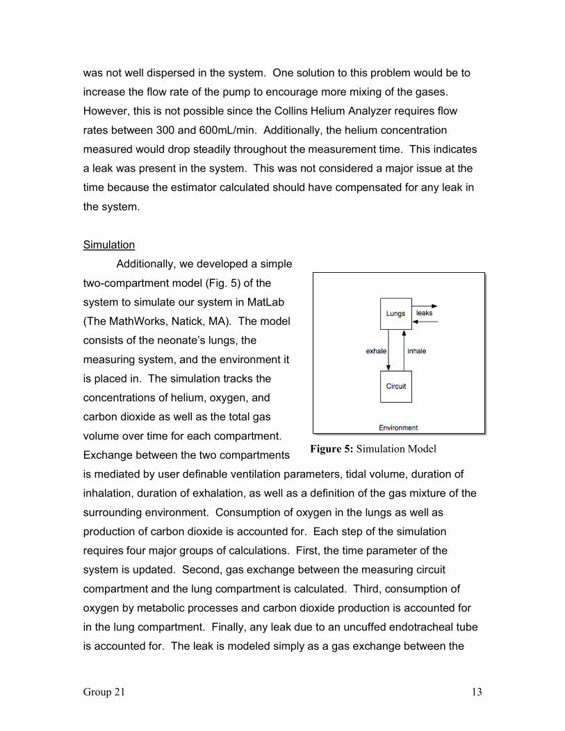
Simulation
Additionally, we developed a simple
two-compartment model (Fig. 5) of the
system to simulate our system in MatLab
(The MathWorks, Natick, MA). The model
consists of the neonate’s lungs, the
measuring system, and the environment it
is placed in. The simulation tracks the
concentrations of helium, oxygen, and
carbon dioxide as well as the total gas
volume over time for each compartment.
Exchange between the two compartments
is mediated by user definable ventilation parameters, tidal volume, duration of
inhalation, duration of exhalation, as well as a definition of the gas mixture of the
surrounding environment. Consumption of oxygen in the lungs as well as
production of carbon dioxide is accounted for. Each step of the simulation
requires four major groups of calculations. First, the time parameter of the
system is updated. Second, gas exchange between the measuring circuit
compartment and the lung compartment is calculated. Third, consumption of
oxygen by metabolic processes and carbon dioxide production is accounted for
in the lung compartment. Finally, any leak due to an uncuffed endotracheal tube
is accounted for. The leak is modeled simply as a gas exchange between the
Figure 5: Simulation Model

Group 21 14
lung compartment and the surrounding environment. Each gas exchange is
calculated using the simple equation:
!
Ct+1 =CtVt + Ct,exchangedVexchanged
Vt +Vexchanged
Where Ct indicates the concentration at time t, Vt is the volume of the
compartment at time t, Ct,exchanged is the concentration of the gas in the mixture
being exchanged, and Vexchanged is the volume being exchanged. The sign on
Vexchanged is positive when gas is entering the compartment, and negative when
gas is leaving the compartment. The full implementation of the simulation is
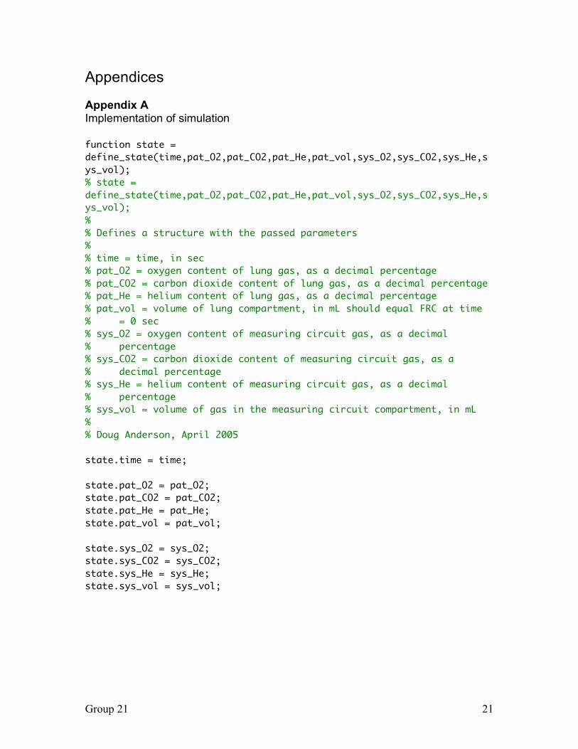
provided in Appendix A.
Simulations were conducted to test both the accuracy of the proposed
method and to assess the amount of oxygen and carbon dioxide in the lungs
over the testing period. Two sets of simulations were run, one using system
parameters described by Schwartz, et al. [8], and the second using the
parameters of our prototype. Ventilation parameters were taken from
Szymankiewicz, et al. [9], as examples of parameters for a normal ventilated
neonate. The rate of oxygen usage was assumed to scale with body mass, and
assumed a resting consumption rate of 300mL/min in a normal 70kg adult [10].
Using an average mass of 1.2kg, this corresponds to an oxygen consumption
rate of 5.14mL/min. For all simulations, the starting concentrations of gas in the
lungs were normal values for adults breathing room air. While this may not be an
accurate starting point, it does offer a common starting point for all simulations.
In each set of simulations, the leak rate was varied to assess the accuracy of our
estimator. Estimates of the FRC were calculated from simulated data at 45 and
60 seconds, mimicing the prototype testing. A summary of simulation
parameters is given in Table 1.

Group 21 15
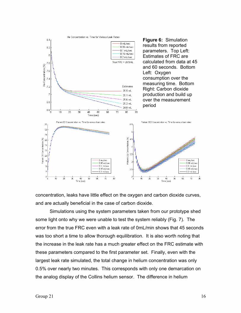
Simulations run using the system parameters from Schwartz, et al. [8],
show that the equation suggested does indeed produce a good estimator of the
equilibrium concentration and, thus, the FRC (Fig. 6). It is worth noting that the
accuracy of the estimator improves as the leak rate gets smaller. This makes
sense in terms of the equation. Figure 6 also shows that the oxygen content of
the lungs stays at a safe level over the testing window. The initial rise in oxygen
levels is due to our choice of starting states. Room air has a much lower oxygen
content than the gas used in our simulated system, so the lungs’ levels rise
dramatically in the first few moments of the simulation. Additionally, the carbon
dioxide content of the lungs falls rapidly over the first few seconds and then rises
steadily over the testing period. While the rise in carbon dioxide over the
duration of the testing is not dangerous, it is something that should be monitored
closely if testing lasts longer than a few minutes. The initial fall in carbon dioxide
is due to the choice of normal lung gas concentrations as the starting point for
the simulation. These concentrations are not truly valid for the ventilation
parameters used in the simulation so the concentrations fall quickly to match the
ventilation parameters. It is worth nothing that, unlike the measured helium
Parameter Referenced Value Prototype Value
System Volume 185 mL [1] 785 mL
Patient FRC 26. 5 mL [1] 30 mL
Tidal Volume 7.14 mL [2] 10 mL
Breathing Rate 48 breaths/min [2] 30 breaths/min
O2 Consumption 5.14 mL/min [3] N/A
Respiratory Quotient 0.8 [3] N/A
Leak Rate Varied, 0-0.2 mL/sec
Starting Gas %, Circuit 36.8% O2, 8.1% He2, 0% CO2
Starting Gas %, Patient 13.15% O2, 0% He2,
5.26% CO2
22% O2, 0% He2,
1% CO2
Table 1: Simulation Parameters
Group 21 16
concentration, leaks have little effect on the oxygen and carbon dioxide curves,
and are actually beneficial in the case of carbon dioxide.
Simulations using the system parameters taken from our prototype shed
some light onto why we were unable to test the system reliably (Fig. 7). The
error from the true FRC even with a leak rate of 0mL/min shows that 45 seconds
was too short a time to allow thorough equilibration. It is also worth noting that
the increase in the leak rate has a much greater effect on the FRC estimate with
these parameters compared to the first parameter set. Finally, even with the
largest leak rate simulated, the total change in helium concentration was only
0.5% over nearly two minutes. This corresponds with only one demarcation on
the analog display of the Collins helium sensor. The difference in helium
Figure 6: Simulation results from reported parameters. Top Left: Estimates of FRC are calculated from data at 45 and 60 seconds. Bottom Left: Oxygen consumption over the measuring time. Bottom Right: Carbon dioxide production and build up over the measurement period

Group 21 17
concentration at our measurement points of 45 and 60 seconds is less than
0.1%. This would have been very hard to differentiate. Oxygen and carbon
dioxide curves for this simulation set are omitted since they provide little useful
information in the absence of an oxygen consumer and carbon dioxide source.
While these simulations are no substitute for real world testing, they
clearly demonstrate the feasibility of this method as well as some potential
pitfalls. The simulations performed using the parameters from previous reports
[8,9,10] show the method can provide a good estimator of the FRC even with
relatively large leak rates. The simulations using the parameters of our prototype
show that the volume of the measuring circuit must be kept low in order to speed
equilibration and increase the change in helium concentration. However, the
volume of the measuring is also constrained by the neonates need for
adequately oxygenated air and low carbon dioxide levels. It is also important to
note that these simulations assume perfect mixing within each compartment, so
these simulations make no predictions about the error associated with
incomplete mixing of the gases.
Safety Issues
Potential safety issues with our device were analyzed using the
DesignSafe software. The bulk of the safety issues were issues all electrical
devices must deal with. We intend to account for these hazards with standard
electrical safety measures and trained service personnel. Another major concern
Figure 7: Helium concentrations simulated from the prototype parameter set for various leak rates

Group 21 18
is the involvement of oxygen gas, which is highly flammable. However, the
device is meant to be used in a hospital environment which already has strict
protocols, such as no smoking, to reduce the danger in a building where oxygen
gas is ubiquitous. Another concern is to maintain good oxygen content in the gas
being inhaled by the patient. Proposed safety devices include sensors within the
circuit to measure the oxygen and carbon dioxide content and methods to add
oxygen and remove carbon dioxide if necessary. This has not been implemented
yet. Additionally, only trained respiratory technicians, nurses, and doctors will
use this device. They will be trained to monitor the patient throughout
measurement and their previous training will dictate how to deal with any
situation that may arise such as hypoxia or hypercapnia. A final concern involves
patients with latex allergies. Our current prototype includes a latex anesthesia
bag which could cause allergic reactions in sensitive patients. In the future we
will use an anesthesia bag made from a hypoallergenic material such as nitrile
rubbers. Additional precautions will include labels and user manuals indicating all
potential hazards. The complete DesignSafe report can be found in Appendix B.
In addition, the procedure posses no environmental hazard as it uses the inert
gas, helium, in its testing.
Conclusions
From the data collected, we conclude that the modified helium dilution
methods proposed by Schwartz, et al., can be effective in measuring the FRC of
ventilated neonates. However, care must be taken to ensure proper mixing of
the gases and to minimize the volume of the measuring circuit within the
constraints of providing adequate oxygen to the neonate and maintaining
appropriate carbon dioxide levels. Sensing circuit volumes that are large with
respect to the volume to be measured increase the error in the estimated volume
due to leaks. Additionally, relatively large sensing circuit volumes make
measuring the change in helium concentration more difficult.
This device has the potential for profound social impact. Determination of
the FRC via helium dilution has the potential to save many lives by eliminating

Group 21 19
the need for more dangerous or complicated procedures. The resulting impact
on the friends and family of critically ill neonates will be most certainly be great.
Recommendations Devices like this are not currently in widespread use, yet, they offer great
potential as a tool to aid physicians in treatment of critically ill neonates. We
recommend that work on this project continue with the following goals:
1. Rebuild the prototype with a smaller sensing circuit volume and a modern
digital helium sensor and retest the device.
2. Incorporate oxygen and carbon dioxide sensors into the sensing circuit to
monitor gas levels and provide feedback to physicians as potential trouble
arises.
3. Incorporate methods to add oxygen to the system and remove carbon
dioxide. In addition to the sensors mentioned above, this will increase the
safety of the device for extended use if necessary.
References 1. “Functional Residual Capacity”, Family Practice Notebook.Com, 2000,
<http://www.fpnotebook.com/LUN99.htm> (January 20 2005). 2. Thiam, DGY. “Pulmonary Function Tests in Paediatrics Part 2: Back to
Basics”, October 2003, <http://www.med.nus.edu.sg/paed/medical_education/postgraduate/Allergy_Pulmonology/pulmonary_function_test2.htm> (January 25 2005).
3. “Application of Continuous Positive Airway Pressure to Neonates via Nasal Prongs, Nasopharyngeal Tube, or Nasal Mask”, AARC Clinical Practice Guideline, 2004, <http://www.rcjournal.com/contents/09.04/09.04.1100.asp> (January 20 2005).
4. “Mechanical Ventilators”, NICU Web, 2002, < http://neonatal.peds.washington.edu/NICU-WEB/vents.stm#Introduction> (January 20 2005).
5. Caitlin, EA. “Mechanical Ventilation in the Neonate”, <http://www.mgh.harvard.edu/children/prof/nicu/ventilation.pdf> (January 20 2005)

Group 21 20
6. “ECMO Program”, Monroe Carell Jr. Children’s Hospital at Vanderbilt, 2005, < http://www.vanderbiltchildrens.com/interior.php?mid=959> (February 10 2005).
7. “Curbing the Costs of Medical Scans, Insurers seek to rein in on fast-growing use of pricey, high-tech MRI’s and CT’s”; Victoria Colliver; Sunday April 24, 2005. <http://sfgate.com/cgi-bin/article.cgi?file=/c/a/2005/04/24/BUGD3CDRAP1.DTL>
8. Schwartz, JG, Fox, WW, Shaffer, TH. “A Method for Measuring Functional Residual Capacity in Neonates with Endotracheal Tubes.” IEEE Trans. Biomed. Engr. May 1978. 25(3): 304-7.
9. Szymankiewicz, M, Vidyasagar, D, Gadzinowski, J. “Predictors of successful extubation of preterm low-birth-weight infants with respiratory distress syndrome.” Pediatr Crit Care Med. 2005. 6(1): 44-9.
10. West, JB. Respiratory Physiology: The Essentials. 6th Ed. Lippincott, Williams & Wilkins. Philadelphia: 2000.
Group 21 21
Appendices Appendix A Implementation of simulation function state = define_state(time,pat_O2,pat_CO2,pat_He,pat_vol,sys_O2,sys_CO2,sys_He,sys_vol); % state = define_state(time,pat_O2,pat_CO2,pat_He,pat_vol,sys_O2,sys_CO2,sys_He,sys_vol); % % Defines a structure with the passed parameters % % time = time, in sec % pat_O2 = oxygen content of lung gas, as a decimal percentage % pat_CO2 = carbon dioxide content of lung gas, as a decimal percentage % pat_He = helium content of lung gas, as a decimal percentage % pat_vol = volume of lung compartment, in mL should equal FRC at time % = 0 sec % sys_O2 = oxygen content of measuring circuit gas, as a decimal % percentage % sys_CO2 = carbon dioxide content of measuring circuit gas, as a % decimal percentage % sys_He = helium content of measuring circuit gas, as a decimal % percentage % sys_vol = volume of gas in the measuring circuit compartment, in mL % % Doug Anderson, April 2005 state.time = time; state.pat_O2 = pat_O2; state.pat_CO2 = pat_CO2; state.pat_He = pat_He; state.pat_vol = pat_vol; state.sys_O2 = sys_O2; state.sys_CO2 = sys_CO2; state.sys_He = sys_He; state.sys_vol = sys_vol;

Group 21 22
function state = sim_dilution(breaths, start_state, tidal, insp_dur, exp_dur, O2_use, rq, leak_rate); % sim_dilution(breaths, start_state, tidal, insp_dur, exp_dur, O2_use, rq, leak_rate); % % Simulates Helium dilution method % % breaths = number of breaths to simulate, must have positive integer value % start_state = structure holding the starting values for the simulation, % first element in returned array of structures equals start_state, % pat_vol in start_state should equal the FRC % tidal = tidal volume, in mL % insp_dur = duration of inspiration, in sec % exp_dur = duration of expiration, in sec % O2_use = rate of oxygen consumption, in mL/sec % rq = respiratory quotient % leak_rate = rate of gas leak/exchange with environment (assumes room % air), in mL/sec % % Doug Anderson, April 2005 state(1) = start_state; index = 2; for i=2:breaths state(index) = inhale(state(index1),tidal,insp_dur,O2_use,rq,leak_rate); index=index+1; state(index) = exhale(state(index-1),tidal,insp_dur,O2_use,rq,leak_rate); index=index+1; end

Group 21 23
function new_state = inhale(curr_state,tidal,duration,O2_use,rq,leak_rate); % new_state = inhale(curr_state,tidal,duration,O2_use,rq,leak_rate); % % Simulates the inhalation step of helium dilution % % curr_state = the current state of the model % tidal = tidal volume, in mL % duration = duration of inhalation, in sec % O2_use = rate of consumption of oxygen, in mL/sec % rq = respiratoy quotient % leak_rate = leak rate, in mL/sec % % Doug Anderson, April 2005 %Advance Time new_time = curr_state.time + duration; %Exchange Gases sys_vol = curr_state.sys_vol - tidal; sys_O2 = ((curr_state.sys_O2*curr_state.sys_vol)- (curr_state.sys_O2*tidal))/sys_vol; sys_He = ((curr_state.sys_He*curr_state.sys_vol)- (curr_state.sys_He*tidal))/sys_vol; sys_CO2 = ((curr_state.sys_CO2*curr_state.sys_vol)- (curr_state.sys_CO2*tidal))/sys_vol; pat_vol = curr_state.pat_vol + tidal; pat_O2 = ((curr_state.pat_O2*curr_state.pat_vol)+(curr_state.sys_O2*tidal))/pat_ vol; pat_He = ((curr_state.pat_He*curr_state.pat_vol)+(curr_state.sys_He*tidal))/pat_ vol; pat_CO2 = ((curr_state.pat_CO2*curr_state.pat_vol)+(curr_state.sys_CO2*tidal))/pa t_vol; %Account for consumption of O2 and creation of CO2 used_O2 = O2_use*duration; made_CO2 = O2_use*duration*rq; pat_O2 = ((pat_O2*pat_vol)-used_O2)/pat_vol; pat_CO2 = ((pat_CO2*pat_vol)+made_CO2)/pat_vol; %Account for leakage pat_O2 = ((pat_O2*pat_vol)- (pat_O2*leak_rate*duration)+(0.22*leak_rate*duration))/pat_vol;

Group 21 24
pat_CO2 = ((pat_CO2*pat_vol)- (pat_CO2*leak_rate*duration)+(0*leak_rate*duration))/pat_vol; pat_He = ((pat_He*pat_vol)- (pat_He*leak_rate*duration)+(0*leak_rate*duration))/pat_vol; new_state = define_state(new_time,pat_O2,pat_CO2,pat_He,pat_vol,sys_O2,sys_CO2,sys_ He,sys_vol); function new_state = exhale(curr_state,tidal,duration,O2_use,rq,leak_rate); % new_state = exhale(curr_state,tidal,duration,O2_use,rq,leak_rate); % % Simulates the exhalation step of helium dilution % % curr_state = the current state of the model % tidal = tidal volume, in mL % duration = duration of inhalation, in sec % O2_use = rate of consumption of oxygen, in mL/sec % rq = respiratoy quotient % leak_rate = leak rate, in mL/sec % % Doug Anderson, April 2005 %Advance Time new_time = curr_state.time + duration; %Exchange Gases sys_vol = curr_state.sys_vol + tidal; sys_O2 = ((curr_state.sys_O2*curr_state.sys_vol)+(curr_state.pat_O2*tidal))/sys_ vol; sys_He = ((curr_state.sys_He*curr_state.sys_vol)+(curr_state.pat_He*tidal))/sys_ vol; sys_CO2 = ((curr_state.sys_CO2*curr_state.sys_vol)+(curr_state.pat_CO2*tidal))/sy s_vol; pat_vol = curr_state.pat_vol - tidal; pat_O2 = ((curr_state.pat_O2*curr_state.pat_vol)- (curr_state.pat_O2*tidal))/pat_vol; pat_He = ((curr_state.pat_He*curr_state.pat_vol)- (curr_state.pat_He*tidal))/pat_vol; pat_CO2 = ((curr_state.pat_CO2*curr_state.pat_vol)- (curr_state.pat_CO2*tidal))/pat_vol; %Account for consumption of O2 and creation of CO2

Group 21 25
used_O2 = O2_use*duration; made_CO2 = O2_use*duration*rq; pat_O2 = ((pat_O2*pat_vol)-used_O2)/pat_vol; pat_CO2 = ((pat_CO2*pat_vol)+made_CO2)/pat_vol; %Account for leakage pat_O2 = ((pat_O2*pat_vol)- (pat_O2*leak_rate*duration)+(0.22*leak_rate*duration))/pat_vol; pat_CO2 = ((pat_CO2*pat_vol)- (pat_CO2*leak_rate*duration)+(0*leak_rate*duration))/pat_vol; pat_He = ((pat_He*pat_vol)- (pat_He*leak_rate*duration)+(0*leak_rate*duration))/pat_vol; new_state = define_state(new_time,pat_O2,pat_CO2,pat_He,pat_vol,sys_O2,sys_CO2,sys_ He,sys_vol);

Group 21 26
Appendix B DesignSafe Report

Group 21 27
Group 21 28

Group 21 29

Group 21 30
Group 21 31

Group 21 32

Group 21 33
Group 21 34
Appendix C Innovation Workbench Questionnaire
Ideation Process
Innovation Situation Questionnaire 1. Brief description of the problem
Functional residula capacity is the volume remaining in the lungs at resting expiratory level. It is the sum of residual volume (volume remaining in the lungs after maximal expiration) and the expiratpry reserve volume (maximum volume of additional air that can can be expired from the end of a normal be expired from the end of a normal expiration).expiration). If functional residual capacity is too small, then blood that enters the lung will exit without effic ient oxygen exchange. Therefore, a device or method is needed to measure the functional residual capacity of a ventilated neonate in order to set the ventilator parameters such that functional residual capacity is large enough to create gas exchange between the lungs and blood, reducing the possibility of insuffic ient oxygenation of the blood and subsequent hypoxia. Knowledge of this information could reduce the number of infants placed on ECMO, a dangerous and expensive last resort to oxygenation.
2. Information about the system 2.1 System name
Measuring Neonatal Funtional Residual Capacity 2.2 System structure
An enclosed system with a way to measure Helium gas volume or concentration. 2.3 Functioning of the system
Measure the functional residual capacity of a neonate lung using helium dilution methods without suspending ventilation.
2.4 System environment In neonatal intensive care units (NICU), ventilators are used to facilitate the respiratory mechanics of infants. The device will connect to existing ventilators in the NICU. In these units, enviromental conditions are tightly regulated.
3. Information about the problem situation 3.1 Problem that should be resolved
An enclosed system is hard to maintain because the ventilator provides a continuous inflow of air and the infant cannot rebreathe from a constant volume for any extended length of time.
3.2 Mechanism causing the problem The helium dilution method requires a closed system to make a true measurement while maintaining ventilator support mandates an open system.
3.3 Undesired consequences of unresolved problem Either ventilator support must be interrupted or a closed system cannot be used, compromising the measurement.
3.4 History of the problem Helium dilution is a common method for measuring lung volume in adults. Unfortunately, the lack of a closed system and inability of infants to cooperate with such tests has prevented it use previously.
3.5 Other systems in which a similar problem exists
3.6 Other problems to be solved Size, ease of use, and safety.

Group 21 35
4. Ideal vision of solution A device which can maintain a closed system without suspending ventilator support.
5. Available resources Ventilators and a Sensormedic adult pulmonary function unit are available for testing the device. Additionally, the helium sensor, a peristaltic pump, and a variety of tubing and connectors is available.
6. Allowable changes to the system The ventilator, pulmonary function device, pump, and helium sensor cannot be internally modified. Any changes must maintain the flow of air to the infant and meet all applicable standards on patient safety and materials for pulmonary equipment.
7. Criteria for selecting solution concepts Safe for repeated and often use. Non-invasive. Quick. Easy. Inexpensive.
8. Company business environment The company has no other products. We anticipate all NICUs will have interest in the product and little competition currently exists in this market.
9. Project data Timeline: Nov. 2004 - Research, Project Definition
Dec. 2004 - Research, Project Definition
Jan. 2005 - Process Development, Materials Definition
Feb. 2005 - Prototype Development
Mar. 2005 - Prototype Development, Testing and Analysis
Apr. 2005 - Testing and Analysis
May. 2005 - Presentation
Team Members
Douglas Anderson - BME Student; Team Leader: Knowledge of web design, simple programming, basic circuit design, Matlab, physiology.
David Lammlein - ME Student: Knowledge of mechanics, dynamics, materials
Janine McKinnon - BME Student: Knowledge of data analysis, Matlab, communication skills, organizational skills
Dr. Paul King - Professor of BME; Advisor
Dr. Bill Walsh - Professor of Pediatrics; Director of Nursuries, Vanderbilt Children's Hospital; Advisor
Contact email: [email protected]
Problem Formulation 1. Build the Diagram

Group 21 36
2. Directions for Innovation 4/3/2005 6:00:37 PM Diagram1 » 1. Find an alternative way to obtain [the] (closed system) that offers the following: provides or enhances [the] (uses inert Helium gas) and (measures FRC), does not influence [the] (maintains ventilator support). 2. Try to resolve the following contradiction: The useful factor [the] (closed system) should be in place in order to provide or enhance [the] (uses inert Helium gas) and (measures FRC), and should not exist in order to avoid hindering [the] (maintains ventilator support). 3. Find an alternative way to obtain [the] (maintains ventilator support) that offers the following: provides or enhances [the] (improves oxygenation), eliminates, reduces, or prevents [the] (lowers oxygenation), is not influenced by [the] (closed system). 4. Find an alternative way to obtain [the] (uses inert Helium gas) that does not require [the] (closed system). 5. Consider transitioning to the next generation of the system that will provide [the] (uses inert Helium gas) in a more effective way and/or will be free of existing problems. 6. Find an alternative way to obtain [the] (improves oxygenation) that does not require [the] (measures FRC) and (maintains ventilator support). 7. Consider transitioning to the next generation of the system that will provide [the] (improves oxygenation) in a more effective way and/or will be free of existing problems. » 8. Find an alternative way to obtain [the] (measures FRC) that offers the following: provides or enhances [the] (improves oxygenation), does not require [the] (closed system). 9. Find a way to eliminate, reduce, or prevent [the] (lowers oxygenation).
Group 21 37
Prioritize Directions 1. Directions selected for further consideration
1. Find an alternative way to obtain [the] (closed system) that offers the following: provides or enhances [the] (uses inert Helium gas) and (measures FRC), does not influence [the] (maintains ventilator support). 1.1. Improve the useful factor (closed system). » 1.2. Obtain the useful result without the use of [the] (closed system). 1.3. Increase effectiveness of the useful action of [the] (closed system). » 1.4. Synthesize the new system to provide [the] (closed system). 1.5. Apply universal Operators to provide the useful factor (closed system). 1.6. Consider resources to provide the useful factor (closed system). 8. Find an alternative way to obtain [the] (measures FRC) that offers the following: provides or enhances [the] (improves oxygenation), does not require [the] (closed system). 8.1. Improve the useful factor (measures FRC). 8.2. Obtain the useful result without the use of [the] (measures FRC). 8.3. Increase effectiveness of the useful action of [the] (measures FRC). » 8.4. Synthesize the new system to provide [the] (measures FRC). 8.5. Apply universal Operators to provide the useful factor (measures FRC). 8.6. Consider resources to provide the useful factor (measures FRC).
2. List and categorize all preliminary ideas We should either discover a new method to create the closed system while maintaining ventilator support or find a way to account for the open system in the FRC calculation.
Develop Concepts 1. Combine ideas into Concepts
Perhaps we can do both, creating a mostly closed system without compromising ventilator support. Use the ventilator to apply pressure to the closed system?
2. Apply Lines of Evolution to further improve Concepts Use a mathematical model to account for the leaks from a mostly closed system. Provide a method for applying the pressure produced by the ventilator to the gas in the closed system without allowing mixing (diaphragm maybe?).
Evaluate Results 1. Meet criteria for evaluating Concepts
The method mentioned above seems to meet all the criteria for solving this problem. 2. Reveal and prevent potential failures
The mostly closed system may not contain enough oxygen for long measurement times, additionally, CO2 buildup is a concern. The helium may leak too quickly to calculated a reasonable estimate of the FRC. The diaphragm like portion could be too stiff and not

Group 21 38
transfer the pressure well enough to maintain ventilator support. 3. Plan the implementation
A prototype of the new system will be constructed in heavily tested before it is ever tried on an infant. The system will be tested without ventilator support with a syringe or other known volume acting for the FRC. It will also be tested on adults who do not need ventilator support to test its ability to measure a dynamic volume. Finally, a known volume in a distensible container will be used to test the system when ventilator support is applied to make sure the system for transferring the pressures functions properly and measurements can still be made. Only then will testing on infants be attempted.