critical overview of nitinol surfaces and their...
TRANSCRIPT

Available online at www.sciencedirect.com
Acta Biomaterialia 4 (2008) 447–467
www.elsevier.com/locate/actabiomat
Review
Critical overview of Nitinol surfaces and their modificationsfor medical applications
S. Shabalovskaya a,b,*, J. Anderegg b, J. Van Humbeeck a
a Katholieke Universiteit Leuven, MTM Kasteelpark Arenberg 44, B-3001 Leuven, Belgiumb Ames Laboratory, Ames, IA 50011, USA
Received 18 July 2007; received in revised form 16 November 2007; accepted 10 January 2008Available online 6 February 2008
Abstract
Nitinol, a group of nearly equiatomic shape memory and superelastic NiTi alloys, is being extensively explored for medical applica-tions. Release of Ni in the human body, a potential problem with Nitinol implant devices, has stimulated a great deal of research on itssurface modifications and coatings. In order to use any of the developed surfaces in implant designs, it is important to understandwhether they really have advantages over bare Nitinol. This paper overviews the current situation, discusses the advantages and disad-vantages of new surfaces as well as the limitations of the studies performed. It presents a comprehensive analysis of surface topography,chemistry, corrosion behavior, nickel release and biological responses to Nitinol surfaces modified mechanically or using such methods asetching in acids and alkaline solutions, electropolishing, heat and ion beam treatments, boiling in water and autoclaving, conventionaland ion plasma implantations, laser melting and bioactive coating deposition. The analysis demonstrates that the presently developedsurfaces vary in thickness from a few nanometers to micrometers, and that they can effectively prevent Ni release if the surface integrityis maintained under strain and if no Ni-enriched sub-layers are present. Whether it is appropriate to use various low temperature pre-treatment protocols (6160 �C) developed originally for pure titanium for Nitinol surface modifications and coatings is also discussed.The importance of selection of original Nitinol surfaces with regard to the performance of coatings and comparative performance ofcontrols in the studies is emphasized. Considering the obvious advantages of bare Nitinol surfaces for superelastic implants, detailsof their preparation are also outlined.� 2008 Acta Materialia Inc. Published by Elsevier Ltd. All rights reserved.
Keywords: Nitinol; Surface modifications; Biocompatibility; Corrosion; Ni release
Contents
1. Introduction . . . . . . . . . . . . . . . . . . . . . . . . . . . . . . . . . . . . . . . . . . . . . . . . . . . . . . . . . . . . . . . . . . . . . . . . . . . . . . . 4482. Bare Nitinol surfaces and their modifications . . . . . . . . . . . . . . . . . . . . . . . . . . . . . . . . . . . . . . . . . . . . . . . . . . . . . . . . 449
1742-7
doi:10.
* CoE-m
2.1. Mechanically modified surfaces and biological responses . . . . . . . . . . . . . . . . . . . . . . . . . . . . . . . . . . . . . . . . . . 4492.2. Chemically and electrochemically modified surfaces . . . . . . . . . . . . . . . . . . . . . . . . . . . . . . . . . . . . . . . . . . . . . . 4502.3. Heat-treated surfaces . . . . . . . . . . . . . . . . . . . . . . . . . . . . . . . . . . . . . . . . . . . . . . . . . . . . . . . . . . . . . . . . . . . . 4512.4. Biological responses to bare Nitinol surfaces modified using heat treatments, chemical and electrochemical approaches 453
2.4.1. Endothelial and smooth muscle cell responses . . . . . . . . . . . . . . . . . . . . . . . . . . . . . . . . . . . . . . . . . . . . . 4542.4.2. Thrombogenic potential, protein adsorption and platelet adhesion . . . . . . . . . . . . . . . . . . . . . . . . . . . . . . 455
061/$ - see front matter � 2008 Acta Materialia Inc. Published by Elsevier Ltd. All rights reserved.
1016/j.actbio.2008.01.013
rresponding author. Address: Ames Laboratory-DOE, Room 324, Wiehelm Hall, Ames, IA 50011, USA.ail addresses: [email protected], [email protected] (S. Shabalovskaya).

448 S. Shabalovskaya et al. / Acta Biomaterialia 4 (2008) 447–467
3. Surface modifications with ion and energy sources . . . . . . . . . . . . . . . . . . . . . . . . . . . . . . . . . . . . . . . . . . . . . . . . . . . . 457
3.1. Conventional ion implantation and electron beam . . . . . . . . . . . . . . . . . . . . . . . . . . . . . . . . . . . . . . . . . . . . . . . 4573.2. Plasma immersion ion implantation . . . . . . . . . . . . . . . . . . . . . . . . . . . . . . . . . . . . . . . . . . . . . . . . . . . . . . . . . 4583.2.1. Oxygen implantation. . . . . . . . . . . . . . . . . . . . . . . . . . . . . . . . . . . . . . . . . . . . . . . . . . . . . . . . . . . . . . . 4583.2.2. Diamond-like carbon . . . . . . . . . . . . . . . . . . . . . . . . . . . . . . . . . . . . . . . . . . . . . . . . . . . . . . . . . . . . . . 4593.2.3. Biological responses to plasma ion immersion implanted surfaces . . . . . . . . . . . . . . . . . . . . . . . . . . . . . . . 460
3.3. Laser surface melting. . . . . . . . . . . . . . . . . . . . . . . . . . . . . . . . . . . . . . . . . . . . . . . . . . . . . . . . . . . . . . . . . . . . 460
4. Other surfaces . . . . . . . . . . . . . . . . . . . . . . . . . . . . . . . . . . . . . . . . . . . . . . . . . . . . . . . . . . . . . . . . . . . . . . . . . . . . . . 4604.1. Sol–gel and hydrogen peroxide surface treatments . . . . . . . . . . . . . . . . . . . . . . . . . . . . . . . . . . . . . . . . . . . . . . . 4604.2. Bioactive surfaces . . . . . . . . . . . . . . . . . . . . . . . . . . . . . . . . . . . . . . . . . . . . . . . . . . . . . . . . . . . . . . . . . . . . . . 461
5. Nitinol surface under strain . . . . . . . . . . . . . . . . . . . . . . . . . . . . . . . . . . . . . . . . . . . . . . . . . . . . . . . . . . . . . . . . . . . . 4636. Conclusions and outlook . . . . . . . . . . . . . . . . . . . . . . . . . . . . . . . . . . . . . . . . . . . . . . . . . . . . . . . . . . . . . . . . . . . . . . 464
Acknowledgments . . . . . . . . . . . . . . . . . . . . . . . . . . . . . . . . . . . . . . . . . . . . . . . . . . . . . . . . . . . . . . . . . . . . . . . . . . . 465References . . . . . . . . . . . . . . . . . . . . . . . . . . . . . . . . . . . . . . . . . . . . . . . . . . . . . . . . . . . . . . . . . . . . . . . . . . . . . . . . . 465
1. Introduction
Surface modification and coating of Nitinol (an acro-nym for NiTi Naval Ordnance Laboratory), a family ofnearly equiatomic NiTi alloys with shape memory andsuperelastic properties, is a subject of numerous recentstudies directed at improving the material’s corrosion resis-tance as well as its biocompatibility through elimination ofNi from the surface. This chemical element is known to beallergenic and toxic, though essential for the human body.Although it has been shown that the amount of Ni recov-ered in biological studies in vitro may be either very lowfrom the beginning or drop to undetectable levels after abrief exposure to biological environments [1,2], ‘the nickelcase’ keeps reappearing. Thus, the recent results obtainedon commercial ready-to-use orthodontic wires showed thatthe Ni release varied in a wide range from 0.2 to 7 lg cm�2
[3]. Moreover, it has been reported that the Ni release canactually significantly increase with time [4–7], maintaininga high level up to 8 weeks and even for a few months[6,7], indicating the need for better understanding of thematerial/surface interface. Based on the number of pub-lished papers on Nitinol surfaces, especially recently, onemight conclude that this issue indeed deserves seriousattention. Various techniques and protocols have beenused for surface treatments; among them mechanical andelectrochemical treatments, chemical etching, heat treat-ments, conventional and plasma ion immersion implanta-tion, laser and electron-beam irradiation, design ofbioactive surfaces, and a proper technique can easily be lostin that jungle of publications. Some of the procedures thatwere developed originally for pure Ti and their applicationto NiTi not only may not bring any improvement but,rather, can cause surface damage because of inevitable Niinvolvement. Mechanisms of oxide formation on NiTi sur-faces vary, depending on media, temperature and irradia-tion. The interpretations of the Ti and Ni elementaldepth profiles in Nitinol differ because of the possibilityof preferential sputtering of one of the alloy componentsthat depends on the angles and energies of sputtering ionbeams [8]. The absence of a precise description of atomic
diffusion on the interfaces makes this situation even morecomplex.
An interesting development is that X-ray photoelectronspectroscopy (XPS) and Auger spectroscopy have becomeroutine methods, and most of the recently published stud-ies on Nitinol have been done with an impressive list oftechniques, including even energy dispersive X-ray spec-troscopy (EDS) [3,9–11], unsuitable for surface analysis.Despite a great number of new publications, some of whichinclude detailed XPS surface studies, their analyses are verydifficult especially when relevant to biological responses.Thus, instead of providing absolute values of elementalconcentrations for Ni and Ti, various relative values arepresented that do not give objective information on Nitinolsurface chemistry. The important experimental details suchas the Ar ion sputtering rates, the electron escape anglesassociated with a certain surface depth in XPS studies,the scanning rates in cyclic potential polarization, etc. arequite often missing. Because the selected scanning rates inpotentiodynamic (PD) cyclic potential polarization aresometimes as much as 3–30 times higher [12–14] than thoserecommended by the American Society for Testing andMaterials (ASTM) for testing medical devices [15], theresults obtained might easily be overrated, especially withregard to localized corrosion resistance. Very little or noattention has been paid thus far to the selection of the ori-ginal NiTi surfaces subjected to modification. As a result,the surfaces developed either did not perform in the wayin which they were expected to, or their performance wasoverestimated because it was compared with the poorestbare Nitinol surfaces. Sometimes surface modificationtechniques are combined with procedures employed forthe design of optimal shape memory and superelasticity.The consequence is that not only is the surface compositionmodified, but so also is the bulk of Nitinol; as a result,important parameters for medical applications such asshape recovery temperatures and mechanical propertiesare altered.
All the studies of surface modifications of Nitinol havebeen aimed at improving its corrosion behavior. It has beenshown that localized corrosion resistance of bare Nitinol

Fig. 1 Topography of NiTi samples mechanically polished (1 lm finish) inaustenitic phase at room temperature as observed in atomic forcemicroscopy (AFM). Stress caused by mechanical polishing inducedmartensitic transformation and surface relief. Four grains of differentorientation are visible as well as inclusions (with bright tips), some ofwhich are located near the grain boundaries.
S. Shabalovskaya et al. / Acta Biomaterialia 4 (2008) 447–467 449
may vary significantly, depending on its surface state [7,16–18]. In PD and potentiostatic (PS) potential polarization,Nitinol surfaces prepared appropriately do not break orpit up to 800 mV and even 1300 mV applied potentials.However, in scratch corrosion tests when surface damageis caused mechanically, the repassivation ability of Nitinolhappens to be inferior to that of pure Ti, though compara-ble with the scratch healing ability of stainless steel. Thepitting potentials of NiTi determined in scratch tests arelow (from 150 to 300 mV) compared with PD and PSpolarization, and this is the problem to be targeted in thedevelopment of Nitinol surface modifications.
The present review analyzes the current situation withNitinol surface modifications and its progress during thepast 7 years. Earlier publications on this subject havebeen covered in previous reviews [18,19], and moredetailed information on Nitinol biocompatibility is pre-sented elsewhere [20]. The advantages and disadvantagesof the surfaces developed and the limitations of the stud-ies conducted are discussed, and the surface performancesin Ni release, corrosion and biocompatibility are com-pared. For the reasons mentioned above, when analyzingthe chemistry of bare Nitinol surfaces, the authors willoften refer to their own papers on XPS studies, whichwere conducted at surface-sensitive angles and providedthe actual elemental concentrations pertinent to biologicalresponses.
2. Bare Nitinol surfaces and their modifications
2.1. Mechanically modified surfaces and biological responses
Traditional surface treatments for biomaterials includemechanical polishing (Mp), electropolishing (Ep), chemicaletching (Ce) in acid solutions, heat treatments (Ht) undervarious conditions, sandblasting and short pinning.Because of its proprietary nature, information on Nitinolsurface treatments typically is not disclosed. It will be dem-onstrated in the following sections that, in the absence ofstandard surface treatment protocols for Nitinol, mechan-ically ground or ‘polished-to-luster’ surfaces are often usedin biological studies as well as for coating substrates. Thesesurfaces, however, are known for their poor reproducibilityin corrosion resistance [16,21], and their biological perfor-mance is inferior to that of the surfaces additionally treated[22,23]. The elemental composition of Mp surfaces (1 lmfinish) depends on the sample polishing, cleaning and han-dling procedures. The total concentration of surface con-taminants such as Ca, Na, Mg, Si, P and Cl, originatingfrom polishing or cleaning solutions or from calcium pow-dered gloves, varied from 1 to 8 at.%, thus affecting theconcentrations of metals on the surface. The Ni concentra-tions in the 1–4 at.% and Ti in the 8–13 at.% ranges deter-mined by XPS analysis at a 20� surface-sensitive angle arereported in Refs. [24,25]. The rest are mostly carbon andoxygen, the inevitable surface components. It follows fromthe literature analysis that Mp surfaces with a contamina-
tion level significantly higher than that mentioned above(�30 at.%) are also used in the studies [2].
One of the reasons behind the inconsistent corrosionperformance of Mp surfaces could be the presence of resid-ual defective surface layers induced, for instance, by elec-tro-discharge machining (EDM). It was demonstratedthat EDM caused a 5–22-lm-thick melting zone, 50% ofwhich consisted of TiC [26]. The resulting internal stressesgenerated cracks descending from the melting zone into thedepth, as well as propagating in the direction parallel to thesurface.
Additional factors contributing to the inconsistent cor-rosion behavior of Mp surfaces could be the residual plas-tic deformation associated with grinding, the inevitablescratches left by hard inclusion particles emerging fromthe Nitinol surface, as well as the chemical heterogeneityof these surfaces inheriting all inclusions from the bulk ofthe material. The biological performances of Mp surfacescan also be compromised by alteration of surface topogra-phy due to martensitic relief induced during grinding andpolishing (Fig. 1). Because of these and possibly other,not yet understood, causes, visually smooth but intrinsi-cally defective Nitinol surfaces prepared by mechanicalpolishing may not be an optimal option either for controlsin the surface studies or for coating substrates.
Sandblasting, shot peening and grooving of NiTi sur-faces were used to induce specific topography and rough-ness in order to facilitate cell adhesion, proliferation andmigration [27–29]. Aiming at faster endothelialization ofendovascular prostheses and healing process after implan-tation, the response of endothelial cells (EC) to surfacegrooving was explored [29]. It was shown that grooved

450 S. Shabalovskaya et al. / Acta Biomaterialia 4 (2008) 447–467
NiTi surfaces promoted migration of EC, up to 64.6%(p < 0.0001) when compared with smooth controls. Thecells aligned with the grooves elongated and became morenumerous on grooved surfaces. The almost doubled migra-tion rates of EC upon NiTi surface grooving were observedas compared with metallic surfaces ordinarily used forendovascular stents and yet, the proliferative cell response,equally important to the endothelialization, was not inves-tigated. It was shown, however, that the ability of thesmooth electropolished NiTi surface and that of themechanically polished 600 grit finish (Mp600) to affectEC were different [22]. The Mp600 surfaces noticeably sup-pressed cells by �30%, compared with the smoothest elec-tropolished surfaces and with the controls (asdemonstrated later in this review).
The performance of short peened surfaces was not sta-tistically different (p < 0.05) from the surfaces polished toa 1-lm finish in the cytotoxicity and hemolysis tests onmouse fibroblast [28]. Using human gingival fibroblast cellsand rat calvaria osteoblasts, the authors of Ref. [27] evalu-ated the effect of different surface roughness induced bymechanical polishing (80, 400 and 2400 grit finish) on cel-lular adhesion and maintenance of cellular functions. Inter-estingly, these two types of cells revealed quite differentpatterns in their response to roughness variation. Whilethe adhesion rates of osteoblasts were not affected byroughness increase, fibroblast adhesion was reduced signif-icantly (p > 0.05) on rough NiTi surface (400 grit finish).The capability for normal growth and proliferation (viabil-ity) of fibroblasts suffered owing to the effect of roughnesswhile, on the contrary, osteoblast viability was reduced onthe smoothest surface (2400 grit). As the authors of Ref.[27] pointed out, the alteration of NiTi surface roughnesson a micron scale showed that there might be a roughnessthreshold (between 0.08 and 1 lm) over which fibroblastproliferation becomes difficult (Fig. 2). However, it is pos-sible that there also may be a limitation of degree of surface
Fig. 2 Fibroblast and osteoblast viability determined based on MTT assay(optical density) on 2, 4 and 7 days of culture incubation with NiTi sampleswith 400 grit and 2400 grit surface finish. The figure was adapted andreprinted from Ref. [27] with the permission of IOS Press and the authors.
smoothness, as osteoblast viability was dramatically inhib-ited by the 2400 grit finish surfaces.
In the case of Nitinol, sandblasting is also employed forthe removal of black surface oxide resulting from the man-ufacturing process, which involves multiple annealing inair. This wire is further drawn to obtain a smooth surfaceof a light gray color. The fine drawn wires made by thismanufacturing process showed inconsistent corrosion per-formance [16,30–32], which was attributed to the fact thatthe sandblasting was unable to eliminate completely theoriginal scale on a microscopic level, and also becausethe new surface defects were formed during the followingfine drawing of sandblasted wires of irregular surfacetopography. As far as the corrosion resistance of as-received wires under strain is concerned, it did not deterio-rate when a 4% strain in tension mode was applied [16].
2.2. Chemically and electrochemically modified surfaces
The electrochemistry of Nitinol is poorly explored. Untilrecently, there have been no studies on electropolishing andanodizing of this material. This situation is graduallyimproving with the publications of papers on electropolish-ing processes in various electrolytes [33–36], and anodizingin various solutions and voltage regimes [14,36]. The effectof chemical etching (passivation) in HF + HNO3 aqueoussolutions on Nitinol surface chemistry has also been stud-ied [24,25]. The effect of boiling in water and treatmentsin autoclave (hydrothermal treatment) on reducing the Nirelease from NiTi have been explored as well [24,25,37,38].
Electropolishing and chemical etching of Nitinol areknown to be efficient for the elimination of defective sur-face layers and surface oxidizing. Owing to a gain in totalenergy caused by the differences in the enthalpy of forma-tion of Ti and Ni oxides [39], the preferential oxidationof Ti on Nitinol surface always occurs. As a result, bareNitinol surfaces are built from Ti oxides with Ni concentra-tions from �2 to 7 at.%, depending on the electrolytes andregimes employed. Chemical etching and electropolishingof NiTi can be used for surface structuring as well. As, ingeneral, Nitinol inclusions tend to be distributed relativelyuniformly in the bulk of the material, their removal duringchemical etching leaves a structured surface that may bebeneficial for cell attachment and locomotion (Fig. 3a).This could be a better alternative to the grooved surfacesdiscussed in the previous section. The electrolytic etchingexplored in Ref. [40] induced highly porous NiTi surfacesthat might lead to enhanced Ni release. Surprisingly, elec-tropolishing of Nitinol can also be used to cause a slightstructuring to promote cell attachment. Originally smoothsurfaces, mechanically polished in austenitic phase at roomtemperature and then electropolished in martensite phaseat low temperatures, lose their smoothness and acquire astep-like structure (Fig. 3d) upon martensitic transforma-tion [40,41]. This new appearance, called ‘relief’, is due tothe shear nature of Nitinol phase transformation associ-ated with a uniform shift of atomic planes.

Fig. 3 SEM images of NiTi surfaces chemically etched in the HF + HNO3
aqueous solutions for (a) wire and (b) disk samples, and electropolished;(c) Ep1 at room temperature in austenite; (d) Ep2 at low temperatures inmartensite phases. Carbide and oxide inclusion particles can be seen in ablack and white contrast, respectively.
S. Shabalovskaya et al. / Acta Biomaterialia 4 (2008) 447–467 451
Electropolished surfaces are more heterogeneous thanchemically etched ones because, similarly to Mp surfaces,they retain all phases inherited from the alloy bulk.Fig. 3 also illustrates the presence of carbide particles visi-ble in a black contrast (c and d) and oxide inclusions inwhite contrast (b and c). Higher chemical heterogeneityof Ep surfaces in principle might cause their more inferiorcorrosion performance compared with chemically etchedsurfaces, especially in a long-term run. The breakdownpotentials reported for cyclically polarized [41,42], Ep, Ceand CeWb surfaces [17,32,43–45] were in the range from800 mV to 1400 mV versus SCE. The surface oxide filmsobtained after chemical etching and Ep were only a fewnanometers thick [25,34], but most of the Ti was presentonly in the oxidized +4 state, though Ni was found in bothoxidized +3 and elemental states [24,25,34]. Oxidation ofNi and Ti on the surface can be promoted by boiling inwater (Wb) and by autoclaving in steam and water underpressure [24]. This gentle treatment assisting atomic diffu-sion, and Ni release into boiling water during treatmentitself, resulted in a thicker oxide film (10–20 nm), a morestoichiometric oxide composition, a reduced Ni surfaceconcentration (0–1 at.%), and elimination of Ni release[38]. An interesting aspect of Nitinol surfaces with thinoxide layers is the combination of amorphous and nano-crystalline phases [25,34,45]. The corrosion resistance ofelectropolished, Ce and CeWb surfaces did not deteriorateunder the strains of up to 6–8% in tension mode [44,45].
The first evaluations of the anodizing process in Nitinolindicated that it was different from that reported for pure
Ti [14,36]. Although anodizing was employed to generatethicker oxide films and modify surface topography, nothick films could be obtained on NiTi in acetic acid electro-lyte, and in 0.1 M sulfuric acid, as well as in alkaline solu-tions [14]. In the latter case, a continuous exfoliation of theanodizing products from the surface occurred. However, inthe acetate and borate buffer solutions, the proper anodiz-ing conditions were realized, as reported in Ref. [14]. Theresulting 5 lm oxide films had 2 at.% and 7 at.% of Ni onthe surface for the acetate and borate buffers, respectively,and had amorphous structures. Unfortunately, the anodiz-ing [14] did not reduce the Ni surface content, and also thesurfaces obtained using an optimized anodizing regimewere heavily cracked. In the PD cyclic potential polariza-tion, the scanning rate employed was 5 mV s�1, i.e., 30-foldhigher than recommended by ASTM standards(0.167 mV s�1) for testing medical devices [15]. As pit initi-ation is a very slow process, those PD tests may not be sat-isfactory for the evaluation of localized corrosionresistance [19,30].
A 10-fold reduction in the Ni surface concentration(from 3.48 to 0.36 at.%), a similar increase in polarizationresistance up to 3.6 MX, and an order of magnitude lowercurrent density were registered in the oxide films obtainedupon anodizing of 1 lm finish samples in acetic acid [36].However, the authors reported on pits when oxide filmthickness reached 20–25 nm. Anodizing in a 0.9% salinesolution using a slow and repeated potential sweeping inthe voltage range of oxygen evolution dramaticallyimproved the corrosion resistance [41,42,45]. Similarresults were reported also on anodizing in a Na2SO4 solu-tion [34]. A significant reduction in density of passive cur-rent, nobler corrosion potentials, and a 10–20-fold increasein polarization resistance up to 67 MX were observed in theanodized Ce and CeWb samples. These latter surfaces alsodid not exhibit any cracking upon 6% strain in corrosivesolution [45]. This improvement in corrosion performanceupon anodizing was attributed to complete oxidation ofNi in the external surface layers and partial removal ofinclusions from the surface due to preferential dissolutionof material surrounding inclusions with modified chemicalcomposition.
2.3. Heat-treated surfaces
Heat treatments of Nitinol in air, argon and partiallyreduced atmosphere have been explored again and againfor surface oxidation, aiming at prevention of Ni release[23,25,34,37,46–49] in the biological environment. It isimportant to understand that heat treatments of Nitinolare also an essential part of alloy processing and devicemanufacturing. Temperatures about 700 �C are employedfor wire annealing during its drawing; the temperatures ina 450–550 �C interval are used to design optimal shapememory and superelasticity, and for shape setting proce-dure. Heat treatments at temperatures <300 �C are custom-arily associated with the processes of modifications of

Fig. 4 Typical Ni 2p XPS spectra for NiTi collected at a 20� electronescape angle. (1) Mp; (2) MpHt (520 �C � 20 min); (3) Ep1WbHt; (4)CeWbHt (heat treatments in air); (5) annealed in argon (500 �C � 20 min).Ni in elemental state (peaks at �853 eV and �870 eV) is obvious only forMp surface. The rest of the peaks correspond to a Ni+3 oxidation state[25].
452 S. Shabalovskaya et al. / Acta Biomaterialia 4 (2008) 447–467
Nitinol surface and are also sometimes employed in thefabrication of stent grafts for attaching polyester fabric.Not only may the above heat treatments modify the phasecomposition of the bulk, but they also modify the Nitinoltendencies in formation of either Ti- or Ni-rich surfacesub-layers. The coexistence of various chemical compoundssuch as NixTiy with the binary and ternary metal oxides aswell as oxi-carbides in the NiTi surface layers may invitegalvanic corrosion. Ni-rich phases, Ni-enriched matrixaround Ti-based inclusions, as well as free metallic Ni serveas reservoirs for potential Ni release, especially upon sur-face damage. Differences in the history of material process-ing cause both significant variations in Ni surface content[25] and different patterns in Ni release, discussed in detailselsewhere [20]. Ni accumulated during material processingin surface sub-layers causes Ni release that is increasingwith time of exposure to corrosive solutions [7], in contrastto the well-known decrease reported, for instance, in [1,2].
The above-cited surface studies largely disagree as tothe composition of new phases formed during Ht in thesurface sub-layers, most probably because of a differentprehistory of material processing. However, to have a gen-eral idea of the effect of Ht on Nitinol surface chemistry, itis worthwhile to analyze at least some of the results. Thus,after oxidation in air at 300–500 �C for only 30 min, TiO,pure Ni and NiTi in B2 phase were detected in externalsurface layers at 1� glancing X-ray diffraction (GXRD)angle [47]. After oxidation at 600 �C, different phases wereobserved: TiO2, Ni and Ni3Ti. Beneath the Ni-rich Ni3Tilayer, the austenitic B2 and martensitic NiTi phase werepresent simultaneously (10� GXRD angle) implying analteration of shape recovery temperatures of the bulkmaterial adjacent to the interface. In agreement with[47], some free Ni has been reported after Ht at tempera-tures of 300–500 �C [23]. Ni concentration increased withthe increase in Ht temperature from 300 to 400 �C, prob-ably because of the outward Ni diffusion and its accumu-lation in the external surface sub-layers. The temperatureincrease up to 600 �C [23] was accompanied by completesurface Ni oxidation (+3) and by a decrease in Ni surfaceconcentration. Simultaneously, the oxide thicknessincreased 20-fold (�0.53 lm) compared with the 400 �Ctreated samples (0.028 lm) [49]. Fig. 4 shows the degreeof Ni oxidation depending on surface treatment [25].One can see that Ni in the elemental, non-oxidized state(a 2p3/2 peak at �853 eV) is obvious only in the Mp sam-ples (curve 1). The total Ni content is minimal on the sur-faces of CeWbHt and the samples annealed in argon(curves 4,5).
Fig. 5 illustrates the elemental distribution into thedepth of NiTi surface after Ht at 500 �C and 600 �C inair [47]; no sputtering rate was reported in the study, how-ever. It can be seen that the Ni concentration in the exter-nal surface layers is �8 at.%, i.e., similar to that observedin MpHt samples [25]. The temperature increase up to600 �C induced a Ti oxide film at least 5-fold thicker andcaused accumulation of Ni on the interface with the bulk
(a clear peak in the Ni depth profile) at the sputtering timeshigher than 16,000 s. The accumulation of Ni below thesurface after Ht, however, can be eliminated through preli-minary chemical etching. The resulting pre-treated surfaceshad a very low Ni surface content <1 at.%; they weredepleted of Ni to a depth of up to �90 nm [25], and dem-onstrated a negligible Ni release [20,22]. The existence of Nidepleted external surface layers was also reported after Htof Nitinol in a partially reduced oxygen atmosphere[37,48].
Aiming at medical applications, thin films of NiTi(10 nm�1 lm) obtained by sputtering in amorphous formand crystallized by annealing (500 �C in a vacuum of10�6 Torr) were evaluated in the biological assays [50],which will be discussed in the next section.
It is appropriate to mention here the structural states ofthe external surface layers resulting from the chemical andheat treatments of Nitinol. The GXRD studies indicatethat Ti-based oxides are present on Nitinol surfaces in var-ious phase states, namely, amorphous, anatase, rutile andbrukite, as it was measured for CuKa radiation at a 0.4�grazing angle [45]. While Ce surfaces are essentially amor-phous, the heat-treated ones are a mixture of various crys-tal structures. No attempts have yet been made to clarifythe conditions that would cause the formation of a prefer-ential oxide structure.
The examination of surface topography of all Nitinolsurfaces that participated in the studies [45,51] revealed a

0 4000 8000 120000
10
20
30
40
50
60
70
Ni2p3/2
C1s
O1s
Ti2p
Con
cent
ratio
n [a
t. %
]
Sputtering time [s]
0 10000 20000 300000
10
20
30
40
50
60
70
Ni2p3/2
C1s
O1s
Ti2p
Con
cent
ratio
n [a
t. %
]
Sputtering time [s]
Fig. 5 XPS depth profiles of NiTi heat treated for 30 min in air at (a)500 �C and (b) 600 �C. A clearly pronounced peak in the Ni depth profileindicates accumulation of Ni in the internal surface layers [47]. There isalso a small Ni peak (�8 at.%) detected in the external surface layers.
S. Shabalovskaya et al. / Acta Biomaterialia 4 (2008) 447–467 453
gradual increase in crystallinity with heat treatments(Fig. 6). Thus the size of the nano-crystals increased from�2 nm (Ce and CeWb) to 20–30 nm upon heat treatment,together with surface roughness [51]. In fact, the CeWb sur-faces (Fig. 6b) look smoother than Ce (Fig. 6a) because ofuniform growth of nano-crystals filling all the surface cav-ities. Except for occasional elevations and high frequencynoise, the Ce finish state did not show any crystallinegrowth. The detected noise, however, was steady and per-fectly reproducible. The authors believe that this signal dis-turbance in the case of Ce surface was due to electrostaticinteraction between the negatively charged phosphorusdipped tip and a Nitinol surface which is not sufficientlypassivated. Modification of the tip with non-ionic or posi-tively charged elements (in progress) would help to verifyelectrostatic nature of this interaction.
It should be emphasized that the effect of heat treat-ments on Nitinol corrosion resistance depends on the stateof the original surface subjected to Ht and the thickness ofthe resulting oxide films. For example, Ep wires exhibitedsevere deterioration in corrosion resistance after Ht if their
surface oxide thickness was in the interval 0.1–10 lm [52].However, the oxides with thickness out of this range pro-vided good corrosion protection, though most of them stillcracked on being subjected to 3% deformation. The corro-sion resistance of Ce and Wb wires improved after heattreatment at 500–520 �C for 15–20 min simulating shapesetting procedure. It can be seen from Fig. 7 that the cur-rent density in a passive range of potentials is reduced byalmost two orders of magnitude. Both the increased thick-ness of the oxide film and the elimination of Ni-rich surfacelayers through a pre-treatment contributed to the dramaticimprovement observed in corrosion resistance. These sur-faces, however, did not show stable corrosion performanceunder strain [45].
2.4. Biological responses to bare Nitinol surfaces modified
using heat treatments, chemical and electrochemical
approaches
The biocompatibility of porous and non-porous NiTifor dental, maxillofacial and orthopedic applications is welldocumented [18–20]. The recent observations related to theground and fine finished Nitinol surfaces mentioned in Sec-tion 2.1 of the present review also supports this conclusion.Thus fibroblastic and osteoblastic cells can adhere, live,proliferate and differentiate themselves on NiTi surfaces[27]. As far as the osteoconductivity of these surfaces isconcerned, it should be discussed in connection with hemo-compatibility. Quoting from Ref. [53]:
The wound site is fist occupied by a blood clot. Connec-tive tissue cells migrate through the remnants of the clotstill attached to the implant surface, which will havebeen modified by both ion and protein exchange mech-anisms as well as blood-borne cell activities, and the cel-lular and humoral components of blood, such asplatelets and fibrinogen, will have been already inter-acted with the implant surface before osteogenic cellsinvade the wound site.
A lot of experience and knowledge have been accumu-lated in the rapidly developing area of application of super-elastic stents for treatment of diseased blood vessels.
The issues related to Nitinol stent biocompatibility andtheir failures were discussed recently elsewhere [20]. Twomajor clinical complications with endovascular stents andstent grafts are subacute stent thrombosis and neointimalproliferation, which lead to restenosis. Restenosis is definedas a repeated narrowing of operated blood vessels with>50% luminal closure. Based on clinical studies, restenosisrates range from 8% to 10% for ideal lesions, and up to 30–50% for complex conditions, or associated pathologies [54].Although the exact mechanisms of thrombosis and resteno-sis are still being investigated, both involve activation of ablood coagulation cascade – that is, activation of plateletsand their aggregation through binding to fibrinogenadsorbed to the implant surface. The activated plateletsrelease a growth factor (a potential stimulator of smooth

Fig. 6 Topography of various NiTi surfaces: (a) chemically etched (Ce); (b) chemically etched and boiled in water for 20 min (CeWb); (c) additionally heattreated at 500 �C for 20 min (CeWbHt). Adapted and reprinted from Ref. [51].
Fig. 7 PD cyclic polarization for CeWb (lower curve) and CeWbHt NiTidisk samples. The drop in the current at the potentials >1.2 V is related tooxygen evolution. SEM evaluation indicated no traces of pitting aftercorrosion testing [41,45].
454 S. Shabalovskaya et al. / Acta Biomaterialia 4 (2008) 447–467
muscle cell (SMC) proliferation), which results in intimalhyperplasia. From the objective of preventing extensiveproliferation of SMC, fast stent endothelialization is bene-ficial [29].
2.4.1. Endothelial and smooth muscle cell responses
Bare Nitinol surfaces were evaluated in a number ofrecent biocompatibility studies [22,23,28,50,55]. In orderto be able to compare the biological performances of differ-ent surfaces, the inevitable subtle differences in samplepreparation and testing techniques should be avoided. Thiscan be done effectively only within a single study. Theresponses of human vascular endothelial cells (HMVEC)
to the surfaces Ht at various temperatures (300, 400 and600 �C � 30 min) were compared with 1 lm finish surfaces[23]. In turn, the performance of the latter surface was com-pared with that of Ep, Ht (400 and 600 �C � 30 min) andshot peened in the studies on hemocompatibility [28].And a more complete set of surfaces that included Mp,Ce, CeWb, Ht, and surfaces electropolished, in austeniteand martensite phases, was investigated in [22]. All surfacesevaluated in the latter study caused a very low Ni release(0–11 ng ml�1 per 1 cm2 of the sample surface area) intothe biological medium for human vascular EC and inducedno, or only a mild toxic, effect on these cells (Fig. 8).
The comparative proliferation and migration ofHMVEC and SMC on amorphous and crystalline surfacesof Nitinol thin films was evaluated in another study [50].The fact that EC and SMC attached to NiTi thin filmsand proliferated in the absence of plasma proteins [50] isalso an indication of the absence of material toxicity. Ithas also been shown that SMC proliferated significantlymore slowly than EC but migrated faster. Another interest-ing observation from this study was that crystalline filmssomehow promoted SMC migration compared with amor-phous state. Comparing the cell proliferation rates, theauthors of Ref. [50] concluded that EC proliferated fasteron crystalline surfaces than on amorphous ones. However,the corresponding standard deviations were overlapping,and no statistical analysis was provided. It is worth men-tioning that SMC attached and proliferated exclusivelyalong the expandable mesh of NiTi film with no contactwith smooth glass, thus pointing to the importance ofdirecting cell locomotion.

0
20
40
60
80
100
120
140
Mp Ce
CeWb
CeWbH
tEp1 Ep2
Teflo
n
Rel
ativ
e to
xici
ty, %
Fig. 8 Toxicity of Nitinol surfaces to the HMVEC based on mitochon-drial succinic dehydrogenase (SDH) activity compared with negativecontrol Teflon. Surface designations: Mp, mechanically polished 600 gritfinish; Ce, etched in 1HF + 4HNO3 aqueous solution; CeWb, additionallyboiled in distilled water for 30 min; CeWbHt, additional heat treatment at520 �C for 15 min, mimicking shape setting procedure; Ep1 and Ep2,electropolished in austenite phase at room temperature and in martensiteat �T = 45 �C. There is only a mild toxic effect observed on Mp (600 gritfinish) and Ce samples (p < 0.05) [22].
0
50
100
150
200
250
300
350
400
Mp6
00 Ce
CeWb
CeWbH
t
MpH
tEp1 Ep2 Ni Ti
AuGlas
s
Sample group
Ad
sorb
ed f
ibri
no
gen
(n
g /
cm2 )
c c
bc
a
bc
cc c c
ab
Mp1
μm
Fig. 9 Fibrinogen adsorption to various NiTi surfaces compared withpure Ni and Ti, and with positive controls gold (Au) and glass. Only twodistinct sample groups ‘a’ and ‘c’ can be selected (p < 0.05). The samplegroups labeled ‘ab’ and ‘bc’ can be equally assigned to either group [22,51].
Fig. 10 Fibrinogen adsorption to various bare NiTi surfaces and OCPplotted vs Ti surface concentrations (Mp600 finish, 5.7; Mp1lm, �10;Ep2, 13; Ep1 and Ce, 15, CeWb, 16.1; MpHt, 18.9; CeWbHt, 21.6 at.% Ti)[22,51]. With the exception of highly defective surfaces (Mp600 grit finish),fibrinogen amounts found on NiTi are in direct proportion to the Ticoncentrations. A correlation between adsorbed fibrinogen and OCPindicates that a gradual alteration in surface charge governs fibrinogenadsorption.
S. Shabalovskaya et al. / Acta Biomaterialia 4 (2008) 447–467 455
In another study [23], it was shown that Mp and MpHtsurfaces (at 300, 400 and 600 �C) altered the patterns ofimmunostaining and increased EC permeability, resultingin reduced epithelium inertness. The oxidative stress levelsof EC varied with the least damage (170%) caused byMpHt (600 �C) samples, and the most damage (270%),compared with control gelatine (100%), was caused byMp samples.
2.4.2. Thrombogenic potential, protein adsorption and
platelet adhesion
Controversies exist in the literature regarding the throm-bogenic potential of bare Nitinol surfaces. One group ofresearchers provided data from an ex vivo study indicatingthat Nitinol was significantly less thrombogenic than stain-less steel [55], while another group found that stainless steelhas the lowest thrombogenicity among metals and alloys[56]. These controversies may be related to differences inNitinol surface chemistry and topography that result fromvarious surface treatment protocols. It should be men-tioned that in the study [55] aimed at clarifying this impor-tant issue, the sterilization procedures employed for NiTiand stainless steel were different: ethylene oxide and ‘c ster-ilization’, respectively. It is known by now that the effect ofsterilization on Nitinol surface chemistry can be as power-ful as that of surface treatment itself [19,24,57]; and for thisreason sterilization is, in fact, a final surface treatment.
In a study on serum protein adsorption [20,22], fibrino-gen adsorption to Nitinol surfaces varied in a wide rangefrom 130 to 300 ng cm�2 (Fig. 9), suggesting that Nitinolmay have variable thrombogenic potential. Albuminadsorption was significantly lower. Interestingly, surfacesEp in different electrolytes differed by as much as twice inalbumin adsorption, which also suggests a possibility inthe manipulation of Nitinol thrombogenicity. No obviouscorrelations between protein adsorption and surfaceroughness or oxide thickness within the whole complex ofthe studied surfaces were revealed. However, the connec-
tion between increased crystallinity in the row of the Ce,CeWb and CeWbHt surfaces depicted in Fig. 6 and adsorp-tion of fibrinogen (from 150 to 300 ng cm�2) seems to beevident. Also, a link between fibrinogen adsorption andTi surface concentration was established (Fig. 10). Forexample, a NiTi surface with the maximal Ti content of�22 at.% adsorbed the highest fibrinogen amount, equalto that of pure Ti. Further investigation into chemicaland electrochemical surface properties [20,45] suggestedthat the amount of adsorbed fibrinogen is correlated withopen circuit potentials (OCP), and thus also with surfacecharge [58]. Thus, as the negative charge on a Nitinol sur-face increases (i.e., more negative OCP), fibrinogen adsorp-tion decreases proportionately. It is worthwhile mentioninghere the electrostatic disturbance detected on Ce samples(Fig. 6a), which also belong to group c (p < 0.05) of the

Fig. 11 Percentage of platelets in the spread state on each type of NiTisurfaces. POL, polished (1 lm finish); EP, electropolished; HT400,30 andHT600,30, heat treated for 30 min at 400 and 600 �C, respectively; 316 L,polished stainless steel; Ti, polished Ti; TCP, tissue culture plastic control.The percentage of spread platelets varied significantly (p < 0.05) [28].Reproduced by permission from the authors and publisher.
�W
iley
Per
iod
ical
s,In
c20
03
456 S. Shabalovskaya et al. / Acta Biomaterialia 4 (2008) 447–467
Nitinol surfaces with the lowest amount of fibrinogenabsorbed (Fig. 9).
Protein adsorption to NiTi alloys has also been studiedby Michiardi et al. [59], and it was deduced that albuminadsorption was directly proportional to the polar compo-nent of surface energy and inversely proportional to theconcentration of Ni in the bulk alloy. This latter variedonly within 1 at.% (49.5–50.5 at.% Ni), and it is not clearfor the moment how this very small difference in Ni con-centration within the bulk of the alloys could affect theNi surface content.
It was previously hypothesized that the presence of Nion a surface may encourage adsorption of albumin to NiTi[18,22], as albumin is known for its affinity to Ni [60]. Theresults on albumin adsorption to Nitinol wires with vari-able Ni surface concentrations [61] seem to support thishypothesis.
In a study on NiTi hemocompatibility [28], Mp and Epsurfaces revealed a twofold higher percentage of spreadplatelets than MpHt samples heated at 400 �C (Fig. 11).And the platelet spreading was completely eliminated after
Fig. 12 SEM images of human platelets adhered to NiTi surfaces [22]: (a) Ce; (bplatelets deposited on a thin base layer of fully spread platelets alter their mstructures were observed on CeWbHt surfaces when secondary platelets were trthat human platelets can form a monolayer of fully spread cells on NiTi surfaimplants [62].
the Ht at 600 �C. The reduced platelet activation upon Htwas attributed in that study to a lower Ni release due to thebinding of elemental Ni into compounds such as Ni(OH)2.This assumption, nevertheless, needs to be proven throughthe estimation of actual Ni release. Moreover, it is notentirely clear at the moment whether a complete absenceof fully spread platelets is an advantage or disadvantageof Ht surfaces, as the importance of a monolayer of fullyspread platelets for clinical compatibility of a biomaterialhas been shown [62].
In agreement with Armitage et al. [28], platelet spread-ing and aggregation on Nitinol varied, depending on sur-face treatments [20,22,51]. Thus, fully spread plateletsforming a base layer (Fig. 12) coexist with non-activatedround platelets in the case of Ce samples (a); with activatedplatelets in a spread dendritic state for Ce and boiled inwater samples (CeWb) as well as EP samples (b); and withplatelets that aggregated in thrombi (c) after heat treatment(CeWbHt). The Ni content in the above-mentionedsequence of surfaces decreased from �7 to 2.2 at0.9 at.%, pointing to a possible link between higher Ni sur-face concentration and lower Nitinol thrombogenicity.
There is another important factor that must be consid-ered in this discussion – an alteration in surface topogra-phy. As one can see from Fig. 12, the major features ofthe surface topography on a micron scale induced by chem-ical etching (crater-like indentions) are preserved after boil-ing in water and heat treatment. However, on the nano-scale there are dramatic differences associated with thegrowth and development of nano-crystal structure(Fig. 6). The size of the nano-crystals is increased uponheat treatment from �2 nm (Ce and CeWb) to 20–30 nm.The growth of nano-crystals caused a gradual increase inthe surface area available for protein adsorption. Thistopography alteration could not affect platelet adhesiondirectly because the new surface features introduced wereof nano-scale and the platelets’s diameter in their restingstate is 3 lm. The increase in surface area (caused bynano-crystalline growth) within the same original surfacetopography (Ce) resulted in a gradual increase in fibrino-gen adsorption in the sequence of the surfaces Ce, CeWb
) CeWb; (c) CeWbHt. Sample designation the same as in Fig. 8. Secondaryorphology from (a) round to (b) spread dendritic. Finally, thrombus-likeapped by the sticky patches of the base layer after Ht (c). (a) and (b) showces that is considered to be important for the clinical biocompatibility of

S. Shabalovskaya et al. / Acta Biomaterialia 4 (2008) 447–467 457
and CeWbHt (Fig. 9) and subsequent platelet adhesion andactivation (Fig. 12). The activated platelets released cyto-kines which, in turn, activated other platelets, which werenot directly in contact with the surface and caused cellaggregation in thrombi (Fig. 12c). In agreement with theseobservations on Nitinol, it was shown for pure Ti thatincreasing complexity of the surface microstructureenhanced platelet activation both at the implant surfaceand in the bulk compartment (not directly on the surface)[53]. It was also demonstrated that surface topographyhad a greater impact than oxide thickness on most cellularreactions involved with leukocyte adhesion [63], and theconditions for healing or rejection of implants can be setout during the first hours of implantation when leukocyteadhesion to surfaces is increased.
Pure titanium and its alloys are widely used in the ortho-pedic and dental industries as endosseous implants, whichachieve endosseous integration and enable load transferfrom implant to bone. The higher thrombogenicity of Ti-based materials such as TiN and TiNbZr [64] comparedwith stainless steel is regarded as their advantage becauseof the important role that activated platelets play in earlywound healing events, recruiting other cells into healingcompartment through a variety of platelet derived cyto-kines and intracellular signaling. These signals guide migra-tion of osteogenic cells towards the implant surface,improving osteointegration. Similar considerations are truefor Nitinol surfaces.
The whole spectrum of morphological shapes of plate-lets observed on bare Nitinol surfaces, including round,pseudopodial, platelet aggregation and also full spreadingprovide surfaces that may be suitable for various needs,from non-thrombogenic to high-thrombogenic. It is impor-tant to note that while stent applications rely on the non-thrombogenicity of Nitinol, its use for occlusion devicesrequires, in fact, enhanced thrombogenicity [65], which isachieved currently through the use of polyester textile fab-ric incorporated into implant devices. It is clear from theanalysis that the selection of an appropriate surface treat-ment for Nitinol could eliminate the use of fabric associ-ated with chronic inflammation and also simplify implantdesign.
Based on the presented analysis of in vitro studies, it isobvious that the mentioned disagreements regarding thethrombogenicity of Nitinol [55,56], are due to its variablethrombogenic potential that depends on surface chemistry.The in vivo results of recent animal and clinical studiesreviewed in Ref. [20] showed that the biological perfor-mances of various bare Nitinol implant devices were simi-lar to or better than that of stainless steel [65–67].
Concluding this section, it should be emphasized thatbare Nitinol surfaces can be designed with an impressivearray of topographies, desirable oxide thickness, variouschemistry and corrosion resistance, and biological proper-ties suitable for all types of implantation. To improve boththe immediate response to Nitinol implantation and alsolong-term implant performance, it is necessarily, however,
to understand its biological properties better, in particularthe specifics of the interactions of Nitinol surfaces withplasma proteins and platelets. Deeper insight into the elec-tronic structure of Nitinol surface oxides, their conductiv-ity/reactivity, nano-structure and defects (oxygenvacancies and Ti d-band occupancy), surface charge andoxide stoichiometry is needed. It is also possible that thecatalytic nature of Nitinol surface elements, Ni and Ti,and their oxides may contribute to the biological responses.So far, there is ‘progress’ only in the evaluation of surfacecompositions of Nitinol which, however, together with thecorresponding surface treatments, were not disclosed inmost of the available studies. In spite of this, there is realprogress because the important correlations between bio-logical responses and surface energy, topography and elec-trochemical properties were revealed. This first evidencehas already given important clues to understanding thehemocompatibility and osteoconductivity of Nitinolsurfaces.
3. Surface modifications with ion and energy sources
3.1. Conventional ion implantation and electron beam
Conventional ion implantation is a line-of-sight processin which ions are extracted from plasma, accelerated, andbombarded into a device. In the case of a non-planardevice, manipulation is required to implant all its sides uni-formly. This adds complexity, which is exacerbated by theneed to provide adequate heat sinks to limit the rise in tem-perature during implantation [68].
A systematic study of the effects of the implantation ofions of oxygen (O), carbon (C), copper (Cu), zinc (Zn), zir-conium (Zr) and molybdenum (Mo), and of electron-beamirradiation on Nitinol surface chemistry and its mechanicaland shape memory properties for medical applications wasconducted recently [69]. The pulsed electron-beam modi-fied the surface as deep as �5 nm, exceeding the beam pen-etration three to four times. The implantation of oxygenand carbon altered the material to a depth of 20–30 nm.The thickness of surface layers formed after implantationof Cu, Ti, Zr and Mo ions also varied. Thus, the implanta-tion of Zr resulted in �15% increase in Zr concentration ata depth of 20 nm, as well as in a Ti depletion with its min-imal concentration at 30 nm below the surface (Fig. 13). Itis interesting that, despite the relatively low implantationtemperatures (150–200 �C), the shapes of the Ti and Nidepth profiles observed after Zr implantation were similarto those obtained after heat treatments at 600 �C(Fig. 5b). Both the electron-beam irradiation and the ionimplantation lead to a depletion of Ni at Nitinol surfaces.The analysis of phase composition performed usingGIXRD revealed that the modified layers were the complexcomposites of both carbides and oxides of implanted ionsand the secondary phases of Ni–Ti or Ni–Ti–Me. Forinstance, the Ti implantation (1.4 � 1017 cm�2 flux)induced Ti-rich phases on the interface with the bulk at a

Fig. 13 Elemental Auger depth profiles for NiTi surface implanted withZr. The distribution of Ti and Ni into surface depth is similar to thatobserved on heat-treated samples in Fig. 5b. The peaks in the Zr (20 nm)and Ni (33 nm) depth profiles indicate an accumulation of these elementsin the surface, and a minimum in the Ti depth profile (30 nm) impliesdepletion. Adapted and reproduced by permission from the authors [70].
458 S. Shabalovskaya et al. / Acta Biomaterialia 4 (2008) 447–467
depth of 200–300 nm (�70%NiTi(B2) + �20%(Ti2Ni andTi4Ni2O) + �10%TiC). The implantation of Zr, however,induced additionally Ni-rich phases at a depth of1–1.5 lm (40% NiTi(B2) + 20%(Ti2Ni + Ti4Ni2O) +610%ZrO2 + 30%(Ni3Zr + Ni11Zr9) + 5%TiC). As onecan see, depending on the type of implanted ions, eitherNi- or Ti-rich phases can be formed in the surface depth.Although the employed methods altered NiTi surface com-position to a depth of �1.5 lm, they did not cause deteri-oration of the shape memory effect, which could beattributed to the samples being massive. However, themechanical properties, the regularities in accumulationand recovery of martensitic (superelastic) and the plasticdeformations were affected. The shape recovery tempera-tures were slightly shifted to lower temperatures by �10�in the implanted alloy and by �30� in the electron-beam-treated alloys. The corrosion performance of the modifiedsurfaces in the strain-free state improved, and Ni releasewas significantly reduced as compared with the originalEp state. However, the relaxation of internal stressesinduced, for example, by electron-beam irradiation, pro-ceeded through surface cracking when loads within elasticlimits (<1%) were applied.
The study discussed [69] did not explore the issue on sur-face amorphization pertinent to surface hardening and fric-tion coefficient lowering (i.e., higher resistance to wear).From the earlier studies aimed at biomedical applications
[70], it followed, however, that the nitrogen implantationwith the doses in excess of 1015 ions cm�2 induced amor-phous regions within both martensitic and austenitic sam-ples. A similar amorphization effect was observed uponshort peening of the NiTi surface with no detectable influ-ence on the transformation temperatures, as measured bydifferential scanning calorimetry [70]. In both cases, amor-phization was attributed to the introduction of latticedefects due to either plastic deformation (peening) or iondisplacement (implantation). No further development ofthis issue in connection with medical applications wasundertaken.
3.2. Plasma immersion ion implantation
In plasma immersion ion implantation (PIII), a device isplaced directly in ion plasma and then pulse-biased to highnegative potentials. As a plasma sheath is formed aroundthe entire device, the ions bombard all sides of the deviceuniformly, in contrast to conventional ion implantation.However, at the high fluencies needed for most materialprocessing applications (1016–1017 atoms cm�2), the com-petition between ion implantation and sputtering limitsthe maximum retained dose, especially in the case ofdevices with curved surfaces [68]. A number of papers havebeen published recently on the studies of PIII surfaces ofNiTi for medical applications.
3.2.1. Oxygen implantation
The effects of oxygen, nitrogen, argon ion and C2H2
plasmas on surface modification of Nitinol were explored[71–79,81–84]. PIII carried out at the 20–50 kV target volt-ages with the incident dose of 1016–1017 atoms cm�2,resulted in the alteration of the chemical composition ofNitinol surfaces to a depth from a few hundred nanometersto 2 lm. Although the researchers intended to develop Ni-free surfaces, the surfaces that emerged from the implanta-tion were actually enriched with Ni, even though the aver-age Ni concentration in the external surface layers was �5–7 at.%. The latter is higher than could be obtained after tra-ditional surface treatments such as Ce or Ep combined witha treatment in water or Ht, as shown in Sections 2.2 and2.3.
The distributions of elements in the surface layersobserved in various studies [71–74] were quite different.For example, Ni could be accumulated either in external(�15 nm [74] and �80 nm [72]) or in internal surface layers>1000 nm [71]. The differences in the NiTi substrates usedin the studies (Ar+ sputtered, HF pickled, heat treated, Mpto luster or ‘shiny’) make the analysis difficult. Someauthors employed heat treatments before implantation[71], others afterwards [74].
It is appropriate to discuss here the Ar ion sputteringrates used in the elemental depth profiles. The informationon elemental distribution in external surface layers that isimportant for understanding of biological responses is lostwhen high sputtering rates P20–30 nm min�1 [71,74] are
S. Shabalovskaya et al. / Acta Biomaterialia 4 (2008) 447–467 459
employed in surface studies. This factor has been com-pletely ignored, however. In most papers, the employedsputtering rates are not even listed [47,72,73,76,77,79].Non-destructive XPS depth profiles obtained by means ofvariation of electron escape angles in a 15–85� range betterrepresent the elemental distribution in the external surfacelayers 610 nm, because they do not involve preferential ionsputtering. Additionally, they do not require a calibrationof sputtering rates that would need to adhere to appropri-ate standards not readily available. To avoid a loss ofinformation on the external surface layers while studyingthick surfaces, combinations of different sputtering ratesor a combination of non-destructive depth profiling withAr ion sputtering can be used.
The analysis of chemical species showed that Ti and Niwere detected in all possible oxidation states before andafter oxygen implantation, and there was a significant dif-ference in the depth of oxide location [71]. Thus, in the ori-ginal CeMpHt state, for example, the oxides were detectedat a depth of �50 nm after implantation. The same oxidescould be found in the deeper surface layers: Ni oxides downto 30–180 nm, and Ti oxides located even deeper (150–800 nm). The degree of Ni oxidation also increased afterimplantation. TEM studies of the PIII samples showedthat, despite the ‘low’ temperatures involved (Troom =270 �C [71,73,78]), the phase composition of the materialadjacent to the surface was altered significantly. It was, infact, similar to that observed upon conventional implanta-tion. For instance, in addition to a Ti3Ni4 phase in the ori-ginal Ht samples, an external TiO2 layer of thickness800 nm and an adjacent Ti4Ni2O layer of thickness�350 nm were found after PIII [71]. The situation wasquite different when NiTi was not Ht before PII implanta-tion: instead of the Ti-enriched Ti4Ni2O phase, the Ni-richlayers constituted from Ni4Ti3 and Ni2Ti phases were bur-ied within the surface [72].
An important observation is that oxygen PIII mightresult in surface damage when doses such as3 � 1017 ion cm�2 are used [12]. The major advantages ofoxygen implanted surfaces according to the authors[12,71] were surface smoothing, slightly improved corro-sion wear, decrease in passive current density and a ten-dency to pitting. The authors would like to comment thatthe conclusion on ‘general improvement of corrosion resis-tant’ is questionable because the lowest current density inthe passivity region was still detected in untreated samples[12]. Moreover, the fact that the PIII samples revealed verylow breakdown potentials (200 mV) similar to those ofuntreated samples implies that a desirable surface improve-ment aiming at medical applications was not achieved.
3.2.2. Diamond-like carbon
Due to such properties as extreme hardness, low frictioncoefficient, chemical inertness, high corrosion resistanceand excellent biocompatibility, diamond-like carbon(DLC) coatings are actively under development for variousapplications, including prevention of Ni release from NiTi.
Various regimes and gases (acetylene C2H2 and benzeneC6H6) are used for PIII, as well as direct coating depositionwith no pulse-biased voltage [76–79,81–84]. It was reportedthat direct coating deposition resulted in delamination ofimplanted surface layers [81]. To improve DLC film adhe-sion to metal surfaces, SiC as an interlayer, is used[5,81,83]. PIII implantation of Nitinol with carbon or nitro-gen, as well as direct C2H2 plasma deposition, decreased Niconcentration to a depth of �10–50 nm, especially when600 �C annealing followed implantation [76,77]. The PIII-treated samples revealed surface sub-layers enriched withcarbon [76] or nitrogen [75] to a depth of 70–100 nm. As aresult, an undesirable twofold increase in hardness and sig-nificant alteration in Young’s modulus were observed[78,79,81]. The modified surfaces showed improved corro-sion behavior in PD tests and did not breakdown up toP1100 mV in simulated body fluid [77,78,81,84], while thecontrols mechanically polished to mirror had very low Ebd(230–272 mV). The authors of Ref. [76] pointed out that‘the surfaces of original samples completely corrodedwhereas only 25–30 lm size holes were formed on theimplanted samples’. Although those ‘holes’ cannot be com-pared with the devastating corrosion demonstrated by theoriginal surfaces (related to poor surface quality), they stillindicate pitting of PIII-treated Nitinol. Furthermore, thehuge missing areas in the corroded control samples (Fig. 4in Ref. [76]) point to a significant weight loss; one wouldexpect high Ti content in the corrosive solution, but it turnedout to be very low. Another important consideration is thatannealing at 600 �C for 5 h employed after PIII is, in fact, anadditional treatment for the bulk and surface of Nitinol. Itseffect should be evaluated separately from the PIII in order toarrive at appropriate conclusions. The corrosion resistanceof bare Nitinol surfaces similar or better than that obtainedafter PIII was reported in a number of studies[17,32,34,43,85]. It is also questionable whether thedescribed modified surfaces will retain their stability andintegrity under strain in corrosive environments and whetherthey will repassivate upon damage caused by a scratch.
As far as Ni release is concerned, DLC coatings of�1 lm thickness did in fact reduce the Ni release to analmost undetectable level in the short-term studies [5],and by a factor of 5 after 6 months. It is important to note,however, that the Ni release from the original material,orthodontic wires and cold rolled plates, increased duringa 2-week exposure [5,7,43], contrary to its decrease withtime of exposure reported in Refs. [1,2,45,86]. The differentpatterns in the Ni release can be explained by the differentnature of the materials, lab-like alloys vs commercial heav-ily processed alloys. Ni is always accumulated in the sur-face sub-layers during Nitinol processing, owing to theformation of thicker Ti surface oxides, and Ni diffusionto the surface during those procedures at elevated temper-atures. Since these Ni-enriched layers were not removed bymachining, etching, etc., they were eventually buried dur-ing implantation. These buried layers present a potentialsource of Ni release in a corrosive environment.

0
5
10
15
20
25
30
2 4 6 8Day
Nu
mb
er o
f vi
able
cel
ls (
X10
000) NiTi
NiTi-N
NiTi-O
Empty-well
Fig. 14 Osteoblast proliferation (p < 0.05) detected on 2, 4, 6 and 8 daysof exposure to NiTi samples (controls, polished to ‘shiny’; N and Oimplanted) and ‘empty wells’ – cells with no samples [74,75]. Adapted fromRef. [74] and reproduced with permission from the authors and publisher.
�W
iley
Per
iod
ical
s,In
c.20
05
460 S. Shabalovskaya et al. / Acta Biomaterialia 4 (2008) 447–467
3.2.3. Biological responses to plasma ion immersion
implanted surfaces
Analysis of biological responses to the PIII-treated sur-faces [72–79] led to the conclusion that the results areambiguous. Thus the total fibrinogen adsorption wasslightly reduced on the DLC and oxygen implanted sam-ples, but the undesirable denaturated portion of fibrinogenincreased significantly (p < 0.05) compared with theuntreated state [73]. Surprisingly, the increase in the amountof denaturated fibrinogen did not alter the platelet adhe-sion. Platelet spreading was reduced on DLC-coated sur-faces [84] compared with polished controls (1 lm finish),though the origin of the platelets (human vs. animal) wasnot specified. Although in Ref. [84] it was claimed that thenumber of platelets adhering to DLC-coated NiTi wasreduced, a statistical analysis to support this conclusionwas not presented. In another study, the activity of alkalinephosphate, a marker enzyme of mature bone-forming cells,measured after 7 days exposure did not reveal any statisti-cally significant differences between the implanted andnon-implanted samples [72]. Further, the evaluation ofdeath of bovine marrow cells based on LDH release [72]showed that there was no statistically significant variationamong the following samples: PIII and non-implantedNiTi, stainless steel (positive control) and pure Ni as nega-tive control (see information on statistical analysis in thepaper itself [72]). The fact that this test did not distinguishbetween the negative and positive controls is of concern.It should also be admitted that the non-implanted NiTisamples for biological studies [72] were subjected to a com-plex protocol with no justifications or evaluation of theresulting surface chemistry (Ar ion sputtered, sterilized insteam, pickled in 0.1 M HCl and soaked in PBS).
The conclusion on the cytocompatibility of the PIII-developed surfaces was based on a study on osteoblasts[75,77,79]. The results [74,75] displayed in Fig. 14 indicatethat, by the eighth day, the number of viable osteoblastsexposed to the samples implanted with oxygen was reducedby 57% compared with the negative control, and the sam-ples implanted with nitrogen and carbon suppressed osteo-blast proliferation by �20–25%. In contrast, C2H2
deposition [77] caused �25–45% osteoblast stimulation,indicating that the response might not be a neutral one.A slightly better growth of human gingival fibroblastswas detected on a �1-lm-thick DLC coating comparedwith the control untreated orthodontic arch wires [83].
3.3. Laser surface melting
Laser surface melting (LSM) of Nitinol using eitherargon or dry air as a shielding gas was explored in Ref.[2]. Based on XRD measurements (not specified, eitherbulk- or surface-sensitive), the authors reported that theoriginal Mp samples were ‘mainly’ composed of a singleB2 phase (austenite). After LSM in argon, Nitinol revealednew phases, such as Ti2Ni, TiNi3 and martensite B190. Thephase composition was different after LSM in dry air, when
TiO2 and Ti4Ni2O phases were observed in internal surfacelayers. Surprisingly enough, in spite of a surface oxidethickness of 70 nm, Ni was detected only in the elemental(non-oxidized) state, and Ti was present in all its chemicalvarieties (+2, +3, +4 and 0), implying poor oxidation con-ditions. The authors wonder whether this could be relatedto the heavily contaminated surfaces displaying up to 30%of Ca, Mg and P.
Despite a great difference in the Ni surface contentbetween the original (�20 at.%) and treated (�6 at.%)states deduced from the depth profiles (Fig. 6 in Ref. [2]),the difference in Ni release was significant only on the firstday of sample exposure to Hanks’ solution. It was alsoshown that, after LSM, the corrosion current in the passiv-ity region in PD tests could be reduced by two to threeorders of magnitude, similarly to the effect observed onthe heat-treated samples depicted in Fig. 7 in the presentpaper. This dramatic improvement was ascribed to themelting of inclusions, as it had been discussed in [41].The presence of the B190 martensite after LSM is an indica-tion of an alteration of chemical composition in thesurface.
4. Other surfaces
4.1. Sol–gel and hydrogen peroxide surface treatments
Attempts were made to form Ti-rich layers on NiTithrough deposition of Ti from various solutions or throughthe chemical treatments developed originally for pure Ti[87–92]. Thus, the sol–gel-derived NiTi surfaces exploredin two studies [88,92] exhibited a satisfactory corrosionbehavior and were depleted of Ni. However, their perfor-mance was not better than that observed with bare Nitinolsurfaces prepared appropriately. The examination of ele-mental depth profiles of NiTi [89] treated in 10 M NaOHaqueous solution (60 �C � 24 h) revealed that �30% ofNi was present on the surface: the amount equal to that

S. Shabalovskaya et al. / Acta Biomaterialia 4 (2008) 447–467 461
of Ti. The Nitinol surfaces with the Ni concentration in thisrange caused a toxic effect similar to that of pure Ni [38].Long exposures of NiTi to temperatures of 60–160 �C invarious chemical environments [4,87,89,90] is fraught withsurface Ni enrichment. A possible alteration of Nitinolshape recovery temperatures that must be attached to thebody temperature is another consideration.
Oxidation in hydrogen peroxide [90,91] was exploredagain for NiTi surface treatments. As was found in [90],boiling for 2 h in a 30% H2O2 solution resulted in forma-tion of ‘titania scale relatively depleted in Ni with a500 nm thickness and some micro-cracks; Ti was presenton the surface as a mixture of oxidation states; Ni oxidepeak could not be found’. The authors also claimed thattheir treatment reduced the Ni surface content from 47.5to 6.7 at.%. It is obvious that the oxide film obtained wasfar too thick to keep surface integrity, even without exter-nal stress applied, and extensive cracking resulted. It ismost surprising, however, that the Ni surface concentra-tion of 47.5 at.% was reported for a nearly equiatomicalloy. If it were true, it would be Nitinol with no surfaceat all. If the electron escape angle, not specified in the studyperformed using VG ESCALAB, were bulk – rather thansurface-sensitive, it could partially explain the striking Nisurface concentration reported.
Powder immersion reaction assisted coating (PIRAC)was used for design of a TiN coating [93]. A surface result-ing from this procedure was composed of two layers, exter-nal TiN (100–400 nm) and internal TiNi2 (600–1000 nm).The thickness of these layers varied, depending on the tem-perature of the PIRAC treatment and was efficient enoughto eliminate the Ni release. However, the PIRAC sampleswith thinner coating that may be of interest did not repass-ivate in cyclic potential polarization, implying a new prob-lem induced by coating. Thicker coatings improvedlocalized corrosion performance but not to the levelreported for bare Nitinol with optimized surfaces [16,32].
4.2. Bioactive surfaces
There are continuing efforts in the development of newNiTi surfaces to ensure their low thrombogenicity [94–96]as well as the reliable performance of material in orthope-dics [97–100]. Summarizing the results on the comparativethrombogenicity study of coated NiTi stents [94], theauthors concluded:
Heparin coating and passivation in HNO3 did not causesignificant effect compared with Mp control stents; mildreduction in thrombogenicity was observed with Ep,sandblasted and ceramic coated stents; certain beneficialeffect in the case of polyurethane polymer; and for clin-ical use, Nitinol stents should be at least electropolished.
In another study of bioactive surfaces [95], an uncoatedNiTi surface (the finish was not specified) performed betterthan coated ones. It showed a higher EC coverage, impor-
tant for fast endothelialization, as compared with r-hirudinand heparin-coated stent material. Heparin-coated surfacewas unable to sustain EC adhesion even after 48 h of incu-bation, though heparin immobilization is considered highlydesirable for improved thrombogenicity [101,102]. Here theauthors would like to make a comment regarding polymercoatings. Despite a beneficial effect of polymer coating onthrombogenicity observed in [102], and, as we believe, alsoon mechanical compatibility with Nitinol superelasticity,they do degrade in corrosive environments. Additionally,polymer damage by scratch would uncover a substratematerial, create crevice conditions and thus aggravate cor-rosion. Moreover, a polymer coating discussed in [94]exhibits high porosity with the pore size up to �20 lm, per-fect for crevice. As for the reduction in corrosion rateclaimed in that study, a 30-lm-thick polymer film did resultin its reduction from 275 lm yr�1 (bare NiTi surface) to13 lm yr�1 (polymer coated). However, this comparisonwith a highly defective 600 grit finish ground surface ishardly justified. The corrosion rates for bare Nitinolreported so far are in the range 0.1–2.26 lm yr�1 [43,103],much lower than that of a coated with polymer.
Bare Nitinol surfaces are known for their close appo-sition to the bone and excellent biocompatibility[18,19,86]. A Nitinol surface is capable of inducing CaPlayers just from physiological solutions such as Hanks’,mimicking extracellular body fluids [1]. However, thereare single cases pointing to delayed bone formation,abnormal bone remodeling, and a little bone/implantcontact even after a few months of NiTi implantation[104,105]. Although those mentioned cases may berelated to specifics of the material used in those studies(see discussion in Ref. [20]), there are ongoing studieson the deposition of highly biocompatible CaP layerson Nitinol.
Many techniques have been employed for CaP andhydroxyapatite deposition onto metal surfaces: ion-beamsputtering, chemical vapor deposition, sol–gel coatings,electrophoresis, electrochemical deposition and plasmaspraying [82]. The most popular among these is the classi-cal plasma-spray technique, resulting in NiTi surfaces con-sisting of rough dense layers of globular particles. Thistechnique, however, involves thermal stress and is alsonot suitable for implants with complex geometry. Chemicalmethods such as the dip immersion technique, free of thesedisadvantages, are also actively explored. To enhance CaPdeposition onto metal surfaces, preliminary treatments areemployed aiming at designing a TiO2 oxide surface layer.These pre-treatments combine soaking or boiling in a30% hydrogen peroxide solution with subsequent soakingfor many hours in KOH or Ca(OH)2 solutions at varioustemperatures from room temperature to 160 �C. After thispre-treatment, the article is soaked for many hours in over-saturated calcium phosphate solutions, again at elevatedtemperatures of up to �160 �C. The resulting precipitatinglayers P1 lm thick typically consist of calcium phosphatewith some hydroxyapatite [97–99].

0
1
2
3
4
5
6
7
8
9
10
3 daysweek
21weeks
3weeks
4weeks
5weeks weeks weeks weeks
6 7 8
Nic
kel m
g/l
Fig. 16 Ni release from NiTi samples into PBS at 7.4 pH measured usingatomic absorption spectroscopy (±1 ng ml�1) [6]. From left to right:etched in alkaline solution mentioned above (pre-treatment for CaPdeposition); finally CaP coated; and as-received states. The lowest Nirelease was detected for as-received samples; it dramatically increased aftersurface pre-treatment and was reduced, but not to the original level, whenthe CaP coating was deposited. It is obvious that the pre-treatmentprotocol employed induced accumulation of Ni on the surface. Adaptedfrom Ref. [6] and reproduced with the permission of Bogdanski.
462 S. Shabalovskaya et al. / Acta Biomaterialia 4 (2008) 447–467
CaP coating on NiTi obtained using the dip immersiontechnique [100] was extensively evaluated in the biologicalstudies [6]. The Ni release induced by the surfaces emergingfrom these studies was very high and lasting, and the bio-logical responses to those surfaces were rather negative[6,100,106,107]. The designed surfaces caused activationand spreading of PMN (polymorphonuclear neutrophilegranulocytes) lymphocyte cells, inhibited apoptosis ofgranulocytes and increased expression of intracellularadhesion molecule ICAM-1. They induced the enhancedrelease of inflammatory cytokines (Fig. 15) by the periphe-ral blood mononuclear cells PBMC (40-fold for TNFa) aswell as by PMN cells compared with an as-received NiTiand plasma spray coated surface. Based on the results ofa comparative study on cytokine release caused by variousNi concentrations in the model NiCl2 solutions, theauthors concluded that all the adverse biological responseswere due to specific sharp-edged plate morphology of theobtained coating. In her dissertation, however, Bogdanski[6] mentioned the possibility of Nitinol surface enrichmentwith Ni, but this possibility was not explored. Since the Nirelease induced by Nitinol surfaces in the studies discussedwas three orders of magnitude higher than the natural Nilevel in the human basal serum, from 1 to 6 ng ml�1
[108], we took the liberty of analyzing this case in moredetail.
In contrast to the well-known patterns of Ni release,when it vanishes after a couple of weeks of exposure ofNiTi samples to biological environments [1,2,20,38,86],the observed Ni release increased with time (Fig. 16). Bythe end of the eighth week, it increased by �50% for bothpre-treated and finally CaP-coated samples. And a very lowNi release was detected from the as-received samples,pointing to the effect of the surface pre-treatmentsemployed in the study. As far as the absolute values areconcerned, the Ni release from the pre-treated NiTi sam-ples (�5000–9000 ng ml�1) was at least three orders ofmagnitude higher than those observed with bare NiTi sur-
0
1000
2000
3000
4000
5000
6000
7000
8000
Control as-received CaP-coated
pg
/ml
IL-1ra
IL-2
IL-6
IL-8
IL-10
TNF-αGM-CSF
IFN-γ
Fig. 15 Cytokine release by PBMC [106]. From left to right: control cellsalone (no metal samples); cells exposed to NiTi in as-received; and finallyCaP-coated states. Cytokine release for CaP-coated surface is significantly(p < 0.05) higher than it was observed for the as-received state. Unfor-tunately, cytokine release for pre-treated surfaces was not evaluated. Itcould provide better insight into the nature of cytokine release. Adaptedfrom Ref. [6] and reproduced with the permission of Bogdanski.
faces (0–11 ng ml�1), and as much as five times higher thanthat in pure Ni samples [22]. It is worth mentioning theother studies where Ni release also increased with time.For instance, the absolute values of Ni release detectedafter one to a few months exposure [7,43] were at least1000 times lower than those reported in [6].
The Ni concentrations in the 460–1000 ng ml�1 rangeinduced by bare NiTi surfaces caused only a slight �30%secretion of inflammatory cytokines interleukin IL 1b andTNF-alpha by activated monocytes [109]. The Ni concen-trations of 5000–9000 ng ml�1 observed in Ref. [6] fall inthe range of lethal, as defined for the EC [109]. To under-stand this dramatic Ni release and to demonstrate the chal-lenge NiTi presents if it is not treat properly, the authorsprepared NiTi surfaces following the protocols for surfacepre-treatment [6,100] and examined the surfaces usingXPS, Auger and SEM, as described in Ref. [25].
The examination of surfaces in SEM revealed an exter-nal flake-like layer <1 lm thick (Fig. 17a and b) and asmooth internal layer. The latter became obvious only afterAr ion sputtering (Fig. 17c). The external rather porouslayer was occasionally cracked, discontinuous and defec-tive at surface indentions. The internal layer that partiallycleaved by sputtering along the grain boundaries lookeddenser. After brief Ar ion etching for only 2 min, the chem-ical composition averaged from three Auger spectra, suchas those presented in Fig. 18, was C35Ti20Ni12O33 for theexternal flake-like surface layer (in agreement with XPSresults) and C8Ti11Ni58O22 for the internal denser layers.The stoichiometric TiO2 (Ti11O22) composition of the bur-ied surface sub-layer indicates that the goal of the pre-treat-ment has been in fact achieved. However, this perfectstoichiometry also implies that Ni (�58 at.%) is presentin the elemental state. At a surface depth <10 nm, bothNi and Ti were completely oxidized, as followed fromnon-destructive XPS depth profiling. Elemental Ni fromthe buried surface sub-layers can easily diffuse through
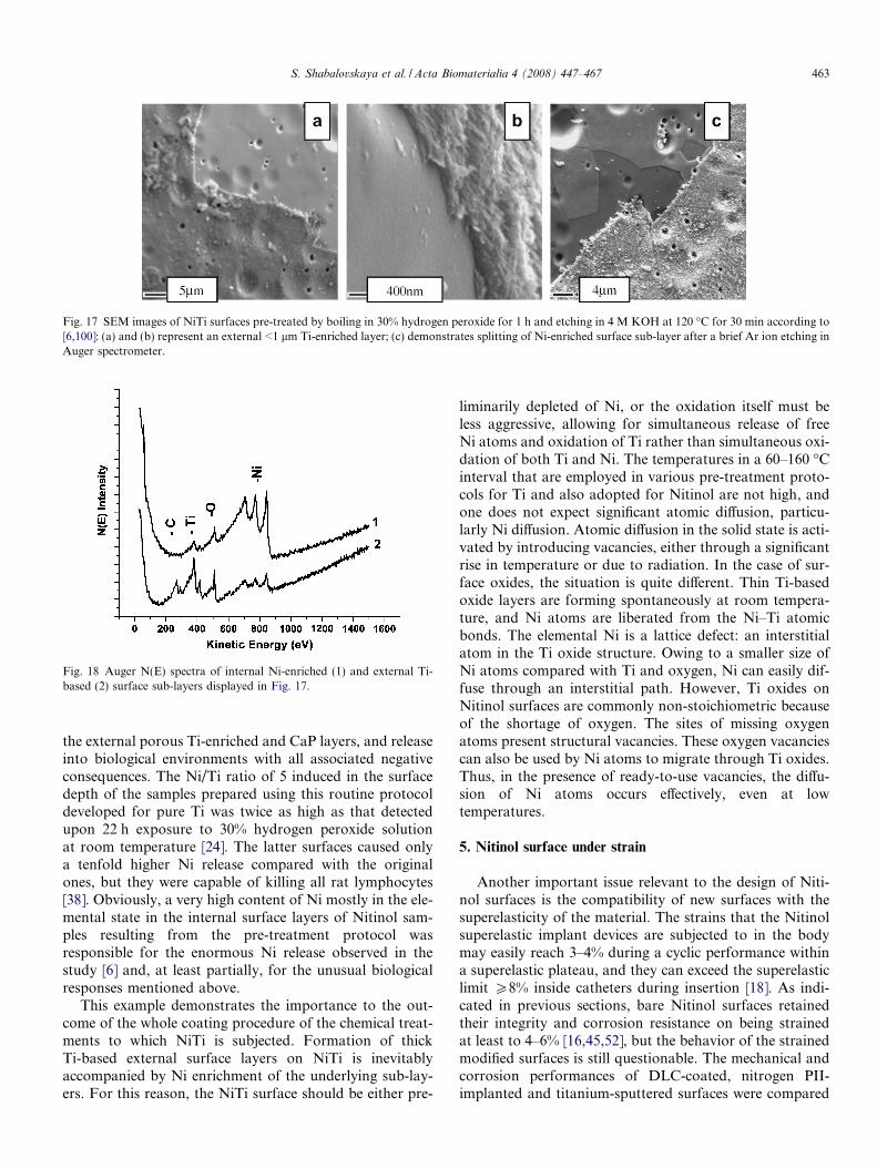
Fig. 17 SEM images of NiTi surfaces pre-treated by boiling in 30% hydrogen peroxide for 1 h and etching in 4 M KOH at 120 �C for 30 min according to[6,100]: (a) and (b) represent an external <1 lm Ti-enriched layer; (c) demonstrates splitting of Ni-enriched surface sub-layer after a brief Ar ion etching inAuger spectrometer.
Fig. 18 Auger N(E) spectra of internal Ni-enriched (1) and external Ti-based (2) surface sub-layers displayed in Fig. 17.
S. Shabalovskaya et al. / Acta Biomaterialia 4 (2008) 447–467 463
the external porous Ti-enriched and CaP layers, and releaseinto biological environments with all associated negativeconsequences. The Ni/Ti ratio of 5 induced in the surfacedepth of the samples prepared using this routine protocoldeveloped for pure Ti was twice as high as that detectedupon 22 h exposure to 30% hydrogen peroxide solutionat room temperature [24]. The latter surfaces caused onlya tenfold higher Ni release compared with the originalones, but they were capable of killing all rat lymphocytes[38]. Obviously, a very high content of Ni mostly in the ele-mental state in the internal surface layers of Nitinol sam-ples resulting from the pre-treatment protocol wasresponsible for the enormous Ni release observed in thestudy [6] and, at least partially, for the unusual biologicalresponses mentioned above.
This example demonstrates the importance to the out-come of the whole coating procedure of the chemical treat-ments to which NiTi is subjected. Formation of thickTi-based external surface layers on NiTi is inevitablyaccompanied by Ni enrichment of the underlying sub-lay-ers. For this reason, the NiTi surface should be either pre-
liminarily depleted of Ni, or the oxidation itself must beless aggressive, allowing for simultaneous release of freeNi atoms and oxidation of Ti rather than simultaneous oxi-dation of both Ti and Ni. The temperatures in a 60–160 �Cinterval that are employed in various pre-treatment proto-cols for Ti and also adopted for Nitinol are not high, andone does not expect significant atomic diffusion, particu-larly Ni diffusion. Atomic diffusion in the solid state is acti-vated by introducing vacancies, either through a significantrise in temperature or due to radiation. In the case of sur-face oxides, the situation is quite different. Thin Ti-basedoxide layers are forming spontaneously at room tempera-ture, and Ni atoms are liberated from the Ni–Ti atomicbonds. The elemental Ni is a lattice defect: an interstitialatom in the Ti oxide structure. Owing to a smaller size ofNi atoms compared with Ti and oxygen, Ni can easily dif-fuse through an interstitial path. However, Ti oxides onNitinol surfaces are commonly non-stoichiometric becauseof the shortage of oxygen. The sites of missing oxygenatoms present structural vacancies. These oxygen vacanciescan also be used by Ni atoms to migrate through Ti oxides.Thus, in the presence of ready-to-use vacancies, the diffu-sion of Ni atoms occurs effectively, even at lowtemperatures.
5. Nitinol surface under strain
Another important issue relevant to the design of Niti-nol surfaces is the compatibility of new surfaces with thesuperelasticity of the material. The strains that the Nitinolsuperelastic implant devices are subjected to in the bodymay easily reach 3–4% during a cyclic performance withina superelastic plateau, and they can exceed the superelasticlimit P8% inside catheters during insertion [18]. As indi-cated in previous sections, bare Nitinol surfaces retainedtheir integrity and corrosion resistance on being strainedat least to 4–6% [16,45,52], but the behavior of the strainedmodified surfaces is still questionable. The mechanical andcorrosion performances of DLC-coated, nitrogen PII-implanted and titanium-sputtered surfaces were compared

Fig. 19 SEM images of DLC (a and c) and nitrogen plasma implanted (band d) NiTi surfaces. The images (c) and (d) were acquired duringdeformation in a tension mode to 3.2% and 5.6% local strains, respec-tively. Adapted and reprinted from Ref. [44].
464 S. Shabalovskaya et al. / Acta Biomaterialia 4 (2008) 447–467
with non-coated electropolished NiTi [44,78]. Samples werestrained to 1%, �3% and 5.6% of the local strains in ten-sion mode within the stress–strain plateau, and above theplateau to 8%. In contrast to Ep substrate material thatretained an intact surface, the coated surfaces alreadybegan cracking at very low strains (Fig. 19). The firstcracks were observed at 1% strain for nitrogen-implanted(0.2 lm coating thickness), at �3% for DLC-coated(0.85 lm) and Ti-sputtered (1.1 lm) surfaces. The evalua-tion of corrosion resistance in this study showed that, incontrast to the original bare surface, the coated or modifiedNiTi surfaces, already in a strain-free state, might not havepassivity, and their corrosion resistance deteriorated dra-matically under strain. Alteration of the surface topogra-phy and heterogeneous distribution of local strains on thesubstrate associated with the nature of martensitic phasetransformation (responsible for shape memory effect), aswell as NiTi surface particulates, undermine the adhesionand cohesion of coatings and lead eventually to coatingdisintegration at the strain levels within the superelasticNitinol plateau.
6. Conclusions and outlook
From the analysis presented in this review, it is clear that,using the developed approaches, Nitinol surface can bemodified into various depths from nanometers to microme-ters, and its coating thickness can be extended up to�30 lm. It is questionable, however, whether micrometerdimensions are desirable, especially for the thin profile car-diovascular implants devices where stent wall thickness, forinstance, is being reduced to 30–50 lm to obtain better com-patibility. Another complication to do with Nitinol is thatcertain designed surfaces did not comply with material
superelasticity. Thick surfaces built from native oxides orresulting from surface modifications and coatings crackedupon low strains (<1%), and their corrosion resistance dete-riorated. It is obvious that a final conclusion regarding theadvantages of newly developed surfaces has to be madebased on corrosion tests performed under strain. Finally,as far as the improvement in Nitinol corrosion resistanceis concerned, the conducted studies of newly developed sur-faces in their vast majority did not use PS or scratch testspertinent to localized corrosion. For this reason, no conclu-sion can be made on the effect of the analyzed surface mod-ifications on the Nitinol’s localized corrosion resistance. Itseems at this point that the scratch-healing ability of Niti-nol, inferior to that of Ti, depends on the nature of thematerial, on the concentration, size and volume distributionof inclusions rather than on the surface oxide.
The reviewed studies indicate that NiTi surfaces asmodified using various procedures that involve tempera-tures from �160 to 600 �C (heat treatments, conventionaland plasma ion implantation as well as LSM) could beeither depleted of Ni or enriched with it, and, respectively,surface sub-layers could be composed of either Ti or Ni-based phases. Segregation of Ni in any form in NiTi sur-face sub-layers caused by material processing and surfacetreatments/pre-treatments or modification should be elim-inated to ensure non-toxic implant behavior in the body.The lasting Ni release increasing with time observed incertain studies is an indication of a Ni enrichment ofthe internal surface layers. It is obvious from the analysisthat the long-term exposures to low temperature protocols(�100–160 �C) developed originally for pure Ti are notsuitable for Nitinol because of the inevitable involvementof Ni.
As follows from the present review, there is obviously alack of understanding of the importance of the quality ofthe original surfaces subjected to modification. ‘Non-defined’ NiTi surfaces cannot be used as controls in thestudies, because the meaning of a reference is lost. Theanalysis presented does not allow for arriving at definiteconclusions regarding the biocompatibility of the reviewedsurfaces because the corresponding biological studies areeither not available for the moment or not conclusive. Inthose cases when the corresponding data were available,bare Nitinol surfaces seemed to perform better than or atleast similarly to the modified or coated surfaces bothin vitro and in vivo. Considering the important limitationsof artificial surfaces related to their incompatibility withsuperelasticity and poor adjustment to various Nitinol sur-face topographies, further studies into improvement andoptimization of bare surfaces are needed. Chemical andelectrochemical approaches combined with a hydrothermalapproach provided bare surfaces of various topographiesand thickness P20 nm, and resulted in a negligible Nirelease, stable corrosion performance and good/excellentbiological responses. It is obvious from the analysis thatthe biological potential of bare Nitinol surfaces is not fullyexhausted. A new aspect of native Nitinol surfaces related

S. Shabalovskaya et al. / Acta Biomaterialia 4 (2008) 447–467 465
to the possibility of manipulation with their thrombogenic-ity pertinent to both cardiovascular and osseogenic implantdevices also merits a deeper insight. It seems at this pointthat, for that purpose, many other surface propertiesbesides surface chemical composition, must be exploredsystematically. Among these are: surface oxide hydrationdegrees, types of conductivity, surface charge, structureand possibly catalytic activity of Nitinol surfaces composedof very well-known catalysts (Ti, Ni and their oxides),structural defects caused by oxygen deficiency and theireffects on the electronic structure of Ti surface oxides mod-ified by the presence of Ni.
Acknowledgments
The Research Fund of K.U. Leuven is acknowledgedfor financial support. The authors also grateful to L. Meis-ner, D. Bogdanski, K. Cheung, and C. Hebing, who gener-ously contributed the materials for this review as well as toG.K., who volunteered to edit this multidisciplinary ‘pro-ject’. We also acknowledge the useful discussions withG. Rondelli and his contribution to this review, as well asB. Harmon for his constant interest and encouragements.This manuscript was also authored by Iowa State Univer-sity of Science and Technology under Contract No. DE-AC02–07CH11358 with the US Department of Energy.
References
[1] Wever D, Velderhuizen A, De Vries J, Busscher H, Uges D, VanHorn J. Electrochemical and surface characterization of NiTi alloy.Biomaterials 1998;19:761–9.
[2] Cui Z, Man H, Yang X. The corrosion and nickel release behaviorof laser surface-melted NiTi shape memory alloys in Hanks solution.Surf Coat Technol 2005;192:347–53.
[3] Arndt M, Bruck A, Scully T, Jager A, Borauel C. Nickel ion releasefrom orthodontic NiTi wires under simulation of realistic in-situ
conditions. J Mater Sci 2005;40:3659–67.[4] Sui J, Cai W. Effect of diamond-like carbon (DLC) on the properties
of the NiTi alloys. Diamond Relat Mater 2006;15:1720–6.[5] Kobayashi S, Ohgoe Y, Ozeki K, Sato K, Sumiya T, Hirakuri K.
Diamond-like carbon coatings on orthodontic archwires. DiamondRelat Mater 2005;14:1094–7.
[6] Bogdanski D. Untersuchungen zur biocompatibilitat and bio-functionalitat von implantatmaterialien am beispiel von nickel–titan-formgedachtnislegierungen. Dissertation, Ruhr-UniversitatBochum: Germany; 2005.
[7] Clarke B, Carroll W, Rochev Y, Hynes M, Bradley D, Plumley D.Influence of Nitinol wire surface treatment on oxide thickness andcomposition and its subsequent effect on corrosion resistance andnickel ion release. J Biomed Mater Res 2006;79A:61–70.
[8] Chernish V, Tuboltsev V, Kalikauskas V. Angular distribution of Niand Ti atoms sputtered from NiTi alloy under He+ and Ar+ ionbombardment. Nucl Instrum Methods Phys Res 1998;B140:303–10.
[9] Eliades T, Zinelis S, Papadopoulos M, Eliades G, Athanasiou A.Nickel content of as-received and retrieved NiTi and stainless steelarchwires: assessing the nickel release hypothesis. Angle Orthodont2004;74:151–4.
[10] Oliveira A, Giacomelli C, Spinelli A. Microstructure and surfacecomposition effects on transpassivation of NiTi wires for implantpurposes. J Braz Chem Soc 2005;16:131–8.
[11] Prymak O, Bogdanski D, Koller M, Esenwein S, Muhr G,Beckmann F, et al. Morphological characterization and in vitro
biocompatibility of a porous nickel–titanium alloy. Biomaterials2005;26:5801–7.
[12] Tan L, Dodd R, Crone W. Corrosion and wear-corrosion behaviorof NiTi modified by plasma source ion implantation. Biomaterials2003;24:3931–9.
[13] Huang H. Surface characterization and corrosion resistance ofnickel–titanium orthodontic archwires in artificial saliva of variousdegree of acidity. J Biomed Mater Res 2005;74A:629–39.
[14] Kawakita J, Startmann M, Hassel W. High voltage pulse anodiza-tion of a NiTi shape memory alloy. J Electrochem Soc 2007;154:C294–8.
[15] ASTM G61-86. Annual Book of ASTM Standards: metals, testmethods and analytical procedures, vol. 3.02. Philadelphia, PA:American Society for Testing and Materials; 1995.
[16] Rondelli G, Vicentini B. Evaluation by electrochemical tests ofpassive film stability of equiatomic Ni–Ti alloy also in the presenceof stress induced martensite. J Biomed Mater Res 2000;51:47–54.
[17] Venugopalan R, Trepanier C. Assessing the corrosion behavior ofNitinol for minimally invasive device design. Minim Invasive TherAllied Technol 2000;9:67–75.
[18] Shabalovskaya S. Surface, corrosion and biocompatibility aspects ofNitinol as an implant material. Bio Med Mater Eng 2002;12:69–109.
[19] Shabalovskaya S. Physicochemical and biological aspects of Nitinolas a biomaterial. Int Mater Rev 2001;4:233–50.
[20] Shabalovskaya S, Van Humbeeck J. Biocompatibility of shapememory alloys. In: Yoneyama T, Miyazaki S, editors. Shapememory alloys for medical applications. Cambridge, UK: WoodheadPublishing Limited; 2008.
[21] Villermaux F, Tabrizian M, Yahia L, Czeremuszkin G, Piron D.Corrosion resistance improvement by plasma coating. Bio MedMater Eng 1996;6:241–54.
[22] Shabalovskaya S, Wataha J, Anderegg J, Hauch K, Cunnick J.Surface treatments and biocompatibility of Nitinol. In: Proceedingsof international conference on shape memory and superelastictechnologies. Germany: Baden–Baden; 2004. p. 367–73.
[23] Plant S, Grant D, Leach L. Behavior of human endothelial cells onsurface modified NiTi alloy. Biomaterials 2005;26:5359–67.
[24] Shabalovskaya S, Anderegg J. Surface spectroscopic characteriza-tion of TiNi nearly equiatomic shape memory alloys for implants. JVac Sci Technol 1995;A13:2624–32.
[25] Shabalovskaya S, Anderegg J, Laabs F, Thiel P, Rondelli G. Surfaceconditions of Nitinol wires, tubing, and as-cast alloys: the effect ofchemical etching, aging in boiling water, and heat treatment. JBiomed Mater Res 2003;65B:193–203.
[26] Theisen W, Schuermann A. Electro discharge machining of Nickel–Titanium shape memory alloys. Mater Sci Eng 2004;A378:200–4.
[27] Wirth C, Comte V, Lagneau C, Exbrayat P, Lissac M, Jaffrezic-Renault N, Ponsonnet L. Nitinol surface roughness modulatesin vitro cell response: a comparison between fibroblasts andosteoblasts. Mater Sci Eng 2005;C25:51–60.
[28] Armitage D, Parker T, Grant D. Biocompatibility and haemocom-patibility of surface – modified NiTi alloys. J Biomed Mater Res2003;66A:129–37.
[29] Palmaz J, Benson A, Sprague E. Influence of surface topography onendothelialization of intravascular metallic materials. J Vasc Inter vRadiol 1999;10:439–44.
[30] Rondelli G, Vincentini B. Localized corrosion behavior in simulatedbody fluids of commercial Ni–Ti orthodontic wires. Biomaterials1999;20:785–92.
[31] Carroll W, Kelly M, Brien B. Corrosion behavior of Nitinol wires inbody fluids environment. In: Proceedings of international conferenceon shape memory and superelastic technologies. Antwerpen; 1999.p. 240–9.
[32] Shabalovskaya S, Rondelli G, Anderegg J, Xiong J, Wu M.Comparative corrosion performance of black oxide, sandblasted,and fine-drawn Nitinol wires in potentiodynamic and potentiostatic

466 S. Shabalovskaya et al. / Acta Biomaterialia 4 (2008) 447–467
tests: effects of chemical etching and electropolishing. J BiomedMater Res 2004;69B:223–31.
[33] Pohl M, Heßing C, Frenzel J. Electrolytic processing of NiTi shapememory alloys. Mater Sci Eng 2004;A378:191–9.
[34] Barison S, Cattarin S, Daolio S, Musiani M, Tuissi A. Character-ization of surface oxidation of nickel–titanium alloy by ion-beamand electrochemical techniques. Electrochem Acta 2004;50:11–8.
[35] Fushimi K, Startmann M, Hassel A. Electropolishing of NiTi shapememory alloys in methanolic H2SO4. Electrochem Acta2006;52:1290–5.
[36] Shi P, Cheng F, Man H. Improvement in corrosion resistance ofNiTi by anodization in acetic acid. Mater Lett 2007;61:2385–8.
[37] Michiardi A, Aparicio C, Planell J, Gil F. New oxidation treatmentof NiTi shape memory alloys to obtain Ni-free surfaces and toimprove biocompatibility. J Biomed Mat Res 2006;77B:249–56.
[38] Shabalovskaya S. On the nature of biocompatibility and medicalapplications of NiTi shape memory alloys. Bio Med Mater Eng1996;6:267–90.
[39] Wagman D, Evans V, Parker V, Schumm R, Halow I, Bailey S,Churney K. J Phys Chem Ref Data 1982;Suppl. 2:11.
[40] Pohl M, Hebing C, Frenzel J. Electrolytic processing of shapememory alloys. Mater Corros 2002;53:673–9.
[41] Shabalovskaya S, Anderegg J, Rondelli G, Xiong J. The effect ofsurface particulates on the corrosion resistance of Nitinol wire. In:Proceedings of international conference on shape memory andsuperelastic technologies. Asilomar; 2003. p. 399–408.
[42] Brown S. On methods used for corrosion testing of NiTi. In:Proceedings of international conference on shape memory andsuperelastic technologies. Asilomar; 2000. p. 271–277.
[43] Cisse O, Savagodo O, Wu M, Yahia L. Effect of surface treatment ofNiTi alloy on its corrosion behavior in Hank’s solution. J BiomedMater Res 2002;61:339–45.
[44] Hebing C, Frenzel J, Pohl M, Shabalovskaya S. Effect of martensitictransformation on the performance of coated NiTi surfaces. MaterSc Eng 2007.
[45] Shabalovskaya S, Rondelli G, Anderegg J, Van Humbeeck J.Chemical and electrochemical properties of Nitinol surface oxidesand their correlation with biological responses. Abstract. In:International conference on shape memory and superelastic tech-nologies. Tsukuba; December 2007.
[46] Chuprina V. A study on oxidation of titanium nickelide. Phasecomposition of the scale. Powder Metall 1989;318:57–61.
[47] Firstov G, Votchev R, Kumar H, Blanpain B, Van Humbeeck J.Surface oxidation of NiTi shape memory alloys. Biomaterials2002;23:4863–71.
[48] Heßing C, Gabert S, Pohl M, Parezanovic I. High-temperaturecorrosion of NiTi shape memory alloys. Mat-Wiss Werkstofftech2004;35:332–7.
[49] Zhu L, Pino J, Pelton A. Oxidation of Nitinol. In: Proceedings ofinternational conference on shape memory and superelastic tech-nologies. USA: Asilomar; 2003. p. 357–66.
[50] Menchaka L, Lam H, Leong I, Li S, Johnson D. Endothelial andsmooth muscle cell growth on titanium nickel thin film. In:Proceedings of international conference on shape memory andsuperelastic technologies. Germany: Baden–Baden; 2004. p. 381–6.
[51] Shabalovskaya S, Anderegg J, Rondelli G, Vanderlinden W, DeFeyter S. Comparative in vitro performances of bare Nitinolsurfaces. Biomed Mater Eng 2008;18:1–14.
[52] Trepanier C, Zhu L, Fino J, Pelton A. Corrosion resistance ofoxidized Nitinol. In: Proceedings of international conference onshape memory and superelastic technologies. USA: Asilomar; 2003.p. 367–74.
[53] Park J, Gemmel H, Davies J. Platelet interactions with titanium:modulation of platelet activity by surface topography. Biomaterials2001;22:2671–82.
[54] Thierry B, Tabrizian M. Biocompatibility and biostability ofmetallic endovascular implants: state of art and perspectives. JEndovasc Ther 2003;10:807–24.
[55] Thierry B, Merhi Y, Bilodeau L, Trepanier C, Tabrizian M. Nitinolversus stainless steel stents: acute thrombogenicity study in anex vivo porcine model. Biomaterials 2002;23:2997–3005.
[56] Palmaz J, Bailey D, Marton D, Sprague E. Influence of stent designand material composition on procedure outcome. Basic Sci RevArticle 2002:1031–40.
[57] Thierry B, Tabrizian M, Savadogo O, Yahia L. Effects of steriliza-tion processes on NiTi alloy: surface characterization. J BiomedMater Res 2000;49:88–98.
[58] Schmutz P, Frankel G, Serry F. Corrosion studies with AFM, Part1: characterization of potential inhomogeneities on passive surfacesby surface potential imaging. Veeco Instruments Inc., n5, AN87-91;2005.
[59] Michiardi A, Aparicio C, Ratner B, Planell J, Gil J. The influence ofsurface energy on competitive protein adsorption on oxidized NiTisurfaces. Biomaterials 2006;29:586–94.
[60] Lucassen M, Sarkar B. Nickel-binding constituents of human bloodserum. J Toxic Environ Health 1997;5:897–905.
[61] Clarke B, Kingshott P, Hou X, Rochev Y, Gorelov A, Carroll W.Effect of Nitinol wire surface properties on albumin adsorption.Acta Biomater 2007;3:103–11.
[62] Goodman S. Sheep, pig, and human platelet-material interactionswith model cardiovascular devices. J Biomed Mater Res1999;45:240–50.
[63] Ericksson C, Lausmaa J, Nygren H. Interaction between humanwhole blood and modified TiO2 – surfaces: influence of surfacetopography and oxide thickness on leukocyte adhesion and activa-tion. Biomaterials 2001;22:1987–96.
[64] Hong J, Andersson J, Ekdahl K, Elgue G, Axen N, Larsson R,Lilsson B. Titanium is highly thrombogenic biomaterial: possibleimplications for osteogenesis. Thromb Haemost 1999;82:58–64.
[65] Sigler M, Paul T, Grabitz R. Biocompatibility screening in cardio-vascular implants. Z Kardiol 2005;94:383–91.
[66] Janke T, Voshage G, Muller-Hulsback S, Grimm J, Heller M,Brossmann J. Endovascular placement of self-expending Nitinol coilstents for the treatment of Femoropopliteal obstructive disease. JVasc Interv Radiol 2002;13:257–66.
[67] Sabeti S, Schillinger M, Amighi J, Sherif C, Mlekush W, Ahmadi R,Minar E. Primary patency of femoropopliteal arteries treated withNitinol versus stainless steel self-expendindg stents: propensityscore-adjusted analysis. Radiology 2004;232:516–21.
[68] Anders A, editor. Handbook of plasma immersion ion implantationand deposition. New York: J Wiley & Sons, Inc.; 2000. p. 1–19.
[69] Lotkov A, Meisner L, Grishkov V. Titanium nickelide-based alloys:surface modifications with ion beam, plasma flows and chemicaltreatment. Phys Metals Metall 2005;99:508–19.
[70] Grant D, Green S, Wood J. The surface performance of shot peenedand ion implanted NiTi shape memory alloys. Acta Metall Mater1995;43:1045–51.
[71] Tan L, Crone W. Surface characterization of NiTi modified byplasma source ion implantation. Acta Mater 2002;50:4449–60.
[72] Shevchenko N, Pham M, Maitz M. Studies of surface modified NiTialloys. Appl Surf Sci 2004;235:126–31.
[73] Maitz M, Shevchenko N. Plasma-immersion ion-implanted Nitinolsurface with depressed Ni concentration for implants in blood. JBiomed Mater Res 2006;76A:356–65.
[74] Yeung K, Poon R, Liu X, Ho P, Chung C, Chu P, Lu W, et al.Investigation of nickel suppression and cytocompatibility of surface-treated Ni–Ti shape memory alloys by using plasma immersion ionimplantation. J Biomed Mater Res 2005;72A:238–45.
[75] Yeung K, Poon R, Liu X, Ho J, Chung C, Chu P, et al.Improvement of corrosion resistance, mechanical properties andcyto-compatibility of NiTi shape memory alloys using nitrogen, andoxygen plasma immersion ion implantation. J Biomed Mater Res2005;75A:256–67.
[76] Poon R, Ho J, Luk C, Liu X. Improvement of corrosion resistanceof NiTi orthopedic materials by carbon plasma immersion ionimplantation. Nucl Instrum Methods Phys Res 2006;B242:270–4.

S. Shabalovskaya et al. / Acta Biomaterialia 4 (2008) 447–467 467
[77] Poon R, Yeung K, Liu X, Chu P, Chung C, Lu W, et al. Carbonplasma immersion ion implantation of NiTi shape memory alloys.Biomaterials 2005;26:2265–72.
[78] Heßing C, Pohl M, Frenzel J. Coated NiTi shape memory alloysunder strain. In: Proceedings of international conference on shapememory and superelastic technologies. Germany: Baden–Baden;2004. p. 301–7.
[79] Poon R, Liu X, Chung C, Chu P. Surface and corrosion charac-teristics of carbon plasma implanted and deposited nickel–titaniumalloy. J Vac Sci Tech 2005;A23:525–30.
[81] Sui J, Cai W. Mechanical properties and anti-corrosion behavior ofthe diamond-like carbon films. Surf Coat Technol 2006;201:1323–7.
[82] Brunette D, Tengvall P, Textor M, Thomsen P. Titanium inmedicine. Berlin: Springer; 2001.
[83] Ohgoe Y, Kobayashi S, Kobayashi O, Aoki H, Nakamori H,Hirakuri K. Reduction effect of nickel ion release on diamond-likecarbon film coated onto an orthodontic archwire. Thin Solid Films2006;497:218–22.
[84] Cheng Y, Zheng Y. The corrosion behavior and hemocompatibilityof TiNi alloys coated with DLC by plasma based ion implantation.Surf Coat Technol 2006;200:4543–8.
[85] Shih C, Lin K, Chen Y, Su Y. Increased corrosion resistance of stentmaterial by converting surface film of polycrystalline oxide intoamorphous. J Biomed Mater Res 2000;52:323–32.
[86] Ryhanen J, Niemi E, Serlo S, Niemela E, Sandvik P, Pernu H, SaloT. Biocompatibility of nickel–titanium metal and its corrosionbehaviour in human cell cultures. J Biomed Mater Res1997;35:451–7.
[87] Cheng F, Shi P, Man H. A preliminary study of TiO2 deposition onNiTi by a hydrothermal method. Sci Coat Technol 2004;187:26–32.
[88] Cheng F, Shi P, Man H. Anatase coating on NiTi via a low-temperature sol–gel route for improving corrosion resistance.Scripta Mater 2004;51:1041–5.
[89] Chu C, Chung C, Pu Y, Lin P. Graded surface structure inchemically polished NiTi shape memory alloy after subsequentNaOH treatment. Scripta Mater 2005;52:1117–23.
[90] Chu C, Chung C, Chu P. Surface oxidation of NiTi shape memoryalloy in boiling aqueous solution containing hydrogen peroxide.Mater Sci Eng 2006;A417:104–9.
[91] Chu C, Chung C, Zhou J, Pu Y, Lin P. Fabrication andcharacteristics of bioactive film sodium titanate/titania graded filmon NiTi shape memory alloy. J Biomed Mater Res 2005;75A:595–602.
[92] Liu J, Shi F, Yang D. Characterization of sol–gel-derived TiO2 andTiO2–SiO2 films for biomedical applications. J Mater Sci Tech2004;20:550–4.
[93] Starovsky D, Gotman I. Corrosion behavior of titanium nitridecoated Ni–Ti shape memory surgical alloy. Biomaterials 2001;22:1853–9.
[94] Tepe G, Schmehl J, Wendel H, Schaffner S, Heller S, Gianotti M,Claussen C, Duda S. Reduced thrombogenicity of Nitinol stents –in vitro evaluation of different surface modifications and coatings.Biomaterials 2006;27:643–50.
[95] Kong X, Grabitz W, Van Oeveren W, Klee D, Van Kooten T,Freudenthal F, Qing M, et al. Effect of biologically active coating onbiocompatibility of Nitinol devices designed for the closure of intra-atrial communications. Biomaterials 2002;23:1775–83.
[96] Mazumder MM, De S, Trigwell S, Ali N, Mazumder MK, Mehta J.Corrosion resistance of polyurethane-coated Nitinol cardiovascularstents. J Biomater Sci Polym Edn 2003;14:1351–62.
[97] Chen M, Yang X, Hu R, Cui Z, Man H. Bioactive NiTi shapememory alloy used as bone bonding implant. Mater Sci Eng2004;C24:497–502.
[98] Schlegel P. In vivo biocompatibility studies of different calcium–phosphate surfaces for implant bone integration. Dissertation,University Zurich, Davos, Switzerland; 2004.
[99] Shi P, Geng F, Cheng F. Preparation of titania-hydroxyapatitecoating on NiTi via a low-temperature route. Mater Lett2006;60:1996–9.
[100] Choi J, Bogdanski D, Koller M, Esenwein S, Muller D, Muhr G,Epple M. Calcium phosphate coating of nickel–titanium shape-memory alloys. Coating procedure and adherence of leukocytesand platelets. Biomaterials 2003;24:3689–96.
[101] Hardhammer P, Van Beusekom H, Emanuelsson H. Reductionthrombotic events with heparin coated Palmatz–Schatz stents innormal coronary arteries. Circulation 1996;93:423–30.
[102] Serruys P, Emanuelsson H, Van de Giessen W. Heparin-coatedPalmatz–Schatz stents in human coronary arteries. Early outcome ofthe Benestent-II pilot study. Circulation 1996;93:412–22.
[103] Vanderkerkhove R, Temmerman E. Electrochemical research on thecorrosion of NiTi. In: Proceedings of international conference onshape memory and superelastic technologies. Belgium: Antwerpen;1999. p. 129–141.
[104] Berger-Gorbet M, Broxup B, Rivard C, Yahia L. Biocompatibilitytesting of NiTi screws using immunohistochemistry of sectioncontaining metallic implants. J Biomed Mater Res 1996;32:243–8.
[105] Takeshita F, Takata H, Ayukawa Y, Suetsugu T. Histomorpho-metric analysis of the response of rat tibia to shape memory alloy(Nitinol). Biomaterials 1997;18:21–5.
[106] Bogdanski D, Esenwein S, Epple M, Koller M. Inhibition ofPMN apoptosis after adherence to dip-coated calcium phosphatesurfaces on a NiTi shape memory alloy. Biomaterials 2004;25:4627–32.
[107] Bogdanski D, Epple M, Esenwein S, Muhr G, Petzoldt V, PrymakO, et al. Biocompatibility of calcium phosphate-coated and ofgeometrically structured nickel–titanium (NiTi) by in vitro testingmethods. Mater Sci Eng 2004;A378:527–31.
[108] Assad M, Chernyshov A, Jarzem P, Leroux M, Cillard C, CharetteS, Rivard C. Porous titanium–nickel for intervertebral fusion in asheep model: Part 2. Surface analysis and nickel release assessment. JBiomed Mater Res 2003;64B:121–9.
[109] Wataha J, Lewis J, Volkman K, Lockwood P, Messer R, BouillaguetS. Sublethal concentrations of Au(III), Pd(II), and Ni(II) differen-tially alter inflammatory cytokine secretion from activated mono-cytes. J Biomed Mater Res 2004;69B:11–7.