lower respiratory tract infection - iacld.iriacld.ir/dl/modavan/bacteriology/lowerrespiratory... ·...
TRANSCRIPT

Lower Respiratory Tract Infection

- Clinical Diseases and etiologic agents
-Sample Collection and transport
-Direct Smear
(interpretation and Report) -Culture methods
(interpretation and Report)

“ The culture of lower respiratory specimens may result in more
unnecessary microbiologic effort than any other type of specimen.”
Raymond C Bartlett

Lower Respiratory Tract Infections Epidemiology
• Pneumonia is the sixth leading cause of death in US
• Increasing numbers of patients at risk – Aging population
– Increase in patients with immunocompromising conditions

Lower respiratory infections
• Overtreatment has lead to resistance
– Multidrug resistant Streptococcus pneumoniae
– Resistance among hospital acquired pathogens such as Acinetobacter, Pseudomonas aeruginosa E.coli K.pneumonia (ESBLs) MRSA and others

Categories of Lower Respiratory Tract Infections
• Acute bronchitis
• Community acquired pneumonia
• Hospital acquired pneumonia
• Pneumonia in the immunocompromised host
Acute Bronchitis
•Part of or preceded by an URTI
•Sputum is often clear at the onset but may become purulant
•Etiologic agents:
- adults viral
- Infant and preschool children: bordetella pertusis

Chronic bronchitis
Common condition affecting about 10-30% adults
Coughing up sputum on most days during at least 3 consecutive months for more than 2 successive years
Viruses are frequent causes
Non encapsulated H.influ/S.pneumoniae/M.catharalis

Bronchiolitis
Human metapneumovirus
RSV 40-80% cases

Pneumonia
*Children 2 month to 5 years 80% viral (RSV/Parainflu/Adeno/Metapneumovirus/C.pneumoniae/M.pneumoniae)
*Children 5-14 years 70 % viral and up to 80% of cases mixed infection
*Young adults<30 years M.pneumoniae/Influ/C.pneumoniae


Hospital Acquired Pneumonia
*5-10 % hospital in – patients develop infection during administration to ICU.
*80% of hospital associated pnemonia is linked with mechanical ventilation(VAP)
*10-30% of critical care patients develop VAP
*VAP is the most common and fatal infection of ICU
*VAP may account for up to 60% of all HAI


Organism isolated from tracheal tube aspirates in Milad Hospital
organism No(%)
K. pneumoniae
P. aeruginosa
A. baumannii
S. aureus
S. marsescense
E. coli
Enterobacter spp
Candida spp
Others
67(20)
62(18.5)
60(18)
51(15.5)
32(9.5)
22(6.6)
10(3)
5(1.5)
26(7.7)
Rhbar and Hajia accepted for publication in ICHE
Aspiration pneumonia
Anaerob
S.aureus
Enterobacteriacae
Pseudomona
H.influ
Legionella
Acinetobacter
Moraxella

Chronic LRTI
*M.tuberculosis
*M.avium intracellular
*Actinomyces/Nocardia
*Fungi

Cystic Fibrosis
*Mucoid Pseudomona
*S.aureus
*H.influ
*Burkhholderia cepacia
RSV/Flu/Aspergylus

Immunocoprommised patients
*Neoplasm
*Transplant Recipents
*HIV

Community Acquired Pneumonia Diagnosis
Available Test Methodologies
• Sputum Gram stain and culture
• Blood cultures
• Serologic studies
• Antigen detection tests
• Nucleic acid amplification tests

Specimen collection

Sputum Collection • Proper patient instruction
– Food should not have been ingested for 1-2 h prior to expectoration
– The mouth should be rinsed with saline or water
– Avoid using mouth wash solution
– Patient should breathe and cough deeply
– Patient should expectorate into a sterile container
• Transport container immediately to lab
• Perform Gram stain and plant specimen as soon as possible
Sputum collection • Sputum of less than 2ml should not be
processed unless obviously purulent
• Only 1 sputum per 24hr .submitted
• Transportation in <2 hr is recommended with refrigeration if delays anticipated.
• Handle all samples using universal precautions.
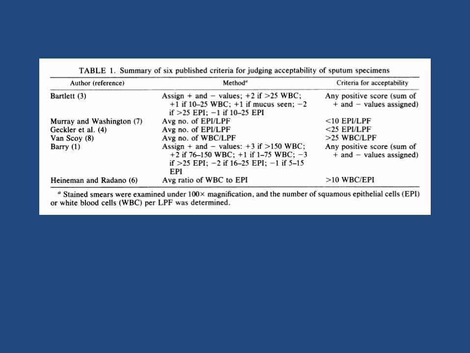
• Some scoring system should be used to reject specimen that re oral contamination.

Induced sputum *Patients who are unable to produce sputum may be
assisted by respiratory therapy technician. Aerosol induced specimen are collected by allowing the patient to breath aerosolized droplets of a solution of 15% sodium chloride and 10% glycerin for approximately 10 minute . Obtaining such specimen may avoid the need for a more invasive procedures ,such as bronchoscopy or needle aspiration, in many cases.
*Specimen resemble saliva
*Should be accepted in the laboratory without prescreening
*High diagnostic value in pneumocystis,mycobacterium and fungi

Gastric aspiration
• The gastric aspiration is used exclusively for isolation of acid-fast bacilli and may be collected from patients who are unable to produce sputum, particularly young children. The relative resistance of mycobacteria allows them to remain viable for a short period. Gastric lavage must be delivered to the lab immediately so that the acidity can be neutralized. Specimen can be first neutralized and then transported if immediate delivery is not possible.

IDSA Practice Guidelines Diagnostic Tests for CAP
• Outpatients – Empiric therapy with a macrolide, doxycycline, or
a fluoroquinolone
• Hospitalized patients with CAP – Gram stain and culture of sputum
– 2 pretreatment blood cultures
– Studies for Mtb, Legionella in select patients
Bartlett JG. 2003. Clin Infect Dis 37:1405-33

ATS Guidelines Diagnostic Tests for CAP
• Empiric therapy for outpatients – Macrolide or tetracycline
• Hospitalized patients with CAP – 2 sets of pre-treatment blood cultures – Pleural fluid Gram stain/culture when appropriate – Studies for Legionella, Mtb, fungi in select patients – Sputum Gram stain/culture only if resistant or unusual
pathogen is suspected – Avoid extensive testing ATS. 2007. Clin Infect Dis 44:S27-72

Hospital Acquired Pneumonia Diagnosis
• American College of Chest Physicians: Clinical findings are not sufficient for definitive diagnosis
• Qualitative culture or endotracheal sputum has poor predictive value
• Endotracheal aspiration or suction samples should be treated as sputum by laboratory(GNB clonization)
• Bronchoscopy is recommended by many pulmonologists – Bronchial washes (contamination with URT flora) – Protected specimen brushing(Aspiration pneumonia) – Bronchoalveolar lavage specimens (BAL) – Transbronchial biopsy
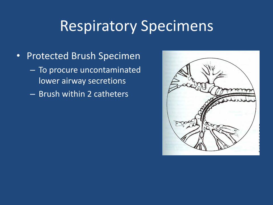
Respiratory Specimens
• Protected Brush Specimen
– To procure uncontaminated lower airway secretions
– Brush within 2 catheters

Respiratory Specimens
• Bronchoalveolar Lavage (BAL)
– Samples large area of the lung
– Performed using a bronchoscope
– 100 to 250 ml of saline injected
– Injected saline along with secretions is collected by aspiration
• Transthoracic Aspiration
– Involves percutaneous introduction of a needle directly into the infiltrate

Direct Gram Smear

Specimen Acceptability Criteria Gram Stain Smear
Prescreening for adequacy

*Expectorated sputum *Endotracheal aspiration or suction

Sputum Gram Stain
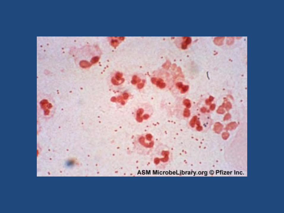
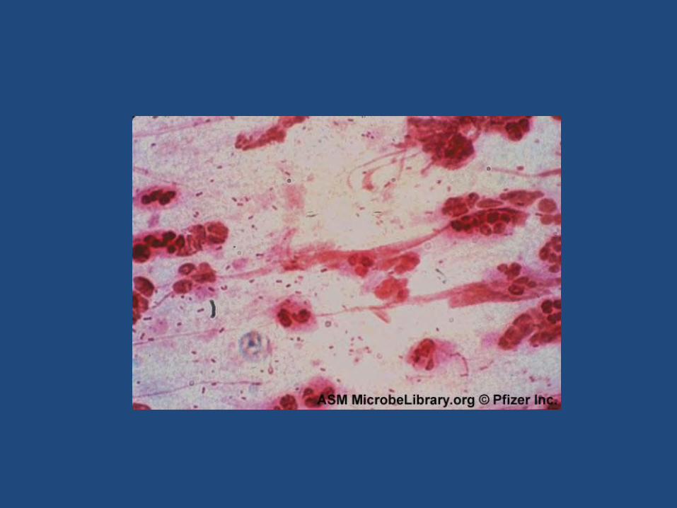
Good quality specimens
• Quantify number and types of inflammatory cells
• Note presence of bronchial epithelial cells
• Concentrate on areas with WBCs when looking for organisms


WBC
Number of WBC may not relevant because *many patients are severely neutropenic from
disease or treatment
*Defect in effective inflamatory response such as Immunocompromosed patients (bone
marrow implant)
*Legionella pneumonia sputum
*Forign body (endotracheal catheter)
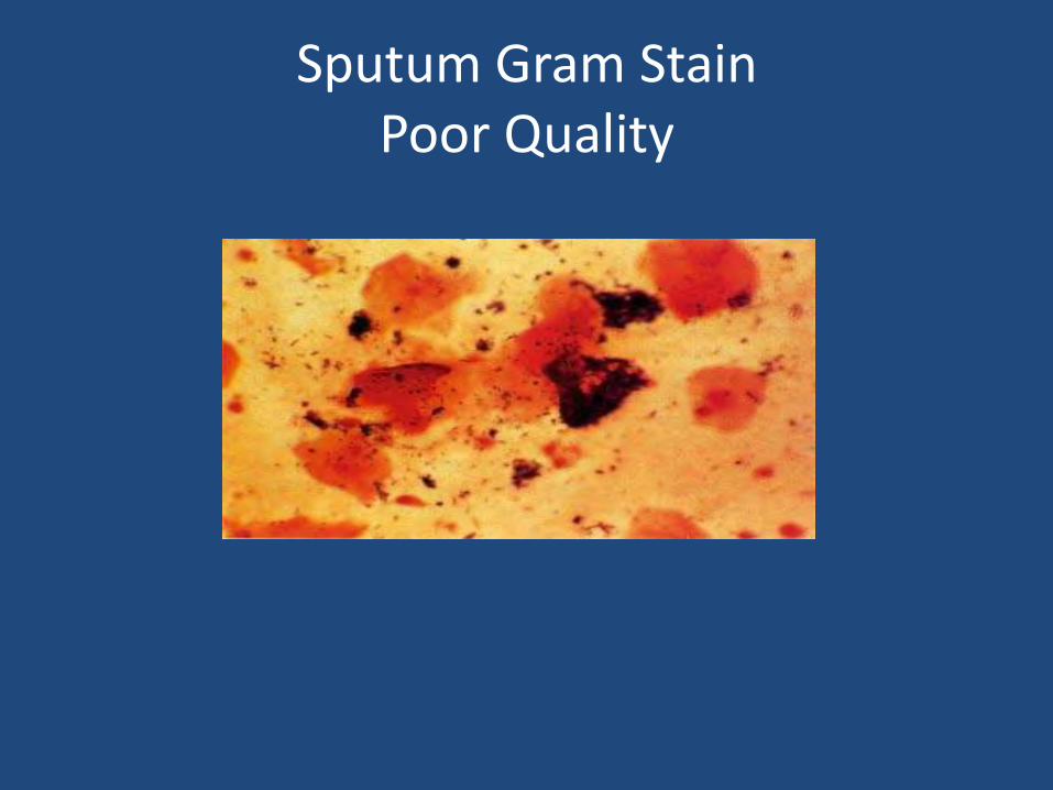
Sputum Gram Stain Poor Quality


Sputum Gram Stain Good Quality


Bronchoalveolar Lavage (BAL) Specimen Acceptability
• Microscopic examination of Gram-stained smear
– Acceptable
• <1% of cells present are squamous epithelial cells
– Unacceptable
• >1% of cells present are squamous epithelial cells
• Presence of ciliated columnar bronchial epithelial cells/goblet cells or pulmonary macrophage in specimen obtained by brochoscop or BAL indicates a specimen from LRT

Gram Stain Smear
Interpretation and Report

-No squamous epithelial cell seen -Few squamous epithelial cell seen -Abundant (many) squamous epithelial cell seen indicating oropharyngeal contamination -Contamination with saliva please submit another specimen -No WBC Seen

Smears with predominant EP and without WBC No need to culture and report of organisms
Smears with EP and WBC
-Predominant organism Gram negative bacilli predominant organism seen on Gram smear
-Presence of other organisms
Mixed bacterial morphotypes also present on Gram smear
-Without predominant organism Mixed bacterial morphotypes seen on Gram smear
-Without organism No organisms seen
-One morphotype Report morphotype as few/moderate and many

Gram Stain Reports
• Be as descriptive as possible
– Moderate neutrophils
– Moderate Gram positive diplococci suggestive of Streptococcus pneumoniae
– Few bacteria suggestive of oral flora
• Keep report short—avoid line listing of all morphotypes present

Culture
Routine culture
• Blood Agar
• Chocolate Agar
• Mac Conkey
• Thioglycolate(for aspirated specimen biopsy)

Routine culture
• Because of contaminating oral flora ,sputum specimens ,specimens obtained by bronchial washing, and lavage trachestomy, or endotracheal tube aspirates are not inoculated to enriched broth or incubated anaerobically.
• Only specimens obtained by percutaneous aspiration (including transtracheal aspiration )and by protected bronchial brush are suitable for anaerobic culture

Culture
• Transtracheal and percutaneous lung aspiration material may be inoculated to enriched thioglycollate ,as well as to solid media. For suspected cases of legionella disease buffered charcoal yeast extract (BCYE) agar and selective BCYE are inoculated.

Culture
• Sputum specimens from patients known to have cystic fibrosis should be inoculated to selective agar ,such as manitol salt agar for recovery of S .aureus and selective horse blood-bacitracin ,incubated anaerobically and aerobically ,for recovery of H.influenzae that may be obscured by the mucoid P.aeroginosa on routine media.

Bronchoscopy Specimens Processing • Bronchoscopy brush protected
– Aerobic bacterial culture and Gram stain – Anaerobic bacterial culture – Limited volume
• Bronchoscopy brush, unprotected – No anaerobic culture – Limited volume
• Bronchial washings – Useful only for pneumonia caused by strict pathogens – Reasonable requests: Mtb, Fungi, Legionella, Pneumocystis
• Bronchoalveolar lavage – No anaerobe culture

Bronchoscopy Samples Quantitative Methods
PSB or BAL Baselski and Wunderink. 1994. Clin Micro Rev 7:546. vortex 30-60s Final dilutions
Plate 0.1 ml Chocolate, blood 1:10
Dilute
0.1 ml to 9.9 ml saline Plate 0.1 ml Chocolate 1:1000
blood
Dilute 0.1 ml to 9.9 ml
saline
Plate 0.1 ml Chocolate 1:100,000
blood

Bronchoscopy Samples Quantitative Methods
Calibrated loop method
Baselski and Wunderink. 1994. Clin Micro Rev 7:547
PSB vortex 30-60 s BAL
Plate 0.1 ml
Chocolate Chocolate Chocolate
Final Dilutions
1:10 1:100 1:1000
Plate 0.01 ml Plate 0.001 ml
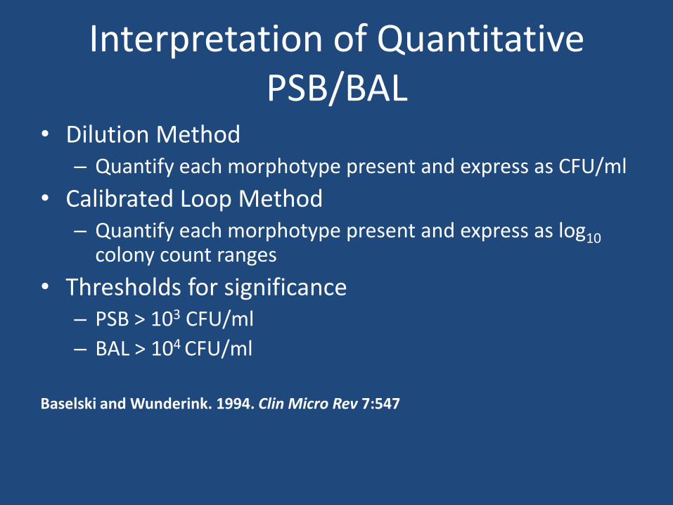
Interpretation of Quantitative PSB/BAL
• Dilution Method – Quantify each morphotype present and express as CFU/ml
• Calibrated Loop Method – Quantify each morphotype present and express as log10
colony count ranges
• Thresholds for significance – PSB > 103 CFU/ml
– BAL > 104 CFU/ml
Baselski and Wunderink. 1994. Clin Micro Rev 7:547

Special notes
Specimen without EP or few EP

Heavy or moderate (predominant) growth or colony resemble S.pneumoniae
α hemolytic Streptococci
Diagnostic tests
S.pneumoniae α hemolytic Streptococci as normal microbial isolated

S.pneumoniae S.pneumoniae mucoid colony

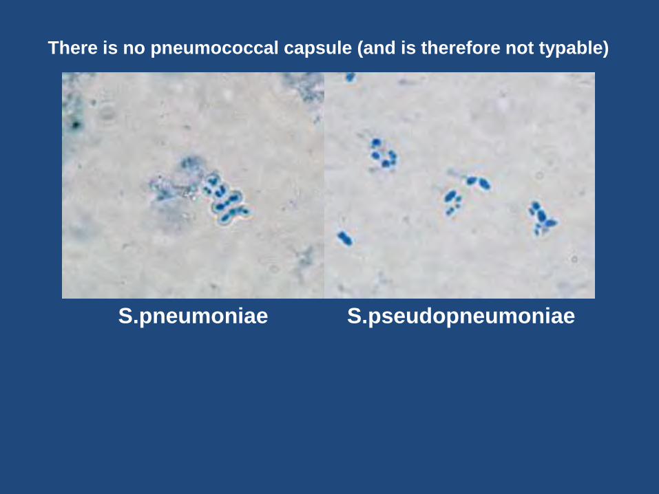
S.pneumoniae S.pseudopneumoniae

S.pneumoniae S.pseudopneumoniae

S.pneumoniae
S.pseudopneumoniae
Ambient atmospher 5% Co2

S.pneumoniae S.pseudopneumoniae
It is not soluble in bile
S.pseudopneumoniae S.pneumoniae
There is no pneumococcal capsule (and is therefore not typable)

β hemolytic Streptococci Mixed with other colony or few colony Good growth
Isolation and subculture(stabing)
Diagnosis tests
Group A Streptococci Isolated Serotyping for other
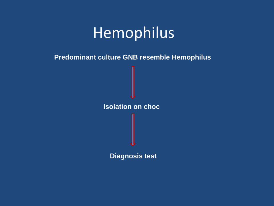
Hemophilus
Predominant culture GNB resemble Hemophilus
Isolation on choc
Diagnosis test

Nieserria and Moraxella Predominant culture GNDB or GNDB in Gram smear
Diagnosis test

Other Gram Negative

Staphylococci
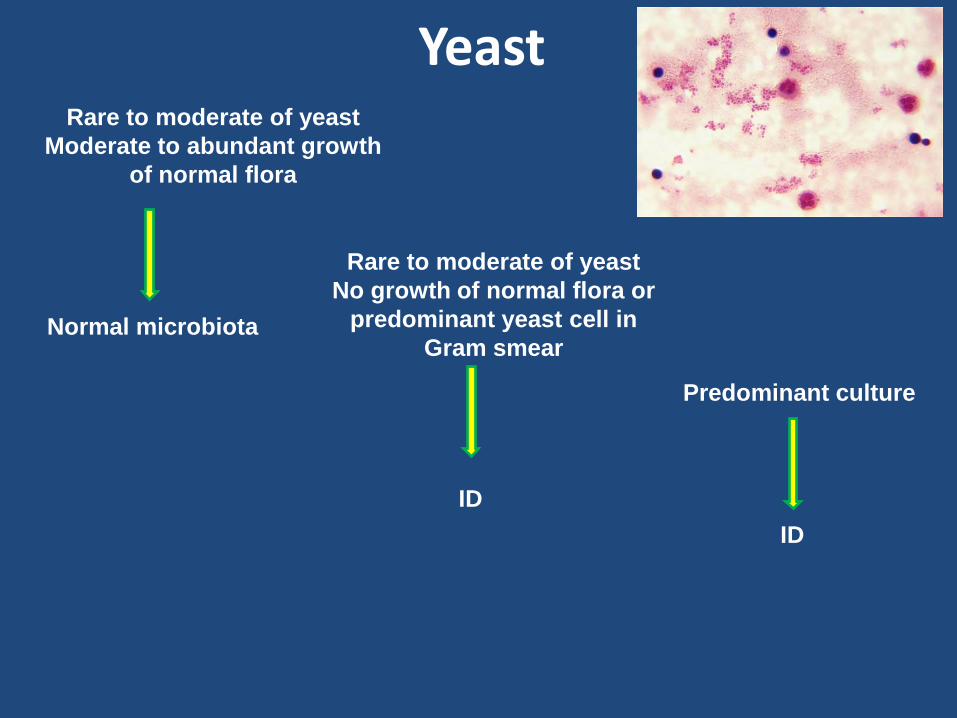
Yeast Rare to moderate of yeast
Moderate to abundant growth
of normal flora
Rare to moderate of yeast
No growth of normal flora or
predominant yeast cell in
Gram smear
Predominant culture
Normal microbiota
ID
ID

Special notes
Specimen with many EP and WBC

predominant organisms in gram stain not seen No need to additional diagnosis and complementary culture
One organism seen in gram stain -Growth on culture ID / AST
-Normal flora no need to ID
-Predominant GNB in gram smear and isolation of one type colony or predominant colony
ID / AST
-Two or more colony type of GNB without predominant type
Multiple species of gram negative bacilli, no predominant organism isolated
Culture without predominant organisms Mixed bacterial morphotypes

*Examine all sputum specimen microscopically *Report microscopic finding quantitavely *Use microscopic criteria for accetability *compare Gram stain and culture results
*Oganisms seen in the smear don’t grow in culture *Organism that grow in moderate quantity or less are not
seen in the Gram smear *List both the Gram smear and culture results together in
the same report
College of American Pathologist Recommendation
