letters to the editor - ucm · letters to the editor primary t-cell immunodeficiency with...
TRANSCRIPT

Letters to the Editor
Primary T-cell immunodeficiencywith functional revertant somaticmosaicism in CD247
To the Editor:T lymphocytes detect antigens with the T-cell receptor (TCR)
composed of a variable heterodimer (either ab or gd), 2 invariantheterodimers (CD3gε and CD3dε), and an invariant homodimer(CD247 or zz).1 Because of the crucial role of TCR signaling inthymic selection, mutations in TCR, CD3, or CD247 selectivelyimpair T-cell development, albeit to different degrees: deficiencyof CD3d or CD3ε, but not of CD3g or CD247, causes severe T-celllymphopenia. Their clinical outcome is also disparate, becauseCD3g deficiency does not require urgent transplantation. Thus,TCR immunodeficiencies display a range of phenotypes andcareful differential diagnosis is essential for appropriate therapy.
We describe an infant born to consanguineous parentswith early-onset chronic cytomegalovirus infection, severeimmunodeficiency, and extremely low surface TCR levels. Herimmunologic characterization at age 11 months is summarized inTable E1 in this article’s Online Repository at www.jacionline.
FIG 1. Patient characterization. A, Peripheral blood
indicated subsets. Numbers indicate % MFI relativ
the indicated CD. Numbers indicate % iCD2472 or
sorted T cells. Numbers indicate % normalized ban
predicted proteins. EC, Extracellular; IC, intracellular; M
TM, transmembrane; WB, Western blot. ***P <_ .001.
org. Briefly, she showed low T- and B-cell counts, selective severeCD41 T-cell lymphopenia (Fig 1, A), low recent thymicemigrants, and naive T cells and poor TCRVb repertoire, allsuggestive of a defect in T-cell development despite a normal-sized thymus (see Fig E1 in this article’s Online Repository atwww.jacionline.org).
Surface TCR expression was markedly reduced in both ab andgd T cells (Fig 1, B), suggesting a defect in an invariant chain ofthe TCR complex. Intracellular (i) flow cytometry showed normalCD3g, d, and ε expression but almost absolute absence of CD247(Fig 1, C). cDNA sequencing revealed a homozygous T-to-C mu-tation at position 12 of exon 1 of CD247 as reported recently2
(NCBI/ClinVar: rs672601318), which causes loss of the initiationcodon and therefore prevents translation. The patient’s parentsand 4 additional family members were asymptomatic butheterozygous for the mutation (see Fig E2 in this article’s OnlineRepository at www.jacionline.org). Interestingly, surface TCRexpression in mutation carriers was reduced 2-fold (Fig 1, B),revealing a clear correlation between surface TCR and CD247genotype, which was useful for diagnosis and genetic counseling.
Notably, a few T cells in the patient expressed surface TCR levels(Fig 1, A) comparable to those of carriers (CD3εhigh), which
phenotype. B, Surface CD3ε expression in the
e to controls. C, Intracellular (i) expression of
iCD2471 in iCD3d1 (T) cells. D, CD247 WB of
d intensity relative to control. E, Mutations and
FI, mean fluorescence intensity; SP, signal peptide;
347

FIG 2. Impaired TCR-induced signaling in primary (A and B) and cultured T cells (C and D). % CD691 cells
(Fig 2, A) or CFSE dilution (Fig 2, B) after stimulation with anti-CD3ε for 24 hours or 5 days, respectively.
C, Representative pZAP-70 and pERK levels after stimulation with anti-CD3ε (left) and MFI 1 SEM
relative to unstimulated cells of 4 independent experiments (right). D, T-cell growth in allogeneic cultures.
CFSE, Carboxyfluorescein diacetate succinimidyl ester; pERK, phospho-extracellular signal–regulated ki-
nase. *P <_ .05.
J ALLERGY CLIN IMMUNOL
JANUARY 2017
348 LETTERS TO THE EDITOR
correlated with the rare CD2471 T-cell subset (0.2%, Fig 1, C). Toconfirm this correlation, T cells from the patient were culturedin allogeneic cultures, where CD3εhigh T cells became prominent(see Fig E3 in this article’s Online Repository at www.jacionline.org). Fluorescence-activated cell sorting and CD247 Western blotanalysis confirmed that the patient’s CD3εhigh T cells had recoveredCD247 expression (Fig 1, D). RNA sequencing of CD3εhigh T cellsrevealed 2 independent somatic mutations at or near the germlinemutation: a reversion (c.2T>C>T) and a second-site mutation(c.-8A>T) that generates an alternative in-frame initiation codon, 3codons upstream of the original ATG (Fig 1, E). Given the lowfrequency of revertant T cells in vivo, it seems improbable that asingle cell would carry both somatic mutations.
Collectively, these results show that a very small percentage ofthe patient’s T cells had undergone somatic mutations able torevert the inherited mutation, allowing CD247 protein synthesisand thus higher surface TCR expression. Revertant T cellsshowed diversity in TCRCb1, CD4, and CD8 expression (datanot shown), suggesting that the reversion events occurred early indevelopment.
CD69 upregulation and short-term proliferation of primaryT cells after anti-CD3 antibody stimulation was impaired in thepatient and reduced in carriers (Fig 2, A and B). ZAP-70 andextracellular signal-regulated kinase (ERK) phosphorylationwas also impaired in CD247-deficient T cells, whereas revertantT cells displayed carrier phosphorylation levels (Fig 2, C). These
results indicate that the reversions could partially rescue TCRsignaling in vitro. In contrast, the patient’s T cells (both CD3εlow
and CD3εhigh) readily proliferated when cultured with allogeneiccells and IL-2 (Fig 2, D), suggesting that their TCR signalingdefect could be overcome if a long-term TCR stimulus togetherwith continuous IL-2 supply were present. This is in line withthe in vivo expansion of the patient’s CD81 T cells being drivenby chronic cytomegalovirus infection, which, in turn, wouldexplain their exhaustion and reduced proliferative responsein vitro compared with their CD41 counterparts. Revertant T cellswere capable of expansion in vitro (Fig 2, D) and also, but lessefficiently, in vivo (data not shown), where they did not sufficeto repopulate the T-cell compartment.
The immunologic phenotype of this new patient resembled thatof 2 other reported cases of CD247 deficiency (see Table E2 andFig E4 in this article’s Online Repository at www.jacionline.org).In both cases, CD3εlow and CD3εhigh T cells were also identified.The first study3 reported 3 second-site somatic mutations inCD247 that partially rescued TCR expression but not function,as measured solely by anti-CD3–induced ZAP-70 phosphoryla-tion. No molecular analysis for somatic mutations was reportedfor the second patient.4 Thus, the presence of revertants alongwith strongly reduced surface TCR expression is pathognomonicof CD247 deficiency.5,6
The new case described here showed complete CD247 proteindeficiency due to loss of the initiation codon. The disorder was
J ALLERGY CLIN IMMUNOL
VOLUME 139, NUMBER 1
LETTERS TO THE EDITOR 349
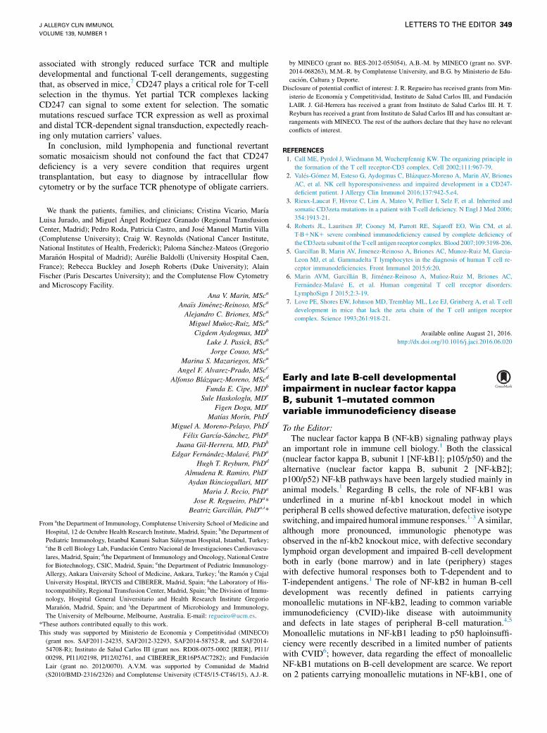
associated with strongly reduced surface TCR and multipledevelopmental and functional T-cell derangements, suggestingthat, as observed in mice,7 CD247 plays a critical role for T-cellselection in the thymus. Yet partial TCR complexes lackingCD247 can signal to some extent for selection. The somaticmutations rescued surface TCR expression as well as proximaland distal TCR-dependent signal transduction, expectedly reach-ing only mutation carriers’ values.
In conclusion, mild lymphopenia and functional revertantsomatic mosaicism should not confound the fact that CD247deficiency is a very severe condition that requires urgenttransplantation, but easy to diagnose by intracellular flowcytometry or by the surface TCR phenotype of obligate carriers.
We thank the patients, families, and clinicians; Cristina Vicario, Mar�ıa
Luisa Jurado, and Miguel �Angel Rodr�ıguez Granado (Regional Transfusion
Center, Madrid); Pedro Roda, Patricia Castro, and Jos�e Manuel Martin Villa
(Complutense University); Craig W. Reynolds (National Cancer Institute,
National Institutes of Health, Frederick); Paloma S�anchez-Mateos (Gregorio
Mara~n�on Hospital of Madrid); Aur�elie Baldolli (University Hospital Caen,
France); Rebecca Buckley and Joseph Roberts (Duke University); Alain
Fischer (Paris Descartes University); and the Complutense Flow Cytometry
and Microscopy Facility.
Ana V. Marin, MSca
Ana€ıs Jim�enez-Reinoso, MSca
Alejandro C. Briones, MSca
Miguel Mu~noz-Ruiz, MSca
Cigdem Aydogmus, MDb
Luke J. Pasick, BSca
Jorge Couso, MSca
Marina S. Mazariegos, MSca
Angel F. Alvarez-Prado, MScc
Alfonso Bl�azquez-Moreno, MScd
Funda E. Cipe, MDb
Sule Haskologlu, MDe
Figen Dogu, MDe
Mat�ıas Mor�ın, PhDf
Miguel A. Moreno-Pelayo, PhDf
F�elix Garc�ıa-S�anchez, PhDg
Juana Gil-Herrera, MD, PhDh
Edgar Fern�andez-Malav�e, PhDa
Hugh T. Reyburn, PhDd
Almudena R. Ramiro, PhDc
Aydan Ikinciogullari, MDe
Maria J. Recio, PhDa
Jose R. Regueiro, PhDa*
Beatriz Garcill�an, PhDa,i*
From athe Department of Immunology, Complutense University School of Medicine and
Hospital, 12 de Octubre Health Research Institute, Madrid, Spain; bthe Department of
Pediatric Immunology, Istanbul Kanuni Sultan S€uleyman Hospital, Istanbul, Turkey;cthe B cell Biology Lab, Fundaci�on Centro Nacional de Investigaciones Cardiovascu-
lares, Madrid, Spain; dthe Department of Immunology and Oncology, National Centre
for Biotechnology, CSIC, Madrid, Spain; ethe Department of Pediatric Immunology-
Allergy, Ankara University School of Medicine, Ankara, Turkey; fthe Ram�on y Cajal
University Hospital, IRYCIS and CIBERER, Madrid, Spain; gthe Laboratory of His-
tocompatibility, Regional Transfusion Center, Madrid, Spain; hthe Division of Immu-
nology, Hospital General Universitario and Health Research Institute Gregorio
Mara~n�on, Madrid, Spain; and ithe Department of Microbiology and Immunology,
The University of Melbourne, Melbourne, Australia. E-mail: [email protected].
*These authors contributed equally to this work.
This study was supported by Ministerio de Econom�ıa y Competitividad (MINECO)
(grant nos. SAF2011-24235, SAF2012-32293, SAF2014-58752-R, and SAF2014-
54708-R); Instituto de Salud Carlos III (grant nos. RD08-0075-0002 [RIER], PI11/
00298, PI11/02198, PI12/02761, and CIBERER_ER16P5AC7282); and Fundaci�on
Lair (grant no. 2012/0070). A.V.M. was supported by Comunidad de Madrid
(S2010/BMD-2316/2326) and Complutense University (CT45/15-CT46/15), A.J.-R.
by MINECO (grant no. BES-2012-055054), A.B.-M. by MINECO (grant no. SVP-
2014-068263), M.M.-R. by Complutense University, and B.G. by Ministerio de Edu-
caci�on, Cultura y Deporte.
Disclosure of potential conflict of interest: J. R. Regueiro has received grants from Min-
isterio de Econom�ıa y Competitividad, Instituto de Salud Carlos III, and Fundaci�on
LAIR. J. Gil-Herrera has received a grant from Instituto de Salud Carlos III. H. T.
Reyburn has received a grant from Instituto de Salud Carlos III and has consultant ar-
rangements with MINECO. The rest of the authors declare that they have no relevant
conflicts of interest.
REFERENCES
1. Call ME, Pyrdol J, Wiedmann M, Wucherpfennig KW. The organizing principle in
the formation of the T cell receptor-CD3 complex. Cell 2002;111:967-79.
2. Val�es-G�omez M, Esteso G, Aydogmus C, Bl�azquez-Moreno A, Marin AV, Briones
AC, et al. NK cell hyporesponsiveness and impaired development in a CD247-
deficient patient. J Allergy Clin Immunol 2016;137:942-5.e4.
3. Rieux-Laucat F, Hivroz C, Lim A, Mateo V, Pellier I, Selz F, et al. Inherited and
somatic CD3zeta mutations in a patient with T-cell deficiency. N Engl J Med 2006;
354:1913-21.
4. Roberts JL, Lauritsen JP, Cooney M, Parrott RE, Sajaroff EO, Win CM, et al.
T-B1NK1 severe combined immunodeficiency caused by complete deficiency of
theCD3zeta subunit of the T-cell antigen receptor complex.Blood 2007;109:3198-206.
5. Garcillan B, Marin AV, Jimenez-Reinoso A, Briones AC, Munoz-Ruiz M, Garcia-
Leon MJ, et al. Gammadelta T lymphocytes in the diagnosis of human T cell re-
ceptor immunodeficiencies. Front Immunol 2015;6:20.
6. Marin AVM, Garcill�an B, Jim�enez-Reinoso A, Mu~noz-Ruiz M, Briones AC,
Fern�andez-Malav�e E, et al. Human congenital T cell receptor disorders.
LymphoSign J 2015;2:3-19.
7. Love PE, Shores EW, Johnson MD, Tremblay ML, Lee EJ, Grinberg A, et al. T cell
development in mice that lack the zeta chain of the T cell antigen receptor
complex. Science 1993;261:918-21.
Available online August 21, 2016.http://dx.doi.org/10.1016/j.jaci.2016.06.020
Early and late B-cell developmentalimpairment in nuclear factor kappaB, subunit 1–mutated commonvariable immunodeficiency disease
To the Editor:The nuclear factor kappa B (NF-kB) signaling pathway plays
an important role in immune cell biology.1 Both the classical(nuclear factor kappa B, subunit 1 [NF-kB1]; p105/p50) and thealternative (nuclear factor kappa B, subunit 2 [NF-kB2];p100/p52) NF-kB pathways have been largely studied mainly inanimal models.1 Regarding B cells, the role of NF-kB1 wasunderlined in a murine nf-kb1 knockout model in whichperipheral B cells showed defective maturation, defective isotypeswitching, and impaired humoral immune responses.1-3 A similar,although more pronounced, immunologic phenotype wasobserved in the nf-kb2 knockout mice, with defective secondarylymphoid organ development and impaired B-cell developmentboth in early (bone marrow) and in late (periphery) stageswith defective humoral responses both to T-dependent and toT-independent antigens.1 The role of NF-kB2 in human B-celldevelopment was recently defined in patients carryingmonoallelic mutations in NF-kB2, leading to common variableimmunodeficiency (CVID)-like disease with autoimmunityand defects in late stages of peripheral B-cell maturation.4,5
Monoallelic mutations in NF-kB1 leading to p50 haploinsuffi-ciency were recently described in a limited number of patientswith CVID6; however, data regarding the effect of monoallelicNF-kB1 mutations on B-cell development are scarce. We reporton 2 patients carrying monoallelic mutations in NF-kB1, one of
METHODS
Case reportThe case has been reported in full previously, in connection with her natural
killer (NK)-cell dysfunctions in Val�es-G�omez et al.E1 In addition, and because
of the association of reduced CD247 expression with several autoimmune
disorders, we analyzed serum samples from the patient and several carriers
(IV.1, IV.2, IV.5, IV.7, IV.8, IV.10, IV.11, IV.14, IV.15, IV.16) to determine
antinuclear antibodies, thyroid autoantibodies (antithyroglobulin and
antithyroperoxidase), antibodies against surface antigens, and intracytoplas-
mic myeloperoxidase and proteinase 3 in neutrophils, which were all found
to be negative. Thus, carriers do not seem to be at risk of autoimmunity, despite
their slightly impaired TCR functionality. However, the patient showed
several positive direct Coombs tests results (November 2013 to June 2014),
which became negative after lymphoid engraftment, thus compatible with
asymptomatic subtle hemolytic anemia possibly related to the disease itself
or to the chronic cytomegalovirus infection. The study was conducted
according to the principles expressed in the Declaration of Helsinki and
approved by the Institutional Research Ethics Committees of the hospitals
involved. All participants or their guardians provided informed consent for
the collection of samples and subsequent analyses.
PBMC isolation and cell culturePBMCs from the patient, her family, and healthy controls (age-matched
whenever possible) were isolated by centrifugation on a Ficoll-Paque PLUS
(GE Healthcare, Little Chalfont, United Kingdom) gradient. Polyclonal T-cell
lines were generated by stimulation at day 0 with 1 mg/mL PHA
(Sigma-Aldrich, St Louis, Mo), and coculture with irradiated allogeneic
feeder cells weekly (PBMC and EBV-transformed B cells, 40 and 65 Gy,
respectively) at 1:2 ratio in Iscove’s Modified Dulbecco’s Medium (IMDM)
(GE Healthcare) supplemented with 40 IU/mL recombinant human IL-2
(provided by Craig W. Reynolds, Frederick Cancer Research and
Development Center, National Cancer Institute, National Institutes of
Health, Frederick, Md), 10% AB1 human serum, and 1% L-glutamine and
Antibiotic-Antimycotic (Life Technologies, Carlsbad, Calif). Cell growth
was calculated weekly as the ratio of recovered versus seeded cells, and
long-term growth plots were estimated as projections thereof.
ImmunophenotypeMultiparametric flow cytometry was performed with mAbs against CD3ε
(UCHT-1 and S4.1), CD4 (13B8.2), CD45RA (ALB11), and CD45RO
(UCHL-1) from Beckman Coulter (Brea, Calif); CD3d (EP4426), CD3g
(EPR4517), and CD3ε (EPR5361(2)) from Abcam (Cambridge, United
Kingdom); CD247 (6B10.2) from Biolegend (San Diego, Calif); CD247
(H146-968) from Thermo Fisher Scientific (Waltham, Mass); abTCR (BMA
031) from Miltenyi Biotec (Bergisch Gladbach, Germany); and gdTCR
(11F2), CD31 (WM59), CD27 (M-T271), CD56 (B159), and CD8 (RPA-T8)
from BD Biosciences (San Jose, Calif). For unlabeled antibodies, an
additional step with phycoerythrin-conjugated anti-mouse IgG (H 1 L)
from Beckman Coulter or anti-rabbit IgG (H1L) from Life Technologies was
performed. For intracellular staining, cells were fixed with 2%
paraformaldehyde and permeabilized with 0.5% saponin. Data were acquired
with a FACSCalibur flow cytometer (BD Biosciences) and analyzed with
FlowJo software (TreeStar, Ashland, Ore).
T-cell functionTo analyze CD69 induction after TCR engagement, 0.23 106 PBMCswere
plated in flat-bottom96-well plates and stimulated for 24 hours with 10mg/mL
of plastic-coated anti-CD3ε mAb (UCHT-1 from BD Biosciences). CD69
induction was analyzed by flow cytometry with anti-CD69 (L-78 from BD
Biosciences). Proliferation was measured by dilution of the cell tracer
carboxyfluorescein diacetate succinimidyl ester (Sigma Aldrich). Briefly,
cells were stained with 1 mM carboxyfluorescein diacetate succinimidyl ester
and stimulated with 1 mg/mL UCHT-1 (eBioscience, San Diego, Calif) for
5 days. Phosphorylation of ZAP-70 and extracellular signal-regu
was determined by intracellular flow cytometry after stimulation o
cultured cells with 20 mg/mL anti-CD3ε mAb (OKT3 from eBi
48C for 30 minutes cross-linked with 10 mg/mL goat F(ab’)2immunoglobulin (H 1 L) (Beckman Coulter) at 37 8C for 1
Phosphorylated (p) proteins were detected by intracellular flow
with rabbit antibodies against pERK (Thr202/Tyr204) and
(Tyr319)/pSyk (Tyr352) from Cell Signaling (Danvers, Mass)
step with phycoerythrin-labeled anti-rabbit antibody (Life Te
was performed.
TCRb clonalityClonality at the TCRb locus was studied using a commercial
Diagnostica, Granada, Spain, EC-certified for clinical u
amplifies genomic TCR VbJb rearrangements using 2 specific
conserved V- and J-flanking regions. Polyclonal (healthy don
DNA was included for reference. Amplimers were separated an
in an ABI Prism Genetic Analyzer 3110 using GeneMapper
Applied Biosystems (Foster City, Calif).
CD3 and CD247 sequence analysisGenomic DNA and RNA were obtained from peripheral blo
T cells, or sorted CD3εhigh-expressing cells. Primers for CD3G an
CD3D have previously been described in Recio et alE2 and Gil et a
cDNA was amplified using specific exon 1–flanking primers
59 ACACCCCAAACCCTCAAACCTC 39; Reverse: 59 AGGAGGTTTGAAGGAG 39) and PCR products were sequenced. For CD2
cDNA was amplified by PCR using specific primers (Forward:
TCTCCACAGTCCTCCACTTCCTG39; Reverse: 59GATCCGCTAGGAAGGCTTTAGCATGCC 39). DNA fragments were clone
1.2 plasmid (CloneJET PCRCloning Kit, Life Technologies) and
into DH5a Escherichia coli strain. Colonies containing recombina
were also sequenced. CD247 haplotypes were determined by a
Sequence Tag Sites in the genetic interval that contains the CD2
chromosome 1q24.2, essentially as described in Recio et al.E2
Western blotCells were lysed in buffer containing 0.5%Brij96v; 50mg of ce
resolved by SDS-PAGE, transferred into polyvinylidene fluoride
and developed with anti–a-tubulin (B5-1-2 clone, Sigma Aldri
rabbit anti-CD247 448 antiserum (specific for the last 34 amino
C-terminal region), kindly provided by Balbino Alarc�on, CentroMolecular Severo Ochoa, UAM-CSIC, Madrid, Spain, and
described in San Jose et al.E4 Blots were visualized using an Odys
imaging system and quantified using Image Studio software
LI-COR Biosciences, Lincoln, Neb).
Statistical analysisTo assess statistical significance, the 2-tailed Student t tes
ANOVAwith Bonferroni multiple comparison test was performe
REFERENCES
E1. Val�es-G�omez M, Esteso G, Aydogmus C, Bl�azquez-Moreno A
Briones AC, et al. NK cell hyporesponsiveness and impaired deve
CD247-deficient patient. J Allergy Clin Immunol 2016;137:942-5.e
E2. Recio MJ, Moreno-Pelayo MA, Kilic SS, Guardo AC, Sanal O,
et al. Differential biological role of CD3 chains revealed by human
ciencies. J Immunol 2007;178:2556-64.
E3. Gil J, Busto EM, Garcillan B, Chean C, Garcia-Rodriguez MC, Dia
et al. A leaky mutation in CD3D differentially affects alphabeta and
J ALLERGY CLIN IMMUNOL
VOLUME 139, NUMBER 1
LETTERS TO THE EDITOR 349.e1
lated kinase
f 0.33 106
oscience) at
anti-mouse
0 minutes.
cytometry
pZAP-70
. A second
chnologies)
kit (Master
se), which
primers for
or) control
d analyzed
V 4.0 from
od, cultured
dCD3E and
l.E3 CD247
(Forward:
GCAGGA
47 cloning,
59 GGAGAGGCCGCA
d into pJET
transformed
nt plasmids
nalysis of 4
47 gene on
ll lysatewas
membranes,
ch) and the
acids of its
de Biolog�ıapreviously
sey infrared
(both from
t or 1-way
d (*P <_ .05;
**P <_ .01; ***P <_ .001). Error bars represent SEM.
, Marin AV,
lopment in a
4.
Allende LM,
immunodefi-
z-Alderete A,
gammadelta

T cells and leads to a Talphabeta-Tgammadelta1B1NK1 human SCID. J Clin
Invest 2011;121:3872-6.
E4. San Jose E, Sahuquillo AG, Bragado R, Alarcon B. Assembly of the TCR/CD3
complex: CD3 epsilon/delta and CD3 epsilon/gamma dimers associate
indistinctly with both TCR alpha and TCR beta chains: evidence for a double
TCR heterodimer model. Eur J Immunol 1998;28:12-21.
E5. Schatorje EJ, Gemen EF, Driessen GJ, Leuvenink J, van Hout RW, de Vries E.
Paediatric reference values for the peripheral T cell compartment. Scand J
Immunol 2012;75:436-44.
E6. Rieux-Laucat F, Hivroz C, Lim A, Mateo V, Pellier I, Selz F, et al. Inherited and
somatic CD3zeta mutations in a patient with T-cell deficiency. N Engl J Med
2006;354:1913-21.
E7. Roberts JL, Lauritsen JP, Cooney M, Parrott RE, Sajaroff EO, Win CM, et al.
T-B1NK1 severe combined immunodeficiency caused by complete deficiency of
theCD3zeta subunitof theT-cellantigen receptorcomplex.Blood2007;109:3198-206.
E8. Ikinciogullari A, Kendirli T, Dogu F, Egin Y, Reisli I, Cin S, et al. Peripheral
blood lymphocyte subsets in healthy Turkish children. Turk J Pediatr 2004;46:
125-30.
E9. Morbach H, Eichhorn EM, Liese JG, Girschick HJ. Reference values for
B cell subpopulations from infancy to adulthood. Clin Exp Immunol 2010;162:
271-9.
E10. Aksu G, Genel F, Koturoglu G, Kurugol Z, Kutukculer N. Serum immunoglobulin
(IgG, IgM, IgA) and IgG subclass concentrations in healthy children: a study
using nephelometric technique. Turk J Pediatr 2006;48:19-24.
J ALLERGY CLIN IMMUNOL
JANUARY 2017
349.e2 LETTERS TO THE EDITOR

FIG E1. Patient characterization. A, Reduced % of recent thymic emigrants defined as CD41 CD45RA1
CD311 lymphocytes (age-matched control range,E5 77%-96%). B, Reduced naive T-cell compartment.
T-cell maturation stages in CD41 and CD8bright cells were defined as naive (CD45RO2CD271), central
memory (CD45RO1CD271), effector memory (CD45RO1CD272), or effector (CD45RO2CD272). Control in
Fig E1, A and B, was a 20-year-old healthy donor. C, Reduced TCRb clonality in PBMCs. D, Chest computed
axial tomography showing normal-sized thymus.
J ALLERGY CLIN IMMUNOL
VOLUME 139, NUMBER 1
LETTERS TO THE EDITOR 349.e3

FIG E2. CD247 deficiency due to a homozygous mutation. A, CD247 gene and protein, including the germ-
linemutation (NCBI/ClinVar: rs672601318, NM_000734.3:c.2T>C and NG_007384.1:g.5146T>C in exon 1) and
the next in-frame ATG. B, Top, CD247 mutation and haplotypes based on the indicated microsatellite
markers (black 5 disease-associated chromosome). Bottom, pedigree with haplotypes for studied
individuals. Circles, squares, and diamonds indicate females, males, or unknown sex, respectively.
J ALLERGY CLIN IMMUNOL
JANUARY 2017
349.e4 LETTERS TO THE EDITOR
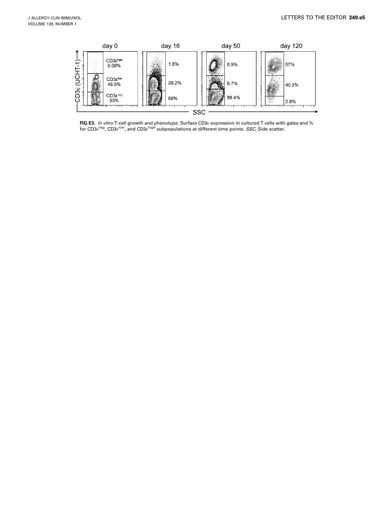
FIG E3. In vitro T-cell growth and phenotype. Surface CD3ε expression in cultured T cells with gates and %
for CD3εneg, CD3εlow, and CD3εhigh subpopulations at different time points. SSC, Side scatter.
J ALLERGY CLIN IMMUNOL
VOLUME 139, NUMBER 1
LETTERS TO THE EDITOR 349.e5
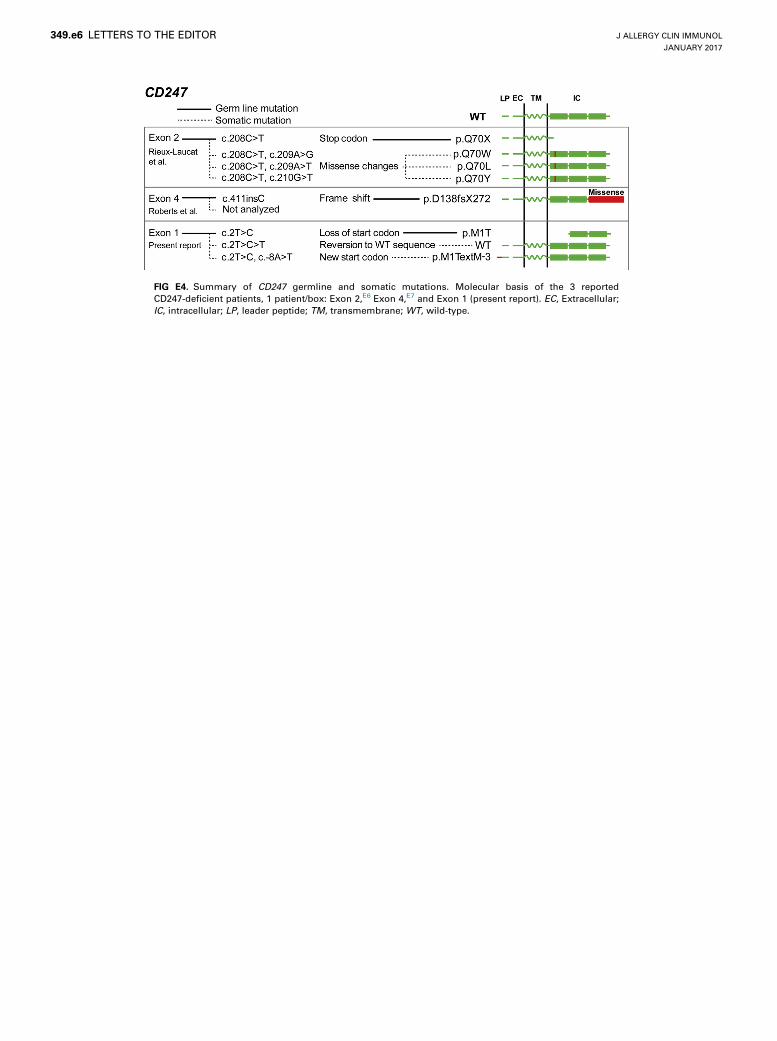
FIG E4. Summary of CD247 germline and somatic mutations. Molecular basis of the 3 reported
CD247-deficient patients, 1 patient/box: Exon 2,E6 Exon 4,E7 and Exon 1 (present report). EC, Extracellular;
IC, intracellular; LP, leader peptide; TM, transmembrane; WT, wild-type.
J ALLERGY CLIN IMMUNOL
JANUARY 2017
349.e6 LETTERS TO THE EDITOR
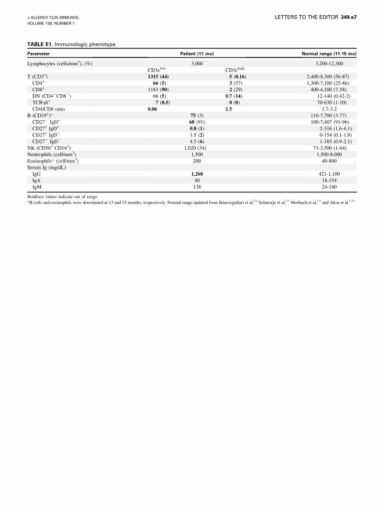
TABLE E1. Immunologic phenotype
Parameter Patient (11 mo) Normal range (11-15 mo)
Lymphocytes (cells/mm3), (%) 3,000 3,200-12,300
CD3εlow CD3εhigh
T (CD3+) 1315 (44) 5 (0.16) 2,400-8,300 (56-87)
CD4+ 66 (5) 3 (57) 1,300-7,100 (25-86)
CD8+ 1183 (90) 2 (29) 400-4,100 (7-58)
DN (CD42CD82) 66 (5) 0.7 (14) 12-140 (0.42-2)
TCRgd+ 7 (0.5) 0 (0) 70-630 (1-10)
CD4/CD8 ratio 0.06 1.5 1.7-3.2
B (CD19+)* 75 (3) 110-7,700 (3-77)
CD272 IgD+ 68 (91) 100-7,407 (91-96)
CD27+ IgD+ 0.8 (1) 2-316 (1.6-4.1)
CD27+ IgD2 1.5 (2) 0-154 (0.1-1.9)
CD272 IgD2 4.5 (6) 1-185 (0.9-2.1)
NK (CD56+ CD16+) 1,020 (34) 71-3,500 (1-64)
Neutrophils (cell/mm3) 1,500 1,500-8,000
Eosinophils* (cell/mm3) 200 40-800
Serum Ig (mg/dL)
IgG 1,260 421-1,100
IgA 40 18-154
IgM 138 24-180
Boldface values indicate out of range.
*B cells and eosinophils were determined at 13 and 15 months, respectively. Normal range updated from Ikinciogullari et al,E8 Schatorje et al,E5 Morbach et al,E9 and Aksu et al.E10
J ALLERGY CLIN IMMUNOL
VOLUME 139, NUMBER 1
LETTERS TO THE EDITOR 349.e7

TABLE E2. Comparative clinical and immunologic features of CD247-deficient patients
Feature Present report Rieux-Laucat et alE6 Roberts et alE7
Sex Female Male Female
Onset 2 mo 4 mo 4 mo
Age at diagnosis (mo) 11 10 11
HSCT (mo) Haploidentical (19) Haploidentical (30) Haploidentical (12, 16)
Present age 33 mo� 13 y* 4 y*�Cytomegalovirus 1 2 1Germline mutation
Type Truncation Truncation Insertion
cDNA c.2T>C c.208C>T c.411insC
Protein p.M1T p.Q70X p.D138fsX272
Serum immunoglobulin levels High (IgG) High (IgG, -A, -M, -E) High (IgG,� -A, -M)
T-cell lymphopenia Mild Mild Mild
CD3εhigh
% of T cells 0.36 10 0.6
Lineage CD4 and CD8 CD4 CD4 and CD8
Somatic mutations Yes (2) Yes (3) NA
Revertant Yes No NA
Second site Yes Yes NA
Functional Yes (5 carriers) No NA
CD3εlow
% of T cells 99.6 90.0 99.4
CD4/CD8 ratio Inverted (0.06) Inverted (0.23) Inverted (0.85)
Functional No No No
IVIG, Intravenous immunoglobulin; NA, not analyzed.
*Alain Fischer, Paris Descartes University, and Rebecca Buckley and Joseph Roberts, Duke University (personal communication, 2015).
�Exitus at.�Receiving IVIG.
J ALLERGY CLIN IMMUNOL
JANUARY 2017
349.e8 LETTERS TO THE EDITOR