learning to decode your blood test results for cll
TRANSCRIPT

Learning to Decode Your Blood Test Results for CLL
June 29, 2021
10:00 AM PT, 11:00 AM MT, 12:00 PM CT, 1:00 PM ET

This program was made possible by grant support
from

Speakers
Welcome: Patricia KoffmanCo-Founder and Communications Director CLL Society
Speaker: Susan Leclair, PhD, CLS (NCA) Chancellor Professor EmeritaUniversity of Massachusetts Dartmouth
Moderator: Brian Koffman, MDCM (retired), MS Ed Executive Vice President and Chief Medical OfficerCLL Society

A QUICK RUN DOWN OF ALL 25,000!
§ Not really§ But I did want to start off by saying this is a BIG subject and we will only look at a few of the
more common tests.
§ But first – a word from realityTests live in a real world that is bounded by
Preanalytical Aspects Analytical Aspects Post-Analytical Aspectspatient preparation issues instrumentation reporting mechanisms (to whom, when)time of collection reagent quality reflex testing protocolsconditions of collection specificity & sensitivity presence/absence of interpretationtransport &storage technique/method wrong test requestedconfounding meds locationwrong test requested patient population. fingerstick vs. venous
Each of these attributes influences the value of the test result – consistency is key.

WHITE CELL VALUES§ We do not count white cells.
§ We count nuclei so nucleated red blood cells are counted here – corrected white cell count
§ 5 major cell lines present in the peripheral blood
§ Neutrophils – present all the time§ Lymphocytes – present all the time§ Monocytes – present occasionally § Eosinophils – present occasionally§ Basophils – present rarely

WHITE CELL VALUES§The number will bounce around all day long in response to your environment.
§ Want your granulocytes to increase?§ Exercise (walk up the stairs) for a few minutes before getting your blood drawn
§ Half of your granulocytes usually marginate along the walls of the blood vessels. Exercise “shakes” them off putting them into the circulating pool for about 15-20 minutes.
§ Have a panic attack – reaction to stressAdrenaline will also take cells away from the marginating pool
§ Be on Steroids

WHITE CELL VALUES§Two ways to report white cells by type
§ Circa 1900 – the traditional differential§ Look at the first 100 random white blood cells you see using a microscope and your own
trained eyes.§ Some people are better than others§ Some days and better than others§ If there are 7500 white cells in a microliter of blood and you count 100 of them - report in
percentages§ What are the odds that you will fin what is important?§ How can you tell which cell line is increased or decreased?
§ Circa – 1980s with the advent of multi-channel instruments§ Counts the exact number of cells in a specific volume of blood.§ Count somewhere between 20,000 and 50,000

WHITE CELL VALUES§ The best differential is the ABSOLUTE differential.
§ Counts the exact number of cells in a specific volume of blood.
§ Percentages cannot tell which cell line is increased or decreased.White cell count % neutrophils % lymphocytes A.N.C.
Absolute neutrophil Count
A.L.C.Absolute lymphocyte count
2.0x10⁹/L 63 37 1.2x10⁹/L 0.74x10⁹/L
4.0x10⁹/L 63 37 2.5x10⁹/L 1.4x10⁹/L
8.0x10//L 63 37 5.0x10⁹/L 2.8x10⁹/L
16.0x10⁹/L 63 37 10.0x10⁹/L 3.7x10⁹/L

WHITE CELL VALUES§Then why use both?
§ Absolute§ You get a real number of cells by cell line. And there is NO way to confuse which cell
line is increased/decreased
§ Percentage§ There is nothing better to assess the quality of the cells than having someone who
knows what they are doing look at them.
§ So doing both gives you a more complete picture of the cells and what they have been doing

WHITE CELL VALUES§ For example§ Both of these are the same cell. One is exhausted and on the brink of
death itself. No instrument can tell them apart.

WHITE CELL VALUES§ Is anyone better than another?
§ Neutrophils (once known as granulocytes)§ Most common cell§ Exists in the marrow, the two pools in the peripheral blood and in the tissues§ Phagocytize dead/dying cells and any foreign item (particle or droplet)§ Incites, controls, and participates in the inflammatory process§ Determines acute or chronic inflammation§ Most varied morphology
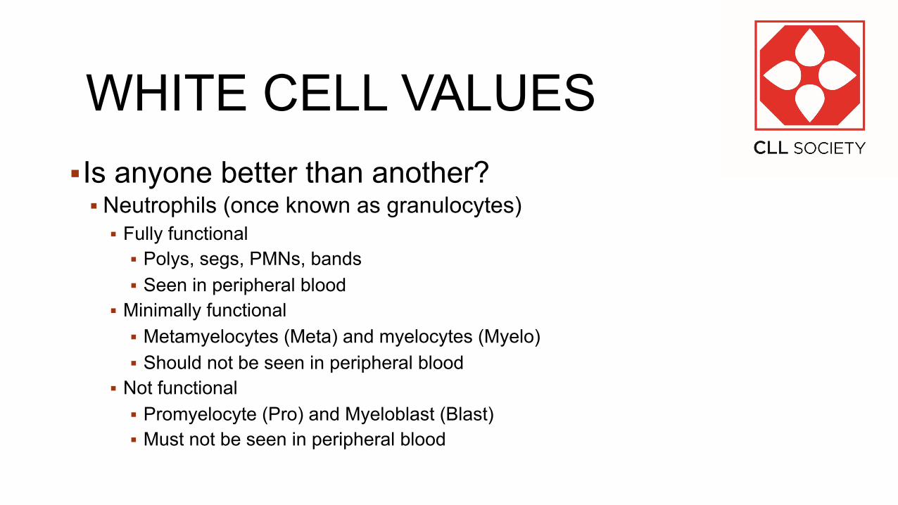
WHITE CELL VALUES§Is anyone better than another?
§ Neutrophils (once known as granulocytes)§ Fully functional
§ Polys, segs, PMNs, bands§ Seen in peripheral blood
§ Minimally functional§ Metamyelocytes (Meta) and myelocytes (Myelo)§ Should not be seen in peripheral blood
§ Not functional§ Promyelocyte (Pro) and Myeloblast (Blast)§ Must not be seen in peripheral blood

WHITE CELL VALUES§Is anyone better than another?
§ Monocytes§ Usually found in the tissues – uses blood stream to move from one place to another§ Exists in the marrow, only one pool in the peripheral blood and in the tissues§ Phagocytize dead/dying cells and any foreign item (particles only)§ Processes antigens for the T cell to recognize§ Will be increased after any trauma

WHITE CELL VALUES§Is anyone better than another?
§ Lymphocytes§ Found in lymph nodes, lymphatics, peripheral blood, and bone marrow§ Circulate freely between the nodes/lymphatics and the peripheral blood/marrow§ Cannot be differentiated using light microscopy§ Could comment on size (small, medium, large)§ Assumed an unusual looking lymphocytes WAS damaged in some fashion (atypical)
§ Did not have a known function until the mid 1960s (Robert Good – Minnesota – Nobel Prize)
§ Were separated by function – T, B, and NK cells§ Realized that “atypical” cells were in fact reacting to the presence of a foreign antigen and
were defenders not the illness – new Name Reactive Lymphocytes§ Sadly – many people refuse to update to the correct name – apathy?

WHITE CELL VALUES§ Lymphocytes
§ Small lymphocytes § Usually B cells § Resting from any action so can be naïve or
memory§ Medium lymphocytes
§ Can be T, B or NK cells§ If it has granules, more likely to be t or NK
§ Large lymphocytes§ Can be T or NK
§ Reactive lymphocyte§ If B cell, than larger cytoplasm for antibody
production§ If T or NK cell, less cytoplasm and more granules

WHITE CELL VALUES§ Smudge cell
§ Can be any cell line§ Usually
§ A cell that is very fragile and cannot withstand the collection and processing.§ Frequently seen in CLL
§ A cell that has died in the course of some reaction/inflammatory response etc. § If a monocyte or neutrophil, can just have died if
in a circle.§ If a neutrophil, then it is called a neutrophil trap
or net. The cell has exploded itself in order to make the largest area filled with protein braking or killing enzymes.

PLATELETS§The number will bounce around all day long in response to your environment.§ When performed manually – very difficult so the accepted range is +/-
50,000. § When performed by instrument, the accepted range is = +/- 20,000
§ As the instruments got better, the acceptable range has moved from 150 –500 to 150 – 450 to 130 – 400 to even smaller ranges for some facilities (150 – 350)

PLATELETS§PDW – platelet distribution width
§ Similar to the RDW§ Mathematical description of size variation§ Do we care?
§ Partially§ Larger platelets suggest some type of inflammation, overuse, or drug response§ Smaller platelets suggest deficiencies similar to microcytic red cells (iron, B6,
hypothyroidism)§ Why not?
§ The most important things about platelets is their function. We have a few, not very precise tests for platelets because their function is in such a complex situation we cannot replicate it – size of capillary damage, type of damage (smooth vs. ragged), integrity of vessel walls, signaling from localized cells, eternal conditions (heat/cold, pressure, etc.)

PLATELETS§ Bone marrow cell – megakaryocyte, get larger and then breaks off pieces
of the cytoplasm§ Those pieces then need to reorganize themselves§ Large platelets are usually not well organized and function less efficiently

PLATELETS§When not stimulated
§ Platelets are small disc shaped pieces of cytoplasm§ When stimulated, they wring themselves out like a
sponge, spreading contents into the area.§ Their protrusions interlace forming a lattice structure.§ They ADHERE to damaged walls.§ Once “laced” they AGGREGATE and tighten§ Other Contents are stimulants for the clotting process.
§ Platelet lack or lessened function is seen in§ Small blood vessel bleeding (gums, mucous membranes,
skin, etc. § Not big clots
§ Drugs – aspirin, clopidogrel, ticagrelor, ticlopidine

PLATELETS
§Anti- Platelet Drugs
§ Aspirin usually low dose (81mg) but can be 325mg.
§ Clopidogrel – Plavix§ Ticagrelor – Brilinta§ Prasugrel (Effient)§ Dipyridamole/aspirin (Aggrenox)§ Ticlopidine (Ticlid)§ Eptifibatide (Integrilin)

Normal Lab ValuesFind Information on CLLSOCIETY.ORG

Normal Lab Values

Normal Lab Values

Keeping Track of Your Lab ResultsDownload the Template to Keep Track of Your Lab History

Keeping Track of Your Lab Results
Allows for a broader view of your long-term trending history for all key CLL lab components (CBC, Absolute Lymphocytes, and more)

Example Lab Tracking Form

Audience Questions & Answers

This program was made possible by grant support
from

Thank You for Attending!
Please take a moment to complete our post-event survey, your feedback is important to us
Join us in August for our webinar on PI3K Inhibitors
CLL Society is invested in your long life. Please invest in the long life of the CLL Society by supporting our work
cllsociety.org/donate-to-cll-society/30