laminitis means inflammation of the laminae. the laminae is a layer of tissue that carries blood to...
TRANSCRIPT
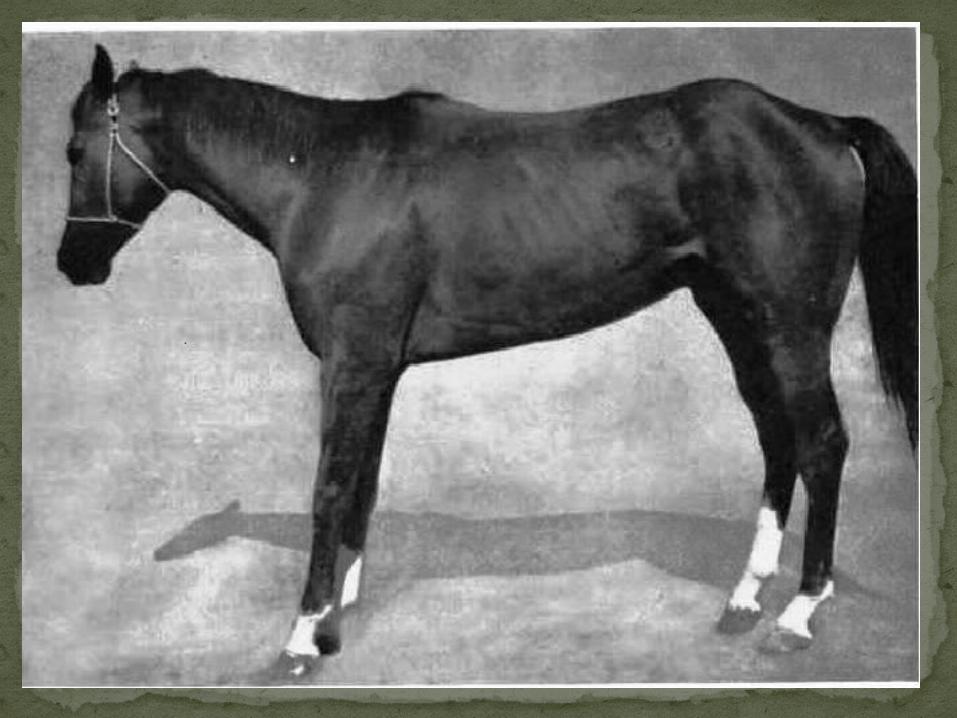
Laminitis in horse
Laminitis means inflammation of the laminae. The laminae is a layer of tissue that carries blood to all the components of the hoof. The laminae attach to the hoof wall and to the coffin bone. Laminitis is a result of problems in these connective sites.
Laminitis(Founder)
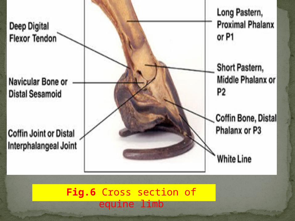
Fig.6 Cross section of equine limb

Fig.7 The horny hoof wall from a horse. The white arrow identifies a vertical groove of horny lamina that interlocks with the sensitive lamina.

Acute laminitis is defined as: The initial onset of laminitis and lasts for variable periods of time, may progress to chronic laminitis.
Chronic stable laminitis is defined as: Laminitis that the coffin bone becomes stable. Healing has begun.
Chronic unstable laminitis is defined as: Laminitis that requires frequent and prolonged treatment because the coffin bone continues to rotate and or sink.
Types of laminitis
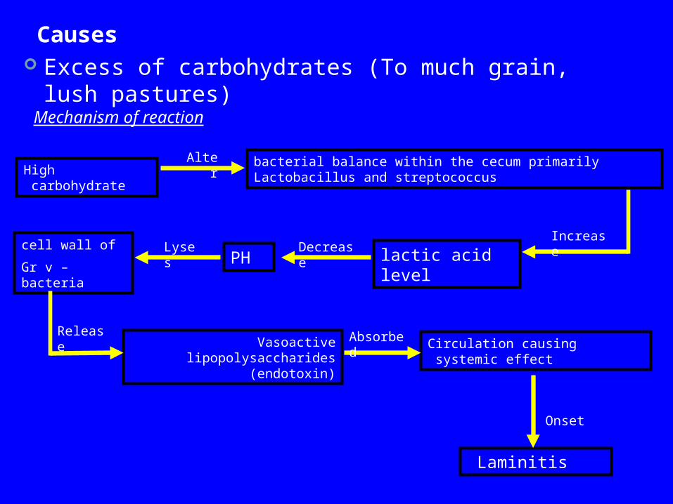
Causes Excess of carbohydrates (To much grain, lush
pastures)Mechanism of reaction
cell wall of
Gr v – bacteria
Vasoactive lipopolysaccharides (endotoxin)
Circulation causing systemic effect
Laminitis
bacterial balance within the cecum primarily Lactobacillus and streptococcus
lactic acid level
High carbohydrate
PH
Alter
IncreaseDecreaseLyses
Release Absorbed
Onset

Excessive weight (Draft horses are prone to laminitis of this cause)
GI problemsEndometritis or severe systemic infectionA mare may develop this type of laminitis
shortly after foaling as a result of infection arising from retention of part of fetal membranes or of a uterine infection.
Prolonged transportationExcessive work on hard surfaceBedding containing black walnut shavingsIngestion of cold water when horse is
overheated

StiffnessLamenessStanding on heels Reluctance to moveHeat in the hoovesIncrease of fetlock pulseSensitivity to hoof testers
Symptoms
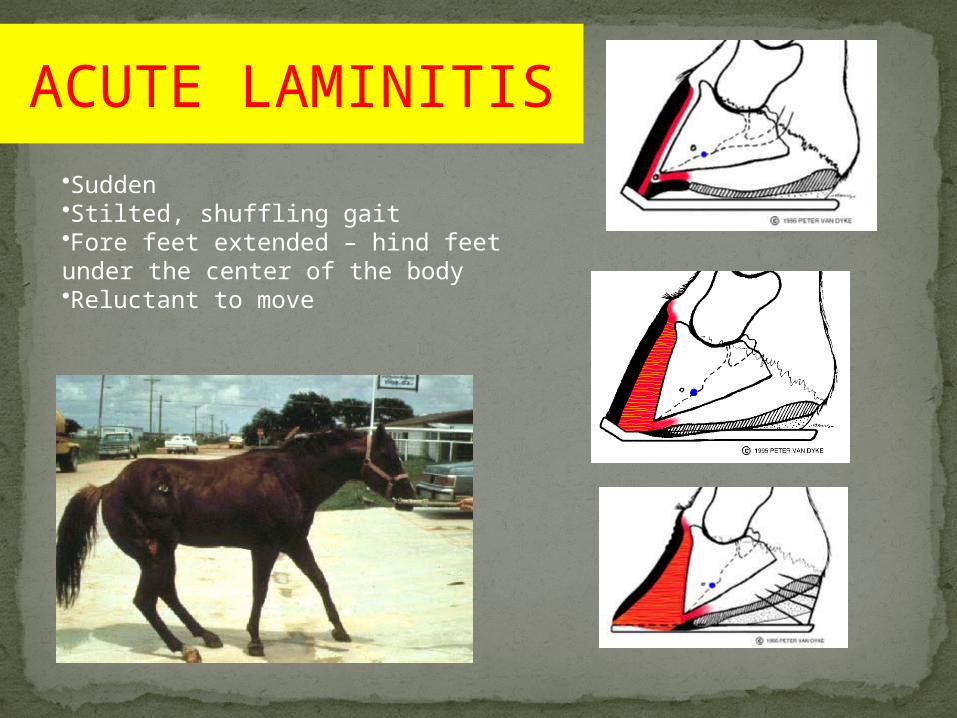
•Sudden •Stilted, shuffling gait•Fore feet extended – hind feet under the center of the body •Reluctant to move
ACUTE LAMINITIS
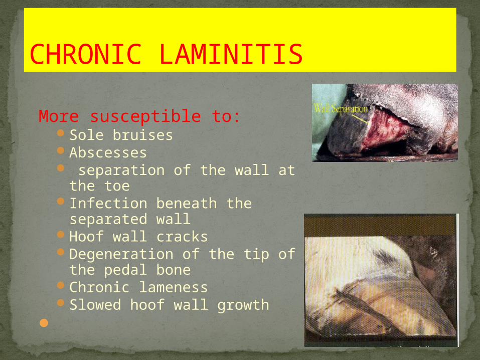
More susceptible to:Sole bruisesAbscesses separation of the wall at the
toeInfection beneath the
separated wallHoof wall cracksDegeneration of the tip of
the pedal boneChronic lamenessSlowed hoof wall growth
CHRONIC LAMINITIS

Difficult in diagnosis but the below may lead to diagnosis.
1.history(feed with high carbohydrate ,prolonged transportation, GI probelms, reproductive problems).
2.clinical signs.3.radiograph.
diagnosis
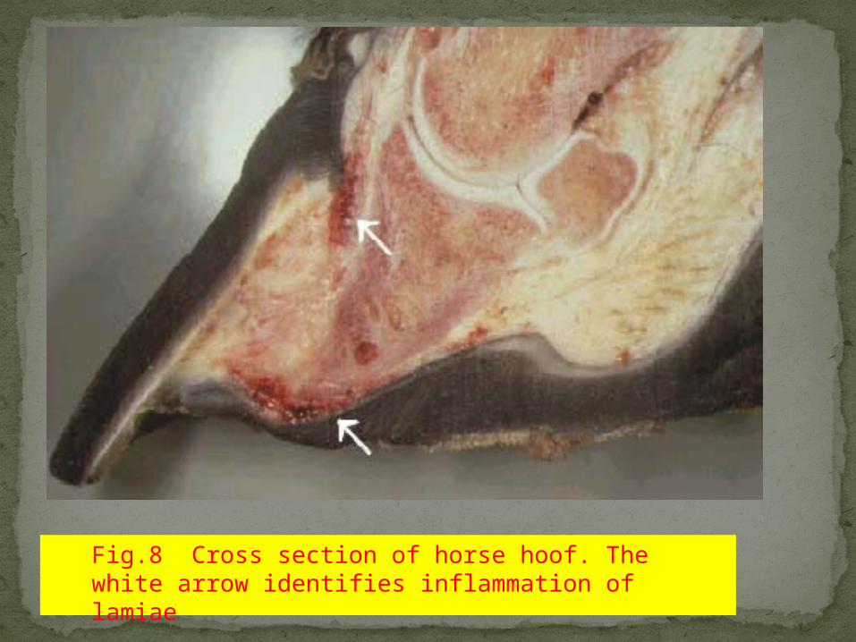
Fig.8 Cross section of horse hoof. The white arrow identifies inflammation of lamiae

Single limb cryotherapy trial.Using a rubber boot (Bigfoot Ice Boots) one forelimb was immersed in ice and water (mean temperature 0.5- 1.7°C) for the 48 hour experimental period. The mean internal hoof temperature was 3.5-0.9°C. Laminitis occurred only in the noncooled untreated limbs. The cooled limbs did not develop clinical laminitis and had significantly reduced lamellar histological damage.

Dorsopalmar venogram of a normal foot made with digital equipment.Most of the vessels are large veins. DA = digital artery with ‘string of beads’ appearance. C = Coronary venous plexus. SLP = sub-lamellar plexus. DP = distal phalanx. SP = sole papillae.
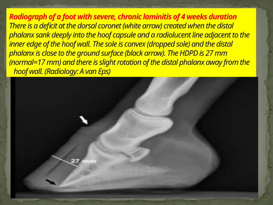
Radiograph of a foot with severe, chronic laminitis of 4 weeks durationThere is a deficit at the dorsal coronet (white arrow) created when the distal phalanx sank deeply into the hoof capsule and a radiolucent line adjacent to the inner edge of the hoof wall. The sole is convex (dropped sole) and the distal phalanx is close to the ground surface (black arrow). The HDPD is 27 mm (normal=17 mm) and there is slight rotation of the distal phalanx away from the hoof wall. (Radiology: A van Eps)
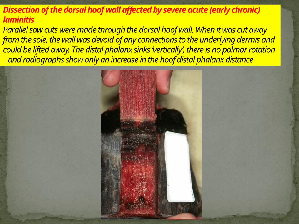
Dissection of the dorsal hoof wall affected by severe acute (early chronic) laminitis.Parallel saw cuts were made through the dorsal hoof wall. When it was cut away from the sole, the wall was devoid of any connections to the underlying dermis and could be lifted away. The distal phalanx sinks ‘vertically’, there is no palmar rotation and radiographs show only an increase in the hoof distal phalanx distance.
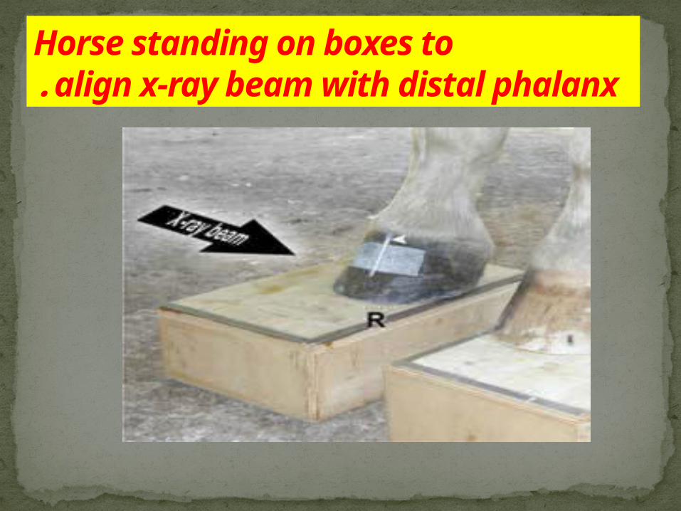
Horse standing on boxes toalign x-ray beam with distal phalanx.
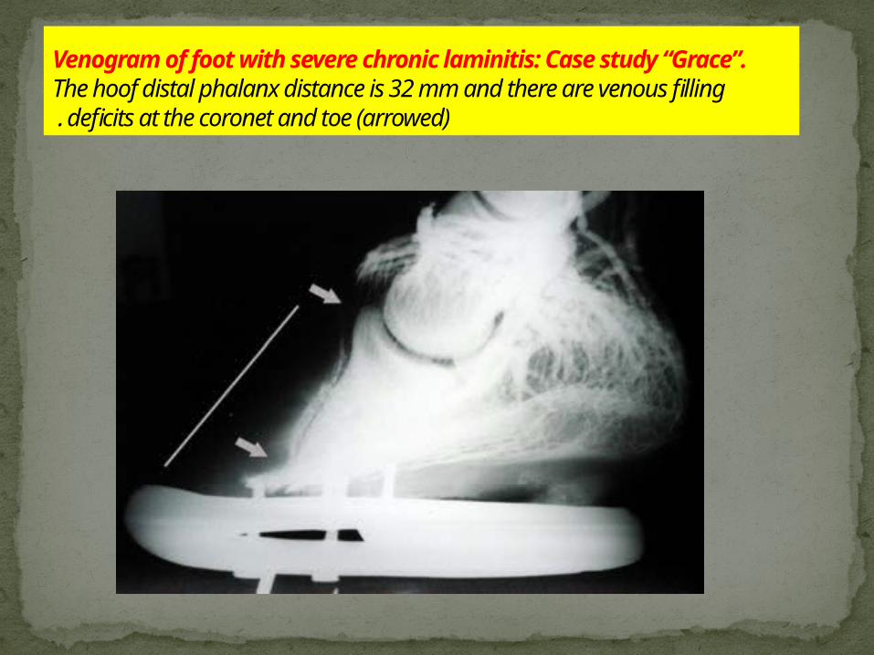
Venogram of foot with severe chronic laminitis: Case study “Grace”.The hoof distal phalanx distance is 32 mm and there are venous filling deficits at the coronet and toe (arrowed).

Encouraging the horse to lie down to relieve
pressure on the hoof
Imposing dietary restrictions to prevent
overeating
Administering fluids if the horse is ill or
dehydrated
Administration of pain medications
Treatment