equine laminitis - evzequine laminitis christopher c. pollitt, bvsc, phd laminitis, failure of the...
TRANSCRIPT
EC
SA
3
quine Laminitishristopher C. Pollitt, BVSc, PhD
Laminitis, failure of the distal phalanx to maintain its attachment to the lamellae of theinner hoof wall, causes unrelenting pain and a characteristic lameness. During adevelopmental phase, pathology in organs anatomically remote from the foot generateslaminitis trigger factors that circulate to cause separation and disorganization of hooflamellar anatomy. Laminitis, linked to seasonal variations in grass fructan concentra-tion can be induced experimentally using oligofructose, a closely related compound.There is a strong correlation between the degree of lameness and the severity ofhistopathology in lamellar samples from horses with laminitis. Matrix metalloproteinaseenzymes (MMPs), responsible for normal enzymatic remodeling of the epidermallamellae, appear to be accidentally recruited in the pathogenesis of the laminitis.MMP-2 and MMP-9 have been isolated from normal lamellar tissues and in increasedquantities from lamellar tissues affected by laminitis. Although epidermal cells of otherspecies readily increase their production of MMP when exposed to cytokines this is notthe case with equine lamellar explants. An enzymatic theory of laminitis etiology basedon lamellar MMP activation challenges the alternative view that laminitis developsbecause of vascular pathology affecting the circulation of the foot. Epidermal cellnecrosis, intravascular coagulation, edema and other evidence of ischemia cannot beidentified in tissue affected by early carbohydrate-induced laminitis. In fact, laminitisdoes not occur if the foot is in a state of vasoconstriction during the developmentalphase suggesting that exogenous trigger factors cause laminitis when they reach thelamellar tissues via dilated blood vessels. Indeed acute laminitis is prevented in a singlecooled limb while laminitis develops in the three remaining limbs maintained at roomtemperature. Small explants of cultured, hoof lamellar tissue, an in vitro model forlaminitis, show that a substance(s) (a potential exogenous laminitis trigger factor) in thesupernatant of cultures of Streptococcus bovis activates equine hoof MMP-2 andcauses lamellar separation. This is taken as evidence for a bacterial pathogenesis oflaminitis since the population of S. bovis, the microorganism responsible for rapidfermentation of carbohydrate in the equine hindgut, explodes exponentially during thedevelopmental phase. Chemical inhibitors block the activity of the laminitis MMPs invitro and have the potential to prevent field cases of laminitis. At acute laminitis onsetmany of the ultrastructural plaques (hemidesmosomes, HDs) that attach lamellar basalcells to the basement membrane of the connective tissue of the distal phalanx, areabsent or disrupted. This is accompanied by BM separation, cytoskeleton damage androunding of basal cell nuclei. The magnitude of HD loss in lamellar basal cells, affectedby laminitis directly correlates to the dose of carbohydrate used to induce it. Soundlamellar architecture depends on the structural integrity of HDs, a fact illustrated whena newborn foal, lacking in plectin (one of the HD intracytoplasmic plaque proteins),developed laminitis as soon as it was ambulatory. The weight-bearing basement mem-brane of the hoof lamellar dermal epidermal interface is unique to the anatomy ofdigitigrade equids. However the lamellae are dependent on MMP enzymes for tissueremodeling thus making horses susceptible in situations that accidentally precipitateMMP activation, HD failure, basement membrane disadhesion and ultimately laminitis.Clin Tech Equine Pract 3:34-44 © 2004 Elsevier Inc. All rights reserved.
KEYWORDS equine laminitis, matrix metalloproteinase (MMP), basement membrane, Strepto-coccus bovis, hemidesmosome, fructan
chool of Veterinary Science, The University of Queensland, Australia.ddress reprint requests to C.C. Pollitt, BVSc, PhD, The University of Queensland, Department of Companion Animal Science, School of Veterinary Science,
Brisbane, Queensland 4072, Australia. E-mail: [email protected]
4 1534-7516/04/$-see front matter © 2004 Elsevier Inc. All rights reserved.doi:10.1053/j.ctep.2004.07.003
Equine laminitis 35
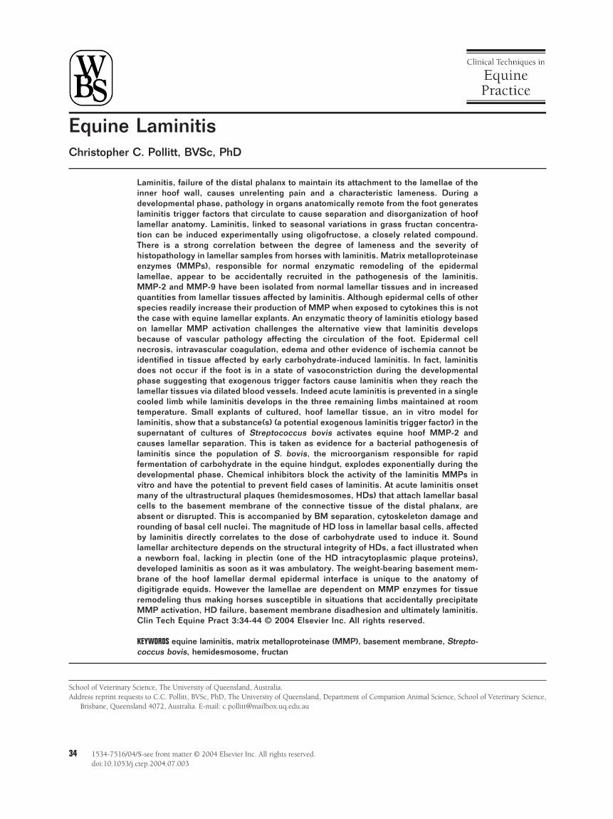
Figure 1 Horse with severe laminitis in both front feet showing typical laminitis gait. The hind feet are placed as far
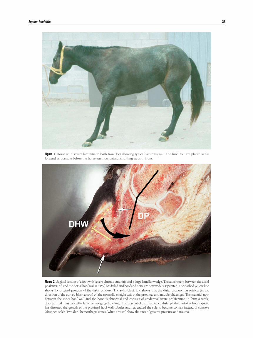
forward as possible before the horse attempts painful shuffling steps in front.Figure 2 Sagittal section of a foot with severe chronic laminitis and a large lamellar wedge. The attachment between the distalphalanx (DP) and the dorsal hoof wall (DHW) has failed and hoof and bone are now widely separated. The dashed yellow lineshows the original position of the distal phalanx. The solid black line shows that the distal phalanx has rotated (in thedirection of the curved black arrow) off the normally straight axis of the proximal and middle phalanges. The material nowbetween the inner hoof wall and the bone is abnormal and consists of epidermal tissue proliferating to form a weak,disorganized mass called the lamellar wedge (yellow line). The descent of the unattached distal phalanx into the hoof capsulehas distorted the growth of the proximal hoof wall tubules and has caused the sole to become convex instead of concave
(dropped sole). Two dark hemorrhagic zones (white arrows) show the sites of greatest pressure and trauma.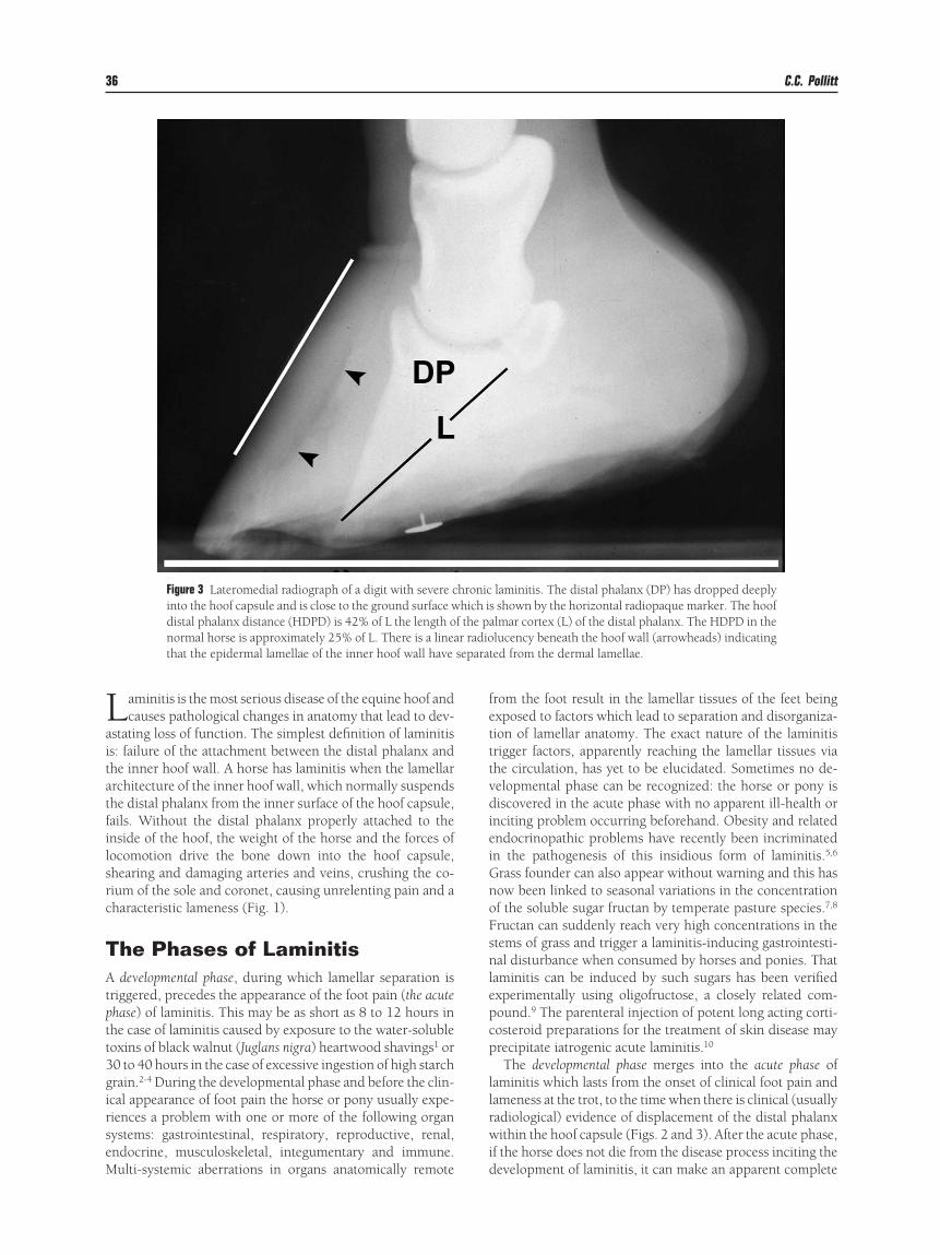
Laitatfilsrc
TAtptt3girseM
fetttvdieiGnoFsnlepcp
llrwi
separa
36 C.C. Pollitt
aminitis is the most serious disease of the equine hoof andcauses pathological changes in anatomy that lead to dev-
stating loss of function. The simplest definition of laminitiss: failure of the attachment between the distal phalanx andhe inner hoof wall. A horse has laminitis when the lamellarrchitecture of the inner hoof wall, which normally suspendshe distal phalanx from the inner surface of the hoof capsule,ails. Without the distal phalanx properly attached to thenside of the hoof, the weight of the horse and the forces ofocomotion drive the bone down into the hoof capsule,hearing and damaging arteries and veins, crushing the co-ium of the sole and coronet, causing unrelenting pain and aharacteristic lameness (Fig. 1).
he Phases of Laminitisdevelopmental phase, during which lamellar separation is
riggered, precedes the appearance of the foot pain (the acutehase) of laminitis. This may be as short as 8 to 12 hours inhe case of laminitis caused by exposure to the water-solubleoxins of black walnut (Juglans nigra) heartwood shavings1 or0 to 40 hours in the case of excessive ingestion of high starchrain.2-4 During the developmental phase and before the clin-cal appearance of foot pain the horse or pony usually expe-iences a problem with one or more of the following organystems: gastrointestinal, respiratory, reproductive, renal,ndocrine, musculoskeletal, integumentary and immune.
Figure 3 Lateromedial radiograph of a digit with severe cinto the hoof capsule and is close to the ground surface wdistal phalanx distance (HDPD) is 42% of L the length onormal horse is approximately 25% of L. There is a lineathat the epidermal lamellae of the inner hoof wall have
ulti-systemic aberrations in organs anatomically remote d
rom the foot result in the lamellar tissues of the feet beingxposed to factors which lead to separation and disorganiza-ion of lamellar anatomy. The exact nature of the laminitisrigger factors, apparently reaching the lamellar tissues viahe circulation, has yet to be elucidated. Sometimes no de-elopmental phase can be recognized: the horse or pony isiscovered in the acute phase with no apparent ill-health or
nciting problem occurring beforehand. Obesity and relatedndocrinopathic problems have recently been incriminatedn the pathogenesis of this insidious form of laminitis.5,6
rass founder can also appear without warning and this hasow been linked to seasonal variations in the concentrationf the soluble sugar fructan by temperate pasture species.7,8
ructan can suddenly reach very high concentrations in thetems of grass and trigger a laminitis-inducing gastrointesti-al disturbance when consumed by horses and ponies. That
aminitis can be induced by such sugars has been verifiedxperimentally using oligofructose, a closely related com-ound.9 The parenteral injection of potent long acting corti-osteroid preparations for the treatment of skin disease mayrecipitate iatrogenic acute laminitis.10
The developmental phase merges into the acute phase ofaminitis which lasts from the onset of clinical foot pain andameness at the trot, to the time when there is clinical (usuallyadiological) evidence of displacement of the distal phalanxithin the hoof capsule (Figs. 2 and 3). After the acute phase,
f the horse does not die from the disease process inciting the
laminitis. The distal phalanx (DP) has dropped deeplys shown by the horizontal radiopaque marker. The hooflmar cortex (L) of the distal phalanx. The HDPD in thelucency beneath the hoof wall (arrowheads) indicating
ted from the dermal lamellae.
hronichich i
f the par radio
evelopment of laminitis, it can make an apparent complete
rpctgdrlopcglutfltwmpltsndtaf
ce
TGAcbMrSsm
GDctasonbmS
Equine laminitis 37
ecovery or develop palmar/plantar displacement of the distalhalanx, the hallmark of chronic laminitis. The chronic phasean last indefinitely with clinical signs ranging from persis-ent, mild lameness, continued severe foot pain, further de-eneration of lamellar attachments, recumbency, hoof walleformation and sloughing of the hooves.11 It is important toealize that the process initiating the destruction of the lamel-ar attachment apparatus begins to operate during the devel-pmental phase before the first clinical sign of laminitis, footain, is apparent. During the developmental phase the spe-ific problems of the horse often have to be attended to ur-ently (eg, acute abdomen, grain overload acidosis, electro-yte imbalance, rhabdomyolysis, retained placenta) andnfortunately the feet are often left out of therapeutic equa-ion until the first signs of foot pain (shifting weight from oneoot to the other) appear. By the time foot pain is apparentamellar pathology is underway. In other words foot pain ishe clinical sign that lamellar disintegration is occurring. Toait and see if foot pain is the sequel to a metabolic crisis is toiss the opportunity to prevent or at least ameliorate lamellarathology. There is a good correlation between the severity of
aminitis histopathology, as seen with the microscope, andhe degree of lameness [using the Obel grading system2]hown by the horse.3 When a horse first starts to show lami-itic pain, the anatomy of the hoof wall lamellae is beingestroyed. The higher the lameness grade, the more severehe microscopic damage. Any activity that places stress on anlready weakened lamellar attachment apparatus (such as
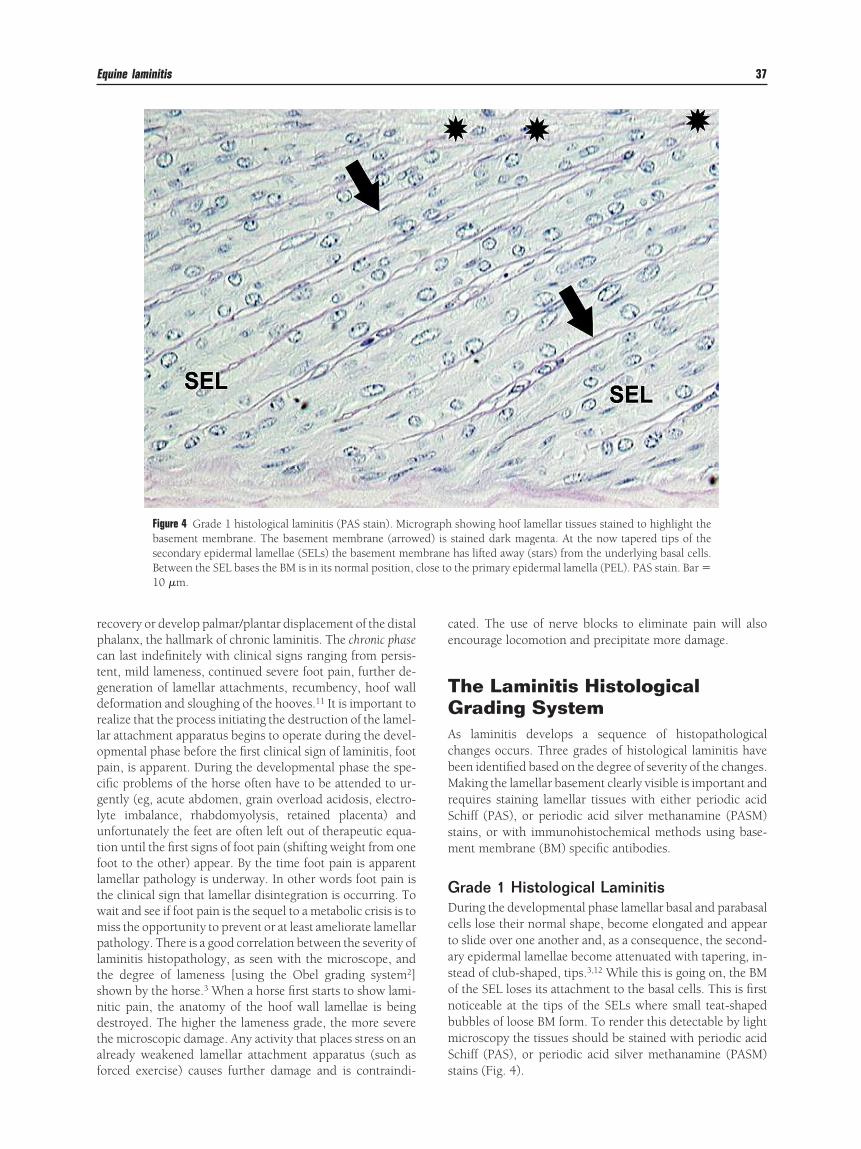
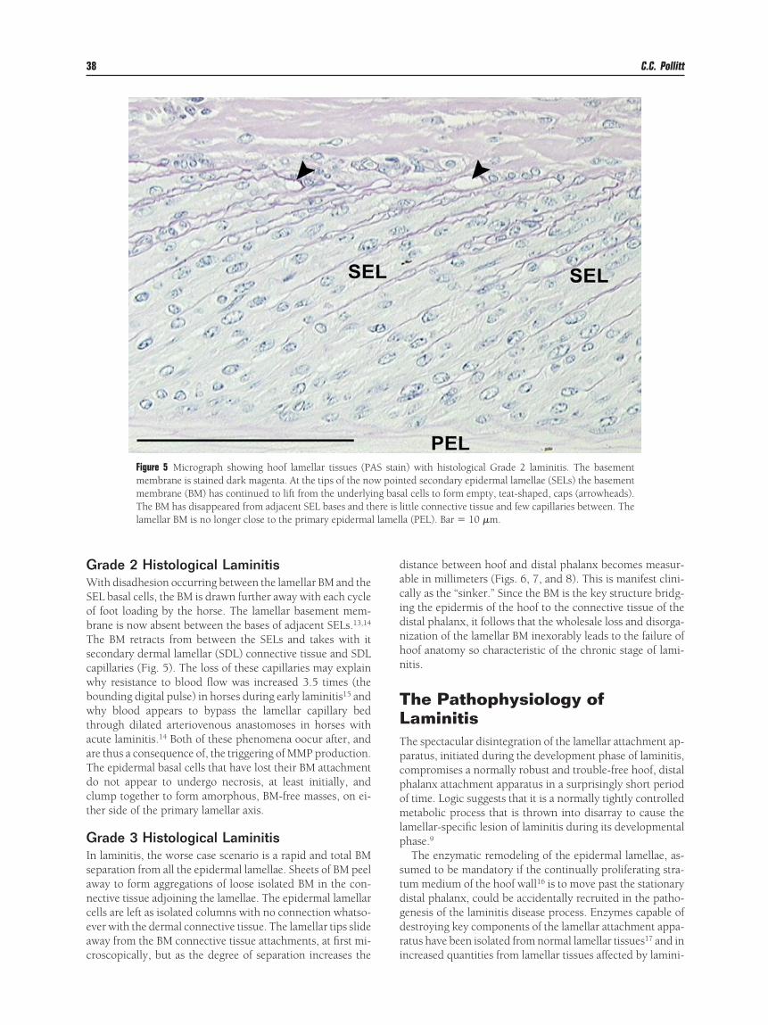
Figure 4 Grade 1 histological laminitis (PAS stain). Micrbasement membrane. The basement membrane (arrowsecondary epidermal lamellae (SELs) the basement memBetween the SEL bases the BM is in its normal position, c10 �m.
orced exercise) causes further damage and is contraindi- s
ated. The use of nerve blocks to eliminate pain will alsoncourage locomotion and precipitate more damage.
he Laminitis Histologicalrading System
s laminitis develops a sequence of histopathologicalhanges occurs. Three grades of histological laminitis haveeen identified based on the degree of severity of the changes.aking the lamellar basement clearly visible is important and
equires staining lamellar tissues with either periodic acidchiff (PAS), or periodic acid silver methanamine (PASM)tains, or with immunohistochemical methods using base-ent membrane (BM) specific antibodies.
rade 1 Histological Laminitisuring the developmental phase lamellar basal and parabasalells lose their normal shape, become elongated and appearo slide over one another and, as a consequence, the second-ry epidermal lamellae become attenuated with tapering, in-tead of club-shaped, tips.3,12 While this is going on, the BMf the SEL loses its attachment to the basal cells. This is firstoticeable at the tips of the SELs where small teat-shapedubbles of loose BM form. To render this detectable by lighticroscopy the tissues should be stained with periodic acid
chiff (PAS), or periodic acid silver methanamine (PASM)
showing hoof lamellar tissues stained to highlight thestained dark magenta. At the now tapered tips of thehas lifted away (stars) from the underlying basal cells.the primary epidermal lamella (PEL). PAS stain. Bar �
ographed) isbranelose to
tains (Fig. 4).
GWSobTscwbwtaaTdct
GIsanceac
dacidnhn
TLTpcpomlp
stdgdr
l lamel
38 C.C. Pollitt
rade 2 Histological Laminitisith disadhesion occurring between the lamellar BM and the
EL basal cells, the BM is drawn further away with each cyclef foot loading by the horse. The lamellar basement mem-rane is now absent between the bases of adjacent SELs.13,14
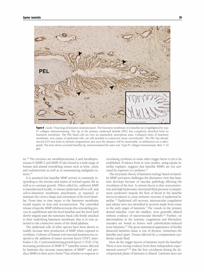
he BM retracts from between the SELs and takes with itecondary dermal lamellar (SDL) connective tissue and SDLapillaries (Fig. 5). The loss of these capillaries may explainhy resistance to blood flow was increased 3.5 times (theounding digital pulse) in horses during early laminitis15 andhy blood appears to bypass the lamellar capillary bed
hrough dilated arteriovenous anastomoses in horses withcute laminitis.14 Both of these phenomena oocur after, andre thus a consequence of, the triggering of MMP production.he epidermal basal cells that have lost their BM attachmento not appear to undergo necrosis, at least initially, andlump together to form amorphous, BM-free masses, on ei-her side of the primary lamellar axis.
rade 3 Histological Laminitisn laminitis, the worse case scenario is a rapid and total BMeparation from all the epidermal lamellae. Sheets of BM peelway to form aggregations of loose isolated BM in the con-ective tissue adjoining the lamellae. The epidermal lamellarells are left as isolated columns with no connection whatso-ver with the dermal connective tissue. The lamellar tips slideway from the BM connective tissue attachments, at first mi-
Figure 5 Micrograph showing hoof lamellar tissues (PAmembrane is stained dark magenta. At the tips of the nomembrane (BM) has continued to lift from the underlyiThe BM has disappeared from adjacent SEL bases and thlamellar BM is no longer close to the primary epiderma
roscopically, but as the degree of separation increases the i
istance between hoof and distal phalanx becomes measur-ble in millimeters (Figs. 6, 7, and 8). This is manifest clini-ally as the “sinker.” Since the BM is the key structure bridg-ng the epidermis of the hoof to the connective tissue of theistal phalanx, it follows that the wholesale loss and disorga-ization of the lamellar BM inexorably leads to the failure ofoof anatomy so characteristic of the chronic stage of lami-itis.
he Pathophysiology ofaminitis
he spectacular disintegration of the lamellar attachment ap-aratus, initiated during the development phase of laminitis,ompromises a normally robust and trouble-free hoof, distalhalanx attachment apparatus in a surprisingly short periodf time. Logic suggests that it is a normally tightly controlledetabolic process that is thrown into disarray to cause the
amellar-specific lesion of laminitis during its developmentalhase.9
The enzymatic remodeling of the epidermal lamellae, as-umed to be mandatory if the continually proliferating stra-um medium of the hoof wall16 is to move past the stationaryistal phalanx, could be accidentally recruited in the patho-enesis of the laminitis disease process. Enzymes capable ofestroying key components of the lamellar attachment appa-atus have been isolated from normal lamellar tissues17 and in
n) with histological Grade 2 laminitis. The basementted secondary epidermal lamellae (SELs) the basement
al cells to form empty, teat-shaped, caps (arrowheads).ittle connective tissue and few capillaries between. Thela (PEL). Bar � 10 �m.
S staiw poinng basere is l
ncreased quantities from lamellar tissues affected by lamini-
ttham
swicmlwrcstt
rcslibd
cemv
lnctmmmaidwacadld
Ti
Equine laminitis 39
is.18 The enzymes are metalloproteinase-2 and metallopro-einase-9 (MMP-2 and MMP-9) also found in a wide range ofuman and animal remodeling tissues such as bone, jointsnd endometrium as well as in metastasizing malignant tu-ors.19
It is assumed that lamellar MMP activity is constantly re-ponding to the stresses and strains of normal equine life asell as to constant growth. When called for, sufficient MMP
s manufactured locally, to release epidermal cell-to-cell, andell-to-basement membrane attachments, as required, toaintain the correct shape and orientation of the hoof lamel-
ae. From time to time injury to the basement membraneould require its lysis and reconstruction. The controlled
elease of specific MMP inhibitors keeps this remodeling pro-ess in equilibrium and the hoof lamellae and the hoof itselflowly migrate past the stationary basal cells firmly attachedo their underlying basement membrane that is in turn at-ached to the connective tissue of the distal phalanx.
The epidermal cells of other species have been shown toeadily increase their production of MMP when exposed toytokines. Cultures of human oral mucosal keratinocytes re-pond to the addition of tumor necrosis factor (TNF), inter-eukin-1 (IL-1) and transforming growth factor-1 (TGF-1) byncreasing production of MMP-9.20 Lamellar tissues affectedy laminitis also increase transcription of MMP17 and pro-
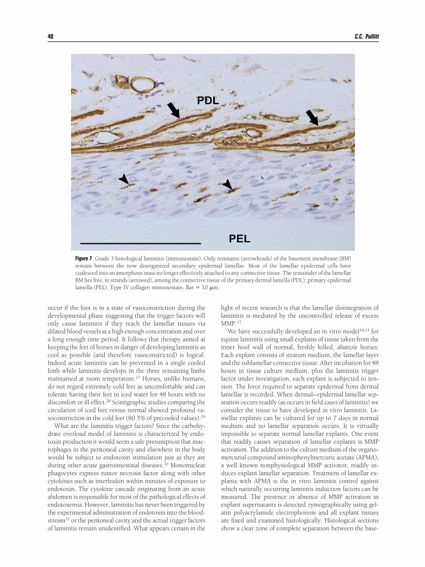
Figure 6 Grade 3 histological laminitis (immunostain). TIV collagen immunostaining. The tip of the primarybasement membrane. The PEL basal cells are now anmembrane, now empty of epidermal cells, are still attamoved 0.03 mm from its dermal compartment and soograph. The inset shows a normal lamellar tip, immunosta�m.
uce MMPs in their active forms18 but whether in response to v
irculating cytokines or some other trigger factor is yet to bestablished. Evidence from in vitro studies, using equine la-ellar explants, suggests that lamellar MMPs are not acti-
ated by exposure to cytokines.21
The enzymatic theory of laminitis etiology based on lamel-ar MMP activation challenges the alternative view that lami-itis develops because of vascular pathology affecting theirculation of the foot. A current theory is that venoconstric-ion and high hydrostatic interstitial fluid pressure (compart-ent syndrome) impede the flow of blood in the lamellaricrocirculation to cause ischemic necrosis of epidermal la-ellae.15 Epidermal cell necrosis, intravascular coagulation
nd edema were not identified in sections made from tissuen the early stages of laminitis.3 The vessels in the primaryermal lamellae, even the smallest, were generally dilatedithout evidence of microvascular thrombi.22 Further, no
bnormalities in the systemic coagulation and fibrinolyticascades are found in horses with carbohydrate-inducedcute laminitis.23 The gross anatomical appearance of freshlyissected laminitis tissue is one of dryness. Sometimes the
amellae peel apart. Tissues affected by a compartment syn-rome exude fluid.How do the trigger factors of laminitis reach the lamellae?
here is now strong evidence from three independent exper-mental sources24-26 that the foot circulation during the de-
ment membrane of a lamellar tip is highlighted by typemal lamella (PEL) has completely detached from itsched, amorphous mass. Collapsed tubes of basement
connective tissue (arrowheads). The PEL has alreadydistance will be measurable, in millimeters on a radio-he same way. Type IV collagen immunostain. Bars � 10
he baseepiderunatta
ched ton theined t
elopmental phase of laminitis is dilated. Laminitis does not
ododakcIlmdtdcs
dtrwdpceaetso
llM
eiEahfslacmmitamadpwmeaa
�m.
40 C.C. Pollitt
ccur if the foot is in a state of vasoconstriction during theevelopmental phase suggesting that the trigger factors willnly cause laminitis if they reach the lamellar tissues viailated blood vessels at a high enough concentration and overlong enough time period. It follows that therapy aimed ateeping the feet of horses in danger of developing laminitis asool as possible (and therefore vasoconstricted) is logical.ndeed acute laminitis can be prevented in a single cooledimb while laminitis develops in the three remaining limbs
aintained at room temperature.27 Horses, unlike humans,o not regard extremely cold feet as uncomfortable and canolerate having their feet in iced water for 48 hours with noiscomfort or ill effect.28 Scintigraphic studies comparing theirculation of iced feet versus normal showed profound va-oconstriction in the cold feet (80.5% of precooled values).29
What are the laminitis trigger factors? Since the carbohy-rate overload model of laminitis is characterized by endo-oxin production it would seem a safe presumption that mac-ophages in the peritoneal cavity and elsewhere in the bodyould be subject to endotoxin stimulation just as they areuring other acute gastrointestinal diseases.30 Mononuclearhagocytes express tumor necrosis factor along with otherytokines such as interleukin within minutes of exposure tondotoxin. The cytokine cascade originating from an acutebdomen is responsible for most of the pathological effects ofndotoxemia. However, laminitis has never been triggered byhe experimental administration of endotoxin into the blood-tream31 or the peritoneal cavity and the actual trigger factors
Figure 7 Grade 3 histological laminitis (immunostain). Oremain between the now disorganized secondary epicoalesced into an amorphous mass no longer effectively aBM lies free, in strands (arrowed), among the connectivelamella (PEL). Type IV collagen immunostain. Bar � 10
f laminitis remain unidentified. What appears certain in the s
ight of recent research is that the lamellar disintegration ofaminitis is mediated by the uncontrolled release of excess
MP.17
We have successfully developed an in vitro model18,21 forquine laminitis using small explants of tissue taken from thenner hoof wall of normal, freshly killed, abattoir horses.ach explant consists of stratum medium, the lamellar layernd the sublamellar connective tissue. After incubation for 48ours in tissue culture medium, plus the laminitis triggeractor under investigation, each explant is subjected to ten-ion. The force required to separate epidermal from dermalamellae is recorded. When dermal–epidermal lamellar sep-ration occurs readily (as occurs in field cases of laminitis) weonsider the tissue to have developed in vitro laminitis. La-ellar explants can be cultured for up to 7 days in normaledium and no lamellar separation occurs. It is virtually
mpossible to separate normal lamellar explants. One eventhat readily causes separation of lamellar explants is MMPctivation. The addition to the culture medium of the organo-ercurial compound aminophenylmercuric acetate (APMA),well known nonphysiological MMP activator, readily in-
uces explant lamellar separation. Treatment of lamellar ex-lants with APMA is the in vitro laminitis control againsthich naturally occurring laminitis induction factors can beeasured. The presence or absence of MMP activation in
xplant supernatants is detected zymographically using gel-tin polyacrylamide electrophoresis and all explant tissuesre fixed and examined histologically. Histological sections
mnants (arrowheads) of the basement membrane (BM)lamellae. Most of the lamellar epidermal cells haveto any connective tissue. The remainder of the lamellarf the primary dermal lamella (PDL); primary epidermal
nly redermalttachedtissue o
how a clear zone of complete separation between the base-
mTb
vTknnlopemcoppo
neib
rnbrwg9ttTcla
Equine laminitis 41
ent membrane and the basal cells of the epidermal lamellae.his is a characteristic of in vitro laminitis and resembles theasement membrane lesion of natural in vivo laminitis.We have used the in vitro laminitis explant model to in-
estigate most of the proposed causes of equine laminitis.he equine lamellae have tested resistant to virtually allnown cytokines, tissue factors and prostaglandins. Gram-egative bacterial endotoxin, extract of black walnut (Juglansigra) and even anaerobic culture conditions fail to induceamellar separation or significant MMP activation. There isne notable exception however. A factor present in the su-ernatant of cultures of Streptococcus bovis isolated from thequine cecum activates equine hoof MMP-2 and causes la-ellar separation.21 During grain overload S. bovis is the prin-
ipal microorganism responsible for the rapid fermentationf carbohydrate to lactic acid in the equine hindgut. In theresence of virtually unlimited substrate its population ex-lodes exponentially. We are currently investigating the role
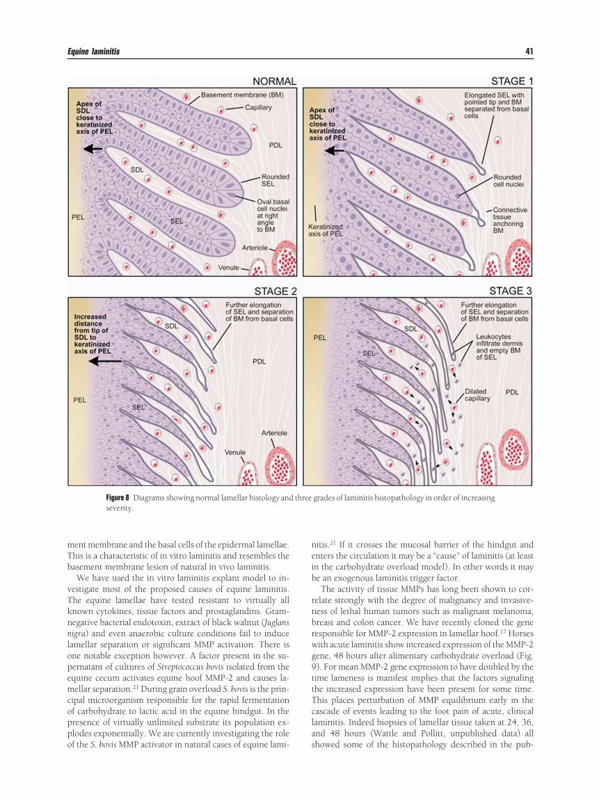
Figure 8 Diagrams showing normal lamellar histology andseverity.
f the S. bovis MMP activator in natural cases of equine lami- s
itis.21 If it crosses the mucosal barrier of the hindgut andnters the circulation it may be a “cause” of laminitis (at leastn the carbohydrate overload model). In other words it maye an exogenous laminitis trigger factor.The activity of tissue MMPs has long been shown to cor-
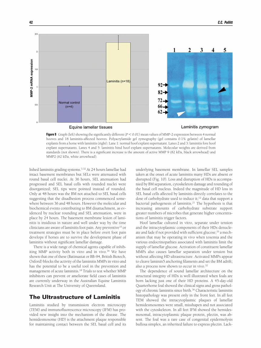
elate strongly with the degree of malignancy and invasive-ess of lethal human tumors such as malignant melanoma,reast and colon cancer. We have recently cloned the geneesponsible for MMP-2 expression in lamellar hoof.17 Horsesith acute laminitis show increased expression of the MMP-2ene, 48 hours after alimentary carbohydrate overload (Fig.). For mean MMP-2 gene expression to have doubled by theime lameness is manifest implies that the factors signalinghe increased expression have been present for some time.his places perturbation of MMP equilibrium early in theascade of events leading to the foot pain of acute, clinicalaminitis. Indeed biopsies of lamellar tissue taken at 24, 36,nd 48 hours (Wattle and Pollitt, unpublished data) all
grades of laminitis histopathology in order of increasing
threehowed some of the histopathology described in the pub-
lirpdOswbipnctdl
isOhmiaR
TL(vhf
utdntSdbigt
aaavsMwta
sbQohThwms
42 C.C. Pollitt
ished laminitis grading systems.3,32 At 24 hours lamellae hadntact basement membranes but SELs were attenuated withound basal cell nuclei. At 36 hours, SEL attenuation hadrogressed and SEL basal cells with rounded nuclei wereisorganized; SEL tips were pointed instead of rounded.nly at 48 hours was the BM not attached to SEL basal cells
uggesting that the disadhesion process commenced some-here between 36 and 48 hours. However the molecular andiochemical events contributing to BM disattachment, as ev-
denced by nuclear rounding and SEL attenuation, were inlace by 24 hours. The basement membrane lesion of lami-itis is insidious in nature and well under way by the timelinicians are aware of laminitis foot pain. Any preventive33 orreatment strategies must be in place before overt foot painevelops if horses are to survive the development phase of
aminitis without significant lamellar damage.There is a wide range of chemical agents capable of inhib-
ting MMP activity both in vitro and in vivo.34 We havehown that one of these (Batimastat or BB-94, British Biotech,xford) blocks the activity of the laminitis MMPs in vitro andas the potential to be a useful tool in the prevention andanagement of acute laminitis.18 Trials to test whether MMP
nhibitors can prevent or ameliorate field cases of laminitisre currently underway in the Australian Equine Laminitisesearch Unit at The University of Queensland.
he Ultrastructure of Laminitisaminitis studied by transmission electron microscopyTEM) and immunofluorescence microscopy (IFM) has pro-ided new insight into the mechanism of the disease. Theemidesmosome (HD) is the attachment plaque responsible
Figure 9 Graph (left) showing the significantly different (Phooves and 18 laminitis-affected hooves. Polyacrylamexplants from a horse with laminitis (right). Lane 1: normexplant supernatants. Lanes 4 and 5: laminitis hind hostandards (not shown). There is a significant increase isMMP2 (62 kDa, white arrowhead).
or maintaining contact between the SEL basal cell and its b
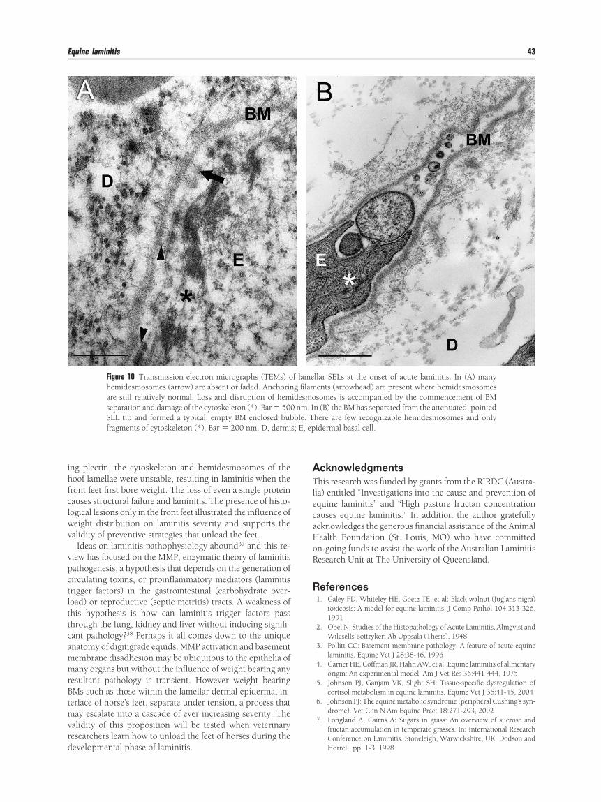
nderlying basement membrane. In lamellar SEL samplesaken at the onset of acute laminitis many HDs are absent orisrupted (Fig. 10). Loss and disruption of HDs is accompa-ied by BM separation, cytoskeleton damage and rounding ofhe basal cell nucleus. Indeed the magnitude of HD loss inEL basal cells affected by laminitis directly correlates to theose of carbohydrate used to induce it;12 data that support aacterial pathogenesis of laminitis.21 The hypothesis is that
ncreasing amounts of carbohydrate substrate supportreater numbers of microbes that generate higher concentra-ions of laminitis trigger factors.
Hoof lamellae cultured in vitro, separate under tensionnd the intracytoplasmic components of their HDs denucle-te and fade if not provided with sufficient glucose;35 a mech-nism that may be operating in vivo when toxemia and thearious endocrinopathies associated with laminitis limit theupply of lamellar glucose. Activation of constituent lamellarMPs also causes lamellar separation under tension butithout affecting HD ultrastructure. Activated MMPs appear
o cleave laminin5 anchoring filaments and set the BM adrift;lso a process now shown to occur in vivo.32
The dependence of sound lamellar architecture on thetructural integrity of HDs is well illustrated when foals areorn lacking just one of their HD proteins. A 45-day-olduarterhorse foal showed the clinical signs and gross pathol-gy of chronic laminitis since birth.36 Characteristic laminitisistopathology was present only in the front feet. In all feetEM showed the intracytoplasmic plaques of lamellaremidesmosomes were small, misshapen and not associatedith the cytoskeleton. In all feet IFM showed the hemides-osomal, intracytoplasmic plaque protein, plectin, was ab-
ent. The foal was a rare case of congenital epidermolysis
1) mean values of MMP-2 expression between 4 normall zymography (gel contains 0.1% gelatin) of lamellarf explant supernatant. Lanes 2 and 3: laminitis fore hooflant supernatants. Molecular weights are derived fromount of active MMP 9 (82 kDa, black arrowhead) and
� 0.0ide geal hooof expthe am
ullosa simplex, an inherited failure to express plectin. Lack-
ihfclwv
vpctlttcammrBtmvrd
ATlecaHoR
R
s; E, ep
Equine laminitis 43
ng plectin, the cytoskeleton and hemidesmosomes of theoof lamellae were unstable, resulting in laminitis when the
ront feet first bore weight. The loss of even a single proteinauses structural failure and laminitis. The presence of histo-ogical lesions only in the front feet illustrated the influence ofeight distribution on laminitis severity and supports thealidity of preventive strategies that unload the feet.
Ideas on laminitis pathophysiology abound37 and this re-iew has focused on the MMP, enzymatic theory of laminitisathogenesis, a hypothesis that depends on the generation ofirculating toxins, or proinflammatory mediators (laminitisrigger factors) in the gastrointestinal (carbohydrate over-oad) or reproductive (septic metritis) tracts. A weakness ofhis hypothesis is how can laminitis trigger factors passhrough the lung, kidney and liver without inducing signifi-ant pathology?38 Perhaps it all comes down to the uniquenatomy of digitigrade equids. MMP activation and basementembrane disadhesion may be ubiquitous to the epithelia ofany organs but without the influence of weight bearing any
esultant pathology is transient. However weight bearingMs such as those within the lamellar dermal epidermal in-erface of horse’s feet, separate under tension, a process thatay escalate into a cascade of ever increasing severity. The
alidity of this proposition will be tested when veterinaryesearchers learn how to unload the feet of horses during the
Figure 10 Transmission electron micrographs (TEMs) ohemidesmosomes (arrow) are absent or faded. Anchorinare still relatively normal. Loss and disruption of hemseparation and damage of the cytoskeleton (*). Bar � 500SEL tip and formed a typical, empty BM enclosed bubfragments of cytoskeleton (*). Bar � 200 nm. D, dermi
evelopmental phase of laminitis.
cknowledgmentshis research was funded by grants from the RIRDC (Austra-
ia) entitled “Investigations into the cause and prevention ofquine laminitis” and “High pasture fructan concentrationauses equine laminitis.” In addition the author gratefullycknowledges the generous financial assistance of the Animalealth Foundation (St. Louis, MO) who have committedn-going funds to assist the work of the Australian Laminitisesearch Unit at The University of Queensland.
eferences1. Galey FD, Whiteley HE, Goetz TE, et al: Black walnut (Juglans nigra)
toxicosis: A model for equine laminitis. J Comp Pathol 104:313-326,1991
2. Obel N: Studies of the Histopathology of Acute Laminitis, Almgvist andWilcsells Bottrykeri Ab Uppsala (Thesis), 1948.
3. Pollitt CC: Basement membrane pathology: A feature of acute equinelaminitis. Equine Vet J 28:38-46, 1996
4. Garner HE, Coffman JR, Hahn AW, et al: Equine laminitis of alimentaryorigin: An experimental model. Am J Vet Res 36:441-444, 1975
5. Johnson PJ, Ganjam VK, Slight SH: Tissue-specific dysregulation ofcortisol metabolism in equine laminitis. Equine Vet J 36:41-45, 2004
6. Johnson PJ: The equine metabolic syndrome (peripheral Cushing’s syn-drome). Vet Clin N Am Equine Pract 18:271-293, 2002
7. Longland A, Cairns A: Sugars in grass: An overview of sucrose andfructan accumulation in temperate grasses. In: International ResearchConference on Laminitis. Stoneleigh, Warwickshire, UK: Dodson and
llar SELs at the onset of acute laminitis. In (A) manyents (arrowhead) are present where hemidesmosomessomes is accompanied by the commencement of BM
n (B) the BM has separated from the attenuated, pointedhere are few recognizable hemidesmosomes and onlyidermal basal cell.
f lameg filam
idesmonm. I
ble. T
Horrell, pp. 1-3, 1998
11
1
1
1
1
1
1
1
1
2
2
2
2
2
2
2
2
2
2
3
3
3
3
3
3
3
33
44 C.C. Pollitt
8. Watts K: Information on the current research and prevention of grassfounder in horses: http://www.safergrass.org
9. Pollitt CC, Kyaw-Tanner M, French KR, et al: Equine laminitis: In-depth. New Orleans, LA: American Association of Equine Practitioners49th Annual Convention, pp. 21-25, 2003
0. Eustace RA, Redden RR: Iatrogenic laminitis. Vet Rec 126:586, 19901. Hunt RJ: A retrospective evaluation of laminitis in horses. Equine Vet J
25:61-64, 19932. French KR, Pollitt CC: Equine laminitis: Loss of hemidesmosome ul-
trastructure correlates to dose in an oligofructose induction model: Anultrastructural study. Equine Vet J 36:230-235, 2004
3. Pollitt CC, Daradka M: Equine laminitis basement membrane pathol-ogy: Loss of type IV collagen, type VII collagen and laminin immuno-staining. Equine Vet J Suppl 26:139-144, 1998
4. Hood DM, Amoss MS, Hightower D, et al: Equine Laminitis 1: Radio-isotopic analysis of the haemodynamics of the foot during the acutedisease. J Equine Med Surg 2:439-444, 1978
5. Allen D Jr, Clark ES, Moore JN, et al: Evaluation of equine digitalStarling forces and hemodynamics during early laminitis. Am J Vet Res51:1930-1934, 1990
6. Daradka M, Pollitt CC: Epidermal cell proliferation in the equine hoofwall. Equine Vet J 36:236-241, 2004
7. Kyaw-Tanner M, Pollitt CC: Equine laminitis: Increased transcriptionof matrix metalloproteinase-2 (MMP-2) occurs during the develop-mental phase. Equine Vet J 36:221-225, 2004
8. Pollitt CC, Pass MA, Pollitt S: Batimastat (BB-94) inhibits matrix met-alloproteinases of equine laminitis. Equine Vet J Suppl 26:119-124,1998
9. Birkedal-Hansen H: Proteolytic remodeling of extracellular matrix.Curr Opin Cell Biol 7:728-735, 1995
0. Pirilä E: Expression and role of matrix metalloproteinases and the lami-nin-5 gamma-2 chain in wound healing and cell migration, Universityof Helsinki, 2003, PhD dissertation. ISBN 952-91-6624-9
1. Mungall BA, Kyaw Tanner M, Pollitt CC: In vitro evidence for a bacte-rial pathogenesis of equine laminitis. Vet Microbiol 79:209-223, 2001
2. Weiss DJ, Geor RJ, Johnston G, et al: Microvascular thrombosis asso-ciated with onset of acute laminitis in ponies. Am J Vet Res 55:606-612,1994
3. Prasse KW, Allen D Jr, Moore JN, et al: Evaluation of coagulation andfibrinolysis during the prodromal stages of carbohydrate-induced acute
laminitis in horses. Am J Vet Res 51:1950-1955, 19904. Pollitt CC, Davies CT: Equine laminitis: Its development coincides withincreased sublamellar blood flow. Equine Vet J Suppl 26:125-132,1998
5. Robinson NE, Scott JB, Dabney JM, et al: Digital vascular responses andpermeability in equine alimentary laminitis. Am J Vet Res 37:1171-1176, 1976
6. Trout DR, Hornof WJ, Linford RL, et al: Scintigraphic evaluation ofdigital circulation during the developmental and acute phases of equinelaminitis. Equine Vet J 22:416-421, 1990
7. van Eps AW, Pollitt CC: Equine laminitis: Cryotherapy reduces theseverity of the acute lesion. Equine Vet J 36:255-260, 2004
8. Pollitt CC, van Eps AW: Prolonged, continuous distal limb cryotherapyin the horse. Equine Vet J 36:216-220, 2004
9. Worster AA, Gaughan EM, Hoskinson JJ, et al: Effects of external ther-mal manipulation on laminar temperature and perfusion scintigraphyof the equine digit. N Z Vet J 48:111-116, 2000
0. Barton MH, Collatos C, Moore JN: Endotoxin induced expression oftumour necrosis factor, tissue factor and plasminogen activator inhib-itor activity by peritoneal macrophages. Equine Vet J 28:382-389, 1996
1. Hunt RJ, Allen D, Moore JN: Effect of endotoxin administration onequine digital hemodynamics and starling forces. Am J Vet Res 51:1703-1707, 1990
2. French KR, Pollitt CC: Equine laminitis: Cleavage of laminin5 (L5)associated with basement membrane dysadhesion. Equine Vet J 36:242-247, 2004
3. van Eps AW, Pollitt CC: Equine laminitis: Cryotherapy reduces theseverity of the acute lesion. Equine Vet J 36:255-260, 2004
4. Roach DM, Fitridge RA, Laws PE, et al: Up-regulation of MMP-2 andMMP-9 leads to degradation of type IV collagen during skeletal musclereperfusion injury: Protection by the MMP inhibitor, doxycycline. EurJ Vasc Endovasc Surg 23:260-269, 2002
5. French KR, Pollitt CC. Equine laminitis: glucose deprivation and MMPactivation induce dermo-epidermal separation in vitro. Equine Vet J36:261-266, 2004
6. French KR, Pollitt CC: Equine laminitis: Congenital, hemidesmosomalplectin deficiency in a Quarterhorse foal. Equine Vet J 36:299-303,2004
7. Hood D. The hoof project. http://www.hoofproject.com/8. Hood DM: The pathophysiology of developmental and acute laminitis.
Vet Clin North Am Equine Pract 15:321-343, 1999