kou molecular and cellular biology 2003 3186-3201
TRANSCRIPT
MOLECULAR AND CELLULAR BIOLOGY, May 2003, p. 3186–3201 Vol. 23, No. 90270-7306/03/$08.00�0 DOI: 10.1128/MCB.23.9.3186–3201.2003Copyright © 2003, American Society for Microbiology. All Rights Reserved.
Structural and Functional Analysis of Mutations along theCrystallographic Dimer Interface of the Yeast TATA
Binding ProteinHaiping Kou, Jordan D. Irvin, Kathryn L. Huisinga, Madhusmita Mitra,
and B. Franklin Pugh*Department of Biochemistry and Molecular Biology, The Pennsylvania State University,
University Park, Pennsylvania 16803
Received 18 November 2002/Returned for modification 23 December 2002/Accepted 5 February 2003
The TATA binding protein (TBP) is a central component of the eukaryotic transcription machinery and issubjected to both positive and negative regulation. As is evident from structural and functional studies, TBP’sconcave DNA binding surface is inhibited by a number of potential mechanisms, including homodimerizationand binding to the TAND domain of the TFIID subunit TAF1 (yTAFII145/130). Here we further characterizedthese interactions by creating mutations at 24 amino acids within the Saccharomyces cerevisiae TBP crystallo-graphic dimer interface. These mutants are impaired for dimerization, TAF1 TAND binding, and TATAbinding to an extent that is consistent with the crystal or nuclear magnetic resonance structure of these orrelated interactions. In vivo, these mutants displayed a variety of phenotypes, the severity of which correlatedwith relative dimer instability in vitro. The phenotypes included a low steady-state level of the mutant TBP,transcriptional derepression, dominant slow growth (partial toxicity), and synthetic toxicity in combinationwith a deletion of the TAF1 TAND domain. These phenotypes cannot be accounted for by defective interactionswith other known TBP inhibitors and likely reflect defects in TBP dimerization.
Activation of eukaryotic genes is a multistep process, involv-ing the coalescence of promoter-specific activators, chromatin-remodeling complexes, and components of the general tran-scription machinery at promoters. An important part of theactivation process is the removal of inhibitors associated withlatent activators, promoters, and the general transcription ma-chinery. One component of the general transcription machin-ery that is subjected to substantial inhibition is the TATAbinding protein (TBP) (reviewed in reference 75). Virtually allgenes require TBP for function, and its association with pro-moters is generally linked to transcriptional activity (57, 63).Preventing unregulated promoter binding by TBP may be crit-ical for preventing unregulated gene expression. TBP accessmight be prevented in part by nucleosome formation over theTATA box (43, 80). However, many quiescent genes are notderepressed upon histone depletion (83), indicating that otherinhibitory mechanisms might prevent TBP from binding topromoters.
A number of proteins inhibit TBP function. These includethe TAF1 (yTAFII145/130) subunit of TFIID, NC2, Mot1, theSpt3/Spt8 subunits of SAGA, the Ccr4-Not complex, and asecond molecule of TBP in the form of homodimers. Here wefocus on two inhibitory interactions which are directed atTBP’s concave surface: TBP dimerization and the TAF1TAND domain.
TFIID is a multisubunit complex consisting of TBP andTAFs (19, 77, 78). TFIID is required for activated transcriptionbut is intrinsically inhibitory toward TBP-TATA interactions
(78). At least part of this inhibitory activity might reside withinthe amino-terminal TAND domain of the TFIID subunit,TAF1 (11, 52, 70). Mutagenesis studies have delineated Dro-sophila and yeast TANDs as two subdomains, I and II (52, 54).TAND II binds to TBP’s convex surface in the vicinity of helix2 (52). TAND I binds to the concave surface of TBP. Nuclearmagnetic resonance (NMR) imaging of the Drosophila TANDI domain in complex with yeast TBP reveals that TAND Imimics the minor-groove structure of partially unwoundTATA box DNA (65). The surface of TBP that interacts withthe Drosophila TAND I domain has been partially mappedthrough mutagenesis (70), and it agrees well with the NMRstructure of the complex. While the yeast and DrosophilaTAND domains function similarly, it is striking that they arepoorly (�30%) conserved, with yeast TAND being approxi-mately half the size of Drosophila TAND (52). The structure ofthe yeast TAND I domain has not been determined, and whereit interacts along TBP’s concave surface has not been fullydelineated. This is particularly intriguing given the small size ofthe yeast TAND I domain. It might be too small to occupy theentirety of TBP’s concave surface.
If TAND inhibits TBP function in vivo, then deletion of thisdomain is expected to lead to an increase in transcription.However, very few genes increase in expression in ataf1(�TAND) strain (�60 out of �6,000) (24). Mutationsalong TBP’s concave surface result in widespread transcrip-tional derepression, which is augmented in a �TAND strain(24). These results suggest that TAND contributes to tran-scriptional inhibition but does not play a predominant role.
Self-association of TBP into dimers might provide a pre-dominant means of inhibiting its concave DNA binding sur-face. Evidence that yeast and human TBPs autorepress their
* Corresponding author. Mailing address: Department of Biochem-istry and Molecular Biology, The Pennsylvania State University, Uni-versity Park, PA 16803. Phone: (814) 863-8252. Fax: (814) 863-8595.E-mail: [email protected].
3186
at Scientific Library, N
CI-F
CR
DC
on April 20, 2009
mcb.asm
.orgD
ownloaded from
DNA binding activities through dimerization comes from X-ray crystallography, pulldown assays, chemical cross-linking,gel filtration, analytical ultracentrifugation, and mutagenesisstudies and has been inferred from its kinetic profile for DNAbinding (22, 25–27, 42, 44, 45, 47, 69, 72, 81). Consistent witha dimer autorepression mechanism, TBP mutations that impairdimerization cause transcriptional derepression in vivo (24,45). Their effect is dominant to wild-type TBP but is partiallysuppressed by wild-type TBP overexpression (45). TBP over-expression might drive unstable dimer mutants into somewhatmore stable heterodimers with wild-type TBP.
Despite several observations that support a physiologicalrole for TBP dimerization (45), the topic remains controversial(20). Ultimately, we wish to understand how a variety of TBPinhibitory mechanisms are coordinated to regulate TBP. Anecessary step in this effort is the mapping of surfaces on TBPthat are targeted for inhibition in vivo and the identification offactors responsible for that inhibition. In an effort to morecompletely examine the potential physiological significance ofTBP dimerization, we mutated 24 amino acids that comprisethe crystallographic dimer interface. Using in vitro pulldownand electrophoretic mobility assays, we characterized theirabilities to dimerize, bind TAND, and bind TATA DNA. Thebinding data were in good agreement with the crystallographicand NMR structures of these or related complexes.
Next, we sought to determine whether mutations along thecrystallographic dimer interface affect a variety of phenotypesin Saccharomyces cerevisiae that we previously linked to defectsin dimerization (24, 45). These include increased TBP turn-over, transcriptional derepression, partial dominant inhibitionof cell growth (toxicity), and synthetic toxicity in a �TANDstrain. With the collection of mutations throughout the dimerinterface, we observed a correlation between these phenotypesand relative dimer stability measured in vitro. Mutations thatknock out interfaces between TBP and other inhibitors did notgive similar phenotypes. Taken together, the data provide fur-ther support for the notion that TBP dimerizes in vivo and is aphysiologically important negative regulator of gene expres-sion.
MATERIALS AND METHODS
Plasmids. All TBP mutations were created by oligonucleotide-directed mu-tagenesis. The mutations and the integrity of the entire open reading frame wereconfirmed in both the bacterial and yeast expression vectors by DNA sequencing.Escherichia coli plasmids expressing the His-TBP mutants were designatedpET16b-yTBP(xxx), where xxx indicates the mutation. The plasmid pGEX-TFIID-C, which encodes human GST-TBP(core), has been described previously(41). Plasmid pGEX-yTBP(181C) has been described previously (45). The plas-mid pGEX-scTAF1(10-88) was constructed by PCR amplification of the TAF1gene and was inserted in frame with the glutathione S-transferase (GST)-codingsequence of pGEX-3X. Derivatives of this plasmid, pGEX-scTAF1(10-88,D66K) and pGEX-scTAF1(10-88, F23K, D66K), were constructed by oligonu-cleotide-directed mutagenesis. The integrity of the entire TAND coding regionwas verified by DNA sequencing. Plasmids expressing the HA3-TBP mutants inS. cerevisiae were designated pCALF-T(xxx)(PGK), where CALF refers to theCEN/ARS origin, LEU2 marker, Flu3 (or HA3) tagged; T indicates TBP; xxx isthe mutation; and PGK is the promoter controlling HA3-TBP expression. Theplasmid is derived from pDP15-flu3-yTBP (73), in which the SPT15 promoter wasreplaced by the PGK1 promoter, as described previously (45). The plasmidpCALF-T(K145E)(GAL) contains the GAL10 promoter in place of the PGK1promoter as described previously (24). pADH1-lacZ contains the core (lackingthe upstream activation sequence) ADH1 promoter downstream of four glucose-repressible Gal4 binding sites (15). The plasmids TAF1/Ura, TAF1/Trp, and
TAF1(�TAND)/Trp have been described previously as pYN1/TAF145, pYN2/TAF145, and pYN2/taf145(�10-73), respectively (52).
Strains. YTW22 [MAT� ura3-52 trp1-�1 his3-�200 leu2-�1 lys2-801amber ade2-101ocher �spt15::TRP1(pCW16-TBP-WT)] is a TBP plasmid shuffle strain (74).YPH252 (MAT� ura3-52 trp1-�1 his3-�200 leu2-�1 lys2-801amber ade2-101ocher)is wild type for TBP (SPT15) and has been described previously (74). In vivostudies with TAF1 and taf1(�TAND) employed the strain Y13.2 (MAT� ura3-52trp1-�63 leu2,3-112 his3-609 �taf145 pYN1/TAF145), in which pYN1/TAF145was replaced by either pYN2/TAF145 or pYN2/taf145(�10-73) by using theplasmid shuffle assay (52).
Protein purification. All His-tagged TBP mutants were purified as follows.Recombinant E. coli (BL21) cells (500 ml) were grown in YT medium containing0.2 g of ampicillin per liter at 37°C to an optical density at 595 nm (OD595) of 0.7and induced with 20 mg of isopropylthio-�-D-galactoside per liter for 45 min at30°C. Cells were harvested by centrifugation, washed and resuspended to avolume of 10 ml in lysis buffer (25 mM HEPES [pH 7.5], 200 mM potassiumchloride, 12.5 mM magnesium chloride, 10% glycerol, 0.05 mM phenylmethyl-sulfonyl fluoride), and quickly frozen in liquid nitrogen. Cells were thawed andmixed with 0.8 mg of lysozyme per ml for 10 min at 4°C, with 2 M potassiumchloride for 15 min, and with 0.2% IGEPAL-CA630 for 5 min. Extracts weresonicated to reduce viscosity and then centrifuged in an SS34 rotor (RC5Ccentrifuge) at 15,000 rpm for 30 min at 4°C. Supernatants were mixed with 10mM imidazole and 0.5 ml of Ni-nitrilotriacetic acid–agarose for 60 min at 4°C.The slurry was then transferred to a column and then washed with wash buffer(20 mM HEPES [pH 7.5], 1 M potassium chloride, 12.5 mM magnesium chlo-ride, 10% glycerol, 60 mM imidazole, 0.05 mM phenylmethylsulfonyl fluoride).TBP was eluted with TSB buffer (20 mM Tris-acetate [pH 7.5], 0.2 M potassiumglutamate, 2 mM magnesium chloride, 20% glycerol, 0.05 mM phenylmethylsul-fonyl fluoride) containing 1 M imidazole and dialyzed against TSB buffer con-taining 60 mM imidazole, 1 mM dithiothreitol, and 0.5 mM phenylmethylsulfonylfluoride. TBP aliquots were frozen in liquid nitrogen and stored at �80°C. TBPwas judged to be �50% pure by sodium dodecyl sulfate-polyacrylamide gelelectrophoresis (SDS-PAGE) followed by silver staining (see Fig. 2). Proteinsconcentrations were estimated from these gels by using highly purified TBPstandards whose concentrations were determined by total amino acid analysis.
Human and yeast GST-TBP(core) were expressed from pGEX-TFIID-C andpGEX-yTBP(181C), respectively, and purified from 3 liters of recombinant E.coli cells as described above, with the following exceptions. Proteins were ex-tracted with 1 M potassium chloride and 0.1% IGEPAL-CA630 (rather than 2 Mand 0.2%, respectively). Glutathione agarose (1 ml) (Sigma) was used in place ofnickel agarose. The resin was washed with H buffer (20 mM HEPES [pH 7.5], 2mM magnesium chloride, 10% glycerol, 0.1 mM phenylmethylsulfonyl fluoride,and 1 mM dithiothreitol) containing 1 M potassium chloride (H1 buffer) andthen with TSB buffer. The resin-bound GST-TBP(core) was aliquoted, quick-frozen in liquid nitrogen, and stored at �80°C.
GST-TAND was expressed from pGEX-scTAF1(10-88) and purified fromrecombinant E. coli DH5� cells as described above, with the following excep-tions. Induction with isopropylthio-�-D-galactoside was for 2 h. Proteins wereextracted with 0.1 M potassium chloride and 0.07% IGEPAL-CA630 (ratherthan 2 M and 0.2%, respectively). Glutathione agarose (0.75 ml) (Sigma) wasused in place of nickel agarose. The resin was washed sequentially with H.35, H1,and then H.35 buffers. Proteins were eluted in H.35 containing 0.1 M reducedglutathione and dialyzed into H.35 buffer. Aliquots were frozen in liquid nitrogenand stored at �80°C. GST-TAND mutants were purified similarly. Proteins werejudged to be approximately 90% pure by SDS-PAGE and silver staining.
GST-TBP(core) pulldown assay. Reaction mixtures contained 20 mM Tris-acetate (pH 7.5), 75 mM potassium glutamate, 4 mM magnesium chloride, 5%glycerol, 0.1 �g of bovine serum albumin per ml, 4 mM spermidine, 0.025%IGEPAL-CA630, 0.5 �g of heparin per ml, a 5 nM concentration of the indicatedHis-tagged TBP mutant, and 20 nM GST-TBP(core) or GST bound to 2 �l ofglutathione agarose resin, in 500 �l. Reaction mixtures were incubated at 4°C for45 min with mixing. Resins were washed three times, each with 500 �l of reactionbuffer. Bound proteins were eluted and subjected to SDS-PAGE, and TBP wasprobed by immunoblotting with TBP antibodies. Reactions were typically per-formed at least six times, and representative data are shown. TBP was quanti-tated by densitometric scanning of autoradiograms. Relative pulldown was de-termined by subtracting local background and normalizing to a wild-type TBPpulldown present on the same gel.
GST-TAND pulldown assay. Reaction mixtures contained 20 mM Tris-Cl (pH8.3), 150 mM potassium chloride, 12.5 mM magnesium chloride, 10% glycerol,50 �g of bovine serum albumin per ml, 1 mM dithiothreitol, a 300 nM concen-tration of the indicated His-tagged TBP mutant, and 300 nM GST-TAND,GST-TAND(F23K D66K), or GST-TAND(D66K) bound to 10 �l of glutathione
VOL. 23, 2003 STRUCTURE AND FUNCTION OF TBP DIMERS 3187
at Scientific Library, N
CI-F
CR
DC
on April 20, 2009
mcb.asm
.orgD
ownloaded from
agarose resin, in 100 �l. Reaction mixtures were incubated at 4°C for 30 min withmixing. Resins were washed three times, each with 500 �l of reaction buffer.Bound proteins were eluted and subjected to SDS-PAGE, and TBP was probedby immunoblotting with TBP antibodies. TAND was probed with GST antibod-ies and detected by enhanced chemiluminescence (ECL). All reactions wereperformed at least three times, and representative data are shown. TBP wasquantitated by densitometric scanning of autoradiograms. Relative pulldown wasdetermined by subtracting local background and normalizing to a wild-type TBPpulldown present on the same gel.
Electrophoretic mobility shift DNA binding assay. Reaction mixtures con-tained 22 mM Tris-acetate (pH 8.0), 60 mM potassium glutamate, 4 mM mag-nesium chloride, 10% glycerol, 5 �g of bovine serum albumin per ml, 1 mMdithiothreitol, 0.01% IGEPAL-CA630, 4 mM spermidine, 4 �g of poly(dG-dC)per ml, a 30 nM concentration of the indicated His-tagged TBP mutant, and �2nM 32P-labeled TATA double-stranded oligonucleotide (50 bp, 5�-CCCCGACCGGGTGTGACAGTGAGGGGGC TATAAAAGGGGGTGGGGGCGCG-3�) in 10 �l. Reaction mixtures were incubated at 23°C for 40 min, and then 5-�lsamples were loaded onto prerun (100 V, 40 min, 4°C) 15-cm native 6% (60.6:1acrylamide/bisacrylamide ratio) polyacrylamide gels containing 1 TGM buffer(25 mM Tris-Cl [pH 8.3], 190 mM glycine, 1 mM EDTA, 5 mM magnesiumacetate), 2.5% glycerol, and 0.5 mM dithiothreitol in running buffer containing1X TGM. Electrophoresis was continued at 160 V (�35 mA) for 25 min at 4°C.Reactions were performed at least three times, and representative data areshown. The amount of shifted species was quantitated by phosphorimager anal-ysis. Relative binding was determined by subtracting local background and nor-malizing to a wild-type TBP shift present on the same gel.
Plasmid shuffle assay. Strain YTW22 was transformed with the variouspCALF-T(xxx)(PGK) plasmids. Deselection was performed on CSM-Leu platescontaining 50 �g of uracil per ml. Cells were then restreaked onto plates con-taining the same medium with or without 1 mg of 5-fluoroorotic acid (5-FOA)per ml and incubated at 23, 30, or 37°C. Growth was examined daily andcompared to that with wild-type TBP.
Immunoblotting and �-galactosidase assay. Strain YPH252 was transformedwith the various pCALF-T(xxx)(PGK) plasmids and with pADH1-lacZ (�-galac-tosidase assay only) and plated on medium containing CSM-Leu plus 2%glucose(immunoblotting assay) or CSM-Ura-Leu plus 2%glucose (�-galactosidase as-say). Cells were restreaked and used to inoculate 5 ml of liquid medium. Formutants that were partially toxic to cell growth, care was taken to use only thesmaller colonies so as to avoid fast-growing revertants. Cells were grown at 30°Cand 300 rpm. At an OD600 of �1.0, equivalent numbers of cells (�0.5 ml) weretaken for immunoblot analysis. Cells were collected by centrifugation and lysedby vortexing with glass beads and standard SDS protein sample buffer. Hemag-glutinin (HA)-tagged TBP mutants were separated from untagged wild-typeendogenous TBP on a 10% polyacrylamide gel. TBP was then detected byimmunoblotting with TBP antibodies and ECL. TBP levels were quantified bydensitometry of autoradiographic films. A titration of recombinant TBP stan-dards, spiked into a null extract, was used to ensure linearity of the quantitation.
�-Galactosidase assays were performed on equivalent numbers of cells (equiv-alent to 1 ml of cells with an OD600 1.0), using the high sensitivity CPRG(chlorophenol red-�-D-galactopyranoside) substrate, as described previously(15). Data were normalized to a null TBP mutant, which expresses only aminoacids 1 to 81 of TBP. Values represent averages from at least three experiments.
Microarray analysis. The experimental design, procedures, data filtering, andstatistical analysis have been described previously (24). Fold changes (log2) ingene expression for TBP(K145E) and TBP(K145E V161R) mutants are availablefrom the authors upon request. Data for the other mutants are available fromChitikila et al. (24).
Toxicity assay. Strain YPH252 was transformed with the various pCALF-T(xxx)(PGK) plasmids and plated on CSM-Leu agar medium. Cells were theninoculated into CSM-Leu liquid medium, and the OD600 during log phase wasmeasured as a function of time. Appropriate dilutions of samples were made toremain within the linear range of the spectrophotometer. Doubling times werecalculated from changes in OD readings as a function of time. Once cells reachedan OD600 of 1, samples were 10-fold serially diluted and 10 �l was spotted ontoCSM-Leu agar plates. Growth was measured at 30°C and compared to that ofstrains harboring null TBP. Evaluation of the growth rates on the agar plates isdescribed in Table 1.
Synthetic toxicity assay. Strain Y13.2 (containing pYN1/TAF145) was trans-formed with either pYN2/TAF145 or pYN2/TAF145(�10-73). Cells were thenstreaked onto CSM-Trp plates containing 50 �g of uracil per ml for deselectionof pYN1/TAF145. pYN1/TAF145 was then eliminated by streaking onto CSM-Trp medium containing 1 mg of 5-FOA per ml. Cells were then transformed withpRS416 and plated on CSM-Ura-Trp medium. The four strains were then trans-
formed with the various pCALF-T(xxx)(PGK) plasmids by using a high-efficiencylithium acetate transformation protocol (37) and then immediately diluted, andfivefold serially diluted samples (2.5 �l) were plated on CSM-Ura-Trp-Leu agarmedium and incubated at 23, 30, or 37°C. Growth was examined daily as de-scribed in Table 1.
RESULTS
Mutations along TBP’s concave surface decrease TBP self-association. Within the crystallographic TBP dimer, 24 aminoacids of one monomer reside within 4 A of the other monomer(Fig. 1). Of these, 21 are identical in yeast and humans. Theseamino acids form a swath across TBP’s concave surface andextend over the C-terminal stirrup. To identify amino acidsimportant for dimerization, each was mutated to a bulkycharged amino acid (arginine, lysine, or glutamic acid). Full-length yeast TBP mutants were expressed in bacteria as poly-histidine fusions and purified by using metal affinity chroma-tography (Fig. 2). The ability of the wild type and each TBPmutant to dimerize was assayed by using a GST pulldown assayin which GST was fused to the conserved core of either humanor yeast TBP.
Since TBP has a tendency to aggregate nonspecifically,which would register as a false positive in this and other dimer-ization assays, it was essential that the interaction being mea-sured along TBP’s concave DNA binding surface was sensitiveto a competing ligand known to interact with this surface, suchas TATA DNA. As shown in Fig. 3A, TATA DNA inhibitedthe pulldown of TBP, while a corresponding TAAG mutantwas less effective, indicating that the assay is specific.
The pulldown data for the TBP mutants are presented inFig. 3B for human core and Fig. 3C for yeast core and aresummarized in Table 1 and Fig. 4A. In control GST resin-onlyexperiments, little or no binding of the wild type or any of themutants was detected, further confirming that binding is spe-cific. For the GST-TBP(core) pulldown, a range of interactionswas observed with the TBP mutants, which were consistentbetween human core and yeast core. In general, we found thegreatest loss of binding when amino acids that are buriedwithin the crystallographic dimer interface were mutated. Mu-tation of amino acids that appeared to be solvent accessible inthe crystallographic dimer had little or no effect on dimeriza-tion. The strong concordance between the crystallographicstructure and the behavior of these mutants suggests that theTBP self-association being measured in this assay reflects in-teractions occurring in the crystallographic dimer.
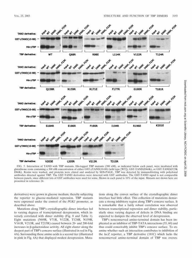
Mutations along TBP’s concave surface decrease TBP-TAND interactions. A GST-TAND pulldown assay was used tomeasure the interaction of amino acids 10 to 88 of the yeastTAF1 TAND domain with the various TBP mutants. Thisconstruct contains TAND I and II, both of which are requiredfor TBP binding (54). To assess the specificity of binding, twoTAND derivatives were generated, containing the F23K andD66K mutations or only the D66K mutation. The double mu-tant is defective in TBP binding (52). F23 resides within TANDI, and D66 resides within TAND II.
As shown in Fig. 5 and summarized in Table 1 and Fig. 4A,yeast TAND makes contact throughout the concave surface ofTBP, in that mutations along this surface largely disruptedTAND binding. Mutations along TBP’s convex C-terminal stir-rup or along one edge of TBP’s concave surface had little effect
3188 KOU ET AL. MOL. CELL. BIOL.
at Scientific Library, N
CI-F
CR
DC
on April 20, 2009
mcb.asm
.orgD
ownloaded from

(Fig. 4B). Overall there was a remarkable concordance of themutagenesis data with what was predicted from the NMR andmutagenesis data for the Drosophila TAND-TBP complex (65,70). Despite low sequence conservation, yeast TAND appearsto be contacting TBP’s concave surface in a manner similar(but probably not identical) to that for Drosophila TAND I.
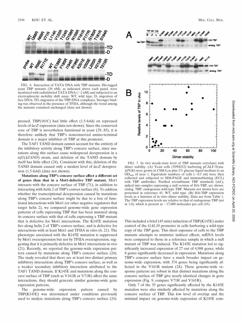
Mutations along TBP’s concave surface decrease TATAbinding. A substantial portion of the dimer interface overlapswith TBP’s DNA binding surface, and dimerization and DNAbinding are competitive events (Fig. 3A). An electrophoreticmobility shift assay was performed to measure TATA bindingby the various mutants (Fig. 6; summarized in Table 1 and Fig.4A). As expected from the structure (48, 50), mutation ofburied or bonded amino acids along the concave surface ofTBP resulted in a loss of DNA binding, while mutation ofother amino acids had little effect. The one exception wasK201, which does not appear to contact DNA in the crystalstructure but is nonetheless important for binding. Similar
observations for this and other equivalent mutations through-out the DNA binding surface of TBP have been previouslyreported (4, 17, 61, 76, 84).
Amino acids along TBP’s crystallographic dimer interfaceare important for growth. As a first step towards assessing thephenotypes of mutations along TBP’s crystallographic dimerinterface, we examined whether such mutants could supportcell viability as the sole source of TBP. Yeast strain YTW22,harboring a deletion of the chromosomal TBP (SPT15) geneand providing wild-type TBP on a Ura-marked plasmid, wasused to exchange the wild-type TBP with the mutant TBPs byplasmid shuffling. As summarized in Table 1 and Fig. 4A,mutations along the concave surface of TBP generally failed tosupport growth. In general, mutations on the convex portion ofthe dimer interface had little effect, with the exception of theTFIIB-defective E186R mutation, which failed to supportgrowth, and R171E and F177R, which caused slow growth.R171 and F177 have genetic interactions with SPT3 (34).
TABLE 1. Properties of TBP mutants
TBP
Stabilitya
Growthb at:[TBP]c �-Gald Toxicitye
Synthetic toxicity f
TT TF TDTAF1 �TAND
23°C 30°C 37°C � TAF1 � TAF1
Null 0 0 0 0 1 1.9 6 6 4 6Wild type 100 100 100 6 6 6 7 1 2.0 6 6 6 6Q68R 58 90 100 5 6 5 6 5 2.2 4 4 6 4N69R 9 20 20 0 0 0 0.4 190 3.2 2 4 0 2V71R 18 90 5 0 0 0 0.7 81 4.5 2 4 0 2
R98E 23 80 50 4 4 4 4 1 2.1 5 6 6 6L114K 41 10 5 0 0 0 7 7 3.9 6 5 4 5V122R 23 40 10 0 0 0 4 44 2.1 6 6 2 4T124R 25 50 10 0 0 0 4 31 2.7 3 3 1 3Q158R 37 90 100 6 6 6 7 8 2.1 6 6 4 6
N159R 14 5 10 0 0 0 0.4 120 3.6 3 3 0 3V161R 8 5 5 0 0 0 0.4 150 4.8 3 3 0 3R171E 34 90 100 3 4 2 4 1 2.0 6 6 6 6F177R 83 130 100 4 4 4 9 1 2.0 6 6 4 6G180R 66 220 100 6 6 4 7 4 1.9 6 6 4 6
T181R 61 110 100 6 6 6 11 1 2.0 6 6 4 6S184R 58 100 100 6 6 4 10 3 2.0 6 6 4 6E186R 66 130 100 0 0 0 8 1 2.1 6 6 1 5F190R 6 5 5 0 0 0 1.1 3 2.3 6 6 4 6I194R 10 5 10 2 2 2 1.2 2 2.2 6 6 4 6
R196E 4 30 5 4 4 5 8 4 4.1 3 3 3 4K201E 23 90 10 4 6 6 7 3 2.0 6 6 6 6V203E 10 10 10 5 5 4 5 6 2.0 6 6 6 6L205R 5 20 10 4 6 4 1.6 3 3.1 6 6 3 6V213R 5 10 10 0 0 0 0.2 110 3.5 3 3 1 3
T215R 9 20 20 0 0 0 0.9 23 2.9 4 4 1 3
a TT, relative dimer stability (from Fig. 3C); TF, relative TBP-TAND stability (from Fig. 5 and reference 24); TD, relative TBP-TATA stability (from Fig. 6). Dataare averaged from multiple repeats (scale of 0 to 100; wild-type value 100).
b Relative colony size (scale of 0 to 6; null value 0 and wild-type value 6) on CSM-Leu solid agar plates at the indicated temperature, after shuffling out wild-typeTBP. Relative values were confirmed by measuring doubling times in liquid medium.
c Relative concentration of HA-tagged TBP in vivo (endogenous untagged TBP value 1.0), as measured in Fig. 7.d Relative �-galactosidase activity (null TBP value 1), as measured in Fig. 8.e Measured in terms of doubling time (hours) for a strain (YPH252) harboring wild-type TBP, as described in Fig. 10.f Relative colony size (0, no growth after 4 days; 1, pinpoint colonies after 4 days; 2, pinpoint colonies after 3 days; 3, pinpoint colonies after 2 days; 4, pinpoint colonies
after 1 day; 5, colonies slightly smaller than those with wild-type TBP after 1 day; 6, colonies the same size as those with wild-type TBP) on CSM-Trp-Ura-Leu solidagar plates at 37°C in strain Y13.2 with the indicated TAF1 alleles [TAF1, wild type, �TAND, TAF1(�10–88)], as described in Fig. 11. Essentially identical results wereobtained at 30 and 23°C (data not shown).
VOL. 23, 2003 STRUCTURE AND FUNCTION OF TBP DIMERS 3189
at Scientific Library, N
CI-F
CR
DC
on April 20, 2009
mcb.asm
.orgD
ownloaded from
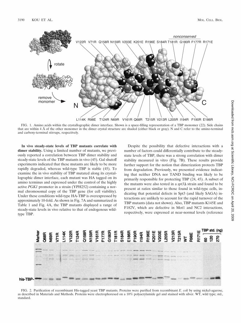
In vivo steady-state levels of TBP mutants correlate withdimer stability. Using a limited number of mutants, we previ-ously reported a correlation between TBP dimer stability andsteady-state levels of the TBP mutants in vivo (45). Gal shutoffexperiments indicated that these mutants are likely to be morerapidly degraded, whereas wild-type TBP is stable (45). Toexamine the in vivo stability of TBP mutated along its crystal-lographic dimer interface, each mutant was HA tagged on itsamino terminus and expressed under the control of the highlyactive PGK1 promoter in a strain (YPH252) containing a nor-mal chromosomal copy of the TBP gene (for cell viability).Under these conditions wild-type HA-TBP is overexpressed byapproximately 10-fold. As shown in Fig. 7A and summarized inTable 1 and Fig. 4A, the TBP mutants displayed a range ofsteady-state levels in vivo relative to that of endogenous wild-type TBP.
Despite the possibility that defective interactions with anumber of factors could differentially contribute to the steady-state levels of TBP, there was a strong correlation with dimerstability measured in vitro (Fig. 7B). These results providefurther support for the notion that dimerization protects TBPfrom degradation. Previously, we presented evidence indicat-ing that neither DNA nor TAND binding was likely to beprimarily responsible for protecting TBP (24, 45). A subset ofthe mutants were also tested in a spt3� strain and found to bepresent at ratios similar to those found in wild-type cells, in-dicating that potential defects in Spt3 (and likely SAGA) in-teractions are unlikely to account for the rapid turnover of theTBP mutants (data not shown). Also, TBP mutants K145E andF182V, which are defective in Mot1 and NC2 interactions,respectively, were expressed at near-normal levels (reference
FIG. 1. Amino acids within the crystallographic dimer interface. Shown is a space-filling representation of a TBP monomer (22). Side chainsthat are within 4 A of the other monomer in the dimer crystal structure are shaded (either black or gray). N and C refer to the amino-terminaland carboxy-terminal stirrups, respectively.
FIG. 2. Purification of recombinant His-tagged yeast TBP mutants. Proteins were purified from recombinant E. coli by using nickel-agarose,as described in Materials and Methods. Proteins were electrophoresed on a 10% polyacrylamide gel and stained with silver. WT, wild type; std.,standard.
3190 KOU ET AL. MOL. CELL. BIOL.
at Scientific Library, N
CI-F
CR
DC
on April 20, 2009
mcb.asm
.orgD
ownloaded from
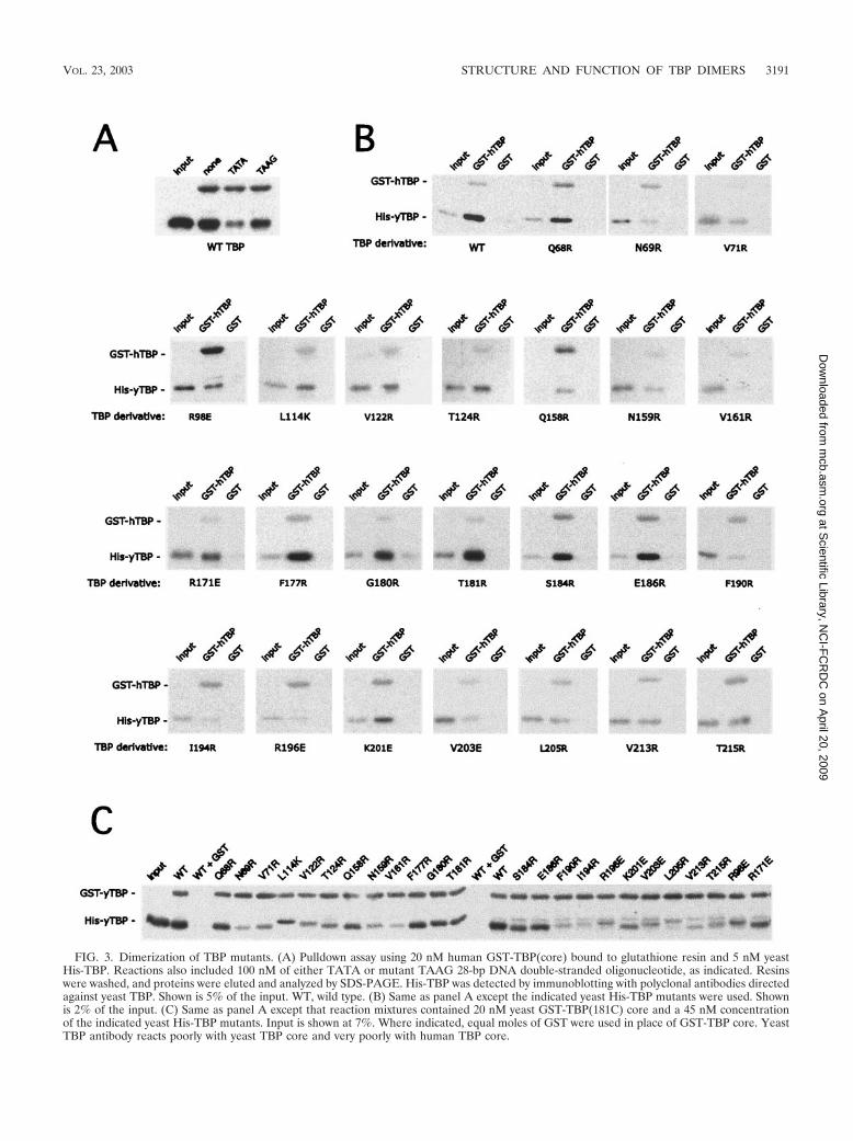
FIG. 3. Dimerization of TBP mutants. (A) Pulldown assay using 20 nM human GST-TBP(core) bound to glutathione resin and 5 nM yeastHis-TBP. Reactions also included 100 nM of either TATA or mutant TAAG 28-bp DNA double-stranded oligonucleotide, as indicated. Resinswere washed, and proteins were eluted and analyzed by SDS-PAGE. His-TBP was detected by immunoblotting with polyclonal antibodies directedagainst yeast TBP. Shown is 5% of the input. WT, wild type. (B) Same as panel A except the indicated yeast His-TBP mutants were used. Shownis 2% of the input. (C) Same as panel A except that reaction mixtures contained 20 nM yeast GST-TBP(181C) core and a 45 nM concentrationof the indicated yeast His-TBP mutants. Input is shown at 7%. Where indicated, equal moles of GST were used in place of GST-TBP core. YeastTBP antibody reacts poorly with yeast TBP core and very poorly with human TBP core.
VOL. 23, 2003 STRUCTURE AND FUNCTION OF TBP DIMERS 3191
at Scientific Library, N
CI-F
CR
DC
on April 20, 2009
mcb.asm
.orgD
ownloaded from

24 and data not shown), indicating that Mot1 and NC2 werenot primarily responsible for preventing TBP turnover.
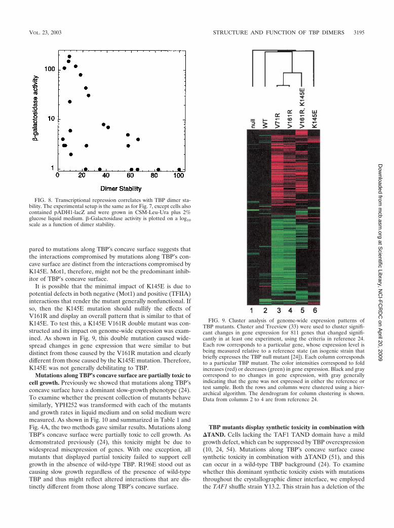
Mutations along TBP’s concave surface cause transcrip-tional derepression. Previously, we and others identified aminoacids along the concave surface of TBP that when mutated leadto transcriptional derepression (15, 21, 24, 36, 45). The level of
derepression correlated with dimer instability (24, 45). To de-termine whether a similar correlation held with a more com-plete set of mutants, we employed the same system, whichincluded the use of a lacZ reporter gene fused to the core(enhancerless) ADH1 promoter (15). Artificial Gal4p bindingsites are located upstream of the promoter. Cells (YPH252
FIG. 4. Summary of the properties of mutations along TBP’s crystallographic dimer interface. (A) Shown are space-filling models of TBPmonomers in the orientation shown in Fig. 1. N and C refer to the amino-terminal and carboxy-terminal stirrups, respectively. Each model is asummary derived from Table 1, in which each of the 24 tested amino acid side chains are color coded if mutations at these sites cause severe (red),moderate (pink), or no (gray) deviations from wild-type behavior. Since V71E but not V71R is defective for TAND binding (24), this residue wascolored red. (B) The NMR structure of the Drosophila TAF1 TAND I backbone (from amino acid 19 to 77) is shown in the context of a space-fillingrepresentation of yeast TBP amino acid side chains that were used in this study (65). The color scheme is the same as that used in panel A. Theview is that of panel A but rotated forward such that the TBP stirrups point inward and the convex seat of the saddle is facing outward.
3192 KOU ET AL. MOL. CELL. BIOL.
at Scientific Library, N
CI-F
CR
DC
on April 20, 2009
mcb.asm
.orgD
ownloaded from
derivatives) were grown in glucose medium, thereby subjectingthe reporter to glucose-mediated repression. TBP mutantswere expressed under the control of the PGK1 promoter, asdescribed above.
Mutations along TBP’s crystallographic dimer interface ledto varying degrees of transcriptional derepression, which in-versely correlated with dimer stability (Fig. 8 and Table 1).Eight mutations (N69R, V71R, V122R, T124R, N159R,V161R, V213R, and T215R) caused between 20- and 200-foldincreases in �-galactosidase activity. All eight cluster along thedeepest part of TBP’s concave surface (illustrated in red in Fig.4A). Surrounding these amino acids is a second tier (illustratedin pink in Fig. 4A) that displayed modest derepression. Muta-
tions along the convex surface of the crystallographic dimerinterface had little effect. This collection of mutations demar-cate a strong inhibitory region along TBP’s concave surface. Itis remarkable that a fairly robust correlation was observedbetween transcriptional repression and dimer stability, partic-ularly since varying degrees of defects in DNA binding areexpected to dampen the observed level of derepression.
TBP’s nonconserved amino-terminal domain has been im-plicated as an inhibitor of TBP-TATA interactions (55, 60) andthus could conceivably inhibit TBP’s concave surface. To ex-amine whether such an interaction contributes to inhibition ofthe lacZ reporter, a TBP derivative (181C) which lacks thenonconserved amino-terminal domain of TBP was overex-
FIG. 5. Interaction of TAND with TBP mutants. His-tagged TBP mutants (300 nM), as indicated below each panel, were incubated withglutathione resin containing a 300 nM concentration of either GST-yTAND(10-88) (wild type [WT]), GST-TAND(D66K), or GST-TAND(F23KD66K). Resins were washed, and proteins were eluted and analyzed by SDS-PAGE. TBP was detected by immunoblotting with polyclonalantibodies directed against TBP. The GST-TAND derivatives were detected with GST antibodies. The GST-TAND signal is not comparablebetween panels, since different lots of GST antibodies were used for some. Shown in each panel is 10% of the input. Mutants not shown here arepresented in reference 24.
VOL. 23, 2003 STRUCTURE AND FUNCTION OF TBP DIMERS 3193
at Scientific Library, N
CI-F
CR
DC
on April 20, 2009
mcb.asm
.orgD
ownloaded from
pressed. TBP(181C) had little effect (1.5-fold) on repressedlevels of lacZ expression (data not shown). Since the conservedcore of TBP is nevertheless functional in yeast (29, 85), it istherefore unlikely that TBP’s nonconserved amino-terminaldomain is a major inhibitor of TBP at this promoter.
The TAF1 TAND domain cannot account for the entirety ofthe inhibitory activity along TBP’s concave surface, since mu-tations along this surface cause widespread derepression in ataf1(�TAND) strain, and deletion of the TAND domain byitself has little effect (24). Consistent with this, deletion of theTAND domain caused only a modest level of lacZ derepres-sion (1.5-fold) (data not shown).
Mutations along TBP’s concave surface affect a different setof genes than that in a Mot1-defective TBP mutant. Mot1interacts with the concave surface of TBP (71), in addition tointeracting with helix 2 of TBP’s convex surface (6). To addresswhether the transcriptional derepression caused by mutationsalong TBP’s concave surface might be due to a loss of func-tional interactions with Mot1 (or other negative regulators thattarget helix 2), we compared genome-wide gene expressionpatterns of cells expressing TBP that has been mutated alongits concave surface with that of cells expressing a TBP mutantthat is defective for Mot1 interactions. The K145E mutationlies along helix 2 of TBP’s convex surface, and is defective forinteractions with at least Mot1 and TFIIA in vitro (6, 21). Thephenotype associated with the K145E mutation is suppressedby Mot1 overexpression but not by TFIIA overexpression, sug-gesting that it is primarily defective in Mot1 interactions in vivo(21). Recently, we reported the genome-wide expression pat-tern caused by mutations along TBP’s concave surface (24).The study revealed that there are at least two distinct primaryinhibitory interactions along TBP’s concave surface, as well asa weaker secondary inhibitory interaction attributed to theTAF1 TAND domain. If K145E and mutations along the con-cave surface of TBP (such as V161R or V71R) affect the sameinteractions, they should generate similar genome-wide geneexpression patterns.
The genome-wide expression pattern caused byTBP(K145E) was determined under conditions previouslyused to analyze mutations along TBP’s concave surface (24).
This included a brief (45-min) induction of TBP(K145E) undercontrol of the GAL10 promoter in cells harboring a wild-typecopy of the TBP gene. This short exposure of cells to the TBPmutants attempts to minimize indirect effects. mRNA levelswere compared to those in a reference sample in which a nullmutant of TBP was induced. The K145E mutation led to sig-nificantly increased expression of 27 out of 4,988 genes, while8 genes significantly decreased in expression. Mutations alongTBP’s concave surface have a much broader impact on ge-nome-wide expression, with 374 genes being significantly af-fected in the V161R mutant (24). These genome-wide re-sponse patterns are robust in that distinct mutations along theconcave surface of TBP give nearly identical changes in geneexpression (Fig. 9, compare V71R and V161R).
Only 7 of the 35 genes significantly affected by the K145Emutation were also similarly affected by mutations along theconcave surface of TBP. This low level of overlap and theminimal impact on genome-wide expression of K145E com-
FIG. 6. Interaction of TATA DNA with TBP mutants. His-taggedyeast TBP mutants (30 nM), as indicated above each panel, wereincubated with radiolabeled TATA DNA (�2 nM) and subjected to anelectrophoretic mobility shift assay. WT, wild type; D, migration offree DNA; TD, migration of the TBP-DNA complexes. Stronger bind-ing was observed in the presence of TFIIA, although the trend amongthe mutants remained unchanged (data not shown).
FIG. 7. In vivo steady-state level of TBP mutants correlates withdimer stability. (A) Yeast cells (YPH252) harboring pCALF-T(xxx-)(PGK) were grown in CSM-Leu plus 2% glucose liquid medium to anOD600 of near 1. Equivalent numbers of cells (�0.5 ml) were thencollected and subjected to SDS-PAGE and immunoblotting (ECL)with TBP antibodies. Purified recombinant TBP standards (std.),spiked into samples expressing a null version of HA-TBP, are shown.endog. TBP, endogenous wild-type TBP. Mutants not shown here arepresented in reference 45. WT, wild type. (B) HA-TBP expressionlevels as a function of in vitro dimer stability. Data are from Table 1.The TBP expression levels are relative to that of endogenous TBP (setat 1.0), which is present at �17,000 molecules per cell (45).
3194 KOU ET AL. MOL. CELL. BIOL.
at Scientific Library, N
CI-F
CR
DC
on April 20, 2009
mcb.asm
.orgD
ownloaded from
pared to mutations along TBP’s concave surface suggests thatthe interactions compromised by mutations along TBP’s con-cave surface are distinct from the interactions compromised byK145E. Mot1, therefore, might not be the predominant inhib-itor of TBP’s concave surface.
It is possible that the minimal impact of K145E is due topotential defects in both negative (Mot1) and positive (TFIIA)interactions that render the mutant generally nonfunctional. Ifso, then the K145E mutation should nullify the effects ofV161R and display an overall pattern that is similar to that ofK145E. To test this, a K145E V161R double mutant was con-structed and its impact on genome-wide expression was exam-ined. As shown in Fig. 9, this double mutation caused wide-spread changes in gene expression that were similar to butdistinct from those caused by the V161R mutation and clearlydifferent from those caused by the K145E mutation. Therefore,K145E was not generally debilitating to TBP.
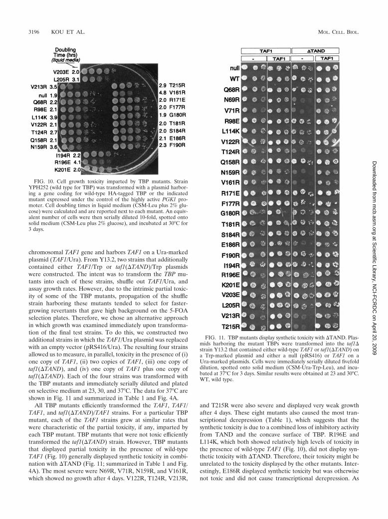
Mutations along TBP’s concave surface are partially toxic tocell growth. Previously we showed that mutations along TBP’sconcave surface have a dominant slow-growth phenotype (24).To examine whether the present collection of mutants behavesimilarly, YPH252 was transformed with each of the mutantsand growth rates in liquid medium and on solid medium weremeasured. As shown in Fig. 10 and summarized in Table 1 andFig. 4A, the two methods gave similar results. Mutations alongTBP’s concave surface were partially toxic to cell growth. Asdemonstrated previously (24), this toxicity might be due towidespread misexpression of genes. With one exception, allmutants that displayed partial toxicity failed to support cellgrowth in the absence of wild-type TBP. R196E stood out ascausing slow growth regardless of the presence of wild-typeTBP and thus might reflect altered interactions that are dis-tinctly different from those along TBP’s concave surface.
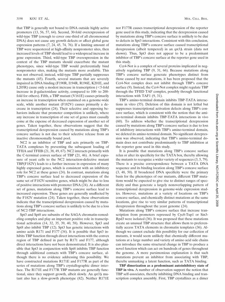
TBP mutants display synthetic toxicity in combination with�TAND. Cells lacking the TAF1 TAND domain have a mildgrowth defect, which can be suppressed by TBP overexpression(10, 24, 54). Mutations along TBP’s concave surface causesynthetic toxicity in combination with �TAND (51), and thiscan occur in a wild-type TBP background (24). To examinewhether this dominant synthetic toxicity exists with mutationsthroughout the crystallographic dimer interface, we employedthe TAF1 shuffle strain Y13.2. This strain has a deletion of the
FIG. 8. Transcriptional repression correlates with TBP dimer sta-bility. The experimental setup is the same as for Fig. 7, except cells alsocontained pADH1-lacZ and were grown in CSM-Leu-Ura plus 2%glucose liquid medium. �-Galactosidase activity is plotted on a log10scale as a function of dimer stability.
FIG. 9. Cluster analysis of genome-wide expression patterns ofTBP mutants. Cluster and Treeview (33) were used to cluster signifi-cant changes in gene expression for 811 genes that changed signifi-cantly in at least one experiment, using the criteria in reference 24.Each row corresponds to a particular gene, whose expression level isbeing measured relative to a reference state (an isogenic strain thatbriefly expresses the TBP null mutant [24]). Each column correspondsto a particular TBP mutant. The color intensities correspond to foldincreases (red) or decreases (green) in gene expression. Black and graycorrespond to no changes in gene expression, with gray generallyindicating that the gene was not expressed in either the reference ortest sample. Both the rows and columns were clustered using a hier-archical algorithm. The dendrogram for column clustering is shown.Data from columns 2 to 4 are from reference 24.
VOL. 23, 2003 STRUCTURE AND FUNCTION OF TBP DIMERS 3195
at Scientific Library, N
CI-F
CR
DC
on April 20, 2009
mcb.asm
.orgD
ownloaded from
chromosomal TAF1 gene and harbors TAF1 on a Ura-markedplasmid (TAF1/Ura). From Y13.2, two strains that additionallycontained either TAF1/Trp or taf1(�TAND)/Trp plasmidswere constructed. The intent was to transform the TBP mu-tants into each of these strains, shuffle out TAF1/Ura, andassay growth rates. However, due to the intrinsic partial toxic-ity of some of the TBP mutants, propagation of the shufflestrain harboring these mutants tended to select for faster-growing revertants that gave high background on the 5-FOAselection plates. Therefore, we chose an alternative approachin which growth was examined immediately upon transforma-tion of the final test strains. To do this, we constructed twoadditional strains in which the TAF1/Ura plasmid was replacedwith an empty vector (pRS416/Ura). The resulting four strainsallowed us to measure, in parallel, toxicity in the presence of (i)one copy of TAF1, (ii) two copies of TAF1, (iii) one copy oftaf1(�TAND), and (iv) one copy of TAF1 plus one copy oftaf1(�TAND). Each of the four strains was transformed withthe TBP mutants and immediately serially diluted and platedon selective medium at 23, 30, and 37°C. The data for 37°C areshown in Fig. 11 and summarized in Table 1 and Fig. 4A.
All TBP mutants efficiently transformed the TAF1, TAF1/TAF1, and taf1(�TAND)/TAF1 strains. For a particular TBPmutant, each of the TAF1 strains grew at similar rates thatwere characteristic of the partial toxicity, if any, imparted byeach TBP mutant. TBP mutants that were not toxic efficientlytransformed the taf1(�TAND) strain. However, TBP mutantsthat displayed partial toxicity in the presence of wild-typeTAF1 (Fig. 10) generally displayed synthetic toxicity in combi-nation with �TAND (Fig. 11; summarized in Table 1 and Fig.4A). The most severe were N69R, V71R, N159R, and V161R,which showed no growth after 4 days. V122R, T124R, V213R,
and T215R were also severe and displayed very weak growthafter 4 days. These eight mutants also caused the most tran-scriptional derepression (Table 1), which suggests that thesynthetic toxicity is due to a combined loss of inhibitory activityfrom TAND and the concave surface of TBP. R196E andL114K, which both showed relatively high levels of toxicity inthe presence of wild-type TAF1 (Fig. 10), did not display syn-thetic toxicity with �TAND. Therefore, their toxicity might beunrelated to the toxicity displayed by the other mutants. Inter-estingly, E186R displayed synthetic toxicity but was otherwisenot toxic and did not cause transcriptional derepression. As
FIG. 10. Cell growth toxicity imparted by TBP mutants. StrainYPH252 (wild type for TBP) was transformed with a plasmid harbor-ing a gene coding for wild-type HA-tagged TBP or the indicatedmutant expressed under the control of the highly active PGK1 pro-moter. Cell doubling times in liquid medium (CSM-Leu plus 2% glu-cose) were calculated and are reported next to each mutant. An equiv-alent number of cells were then serially diluted 10-fold, spotted ontosolid medium (CSM-Leu plus 2% glucose), and incubated at 30°C for3 days.
FIG. 11. TBP mutants display synthetic toxicity with �TAND. Plas-mids harboring the mutant TBPs were transformed into the taf1�strain Y13.2 that contained either wild-type TAF1 or taf1(�TAND) ona Trp-marked plasmid and either a null (pRS416) or TAF1 on aUra-marked plasmids. Cells were immediately serially diluted fivefolddilution, spotted onto solid medium (CSM-Ura-Trp-Leu), and incu-bated at 37°C for 3 days. Similar results were obtained at 23 and 30°C.WT, wild type.
3196 KOU ET AL. MOL. CELL. BIOL.
at Scientific Library, N
CI-F
CR
DC
on April 20, 2009
mcb.asm
.orgD
ownloaded from

E186 interacts with TFIIB, we suspect that the E186R syn-thetic toxicity reflects a mechanism that is distinct from that ofthe others. In general, the synthetic toxicity, while dominant towild-type TBP, was recessive to wild-type TAF1.
DISCUSSION
TBP’s concave surface interacts with multiple factors, whichinclude DNA, the TAND domain of TAF1, Mot1, and a sec-ond molecule of TBP to form a dimer. Here, we have mappedthree of these interactions by creating 24 point mutationsthroughout TBP’s crystallographic dimer interface. The invitro and in vivo properties of these mutants are summarized inFig. 4. The data for all three interactions are in general agree-ment with the NMR-crystallographic structures of these orrelated complexes.
The majority of the mutants have similar phenotypes in vivo.About half do not support cell viability when provided as thesole source of TBP, while several others support weak growth.When the TBP mutants were expressed in a wild-type TBPstrain, their steady-state levels correlated with dimer stability.A similar correlation was observed for a subset of the mutantsexamined in a taf1(�TAND) strain, indicating that defects inTAND interactions were not responsible (24). Six TBP mu-tants that were severely defective in DNA binding in vitro werepresent at high steady-state levels in vivo, which suggests thatdefects in DNA binding were not responsible for the increasedturnover of the TBP mutants. These results provide furthersupport for our previous suggestion that dimerization protectsTBP from degradation in vivo (45).
A set of eight mutations that cluster at the center of TBP’scavity caused high levels of transcriptional derepression. Ad-ditional mutations surrounding this cluster caused modest de-repression. The amount of derepression in vivo correlated withdimer instability measured in vitro. In general, the more severedimerization mutants were partially toxic to cell growth anddisplayed synthetic toxicity in combination with �TAND.Taken together, these data define a major inhibitory patchalong TBP’s concave surface. This region is distinct from apreviously defined NC2 inhibitory surface on TBP’s convexsurface (21). Genome-wide expression profiling, presentedhere and elsewhere (24), indicates that the TAF1 TAND do-main and Mot1 are unlikely to be the primary inhibitors tar-geting TBP’s concave surface. These factors might make minorcontributions. As presented in more detail below, other poten-tial inhibitory mechanisms cannot readily account for the in-hibition of TBP’s concave surface. However, the data are fullyconsistent with a model in which TBP is prevented from bind-ing to accessible promoters by homodimerization. TBP recruit-ment to promoters, which is important for transcription com-plex assembly, would require dimers to dissociate intomonomers.
Potential alternative sources of inhibition along TBP’s con-cave surface. TBP might be inhibited by a number of factors,including the TAF1 TAND domain, Mot1, NC2, Spt3/Spt8, theCcr4-Not complex, TBP’s amino terminal domain, and TBPhomodimerization. Alternatively, TBP could be sequestered atnonpromoter chromosomal sites through direct contact withDNA. The transcriptional derepression caused by mutationsalong TBP’s concave surface could in principle arise from
defects in one or more of these interactions. It is also possiblethat the mutations create novel positive interactions that leadto derepression. Below, we discuss each of these possibilities inlight of the available evidence.
The TAF1 TAND I domain interacts with the concave sur-face of TBP and inhibits TATA binding in vitro (11, 24, 52, 53,70). Therefore, transcriptional derepression observed with mu-tations along TBP’s concave surface could be due to a loss ofTAND interactions. However, several observations indicatethat TAND is not the primary inhibitor. (i) Deletion of theTAND domain does not lead to derepression of the promoterused in this study and has little impact genome wide (24). (ii)Transcriptional derepression caused by mutations along TBP’sconcave surface is unabated in a strain with the TAND domaindeleted, indicating that TAND is not required for transcrip-tional derepression. However, deletion of TAND enhances thelevel of derepression observed with the TBP mutants (24). (iii)Despite the observation that mutations along TBP’s concavesurface impair TAND binding, many of the same mutations donot cause defects in binding full-length TAF1 in yeast crudeextracts (24). These findings indicate that the transcriptionalderepression arising from mutations along TBP’s concave sur-face is not caused solely or predominantly by a loss of TANDinteractions. TAND does play an inhibitory role, but it appearsto be secondary to another inhibitor.
Mot1 uses the energy of ATP hydrolysis to dissociate TBP-DNA complexes (7, 8, 23). Mot1 and TFIIA interact with helix2 on TBP’s convex surface (1, 6, 18, 21, 59). The K145E mu-tation on helix 2 eliminates binding (21). In vivo, phenotypesassociated with K145E are suppressed by Mot1 overexpressionbut not by TFIIA overexpression, which suggests that K145E isprimarily defective in Mot1 interactions (21). Mot1 might alsointeract with TBP’s concave surface (71). Therefore, it is plau-sible that the transcriptional derepression caused by mutationsalong TBP’s concave surface is due to defects in Mot1 inter-actions. A number of observations render this possibility un-likely. First, mutations along TBP’s concave surface do notshow defects in Mot1 binding in coimmunoprecipitation assaysfrom yeast whole-cell extracts (supplement to reference 24).Second, genome-wide expression analysis reveals that genesthat increase in expression with K145E are largely distinct fromthose caused by mutations along TBP’s concave surface. Inaddition, the genome-wide expression pattern of a helix 2 con-cave-surface double mutant (K145E V161R) was approxi-mately additive to the effects of the single mutants. The generallack of overlapping effects of the individual mutants and theadditive effects of the double mutant suggest that the twosurfaces primarily affect different processes.
It has been suggested that TBP is sequestered at high-affinitynonpromoter chromosomal sites in vivo and is liberated byMot1 (28, 68). Since our experiments with the TBP mutantsare performed in a MOT1 strain, TBP should not be seques-tered. However, if Mot1 is inefficient, then there might becompetition between promoter and nonpromoter sites for TBPbinding. Conceivably, mutations along the concave DNA bind-ing surface of TBP could elicit the same effect as Mot1 byselectively destabilizing high-affinity nonproductive TBP-DNAcomplexes, allowing the mutants to assemble productively atpromoters. Several observation are inconsistent with this pos-sibility. First, chromatin immunoprecipitation studies indicate
VOL. 23, 2003 STRUCTURE AND FUNCTION OF TBP DIMERS 3197
at Scientific Library, N
CI-F
CR
DC
on April 20, 2009
mcb.asm
.orgD
ownloaded from
that TBP is generally not bound to DNA outside highly activepromoters (13, 56, 57, 64). Second, 30-fold overexpression ofwild-type TBP (enough to cover one-third of all chromosomalDNA) does not cause any growth defects or affect global geneexpression patterns (7, 24, 45, 74, 76). If a limiting amount ofTBP were sequestered at high-affinity nonpromoter sites, thenincreased levels of TBP should lead to a widespread increase ingene expression. Third, wild-type TBP overexpression in thecontext of the TBP mutants should exacerbate the mutantphenotypes, since wild-type TBP would preferentially bindnonpromoter sites, making the mutants more available. Thiswas not observed; instead, wild-type TBP partially suppressesthe mutants (45). Fourth, several mutants that are severelyimpaired in DNA binding (F190R, I194R, R196E, K201E, andL205R) cause only a modest increase in transcription (�5-foldincrease in �-galactosidase activity, compared to 100- to 200-fold for others). Fifth, a TBP mutant (V161E) causes primarilyan increase in transcription when examined on a genome-widescale, while another mutant (F182V) causes primarily a de-crease in transcription (24). Their selective and opposing be-haviors indicate that promoter competition is unlikely, whereany increase in transcription of one set of genes must causallycome at the expense of decreased expression of another set ofgenes. Taken together, these observations suggest that thetranscriptional derepression caused by mutations along TBP’sconcave surface is not due to their selective release from aninactive chromosomally bound pool.
NC2 is an inhibitor of TBP and acts primarily on TBP-TATA complexes by preventing the subsequent loading ofTFIIA and TFIIB (21, 38, 49, 67). NC2 interacts primarily withthe C-terminal convex surface of TBP (21, 46). A brief expo-sure of yeast cells to the NC2 interaction-defective mutantTBP(F182V) leads to a further increase in expression of manyhighly expressed genes, which is consistent with an inhibitoryrole for NC2 at these genes (24). In contrast, mutations alongTBP’s concave surface lead to decreased expression of thesame set of F182V-sensitive genes, which might be due to lossof positive interactions with promoter DNA (24). At a differentset of genes, mutations along TBP’s concave surface lead toincreased expression. These genes are generally unaffected bythe F182V mutation (24). Taken together, these observationsindicate that the transcriptional derepression caused by muta-tions along TBP’s concave surface is unlikely to be due to a lossof NC2-TBP interactions.
Spt3 and Spt8 are subunits of the SAGA chromatin-remod-eling complex and play an important positive role in transcrip-tional activation (14, 32, 35, 39, 40, 58). However, Spt3 andSpt8 also inhibit TBP (12). Spt3 has genetic interactions withamino acids R171 and F177 (34). It is possible that Spt3 in-hibits TBP function through direct interactions with the convexregion of TBP defined in part by R171 and F177, althoughdirect interactions have not been demonstrated. It is also plau-sible that Spt3 in conjunction with Spt8 inhibits TBP functionthrough additional contacts with TBP’s concave surface, al-though there is no evidence addressing this possibility. Wehave constructed mutations R171E and F177R as part of theseries of mutations along TBP’s crystallographic dimer inter-face. The R171E and F177R TBP mutants are generally func-tional, since they support growth, albeit slowly. An spt3� mu-tant also has a slow-growth phenotype (82). Neither R171E
nor F177R causes transcriptional derepression of the reportergene used in this study, indicating that the derepression causedby mutations along TBP’s concave surface is unlikely to be dueto defects in Spt3 interactions. Consistent with this conclusion,mutations along TBP’s concave surface caused transcriptionalderepression (albeit tempered) in an spt3� strain (data notshown). Thus, Spt3 does not appear to be a predominantinhibitor of TBP’s concave surface at the reporter gene used inthis study.
Ccr4-Not is a complex of several proteins implicated in neg-atively regulating TBP (9, 31, 66). Because mutations alongTBP’s concave surface generate phenotypes distinct fromthose caused by not mutations, it has been proposed that theCcr4-Not complex does not inhibit through TBP’s concavesurface (9). Instead, the Ccr4-Not complex might regulate TBPthrough the TFIID TAF complex, possibly through functionalinteractions with TAF1 (9, 31).
TBP’s amino-terminal domain inhibits TBP-TATA interac-tions in vitro (55). Deletion of this domain is not lethal butsuppresses transcriptional activation defects along TBP’s con-cave surface, which is consistent with the notion that the ami-no-terminal domain inhibits TBP-TATA interactions in vivo(60). To address whether the transcriptional derepressioncaused by mutations along TBP’s concave surface is due to lossof inhibitory interactions with TBP’s amino-terminal domain,we deleted its amino-terminal domain. No significant derepres-sion was observed, indicating that TBP’s amino-terminal do-main does not contribute predominantly to TBP inhibition atthe reporter gene used in this study.
It is possible that mutations along TBP’s concave surfacerelax or alter its specificity for the TATA box, thereby allowingthe mutants to recognize a wider variety of sequences (3, 5, 79).There is a precise correspondence between a TATA DNAsequence and its binding location along TBP’s concave surface(3, 48, 50). If broadened DNA specificity were the primarybasis for the phenotypes of our mutants, different TBP muta-tions would be expected to give rise to distinct ranges of spec-ificity and thus generate a largely nonoverlapping pattern oftranscriptional derepression in genome-wide expression stud-ies. However, mutations at a variety of locations on TBP’sconcave surface, and chemically distinct mutations at the samelocations, give rise to very similar patterns of transcriptionalderepression throughout the yeast genome (24).
Mutations along TBP’s concave surface that increase tran-scription from promoters repressed by Cyc8-Tup1 or Sin3-Rpd3 were isolated (36). It was proposed that these mutationscreate an unusual TBP structure that allows them to preferen-tially access TATA elements in chromatin templates (36). Al-though we cannot exclude this possibility for our collection ofmutants, it would seem unlikely that chemically different mu-tations at a large number and variety of amino acid side chainscan introduce the same structural change in TBP to produce anovel function which can act on hundreds of genes throughoutthe genome. A more parsimonious explanation is that suchmutations prevent an inhibitor from associating with TBP,thereby unmasking a latent function, such as TATA binding.
TBP dimerization as a predominant autoinhibitory state ofTBP in vivo. A number of observation support the notion thatTBP self-associates, thereby inhibiting DNA binding and tran-scription complex assembly. First, TBP crystallizes as a dimer
3198 KOU ET AL. MOL. CELL. BIOL.
at Scientific Library, N
CI-F
CR
DC
on April 20, 2009
mcb.asm
.orgD
ownloaded from
in which the DNA binding and dimerization interfaces overlap(22, 69). Second, analytical ultracentrifugation (20, 30), fluo-rescence anisotropy (72), gel filtration (26, 47), glycerol gradi-ents (42, 47), chemical cross-linking (26, 36, 42, 44, 45), andDNA binding kinetics (25) show that TBP specifically self-associates in vitro. Third, several experiments involving chem-ical cross-linking, gel filtration, and pulldown assays indicatethat TFIID dimerizes (81). Fourth, TFIIA, which plays a mul-tifunctional role in removing TBP-targeted inhibitors, specifi-cally accelerates dimer dissociation and DNA binding of TBP-TFIID (27). Fifth, TBP can be cross-linked into dimers insidehuman tissue culture cells and in yeast cells (45, 81). Sixth,mutations along TBP’s crystallographic dimer interface inhibitTBP dimerization and lead to transcription derepression invivo (24, 45; this study). Taken together, these findings suggestthat TBP-TFIID homodimerization plays an important auto-inhibitory role in vivo.
The notion that TBP dimerizes in vivo has been controver-sial. Since yeast TBP dimerizes in vitro with low affinity (20)and since coimmunoprecipitation (20, 78) and electron micros-copy (2, 16, 62) studies have failed to detect TFIID dimers, thephysiological relevance of this interaction has been questioned.From the available evidence, we surmise that yeast TBP, ingeneral, forms weak dimers (and higher-order structures) invitro, whereas human TBP forms stronger dimers. Conditionsused to purify and/or prepare TFIID for electron microscopymight favor a more monomeric configuration. As noted in onestudy on TBP self-association, solution and handling condi-tions may dictate whether monomers or multimers prevail(72). Since in vitro conditions cannot fully recapitulate thehierarchy of competing protein-protein interactions and theiraffinities as they exist within a living cell, it might be erroneousto conclude anything about the propensity for TBP to dimerizeor not dimerize in vivo based solely on the magnitude of an invitro-determined KD value.
Of the 24 amino acids targeted for mutagenesis in this study,eight mutations (N69R, V71R, V122R, T124R, N159R,V161R, V213R, and T215R) stand out as causing extraordi-narily high levels of transcriptional derepression. These eightamino acids form a continuous surface along the deepest partof TBP’s concave surface. Virtually all are buried in the crys-tallographic dimer and are invariant from yeast to humans.Consistent with these properties, mutation of any of theseamino acids to arginine destabilizes dimer formation in vitro.These mutations also destabilize TATA binding and so mightbe unable to achieve their full derepression potential in vivo.Studies by Geisberg and Struhl with a similar set of mutantssuggest that these mutants nevertheless bind promoter DNA invivo (36).
A second set of mutants (Q68R, L114K, Q158R, S184R,F190R, I194R, R196E, K201E, V203E, and L205R) show in-termediate levels of transcriptional derepression. These aminoacids are also identical in yeast and humans. Consistent withtheir intermediate transcriptional output, several show inter-mediate levels of dimer stability and reside near the solvent-exposed periphery of the crystallographic dimer interface.Three of these mutants (F190R, R196E, and L205R) are bur-ied deep within the interface and are highly defective fordimerization. We suspect that their intermediate behavior isdue to relatively greater defects in essential positive interac-
tions, such as DNA binding, compared to the mutants de-scribed above.
Mutation of the three nonconserved amino acids F177,G180, and T181 had little effect on dimerization and causedlittle derepression. These amino acids are not constrained inthe dimer crystal structure. R98E and R171E appeared to bepartially defective for dimerization. However, the crystal struc-ture suggests that these mutations might enhance dimer sta-bility. The pulldown assay, used to measured dimerization, is acompetition between homodimers present in solution and het-erodimers formed on the resin. If homodimer mutants aremore stable and exchange onto the resin more slowly, fewer ofthese mutants would be retained on the resin. E186 residesalong the TFIIB interface, and so mutation of this amino acidis expected to render TBP largely inactive for transcription.Interestingly, this mutation displays dominant synthetic toxicityin combination with deletion of the TAND domain. The basisfor this is not known, although a TBP(E186R)-TATA complexmight impair transcription complex assembly. TAND mightfunction in this regard to assist in removing nonproductiveTBP from DNA.
Yeast and Drosophila TAF1 TAND I domains have similarinterfaces with TBP. Yeast TAND is about half the size ofDrosophila TAND and is poorly conserved. It was thereforesurprising to find that yeast TAND contacts nearly the samesurface along TBP as Drosophila TAND I. Of the 67 aminoacids present in the Drosophila TAND I NMR structure (65),�50 amino acids reside within the concave surface of TBP.Therefore, we expect the yeast TAND I domain to be mini-mally 50 amino acids in size. The yeast TAND I domain,however, has been demarcated at �28 amino acids (52, 54).Moreover, yeast TAND I is only �10 amino acids away fromTAND II, which is believed to interact with helix 2 of TBP’sconvex surface (52). Based on these facts, it is not clear howyeast TAND I can occupy the same region as DrosophilaTAND I yet still provide a sufficient linker to allow TAND IIto bind helix 2 of TBP. One plausible explanation is that helix3 of Drosophila TAND I is not present in yeast TAND I (Fig.4B). This would allow the polypeptide chain to leave TBP’sconcave surface early and provide a sufficient linker for TANDII. In support of this possibility, the TBP amino acids Q158 andK201, which make contact with Drosophila TAND I helix 3,have no effect on yeast TAND binding when mutated. Alsoconsistent with a smaller binding surface, yeast TAND I has aweaker affinity for TBP than Drosophila TAND I (54).
ACKNOWLEDGMENTS
We thank Lata Chitikila, Diane Alexander, and Kevin Halbe fortechnical assistance and members of the Pugh laboratory for helpfuldiscussions.
This work was supported by NIH grant GM59055.
REFERENCES
1. Adamkewicz, J. I., K. E. Hansen, W. A. Prud’homme, J. L. Davis, andJ. Thorner. 2001. High affinity interaction of yeast transcriptional regulator,Mot1, with TATA box-binding protein (TBP). J. Biol. Chem. 276:11883–11894.
2. Andel, F., III, A. G. Ladurner, C. Inouye, R. Tjian, and E. Nogales. 1999.Three-dimensional structure of the human TFIID-IIA-IIB complex. Science286:2153–2156.
3. Arndt, K. M., S. L. Ricupero, D. M. Eisenmann, and F. Winston. 1992.Biochemical and genetic characterization of a yeast TFIID mutant that alterstranscription in vivo and DNA binding in vitro. Mol. Cell. Biol. 12:2372–2382.
VOL. 23, 2003 STRUCTURE AND FUNCTION OF TBP DIMERS 3199
at Scientific Library, N
CI-F
CR
DC
on April 20, 2009
mcb.asm
.orgD
ownloaded from
4. Arndt, K. M., S. Ricupero-Hovasse, and F. Winston. 1995. TBP mutantsdefective in activated transcription in vivo. EMBO J. 14:1490–1497.
5. Arndt, K. M., C. R. Wobbe, S. Ricupero-Hovasse, K. Struhl, and F. Winston.1994. Equivalent mutations in the two repeats of yeast TATA-binding pro-tein confer distinct TATA recognition specificities. Mol. Cell. Biol. 14:3719–3728.
6. Auble, D. T., and S. Hahn. 1993. An ATP-dependent inhibitor of TBPbinding to DNA. Genes Dev. 7:844–856.
7. Auble, D. T., K. E. Hansen, C. G. Mueller, W. S. Lane, J. Thorner, and S.Hahn. 1994. Mot1, a global repressor of RNA polymerase II transcription,inhibits TBP binding to DNA by an ATP-dependent mechanism. Genes Dev.8:1920–1934.
8. Auble, D. T., D. Wang, K. W. Post, and S. Hahn. 1997. Molecular analysis ofthe SNF2/SWI2 protein family member MOT1, an ATP-driven enzyme thatdissociates TATA-binding protein from DNA. Mol. Cell. Biol. 17:4842–4851.
9. Badarinarayana, V., Y. C. Chiang, and C. L. Denis. 2000. Functional inter-action of CCR4-NOT proteins with TATAA-binding protein (TBP) and itsassociated factors in yeast. Genetics 155:1045–1054.
10. Bai, Y., G. M. Perez, J. M. Beechem, and P. A. Weil. 1997. Structure-functionanalysis of TAF130: identification and characterization of a high-affinityTATA-binding protein interaction domain in the N terminus of yeastTAF(II)130. Mol. Cell. Biol. 17:3081–3093.
11. Banik, U., J. M. Beechem, E. Klebanow, S. Schroeder, and P. A. Weil. 2001.Fluorescence-based analyses of the effects of full-length recombinantTAF130p on the interaction of TATA box-binding protein with TATA boxDNA. J. Biol. Chem. 276:49100–49109.
12. Belotserkovskaya, R., D. E. Sterner, M. Deng, M. H. Sayre, P. M. Lieberman,and S. L. Berger. 2000. Inhibition of TATA-binding protein function bySAGA subunits Spt3 and Spt8 at Gcn4-activated promoters. Mol. Cell. Biol.20:634–647.
13. Bhaumik, S. R., and M. R. Green. 2002. Differential requirement of SAGAcomponents for recruitment of TATA-box-binding protein to promoters invivo. Mol. Cell. Biol. 22:7365–7371.
14. Bhaumik, S. R., and M. R. Green. 2001. SAGA is an essential in vivo targetof the yeast acidic activator Gal4p. Genes Dev. 15:1935–1945.
15. Blair, W. S., and B. R. Cullen. 1997. A yeast TATA-binding protein mutantthat selectively enhances gene expression from weak RNA polymerase IIpromoters. Mol. Cell. Biol. 17:2888–2896.
16. Brand, M., C. Leurent, V. Mallouh, L. Tora, and P. Schultz. 1999. Three-dimensional structures of the TAFII-containing complexes TFIID andTFTC. Science 286:2151–2153.
17. Bryant, G. O., L. S. Martel, S. K. Burley, and A. J. Berk. 1996. Radicalmutations reveal TATA-box binding protein surfaces required for activatedtranscription in vivo. Genes Dev. 10:2491–2504.
18. Buratowski, S., and H. Zhou. 1992. Transcription factor IID mutants defec-tive for interaction with transcription factor IIA. Science 255:1130–1132.
19. Burley, S. K., and R. G. Roeder. 1996. Biochemistry and structural biology oftranscription factor IID (TFIID). Annu. Rev. Biochem. 65:769–799.
20. Campbell, K. M., R. T. Ranallo, L. A. Stargell, and K. J. Lumb. 2000.Reevaluation of transcriptional regulation by TATA-binding protein oli-gomerization: predominance of monomers. Biochemistry 39:2633–2638.
21. Cang, Y., D. T. Auble, and G. Prelich. 1999. A new regulatory domain on theTATA-binding protein. EMBO J. 18:6662–6671.
22. Chasman, D. I., K. M. Flaherty, P. A. Sharp, and R. D. Kornberg. 1993.Crystal structure of yeast TATA-binding protein and model for interactionwith DNA. Proc. Natl. Acad. Sci. USA 90:8174–8178.
23. Chicca, J. J., II, D. T. Auble, and B. F. Pugh. 1998. Cloning and biochemicalcharacterization of TAF-172, a human homolog of yeast Mot1. Mol. Cell.Biol. 18:1701–1710.
24. Chitikila, C., K. L. Huisinga, J. D. Irvin, M. Mitra, and B. F. Pugh. 2002.Interplay of TBP inhibitors in global transcriptional control. Mol. Cell 10:871–882.
25. Coleman, R. A., and B. F. Pugh. 1997. Slow dimer dissociation of the TATAbinding protein dictates the kinetics of DNA binding. Proc. Natl. Acad. Sci.USA 94:7221–7226.
26. Coleman, R. A., A. K. Taggart, L. R. Benjamin, and B. F. Pugh. 1995.Dimerization of the TATA binding protein. J. Biol. Chem. 270:13842–13849.
27. Coleman, R. A., A. K. P. Taggart, S. Burma, J. J. Chicca, I. I., and B. F. Pugh.1999. TFIIA regulates TBP and TFIID dimers. Mol. Cell 4:451–457.
28. Collart, M. A. 1996. The NOT, SPT3, and MOT1 genes functionally interactto regulate transcription at core promoters. Mol. Cell. Biol. 16:6668–6676.
29. Cormack, B. P., M. Strubin, A. S. Ponticelli, and K. Struhl. 1991. Functionaldifferences between yeast and human TFIID are localized to the highlyconserved region. Cell 65:341–348.
30. Daugherty, M. A., M. Brenowitz, and M. G. Fried. 1999. The TATA-bindingprotein from Saccharomyces cerevisiae oligomerizes in solution at micromo-lar concentrations to form tetramers and octamers. J. Mol. Biol. 285:1389–1399.
31. Deluen, C., N. James, L. Maillet, M. Molinete, G. Theiler, M. Lemaire, N.Paquet, and M. A. Collart. 2002. The Ccr4-Not complex and yTAF1[yTaf(II)130p/yTaf(II)145p] show physical and functional interactions. Mol.Cell. Biol. 22:6735–6749.
32. Dudley, A. M., C. Rougeulle, and F. Winston. 1999. The Spt components ofSAGA facilitate TBP binding to a promoter at a post-activator-binding stepin vivo. Genes Dev. 13:2940–2945.
33. Eisen, M. B., P. T. Spellman, P. O. Brown, and D. Botstein. 1998. Clusteranalysis and display of genome-wide expression patterns. Proc. Natl. Acad.Sci. USA 95:14863–14868.
34. Eisenmann, D. M., K. M. Arndt, S. L. Ricupero, J. W. Rooney, and F.Winston. 1992. SPT3 interacts with TFIID to allow normal transcription inSaccharomyces cerevisiae. Genes Dev. 6:1319–1331.
35. Eisenmann, D. M., C. Chapon, S. M. Roberts, C. Dollard, and F. Winston.1994. The Saccharomyces cerevisiae SPT8 gene encodes a very acidic proteinthat is functionally related to SPT3 and TATA-binding protein. Genetics137:647–657.
36. Geisberg, J. V., and K. Struhl. 2000. TATA-binding protein mutants thatincrease transcription from enhancerless and repressed promoters in vivo.Mol. Cell. Biol. 20:1478–1488.
37. Gietz, R. D., and R. H. Schiestl. 1995. Transforming yeast with DNA. Meth-ods Mol. Biol. Cell. Biol. 5:255–269.
38. Goppelt, A., and M. Meisterernst. 1996. Characterization of the basal inhib-itor of class II transcription NC2 from Saccharomyces cerevisiae. NucleicAcids Res. 24:4450–4455.
39. Grant, P. A., L. Duggan, J. Cote, S. M. Roberts, J. E. Brownell, R. Candau,R. Ohba, T. Owen-Hughes, C. D. Allis, F. Winston, S. L. Berger, and J. L.Workman. 1997. Yeast Gcn5 functions in two multisubunit complexes toacetylate nucleosomal histones: characterization of an Ada complex and theSAGA (Spt/Ada) complex. Genes Dev. 11:1640–1650.
40. Grant, P. A., D. Schieltz, M. G. Pray-Grant, D. J. Steger, J. C. Reese, J. R.Yates III, and J. L. Workman. 1998. A subset of TAF(II)s are integralcomponents of the SAGA complex required for nucleosome acetylation andtranscriptional stimulation. Cell 94:45–53.
41. Hagemeier, C., S. Walker, R. Caswell, T. Kouzarides, and J. Sinclair. 1992.The human cytomegalovirus 80-kilodalton but not the 72-kilodalton imme-diate-early protein transactivates heterologous promoters in a TATA box-dependent mechanism and interacts directly with TFIID. J. Virol. 66:4452–4456.
42. Icard-Liepkalns, C. 1993. Binding activity of the human transcription factorTFIID. Biochem. Biophys. Res. Commun. 193:453–459.
43. Imbalzano, A. N., H. Kwon, M. R. Green, and R. E. Kingston. 1994. Facil-itated binding of TATA-binding protein to nucleosomal DNA. Nature 370:481–485.
44. Jackson-Fisher, A. J., S. Burma, M. Portnoy, L. Schneeweis, R. A. Coleman,M. Mitra, C. Chitikila, and B. F. Pugh. 1999. Dimer dissociation and ther-mosensitivity kinetics of the Saccharomyces cerevisiae and human TATAbinding proteins. Biochemistry 38:11340–11348.
45. Jackson-Fisher, A. J., C. Chitikila, M. Mitra, and B. F. Pugh. 1999. A rolefor TBP dimerization in preventing unregulated gene expression. Mol. Cell3:717–727.
46. Kamada, K., F. Shu, H. Chen, S. Malik, G. Stelzer, R. G. Roeder, M.Meisterernst, and S. K. Burley. 2001. Crystal structure of negative cofactor2 recognizing the TBP-DNA transcription complex. Cell 106:71–81.
47. Kato, K., Y. Makino, T. Kishimoto, J. Yamauchi, S. Kato, M. Muramatsu,and T. Tamura. 1994. Multimerization of the mouse TATA-binding protein(TBP) driven by its C-terminal conserved domain. Nucleic Acids Res. 22:1179–1185.
48. Kim, J. L., D. B. Nikolov, and S. K. Burley. 1993. Co-crystal structure of TBPrecognizing the minor groove of a TATA element. Nature 365:520–527.
49. Kim, S., J. G. Na, M. Hampsey, and D. Reinberg. 1997. The Dr1/DRAP1heterodimer is a global repressor of transcription in vivo. Proc. Natl. Acad.Sci. USA 94:820–825.
50. Kim, Y., J. H. Geiger, S. Hahn, and P. B. Sigler. 1993. Crystal structure of ayeast TBP/TATA-box complex. Nature 365:512–520.
51. Kobayashi, A., T. Miyake, Y. Ohyama, M. Kawaichi, and T. Kokubo. 2001.Mutations in the TATA-binding protein, affecting transcriptional activation,show synthetic lethality with the TAF145 gene lacking the TAF N-terminaldomain in Saccharomyces cerevisiae. J. Biol. Chem. 276:395–405.
52. Kokubo, T., M. J. Swanson, J. I. Nishikawa, A. G. Hinnebusch, and Y.Nakatani. 1998. The yeast TAF145 inhibitory domain and TFIIA competi-tively bind to TATA-binding protein. Mol. Cell. Biol. 18:1003–1012.
53. Kotani, T., K. Banno, M. Ikura, A. G. Hinnebusch, Y. Nakatani, M. Kawai-chi, and T. Kokubo. 2000. A role of transcriptional activators as antirepres-sors for the autoinhibitory activity of TATA box binding of transcriptionfactor IID. Proc. Natl. Acad. Sci. USA 97:7178–7183.
54. Kotani, T., T. Miyake, Y. Tsukihashi, A. G. Hinnebusch, Y. Nakatani, M.Kawaichi, and T. Kokubo. 1998. Identification of highly conserved amino-terminal segments of dTAFII230 and yTAFII145 that are functionally inter-changeable for inhibiting TBP-DNA interactions in vitro and in promotingyeast cell growth in vivo. J. Biol. Chem. 273:32254–32264.
55. Kuddus, R., and M. C. Schmidt. 1993. Effect of the non-conserved N-terminus on the DNA binding activity of the yeast TATA binding protein.Nucleic Acids Res. 21:1789–1796.
56. Kuras, L., P. Kosa, M. Mencia, and K. Struhl. 2000. TAF-containing and
3200 KOU ET AL. MOL. CELL. BIOL.
at Scientific Library, N
CI-F
CR
DC
on April 20, 2009
mcb.asm
.orgD
ownloaded from
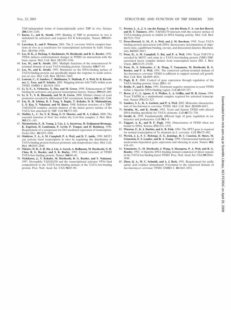
TAF-independent forms of transcriptionally active TBP in vivo. Science288:1244–1248.
57. Kuras, L., and K. Struhl. 1999. Binding of TBP to promoters in vivo isstimulated by activators and requires Pol II holoenzyme. Nature 399:609–613.
58. Larschan, E., and F. Winston. 2001. The S. cerevisiae SAGA complex func-tions in vivo as a coactivator for transcriptional activation by Gal4. GenesDev. 15:1946–1956.
59. Lee, D. K., J. DeJong, S. Hashimoto, M. Horikoshi, and R. G. Roeder. 1992.TFIIA induces conformational changes in TFIID via interactions with thebasic repeat. Mol. Cell. Biol. 12:5189–5196.
60. Lee, M., and K. Struhl. 2001. Multiple functions of the nonconserved N-terminal domain of yeast TATA-binding protein. Genetics 158:87–93.
61. Lee, M., and K. Struhl. 1995. Mutations on the DNA-binding surface ofTATA-binding protein can specifically impair the response to acidic activa-tors in vivo. Mol. Cell. Biol. 15:5461–5469.
62. Leurent, C., S. Sanders, C. Ruhlmann, V. Mallouh, P. A. Weil, D. B. Kirsch-ner, L. Tora, and P. Schultz. 2002. Mapping histone fold TAFs within yeastTFIID. EMBO J. 21:3424–3433.
63. Li, X.-Y., A. Virbasius, X. Zhu, and M. Green. 1999. Enhancement of TBPbinding by activators and general transcription factors. Nature 399:605–609.
64. Li, X. Y., S. R. Bhaumik, and M. R. Green. 2000. Distinct classes of yeastpromoters revealed by differential TAF recruitment. Science 288:1242–1244.
65. Liu, D., R. Ishima, K. I. Tong, S. Bagby, T. Kokubo, D. R. Muhandiram,L. E. Kay, Y. Nakatani, and M. Ikura. 1998. Solution structure of a TBP-TAF(II)230 complex: protein mimicry of the minor groove surface of theTATA box unwound by TBP. Cell 94:573–583.
66. Maillet, L., C. Tu, Y. K. Hong, E. O. Shuster, and M. A. Collart. 2000. Theessential function of Not1 lies within the Ccr4-Not complex. J. Mol. Biol.303:131–143.
67. Mermelstein, F., K. Yeung, J. Cao, J. A. Inostroza, H. Erdjument-Bromage,K. Eagelson, D. Landsman, P. Levitt, P. Tempst, and D. Reinberg. 1996.Requirement of a corepressor for Dr1-mediated repression of transcription.Genes Dev. 10:1033–1048.
68. Muldrow, T. A., A. M. Campbell, P. A. Weil, and D. T. Auble. 1999. MOT1can activate basal transcription in vitro by regulating the distribution ofTATA binding protein between promoter and nonpromoter sites. Mol. Cell.Biol. 19:2835–2845.
69. Nikolov, D. B., S. H. Hu, J. Lin, A. Gasch, A. Hoffmann, M. Horikoshi, N. H.Chua, R. G. Roeder, and S. K. Burley. 1992. Crystal structure of TFIIDTATA-box binding protein. Nature 360:40–46.
70. Nishikawa, J., T. Kokubo, M. Horikoshi, R. G. Roeder, and Y. Nakatani.1997. Drosophila TAF(II)230 and the transcriptional activator VP16 bindcompetitively to the TATA box-binding domain of the TATA box-bindingprotein. Proc. Natl. Acad. Sci. USA 94:85–90.
71. Pereira, L. A., J. A. van der Knaap, V. van den Boom, F. A. van den Heuvel,and H. T. Timmers. 2001. TAF(II)170 interacts with the concave surface ofTATA-binding protein to inhibit its DNA binding activity. Mol. Cell. Biol.21:7523–7534.
72. Perez-Howard, G. M., P. A. Weil, and J. M. Beechem. 1995. Yeast TATAbinding protein interaction with DNA: fluorescence determination of oligo-meric state, equilibrium binding, on-rate, and dissociation kinetics. Biochem-istry 34:8005–8017.
73. Poon, D., A. M. Campbell, Y. Bai, and P. A. Weil. 1994. Yeast TAF170 isencoded by MOT1 and exists in a TATA box-binding protein (TBP)-TBP-associated factor complex distinct from transcription factor IID. J. Biol.Chem. 269:23135–23140.
74. Poon, D., S. Schroeder, C. K. Wang, T. Yamamoto, M. Horikoshi, R. G.Roeder, and P. A. Weil. 1991. The conserved carboxy-terminal domain ofSaccharomyces cerevisiae TFIID is sufficient to support normal cell growth.Mol. Cell. Biol. 11:4809–4821.
75. Pugh, B. F. 2000. Control of gene expression through regulation of theTATA binding protein. Gene 255:1–14.
76. Reddy, P., and S. Hahn. 1991. Dominant negative mutations in yeast TFIIDdefine a bipartite DNA-binding region. Cell 65:349–357.
77. Reese, J. C., L. Apone, S. S. Walker, L. A. Griffin, and M. R. Green. 1994.Yeast TAFIIS in a multisubunit complex required for activated transcrip-tion. Nature 371:523–527.
78. Sanders, S. L., K. A. Garbett, and P. A. Weil. 2002. Molecular characteriza-tion of Saccharomyces cerevisiae TFIID. Mol. Cell. Biol. 22:6000–6013.
79. Strubin, M., and K. Struhl. 1992. Yeast and human TFIID with alteredDNA-binding specificity for TATA elements. Cell 68:721–730.
80. Struhl, K. 1999. Fundamentally different logic of gene regulation in eu-karyotes and prokaryotes. Cell 98:1–4.
81. Taggart, A. K., and B. F. Pugh. 1996. Dimerization of TFIID when notbound to DNA. Science 272:1331–1333.
82. Winston, F., K. J. Durbin, and G. R. Fink. 1984. The SPT3 gene is requiredfor normal transcription of Ty elements in S. cerevisiae. Cell 39:675–682.
83. Wyrick, J. J., F. C. Holstege, E. G. Jennings, H. C. Causton, D. Shore, M.Grunstein, E. S. Lander, and R. A. Young. 1999. Chromosomal landscape ofnucleosome-dependent gene expression and silencing in yeast. Nature 402:418–421.
84. Yamamoto, T., M. Horikoshi, J. Wang, S. Hasegawa, P. A. Weil, and R. G.Roeder. 1992. A bipartite DNA binding domain composed of direct repeatsin the TATA box binding factor TFIID. Proc. Natl. Acad. Sci. USA 89:2844–2848.
85. Zhou, Q. A., M. C. Schmidt, and A. J. Berk. 1991. Requirement for acidicamino acid residues immediately N-terminal to the conserved domain ofSaccharomyces cerevisiae TFIID. EMBO J. 10:1843–1852.
VOL. 23, 2003 STRUCTURE AND FUNCTION OF TBP DIMERS 3201
at Scientific Library, N
CI-F
CR
DC
on April 20, 2009
mcb.asm
.orgD
ownloaded from