knee region bones joint muscles artery & nerves. knee osteology
TRANSCRIPT
Knee region
• Bones• Joint• Muscles• Artery & Nerves

Knee osteology
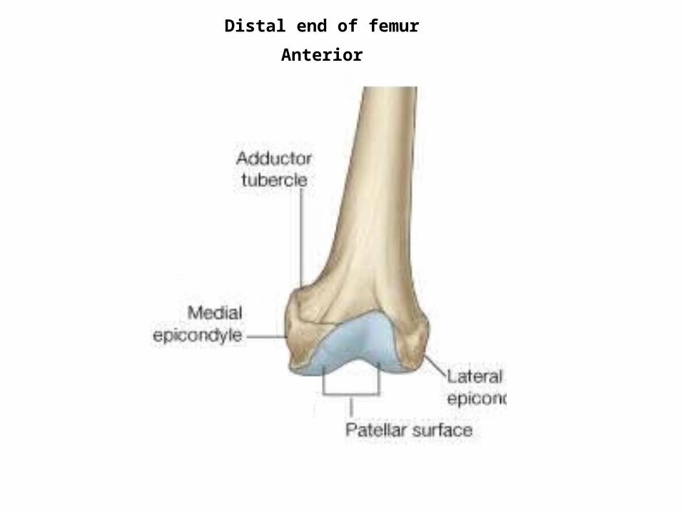
Distal end of femur
Anterior

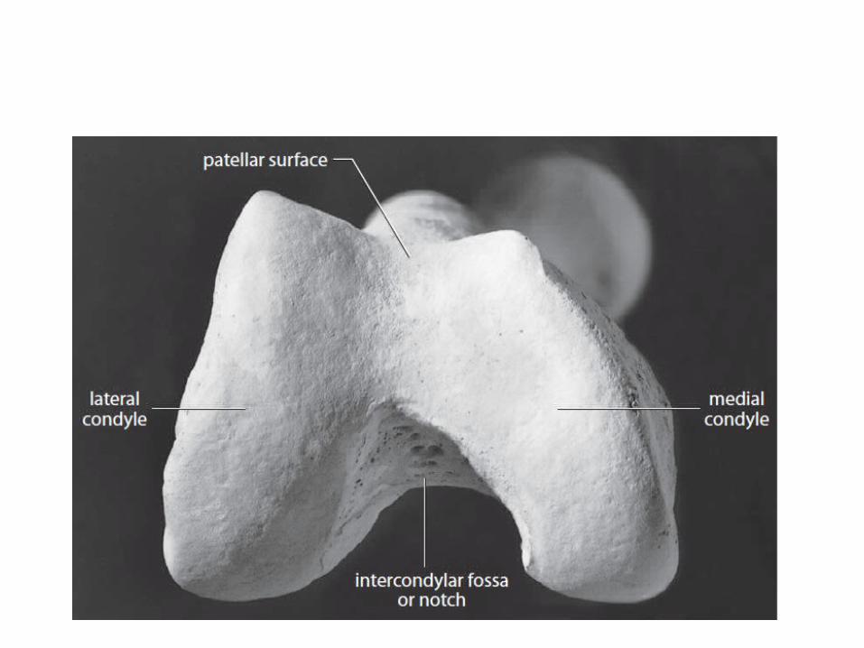
Distal end of femur
Posterior

Femur ant. Femur post.
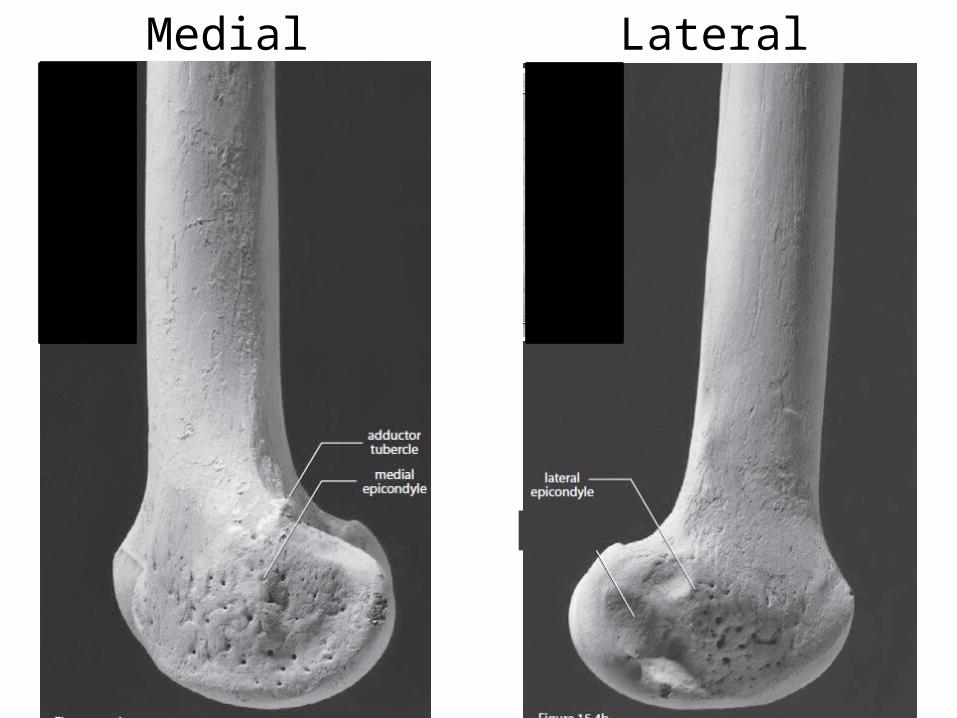
Medial Lateral

Patella
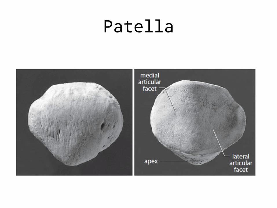
Patella
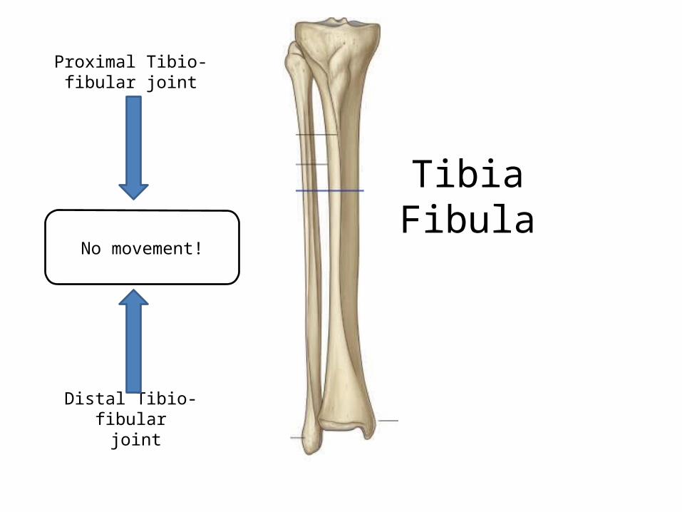
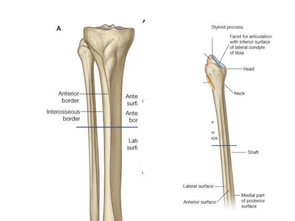
TibiaFibula
Proximal Tibio-fibular joint
Distal Tibio-fibular joint
No movement!
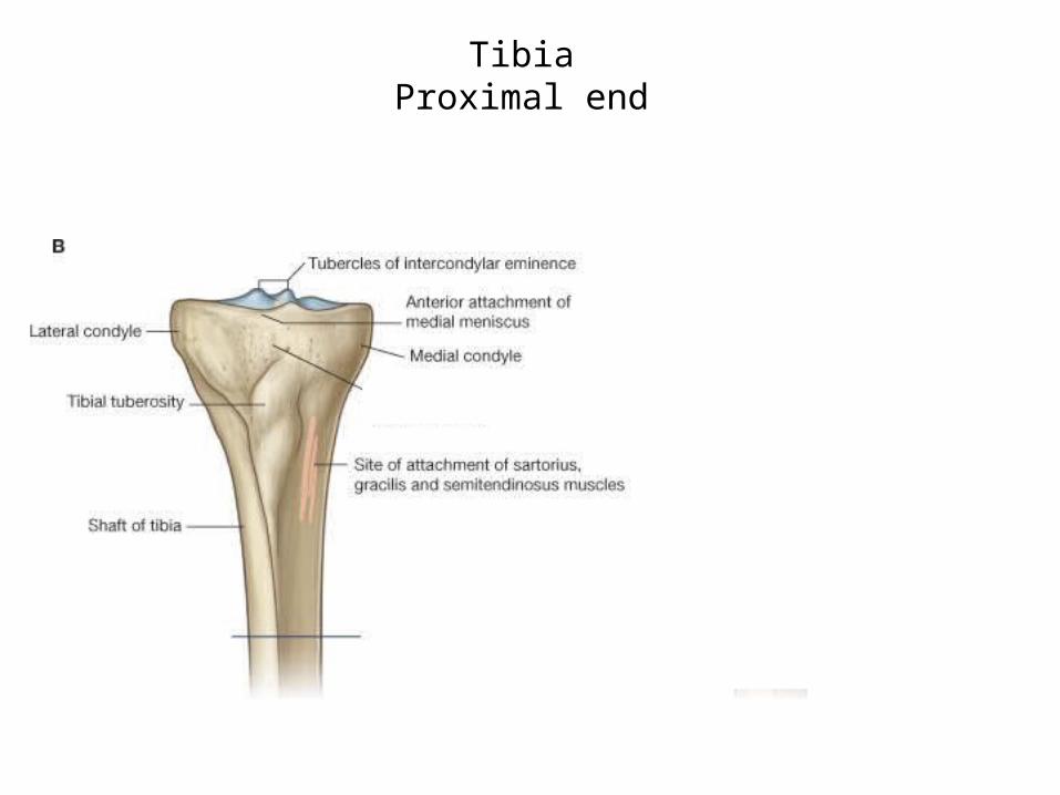
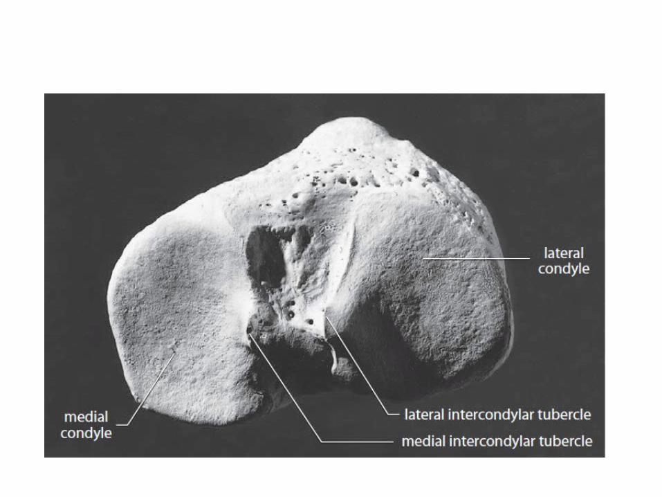
TibiaProximal end
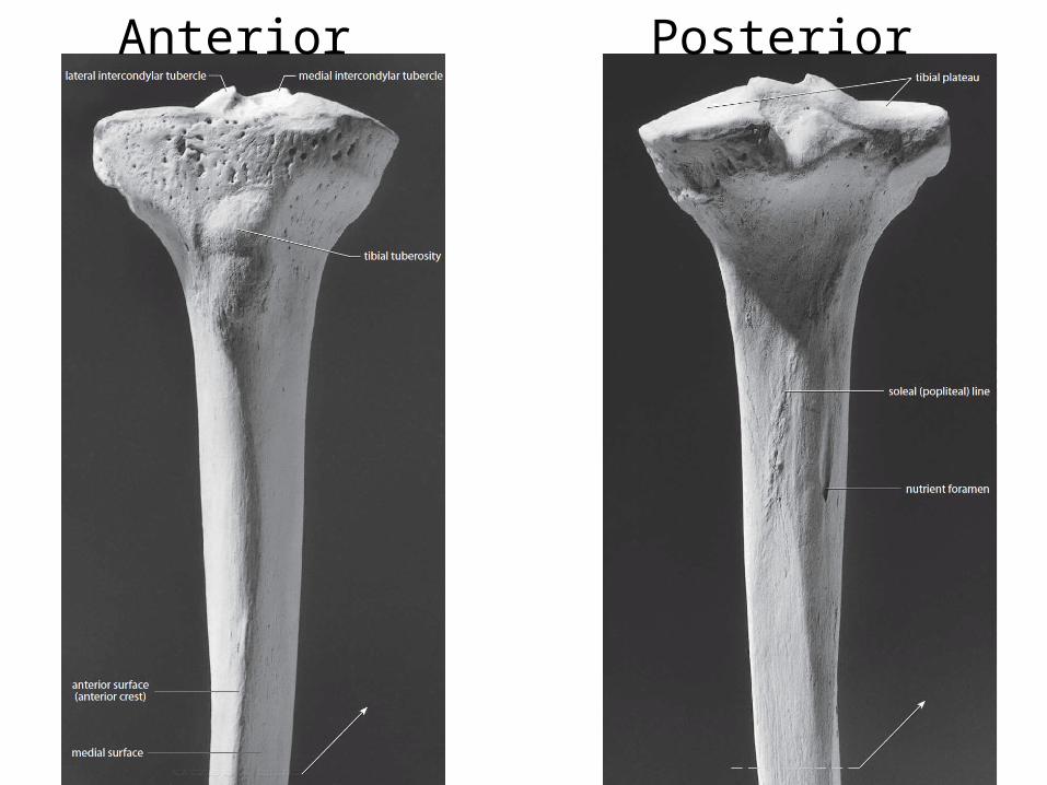
Anterior Posterior
Tibiofemoral (knee) Joint
• Largest and most complex joint
• Actually 3 joints within a single synovial cavity:
1. Laterally: tibiofemoral joint2. Medially: tibiofemoral joint3. Intermediate: patellofemoral joint
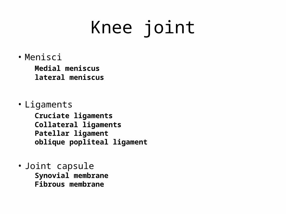
Knee joint
• MenisciMedial meniscuslateral meniscus
• Ligaments Cruciate ligamentsCollateral ligaments Patellar ligamentoblique popliteal ligament
• Joint capsuleSynovial membraneFibrous membrane
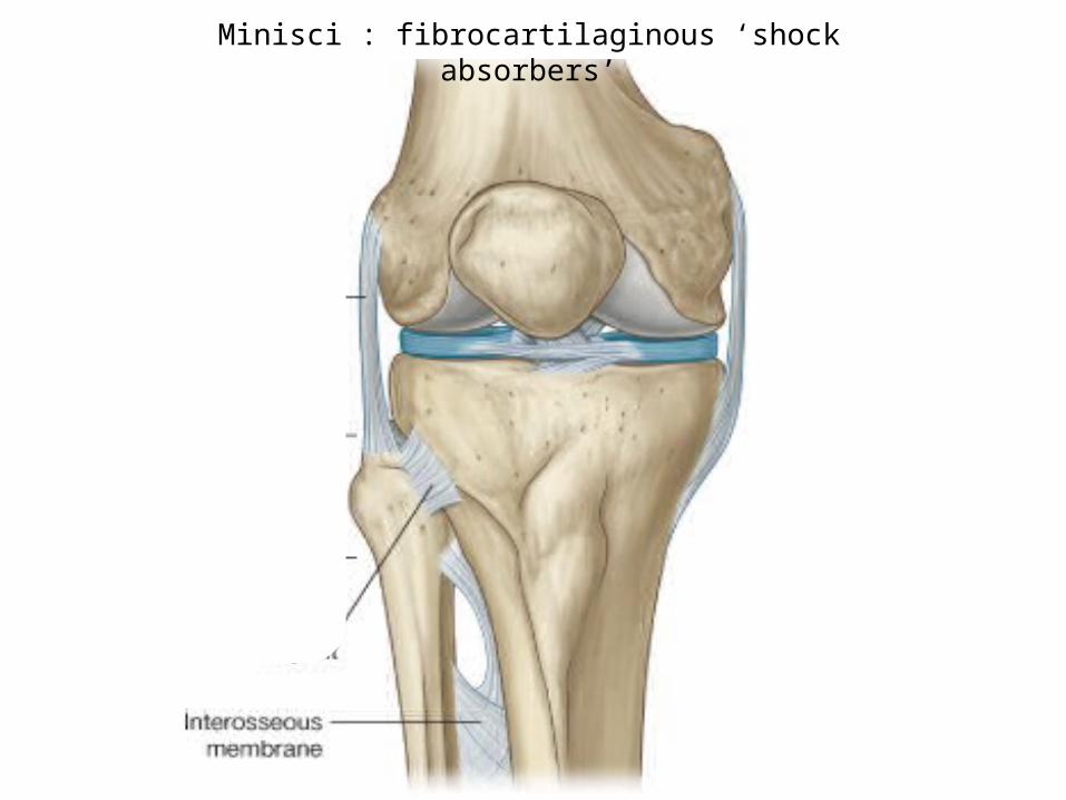
Minisci : fibrocartilaginous ‘shock absorbers’
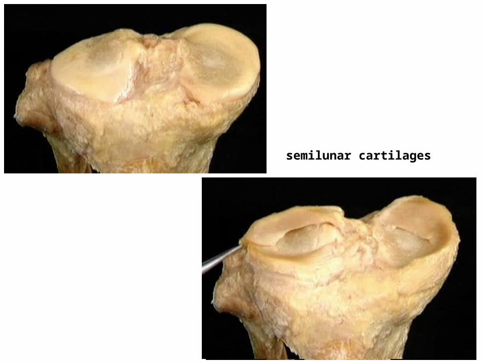
semilunar cartilages
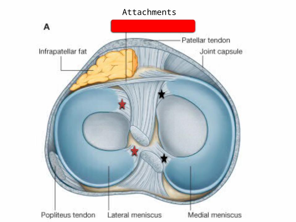
Attachments
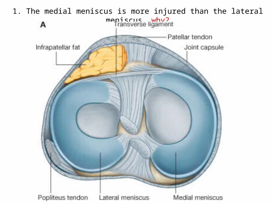
1. The medial meniscus is more injured than the lateral meniscus, why?
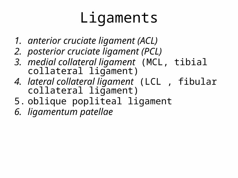
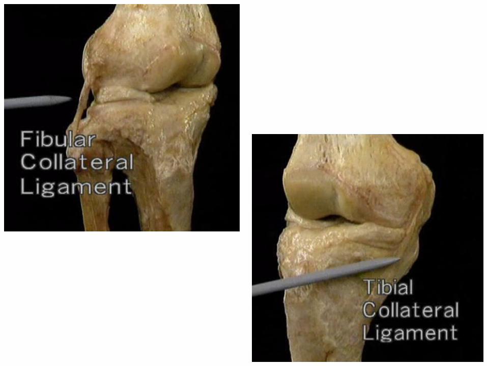
Ligaments1. anterior cruciate ligament (ACL)2. posterior cruciate ligament (PCL)3. medial collateral ligament (MCL, tibial collateral
ligament)4. lateral collateral ligament (LCL , fibular collateral
ligament) 5. oblique popliteal ligament6. ligamentum patellae
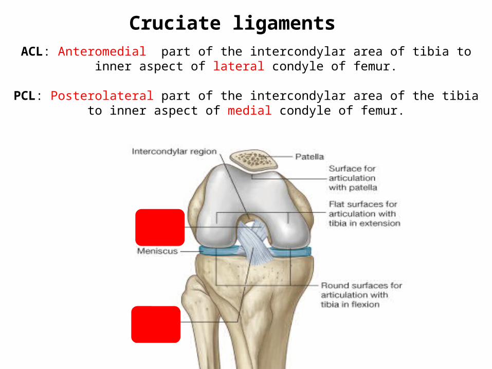
ACL: Anteromedial part of the intercondylar area of tibia to inner aspect of lateral condyle of femur.
PCL: Posterolateral part of the intercondylar area of the tibia to inner aspect of medial condyle of femur.
Cruciate ligaments
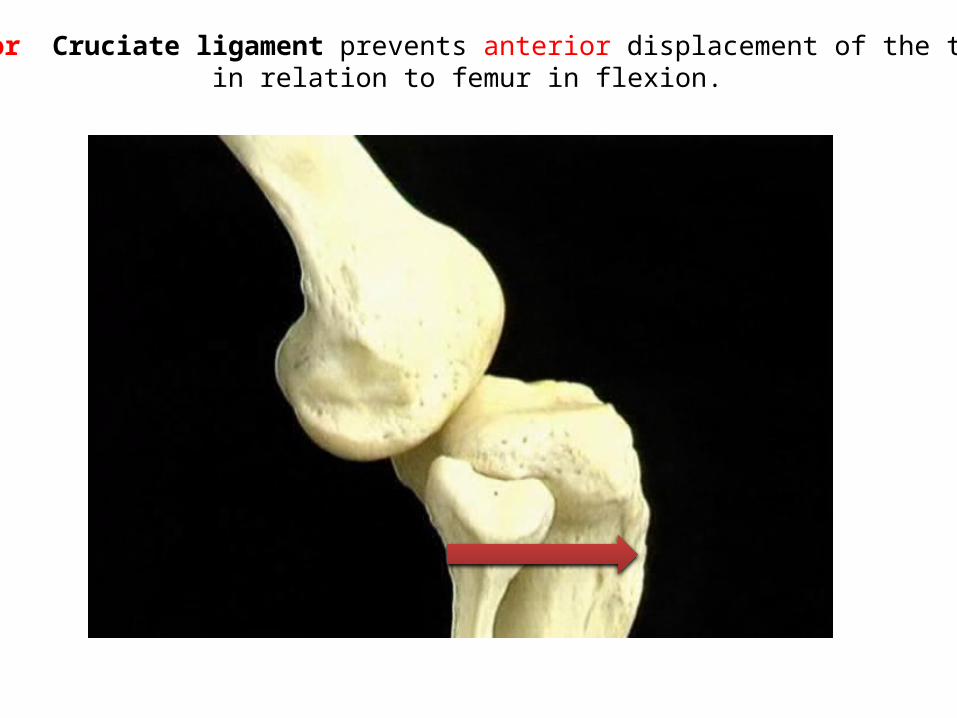
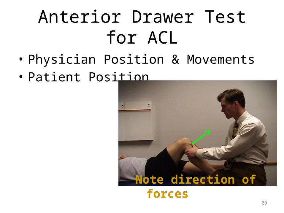
Anterior Cruciate ligament prevents anterior displacement of the tibia in relation to femur in flexion.
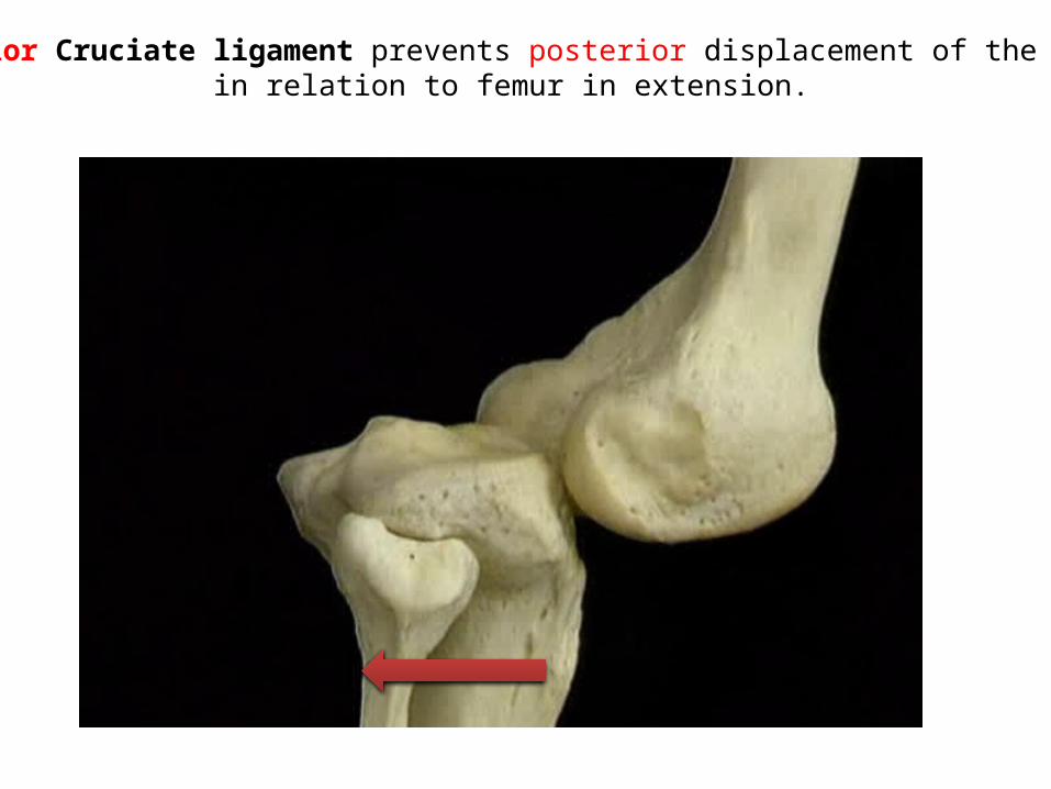
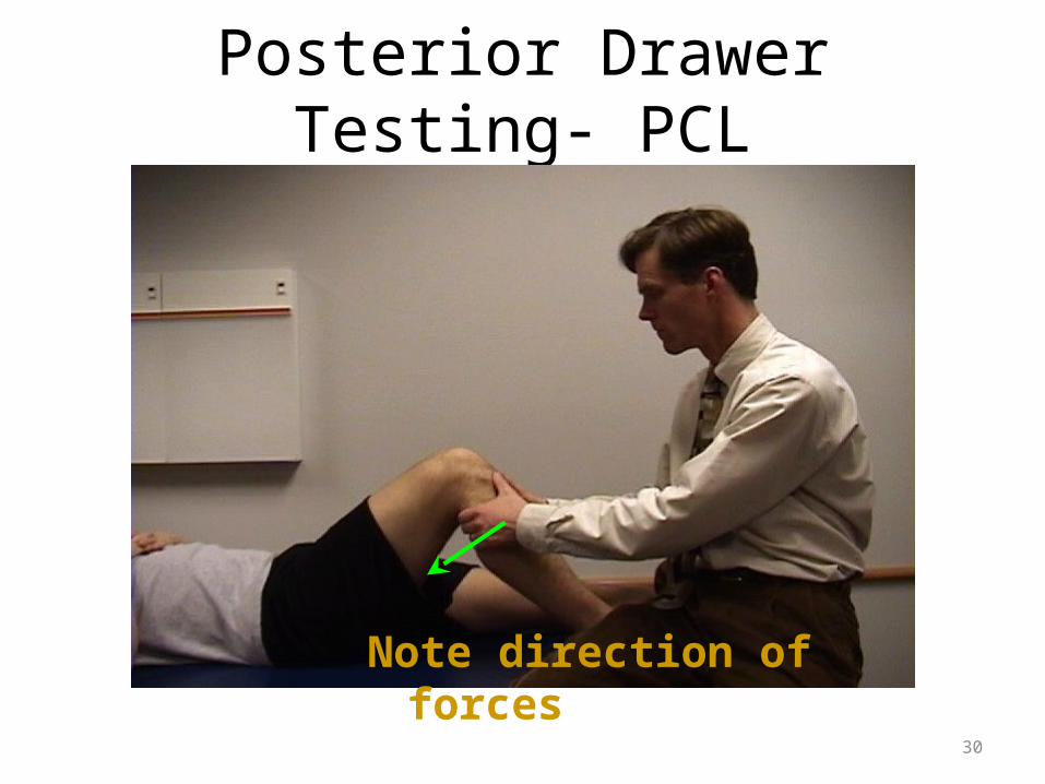
Posterior Cruciate ligament prevents posterior displacement of the tibia in relation to femur in extension.
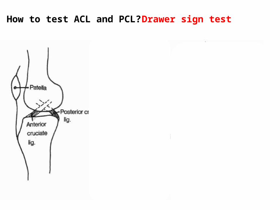
How to test ACL and PCL? Drawer sign test
29
Anterior Drawer Test for ACL
• Physician Position & Movements• Patient Position
Note direction of forces
30
Posterior Drawer Testing- PCL
Note direction of forces
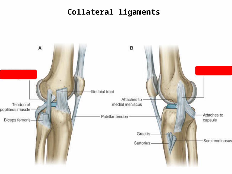
Collateral ligaments
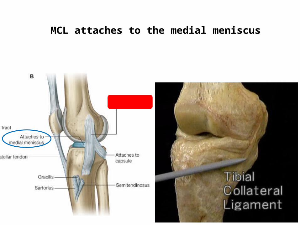
MCL attaches to the medial meniscus
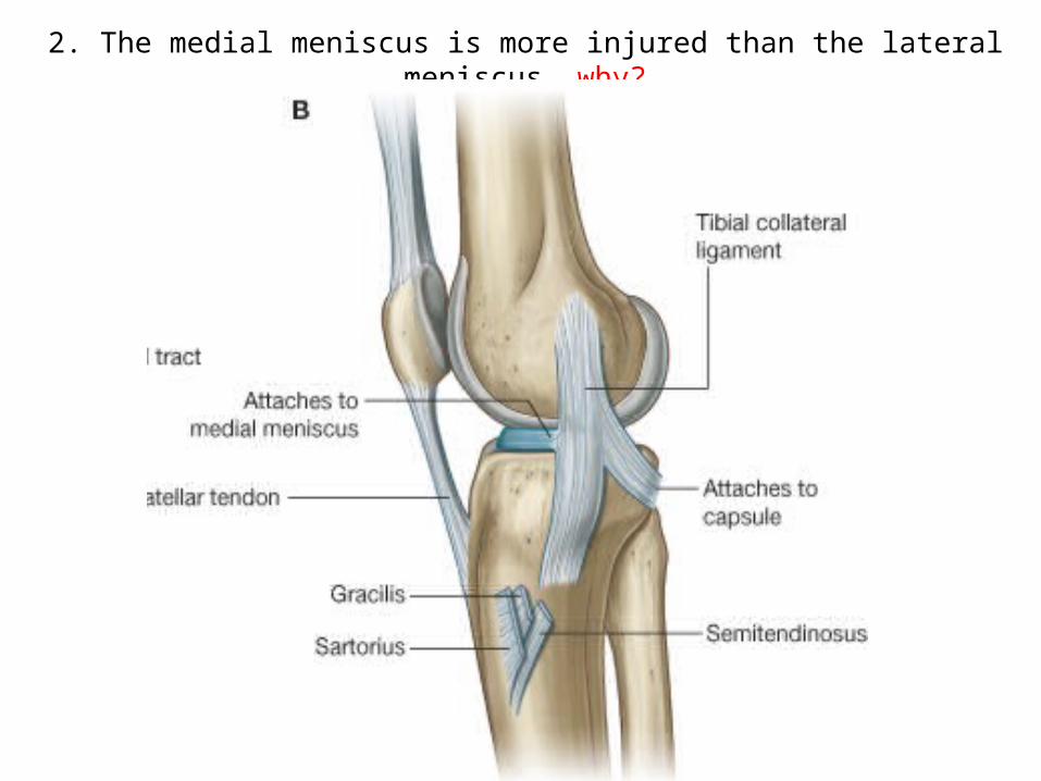
2. The medial meniscus is more injured than the lateral meniscus, why?
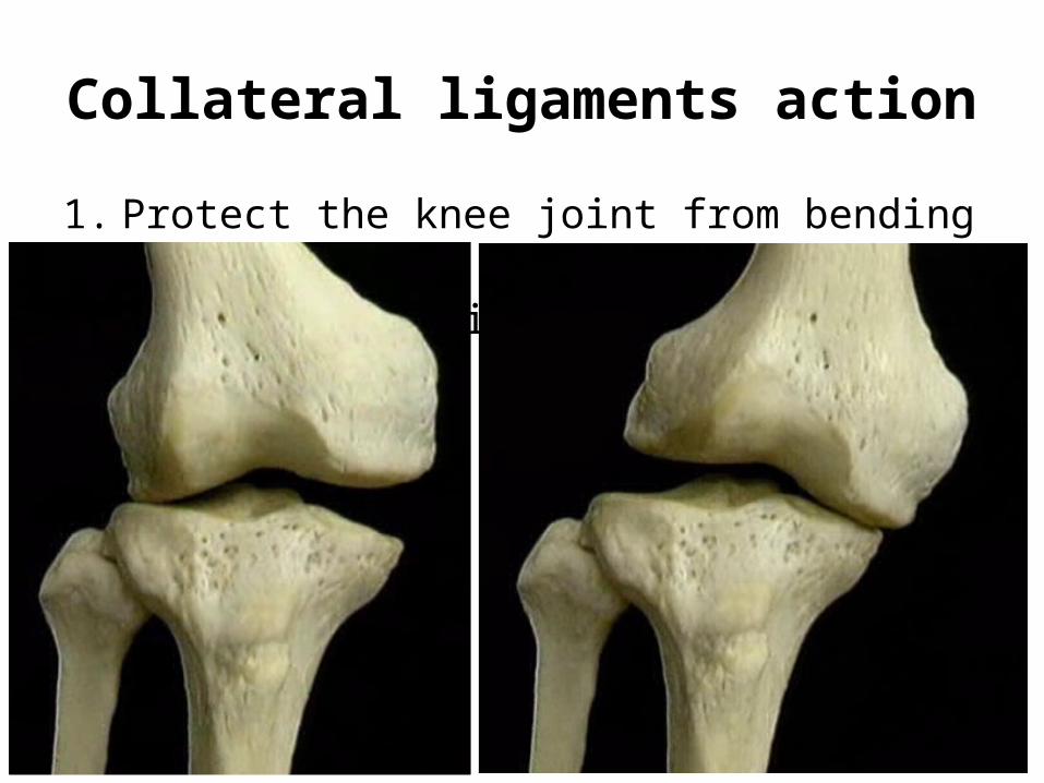
Collateral ligaments action
1. Protect the knee joint from bending side to side. 2. Helps the locking mechanism
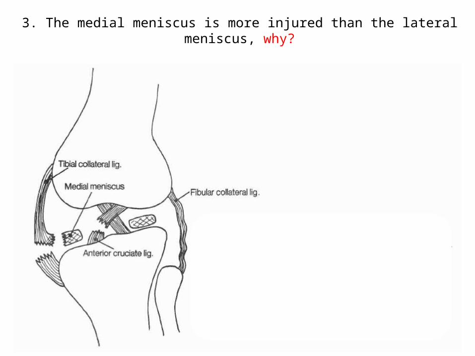
3. The medial meniscus is more injured than the lateral meniscus, why?
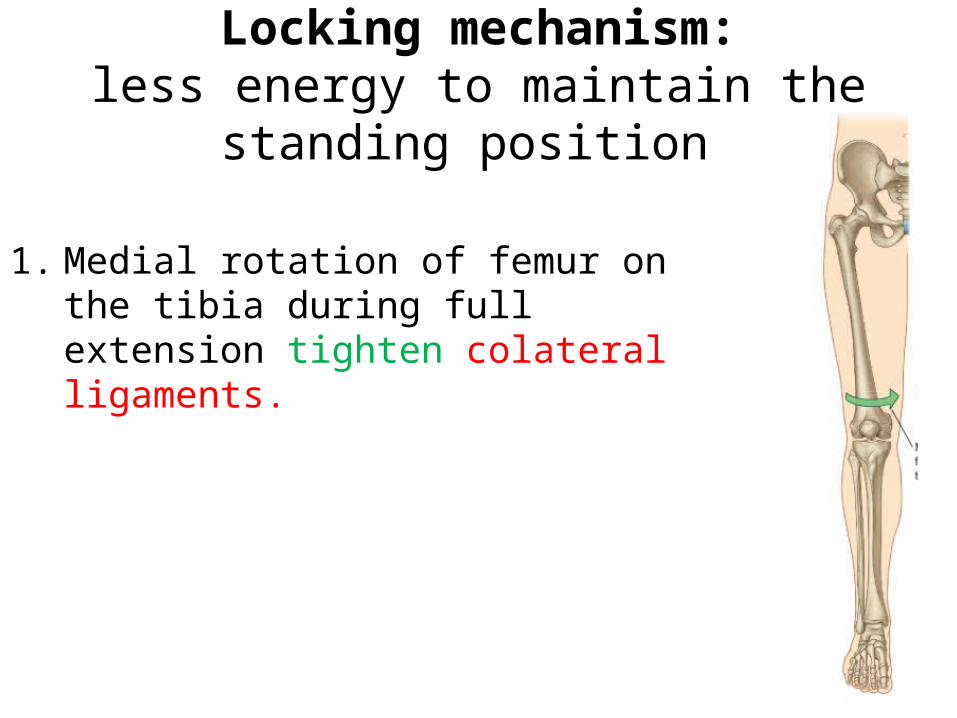
Locking mechanism:less energy to maintain the standing
position
1. Medial rotation of femur on the tibia during full extension tighten colateral ligaments.
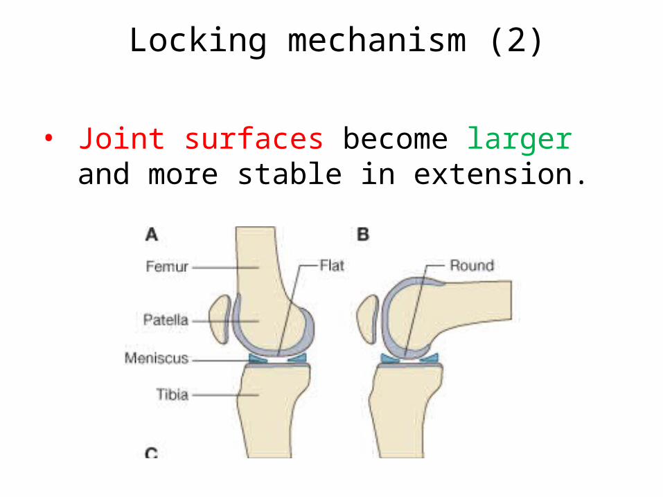
Locking mechanism (2)
• Joint surfaces become larger and more stable in extension.
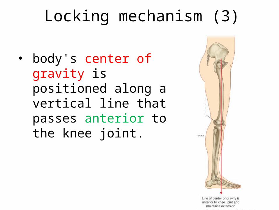
Locking mechanism (3)
• body's center of gravity is positioned along a vertical line that passes anterior to the knee joint.
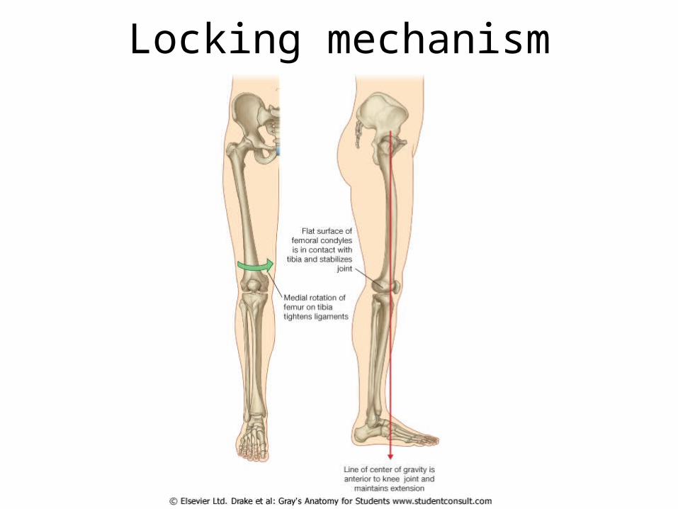
Locking mechanism
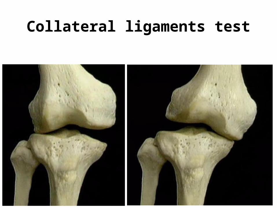
Collateral ligaments test
42
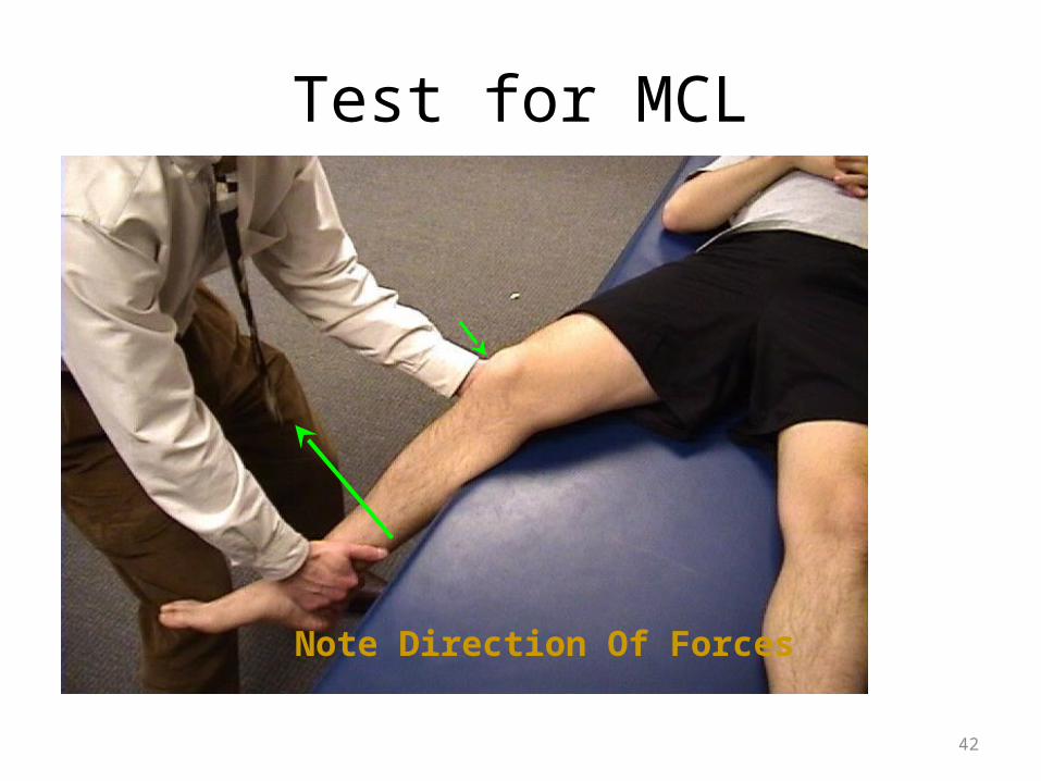
Test for MCL
Note Direction Of Forces
43
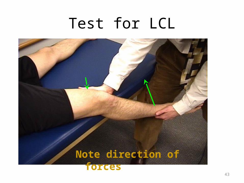
Test for LCL
Note direction of forces
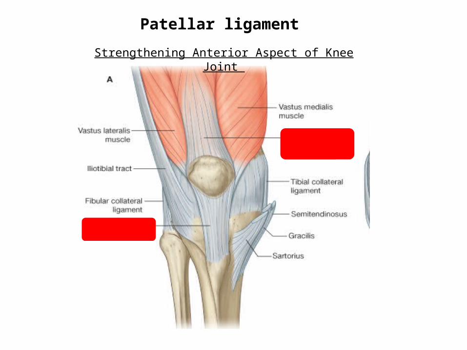
Patellar ligament
Strengthening Anterior Aspect of Knee Joint
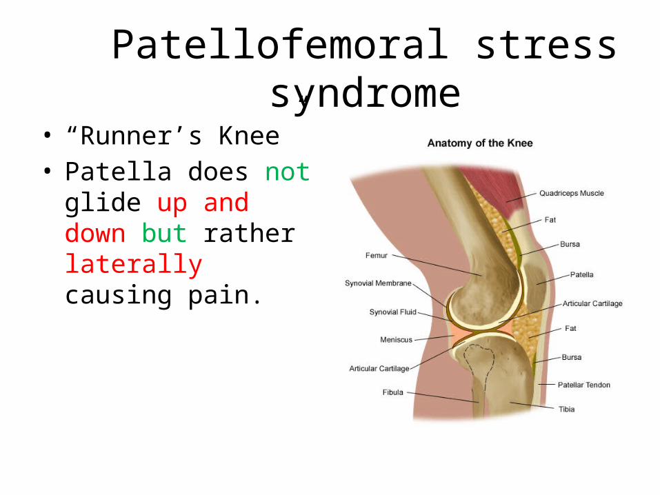
Patellofemoral stress syndrome• “Runner’s Knee”• Patella does not glide
up and down but rather laterally causing pain.
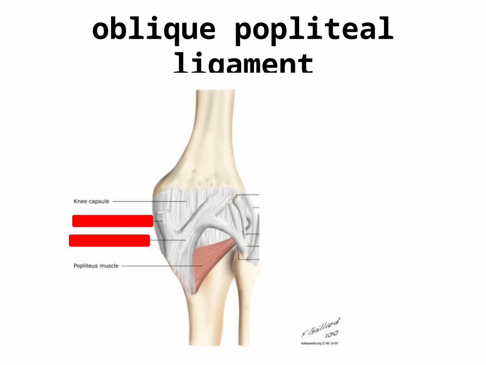
oblique popliteal ligament
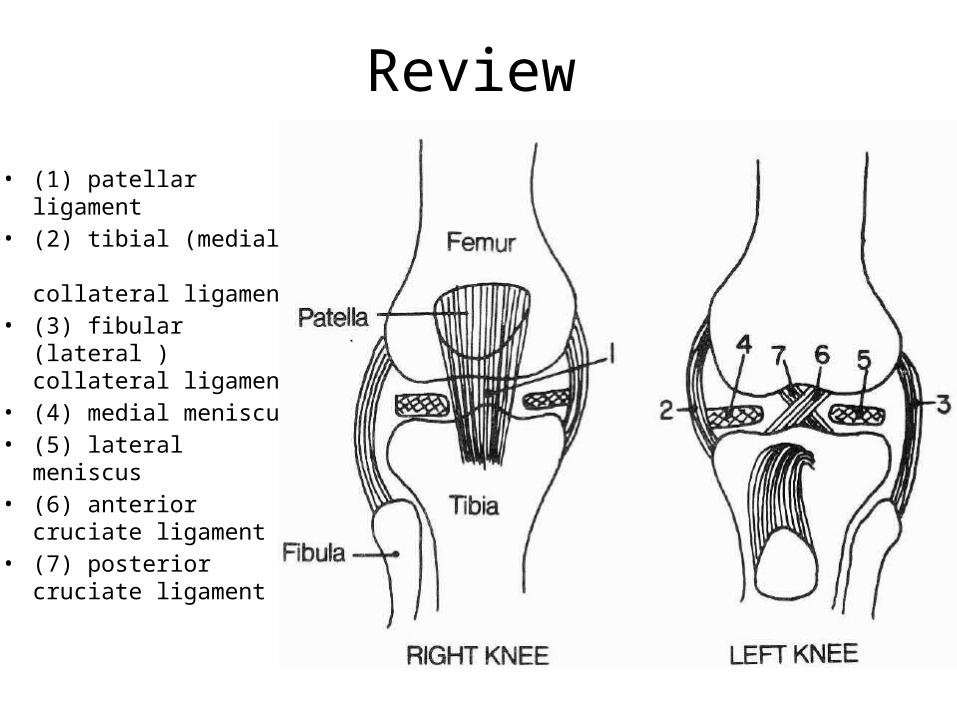
Review
• (1) patellar ligament • (2) tibial (medial)
collateral ligament• (3) fibular (lateral )
collateral ligament• (4) medial meniscus • (5) lateral meniscus• (6) anterior cruciate
ligament • (7) posterior cruciate
ligament
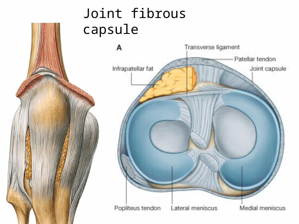
Joint fibrous capsule
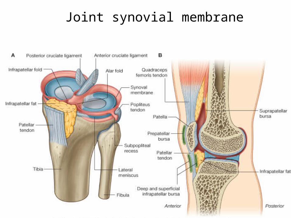
Joint synovial membrane
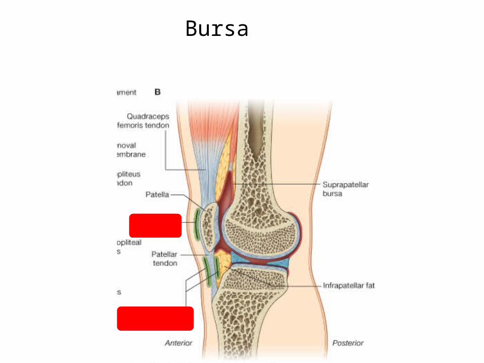
Bursa
• little fluid sacs that helps the muscles and tendons slide freely:
PrepatellarInfrapatellarSuprapatellar
Bursa
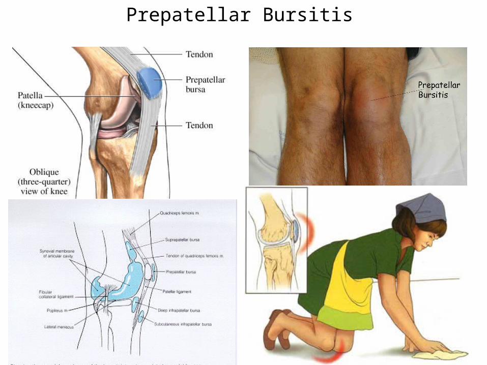
Prepatellar Bursitis