journal bangladesh glaucoma society - bgsbd.net filejournal bangladesh glaucoma society j b g s july...
TRANSCRIPT

JournalBangladesh Glaucoma Society
JBGS
July 2018 Volume 06 Number 02
Contents
Editorial
Angle closure glaucoma – a public health problem 09
Prof. M. Nazrul Islam
Original Article
A Study of Retinal Nerve Fiber Layer (RNFL) Thickness Changes in
Patients of Type 2 Diabetes Melitus with Chronic Kidney Disease (CKD)
Measured by Optical Coherence Tomography 11
Dr. Ruhi Mannan, Dr. Ferdous Hossain,Dr. Samarendranath Adhikary,
Dr. Ashraf Sayeed
Visual outcome after manual small incision cataract surgery
for Phacomorphic glaucoma 15
Dr. Shams Mohammad Noman
Reduction of IOP & other associated ocular parameters following Phaco-
emulsification in ACG 19
Sheikh Mohammed Hossain
Steroid Induced Ocular Hypertension in Post -LASIK Patients:
A Matter of Great Concern 21
Dr. Ummay Kawser, Dr. Siddiqur Rahman, Dr. Ishtiaq Anwar,
Prof. Dr. Md. Hafizur Rahman
Review Article
Pediatric Glaucoma and its Medical Management 24
Prof. Dr. Zakia Sultana Shahid, Prof. Dr.M Hafizur Rahman,
Dr. Md. Abdul Mannan, Dr. Md. Almas Hossain
Angle-closure Glaucoma : The Role of the Lens in the Pathogenesis,
Prevention and Treatment 30
Dr. Md. Safiul Islam Prodhan
Case Report
Bilateral Acute Angle Closure from Topiramate toxicity– 3 case reports 43
Prof. M. Nazrul Islam
Case Presentation: Bilateral Angle-Closure Glaucoma 47
Dr. Shams Mohammad Noman, Dr. Nahid Sultana
BGS News 51

JournalBangladesh Glaucoma Society
Bangladesh Glaucoma Society (BGS) Journal
0301
July 2018, Vol. 6, No. 2
Volume - 06 Number - 02 July 2018
Editor in Chief : Prof. M. Nazrul Islam
Executive Editor : Dr. Muhammad Ziaul Karim

Bangladesh Glaucoma Society (BGS) Journal
0302
July 2018, Vol. 6, No. 2
Journal
Bangladesh Glaucoma Society (JBGS)
Volume - 06, Number-02, July 2018
Published by :
Dr. Md. Safiul Islam Prodhan
Publication & Publicity Secretary
On behalf of the
Bangladesh Glaucoma Society
House # 12A, Road # 05, Dhanmondi, Dhaka
email : [email protected]
website : www.bgsbd.net
Printed at :
New Ekata Computer & Printers
435/A-2, Baro Moghbazar, Dhaka-1217
Tel : 01715444644
Courtesy :

Editorial Board
Chairman : Prof. Sk. M. A. Mannaf
Editor in Chief : Prof. M. Nazrul Islam
Executive Editor : Dr. Muhammad Ziaul Karim
Assistant Editors : Dr. Md. Quamrul Islam Khan
Prof. M. Hafizur Rahman
Dr. Md. Safiul Islam Prodhan
Members
Prof. Ava Hossain
Prof. Syed Maruf Ali
Prof. Md. Arif Mian
Prof. Md. Hassan Shahid
Prof. Md. Shafiqul Islam
Prof. Md. Mizanur Rahman
Prof. Sheikh Md. Hossain
Prof. Zakia Sultana Shahid
Prof. Iftekhar Md. Munir
Dr. Sajedur Rahman
Dr. Md. Musharaf Hossain
Dr. Salma Parvin
Dr. Ruhi Mannan
Advisory Board
Prof. Md. Saleh Uddin
Prof. M A Halim Khan
Prof. Deen Mohd. Noorul Huq
Prof. Sahab Uddin
Prof. Md. Sharfuddin Ahmed
Bang
lades
h Glau
coma
Soc
iety
Address of Correspondence
Executive Editor, JBGS
Harun Eye Foundation Hospital
House # 12A, Road # 05, Dhanmondi, Dhaka
email : [email protected]
website : www.bgsbd.net
Bangladesh Glaucoma Society (BGS) Journal
0303
July 2018, Vol. 6, No. 2

Bang
lades
h Glau
coma
Soc
iety
Bangladesh Glaucoma Society (BGS) Journal
0304
July 2018, Vol. 6, No. 2
Journal of Bangladesh Glaucoma Society (JBGS)
Editorial Policy
The Journal of Bangladesh Glaucoma Society (JBGS) is the official journal of
Bangladesh Glaucoma Society (BGS). JBGS is published regularly and half
yearly.
The JBGS editorial board maintains the following policy:
It is a national level, peer reviewed, open access journal.
JBGS is committed to transparency.
JBGS provides a stimulating forum for discussion of clinical, scientific and
socioeconomic issues of greatest concern to clinicians who care for glaucoma
patients.
Each of the issue tries to present original articles on new approaches to
diagnosis, innovations in pharmacological therapy, surgical technique, and
basic scientific advances in the field of glaucoma that impact on clinical
practice.
JBGS adheres to the highest standards concerning its editorial policies on
publication ethics, scientific misconduct, consent and peer review criteria.
The journal always strictly follows guidance produced by bodies that include
the Committee on Publication Ethics (COPE), the World Association of
Medical Editors (WAME) and the International Committee of Medical Journal
Editors (ICMJE).
JBGS takes all possible misconduct seriously. If an Editor, author or reader
has concerns that a submitted article describes something that might be
considered to constitute misconduct in research, publication or professional
behaviour they should forward their concerns to the journal so that JBGS
editorial body can address the allegations properly.
As an open access journal, JBGS adheres to the Budapest Open Access
Initiative definition of open access. Articles are published to facilitate reuse
of the content and authors retain the copyright.
Although it is an open access journal, no payment information is requested
for any article which is accepted, so that the ability to pay cannot affect
editorial decisions.
Articles submitted to JBGS are subject to peer review. The journal operates
single blind peer review whereby the names of the reviewers are hidden
from the author; which is the traditional and most common method of
reviewing.
JBGS believes that to make the best decision on how to deal with a
manuscript the journal editor should know about any competing interests
that authors may have. There is nothing inherently unethical about a
competing interest but it should be acknowledged and openly stated.

JBGS Review Board
Prof. Ava Hossain
Prof. Sk. M A Mannaf
Prof. Syed Marfu Ali
Prof. M. Nazrul Islam
Prof. Md. Arif Mian
Dr. M Ziaul Karim
Prof. Hassan Shahid
Prof. Shafiqul Islam
Prof. Md. Mizanur Rahman
Prof. Sk. Mohammad Hossain
Prof. Abul Bashar Sheikh
Prof. Iftekhar Md. Munir
Prof. Zakia Sultana Shahid
Dr. Md. Quamrul Islam Khan
Prof. M. Hafizur Rahman
Dr. M A Karim
Dr. Siddiqur Rahman
Dr. Safiul Islam Prodhan
Dr. Mossarraf Hossain
Dr. Ishtiaq Anwar
Dr. Salma Parvin
Dr. Shahnaz Begum
Dr. Ruhi Mannan
Bang
lades
h Glau
coma
Soc
iety
Bangladesh Glaucoma Society (BGS) Journal
0305
July 2018, Vol. 6, No. 2

Executive Committee
2018-2019
President
Dr. Muhammad Ziaul Karim
President Elect
Prof. Md. Mizanur Rahman
General Secretary
Dr. Md. Quamrul Islam Khan
Treasurer
Prof. Dr. Iftekhar Md. Munir
Joint Secretary
Dr. Salma Parvin
Organizing Secretary
Dr. Siddiqur Rahman
Scientific Secretary
Prof. M. Hafizur Rahman
Office Secretary
Dr. Ruhi Mannan
Publication & Publicity Secretary
Dr. Md. Safiul Islam Prodhan
Entertainment Secretary
Dr. Md. Mohasin Baig
Executive Members
Prof. Md. Shafiqul Islam
Prof. Hassan Shahid
Prof. M. Abul Bashar Sheikh
Prof. Sheikh M Hossain
Prof. Kh. Ziaul Islam Md. Ali
Prof. Dr. Zakia Sultana Shahid
Dr. Md. Musharaf Hossain
Dr. M.A. Karim
Dr. Ishtiaque Anwar
Dr. Harun-Ur Rashid
Dr. Mohammad Zafrul HassanBang
lades
h Glau
coma
Soc
iety
Bangladesh Glaucoma Society (BGS) Journal
0306
July 2018, Vol. 6, No. 2

Original papers written in English will be considered
for publication provided these have not been
published previously and are not under consideration
for publication elsewhere.
Conditions for manuscript submission
• All manuscripts will be subjected to peer and
editorial review
• Accepted manuscripts become the property of the
Bangladesh Glaucoma Society Journal.
• The author should obtain written permission from
appropriate authority if the manuscript contains
any table; data or illustration from previously
published in other journals. The letter of
permission should be submitted with manuscript.
• If the photographs are not disguised, permission
from the patient or parents/guardians to print
should accompany the manuscript. Otherwise
identity will be blackened out.
• Rejected manuscripts/electronic copies /
illustrations / photographs will not be returned to
the authors.
• Editors are not responsible for courier/postal failure.
Manuscript preparation
The format of the Bangladesh Glaucoma Society
journal complies with “Uniform Requirements for
Manuscripts Submitted to Biomedical Journals”
published by the International Committee of Medical
Journals Editors in Vancouver British Columbia in
1979, (the widely accepted “Vancouver style”)
published in the Annals of Internal Medicine 1982;
96:766-71. All scientific units should be expressed in
System International (SI) units. Authors are referred
to Annals of International Medicine 1987; 106:114-29
for guidance in the use of SI units. All drugs should
be mentioned in their generic form.
• Should be typed in English and on one side of A4
(290x210cm) size white paper, using Times New
Roman font, size 12, with single space
• There should be one original and two paper copies
and one IBM compatible electronic copy.
• There should be a margin of 2.5 cm at top and
bottom, and 1.2 cm left and right.
• Pages should be numbered in English numerical at
the upper right hand, consecutively, beginning with
the title page.
• Manuscripts should be submitted in the following
order:
Title
should not exceed 100 characters.
Abstract
With a specific format with six sections: Background,
Objective, Methodology, results, Conclusion and
Acknowledgements, Keywords, address of
correspondence (about 350 words maximum). All
these section will be Times New Roman font sixe 12
and italic but not bold. No reference are allowed in
the abstract.
Text
(Introduction, Materials & Methods, results,
Discussion, conclusion).
Acknowledgements
References
Photographs
• In CD/Pen drive
• With appropriate labeling (number in English
numerical, title of photographs and title of
manuscripts.)
Illustrations
• All illustrations should be cited in the text
• Illustration should be numbered in English
numerical and labeled properly, placed
appropriately in relation to text of manuscript.
Tables
• Should be appropriately titled.
• Numbered with Roman numerical in order of text.
• Abbreviation if used, should be explained in foot-
notes.
• Same table should not be repeated as chart.
Placement
• All photographs, illustrations and tables should be
placed in the text in their appropriate places
where their description are given.
Instruction for authors
0307
Bangladesh Glaucoma Society (BGS) Journal July 2018, Vol. 6, No. 2

References
• References from journal should be indicated by
superscript numbers consecutively in the text
(e.g.”…….has been reported1; or as shown by
Rahman2 ) in the order in which they are first
mentioned and should be listed in numerical order
at the end of the article.
• References cited only in tables or legends or
illustrations should be numbered in accordance
with a sequence established by the first mention in
the text.
• Titles of journals should be abbreviated according
to Index Medicus or given in full.
• References must include: (i) all authors, surnames
and initials if there are more than 6 authors, the
first six authors followed by et al; (ii) the full title of
the paper; (iii) the abbreviated or full title of the
journal in italic; (iv) the year of publication; (v) the
volume no will be bold; (vi) the first and last page
numbers followed by full stop.
• References from books must include: (i) authors
name (ii) title of article (iii) editors name/s (iv)
name of the chapter (V)place of publication (vi)
name of publisher (vii)year of publication and page
numbers.
• Documents in electronic format must include: (i)
title (ii) authors name (iii) year of publication (iv)
website address date of access.
Manuscripts Submission
The manuscripts should be submitted to the editor in
chief with a covering letter, mentioning that the work
has not been published or submitted for publication
anywhere else.
Reprints for the authors
2 copies of original journal and five copies of each
article will be provided to the corresponding author
free of cost.
Copy right
No part of the materials published in this journal may
be reproduced, stored or transmitted without prior
written permission of the editorial board.
Instruction for authors
0308
Bangladesh Glaucoma Society (BGS) Journal July 2018, Vol. 6, No. 2

Bangladesh Glaucoma Society (BGS) Journal
Angle closure glaucoma – a public health problem
M N Islam1
0309
Editorial
In this issue of the JBGS, 2 articles as case
presentation has been published on Bilateral Angle-
Closure Glaucoma. Both the article cases are not
common but significantly medical important. If timely
managed by medical or surgical treatment angle
closure can be treated and vision can be saved. The
1st
article Bilateral Acute Angle Closure from
Topiramate toxicity – 3 case reports by M. Nazrul
Islam is of public health importance asTopiramate is
prescribed by medicine specialists, Neurologists,
Otolaryngologist and also by Ophthalmologist.
2nd Case Presentation: Bilateral Angle-Closure
Glaucoma by Dr. Shams Mohammad Noman and Dr.
Nahid Sultana is more of ophthalmologist importance.
We need to know the pathophysiology of angle
closure diseases and how to manage this public health
problem
Acute angle closure glaucoma (AACG) is regarded as
an ocular emergency around the world. The most
accepted terminology now is acute primary angle
closure (APAC). AACG remains – in general– a useful
trigger term to exhume a pattern recognition for the
non specialist.
Primary angle closure spectrum (PACsp) eyes can be
classified as primary angle closure suspects (PACS),
primary angle closure (PAC) or primary angle closure
glaucoma (PACG). The common denominator is an
overcrowding of the anterior segment, with
appositional contact of the iris and trabecular
meshwork. Relative to normal eyes, these eyes
typically have a shallow anterior chamber depth
(ACD), a small corneal diameter (W2W) and a thick,
anteriorly situated lens.
A misconception is that PACsp is exclusive to short,
hyperopic eyes. In fact, it can be found in eyes of
normal or long axial length (AL). An increasing
prevalence of PAC/PACG in axial myopes has been
observed, especially in Asian patients.
Mechanisms underlying acute angle closure is
described by many authors. The identification of the
mechanisms and/or causative factor(s) underlying
acute angle closure (AAC) can lead to better
management of the disease. Ritch and Lowe1 have
described a classification system for the mechanisms
of angle closure that is based on the forces acting at
four successive anatomic levels: the iris (pupil block),
the ciliary body (plateau iris), the lens (phacomorphic
glaucoma) and forces posterior to the lens (malignant
glaucoma). Pupil block, considered the primary
mechanism of AAC, is alleviated by laser peripheral
iridotomy (LPI), which eliminates the pressure
differential between the anterior and posterior
chambers, flatten the iris and widen the angles2,3
.
However, the variable efficacy of LPI to open the
angles coupled with reports of disease progression in
the presence of a patent iridotomy suggests that
mechanisms other than pupil block may be important
in many cases of AAC4,5
.
In the last decade, we have witnessed rapid
advancement in imaging techniques for the anterior
segment. Greater and more precise imaging provided
by ultrasound biomicroscopy (UBM) and anterior
segment optical coherence tomography (ASOCT) has
revealed characteristics of the anterior segment that
have brought about new insights to our understanding
of the various mechanisms involved in angle closure.
For example, using standardized UBM based criteria,
plateau iris was found to be an important mechanism
of angle closure in about 30% of cases in a
predominantly Chinese population6,7
. Using ASOCT
imaging, several novel anatomical risk factors were
identified including greater iris convexity, area and
thickness; smaller anterior chamber width and
volume; and a large lens vault.
Understanding the precise mechanism(s) leading to
AAC may aid in deciding the appropriate approach to
treatment. The presence of a non PB component may
be recalcitrant to an iridotomy and may be relieved
only by a subsequent lens extraction. However, it is

M N IslamAngle closure glaucoma – a public health problem
still difficult to identify which eyes with AAC would
benefit from an LPI and which would not. At present,
LPI remains the primary treatment option for eyes
with AAC in order to first eliminate PB. It is also not
known why some eyes with AAC progress to chronic
angle closure glaucoma stage while others do not. A
clearer understanding of the mechanisms involved in
AAC is the initial step towards development of
personalized care aimed at improving management of
AAC and prevention of disease progression and
blindness from this disorder.
As we watch the ebb and flow of the debate over the
relative contributions of pressure versus vascular
factors in the pathogenesis of glaucoma, it might
perhaps be prudent not to be too dogmatic about the
term acute angle closure glaucoma (AACG).
Microspherophakia with swollen lens can produce
acute glaucoma in younger population. Anterior
Segment OCT and UBM can show the shallow AC ,
increased ant-post diameter of the lens and cofirm the
diagnosis. As authors described it can only be
managed by filtration surgery .
As I mentioned bilateral Acute Angle Closure from
Topiramate toxicity – 3 case reports, published in this
issue of the JBGS, is a public health problem.
Topiramate, a sulfamate-substituted
monosaccharide(Etopira, Topirva, Topmate) primarily
used in the management of migraine, Seizure
disorders and bipolar disease and may produce acute
bilateral angle closure.
The mechanism consists of ciliochoroidal effusion with
anterior rotation of the ciliary body and displacement
of the lens-iris diaphragm. Topiramate may also
disrupt the blood-brain barrier, leading to increased
protein content in cerebrospinal fluid and
simultaneous blood ocular barrier breakdown,
suggesting a common inflammatory mechanism.
Initial treatment consisted of discontinuing topiramate
and administering ocular hypotensive medications.
Use of topical cycloplegia, systemic high-dose
steroids, mannitol, peripheral iridotomy, and choroidal
drainage have been described.
Whatever mechanisms is involved either primary or
secondary from drugs or other diseases, acute angle
closure is a public health problem. The mechanisms
and pathophysiology should be understood even by
non ophthalmologist so they can refer the cases
immediately to proper clinics and hospitals. Early
diagnosis and its judicious treatment can save the
patients eye and save vision almost completely. We
need to act accordingly.
References
1. Ritch R, Lowe RF. In: Ritch R, Shields MB, Krupin T, eds. The
Glaucomas, 2nd edn. St Louis: Mosby, 1996; 801.
2. Snow JT. Value of prophylactic peripheral iridectomy on the
second eye in angle closure glaucoma. Trans OphthalmolSoc U K
1977; 97: 189–191.
3. Edwards RS. Behaviour of the fellow eye in acute angle closure
glaucoma. Br J Ophthalmol 1982; 66: 576–579.
4. Nolan WP, Foster PJ, Devereux JG et al. YAG laser iridotomy
treatment for primary angle closure in east Asian eyes. Br J
Ophthalmol 2000; 84: 1255–1259.
5. Thomas R, Arun T, Muliyil J, George R. Outcome of laser
peripheral iridotomy in chronic primary angle closure glaucoma.
Ophthalmic Surg Lasers 1999; 30: 547–553.
6. Kumar RS, Baskaran M, Chew PT et al. Prevalence of plateau iris
in primary angle closure suspects an ultrasound biomicroscopy
study. Ophthalmology 2008; 115: 430–434.
0310
Author Information:1 Prof. M. Nazrul Islam, Editor in Chief, JBGS

Journal ● Bangladesh Glaucoma Society (BGS)
11
A Study of Retinal Nerve Fiber Layer (RNFL) Thickness Changes in Patients of Type 2 Diabetes Melitus with Chronic Kidney Disease
(CKD) Measured by Optical Coherence Tomography
R Mannan1, F Hossain2, S Adhikary3, A Sayeed4
Authors Information :1Dr. Ruhi Mannan, Senior Medical Officer, Dept. of Ophthalmology BIRDEM General Hospital,Dhaka, E-mail: [email protected]. Ferdous Hossain, Registrar, Dept. of Ophthalmology BIRDEM General Hospital,Dhaka3Dr. Samarendranath Adhikary, Asst. Professor, Dept. of Glaucoma National Institute of Ophthalmology& Hospital,Dhaka4Dr. Ashraf Sayeed, Professor and Head, Dept. of Ophthalmology BIRDEM General Hospital,Dhaka
Original Article
Abstract
Purpose: To evaluate the retinal nerve fiber layer (RNFL) thickness
changes in patients with chronic kidney disease (CKD)with type 2
diabetes mellitus by using optical coherence tomography (OCT).
Methodology: Thirty six eyes of eighteen patients were evaluated.
Among them 18eyes of 9 patients had type 2 diabetes mellitus with
chronic kidney disease (CKD)and were included in (group 1) and 18
eyes of 9 ,age-matched, normal control subjects were included in
(group 2). The RNFL thicknesses were assessed in both the groups.
An RNFL thickness protocol was used to acquire circular scans of 3.4
mm in diameter around the optic nerve. For each eye, RNFL
thicknesses were evaluated in 4 quadrants. All the patients underwent
comprehensive ophthalmologic examination. The mean and quadrant
RNFL thickness values in patients with CKD were compared with the
control group.
Result: In group 1 (CKD with type 2 DM): the mean superior, inferior,
nasal, and temporal RNFL thickness at the disc margin was
164,172,118 and 102 μm, respectively. In group 2 :the mean
superior, inferior, nasal, and temporal RNFL thickness at the disc
margin was 262 ,286,146,and 124 μm, respectively. The RNFL
thickness in CKD with type 2 DM, measured by OCT, was found to be
significantly reduced compared to that of group 2. The average
superior and inferior RNFL thickness was inversely related to age (P�=
0.031,�P�= 0.048, respectively).
Conclusion: The RNFL thickness in chronic kidney disease (CKD
)with type 2 diabetes mellitus (DM), measured by OCT, was found to
be significantly reduced. The presence of CKD and type 2 diabetes
mellitus may lead to inappropriate measurement of glaucomatous optic
neuropathy.
Keywords�: Chronic kidney disease ,�Optical coherence
tomography,�Retinal nerve fiber layer thickness
Introduction
The retinal nerve fiber layer (RNFL) is formed
by retinal ganglion cell axons and represents the
innermost layer of the retina. The nerve fiber
layer collects the visual impulses that begin with the
rods and cones. The thickness of the RNFL increases
toward the optic disc.
Chronic kidney disease (CKD) is usually defined as
kidney damage or reduced glomerular filtration rate
(GFR). Here kidney damage implies pathological
abnormalities or presence of markers of damage.
Normal individuals usually excrete very small amounts
of protein in the urine. Persistently increased protein
excretion is usually a marker of kidney damage. The
excretion of specific types of protein, such as albumin
or low molecular weight globulins, depends on the
type of kidney disease that is present. Increased
excretion of albumin is a sensitive marker for chronic
kidney disease due to diabetes, glomerular disease,
hypertension etc. The risk of cardiovascular disease,
retinopathy, and other diabetic complications is higher
in patients with diabetic kidney disease than in
diabetic patients without kidney disease. The
presence of CKD can be a source of false positive
results and lead to overestimation of glaucomatous
optic neuropathy. Optical coherence tomography
(OCT) is one promising technology which is non-
invasive, non-contact method of giving a cross
sectional image of the retina and its substructures in
a real time mode and in vivo. The resolution of the
OCT image is at about 1–15 µm. It provides details 10
times superior to an ultrasound-B scan. Using the
OCT, the retinal thickness is given by the distance
between the first high reflective layer (that is, the
Journal ● Bangladesh Glaucoma Society (BGS) July 2018, Vol. 6, No. 2
12
vitreoretinal interface) and the retinal pigment
epithelium. The RNFL thickness is calculated from the
reflectivity distribution within the retina, using a
special algorithm.
This particular study was conducted to evaluate the
retinal nerve fiber layer (RNFL) thickness in patients
with chronic chronic kidney disease with type 2
diabetes mellitus by using optical coherence
tomography (OCT).
Methodology
Thirty six eyes of eighteen patients were evaluated
in this study .Among them 9 patients had type 2
diabetes mellitus(DM) with chronic kidney disease
(CKD), were included in (group 1) and 9, age-
matched normal control subjects were included in
(group 2). The RNFL thicknesses were assessed in
both of the groups. An RNFL thickness protocol of
Optovue OCT machine was used to acquire circular
scans of 3.4 mm in diameter around the optic nerve.
For each eye, RNFL thicknesses were evaluated in 4
quadrants. The mean and quadrantic RNFL thickness
values in patients with CKD were compared with the
control group.
It was an observational study, performed at BIRDEM
General Hospital, Dhaka from January 2016 to
December 2016. The study protocol was approved by
the ethical committee of the institute and strictly
followed the Declaration of Helsinki. All the study
subjects were explained the use of OCT. An informed,
written consent was taken from all the study patients.
The study patients had no previous intraocular
surgery, ocular inflammation, trauma, optic
neuropathy, glaucoma or retinopathy. All the patients
underwent comprehensive ophthalmologic
examination at the beginning of the study including
measurement of BCVA, anterior sement evaluation by
slit lamp biomicroscope, gonioscopy, tonometry and
fundoscopy. IOP was measured by using Goldmann
Applanation Tonometer and gonioscopy was
performed by using a Sussman’s 4 mirror gonioscope.
Statistical Analysis
Statistical analysis was performed using commercial
software. Both the study groups were compared using
student's t test. A P value of < 0.05 was considered
statistically significant.
Result
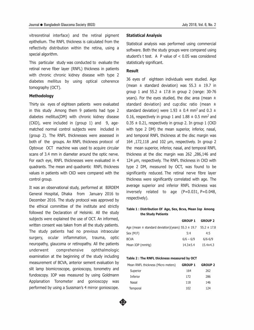
36 eyes of eighteen individuals were studied. Age
(mean ± standard deviation) was 55.3 ± 19.7 in
group 1 and 55.2 ± 17.8 in group 2 (range: 30-76
years). For the eyes studied, the disc area (mean ±
standard deviation) and cup:disc ratio (mean ±
standard deviation) were 1.93 ± 0.4 mm2 and 0.3 ±
0.16, respectively in group 1 and 1.88 ± 0.5 mm2 and
0.35 ± 0.21, respectively in group 2. In group 1 (CKD
with type 2 DM) the mean superior, inferior, nasal,
and temporal RNFL thickness at the disc margin was
164 ,172,118 ,and 102 µm, respectively. In group 2
the mean superior, inferior, nasal, and temporal RNFL
thickness at the disc margin was 262 ,286,146 and
124 µm, respectively. The RNFL thickness in CKD with
type 2 DM, measured by OCT, was found to be
significantly reduced. The retinal nerve fibre layer
thickness were significantly correlated with age. The
average superior and inferior RNFL thickness was
inversely related to age (P=0.031, P=0.048,
respectively).
Table 1 : Distribution Of Age, Sex, Bcva, Mean Iop Among
the Study Patients
GROUP 1 GROUP 2
Age (mean ± standard deviation)(years) 55.3 ± 19.7 55.2 ± 17.8
Sex (M:F) 5:4 4:5
BCVA 6/6 – 6/9 6/6-6/9
Mean IOP (mmHg) 14.3±5.4 15.4±4.3
Table 2 : The RNFL thickness measured by OCT
Mean RNFL thickness (Micro meters) GROUP 1 GROUP 2
Superior 164 262
Inferior 172 286
Nasal 118 146
Temporal 102 124

R MannanA Study of Retinal Nerve Fiber Layer (RNFL) Thickness Changes in Patients of Type 2 Diabetes Melitus with Chronic Kidney Disease (CKD) Measured by Optical Coherence Tomography
13
Fig.1 showing changes in RNFL thickness with age. Here,
(series 1 : patients with CKD & DM, series 2: normal subjects)
Discussion
Our study confirms the excellent reproducibility of
retinal thickness measurements using the OCT. Our
reproducibility values are in good agreement with
several other studies already published in the
literature,2,4,5 Our study was designed to evaluate the
retinal nerve fiber layer (RNFL) thickness changes in
patients with chronic kidney disease (CKD) with type
2 diabetes mellitus by using optical coherence
tomography (OCT). When it occurs over time, a
glaucomatous narrowing of the rim could be missed
or incorrectly measured in primary diagnosis or in
follow up examinations. For the eyes studied the disc
area (mean ± standard deviation) and cup:disc ratio
(mean ± standard deviation) were 1.93 ± 0.4
mm2 and 0.3 ± 0.16, respectively in group 1 and
1.88 ± 0.5 mm2 and 0.35 ± 0.21, respectively in
group 2. In group 1 (CKD with type 2 DM): the
mean superior, inferior, nasal, and temporal RNFL
thickness at the disc margin was 164,172,118 and
102 µm respectively. In group 2 : the mean superior,
inferior, nasal, and temporal RNFL thickness at the
disc margin was 262, 286,146 and 124 µm,
respectively. The RNFL thickness in CKD with type 2
DM, measured by OCT, was found to be significantly
reduced. Although there is paucity of literature and
supportive studies of the same subject, there are
some studies that have considered patients of chronic
renal failure. Additionally, we found a significant
deterioration of the OCT. RNFL thickness
measurements deteriorate with increasing age. This is
not due to lens opacity, because lens opacity also
would affect the measurements of the retinal
thickness. The mean values of RNFL thickness are
within the range already described in the literature.
Schumann5 for example found a RNFL thickness of
91.5 µm in the temporal parapapillary area. In the
study he used a circular OCT scan around the optic
disc with a diameter of 3.37 mm. In another study he
found a RNFL thickness of 126 µm in the temporal
area, but this study included only 26 eyes.6 We found
a highly significant correlation of the RNFL thickness
with age. The RNFL thickness decreased by 0.44.µm
per year. Obviously, about 80% of the changes in
retinal thickness over time are caused by a shrinkage
of the RNFL.The question, whether there is a
decrease in RNFL thickness with age has already
been addressed in several other studies.1,5,9-11 A
direct comparison is only possible with the study of
Schumann et al,5 because they used OCT.
Schumann5 examined 59 eyes of 33 subjects. He
found a RNFL thickness decrease for the peripapillary
RNFL thickness (p <0.03) and the temporal RNFL
thickness (p <0.0001). Like in our study he did not
find any differences between men and women.
Poinooswamy et al11 examined 150 healthy
volunteers of different ages using scanning laser
polarimetry. They found a progressive reduction of
the RNFL thickness with increasing age. The data
presented in their study indicate a significant
reduction of the RNFL thickness of 0.38 µm/year. In
our study we found a very similar value of 0.43 µm
per year. Balazsi et al1 and Mickelberg et al4 counted
the axons of 16 respectively 22 normal human eyes.
They found an axon fibre loss in the optic nerve of
4909 and 5637, respectively, per year. This may be
interpreted as a qualitative confirmation of our
findings, because any loss of axons should lead to a
decrease in RNFL thickness.Jonas et al,9 using red
free photographs, also found a correlation between
the visibility of the retinal nerve fibre bundles and
age. But there are a few studies that did not find a

July 2018, Vol. 6, No. 2Journal ● Bangladesh Glaucoma Society (BGS)
14
correlation between RNFL/retinal thickness and age.
Varma et al8 performed histological examinations of
10 normal enucleated human eyes. They only found a
significant correlation with age in the superior-nasal
and inferior-temporal region. This, however, may be
explained by the small number of eyes examined.
Limitation of the study
If our sample size were bigger, the statistical outcome
would have been more efficient.
Conclusions
The RNFL thickness in chronic kidney disease (CKD)
with type 2 diabetes mellitus (DM), measured by OCT,
was found to be significantly reduced. If our sample
size were larger,we could have concluded the study
more effectively and the statistical outcome would
have been more efficient.The presence of CKD can be
a source of false positive results and lead to
inaccurate measurement of glaucomatous optic
neuropathy.
References
1. Balazsi AG, Rootman J, Drance SM, et al. The effect of age on
the nerve fiber population of the human optic nerve. Am J
Ophthalmol 1984;97:760–6.
2. Hee MR, Izatt JA, Swanson EA, et al. Optical coherence
tomography of the human retina. Arch Ophthalmol 1995;
113:325–32.
3. Fujimoto JG, Pitris C, Boppart SA, et al. Optical coherence
tomography: an emerging technology for biomedical imaging
and optical biopsy. Neoplasie 2000;2:9–25.
4. Mikelberg FS, Drance SM, Schulzer M, et al. The normal human
optic nerve. Axon count and axon diameter
distribution. Ophthalmology 1989;96:1325–8.
5. Schuman JS, Hee MR, Puliafito CA, et al. Quantification of nerve
fiber layer thickness in normal and glaucomatous eyes using
optical coherence tomography. Arch Ophthalmol 1995;
113:586–96.
6. Schuman JS, Pedut-Kloizman T, Hertzmark E, et al.
Reproducibility of nerve fiber layer thickness measurements
using optical coherence tomography. Ophthalmology 1996;
103:1889–98.
7. Dichtl A, Jonas JB, Naumann GO. Retinal nerve fiber layer
thickness in human eyes. Graefes Arch Clin Exp Ophthalmol
1999;237:474–9.
8. Varma R, Skaf M, Barron E. Retinal nerve fiber layer thickness
in normal human eyes. Ophthalmology 1996;103:2114–19.
9. Jonas JB, Nguyen NX, Naumann GO. The retinal nerve fiber
layer in normal eyes. Ophthalmology1989;96:627–32.
10. Mistlberger A, Liebmann JM, Greenfield DS, et al. Heidelberg
retina tomography and optical coherence tomography in
normal, ocular hypertensive, and glaucomatous
eyes. Ophthalmology 1999;106:2027–32.
11. Poinoosawmy D, Fontana L, Wu JX, et al. Variation of nerve
fibre layer thickness measurements with age and ethnicity by
scanning laser polarimetry. Br J Ophthalmol 1997;81:350–4.

Journal ● Bangladesh Glaucoma Society (BGS)
15
Visual outcome after manual small incision cataract surgery for Phacomorphic glaucoma
S M Noman1
Author Information :1Dr. Shams Mohammad Noman
FCPS, DCO
Original Article
Abstract
Aim : To evaluate the visual outcome after manual small incision
cataract surgery (MSICS) as a treatment of phacomorphic glaucoma.
Method : The study included 44 patients with phacomorphic
glaucoma treated by manual small incision cataract surgery with
intraocular lens implantation. Preoperative and postoperative visual
acuity and intraocular pressure have been recorded and compared at
the end of six weeks after surgery.
Result : The mean preoperative intraocular pressure was 35.20 (±
10.86) mm of Hg. There were no s ign i f icant int raoperat ive
complications such as posterior capsular tear or expulsive hemorrhage.
Post operative mean intraocular pressure (IOP) was 12.48 (± 3.45)
mm Hg. There was a statistically significant difference between IOP
at presentation and IOP at last presentation.(P-<O.OOO1).Pre
operative visual acuity in all the affected eyes were perception of light
with projection of rays in all quadrant. Postoperative best corrected
visual acuity was 6/6-6/18 in 28 patients (62.80%), 6/24- 6/36 in 10
patients (23.25%) and � 6/60 in 6 patients (13.95%).
Conclusion : Manual small incision cataract surgery is a safe and
effective method of treatment with minimal or no complications for
phacomorphic glaucoma and the visual outcome and IOP reduction is
satisfactory.
Introduction
Cataract is the most common cause of avoidable
blindness in the world. Bangladesh is one of the
densely populated developing countries having about
700 thousand people blind. Cataract contributes 80 %
of total blindness in Bangladesh1. Limited health care
facility and other socioeconomic factors influence the
patients for late presentation, sometimes with
complication l ike phacolytic and phacomorphic
glaucoma.
The definitive treatment for phacomorphic glaucoma is
cataract extract ion6,8. Surgery in a pat ient of
phacomorphic glaucoma has to face some challenges.
High intraocular pressure, increases the risk of
expulsive haemorrhage, hypermaturity of lens is often
associated with zonulolysis which makes surgery
technically more difficult. Shallow anterior chamber
may cause corneal touch that makes the cornea hazy..
Phacoemulsification is not possible for high intraocular
pressure as well as for the inflammation. Conventional
extracapsular cataract extraction(ECCE) has some
limitations. Manual small incision cataract surgery is
suitable for such phacomorphic glaucoma cases as it is
relatively easy to approach and manage. The aim of
our study is to evaluate the visual outcome and
intraocular pressure after manual small incision
cataract surgery in the management of phamorphic
glaucoma.
Methods
This is a retrospective review of case series and was
conducted at the glaucoma clinic of Chittagong Eye
Infirmary and Training Complex, Bangladesh and was
approved by institutional review board. A total of 43
patients with phacomorphic glaucoma were included
in this study. The patients with inaccurate perception
of light, combined mechanism where primary angle
closure glaucoma was also associated, zonular dialysis
and subluxation of lens where intraocular lens
implantation was not possible were excluded from the
study. Most of the patients in this study group came
f r om r emo te a r ea w i t h poo r s o c i o e conom i c
background.
All of the patients presented with gradual loss of
vision followed by sudden acute onset of pain with
sudden loss of residual vision, redness in the affected
eye. The diagnostic features were shallow anterior
chamber and hypermature cataract with perception
of light and raised intraocular pressure. Conjunctival
congestion, corneal edema and inflammation in the
anterior chamber were found in all cases. Lenticular
changes were capsular calcification or thinning,
c o r t i c a l l i q u e f a c t i o n , s w e l l i n g o f t h e l e n s ,
phacodonesis due to zonular weakness. All patients
were treated medically prior surgery to reduce
Journal ● Bangladesh Glaucoma Society (BGS) July 2018, Vol. 6, No. 2
16
inflammation and intraocular pressure.
All 44 surgeries were done by a single surgeon.
Raised intraocular pressure was usually with systemic
carbonic anhydrase inhibitor or hyperosmotic agents
pr ior to surgery to soften the eye bal l . Ocular
inflammation was reduced with frequent topical
steroid usage.
Peribulbar block was given in all cases with short and
long acting anesthetic agents. Superior rectus briddle
suture was placed and superior limbus and adjacent
conjunctiva were exposed. Fornix based conjunctival
flap was made in the superior part and bleeding blood
vessels were cauterized with wet field bipolar cautery.
A partial thickness 6mm scleral incision was made
2mm behind the l imbus and scleral tunnel was
created up to 1mm of the clear cornea. Anterior
chamber entry was done with 3.2 mm keratome.
Reformation of the anterior chamber was done to
create an environment for easy manipulation for the
next step. A small perforation was made in the upper
part of anterior capsule using a bent 26 G needle
attached with a syringe and aspiration of the liquid
cortex was done. The capsular bag was then inflated
with viscoelastic substance and either continuous
curvi l inear capsulorrhexis (where possible) or
canopener capsulotomy was done.
The tunnel was enlarged on either side up to 6 mm
with the help of crescent knife. The nucleus was
prolapsed in the anter ior chamber by rotat ion
technique and removed by irrigating vectis. After
aspiration of remaining cortex with simco cannula,
anterior chamber and capsular bag was reformed with
viscoelastic substances. A 6 mm PMMA lens was then
inserted in the capsular bag and proper positioning
was done by dialer. Aspiration of viscoelastic material
was done and anterior chamber was reformed with
ringer lactate solution. Self sealed limbal wound was
covered with conjunctival flap.
Post operatively all patients were treated with topical
cycloplegic, steroid and antibiotic. Total ophthalmic
examination was done on first post operative day and
then one week and s ix weeks after operat ion.
Detailed ophthalmic examination was done in each
follow up.
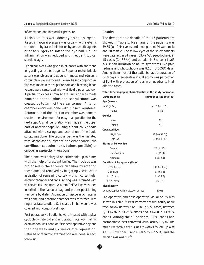
Results
The demographic detai ls of the 43 patients are
showed in Table 1: Mean age of the patients was
59.65 (± 10.44) years and among them 24 were male
and 20 female. The fellow eyes of the study patients
were cataract in 24 cases (53.49 %), pseudophakic in
15 cases (34.88 %) and aphakic in 5 cases (11.63
%). Mean duration of acute symptoms like pain
redness and photophobia was 8.18(±3.60SD) days.
Among them most of the patients have a duration of
0-10 days. Preoperative visual acuity was perception
of light with projection of rays in all quadrants in all
affected cases.
Table 1: Demographic characteristics of the study population
Demographics Number of Patients (%)
Age (Years)
Mean (± SD) 59.65 (± 10.44)
Range 40-85
Gender
Male 23
Female 20
Operated Eye
Right Eye 20 (46.52 %)
Left Eye 23 (53.48 %)
Status of Fellow Eye
Cataract 23 (53.49)
Pseudophakia 15 (34.88)
Apahakia 5 (11.63)
Duration of Symptoms (Days)
Mean (± SD) 9.18 (± 3.60)
0-10 Days 31 (69.8)
11-16 days 11 (25.6)
17-22 days 2 (4.7)
Visual acuity
Light perception with projection of rays 100%
Pre-operative and post-operative visual acuity was
shown in Table-2: Best corrected visual acuity at six
week follow up was ≥ 6/18 in 62.80% cases, between
6/24-6/36 in 23.25% cases and < 6/60 in 13.95%
cases. Among the al l pat ients 86% cases had
postoperative best corrected visual acuity ³ 6/36. The
mean refractive status at six weeks follow up was
+1.50D cylinder (range +0.5 to +2.5 D) and the
median axis was 1800.

S M NomanVisual outcome after manual small incision cataract surgery for Phacomorphic glaucoma
17
Table 2 : Comparison of pre-operative and postoperative
visual acuity
Visual Acuity Pre-Op Post-op
Uncorrected Best Corrected
6/6-6/18 0 (0.0%) 28 (62.80%)
6/24-6/36 0 (0.0%) 10 (23.25%)
< 6/60 43 (100.0%) 6 (13.95%)
The pre operative intraocular pressure ranges from
22-60 mm Hg with the mean of 36.23 (± 10.86) mm
Hg. Post operative intraocular pressure ranges from
5-22 mm Hg with the mean of 12.5 (±3.45) mm Hg.
(Table: 3).
Table 3: Comparison of pre-operative and postoperative IOP
IOP mmHg Preoperative
Mean 35.20 (± 10.86)
Range 22 - 60
20 - 29 11 (25.6 %)
30 - 39 13 (30.2 %)
40 - 49 13 (30.2 %)
50 - 60 6 (14.0 %)
IOP mmHg Postoperative
Mean 12.48 (± 3.45)
Range 5 - 22
5 - 8 6 (14.0%)
9 - 12 18 (41.9%)
13 - 16 16 (37.2%)
17 - 22 3 (7.0%)
Discussion
The result of this study showed good visual outcome
after manual small incision cataract surgery in
patients with phacomorphic glaucoma. Phacomorphic
glaucoma is caused by an obstruction of pupil by
swollen lens. Extracapsular cataract extraction needs
large incision and more surgical manipulation. So
there is always a high risk of expulsive hemorrhage
and severe post operative inflammation.
Phacoemulsification is not suitable in phacomorphic
glaucoma, because the nucleus is swollen, very
shallow anterior chamber, high IOP, compromised
zonules as well as severe inflammation in almost all
cases. There is also a risk of endothelial damage,
zonular dialysis, and posterior capsular tear. But in
MSICS causing less stress on the zonules, does not
need expensive equipments and anterior chamber is
more stable due to shelving scleral wound. In this
study MSICS gives satisfactory uncorrected vision as it
has a low range of post operative astigmatism.
At six weeks visit, 28 patients (62.80%) had best
corrected visual acuity (BCVA) of ≥ 6/18, 10 patients
(23.25%) had BCVA between 6/24-6/36 and 6
patients had<6/60. This compares favorably with
other series in which ECCE was performed in the lens
induced glaucoma4,12,19,20. Post operative visual
acuity was not appreciable in 6 patients in comparison
to others. The reason behind the poor visual
Figure-1 : Phacomorphic glaucoma at presentation
Figure-2 : Phacomorphic glaucoma -6 weeks after surgery
outcome in this group was late presentation of the
patient which causes more inflammation and corneal
decompensation due to prolong raised pre operative
intraocular pressure. The mean post operative
astigmatism of our patient is comparable to a series
where MSICS was performed in 191 eyes of lens
induced glaucoma where the mean astigmatism was
1.20D20. Most of the cases in our study the steep
axis was 1800, whose vision were improved with
refraction, possibly due to relaxation caused by the
superior scleral incision.

July 2018, Vol. 6, No. 2Journal ● Bangladesh Glaucoma Society (BGS)
18
The result of post operative visual acuity in our study
group is also similar to the result of Venkatesh et al
where they showed the post operative outcomes of
33 patients after MSICS in lens induced glaucoma
cases.23 The result is also very much similar with the
study of Ramakrishanan R who showed the post
operative visual outcome as well as IOP control after
MCECS in phacomorphic glaucoma cases.24 Post
operative IOP in all cases was controlled without the
need for long term anti-glaucoma medications. This is
similar to other studies where ECCE performed for
lens induced glaucoma.4,12,19
Conclusion
In a developing country like Bangladesh, phacomorphic
glaucoma is not an uncommon disease due to limited eye
care facilities, ignorance and also economical barrier. Our
study demonstrates that, MSICS is a safe and effective
treatment for the patient with phacomorphic glaucoma
due to satisfactory post operative visual outcome and
adequate control of intraocular pressure without anti
glaucoma medication.
References
1. B.P. Dineen, R.R.A. Bourne, S.M Al i , D.M Noarul Huq,
G.J.Johnson. Prevalence and causes of blindness and visual
impairment in Bangladesh adults: result of the National
B l indness and Low Vis ion Survey of Bangladesh. Br J
Ophthalmol 2003; 87:820–828.
2. Murthy GV, Gupta SK, Bachani D, et al. Current estimates of
blindness in India. Br J Ophthalmol 2005;89:257–60.
3. Dandona L, Dandona R, Naduvilath T, et al. Is the current eye-
care policy focus almost exclusively on cataract adequate to
deal with blindness in India? The Lancet 1998;74:341–3.
4. Jose R. National Programme for control of blindness. Indian J
Community Health 1997; 3:5–9.
5. Minassian D, Mehra V. 3. 8 million blinded by cataract each
year: Projections from the first epidemiological study of
incidence of cataract blindness in India, Br J Ophthalmol
1990;74:341–3.
6. Lane SS, Kopietz LA, Lindquist TD, et al. Treatment of
phacolytic glaucoma with extracapsular cataract extraction.
Ophthalmology 1988; 95:749-3.
7. Dada VK, Sindhu N. Management of cataract- A revolutionary
change that occurred during last two decades. J Indian Med
Association 1999;97:313–7.
8. Gogate PM, Deshpande M, Wormald RP, et al. Extracapsular
cataract surgery compared with manual small incision cataract
surgery in community eye care setting in western India: a
randomised controlled trial. Br J Ophthalmol 2003;87:667–72.
9. Venkatesh R, Muralikrishnan R, Civerchia L, et al. Outcomes of
high volume cataract surgeries in a developing country. Br J
Ophthalmol 2005;89:1079–83.
10. Natchiar G, DabralKar T. Manual small incision suture less
cataract surgery-An alternative technique to instrumental
phacoemulsification. Operative Techniques Cataract Refract
Surg 2000;3:161–70.
11. Muralikrishnan R, Venkatesh R, Prajna NV, et al. Economic
Cost of Cataract Surgery Procedures in an Established Eye
Care Centre in Southern India. Ophthalmic Epidemiol
2004;11:369–80.
12. Mandal AK, Gothwal VK. Intraocular pressure control and
visual outcome in patients with phacolytic glaucoma managed
by extracapsular cataract extraction with or without posterior
chamber intraocular lens implantation. Ophthalmic Surg Lasers
1998;29:880-9.
13. Oxford Cataract Treatment and Evaluation Team. Use of
grading system in evaluation of complications in a randomized
controlled trial. Br J Ophthalmol 1986;70:411–4.
14. Flocks M, Littwin CS, Zimmerman LE. Phacolytic glaucoma: a
clinicopathologic study of one hundred thirty-eight cases of
glaucoma associated with hypermature cataract. Arch
Ophthalmol 1955;54:37–45.
15. Epstein DL, Jedziniak JA, Grant WM. Obstruction of outflow by
lens particles and by heavy-molecular-weight soluble lens
proteins. Invest Ophthalmol Vis Sci 1978;17:272–7.
16. Speaker MG, Guerriero PN, Met JA, et al. A case-control study
of risk factors for intraoperative suprachoroidal expulsive
hemorrhage. Ophthalmology1991;98:202-9.
17. Kothari K, Jain SS, Shah NJ. Anterior capsular staining with
Trypan blue for Capsulorrhexis in mature and hypermature
catarac ts . A pre l im inary s tudy. Ind ian J Ophtha lmol
2001;49:177–80.
18. Venkatesh R, Das MR, Prashanth S, et al. Manual small incision
cataract surgery in white cataracts. Indian J Ophthalmol
2005;53:181–4.
19. Prajna NV, Ramakrishnan R, Krishnadas R, et al. Lens induced
glaucomas –Visual results and risk factors for final visual
acuity. Indian J Ophthalmol 1996;44:149–55.
20. Singh G, Kaur J, Mall S. Phacolytic glaucoma--its treatment by
planned extracapsular cataract extraction with posterior
chamber intraocular lens implantation. Indian J Ophthalmol
1994;42:145–7.
21. Gogate PM, Kulkarni SR, Krishniah S,et al. Safety and efficacy
of Phacoemulsification compared with manual small incision
cataract surgery by a randomized control cl inical tr ial.
Ophthalmology 2005;112:869–875.
22. Thomas R, Braganza A, George T, et al. Vitreous opacities in
phacolytic glaucoma. Ophthalmic Surg Lasers 1996;27:839–43.
23. Rengaraj Venkatesh, Colin S H Tan, Thangavel Thirumalai
Kumar, Ravilla D Ravindran. Safety and efficacy of manual
small incision cataract surgery for phacolytic glaucoma Br J
Ophthalmol 2007;91:279–281.
24. Ramkrishanan R.Maheshwari D et a l .Visualprognosis,
intraocular ressure control and complications in phacomorphic
g l aucoma fo l l ow ing manua l sma l l i n c i s i on ca ta rac t
surgery.Indian J Ophthalmol.2010.Jul-Aug;58(4):303-306

Journal ● Bangladesh Glaucoma Society (BGS)
19
Reduction of IOP & other associated ocular parameters following�Phaco-emulsification in ACG
S M Hossain1
Authors Information :1Sheikh Mohammed Hossain, M.S.
Original Article
Purpose : To evaluate the ocular bio-metric parameters in conjunction
with Intraocular Pressure (IOP) reduction after phaco-emulsification.
Design: Prospective Observational study.
Methods: A total of 25 patients who had undergone uneventful
phacoemulsification were included in the study. IOP and ocular bio
metric parameters were checked�pre-operatively and four months post-
operatively using Goldmann applanation tonometry, optical biometry
and anterior segment optical coherence tomography. The relationship
between IOP change and the parameters, including preoperative IOP,
anterior chamber depth, axial length, angle opening and lens thickness
was evaluated.
Results: The mean patient age was 62.3 years. The average change
in IOP was - 3.24 mm Hg. Preoperative IOP, anterior chamber depth,
angle opening in degrees and lens thickness were significantly
associated with IOP change. The axial length was not associated with
IOP reduction.
Conclusion: Preoperative IOP, lens thickness and parameters like
anterior chamber depth and angle opening in degrees were
significantly associated positively with reduced IOP after phaco-
emulsification cataract surgery.
Financial Disclosure: The author has no proprietary or commercial
interest in any of the materials discussed in this article.
Cataract or Clear lens Extraction has been suggested
as a treatment option for different spectra of Primary
angle closure diseases. Terminology to describe PACG
is confusing and this lack of clarity influences how we
think about the disease. Currently 4 angle closure
categories. Three requires specific Gonioscopic
findings. There is no firm agreement on how many
quadrants must have ITC for angle closure to be
present, but current consencus appears to be that at
least 1800 is required.
Progression of Angle Closure with/ or glaucoma
decreases by helping to open the angle and control
the IOP. Besides PACG, the other glaucoma subsets
which have shown best pressure - lowering effects
following phacoemulsification are OHT, PXS, POAG
Dr. Shingleton said “Phaco offers Surgeons another
option individualizing Glaucoma treatment.” However
few studies have examined the relationship between a
reduction in IOP and ocular biometric or pathologic
changes
Aging crystalline lens > Enlarging lens compress the
TMW > Collapses schlemm’s canal as it presses
forward > Outflow channel slowly begins to fail >
Raised IOP> Phaco + PCIOL > Compression released
> Opening of TMW > changes anterior segment
configuration > decrease IOP.
Here we attempt to determine effects of cataract
surgery on IOP, identify ocular biometric parameters
that effect the reduction in IOP after phaco in ACG
Methods
All patients and their parents were informed
accordingly and gave written informed consent to
participate in this study in accordance with
institutional guidelines. In this hospital based
prospective observational study, we included 25
consecutive patients who had non hypertensive IOP
values (10-19 mmHg) before surgery and who were
scheduled to undergo cataract surgery between 3rd
December, 2015 and 15th November, 2016 in
Glaucoma department at National Institute of
Ophthalmology & Hospital, Dhaka, Bangladesh.
Subjects who have been diagnosed with uveitis,
retinal diseases, congenital anomalies, Ocular trauma,
Intra Ocular Surgeries were excluded. 2.2 mm clear
corneal temporal incision phaco-emulsification with
posterior chamber foldable intra ocular lens surgeries
were singularly performed in all subjects. IOP was
measured using Goldmann applanation tonometry 1
day before and 4 months after surgery. Best
Corrected Visual Acuity (BCVA) was measured in
treated eyes 1 day before and 4 months after surgery.
Anterior Chamber Depth (ACD) and Anterior
Chamber Angle Distance (ACAD) were measured
using Anterior Segment Optical Coherence
Tomography ( AS-OCT). Lens thickness and axial
length were measured using an IOL Master. All
biometric values were assessed by single physician.
Anterior chamber depth in millimeter and angle
opening distance in degree were measured using AS-
OCT (Heidelberg Engineering, Germany). Axial length
and lens thickness were measured using IOL master

Journal ● Bangladesh Glaucoma Society (BGS) July 2018, Vol. 6, No. 2
20
(Carl Zeiss, Germany).
Statistical analysis was performed using a
commercially available statistical software package.
The mean values are presented as means (±)
standard deviation. Paired ‘t’ tests were performed to
determine the significance of changes in IOP.
Results
Among the 25 patients Scheduled in the analysis 17
were male and 8 female; the average age was 62.3
years. The average BCVA before surgery was 6/6 -
6/18 6 (24%), 6/24 -6/36 8 (32%), 6/60 –hand
movement 11 (44%) and the average BCVA 4 month
after surgery was 6/6 -6/18 23 (92%), 6/24 -6/36
2(8%), 6/60 – HM 00 respectively.
Table1: Patients characteristics and ocular parameters Pre-
surgery and 4 months after Phaco emulsification
Variables Before surgery After surgery p value
Age(Y) 62.3 62.3
M/F 17/8 17/8
IOP (mm Hg) 16.56±2.9 13.3±2.0 0.001
BCVA
Mode 6/60 6/6
6/6 – 6/18 6(24%) 23(92%)
6/24- 6/36 8(32%) 2(8%)
6/60 – Hand movement 11(44%) 00
ACD(mm) 2.6±0.1 4.0±0.1 0.001
Angle opening in degree 17.0±2.4 30.4±3.1 0.001
Lens thickness(mm) 4.31 N/A
Axial length(mm) 23.12 N/A
Table 2: Postoperative Intraocular Pressure Reduction After
Phacoemulsification in Primary Angle Closure Suspects
Graph: Mean IOP differences between before and after phaco
emulsification (mm Hg)
Fig:1a Pre op
Graph: Changes of IOP before and after surgery
Fig: 1b post op
Table - 1 shows the average axial length before surgery was 23.12
± 0.21 mm. The average lens thickness before surgery was 4.31 ±
0.19 mm. The average anterior chamber depth (ACD) before
surgery was 2.6 ± 0.1 (fig 1a) and post surgery 4.0 ± 0.1 mm (fig
1b). Pre operative average angle opening in degree was 17.0 ± 2.4
(fig 1a) and post operative was 30.4 ± 3.1 (fig 1b).
The average IOP among all the subjects was 16.56 ±
2.91 mmHg and the average change in IOP after 4
months of surgery was -3.24 ± 1.69 mmHg (Table 2).
References
1. Chang TC, Budnez DL, Liu A, et al. Long Term effect of Phaco
emulsification on Intra Ocular pressure using phakic fellow eye as
control. J cataract refract surg. 2012; 38:866-870
2. J Cataract refract Surg. 2015 Aug 41(8): 1725-9. doi:
10.1016/j.jcrs.2014.12.054
3. Shrivastawa A, Singh K: The effect of cataract extraction on Intra
ocular pressure. Curr opin ophthalmol 2010; 21: 118-122
4. Mansberger SL, Gordon Mo, Jampel HD, Bhorade A, Brandt Jd,
Wilson B, Kons MA; Reduction in Intra ocular pressure acute
cataract extration the ocular hypertension treatment study.
Ophthalmology; 2012; 119: 1826-1831
5. Yang HS, Lee J, Choi S: Ocular biometric parameters associated
with intraocular pressure reduction after cataract surgery in
normal eyes. Am J ophthalmol 2013; 156: 89-94.
6. Liu CJ, Chang C-Y, Ko Y-C, Lau L-I; Determinants of long term
intra ocular pressure after phaco emulsification in primary angle
clousure glaucoma. J Glaucoma 2011; 20-566-570.
7.Guan H, Mick A, Porco T, Dolan Bj : Preoperative factors
associated with IOP reduction after cataract surgery. Optom Vis
Sci 2013; 90: 178-184
8. Kim YH, Hyung SM. Effect of cataract extraction in chronic angle-
closure glaucoma patients. J Korean Ophthalmol Soc
2007;48:521- 526.
Pre op IOP
intervals
(mmHg)
Eyes Mean IOP(mmHg)±SD Paired t Test
value
p value
Preop IOP Postop IOPChange at 4
months
10-11 2 10.0±0.0 11.5±0.05 1.50±0.05 3.0 0.205
12-15 7 14.14±1.21 10.85±1.21 -3.28±1.1 -7.81 0.001
16-19 16 18.4±0.89 14.6±1.02 -3.81±0.20 -18.282 0.001
All eyes 25 16.56±2.91 13.32±2.05 -3.24±1.69 -9.58 0.001
10-11 12-15 16-19
10
14.14
18.4
11.510.85
14.6
Preop IOP Postop IOP
Journal ● Bangladesh Glaucoma Society (BGS)
21
Steroid Induced Ocular Hypertension in Post -LASIK Patients: A Matter of Great Concern
U Kawsar1, S Rahman2, I Anwar3, H Rahman4
Authors Information :1Dr. Ummay Kawser, Assistant Professor M H Samorita Medical College & Hospital, Dhaka2Dr. Siddiqur Rahman, Glaucoma Specialist and Lasik Surgeon Vision Eye Hospital, Dhaka3Dr. Ishtiaq Anwar, Glaucoma Specialist and Lasik Surgeon Bangladesh Eye Hospital4Prof. Dr. Md. Hafizur Rahman, Professor and Head of the Department Ad-Din Medical College, Dhaka
Original Article
Abstract
Aim : To investigate the fluctuation of intraocular pressure (IOP) due
totopical steroidafter laser in situ keratomileusis (LASIK).
Methods : LASIK was performedon 156 eyes of 78 patients for
correction of myopia. IOP was measured by Goldmannapplanation
tonometer before and after LASIK. All 78 patients who underwent
LASIK were enrolled as study group. These aimed at comparing the
IOP differences in pre and post-operative changes.
Results : In study group, comparison of preoperative and
postoperative IOP, the amplitude of abnormal mean IOP fluctuations
reached14.4mmHg (ranged from 18. 2 to 32.6 mmHg) for 19 patients.
After receiving treatment with Timolol 0.5%,18 patients got back
their intra ocular pressure within normal range (18.1 mmHg) at 5th
month visit.
Conclusion : Steroid is important for post LASIK inflammation
management. But it induces IOP fluctuations beyond normal range that
we observed in our study. So it is matter of great concern to evaluate
IOP not only before LASIK but also after LASIK. Immediate treatment
to lower IOP can save the sight against POAG (Primary Open Angle
Glaucoma). Unfortunately we noticed POAG for case due to steroid
use after LASIK.
Keywords : Intraocular Pressure; Primary Open Angle Glaucoma;
LASIK, Steroid
Introduction
Refractive procedures have become a popular surgical
option for the treatment of myopia. Laser in Situ
Keratomileusis (LASIK) has become the most
accepted surgical modality for the correction of a wide
range of myopia. LASIK is performed routinely in
more and more patients. LASIK corrects myopia by
altering the thickness and curvature of the central
cornea. Continuous innovations and incorporating
new technology such as latest generation
microkeratomes, femtosecond laser1, and wavefront
technology2 to this field enabled the prosperity and
progress of LASIK over the world. And clinical
outcomes of LASIK also gained traction and studied
amply. Among the vast majority of publications,
postoperative outcomes and visual quality
assessment, especially postoperative complications
have drawn much attention.3,4
Ocular hypertension is such a hot topic in this
domain. Topical corticoid is routinely prescribed for
corneal wound healing in early post - LASIK patients.
However, it may induce elevation of intraocular
pressure (IOP), and serious consequences in steroid-
sensitive patients, such as steroidinduced lamellar
keratitis5, interface fluid syndrome
6, refractive
regression7, visual acuity loss
8, and even visual field
defects9. Early recognition of these signs and
symptoms, followed by proper treatment might be a
brake to ocular hypertension and reverse
deteriorating consequences. Recently, IOP fluctuation
and variation was highlighted as a risk factor for
glaucoma progression10,11
. However, the influence of
abnormal IOP fluctuations on visual performance has
received little attention12
. We conduct this prospective
study to evaluate greater-than-normal IOP
fluctuations in steroid responders after refractive
surgery.
Subjects And Methods
78 patients who had LASIK during January 2016 to
August 2018 in Vision Eye Hospital were enrolled as
study group. Patients previous ocular surface diseases
such as corneal injury or illness, ocular surgery, any
sign of keratoconus, soft contact lens wear during the
2 week prior to presentation, and those who were
pregnant were excluded. None of the 78 patients
reported a history of systemic or ophthalmic diseases.
Journal ● Bangladesh Glaucoma Society (BGS) July 2018, Vol. 6, No. 2
22
All procedures were performed in accordance with the
ethical standards, and informed consent was obtained
from all patients prior to the study.
Laser in Situ Keratomileusis Procedure:LASIK surgery
consisted of two major steps: flap creation and laser
ablation.Flap diameters of 8. 5 to 9. 0 mm were
created with Moria One Plus microkeratome (Moria,
Antony, France) or Amadeus II (Zeimer Group)or
Wavelight FS200 femtosecond laser (Alcon, USA), and
the optical zones ranged from 6.0 to 8. 0 mm in
diameter. All eyes underwent LASIK profiles
(WavelightOculyzer II pentacam) and standard Lasik
Procedure by Allegretto EX500 excimer laser (Alcon,
USA).
Postoperative Management: Routine postoperative
management included topical Levofloxacin 0.5%
sterile eye drop 4 times a day for 4 weeks,
Dexamethasone 0.1% sterile eye drop 4 times a day
for 4 weeks and Carboxymethylcellulose Sodium 1%
sterile eye drop4 times daily for 4 weeks. After the
first post-operative day visit, all patients scheduled
next two follow-up visits at post – 1st week and
4thweek. But patients identified with ocular
hypertension had scheduled additional two more visit
at2ndmonth& 5th month and each of them had
received immediate hypotension therapy, such as
discontinuation of topical corticosteroidsin a rapid
tapering mode within 7 days and adding topical 0. 5%
Timololmaleate twice daily till to normal tension.
Postoperative Outcomes Assessment: Patients were
consecutively evaluated at postoperative 1week and
1month. IOP was measured with Goldmann
applanation tonometry (GAT) and then the corrected
IOP was calculated according to the Ehlers method by
taking into account the postoperative Pachymetry
(measured by ultrasonic Pachymetry: OcuscanRxP,
Alcon, USA) and the IOP measured with GAT13
. All
instruments involved were the same.
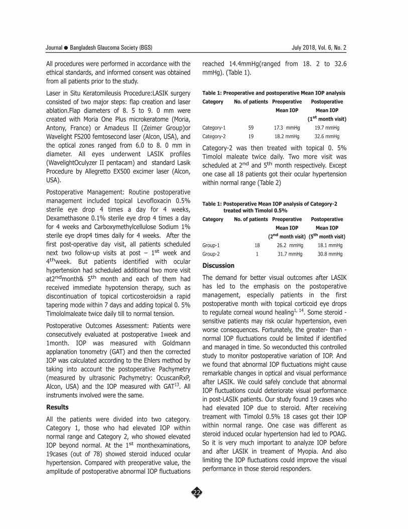
Results
All the patients were divided into two category.
Category 1, those who had elevated IOP within
normal range and Category 2, who showed elevated
IOP beyond normal. At the 1st monthexaminations,
19cases (out of 78) showed steroid induced ocular
hypertension. Compared with preoperative value, the
amplitude of postoperative abnormal IOP fluctuations
reached 14.4mmHg(ranged from 18. 2 to 32.6
mmHg). (Table 1).
Table 1: Preoperative and postoperative Mean IOP analysis
Category No. of patients Preoperative Postoperative
Mean IOP Mean IOP
(1st month visit)
Category-1 59 17.3 mmHg 19.7 mmHg
Category-2 19 18.2 mmHg 32.6 mmHg
Category-2 was then treated with topical 0. 5%
Timolol maleate twice daily. Two more visit was
scheduled at 2nd and 5th month respectively. Except
one case all 18 patients got their ocular hypertension
within normal range (Table 2)
Table 1: Postoperative Mean IOP analysis of Category-2
treated with Timolol 0.5%
Category No. of patients Preoperative Postoperative
Mean IOP Mean IOP
(2nd month visit) (5th month visit)
Group-1 18 26.2 mmHg 18.1 mmHg
Group-2 1 31.7 mmHg 30.8 mmHg
Discussion
The demand for better visual outcomes after LASIK
has led to the emphasis on the postoperative
management, especially patients in the first
postoperative month with topical corticoid eye drops
to regulate corneal wound healing1, 14
. Some steroid -
sensitive patients may risk ocular hypertension, even
worse consequences. Fortunately, the greater- than -
normal IOP fluctuations could be limited if identified
and managed in time. So weconducted this controlled
study to monitor postoperative variation of IOP. And
we found that abnormal IOP fluctuations might cause
remarkable changes in optical and visual performance
after LASIK. We could safely conclude that abnormal
IOP fluctuations could deteriorate visual performance
in post-LASIK patients. Our study found 19 cases who
had elevated IOP due to steroid. After receiving
treament with Timolol 0.5% 18 cases got their IOP
within normal range. One case was different as
steroid induced ocular hypertension had led to POAG.
So it is very much important to analyze IOP before
and after LASIK in treament of Myopia. And also
limiting the IOP fluctuations could improve the visual
performance in those steroid responders.

U KawsarSteroid Induced Ocular Hypertension in Post -LASIK Patients: A Matter of Great Concern
23
References
1. Salomao MQ, Wilson SE. Femtosecond laser in laser in situ
keratomileusis. J Cataract Refract Surg2010; 36(6):1024-1032
2. Steinert RF. History, technology, and meta-analysis.
Ophthalmology 2006; 113(11):1895-1896
3. Zhang ZH, Jin HY, Suo Y, Patel SV, Montés-MicóR, Manche EE,
Xu X. Femtosecond laser versus mechanical microkeratome
laser in situ keratomileusis for myopia: Metaanalysis of
randomized controlled trials. JCataract Refract
Surg2011;37(12):2151-2159
4. Fares U, Suleman H, Al-Aqaba MA, Otri AM, Said DG, Dua HS.
Efficacy, predictability, and safety of wavefront-guided refractive
laser treatment: Metaanalysis. J Cataract Refract
Surg2011;37(8):1465-1475
5. Levinger E, Slomovic A, Bahar I, SlomovicAR. Diagnosis of
steroidinduced elevated intraocular pressure and associated
lamellar keratitis after laser in situ keratomileusis using optical
coherence tomography. JCataract Refract Surg2009;35(2):386-
388
6. Moya Calleja T, Iribarne Ferrer Y, Sanz Jorge A, SedóFernandez
S. Steroid-induced interface fluid syndrome after LASIK. J
Refract Surg2009;25(2):235-239
7. Stewart JM. Mechanism of myopic shift associated with high
IOP after LASIK. J Cataract Refract Surg2006;32(7):1075-1076
8. Frucht - Pery J, Landau D, Raiskup F, Orucov F, Strassman E,
Blumenthal EZ, Solomon A. Early transient visual acuity loss
afterLASIK due to steroid-induced elevation of intraocular
pressure. J Refract Surg2007;23(3):244-251
9. Shaikh NM, Shaikh S, Singh K, Manche E. Progression to end-
stage glaucoma after laser in situ keratomileusis. J Cataract
Refract Surg2002; 28(2):356-359
10. Singh K, Shrivastava A. Intraocular pressure fluctuations: how
much do they matter? CurrOpinOphthalmol2009;20(2):84-87
11. Caprioli J, Coleman AL. Intraocular pressure fluctuation a risk
factorfor visual field progression at low intraocular pressures in
the advanced glaucoma intervention study. Ophthalmology
2008;115(7):1123-1129.e3
12. Xin C, Wang NL, Qiao LY. The influence of persistent high
intraocular pressure on wavefront aberration. Ophthalmol CHN
2007;16:276-278
13. Ehlers N, Bramsen T, Sperling S. Applanation tonometry and
central corneal thickness. ActaOphthalmol(Copenh)
1975;53(1):34-43
14. Du Z, Zhao W, Huang Z, Chang CK, Liu W, Liu X. Inhibition
effect of tetrandrine on haze formation after Epi - LASIK
surgery in rabbits. Curr Eye Res 2011;36(8):699-705

Journal ● Bangladesh Glaucoma Society (BGS)
24
Pediatric Glaucoma and its Medical Management
Z S Shahid1, M H Rahman2, M A Mannan3, M A Hossain4
Authors Information :1Prof. Dr. Zakia Sultana Shahid, Professor Dept. of Opthalmology,
Anwer Khan Modern Medical College & Hospital2Prof. Dr.M Hafizur Rahman, Professor & Head Dept. of
Opthalmology, Addin Medical College & Hospital3Dr. Md. Abdul Mannan, Associate Professor Dept. of Opthalmology,
Anwer Khan Modern Medical College & Hospital4Dr. Md. Almas Hossain, Associate Professor Dept. of Opthalmology,
MAG Osmani Medical College, Sylhet
Review Article
Abstract
Childhood glaucomas are challenging to diagnose. This group of
disease which is heterogeneous in type can cause a rapid loss of
vision of children or even blind. So timely recognition proper and
optimal treatment of pediatric glaucoma is very important.
Fortunately, ophthalmologists often have at their disposal the tools
needed to diagnose and manage these children.
Successful control of IOP is crucial and challenging .Most often
achieved surgically, with medical therapy as a supportive role.
Key Words : Pediatric glaucomas, medical therapy, amblyopia, IOP.
Introduction
Pediatric glaucoma signs vary largely among children
depending on the severity of the IOP (Intraocular
Pressure) rise, suddenness, and the age of the child.
Clinical studies indicate common signs such as corneal
edema, megalo-cornea, buphthalmos, optic nerve
cuping, conjunctival injection, anisometropia, myopia,
strabismus, and amblyopia. At the first year of life,
glaucoma is normally supposed owing to the
symptoms that are associated with secondary corneal
changes. Equally, children who are considerably older
are identified with the disease through the loss of
vision caused by chronic glaucoma as well as signs of
vomiting and pain associated with acute glaucoma.
Similarly, the elevation of IOP is needed to ascertain
the presence of pediatric glaucoma as the range of
normal IOP in childhood approximates the normal
adult range, and it is rare to have measurements
greater than 22 mm Hg or below 10 mm Hg, and
these measurements are essential in the management
of children with glaucoma10
. In infantile glaucoma,
the iris indicates a more anterior insertion than one of
a normal infant, showing altered translucency of the
angle face yielding a unique ciliary body band, sclera
spur and trabecular mesh. Furthermore, corneal
opacification and swelling are common signs of
glaucoma in infants. Children with pediatric glaucoma
can also be seen to withdraw from light, burying their
heads to prevent exposure to light as well as rubbing
of eye.
This article reviews the current and up to date
standards in the treatment of glaucoma among the
pediatric population, focusing on ongoing researches
on medications used in pediatric patients, the current
guidelines of treatment and a provision of
recommendatory effective medication.
Classification
There are many classifications of childhood glaucoma,
but they can simply be classified as Primary, in which
a developmental abnormality of the anterior chamber
(AC) angle only exists. Primary glaucoma in children is
generally divided based on age of onset into primary
congenital glaucoma (PCG), from birth to early
childhood, and juvenile primary open-angle glaucoma
(JOAG), from 4 years to early adulthood. Secondary
glaucoma includes a variety of conditions resulting
from damage to the aqueous outflow system due to
congenital or acquired ocular diseases or systemic
disorders.
The worldwide prevalence of childhood blindness
ranges from 0.03% in high-income countries to
0.12% in undeveloped countries. The causes of
severe visual impairment and blindness are varied and
complex: glaucoma accounts for 4.2%–5.0% of
blindness in the pediatric population.2,4
Medical therapy of Pediatric glaucoma
Medical therapy is usually allocated to a supportive
role in the management of pediatric glaucoma
patients. In children, medical therapy is used to
reduce intraocular pressure temporarily or to clear the
cornea so that surgical therapy, the definitive
treatment for primary congenital glaucoma, can be
undertaken. Those patients who do require long-term
medical therapy usually have intractable disease that

Journal ● Bangladesh Glaucoma Society (BGS) July 2018, Vol. 6, No. 2
25
has not responded adequately to surgical therapy. Our
purpose is to review medical therapy of pediatric
patients. Some children with congenital glaucoma and
elevated intraocular pressure respond well to medical
therapy. In 161 eyes with congenital glaucoma,
medical therapy by itself reduced the intraocular
pressure to less than 21 mm Hg in 12% of eyes in the
short term and 10% of eyes in the long term [1].
When considering medical therapy in pediatric
patients, clinicians should evaluate the risks and
benefits of specific medications, use the minimum
dosages required for therapeutic efficacy, and follow
the patient closely for ocular and systemic side
effects.2,3
Although regulatory agencies worldwide do
not typically include children in antiglaucoma drug
approval studies, clinicians have found several
medications to be useful in treating children with
elevated intraocular pressure.
As a primary approach to childhood glaucoma,
medication alone usually has a provisional efficacy,
particularly in PCG. However, medical treatment
should be considered first-line for JOAG, uveitis-
related glaucoma, and glaucoma after cataract
removal. Different kinds of childhood glaucoma may
respond differently to topical hypotensive drug. In the
case of PCG, universally recognized as a surgical
condition, pharmacologic therapy may help to
alleviate pain and photophobia and to recover corneal
transparency for a better view of the AC angle,
especially before angle surgery.
a ) Beta - Blockers
Beta-blockers can be an excellent choice for lowering
IOP in older children because the drugs may be dosed
bd , depending upon their formulation, and usually
reduce IOP by approximately 25%. In infants and
young children, practitioners should start with the
lowest available dose
Plasma timolol levels in children after treatment with
0.25% timolol greatly exceed those in adults after
instillation of 0.5% timolol, particularly in infants.13
Increased plasma timolol levels in children are
explained by the volume of distribution of the drug,
which is much smaller in children compared with
adults. The ocular volume of the neonate is
approximately half that of the adult, reaching full size
by approximately 2 years, whereas the blood volume
of the neonate as a function of body weight is only a
small fraction of that of the adult
In addition, the infant’s immature metabolic enzyme
systems may prolong the half-life of drugs in the
neonate from two to six times beyond that of the
typical adult.14
Higher plasma levels of drug in infants
and children would be expected to increase the risk of
systemic side effects as compared with adults.
Alarming side effects of timolol therapy, such as
Cheyne-Stokes breathing and apneic spells, have
been reported, especially in infants and younger
children.15-17
Provocation of asthma has been
associated with timolol treatment. Betaxolol, a
selective b1 antagonist, reduces the risk of pulmonary
side effects in adults as compared with timolol, but its
effect on children is not known. Likewise, the effects
of long-term use of any of the topical beta-blockers in
children have not been reported. Timolol in 0.25%
and 0.5% solutions should be used cautiously in
young glaucoma patients. Because of the possibility of
apnea, the drug should be used with extreme caution
in neonates. A detailed pediatric history and
examination to elicit the presence of systemic
abnormalities, such as bronchial asthma and cardiac
disease, should precede the use of timolol. In such
cases, therapy with a beta-blocker is contraindicated.
The use of 0.25% timolol instead of 0.5% timolol is
strongly recommended to reduce the risk of side
effects. Additionally, a significant reduction in the
systemic absorption of timolol has been observed
when performing punctal occlusion and simple eyelid
closure after drop administration.13
Although the
practice of punctal occlusion with the eyelids closed
for at least 1 minute may be considerably more
difficult to execute in children than in adults, simply
blotting off excess drops from the child’s lids may help
to minimize unwanted systemic absorption.13
Once-
daily dosing with timolol 0.25% in a gelforming
solution may similarly help to simplify the medical
regimen.
2. Cholinergic drugs or Miotics
Miotics are generally useful in children only for
constricting the pupil in order to protect the
crystalline lens during surgery and to keep the cleft
open after angle surgery in infants and young
children. Pilocarpine or echothiophate iodide may be
used in select cases of aphakic glaucoma.
Although miotic drugs enhance aqueous outflow
through the trabecular meshwork in normal patients
as well as in glaucoma patients, thereby lowering

Z S ShahidPediatric Glaucoma and its Medical Management
26
intraocular pressure, these drugs are likely not as
effective in developmental glaucoma. This reduced
efficacy in developmental glaucoma is believed to be
attributable to the abnormal insertion of the ciliary
muscle into the trabecular meshwork. In pediatric
patients, the use of pilocarpine (2% applied every 6–8
hours) has been limited.6 These drugs may benefit
aphakic and pseudophakic children with elevated
intraocular pressure, however. In addition, cholinergic
drugs serve a useful purpose in the surgical
management of pediatric glaucoma by causing
pupillary miosis before and after goniotomy.22
The
induced myopia produced by miotic therapy can cause
disabling visual difficulties. A slow-release pilocarpine
membrane delivery system, Ocusert (Alza
Pharmaceuticals, Palo Alto, California), currently not
available from the manufacturer, was helpful in some
young patients24
and the sudden release of
pilocarpine (burst effect) seldom induced myopic
spasms. The long-acting anticholinesterase drugs are
not readily available, are associated with serious
adverse effects, and offer no advantages over
pilocarpine for use in pediatric glaucoma.
Echothiophate iodide (phospholine iodide), usually
administered topically every 12 to 24 hours, is a
potent and relatively irreversible inhibitor of the
enzyme cholinesterase. Ciliary spasm and angle-
closure glaucoma have been
precipitated by the use of echothiophate iodide to
treat esotropia in a child.25
Also, the systemic
absorption of anticholinesterase agents can
significantly reduce the serum cholinesterase and
pseudocholinesterase levels. Affected patients,
particularly children, may show signs of excessive
parasympathetic nervous system activation. These
signs can include generalized weakness, diarrhea,
nausea, vomiting, excessive salivation, and decreased
heart rate. Significantly reduced systemic levels of
cholinesterase and pseudocholinesterase can be
particularly dangerous when surgery is contemplated,
because succinylcholine is commonly used as a
muscle relaxant during general anesthesia.
Succinylcholine is normally quickly hydrolyzed by
systemic cholinesterase at nerve endings. When
serum cholinesterase levels are low, however,
prolonged apnea can result because of this excess of
unhydrolyzed succinylcholine.
3. Alpha-2 Agonists
Brimonidine is absolutely contraindicated in infants
and young children due to its depression of the
central nervous system. The drug also causes
sedation and fatigue in school-aged children, so it
must be used with caution in this population, if at
all12,13
In contrast, even fairly young children usually
tolerate apraclonidine 0.5% well, and the drug can
help achieve a short-term reduction in IOP and can
decrease bleeding at the time of angle surgery.
Several noncomparative case series describing the
use of brimonidine in pediatric glaucoma patients
exist in the ophthalmic literature, whereas the
pediatric use of apraclonidine has not been described.
In 30 patients with a mean age of 10 years,
brimonidine treatment achieved a mean 7% reduction
in intraocular pressure from baseline.18
Two young
children (aged 2 and 4 years) were transiently
unarousable after administration of brimonidine, and
five other children experienced severe fatigue.18
In a
study of 23 patients with a mean age of 8 years, 18%
experienced serious systemic adverse effects
necessitating cessation of the drug.19
Four pediatric
patients have been reported to develop somnolence
after treatment with brimonidine.20
Additionally, a 1-
month-old infant experienced repeated episodes of
coma, characterized by unresponsiveness,
hypotension, hypotonia, hypothermia and bradycardia
after treatment with brimonidine.21
The a 2 agonists
are less often used in pediatric patients compared
with adult patients. The possibility of central nervous
system–mediated side effects is greater with lipophilic
drugs (eg, brimonidine) than for more hydrophilic
drugs (eg, apraclonidine), which are less likely to
cross the blood-brain barrier. Iopidine may help to
minimize intraoperative hyphema precipitated by
goniotomy.22
Brimonidine should be used cautiously in
pediatric patients, and its use should be restricted to
older children.
4. Prostaglandin-related drugs
Latanoprost seems to be quite useful for the
treatment of juvenile open-angle glaucoma and select
cases of primary congenital and aphakic glaucoma.
Prostaglandin-related drugs, specifically latanoprost,
have been evaluated in studies of a variety of
glaucoma diagnoses, including glaucoma associated
with Sturge-Weber syndrome.26-30
In 31 eyes with a
variety of glaucoma diagnoses, 6 (19%) of the
treated eyes responded with a mean reduction in
intraocular pressure of 8.5 mm Hg, which represented

July 2018, Vol. 6, No. 2Journal ● Bangladesh Glaucoma Society (BGS)
27
a 34% decrease from baseline. Most eyes were
nonresponders, however. Subjects who responded
favorably were more likely to have juvenile onset
open-angle glaucoma and to be older than
nonresponders.26
The drug was well tolerated in this
short-term study. In glaucoma associated with Sturge-
Weber syndrome, 17% to 28% of eyes treated with
latanoprost responded with a reduction in intraocular
pressure.27-28
Increased episcleral venous
engorgement was noted, and one patient (6%)
discontinued latanoprost therapy because of
intolerable hyperemia of the conjunctiva.28
Although a
decline in success over time was noted, half of the
patients were controlled at 1 year of follow-up after a
trial of latanoprost as adjunctive therapy.29
Although most children do not respond well to
latanoprost therapy, some children may experience an
appreciable hypotensive effect with treatment.30
Likewise, the once-daily dosing schedule for
latanoprost is convenient. Although local side effects
are infrequent and mild, parents and patients should
be warned about them, including iris pigmentation
changes, eyelash growth, and hyperemia. When
short-term medical therapy is planned, such as before
surgery, these local side effects are usually not a
problem. The prevalence and types of side effects
associated with long-term latanoprost use are not
known, however.
According to Wright, primary infantile glaucoma is
usually treated surgically, although medications have
a definite role as adjuvant therapy, and, rarely, as the
sole form of intervention.7 Enyedi and Freedman
study proposes Latanoprost as a treatment for
pediatric glaucoma. The study indicates that
Latanoprost is a prostaglandin F2 analog, which
decreases IOP by 20-40% with ocular hypertension in
adults or with open-angle glaucoma.12
According to
Enyedi and Freedman, juvenile-onset and older
children with open-angle glaucoma showed significant
effects of ocular hypotensive when treated
latanoprost.12
Similarly, Black et al, study endeavors
to examine the effectiveness of latanoprost in the
treatment of pediatric glaucoma2. Their study focuses
on assessing the lowering effect of IOP when using
Latanoplast and its safety in children, exposing a total
of 115 children with pediatric glaucoma for a period
of 1 month while recording any side-effect
information. Black et al. found out that out of the 115
children participants, 63 of them had IOP data that
could be interpreted and side effects were mild,
confirming the safety of using the drug in pediatric
glaucoma2. Accordingly, Black et al. indicate that
latanoprost has an IOP lowering ability in juvenile and
old children with open-angle glaucoma and a small
percentage of those with aphakic glaucoma2.
5. Carbonic anhydrase inhibitors
In adult patients, the side effects of systemic carbonic
anhydrase inhibitors are well known to clinicians. In
children, growth suppression has been associated
with oral acetazolamide therapy, and infants may
experience severe metabolic acidosis.4,5
Side effects
from the use of systemic carbonic anhydrase
inhibitors in infants and young children are not
commonly reported, although these patients may not
verbalize the occurrence of side effects to their
parents or health care providers. Oral administration
of acetazolamide suspension at a dosage of 10
(range: 5–15) mg/kg/d in divided doses (three times
daily) is usually tolerated by children, reduces
intraocular pressure, and diminishes corneal edema
before surgery.6,7
Topical versus oral carbonic
anhydrase inhibitor therapy has been evaluated for
pediatric glaucoma in a crossover design study.8 The
mean intraocular pressure was reduced by 36% and
27% compared with baseline after treatment with oral
acetazolamide and topical dorzolamide, respectively.
Although not as effective as acetazolamide in this
group of patients, topical dorzolamide caused a
significant reduction in intraocular pressure.8
Furthermore, treatment with topical dorzolamide
caused few side effects. Topical carbonic anhydrase
inhibitors are more widely prescribed compared with
systemic carbonic anhydrase inhibitors. Many
clinicians prefer twice-daily dosing to minimize the
discomfort and inconvenience to the parent and the
child associated with three times daily dosing. Taking
the fixed combination of dorzolamide with timolol
twice a day can simplify the medical regimen by
reducing the number of drops instilled per day and
may be more appropriate for older children.
5. Osmotic drugs
Glycerol is administered orally at a dose of 0.75 to 1.5
g/kg of body weight in a 50% solution.31
The
excessively sweet taste may be partially masked by
chilling the solution over ice or by using fruit juice
(commonly lemon or orange) or flavored water as a
diluent. This drug is not commonly used in the

Z S ShahidPediatric Glaucoma and its Medical Management
28
treatment of developmental glaucoma. Mannitol (20%
solution) is dosed intravenously at 0.5 to 1.5 g/kg of
body weight at approximately 60 drops per minute. A
rapid fall in intraocular pressure occurs in 20 to 30
minutes after drug administration and can last for 4 to
10 hours. Mannitol may also be used to reduce
markedly elevated intraocular pressure before surgery
in patients with developmental glaucomas refractory
to standard medical therapy.
Discussion
Various treatment options have been suggested by
various studies in the treatment of pediatric
glaucoma.1,3-5
As an adjunctive therapy, latanoprost is
tolerated with good IOP response, and although the
response rate is low in the pediatric population, in
those that do respond it is very effective and offers
good 24-hour control. According to Netland, the
frequency of these side effects in children on long-
term therapy is not well known.9 It is also noted in
the studies that timolol has been tested as an
additional medication in uncontrolled pediatric
glaucoma. As in adults, systemic levels of timolol can
be found in pediatric patients after topical dosing, but
at much higher levels and much of the plasma level
increase can be explained by the much smaller
volume of distribution in children as compared to
adults. Furthermore, various studies indicate that oral
carbonic anhydrase inhibitor administration can cause
growth retardation and metabolic acidosis.9 Moreover,
a study of children between age 3 and 12 showed an
effective lowering of IOP in pediatric glaucoma when
comparing the use of systemic administration of
topical dorzolamide and acetazolamide.
Notably, based on the study, epinephrine and
dipivefrin nonspecific adrenergic agonists, are rarely
used in pediatric patients, since, when considered for
medical therapy, systemic side effects may limit their
use in this population. Various researchers have
studied the use of brimonidine, an alpha-2-selective
agonist in the treatment of pediatric populations.
Brimonidine has shown a slight decrease in IOP with
a high rate of central nervous system depression
while in other studies, side effects were sufficient to
merit discontinuation of the therapy.5
In the use of cholinergic drugs, they were the first
medical treatment for glaucoma but are now seldom
used for pediatric patients. Topical use of both
pilocarpine and carbochol can be associated with
cholinergic side effects, including diarrhea,
gastrointestinal cramping, hypotension, headaches,
salivation, syncope, and sweating.4 The degree to
which side effects are experiences is highly
dependent on systemic absorption, which can be
greater in pediatric patients. Since the majority of
pediatric glaucomas result from structural and
developed mental abnormalities of the angle and
associated structures, these drugs may be less
effective in lowering IOP. Furthermore, pilocarpine has
been used to a limited degree in pediatric patients,
and it may be used for induction of miosis pre and
postoperatively for surgical goniotomy. Equally, long
acting anticholinesterases such as echothiophate
iodide are used mostly for the treatment of
accommodative esotropia. Netland indicates that,
since the agents are of poor availability, with no
advantages over pilocarpine and with more serious
side effects, they are seldom used for glaucoma
therapy.9
Conversely, medical treatments of pediatric glaucoma
could include oral and topical carbonic anhydrase
inhibitors, adrenergic agents, prostaglandins, and
miotics. Often, oral carbonic anhydrase inhibitors are
used to lower intraocular pressure in an attempt to
clear the cornea for goniotomy surgery.
Summary
Goals of medical therapy in pediatric glaucoma is to
decrease intraocular pressure temporarily, to clear an
edematous cornea, and to facilitate surgical
intervention.
Most pediatric patients who require long-term medical
therapy have severe disease that has not responded
sufficiently to surgical therapy, and these patients
may experience additional intraocular pressure
reduction with a medical treatment. Oviously before
commencing medical glaucoma therapy in a child,
clinicians need to consider the potential for side
effects very carefully. During treatment, children need
to be closely monitored because they may be at
increased risk of systemic side effects compared with
adults as a result of their reduced body mass and
blood volume for drug distribution. Similar caution
should be exercised when treating the pregnant
glaucoma patient or the nursing mother.
Conclusion and Recommendations
Pediatric glaucomas associated with a relatively poor
surgical outcome, such as secondary glaucoma
associated with aphakia, a medication trial is

July 2018, Vol. 6, No. 2Journal ● Bangladesh Glaucoma Society (BGS)
29
frequently warranted prior to surgical treatment.
Medical therapy is also valuable postoperatively in
controlling glaucoma in children in whom surgery is
only moderately successful. Furthermore, physicians
should be aware of the potential side effects. Topical
carbonic anhydrase inhibitors are both effective and
save adjunctive agents in the treatment of pediatric
glaucoma. Physicians need to be wary when using
topical -blockers during the neonatal period and in
premature infants since there is a definite risk of
bronchospasm and bradycardia. Furthermore, beta-
blockers should not be used in infants or children with
a history of reactive airway disease. In the treatment
of pediatric glaucoma, the consultation with the
patient’s pediatrician and pulmonologist or
cardiologist may be worthwhile if surgical or other
medical options are limited. Parasympathomimetics
and carbon anhydrase inhibitors have also been used
systematically. In addition, side effects noted in
newborns treated with glaucoma medications provide
help in choosing treatment in late stages of
pregnancy.
References
1. Turach ME, Aktan G, Idil A. Medical and surgical aspects of
congenital glaucoma. Acta OphthalmolScand 1995;73:261– 3.
2. Kong L, Fry M, Al-Samarraie M, Gilbert C, Steinkuller PG. An
update on progress and the changing epidemiology of causes of
childhood blindness worldwide. J AAPOS. 2012;16(6):501–507.
3. Palmer EA. How safe are ocular drugs in pediatrics? Ophthalmology
1986;93:1038–40.
4. Gilbert CE, Rahi JS, Quinn GE. Visual impairment and blindness in
children. In: Johnson GJ, Minassian DC, Weale RA, West SK,
editors. The Epidemiology of Eye Disease. 2nd ed. London:
Hodder Arnold; 2003:260–286.
5. Ritch R. Special therapeutic situations. In: Netland PA, Allen RC,
editors. Glaucoma medical therapy: principles and management.
San Francisco, CA7 American Academy of Ophthalmology; 1999.
p. 193– 211.
6. Hass J. Principles and problems of therapy in congenital glaucoma.
Invest Ophthalmol 1968;7:140–6.
7. Shaffer RN. New concepts in infant glaucoma. Trans Ophthalmol
Soc UK 1967;87:581– 5.
8. Portellos M, Buckley EG, Freedman SF. Topical versus oral carbonic
anhydrase inhibitor therapy for pediatric glaucoma. J AAPOS
1998;2:43–7.
9. Boger III WP, Walton DS. Timolol in uncontrolled childhood
glaucomas. Ophthalmology 1981;88:253– 8.
10. McMahon CD, Hetherington Jr J, Hoskins Jr HD, et al. Timolol and
pediatric glaucomas. Ophthalmology 1981;88:249–52.
11. Zimmerman TJ, Kooner KS, Morgan KS. Safety and efficacy of
timolol in pediatric glaucoma. Surv Ophthalmol
1983;28(Suppl):262–4.
12. Hoskins Jr HD, Hetherington Jr J, Magee SK, et al. Clinical
experience with timolol in childhood glaucoma. Arch Ophthalmol
1985;103:1163–5.
13. Passo MS, Palmer EA, Van Buskirk EM. Plasma timolol in glaucoma
patients. Ophthalmology 1984;91: 1361– 3.
14. Harte VJ, Timoney RF. Aspects of the prescribing of drugs for
children. Pharm Int 1981;2(6):132– 5.
15. Burnstine RA, Felton JL, Ginther WH. Cardiorespiratory reaction to
timolol maleate in a pediatric patient: a case report. Ann
Ophthalmol 1982;14:905–6.
16. Bailey PL. Timolol and postoperative apnea in neonates and
young infants. Anesthesiology 1984;61(5):622.
17. Olsen RJ, Bromberg BB, Zimmerman TJ. Apneic spells associated
with timolol therapy in a neonate. Am J Ophthalmol
1979;88:120–2.
18. Enyedi LB, Freedman SF. Safety and efficacy of brimonidine in
children with glaucoma. J AAPOS 2001;5:281– 4.
19. Bowman RJ, Cope J, Nischal KK. Ocular and systemic side effects
of brimonidine 0.2% eye drops (Alphagan) in children. Eye
2004;18:24– 6.
20. Levy Y, Zadok D. Systemic side effects of ophthalmic drops. Clin
Pediatr 2004;43:99– 101.
21. Berlin RJ, Lee UT, Samples JR, et al. Ophthalmic drops causing
coma in an infant. J Pediatr 2001;138: 441– 3.
22. Freedman SF. Medical and surgical treatments for childhood
glaucomas. In: Epstein DL, Allingham RR, Schuman JS, editors.
Chandler and Grant’s glaucoma. 4th edition. Baltimore,
MD7Williams &Wilkins; 1997. p. 609–22.
23. Raab EL. Congenital glaucoma. Persp Ophthalmol 1978;2:35– 41.
24. Pollack IP, Quigley HA, Harbin TS. The Ocusert pilocarpine
system: advantages and disadvantages. South Med J
1976;69:1296– 8.
25. Jones DE, Watson DM. Angle-closure glaucoma precipitated by
the use of phospholine iodide for esotropia in child. Br J
Ophthalmol 1967; 51:783– 5.
26. Enyedi LB, Freedman SF, Buckley EG. The effectiveness of
latanoprost for the treatment of pediatric glaucoma. J AAPOS
1999;3:33– 9.
27. Yang CB, Freedman SF, Myers JS, et al. Use of latanoprost in the
treatment of glaucoma associated with Sturge-Weber syndrome.
Am J Ophthalmol 1998; 126:600– 2.
28. Altuna JC, Greenfield DS, Wand M, et al. Latanoprost pediatric
glaucoma and glaucoma in pregnancy 467 in glaucoma
associated with Sturge-Weber syndrome: benefits and side-
effects. J Glaucoma 1999;8:199 –203.
29. Ong T, Chia A, Nischal KK. Latanoprost in port wine stain related
paediatric glaucoma. Br J Ophthalmol 2003;87:1091– 3.
30. Enyedi LB, Freeman SF. Latanoprost for the treatment of pediatric
glaucoma. Surv Ophthalmol 2002; 47(Suppl):S129– 32.
31. Netland PA, Kolker AE. Osmotic drugs. In: Netland PA, Allen RC,
editors. Glaucoma medical therapy: principles and management.
San Francisco, CA7 American Academy of Ophthalmology; 1999.
p. 133–47.
32. Black AC, Jones S, Yanovitch TL, Enyedi LB, Stinnett SS, et al.
(2009) Latanoprost in pediatric glaucoma--pediatric exposure
over a decade. J AAPOS 13: 558-562.
33. Wright KW (2003) Pediatric ophthalmology and strabismus. New
York: Springer
34. Khitri MR, Mills MD, Ying GS, Davidson SL, Quinn GE (2012) Visual
acuity outcomes in pediatric glaucomas. J AAPOS 16: 376-381.
35. Enyedi LB, Freedman SF (2002) Latanoprost for the treatment of
pediatric glaucoma. Surv Ophthalmol 47 Suppl 1: S129-132.
Journal ● Bangladesh Glaucoma Society (BGS)
30
Angle-closure Glaucoma : The Role of the Lens in thePathogenesis, Prevention and Treatment
M S I Prodhan1
Author Information :1Dr. Md. Safiul Islam Prodhan Associate Professor National Institute of Ophtalmology Dhaka
Review Article
Abstract
Primary angle-closure glaucoma is a major cause of blindness
worldwide. It is a disease of ocular anatomy that is related to
pupillary-block and angle-crowding mechanisms of filtration angle.
Eyes at increased risk for primary angle-closure are small with
decreased axial length, anterior chamber depth, and filtration angle
width, associated with a proportionately large lens. Angle-closure
glaucoma afflicts Asian and Eskimo eyes more frequently than eyes in
other races with similar predisposing dimensions. The treatment of
primary angle closure addresses its causal mechanisms. Laser
peripheral iridotomy equalizes the anterior and posterior pressures and
widens the filtration angle by reducing the effect of pupillary block.
Argon laser peripheral iridoplasty contracts the iris stroma to reduce
angle crowding and is helpful for some affected eyes. Lensectomy
dramatically widens the angle and eliminates pupillary block. Clinical
reports of lensectomy with posterior chamber intraocular lens
implantation in the treatment of acute, chronic, and secondary angle-
closure glaucoma describe very favorable results. The appropriate role
for lensectomy in the management of primary angle closure, however,
remains unproven. Prospective, randomized clinical trials are ongoing
to determine the value and comparative risks and efficacy of
lensectomy versus medical therapy, laser peripheral iridotomy, laser
iridoplasty, and filtration procedures for the treatment of acute and
chronic primary angle closure and for the prevention of chronic angle-
closure glaucoma, both after and in place of laser peripheral iridotomy.
Key words : angle-closure glaucoma, blindness, goniosynechialysis,
iridotomy, lensectomy, phacoemulsification.
I. Introduction
Angle closure is a disorder of ocular anatomy
characterized by closure of the drainage angle by
appositional or synechial approximation of the iris
against the trabecular meshwork, blocking its access
to aqueous humor. The final common result in related
disorders is an elevation of the intraocular pressure
(IOP), due to the secondary impairment of aqueous
humor outflow from the eye, followed by the
development of glaucomatous optic neuropathy.This
review will focus on the role of the lens in the
pathogenesis and treatment of primary and secondary
angle closure.
II. Clinical Types of Angle-closure Glaucoma
Angle-closure disorders can be divided into primary
and secondary groups. Primary angle closure includes
those that are caused by pupillary block, angle
crowding (from plateau iris configuration or anterior
lens position) or a combination of both50. A
classification endorsed by the American Academy of
Ophthalmology subdivides the primary group into
primary angle-closure suspect, primary angle closure
without optic neuropathy (PAC), and primary angle-
closure glaucoma with neuropathy (PACG)4.
Secondary angle-closure disorders are those that
occur in the presence of a second ocular disease such
as with iris neovascularization, uveitis, trauma, or lens
related conditions18.
III. Epidemiology of Primary Angle-closure Glaucoma
It has been estimated that 67 million people
worldwide are affected with a primary glaucoma and
that one-third have PACG95. In European and African
populations primary open-angle glaucoma (POAG)
occurs approximately five times more frequently than
PACG; in Chinese20,21, Mongolians19, and Indians15,
however, the rates of PACG may equal or be greater
than POAG. In Eskimos/ Inuit the prevalence of PACG
is felt to be higher than any other ethnic group12. In
Bangladesh 2% population suffers from glaucoma.
Among these PACG is almost equal to POAG. A
prevalence study reported PACG as occurring more
frequently in Italy than in the rest of the European
population (0.6 - 0.1%)22 In China alone an
estimated 3.5 million people are afflicted with the
disease, and 28 million are estimated to have
occludable drainage angles20. The incidence of PAC
increases with age and is greater in females3. The
annual rate of APAC in Singapore was highest
(68.5/100,000) for elderly Chinese women with a two
times higher rate than in males107. Angle closure is a
disease of older persons, peaking in incidence
between 55 and 70 years of age96. The risk of APAC
Journal ● Bangladesh Glaucoma Society (BGS) July 2018, Vol. 6, No. 2
31
in elderly Singaporeans greater than 60 years of age
was nine times greater compared with the 30 to 59
year old population109. PACG is more visually
destructive than POAG, and it is responsible for the
majority of the bilateral glaucoma-related blindness in
Singapore22. The proportion of those with PACG who
become blind is over 25%, which is double that of
POAG20. Angle closure glaucoma patients, presenting
with an acute attack, are felt to represent only a
minority of patients potentially visually affected by
PACG19. The predominant form of PACG is the
chronic, asymptomatic type, and because the
diagnosis does require gonioscopy, it is likely that a
large proportion of those at risk remain undiagnosed
and untreated93. Even in Singapore, where glaucoma
is responsible for 60% of blindness and the incidence
of angle closure is high, awareness of the problem is
low108.
IV. Mechanisms in Angle-closure Glaucoma
Pupillary block is the most frequent and important
mechanism responsible for angle closure14,97, but in
many cases it is not the only mechanism involved. Iris
angle-crowding may co-exist with pupillary block to
cause the angle closure. In the plateau iris
configuration, the iris is held anteriorly by the ciliary
processes, but a pupillary block component may also
be present132. These pathologic mechanisms exist
because of primary anatomic variations in the size,
position, and relationship of the anterior segment
structures (cornea, iris, ciliary body, lens), or occur
secondary to other acquired ocular pathology. For
example, the lens may shift anteriorly secondary to
trauma or to drugs that result in the formation of fluid
in the supraciliary space107, or the lens may move
anteriorly secondary to expansion of posterior
segment structures (vitreous, subretinal space,
choroid)94. Anterior traction on the peripheral iris
may pull the iris over the trabecular meshwork with
resultant blockage of access to aqueous. This
mechanism of trabecular obstruction occurs with
contraction of neovascular, inflammatory, or
proliferative fibrocellular membranes and is common
in clinical ocular conditions including rubeosis irides,
chronic anterior uveitis, aniridia, and after cataract
surgery in infancy. Because this article will only review
the role of the lens in the pathogenesis of the angle
closure, these mechanisms related to anterior traction
on the iris, which cause angle obstruction
independent of the lens, will not be further discussed.
A. PUPILLARY BLOCK MECHANISM
Aqueous humor (AH) is produced into the posterior
chamber and normally flows anteriorly between the
posterior surface of the iris and the anterior lens
capsule, to enter the anterior chamber through the
pupil, and exits through the trabecular meshwork
(TM). Relative resistance to flow of AH from the
posterior chamber (PC) into the anterior chamber
(AC) is normal and reflected by an estimated pressure
drop of 0.23 mm Hg between the PC and AC35. This
pressure differential may increase greatly when the
dimensions of the iris-lens channel are changed in
such a manner that flow of AH is more impeded113.
The pressure difference between the PC versus the
AC is an important variable that determines the iris
contour. As this pressure increment increases, the iris
becomes more convex. Clinically significant pupillary-
block is present when the increased iris convexity
brings the iris into apposition with the TM or appears
close enough to do so in the future. Extreme anterior
iris-bulging, iris bombe, would be expected with
pressure differentials of 10-15 mm Hg35. The
variables that influence the AH flow through the
‘‘pinch region’’ (iris-lens channel) and influence the
pressure differential and related iris contour have
been studied extensively.35,48,78,103,113 Changing
pupillary size within the normal range of 3 to 7 mm
was determined to have little effect, however, miosis
of 2 mm was predicted to significantly increase the
pressure differential especially in the presence of
increased channel length (O1 mm) or decreased
height (!5 mm)35,113. Increased channel length and
decreased height were associated with increased
pressure increments and were accompanied by the
expected increased iris convexity. Movement of the
iris insertion posteriorly or the lens anteriorly also was
associated with an expected increase in the pressure
differential and iris convexity, which itself lessens the
area of iris--lens canal. Other variables exist and
interact to determine the iris contour, including eye
size, especially the dimensions of the anterior
segment78, lens size and position, iris stroma and iris
musculature characteristics35, ciliary body anatomy,
and physiologic parameters including aqueous humor
flow rate, facility of outflow, vitreous--aqueous fluid
flow, and the effects of accommodation and blinking.
These potentially significant dimensional and
physiologic parameters vary and may become risk
factors of more or less importance in determining iris
contour and the development of pupillary-block in

MSI ProdhanAngle-closure Glaucoma : The Role of the Lens in the Pathogenesis, Prevention and Treatment
32
eyes which become at risk for angle closure. It should
be clear that the lens plays a pivotal role in the
pathogenesis of angle closure secondary to the
pupillary block mechanism. If an iridotomy or
iridectomy is performed the pressure differential
between the AC and PC becomes minimal and iris
deformation secondary to pupillary-block is relieved.
If the lens is removed and posterior synechiae lysed,
the prerequisite anatomical relationship for
irislenticular (pupillary) block is eliminated, and the
anterior chamber angle will widen as the iris leaf
becomes flat and rotates to a more posterior position.
B. ANGLE CROWDING
This mechanism for angleclosure may exist alone, but
more often co-exists with pupillary-block. Angle
crowding can be thought of as the sandwiching of the
peripheral iris between the trabecular meshwork and
some other structure, compared to the pupillary-block
related anterior iris shift secondary to the pressure
differential between the anterior and posterior
chamber. The clinical primary condition possessing
this mechanism is the plateau iris configuration132. In
this condition, anteriorly positioned
ciliary processes prop up the iris anteriorly resulting in
the peripheral iris being held forward in approximation
with the trabecular meshwork103. Depending on the
amount of trabecular obstruction that develops, acute
or chronic angle closure can occur. Indentation
gonioscopy reveals a characteristic double iris hump,
and ultrasound biomicroscopy (UBM) of the ciliary
body reveals the anterior position of the ciliary
processes filling the ciliary sulcus88,133. Gonioscopy
after an iridotomy (LPI) will reveal persistence of the
narrow and occludable peripheral angle. This helps
explain why creating a patent LPI to relieve the
pupillary block component in the presence of this
cause of angle crowding
may not prevent progression to ACG. In plateau iris
configuration, removal of the lens with intraocular
lens implantation increases ACD but does not change
iridociliary apposition. This explains why the
elimination of iris support by the lens also does not
cause the angle to significantly widen126. Age-related
lens changes and other ocular conditions associated
with a forward shift of the anterior lens plane and
decreased ACD can also cause significant angle-
crowding, as seen clinically in nanophthalmos,
microphthalmia, retinopathy of prematurity (ROP),
spherophakia, and axial anterior ectopia lentis.
V. Pathogenesis of Angle-closure Glaucoma and the Role of the Lens
Eyes with primary angle closure have significant
anatomic differences from normal eyes13,75. The
most significant clinical hallmarks of an eye with
angle-closure are the shallow AC and narrow angle.
The mean anterior chamber depth (ACD) in PAC eyes
is approximately 1.8 mm, which is 1 mm shorter than
in normal eyes75,125. Angle closure becomes a rarity
when anterior chamber depth exceeds 2.5 mm76.
Decreased AC volume68,79, small corneal
diameter16,124, and short axial lengths16,124, are all
characteristic of eyes with PACG. The most
satisfactory explanations for the more shallow AC is
the age related increase in lens thickness and more
anterior position of the lens75,76,90. The axial lens
thickness is greater than in normal subjects16,75,76,
and the thicker lenses are significantly more anteriorly
positioned than in normal eyes76,116. Lowe75,76,
estimated that increased lens thickness causes 0.35
mm of AC shallowing, and forward lens position
causes 0.65 mm of shallowing, accounting for the
total of 1 mm difference in AC depth of the smaller
eye compared to the normal eye. Growth of the lens,
with an increase in the number of lens fibers
continuing throughout adult life, results in an increase
in lens thickness and anterior curvature68. Ocular
biometry of Alaskan Eskimos, an ethnic group at high
risk for PAC, confirmed the presence of decreased
anterior chamber angle width, depth, and axial length
associated with increased hyperopia and lens
thickness139. The age-adjusted angle width and ACD
were significantly less than other ethnic groups. When
the biometry of contralateral eyes of patients having
an APAC were studied and compared to population-
based controls, unfavorable dimensions were found
consisting of more shallow anterior chambers and
narrow angles, and thicker lenses. These differences
were considered to explain in part the estimated 50%
risk for APAC in these eyes24. These observations
also explain the tendency of PAC to affect older
patients and its relative rarity in young adults.
Decreased ACD is accelerated in women between the
fourth and fifth decades, which may explain their
greater propensity for PAC71. Biometry and clinical
examination of PAC patients identifies anatomic risk
factors for angle closure and supports the pivotal role
of the lens position and size in the active or potential
mechanism of closure22. These assessments,
however, have not conclusively explained its more
July 2018, Vol. 6, No. 2Journal ● Bangladesh Glaucoma Society (BGS)
33
frequent occurrence in the eyes of certain ethnic
groups (Eskimos and Chinese) with dimensions similar
to eyes in other populations (whites and blacks) with
a less frequent rate of angle closure13,139. Lens
disorders of position and size as seen with lens
dislocation and spherophakia can also result in
pupillary-block and secondary angle-closure. Unlike
PAC, which usually afflicts older patients aged 50
years and above, lens-related secondary angle closure
occurs in patients of all ages. Shallowing of the
anterior chamber occurs from increased thickness and
curvature of the lens and/or forward shifting of the
dislocated lens in the pupil to cause crowding of the
angle and potential pupillary block. With an
intumescent age-related cataract or with lens swelling
after a perforating lens injury, the sheer increased
thickness and bulk of the lens can push the peripheral
iris (angle crowding) against the trabecular
meshwork. As with the plateau-iris syndrome, lens-
induced angle crowding can lead to acute or chronic
angle closure despite a patent LPI102. Argon laser
peripheral iridoplasty (ALPI) has been used to
manage acute phacomorphic angle-closure with
favorable results123,145. Extraction of the abnormal
lens in these conditions is ultimately the only way to
definitively eliminate both the angle-crowding and co-
existing pupillary block.
VI. Current Surgical Treatment Options for Primary
Angle-closure Glaucoma Understanding and caring for
patients with APAC requires repetitive careful clinical
ocular examinations including evaluation of the
filtration angle to determine the mechanism of the
angle-closure and the active stage of the disease. The
treatment of a patient with acute disease should be
followed by care to prevent the development or
worsening of chronic angle closure glaucoma. In a
patient with established synechial angle closure and
advanced glaucomatous optic neuropathy (GON),
active management of the IOP is essential.
Appropriate surgical decisions for angle closure should
be congruous with the patient’s anatomic defects,
offending pathophysiology, and the stage of disease.
Randomized clinical trials are lacking to support the
efficacy of procedures for primary angle closure
glaucoma.
A. LASER PERIPHERAL IRIDOTOMY
Laser peripheral iridotomy (LPI) eliminates the
pressure difference between the anterior and
posterior chambers and is the current standard
treatment to correct pupillary block. It is also a safe
and effective prophylaxis in suspect eyes with
occludable angles secondary to pupillary block,
including fellow eyes of APAC patients at risk for
bilateral angle closure5. Although LPI is an effective
treatment for APAC, with resultant widening of the
filtration angle and reduction of elevated IOP, it is not
reliably protective against chronic angle
closure6,84,105. In a study of the benefit of LPI in
Asian eyes, 100% had resolution of the acute attack
after the LPI, but 58.1% subsequently developed
elevated IOP requiring treatment and 32.7%
eventually needed trabeculectomy for pressure
control6. The median time interval for increase in IOP
was 5.8 months, and most of these eyes which
developed CACG had more than 180 degrees of
peripheral anterior synechiae (PAS). The failure of LPI
to prevent recurrent elevation of the IOP has been
correlated with the amount of PAS present in these
eyes51,84,143.
B. ARGON LASER PERIHERAL IRIDOPLASTY
ALPI is a procedure to induce immediate focal iris
stromal contraction to pull the iris root away from the
angle wall thus widening it. In the long term, it
produces thinning and contraction of the peripheral
iris, giving it a flatter contour. Histopathology
suggests that heat shrinkage of collagen may account
for its short-term effect and contraction of the
induced fibroblastic membrane may be responsible for
its long term effect1. Hence, it is used to reduce
angle crowding in nanophthalmos52, PAC60, lens-
induced angle closure100, and reported to be very
effective in the plateau iris configuration by
eliminating and reducing the amount of residual
appositional angle closure after LPI99. In the initial
treatment of APAC it can assist to lower the IOP by
pulling open the angle, and it can be useful also when
initial medical treatment fails101. A study of 10 APAC
patients treated with ALPI after administration of
pilocarpine and timolol showed a remarkable decrease
in IOP from 59.5 mm Hg to 21.7 mm Hg at 30
minutes and 16 mm Hg at 1 hour post-ALPI65. A
follow-up study was done to confirm the effect was
predominantly from the ALPI by managing eight APAC
patients with ALPI alone and produced similar
results122. In 2000, a randomized controlled
interventional trial comparing ALPI against
conventional systemic IOP-lowering medications in
the first-line treatment of APAC showed the ALPI
treated group had lower IOP than the medically
treated group at 15 minutes, 30 minutes, and 1 hour
after the start of treatment. The differences were
statistically significant. The differences in IOP became

MSI ProdhanAngle-closure Glaucoma : The Role of the Lens in the Pathogenesis, Prevention and Treatment
34
statistically insignificant from 2 hours onwards64. In
chronic PACG, a study with a followup period of 6
months suggested that in eyes which had failed
medical therapy, the effect of iridoplasty may not be
sustained in some eyes even though at least half the
angle was opened at the time of the procedure11. In
another report on the short-term benefit of iridoplasty
after 180 degrees goniosynechialysis in 5 eyes with
chronic PAC and total synechial angle closure,
satisfactory IOP was achieved in 80% (4 eyes), with a
mean follow-up period of 7.6 months61. Re-closure of
the angle occurred in the remaining eye with IOP
elevation. There is a paucity of data on the long-term
effect of iridoplasty in PAC. ALPI alters the peripheral
configuration permanently and may help prevent
subsequent angle closure from peripheral anterior
synechiae formation.
C. ANTERIOR CHAMBER PARACENTESIS
Immediate anterior chamber paracentesis is a
maneuver that rapidly lowers IOP in APAC
providing instantaneous relief of symptoms and
prevention of further optic nerve and trabecular
meshwork damage secondary to the acutely elevated
IOP. The IOP-lowering benefit of a paracentesis may
decrease by 1 hour after the procedure and thus anti
glaucoma medications will become necessary to
sustain the IOP control63. Paracentesis may not break
the pupillary block but can allow the LPI to be
performed sooner. Possible complications include
excessive shallowing of the anterior chamber, lens
trauma, choroidal effusion, and hemorrhage due to
the sudden decompression63.
D. GONIOSYNECHIALYSIS
When extensive synechial angle-closure has occurred,
goniosynechialysis (GSL) is an option for removal of
PAS of recent onset; long-standing PAS are likely to
be associated with permanent trabecular
damage10,59. On its own, the long-term success of
GSL for PAC is unlikely because the procedure does
not address the underlying cause for the synechial
angle closure, be it pupillary-blockor angle-crowding.
Hence, goniosynechialysis is often performed with
other procedures such as LPI120, ALPI61,119 or lens
extraction 62,121 to open the angle in eyes with
minimal to moderate neuronal damage. It can also be
complicated by hyphema, fibrinous inflammation, and
synechial re-closure of the angle120.
E. TRABECULECTOMY
After the onset of permanent trabecular dysfunction,
the surgical management of PACG is in principal
option like POAG59. In medically unresponsive APAC,
trabeculectomy is associated with high risk of post-
operative complications such as shallow anterior
chamber and surgical failure7. Trabeculectomy in
chronic PACG is also associated with higher risk of
failure, postoperative anterior chamber shallowing,
malignant glaucoma, and a significant rate of cataract
formation 1,7,41,45,127 compared to POAG. Even
when the filtration surgery has successfully reduced
the IOP, the ailing trabecular meshwork does not
regain its function, and so the disease is not cured.
The eye must be followed for life to ensure that the
aqueous outflow remains adequate and the IOP
sufficiently controlled to prevent further progression
of the glaucomatous optic neuropathy.
F. LENSECTOMY
Lensectomy in the treatment of PACG has been
actively studied and reported in recent years30,34,39.
Cataract surgery in PACG is generally more
challenging and complicated than in normal eyes or
eyes with POAG because of the shallow AC, large
cataractous lens, and a sometimes atonic pupil after
an acute angle closure attack. The
phacoemulsification procedure offers the advantages
of a smaller self-sealing incision, better maintenance
of the AC intraoperatively with less risk of iris
prolapse, less iris manipulation, better intraocular
maneuver when the pupil is small, faster
postoperative visual rehabilitation, and better
preservation of the superior conjunctiva for future
trabeculectomy if needed.
A. BIOMETRY FOLLOWING LENSECTOMY
Hayashi has shown that after phacoemulsification and
PCIOL implantation, the ACD and angle width in ACG
eyes approximates that of POAG eyes and control
eyes without glaucoma, even though differences of
1.0 mm of anterior chamber depth and 10 degrees of
angle width existed preoperatively34. They thought
that these changes contributed to the significant IOP
reduction seen in the postoperative follow-up period
of 12 months. Another study144, done in chronic
PACG eyes, also showed a mean increase in AC
depth, from 2.04 mm to 3.44 mm, after cataract
extraction and IOL implantation followed by control of
IOP postoperatively. This increase in ACD and angle
width was created by the exchange of the thickened
lens (5 mm) for a 1-mm acrylic or
polymethylmethacrylate lens111.
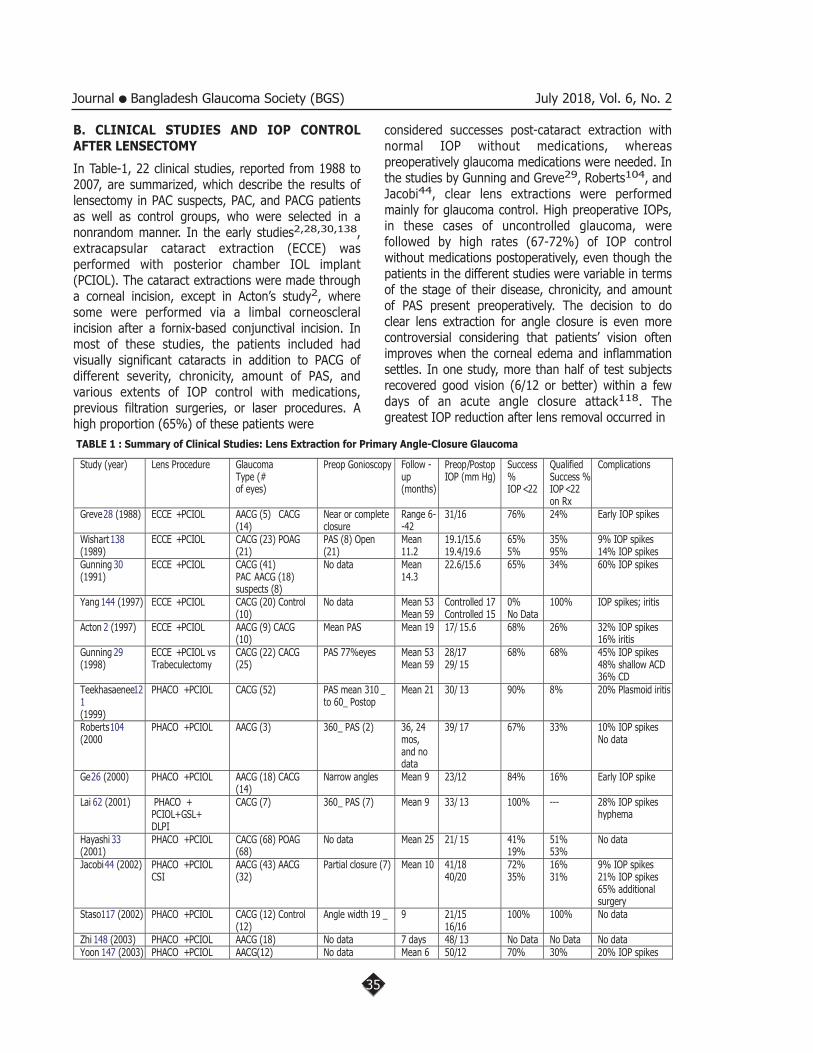
July 2018, Vol. 6, No. 2Journal ● Bangladesh Glaucoma Society (BGS)
35
Study (year) Lens Procedure Glaucoma
Type (#
of eyes)
Preop Gonioscopy Follow -
up
(months)
Preop/Postop
IOP (mm Hg)
Success
%
IOP< 22
Qualified
Success %
IOP< 22
on Rx
Complications
Greve28 (1988) ECCE +PCIOL AACG (5) CACG
(14)
Near or complete
closure
Range 6-
-42
31/16 76% 24% Early IOP spikes
Wishart 138
(1989)
ECCE +PCIOL CACG (23) POAG
(21)
PAS (8) Open
(21)
Mean
11.2
19.1/15.6
19.4/19.6
65%
5%
35%
95%
9% IOP spikes
14% IOP spikes
Gunning 30
(1991)
ECCE +PCIOL CACG (41)
PAC AACG (18)
suspects (8)
No data Mean
14.3
22.6/15.6 65% 34% 60% IOP spikes
Yang 144 (1997) ECCE +PCIOL CACG (20) Control
(10)
No data Mean 53
Mean 59
Controlled 17
Controlled 15
0%
No Data
100% IOP spikes; iritis
Acton 2 (1997) ECCE +PCIOL AACG (9) CACG
(10)
Mean PAS Mean 19 17/ 15.6 68% 26% 32% IOP spikes
16% iritis
Gunning 29
(1998)
ECCE +PCIOL vs
Trabeculectomy
CACG (22) CACG
(25)
PAS 77%eyes Mean 53
Mean 59
28/17
29/ 15
68% 68% 45% IOP spikes
48% shallow ACD
36% CD
Teekhasaenee12
1
(1999)
PHACO +PCIOL CACG (52) PAS mean 310 _
to 60_ Postop
Mean 21 30/ 13 90% 8% 20% Plasmoid iritis
Roberts104
(2000
PHACO +PCIOL AACG (3) 360_ PAS (2) 36, 24
mos,
and no
data
39/ 17 67% 33% 10% IOP spikes
No data
Ge26 (2000) PHACO +PCIOL AACG (18) CACG
(14)
Narrow angles Mean 9 23/12 84% 16% Early IOP spike
Lai 62 (2001) PHACO +
PCIOL+GSL+
DLPI
CACG (7) 360_ PAS (7) Mean 9 33/ 13 100% --- 28% IOP spikes
hyphema
Hayashi 33
(2001)
PHACO +PCIOL CACG (68) POAG
(68)
No data Mean 25 21/ 15 41%
19%
51%
53%
No data
Jacobi44 (2002) PHACO +PCIOL
CSI
AACG (43) AACG
(32)
Partial closure (7) Mean 10 41/18
40/20
72%
35%
16%
31%
9% IOP spikes
21% IOP spikes
65% additional
surgery
Staso117 (2002) PHACO +PCIOL CACG (12) Control
(12)
Angle width 19 _ 9 21/15
16/16
100% 100% No data
Zhi 148 (2003) PHACO +PCIOL AACG (18) No data 7 days 48/ 13 No Data No Data No data
Yoon 147 (2003) PHACO +PCIOL AACG(12) No data Mean 6 50/12 70% 30% 20% IOP spikes
TABLE 1 : Summary of Clinical Studies: Lens Extraction for Primary Angle-Closure Glaucoma
B. CLINICAL STUDIES AND IOP CONTROL AFTER LENSECTOMY
In Table-1, 22 clinical studies, reported from 1988 to
2007, are summarized, which describe the results of
lensectomy in PAC suspects, PAC, and PACG patients
as well as control groups, who were selected in a
nonrandom manner. In the early studies2,28,30,138,
extracapsular cataract extraction (ECCE) was
performed with posterior chamber IOL implant
(PCIOL). The cataract extractions were made through
a corneal incision, except in Acton’s study2, where
some were performed via a limbal corneoscleral
incision after a fornix-based conjunctival incision. In
most of these studies, the patients included had
visually significant cataracts in addition to PACG of
different severity, chronicity, amount of PAS, and
various extents of IOP control with medications,
previous filtration surgeries, or laser procedures. A
high proportion (65%) of these patients were
considered successes post-cataract extraction with
normal IOP without medications, whereas
preoperatively glaucoma medications were needed. In
the studies by Gunning and Greve29, Roberts104, and
Jacobi44, clear lens extractions were performed
mainly for glaucoma control. High preoperative IOPs,
in these cases of uncontrolled glaucoma, were
followed by high rates (67-72%) of IOP control
without medications postoperatively, even though the
patients in the different studies were variable in terms
of the stage of their disease, chronicity, and amount
of PAS present preoperatively. The decision to do
clear lens extraction for angle closure is even more
controversial considering that patients’ vision often
improves when the corneal edema and inflammation
settles. In one study, more than half of test subjects
recovered good vision (6/12 or better) within a few
days of an acute angle closure attack118. The
greatest IOP reduction after lens removal occurred in

MSI ProdhanAngle-closure Glaucoma : The Role of the Lens in the Pathogenesis, Prevention and Treatment
36
acute PAC eyes with uncontrolled IOP
preoperatively44,104,120. This is expected as this
group of patients would have the greatest amount of
pupillary block and appositional angle closure, as well
as the highest baseline IOP. IOP reduction occurred to
an extent that medications were not required
postoperatively, even in eyes with extensive PAS
preoperatively28,121,138. Many eyes were found to
have less PAS after surgery2, suggesting that
gonioscopic examination preoperatively may
overestimate the extent of PAS, when the angle is
narrow. Another possible explanation for finding less
PAS postoperatively, when no additional maneuver
was done during the lens extraction to open the
angle, is the positive pressure of viscoelastic material
and fluid as it is introduced into the eye during the
surgery58. Although gonioscopic details of those who
failed to have IOP control were not described, we
suspect that a residual open angle does not
guarantee successful IOP control. Acton et al’s failure
to achieve IOP control in some subjects occurred
even though only eyes with a maximum of two
quadrants of PAS were included in the study1. The
residual open trabecular meshwork can be potentially
damaged by high IOP, inflammation associated with
an acute attack, or by the adverse effect of
appositional closure. Phacoemulsification and PCIOL
combined with GSL, performed within 6 months of
chronic ACG with an acute attack despite LPI and
argon laser peripheral iridoplasty, has been found to
control IOP without medications in 90.4% of eyes121.
Another study combining phacoemulsification and
PCIOL with limited (inferior) GSL and diode laser
peripheral iridoplasty in the treatment of seven eyes
with cataract and CACG with total synechial angle
closure found a short-term success rate of 100%
during a mean follow-up period of 8.9 months62. The
study by Gunning and Greve 29compared the results
of lens extraction to trabeculectomy without
antimetabolites, except for use of postoperative 5-
fluorouracil in 6 of 25 eyes following surgery.
Longterm IOP control was found to be better in the
trabeculectomy group, but there were also moresight-
threatening complications such as hypotony and
central field loss, poorer visual outcome, and more
surgical reintervention in this group. Sixty percent of
the trabeculectomy eyes required cataract extraction
after a mean postoperative period of 32 months.
When phacoemulsification was compared to
conventional surgical iridectomy (CSI) in the study by
Jacobi, phacoemulsification was found to have more
than doubled the success rate in terms of IOP control
and much lower rate of surgical reintervention than
the CSI group44. Imaizumi subdivided his study
groups into: 1) APAC at first visit without prior
treatment, 2) PACG with earlier laser iridotomy (LI),
and 3) a control group. His results show that the
post-cataract surgery IOPs of group 1 are significantly
lower than the preoperative IOP of group 2 even with
glaucoma medications40. This suggests that lens
extraction lowers IOP as well as, if not better, than
LPI. Lens extraction in PAC, whether clear or
cataractous, not only deepens the AC and opens the
angle but also decreases diurnal IOP variation74 and
improves facility of outflow81. This IOP reduction
following cataract surgery is not observed to the same
extent in POAG eyes. Some studies have found that
IOP control in POAG eyes are largely unaffected by
cataract surgery80,138, whereas others found a
reduction in IOP in the short term that is smaller 104
and less sustained87,104 than that seen in PACG
eyes. The mechanism for IOP reduction in POAG eyes
is unclear.
C. COMPLICATIONS OF LENSECTOMY
Intraocular surgery in patients with angle closure is
more challenging than regular surgery because of the
shallow AC, atonic pupil from the acute attack, and
residual corneal edema. The reviewed studies (Table-
1) of lensectomy for treatment of angle closure
glaucoma report lensectomy, by either ECCE or
PHACO, to be potentially safe in the hands of a skilled
cataract surgeon. The complication causing the most
frequent concern was the immediate postoperative
pressure spike, which occurred in 9--60% of eyes.
Significant postoperative inflammation was seen in
16-40% of eyes reported in at least four
studies2,121,144,147. The addition of GSL may be
associated with increased rates of hyphema, fibrinoid
anterior chamber reaction, IOP spikes, and cystoid
macular edema postoperatively, secondary to the iris
manipulation121. Kubota described the successful
addition of GSL to PHACO and PCIOL, in the presence
of PAS of more than two quadrants, and reported no
significant complications in 5 eyes55. Endothelial cell
damage is common after acute angle closure and
elevated IOP. One study shows that endothelial cell
counts are not significantly diminished following lens
surgery compared to preoperatively40.
VIII. Lens Extraction for Secondary Angle-closure Glaucoma
The lens plays a significant role in the development of
angle closure in other important eye conditions, which
July 2018, Vol. 6, No. 2Journal ● Bangladesh Glaucoma Society (BGS)
37
occur less frequently than PACG but in patients of all
ages96. It is of value to appreciate that the same
mechanisms of pupillary block and angle crowding, as
seen in patients with PACG, also occur in these other
conditions. Angle closure develops in these conditions
when the lens is disproportionately large, when the
eye is abnormally small, when the lens is thickened,
or when the lens becomes subluxated and blocks the
flow of AH through the pupil and/or closes the
narrowed angle by crowding. Treatment choices
should be determined by the underlying pathology
and can include lensectomy when irreversible
anatomic abnormalities exist that are unresponsive to
medical therapy, laser iridoplasty or iridotomy, or
surgical iridectomy.
A. Nanophthalmos
Nanophthalmos is a rare, bilateral, sporadic or familial
condition characterized by small eyes, with adult axial
lengths of less than 20 mm, shallow anterior
chambers with narrow angles and convex irides, small
corneal diameters, high hyperopia, thick sclera and
choroid, high lens/eye volume ratio, high corneal
refractive power, absence of other congenital
malformations, and the frequent occurrence of angle
closure114. The mechanism of the angle-closure in
nanophthalmos relates to the angle-crowding
secondary to the normal-sized lens in a small eye and
anterior segment. This leads to progressive PAS
formation as the iris is forced anteriorly against the
trabecular meshwork by the worsening pupillary-block
and expansion in the posterior segment as the lens
enlarges with age. The mechanism explains the rarity
of long term success in IOP control with LPI alone,
and the beneficial effect of combined LPI and ALPI in
some eyes8. The thickened sclera is felt to impede
venous drainage from the choroid and is
responsible for the uveal effusion that can be
corrected by therapeutic or prophylactic
sclerossclerostomy procedures46. The potential role
for lensectomy in the management of nanophthalmos
is shadowed by an awareness of vision loss and
complications after intraocular surgery in the
past9,86. The potential goals of lensectomy are for
cataract removal, to deepen the AC, and widen the
angle to halt and prevent progressive closure.
Successful cataract removal with phacoemulsification
and PCIOL implantation in nanophthalmos is now
reported77,142. If medical therapy, LPI, ALPI,
combined with therapeutic sclerostomy procedures for
uveal effusions fail to halt progressive angle
closure53, lensectomy with PCIOL implantation with
prophylactic sclerostomy procedures should be
considered.
B. Retinopathy of Prematurity
Retinopathy of prematurity (ROP) occurs in premature
infants with the risk of occurrence inversely related to
the birth weight, reaching 90% for infants less than
750 g. Affected eyes may be small depending on the
stage of the active disease67, possess steep
corneas36, shallow anterior chamber depths36,
narrow angles32, proportionately large lenses, and
exhibit progressive lenticular myopia27. Secondary
angle closure associated with anterior displacement of
the iris-lens diaphragm is common (30%) in advanced
ROP with onset in infancy. Closure of the angle is
secondary to angle crowding, a belief supported by
the clinical failure of iridectomy only to open the angle
and improve IOP control91,131. The angle-closure
may be of acute onset or occur more slowly and
cause CACG82. Progressive growth and forward
movement of the lens secondary to contraction of
posterior retrolenticular membranes96 are also felt to
contribute to the development of pupillary-block
related angle closure82,91,128,131. Posterior
diversion of AH (ciliary block glaucoma) and
neovascular glaucoma also have been seen in ROP
glaucoma patients17,56,82. In the study by Hartnett
and coworkers of 26 untreated stage IV or V ROP
eyes in 17 children between 4 and 35 months, 65%
had open angles, 35% had 90_ or more of closure
with associated glaucoma in only 3 (12%) eyes32.
The treatment of angle closure associated with ROP
depends on the clinical indication and stage of the
illness. When glaucoma is acquired later in childhood
and in adults, active therapy to treat the pupillary
block with iridectomy69, LPI115, lensectomy82,91,
and with glaucoma procedures 82 have been
successful. PHACO with PCIOL has been performed in
adults with ROP resulting in improved vision and IOP
control in glaucoma patients54.
C. Phacomorphic Glaucoma
In phacomorphic glaucoma, angle-closure and
secondary glaucoma is caused by the enlargement of
the swollen lens as seen after lens trauma and more
frequently in the intumescent age-related cataract.
The need and benefit of lens removal for
improvement of vision and correction of the pupillary
block and crowding mechanisms for angle-closure is
obvious.

MSI ProdhanAngle-closure Glaucoma : The Role of the Lens in the Pathogenesis, Prevention and Treatment
38
D. Ciliochoroidal Expansion Syndromes
Ciliochoroidal expansion places even an eye of normal
size at risk for angle-closure glaucoma. In the average
human eye the vitreous volume is 5,000 ml, the
choroidal volume is 480 ml, and the anterior chamber
volume is about 150 ml94. Hence, it can be
appreciated that minimal sustained expansion of the
choroid can decrease the AC volume and ACD
significantly and create the prerequisite anterior
segment configuration for angle closure. Furthermore,
in these syndromes, separation and thickening of the
ciliary body has been documented by UBM130. The
related anterior rotation of the ciliary body and
relaxation of the zonules would be expected to
contribute further to the development of the angle
closure. Ciliochoroidal expansion syndromes occur
secondary to inflammation, choroidal venous
congestion, choroidal metastases, trauma, drugs,
systemic diseases, from aggressive Panretinal
Photocogulation (PRP), or may be
idiopathic23,49,130. Treatment is directed to the
underlying illness and to management of the related
angle closure. Lensectomy is rarely indicated because
often these conditions can be treated medically or can
resolve spontaneously.
E. Lens Subluxation Glaucoma
Eyes with lens subluxation or dislocation are at
increased risk for secondary glaucoma42. When the
lenticular zonular attachments weaken or detach, the
lens thickens and may move from its normal position.
If the weakened zonules are confined to less than half
of the lens circumference, the thickening of the lens
will occur asymmetrically and create refractive
astigmatism and the lens will remain approximately in
its normal position. This circumstance is seen in
young patients with Marfan syndrome whose eyes are
rarely complicated by lens related glaucoma43. In
older patients with Marfan disease, or in those
conditions where circumferential weakening of the
zonules occurs70,136, the lens becomes mobile and
moves anteriorly with resultant shallowing of the
anterior chamber, increased iris convexity, increased
pupillary block, and risk for acute or chronic ACG137.
The subluxated or dislocated lens may enter the
anterior chamber or acutely obstruct the pupil and
cause an acute attack associated with a flat anterior
chamber. Treatment of glaucoma caused by lens
subluxation is best directed to the lens itself. Surgical
iridectomy or LPI can help by decreasing the iris
convexity and increasing the angle width, and will
prevent the occurrence of acute angle closure but can
not be relied on to definitively treat or prevent this
secondary ACG47,136. These measures can be
considered for prophylaxis for selected young
patients141. Angle-crowding is an important
component causing the angle closure and can be
marginally benefited by ALPI98. The definitive
treatment for lens subluxation related glaucoma is
lensectomy42,47,137. The indication for implantation
of anterior chamber IOL or PCIOL is
controversial47,112,129,137. The addition of
successful GSL combined with lensectomy for the
treatment of this PAC has been reported47, and
deserves continued clinical study.
IX. Conclusion
PACG is a leading cause of blindness and is potentially
preventable. It is projected that 15.7 million will have
ACG in 2010 and 3.9 million will be bilaterally blind
from it92,. The lens plays an essential and pivotal role
in the pathogenesis of primary and secondary ACG.
Clinical studies suggest that lensectomy and PCIOL
implantation for ACG patients may offer successful
IOP control, and maintenance of improved vision.
Lensectomy eliminates pupillary block, widens the
angle to lessen angle crowding thus reducing the
irido-trabecular proximity. Medical management and
LPI remain the most common modes of treatment of
an acute attack but newer approaches including early
lens removal are gaining popularity because of their
potential long-term success in IOP control. After an
acute attack lensectomy is most appropriate or it
should be combined with a filtration procedure.
Randomized, controlled studies are ongoing in Hong
Kong and Singapore and should help clarify this
surgical decision66,83. Although lens extraction for
angle closure is biologically plausible, as of this time,
there is no evidence from good quality randomized
trials or non-randomized studies of the effectiveness
of lens extraction for CACG25. More longitudinal
biomorphometrical studies of PACG eyes treated with
and without lensectomy are needed to determine its
role in the prevention of progressive angle closure
and to determine which patients will benefit from lens
extraction37.
X. Method of Literature Search
A search of the PubMed database was conducted for
the years 1988-2007, using the following key words:
angle closure glaucoma, pupillary block, angle
July 2018, Vol. 6, No. 2Journal ● Bangladesh Glaucoma Society (BGS)
39
crowding, lensectomy, cataract extraction. Additional
references were recovered from bibliographies of the
references.
References
1. The Advanced Glaucoma Intervention Study, 8: Risks of cataract
formation after trabeculectomy. Arch Ophthalmol. 2001; 119:1771--
80
2. Acton J, Salmon JF, Scholtz R. Extracapsular cataract extraction
with posterior chamber lens implantation in primary angle-closure
glaucoma. J Cataract Refract Surg.1997; 23:930--4
3. Alsbirk PH. Anterior depth in Greenland Eskimos I. A population
study of variation with age and sex. Acta Ophthalmol. 1974;
52:551--64
4. American Academy of Ophthalmology, Preferred Practice Pattern.
San Francisco, Primary Angle Closure, 2005
5. Ang LP, Aung T, Chew PT. Acute primary angle closure in an
Asian population: long-term outcome of the fellow eye after
prophylactic laser peripheral iridotomy. Ophthalmology. 2000;
107:2092--6
6. Aung T, Ang LP, Chan SP, Chew PT. Acute primary angle closure:
long term intraocular pressure outcome in Asian eyes. Am J
Ophthalmol. 2001; 131:7--12
7. Aung T, Tow SL, Yap EY, et al. Trabeculectomy for acute angle
closure. Ophthalmology. 2000; 107:1298--302
8. Burgoyne C, Tello C, Katz LJ. Nanophthalmia and chronic angle-
closure glaucoma. J Glaucoma. 2002; 11:525--8
9. Calhoun FP Jr. The management of glaucoma in nanophthalmos.
Trans Am Ophthalmol Soc. 1975; 73:97--119
10. Campbell DG, Vela A. Modern goniosynechialysis for the
treatment of synechial angle-closure glaucoma. Ophthalmology.
1984; 91:1052--60
11. Chew PT, Lim A. Argon laser iridoplasty in chronic angle closure
glaucoma. Int Ophthalmol. 1995; 19:67--70
12. Congdon N, Wang F, Tielsch JM. Issues in the epidemiology and
population-based screening of primary angleclosure glaucoma. Surv
Ophthalmol. 1992; 36:411--23
13. Congdon NA, Youlin Q, Quigley H, et al. Biometry and primary
angle-closure glaucoma among Chinese, white, and black
populations. Ophthalmology. 1997; 104:1489--95
14. Curran E. A new operation for glaucoma involving a new
principle in the etiology and treatment of chronic primary
glaucoma. Arch Ophthalmol. 1920; 49:131--55
15. Dandona L, Dandona R, Mandal P, et al. Angle closure
glaucoma in an urban population in Southern India. The Andhra
Pradesh Eye Disease Study. Ophthalmology. 2000; 107:1710--6
16. Delmarcelle Y, Francois J, Goes F, et al. Clinical ocular biometry
(oculometry). Bull Soc Belge Ophthalmol. 1976; 172:1--608
17. Dhillon B, Butt Z, Fleck B. Rubeotic glaucoma and retinopathy
of prematurity: a case report. J Pediatr Ophthalmol Strabismus.
1992; 29:123--5
18. Foster P, Buhrmann R, Quigley H, et al. The definition and
classification of glaucoma in prevalence surveys. Br J Ophthalmol.
2002; 86:238--42
19. Foster PJ, Baasanhu J, Alsbirk PH, et al. Glaucoma in Mongolia:
a population-based survey in Hovsgol ProvinceProvince, Northern
Mongolia. Arch Ophthalmol. 1996; 114: 1235--41
20. Foster PJ, Johnson GJ. Glaucoma in China: how big is the
problem? Br J Ophthalmol. 2001; 85:1277--82
21. Foster PJ, Oen FT, Machin D, et al. The prevalence of glaucoma
in Chinese residents of Singapore: a crosssectional population
survey of the Tanjong Pagar district. Arch Ophthalmol. 2000;
118:1105--11
22. Foster PJ. The epidemiology of primary angle closure and
associated glaucomatous optic neuropathy. Semin Ophthalmol.
2002; 17:50--8
23. Fraunfelder FW, Fraunfelder FT, Keates EU.
Topiramateassociated acute, bilateral, secondary,angle-closure
glaucoma. Ophthalmology. 2004; 111:109--11
24. Friedman DS, Gazzard G, Foster P, et al. Ultrasonography
biomicroscopy, scheimpflug photography, and novel provocative
tests in contralateral eyes of Chinese patients initially seen with
acute angle closure. Ophthalmology. 2003; 121:633--42
25. Friedman DS, Vedula SS. Lens extraction for chronic
angleclosure glaucoma (review). Cochrane Database of Systematic
Reviews;. Issue 3, art. no. CD005555, 2006.
26. Ge J, Guo Y, Liu Y, et al. New management of angle-closure
glaucoma by phacoemulsification with foldable posterior chamber
intraocular lens implantation. Yan Ke Xue Bao. 2000; 16:22--8
27. Gordon RA, Donzis PA. Myopia asociated with retinopathy of
prematurity. Ophthalmology. 1986; 93:1593--8
28. Greve EL. Primary angle closure glaucoma: extracapsular
cataract extraction or filtering procedure. Int Ophthalmol. 1988;
12:157--62
29. Gunning FP, Greve EL. Lens extraction for uncontrolled angle-
closure glaucoma: long-term follow-up. J Cataract Refract Surg.
1998; 24:1347--56
30. Gunning FP, Greve EL. Uncontrolled primary angle closure
glaucoma: results of early intercapsular cataract extraction and
posterior chamber lens implantation. Int
Ophthalmol. 1991; 15:237--47
31. Harasymowycz P, Papamatheakis D, Ahmed I, et al.
Phacoemulsification and goniosynechialysis in the management of
unresponsive primary angle closure. J Glaucoma. 2005; 14:186--9
32. Hartnett ME, Gilbert MM, Richardson TM, et al. Anterior
segment evaluation of infants with retinopathy of prematurity.
Ophthalmology. 1990; 97:122--30
33. Hayashi K, Hayashi H, Nakao F, Hayashi F. Effect of intraocular
surgery on intraocular pressure control in glaucoma patients. J
Cataract Refract Surg. 2001; 27:
1779--1786
34. Hayashi K, Hayashi H, Nakao F, Hayashi F. Changes in anterior
chamber angle width and depth after intraocular lens implantation
in eyes with glaucoma. Ophthalmology. 2000; 107:698--703
35. Heys JJ, Barocas VH, Taravella MJ. Modeling passive mechanical
interaction between the aqueous humor and iris. J Biomech Eng.
2001; 123:540--7
36. Hittner HM, Rhodes LM. McPherson. Anterior segment
abnormalities in cicatricial retinopathy of prematurity. Trans Am

Angle-closure Glaucoma : The Role of the Lens in the Pathogenesis, Prevention and Treatment MSI Prodhan
40
Acad Ophthalmol. 1979;86:803--16
37. Ho CL, Walton DS, Pasquale LR. Lens extraction for angle-
closure glaucoma. Int Ophthalmol Clin. 2004; 44: 213--28
38. Ho CL. Symposium on lens extraction in acute glaucoma. Asian
Pacific J Ophthalmol. 2001; 13:19
39. Hoh ST, Gazzard GA, Oen F, et al. Laser Iridotomy versus Primary
Phacoemulsification. Asia Pacif J Ophthalmol. 2001; 13:22--3
40. Imaizumi M, Takaki Y, Yamashita H. Phacoemulsification and
intraocular lens implantation for acute angle closure not treated or
previously treated by laser iridotomy.
J Cataract Refract Surg. 2006; 32:85--90
41. Inaba Z. Long-term results of trabeculectomy in the Japanese:
an analysis by lifetable method. Jpn J Ophthalmol. 1982;26:361--
7342. Inatani M, Tanihara H, Honjo M, et al. Secondary glaucoma
associated with crystalline lens subluxation. J Cataract Refract Surg.
2000;26:1533--6
43. Izquierido NJ, Traboulsi EI, Enger C, Maumenee IH. Glaucoma
in the Marfan syndrome. Tr Am Soc. 1992; 80: 111--22
44. Jacobi PC, Dietlein TS, Luke C, et al. Primary
phacoemulsification and intraocular lens implantation for acute
angleclosure glaucoma. Ophthalmology. 2002; 109:1597--603
45. Jain IS, Gupta A, Dogra MR. Surgery in angle closure glaucoma:
late complications. Glaucoma. 1984; 6:54--7
46. Jin JC, Anderson DR. Laser and unsutured sclerotomy in
nanophthalmos. Am J Ophthalmol. 1990; 109:575--80
47. Kanamori A, Nakamura M, Matsui N, et al. Goniosynechialysis
with lens aspiration and posterior chamber intraocular lens
implantation for glaucoma in spherophakia.
J Cataract Refract Surg. 2004; 30:513--6
48. Kessler J. The resistance to deformation of the tissue of the
peripheral iris and the space of the angle of the anterior chamber.
Am J Ophthalmol. 1956; 42:734--6
49. Khawly JA, Shields MB. Metastatic carcinoma manifesting as
angle-closure glaucoma. Am J Ophthalmol. 1994; 118: 116-7
50. Kim YY, Jung HR. Clarifying the nomenclature for primary
angle-closure glaucoma. Surv Ophthalmol. 1997; 42(2): 125-36
51. Kim YY, Jung HR. Dilated, miotic-resistant pupil and laser
iridotomy in primary angle-closure glaucoma. Ophthalmologica.
1997; 211:205--8
52. Kimbrough RL, Trempe CS, Brockhurst RJ, Simmons RJ. Angle-
closure glaucoma in nanophthalmos. Am J Ophthalmol. 1979;
88:572--9
53. Kocak I, Atlintas AG, Yalvac IS, et al. Treatment of glaucoma in
young nanophthalmic patients. Int Ophthalmol. 1997; 20:107--11
54. Krolicki TJ, Tasman W. Cataract extraction in adults with
retinopathy of prematurity. Arch Ophthalmol. 1995; 113: 173--7
55. Kubota T, Toguri I, Onizuka N, Matsuura T. Phacoemulsification
and intraocular lens implantation for angle closure glaucoma after
the relief of pupillary block. Ophthalmologica. 2003; 217:325--8
56. Kushner BJ. Ciliary block glaucoma in retinopathy of
prematurity. Arch Opthalmol. 1982; 100:1078--9
57. Lai J, Tham C, Chan J. The clinical outcomes of cataract
extraction by phacoemulsification in eyes with primary angle-
closure glaucoma and co-existing cataract: a prospective series. J
Glaucoma. 2006; 15(1):47--52
58. Lai J, Tham C, Chan J. The clinical outcomes of cataract
extraction by phacoemulsification in eyes with primary angle
closure glaucoma and co-existing cataract: a prospective case
series (letter). J Glaucoma. 2006; 15(4):346
59. Lai J, Tham C, Lam D. Incisional Surgery for angle closure
glaucoma. Sem Ophthalmol. 2002; 17(2):92--9
60. Lai JS, Tham CC, Chua JK, et al. Laser peripheral iridoplasty as
initial treatment of acute attack of primary angle-closure: a long-
term follow-up study. J Glaucoma. 2002;11:484--7
61. Lai JS, Tham CC, Chua JK, Lam DS. Efficacy and safety of
inferior 180 degrees goniosynechialysis followed by diode laser
peripheral iridoplasty in the treatment of chronic angle-closure
glaucoma. J Glaucoma. 2000; 9:388--91
62. Lai JS, Tham CC, Lam DS. The efficacy and safety of combined
phacoemulsification, intraocular lens implantation, and limited
goniosynechialysis, followed by diode laser peripheral iridoplasty, in
the treatment of chronic angle-closure glaucoma. J Glaucoma.
2001; 10:309--15
63. Lam DS, Chua JK, Tham CC, Lai JS. Efficacy and safety of
immediate anterior chamber paracentesis in the treatment of acute
primary angle-closure glaucoma: a pilot study. Ophthalmology.
2002; 109:64--70
64. Lam DS, Lai JS, Tham CC, et al. Argon laser peripheral
iridoplasty versus conventional systemic medical therapy in
treatment of acute primary angle-closure glaucoma: a
proprospective, randomized, controlled trial. Ophthalmology. 2002;
109:1591--6
65. Lam DS, Lai JS, Tham CC. Immediate argon laser peripheral
iridoplasty as treatment for acute attack of primary angle-closure
glaucoma: a preliminary study. Ophthalmology. 1998;105:2231--6,
1998.
66. Lam DS, Tham C, Lai J, et al. Current approaches to the
management of acute primary angle closure. Curr Opin
Ophthalmol. 2007;18:146--51
67. Laws DE, Haslett R, Ashby D, et al. Axial length in infants with
retinopathy of prematurity. Eye. 1994;8:427--30
68. Lee DA, Brubaker RF, Ilsrup DM. Anterior chamber dimensions
in patients with narrow angles and angleclosure glaucoma. Arch
Ophthalmol. 1984; 102:46--50
69. Lee GA, Lee LA, Gole GA. Angle-closure glaucoma after laser
treatment for retinopathy of prematurity. J AAPOS. 1998; 2:383--4
70. Lieberman TW, Podos SM, Hartstein J. Acute glaucoma, ectopia
lentis and homocystinuria. Am J Ophthalmol. 1966; 61:252--5
71. Lim KJ, Hyung SM, Youn DH. Ocular dimensions with aging in
normal eyes. Korean J Ophthalmol. 1992; 6:19-- 31
72. Lim L, Aung T, Husain R, et al. Acute primary angle closure:
configuration of the drainage angle in the first year after laser
peripheral iridotomy. Ophthalmology. 2004;
111(8):1470--4
73. Lim L, Husain R, Gazzard G. Cataract progression after
prophylactic lasr peripheral iridotomy: potential implications for the
prevention of glaucoma blindness. Ophthalmology. 2005;
112:1355--9
74. Liu C, Cheng C, Wu C, et al. Factors predicting intraocular
pressure control after phacoemulsification in angle closure
glaucoma. Arch Ophthalmol. 2006;124:1390--4
75. Lowe RF. Aetiology of the anatomical basis for primary angle
closure glaucoma. Br J Ophthalmol. 1970;54: 161--9

Journal ● Bangladesh Glaucoma Society (BGS) July 2018, Vol. 6, No. 2
41
76. Lowe RF. Causes of shallow anterior chamber in primary angle-
closure glaucoma. Am J Ophthalmol. 1969;67:87--93
77. Mandal AK. Cataract surgery with primary posterior chamber
intraocular lens implantation in nanophthalmos. Ophthalmic Surg
Lasers. 2001;32:333--578. Mapstone R. Precipitation of angle
closure. Br J Ophthalmol. 1974; 58:36--54
79. Markowitz SN, Morin JD. Angle-closure glaucoma: relation
between lens thickness, anterior chamber depth and age. Can J
Ophthalmol. 1984; 19:300--2
80. McGuigan LJ, Gottsch J, Stark WJ, et al. Extracapsular cataract
extraction and posterior chamber lens implantation in eyes with
preexisting glaucoma. Arch Ophthalmol. 1986; 104:1301--8
81. Meyer MA, Savitt MC, Kopitas E. The effect of
phacoemulsification on aqueous outflow facility. Ophthalmology.
1997;104:1221—7 82. Michael AJ, Pesin SR, Katz LJ, Tasman WS.
Management of late-onset angle-closure glaucoma associated with
retinopathy of prematurity. Ophthalmology. 1991; 98:1093--8
83. Nolan W. Lens extraction in primary angle closure. Br J
Ophthalmol. 2006; 90(1):1--2
84. Nolan WP, Foster PJ, Devereux JG, et al. YAG laser iridotomy
treatment for primary angle-closure glaucoma in East Asian eyes.
Br J Ophthalmol. 2000; 84:1255--9
85. Nonaka A, Kondo T, Kikuchi M, et al. Cataract surgery for
residual angle closure after peripheral laser iridotomy.
Ophthalmology. 2005; 112:974--9
86. O’Grady RB. Nanophthalmos. Am J Ophthalmol. 1971; 71:
1251-3
87. Obstbaum SA. Glaucoma and intraocular lens implantation. J
Cataract Refract Surg. 1986; 12:257--61
88. Pavlin CJ, Ritch R, Foster FS. Ultrasound biomicroscopy in
plateau iris syndrome. Am J Ophthalmol. 1992; 113: 390--5
89. Pei TH. Glaucoma in China. Int Ophthalmol. 1995; 8:47-5490.
Cl Phillips. Aetiology of angle-closure glaucoma. Br J Ophthalmol.
1972; 56:248-53
91. Pollard ZF. Lensectomy for secondary angle-closure glaucoma in
advanced cicatricial retrolental fibroplasia. Ophthalmology. 1984;
91:395--8
92. Quigley H, Broman AT. The number of people with glaucoma
worldwide in 2010 and 2020. Br J Ophthalmol. 2006; 90:262--7
93. Quigley HA, Congdon NG, Friedman DG. Glaucoma in China
(and worldwide): changes in established thinking will decrease
preventable blindness. Br J Ophthalmol.
2001; 85:1271--2
94. Quigley HA, Freidman DS, Congdon NG. Possible mechanisms
of primary angle- closure glaucoma and malignant glaucoma. J
Glaucoma. 2003; 12:167--80
95. Quigley HA. The number of persons with glaucoma worldwide.
Br J Ophthalmol. 1996; 85:1277--82
96. Ritch R, Chang BM, Liebmann JM. Angle closure in younger
patients. Ophthalmology. 2003; 110:1880--9
97. Ritch R, Lowe RF. Angle closure glaucoma: mechanisms and
epidemiology. In Ritch R, Shields MB, Krupin T (eds): The
Glaucomas. St. Louis, CV Mosby, 1996, pp. 801--19
98. Ritch R, Solomon LD. Argon laser peripheral iridoplasty for
angle-closure glaucoma in siblings with Weill-Marchesani syndrome.
J Glaucoma. 1992; 1:243--7
99. Ritch R, Tham CC, Lam DS. Argon laser peripheral iridoplasty:
an update. Surv Ophthalmol. 2007; 52(3):279--88
100. Ritch R, Tham CC, Lam DS. Long-term success of argon laser
peripheral iridoplasty in the management of plateau iris syndrome.
Ophthalmology. 2004; 111:104--8
101. Ritch R. Argon laser treatment for medically unresponsive
attacks of angle-closure glaucoma. Am J Ophthalmol. 1982;
94:197--204
102. Ritch R. Assessing the treatment of angle closure.
Ophthalmology. 2003; 110:1867-8
103. Ritch R. Plateau iris is caused by abnormally positioned ciliary
processes. J Glaucoma. 1992; 1:23--6
104. Roberts TV, Francis IC, Lertusumitkul S, et al. Primary
phacoemulsification for uncontrolled angle-closure glaucoma. J
Cataract Refract Surg. 2000; 26:1012--6
105. Rosman M, Aung T, Ang LP, et al. Chronic angle-closure with
glaucomatous damage: long term clinical course in a North
American population and comparison with an Asian population.
Ophthalmology. 2002; 109:2227--31
106. Sakai H, Ishikawa H, et al. Prevalence of ciliochoroidal effusion
after prophylactic laser iridotomy. Am J Ophthalmol. 2003;
136:537--8
107. Sankar PS, Pasquale LR, Grosskreutz CL. Uveal effusion and
secondary angle-closure glaucoma associated with topiramate use.
Arch Ophthalmol. 2001; 119:1210--1
108. Saw SM, Gazzard G, Freidman D, et al. Awareness of
glaucoma, and health beliefs of patients suffering primary acute
angle closure. Br J Ophthalmol. 2003; 87:446--9
109. Seah SK, Foster PJ, Chew PT, et al. Incidence of acute primary
angle-closure glaucoma in Singapore. Arch Ophthalmol. 1997;
115:1436--40
110. Seang-Mei S, Gazzard G, Friedman D. Interventions for angle-
closure glaucoma: an evidence-based update. Ophthalmology.
2003; 110:1869--79
111. Sickenberg M, Gonvers M, van Melle G. Change in
capsulorhexis size with four foldable loop-haptic lenses over 6
months. J Cataract Refract Surg. 1998; 24:925--30
112. Siganos DS, Signaos CS, Popescu CN, Margaritis VN. Clear lens
extraction and intraocular lens implantation in Marfans syndrome. J
Cataract Refract Surg. 2000; 26:
781--4
113. Silver DM, Quigley HA. Aqueous flow through the iris- lens
channel: estimates of differential pressure between the anterior
and posterior chambers. J Glaucoma. 2004; 13:
100--7
114. Singh OS, Simmons RJ, Brockhurst RJ, Trempe CL.
Nanophthalmos. A perspective on identification and therapy.
Ophthalmology. 1982; 89:1006--12115. Smith J, Shivitz I. Angle-
closure glaucoma in adults with cicatricial retinopathy of
prematurity. Arch Ophthalmol. 1984; 102:371--2
116. Sorsby A, Benjamin B, Davey JB, et al. Emmetropia and its
aberrations. Spec Rep Ser med Res Council. London, Her Majestys
Stationery Office, 1956. p 253.
117. Staso SD, Sabetti L, Taverniti L, et al. Phacoemulsification and
intraocular lens implant in eyes with primary angleclosure
glaucoma: our experience. Acta Ophthalmol Scan Suppl. 2002;
136:17--8
Angle-closure Glaucoma : The Role of the Lens in the Pathogenesis, Prevention and Treatment MSI Prodhan
42
118. Tan GSW, Hoh S-T, et al. Visual Acuity after Acute Primary
Angle Closure and Considerations for Primary Lens Extraction. Br J
Ophthalmol. 2006; 90:14--6
119. Tanihara H, Nagata M. Argon laser gonioplasty following
goniosynechialysis. Graefes Arch Clin Exp Ophthalmol. 1991;
229:505--7
120. Tanihara H, Nishinaki K, Nagata M. Surgical results and
complications of goniosynechialysis. Graefes Arch Clin Exp
Ophthalmol. 1992; 230:309--13
121. Teekhasaenee C, Ritch R. Combined phacoemulsification and
goniosynechialysis for uncontrolled chronic angleclosure glaucoma
after acute angle closure glaucoma. Ophthalmology. 1999;
106:669--74
122. Tham CC, Lai JS, Lam DS. Immediate argon laser peripheral
iridoplasty for acute attack of PACG (addendum to previous report).
Ophthalmology. 1999; 106: 1042--3
123. Tham CC, Lai JS, Poon AS, et al. Immediate argon laser
peripheral iridoplasty (ALPI) as initial treatment for acute
phacomorphic angle-closure (phacomorphic glaucoma) before
cataract extraction: a preliminary study. Eye. 2005; 19(7):778--83
124. Tomlinseon A, Leighton DA. Ocular dimensions in the heredity
of angle-closure glaucoma. Br J Ophthalmol. 1973; 57:475--86
125. Tornquist R. Chamber depth in primary acute glaucoma. Br J
Ophthalmol. 1956; 40:421--9
126. Tran HV, Liebmann JM, Ritch R. Iridociliary apposition in
plateau iris syndrome persists after cataract extraction. Am J
Ophthalmol. 2003; 135:40--3
127. Trope GE, Pavlin CJ, Bau A, et al. Malignant glaucoma. Clinical
and ultrasound biomicroscopic features. Ophthalmology. 1994;
101:1030--5
128. Ueda N, Ogino N. Angle-closure glaucoma with pupillary block
mechanism in cicatricial retinopathy of prematurity.
Ophthalmologica. 1988; 196:15--8
129. Vadala´ P, Capozzi P, Fortunato M, et al. Intraocular lens
implantation in Marfans syndrome. J Pediatr Ophthalmol
Strabismus. 2000; 37:206--8
130. Waheeb S, Feldman F, Velos P, Pavlin CJ. Ultrasound
biomicroscopic analysis of drug-induced bilateral angleclosure
glaucoma associated with supraciliary choroidal
effusion. Can J Ophthalmol. 2003; 38:299--302
131. Walton DS. Retrolental fibroplasia with glaucoma. In Chandler
PA, Grant WM (eds): Glaucoma. Philadelphia, Lea & Fiebinger, 2nd
ed 1979, pp. 528--9
132. Wand M, Grant WM, Simmons RJ, Hutchinson BT. Plateau iris
syndrome. Trans Am Acad Ophthalmol Otolaryngol. 1997; 83:122--
30
133. Wand M, Pavlin CJ, Foster FS. Plateau iris syndrome:
ultrasound biomicroscopic and histologic study. Ophthalmic Surg.
1993; 24:129--31
134. Wang JK, Lai PC. Unusual presentation of angle-closure
glaucoma treated by phacoemulsification. J Cataract Refract Surg.
2004; 30:1371--3
135. West RH. Creeping angle closure glaucoma. The influence of
iridotomy and iridectomy. Aust NZ J Ophthalmol. 1992; 20:23--8
136. Willi M, Kut L, Cotlier E. Pupillary-block glaucoma in the
Marchesani syndrome. Arch Ophthalmol. 1973; 90: 504--8
137. Willoughby CE, Wishart PK. Lensectomy in the management of
glaucoma in spherophakia. J Cataract Refract Surg. 2002;
28:1061—4
138. Wishart PK, Atkinson PL. Extracapsular cataract extraction and
posterior chamber lens implantation inpatients with primary chronic
angle-closure glaucoma: effect on intraocular pressure control. Eye.
1989; 3:706-- 712
139. Wojciechowski OD, Congdon NM, Anninger W. Age, gender,
biometry, refractive error, and the anterior chamber angle among
Alaskan Eskimos. Ophthalmology. 2003; 110:365--75
140. Wong TY, Foster PJ, Seah SKL, Chew PTK. Rates of hospital
admissions for primary angle closure glaucoma among Chinese,
Malays, and Indians in Singapore. Br J Ophthalmol. 2000; 84:990--2
141. Wright KW, Chrousos GA. Weill-Marchesani syndrome with
bilateral angle-closure glaucoma. J Pediatr Ophthalmol Strabismus.
1985; 22:129--32
142. Wu W, Dawson DG, Sugar A, et al. Cataract surgery in patients
with nanophthalmos: results and complications. J Cataract Refract
Surg. 2004; 30:584--90
143. Treatment of primary angle-closure glaucoma by argon laser
iridotomy: a long-term follow-up. Jpn J Ophthalmol. 1985; 29:1--12
144. Yang CH, Hung PT. Intraocular lens position and anterior
chamber angle changes after cataract extraction in eyes.
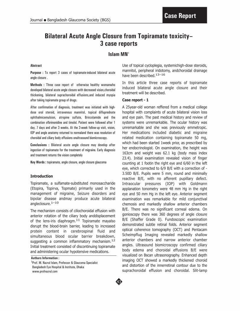
Journal ● Bangladesh Glaucoma Society (BGS)
43
Bilateral Acute Angle Closure from Topiramate toxicity–3 case reports
Islam MN1
Authors Information :1Prof. M. Nazrul Islam, Prefessor & Glaucoma Specialist
Bangladesh Eye Hospital & Institute, Dhaka
www.profnazrul.com
Case Report
Abstract
Purpose : To report 3 cases of topiramate-induced bilateral acute
angle closure .
Methods : Three case report of otherwise healthy womanwho
developed bilateral acute angle closure with decreased vision,choroidal
thickening, bilateral suprachoroidal effusions,and induced myopia
after taking topiramate group of drugs.
After confirmation of diagnosis, treatment was initiated with high-
dose oral steroid, intravenous mannitol, topical difluprednate
ophthalmicemulsion, atropine sulfate, Brinzolamide and the
combination ofbrimonidine and timolol. Patient were followed after 1
day, 7 days and after 3 weeks. At the 3-week follow-up visit, vision,
IOP and angle anatomy returned to normaland there was resolution of
choroidal and ciliary body effusions onultrasound biomicroscopy.
Conclusions : Bilateral acute angle closure may develop after
ingestion of topiramate for the treatment of migraine. Early diagnosis
and treatment returns the vision completely
Key Words : topiramate, angle closure, angle closure glaucoma
Introduction
Topiramate, a sulfamate-substituted monosaccharide
(Etopira, Topirva, Topmate) primarily used in the
management of migraine, Seizure disorders and
bipolar disease andmay produce acute bilateral
angleclosure.1–10
The mechanism consists of ciliochoroidal effusion with
anterior rotation of the ciliary body anddisplacement
of the lens-iris diaphragm.11 Topiramate mayalso
disrupt the blood-brain barrier, leading to increased
protein content in cerebrospinal fluid and
simultaneous blood ocular barrier breakdown,
suggesting a common inflammatory mechanism.12
Initial treatment consisted of discontinuing topiramate
and administering ocular hypotensive medications.
Use of topical cycloplegia, systemichigh-dose steroids,
mannitol, peripheral iridotomy, andchoroidal drainage
have been described.13–16
In this article three case reports of topiramate
induced bilateral acute angle closure and their
treatment will be described.
Case report - 1
A 25year-old woman reffered from a medical college
hospital with complaints of acute bilateral vision loss
and eye pain. The past medical history and review of
systems were unremarkable. The ocular history was
unremarkable and she was previously emmetropic.
Her medications included diabetic and migraine
related medication containing topiramate 50 mg,
which had been started 1week prior, as prescribed by
her endocrinologist. On examination, the height was
163cm and weight was 62.1 kg (body mass index
23.4). Initial examination revealed vision of finger
counting at 1 footin the right eye and 6/60 in the left
eye, which corrected to 6/9 B/E with a correction of -
3.50D B/E. Pupils were 5 mm, round and minimally
reactive B/E, with no afferent pupillary defect.
Intraocular pressures (IOP) with Goldmann
applanation tonometry were 48 mm Hg in the right
eye and 50 mm Hg in the left eye. Anterior segment
examination was remarkable for mild conjunctival
chemosis and markedly shallow anterior chambers
B/E. There was no significant corneal edema. On
gonioscopy there was 360 degrees of angle closure
B/E (Shaffer Grade 0). Fundoscopic examination
demonstrated subtle retinal folds. Anterior segment
optical coherence tomography (OCT) and Pentacam
Scheimpflug Imaging revealed markedly shallow
anterior chambers and narrow anterior chamber
angles. Ultrasound biomicroscopy confirmed ciliary
body edema and choroidal effusions B/E were
visualized on Bscan ultrasonography. Enhanced depth
imaging OCT showed a markedly thickened choroid
and distortion of the innerretinal contour due to the
suprachoroidal effusion and choroidal. Slit-lamp

Journal ● Bangladesh Glaucoma Society (BGS) July 2018, Vol. 6, No. 2
44
photograph at presentation showing a shallow
anterior chamber in the left eye, markedly so
peripheralto the slit photograph.
Case report – 2
A medical student, 22-year-old healthy girl with a
history of migraine presented with severe pain in
both eyes with headache and vomiting. She gave
history of taking oral topiramate 50 mg twice daily 10
days before. She was already treated by two different
ophthalmologist and diagnosed as Primary Acute
Angle Closure Glaucoma. Her primary IOP was 48 and
53 mmHg in right and left eye respectively and Vision
was counting finger in both the eyes. She was given
maximum anti glaucoma therapy. She was also
advised for YAG LPI in her both eyes. On examination
after 10 days I found her visual acuity improved to
6/36 R/E and 6/60 in L/E with myopic correction.
Intraocular pressures (IOPs) were 24 mm Hg R/E and
28 mm Hg L/E. Slit-lamp examination showed mild
conjunctival hyperemia, mild corneal edema,
shallowed anterior chamber, slightly reactive pupils in
both eyes. Gonioscopy revealed angle closure stage
0but with indentation grade 1 for 360, domed iris
convexity, and prominent ciliary processes visible
through the lens, suggestive of anterior displacement
of the iris-lens diaphragm in both eye. The optic discs
was normal. The C:D was 0.45 in both eyes. B-scan
ultrasonography and ultrasound biomicroscopy
showed circumferential ciliochoroidal effusions, which
induced the anterior displacement of the iris-lens
diaphragm seen gonioscopically. Anterir Segment
optical coherence tomography revealed a shallow
anterior chamber and 360-degree iridocorneal contact
in B/E
The topiramate was discontinued and oral
acetazolamide, Steroid and topical Beta blocker and
Brimonidine were given. On 2nd day, her IOPs were
>20 butiridocorneal contact persisted. With
continuous treatment after 3 days IOP came to
normal range <20 mmHg in both eyes. Repeat AS
OCT confirmed the opening of angle B/E with
persistent shallow anterior chamber. The visual acuity
1 week later was 6/9 R/E, 6/12 L/E and IOP was
10mm Hg B/E. She had clear corneas, deep anterior
chambers and complete resolution of the choroidal
effusions, induced myopia resolved and refraction
became normal for distance vision.
Case report - 3
A 52-year-old healthy woman with a history of
migraine presented with bilateral acute onset of pain,
blurred vision and redeye. She gave history of taking
oral topiramate 50mg twice daily 7days before
presentation. Her visual acuity was 6/36 R/E and CF 4
ftin L/E. Intraocular pressures (IOPs) were 34 mm Hg
R/E and 48 mm Hg L/E. Slit-lamp examination
showed conjunctival injection, mild corneal edema,
markedly shallow anterior chamber, and mid-dilated
slightly reactive pupilsin both eyes. Gonioscopy
revealed angle closure stage 0 for 360 and prominent
ciliary processes visible through the lens, suggestive
of anterior displacement of the iris-lens diaphragm in
both eye. The optic discs and maculas were
unremarkable. B-scan ultrasonography and ultrasound
biomicroscopy showed circumferential ciliochoroidal
effusions, which induced the anterior displacement of
the iris-lens diaphragmseen gonioscopically. Anterir
Segment optical coherence tomography revealed a
shallow anterior chamber and 360-degree iridocorneal

Islam MNBilateral Acute Angle Closure from Topiramate toxicity– 3 case reports
45
contact in B/E
The topiramate was discontinued and oral
acetazolamide, Steroid and topical Beta blocker and
Brimonidine were given. On 2nd day, her IOPs were
in the 30s but iridocorneal contact persisted. With
continuous treatment after 3 days IOP came to
normal range <20 mmHg in both eyes. Repeat ASOCT
confirmed the opening of angle B/E with persistent
shallow anterior chamber. The visual acuity 1 week
later was 6/12 R/E, 6/18L/E and IOP was 10mm Hg
B/E. She had clear corneas, deep anterior chambers
and almost complete resolution of the choroidal
effusions. There was very mild induced myopia and
with -.25 Sph power refraction became normal for
distance vision and presbyopic plus power now
needed for near vision.
Discussion
Almost 3% of all patients taking topiramate may have
acute angle closure, myopic shift, idiosyncratic ocular
adverse reactions such as ciliochoroidal detachment,
ciliary body edema, anterior displacement of the lens-
iris diaphragm, increased lens thickening etc.
Other less frequent reactions include periorbital
edema, nystagmus, diplopia, and scleritis. Bilateral
blurred vision is frequently the presenting symptom
and in 85% of the cases IOP elevation occurs within
1- 2 weeks of starting topiramate. Doubling the
recommended dose has caused IOP elevations within
hours.
Treatment consists of discontinuing topiramate and
lowering IOP. Topical miotics exacerbate the condition
and peripheral laser or surgical iridotomy are usually
not helpful, as pupillary block is absent. Choroidal
drainage has been reported effective in normalizing
IOP and deepening the anterior chamber, but this is
invasive and supraciliary effusions can recur. It is
better to try to open the angle to provide time for the
effusion to resorb and the anterior rotation of the
lens-iris diaphragm to resolve spontaneously.
Banta et al1 first reported a case of uveal effusion
andsecondary angle closure associated with
topiramate usein July 2001. The clinical findings of
topiramate-associated angle closure have been well
described. This report aimsto alert clinicians regarding
the possibility of topiramate induced bilateral angle
closure developing secondary tothe use of topiramate
Rhee et a described a 43-year-old patient with
topiramate-associated angle closure with high-
frequency ultrasound evidence of ciliary process
swelling and forward displacement of the lens-iris
diaphragm. In a review of 86 cases of topiramate-
induced angle closure, 6 the onset was reported to
occur a mean of 7 days from the initiation of therapy
with 85% of cases occurringwithin the first 2 weeks of
treatment. Twelve subjects were treated medically
with full recovery. Twenty-three patients stopped the
medication and had complete. Although controversy
exists regarding the exact mechanism of acute
myopia and angle closure after sulfonamide use, most
authors have attributed this to ciliary body swelling.
Thepresence of choroidal effusion suggests an
association between topiramate-induced forward
rotation of the ciliary process and forward
displacement of the lens-iris diaphragm that
contributes to the myopic shift, anterior chamber
shallowing, and resultant angle closure. Although the
exact mechanism is notclear, the fluid movement in
choroidal effusion is potentially related to drug-
induced changes in membrane potential. This adverse
event is theorized to be an idiosyncratic reaction, on
the basis of similar complications seen with other
sulfa-derivativedrugs. Sulfonamide-derived
medications, such as topiramate, are well known to
induce transient myopia throughciliary body edema,
causing relaxation of the zonules, lensthickening and
forward movement of the lens-iris diaphragm.
A mechanism of lens osmotic disturbance and
subsequents welling causing anterior chamber
shallowing was previously postulated. Ultrasound lens
thickness measurements, however, demonstrated that
changes in lens thickness accounted for only a small
amount of the observed anterior chamber depth
decrease. It is now believed that the anterior
chamber shallowing is predominantly due to
ciliochoroidal effusions. Our cases clearly
demonstrates a massively thickened choroid on
enhanced depth OCT imaging. Quigley et al proposed
a mechanism of topiramate associated acute
secondary angle closure and posited that a
developing choroidal effusion exerted pressure on the
vitreous, resulting in resistance to flow through the
vitreousbody. The vitreous in turn compresses the
lens-iris diaphragm, displacing it anteriorly. They
suggested that fluid passing through the vitreous can
July 2018, Vol. 6, No. 2Journal ● Bangladesh Glaucoma Society (BGS)
46
only gain access to the anterior chamber through the
area between the vitreous base and the central
adherence of the anterior hyaloid faceto the posterior
lens. Topical cycloplegic agents help lower IOP by
increasing the diffusional area for fluid to leave the
vitreous cavity by retracting the ciliary processes thus
widening the ciliary body diameter and posteriorly
rotating the lens-iris diaphragm. As the mechanism of
angle closure does not involve pupillary block,
peripheral iridectomy and topical parasym-
pathomimetics are not useful in the treatment of this
type of secondary angle closure and could worsen the
condition by causing further forward rotation of the
lens-iris diaphragm as well as further breakdown of
the blood aqueous barrier. In addition, we advise
caution on treating patients with systemic carbonic
anhydraseinhibitors, as there have been reports of
acetazolamide causing an idiosyncratic reaction
characterized by transientmyopia, ciliary body edema,
uveal effusions, anterior rotation of the lens-iris
diaphragm and acute angle-closure glaucoma.
Management in refractory cases with argon laser
iridoplasty has been reported. Rhee et alfirst reported
the successful use of mannitoland intravenous
methylprednisone to treat patients with this condition,
and suggested that the response tosteroids supported
the theory that ciliochoroidal effusion issecondary to
an inflammatory etiology.
This case reports highlights the need for
physiciansprescribing migraine and weight loss
medications containing topiramate to be aware of the
potentially sight-threatening adverse effect of bilateral
acute angle closure. Patients starting therapy with
these agents should be advised to seek
ophthalmologic care immediately should they develop
decreased vision or eyepain. Our report also supports
that immediate withdrawl of topiramate, use of a
combination of aggressive and prompt oral steroid
treatment as well as topical therapy is useful for the
treatment of topiramate-induced secondary acute
angle closure glaucoma.
References
1. Panday VA, Rhee DJ. Review of sulfonamide-induced
acutemyopia and acute bilateral angle-closure glaucoma.
ComprOphthalmol Update. 2007;8:271–276.
2. Chalam KV, Tillis T, Syed F, et al. Acute bilateral
simultaneousangle closure glaucoma after topiramate
administration: a casereport. J Med Case Rep. 2008;8:1.
3. Chen TC, Chao CW, Sorkin JA. Topiramate induced myopicshift
and angle closure glaucoma. Br J Ophthalmol.
2003;87:648–649.
4. Craig JE, Ong TJ, Louis DL, et al. Mechanism of
topiramateinducedacute onset myopia and angle-closure
glaucoma. AmJ Ophthalmol. 2004;137:193–195.
5. Levy J, Yagev R, Petrova A, et al. Topiramate-inducedbilateral
angle-closure glaucoma. Can J Ophthalmol. 2006;41:221–225.
6. Lin J, Fosnot J, Edmond J. Bilateral angle closure glaucoma ina
child receiving oral topiramate. J AAPOS. 2003;7:66–68.
7. Mansoor Q, Jain S. Bilateral angle-closure glaucoma followingoral
topiramate therapy. ActaOphthalmol Scand. 2005;83:627–628.
8. Medeiros FA, Zhang XY, Bernd AS, et al. Angle-closureglaucoma
associated with ciliary body detachment in patientsusing
topiramate. Arch Ophthalmol. 2003;121:282–285.
9. Nemet A, Nesher R, Almog Y, et al. Bilateral acute angle
closureglaucoma following topiramate treatment. Harefuah.
2002;141:597–599.
10. Banta JT, Hoffman K, Budenz DL, et al. Presumedtopiramate-
induced bilateral acute angle-closure glaucoma.Am J
Ophthalmol. 2001;132:112–114.
11. Rhee DJ, Goldberg MJ, Parrish RK. Bilateral ACG and ciliarybody
swelling from topiramate. Arch Ophthalmol.
2001;119:1723–1725.
12. Tran HV, Ravinet E, Schnyder C, et al. Blood-brain
barrierdisruption associated with topiramate-induced angle-
closureglaucoma of acute onset. KlinischeMonatsbla¨tterfu¨ r
Augenheilkunde.2006;223:425–427.
13. Fraunfelder FW, Fraunfelder FT, Keates EU. Topiramate
associatedacute, bilateral, secondary angle-closure
glaucoma.Ophthalmology. 2004;111:109–111.
14. Parikh R, Parikh S, Das S, et al. Choroidal drainage in the
management of acute angle closure after topiramate toxicity.J
Glaucoma. 2007;16:691–693.
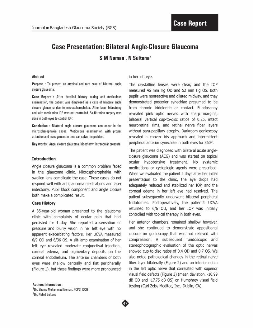


Journal ● Bangladesh Glaucoma Society (BGS)
51
BGS News
4th Asia-Pacific Glaucoma Congress
The 4th Asia Pacific Glaucoma Congress (APGS) 2018 took place at Busan Exhibition and Convention
Centre(BEXCO) from April 13-15,2018 in Korea.
The 4th Asia Pacific Glaucoma Congress (APGS) was the largest glaucoma meeting in Asia Pacific
region.
The 4th Asia Pacific Glaucoma Congress (APGS) participants were 729. The overall feedback on the
educational content was very positive.A team of 38 ophthalmologists from Bangladesh Glaucoma
Society (BGS) attended there. 6 speakers and 11 presented their papers in the symposium.
The 4th Asia Pacific Glaucoma Congress (APGS) was a huge gathering for exchange our knowledge,
views and ideas specially to update the latest glaucoma diagnostic and surgical procedure.
Regis
tration D
esk:
Conta
ct
No. +
31206793411
E-m
ail :
info
@w
orldgla
ucom
a.o
rg
All d
eta
ils w
ill be p
oste
d a
s s
oon
as t
here
available
.
8th World Glaucoma Congress
The 8th World Glaucoma Congress 2019 will be held at the Melbourne Convention &
Exhibition Centre (MCEC) Australia from March 27-30, 2019. Melbourne Convention &
Exhibition Centre (MCEC) is recognized as Australia's Leading Meetings and
Convention Centre. It is set on the banks of the Yarra River. The MCEC is situated
within 20 minutes from the airport and within 15 minutes all Melbourne landmarks
are reached.
