jkoajkoa journal of karnataka orthopaedic association chief editor dr anil k bhat, manipal associate...
TRANSCRIPT

JKOAJournal of Karnataka Orthopaedic Association
Chief EditorDr Anil K Bhat, Manipal
Associate EditorsDr Hitesh H Shah, Manipal
Dr Ashwath Acharya, Manipal
Published byDr Edward Nazareth, Secretary General KOA
On behalf of Karnataka Orthopaedic Association
E-mail: [email protected]
Print ISSN 2454-9010

ii Journal of Karnataka Orthopaedic Association | February 2016 | Volume 4 | Issue 1
Advisory Board
• Dr.M.ShantharamShetty,Mangalore• Dr.JSHegde,Mysore• DrBBPutti,Belgaum• Dr.RRShah,Gulbarga• Dr.RajivNaik,Bangalore• Dr.NareshShetty,Bangalore• Dr.BGSagar,Mysore• DrSharanSPatil,Bangalore• DrManjunathKS,Bangalore• Dr.SureshKorlhalli,Hubli• Dr.Purushotham,Banagalore
Editorial Board Members - Overseas
1. Dr.RamMohan,Manchester,UK2. Dr.A.AnantharamShetty,Kent,UK3. Dr.JamesFernandez,Sheffield,UK
EditorialBoardMembers-National
1. Dr.SRajaSabapathy,Coimbatore2. Dr.Rajagopalan,Pondicherry3. Dr.DhirenGanjwala,Ahmedabad4. Dr.RaghavaDuttMulukutla,Hyderabad5. Dr.Dr.LazarChandy,Kerala6. Dr.Deenadayalan,Coimbatore
Editorial Board Members - State
1. Dr.SharathKRao,Manipal2. DrRajendraBhandankar,Belgavi3. DrAjithKumar,Mangalore4. Dr.MonappaNaik,Manipal5. Dr.SanjayPai,Bangalore6. Dr.ShyamasunderBhat,Manipal7. Dr.KiranAcharya,Manipal8. Dr.ShankarR.Kurpad,Bangalore9. DrBharathKadadi,Bangalore10. Dr.VivekPandey,Manipal
EditorialOffice
DepartmentofOrthopaedics, KasturbaMedicalCollege,Manipal-576104 E-mail:[email protected]@gmail.com
KARNATAKA ORTHOPAEDIC ASSOCIATION
OFFICE BEARERS 20151. DrSharanPatil,President2. Dr.SureshKorlhalli,PresidentElect3. Dr.EdwardL.Nazareth,SecretaryGeneral4. Dr.ManojKumar,JointSecretary5. Dr.RamachandraKamath,Treasurer6. Dr.NithyanandaRao,PastPresident7. Dr.Purushotham,PastSecretaryGeneral
EXECUTIVE COMMITTEE 20151. Dr.V.SridharReddy,Raichur2. Dr.Manjunath.J,Davangere3. Dr.VinodkumarA.C,Bangalore4. Dr.M.J.ShashidharReddy,Bellary5. Dr.B.SachidanandRai,Mangalore6. Dr.Ravish.V.N,Bangalore7. Dr.MohammedIsmailHathiwale,Bijapur8. Dr.G.SrinivasReddy,Bangalore

Journal of Karnataka Orthopaedic Association | February 2016 | Volume 4 | Issue 1 iii
JournalofKarnatakaOrthopaedicAssociation
Editorial
Why and how should we index our journal?
Anil K. Bhat ....................................................................................................................................................................... 1
Review articles
Fractures of Lateral Humeral Condyle in Children
Edward L Nazareth ............................................................................................................................................................ 3
Lateral End Clavicle Fracture: A Review of Current Concepts
Vivek Pandey, Krishna Prasad, Sandesh Madi, Kiran Acharya ........................................................................................ 9
Management of High Energy Tibial Plateau Fractures
Monappa A Naik, Sharath K Rao ...................................................................................................................................... 14
Adult Brachial Plexus Injuries – An Appraisal on its Primary Management
Ashwath M Acharya, Anil K Bhat, Mahesh Kulkarni, KN Jayakrishnan ......................................................................... 21
Modified Stoppa approach for Acetabular Fractures: “Lighting up the tunnel”
M Ajith Kumar, Shailesh Pai ............................................................................................................................................. 31
Original articles
Management of Open Traumatic Extruded Talus
Muthukumar Balaji S, Rohit R K, S Devadoss, A Devadoss ............................................................................................ 35
Intramedullary Interlocking Nailing in Type II and Type III Open Fractures of Tibia – A Clinical Study
Patil NVP, Bellad S H, Anand S R ................................................................................................................................... 39
Surgical Management of Unstable Supracondylar Fractures of Humerus in Children by Closed Reduction
and Percutaneous K Wire Fixation by Crossed Versus Triple Pin Configuration-A Comparative Randomized
Study
C V Mudgal, S F Kammar, Viresh Murgodi ..................................................................................................................... 44
A Study of Functional Outcome of Surgical Management of Floating Knee Among Adults
Suryakanth Kalluraya, V K Bhasme, Vigneshwara B ....................................................................................................... 52
iv Journal of Karnataka Orthopaedic Association | February 2016 | Volume 4 | Issue 1
Technical pearls in commonly performed procedures
Technical Tips in Managing Fingertip Injuries
Hari Venkatramani, Gururaj Hosahalli, S Raja Sabapathy .............................................................................................. 60
Case reports
Solitary Osteochondroma of Scaphoid: A Case Report
Shekar M, Mahadev Jatti, Bharath Kadadi ........................................................................................................................ 64
A Rare Case of Periosteal Chondroma - Distal Phalanx of Toe
Karthik Raj K, Sudarshan A Bhandary, Bhaskar Bhandary, Sachin Shetty ..................................................................... 67
Master class for Postgraduates
Examination of Spine Case
M Shantaram Shetty .......................................................................................................................................................... 70

Journal of Karnataka Orthopaedic Association | February 2016 | Volume 4 | Issue 1 1
Bhat AK: Why and how should we index our journal?
Why and how should we index our journal?
Anil K. Bhat
E-mail: [email protected]
How to cite this article: Bhat AK, Why and how should we index our journal?. J Kar Orth Assoc. 2016 Feb; 4(1): 1-2.
Editorial
From a new editor and editorial board of the KOA journal, I wish you all a very joyous new year. The main objective of this new team would be to take our journal towards making it a peer reviewed and indexed journal which can stand up to a higher scientific scrutiny. To achieve this objective we need to undergo an arduous process and which requires small but significant steps to be covered one by one.In plain words, Indexing is the process of entering information from historical records into an online, searchable database. Indexation of a journal is considered a reflection of its higher scientific quality and will also increase the visibility and accessibility of a journal.
1. Indexing services facilitate the broadest dissemination of information by pointing researchers to articles that are relevant to their field and give our journal wider coverage for easy accessibility to the published articles.
2. This improves our journal’s reputation as a reliable source of high-quality information in our field.
3. Database research is the first activity researchers undertake as part of their study, and they naturally look to established, well-known databases. Thus, being indexed in a known database in our field will help increase our journal’s readership.
For a long-time Index Medicus has been the most comprehensive index of medical scientific journal articles. It is being publication since 1879. Over the years, many other popular indexation services have developed. These include Medline,
PubMed, EMBASE, SCOPUS, EBSCO Publishing’s Electronic Databases, and SCIRUS among others
Importance of publications is being increasingly recognized by the academic institutions. MCI guidelines also recommend indexed publications for teaching faculty in medical colleges. Consequently many more authors would be publishing than ever before.
The single most important factor to achieve indexation would be to improve the quality of scientific content of our articles by improving our research capabilities, methodically documenting our clinical work and writing it up in a succinct manner so as to benefit the medical fraternity at large
The following steps are required to achieve an indexation in most of the indexing services1. Scientific merit of a journal’s content is the
primary consideration in selecting journals for indexing. The validity, importance, originality, and ethical work are the key factors considered in recommending a title for indexing, whatever the intended purpose and audience
2. The journal should demonstrate features that contribute to the objectivity, credibility, and quality of its contents. These features include information about the methods of selecting articles, especially on the explicit process of external peer review, statements indicating adherence to ethical guidelines, evidence that authors have disclosed financial conflicts of interest, timely correction of errata, explicit responsible retractions as appropriate, and opportunity for comments and dissenting opinion
3. Publication must have an ISSN (International Standard Serial Number)
Dr. Anil K. BhatEditor in chief, Journal of KOAProfessor and Head Department of Orthopedics, KMC, Manipal

Bhat AK: Why and how should we index our journal?
2 Journal of Karnataka Orthopaedic Association | February 2016 | Volume 4 | Issue 1
4. Publication content should be issued over time under a common title , is a collection of articles by different authors and is intended to be published indefinitely
5. The journal should state the purpose of its publication and the contents should have a combination of reports of original research, original clinical observations accompanied by analysis and discussion, critical reviews and case reports with discussions
6. Quality of the layout, printing, graphics, and illustrations are all considered in assessing a journal
The process of indexing our journal has begun in all earnest. An ISSN number has been obtained from the National Science library, the number being 2454-9010. This was the first step towards indexing the journal. An eminent group of Orthopaedic surgeons
including both national and international faculty are a part of the editorial board which is also another requirement. Peer review has been started with this issue including a strict check on plagiarism of submitted articles to enhance the scientific content and stress on ethical adherence. This has also lead to rejecting some of the articles this time.
We have come out with a very vibrant issue having a brand new outlay. This issue has a theme of “Current concepts in trauma management ’’ which has covered various aspects of trauma scenarios from acute Brachial plexus injuries to tibial fractures. A separate section for postgraduate teaching is being introduced to discuss an exam topic by an eminent senior faculty.
Today’s progress was yesterday’s plan and if we walk down the right path and we are willing to keep walking, eventually we will make progress.

Edward L Nazareth, et al: Fractures of Lateral HumeralCondyle in Children
Journal of Karnataka Orthopaedic Association | February 2016 | Volume 4 | Issue 1 3
AbstractFractures of lateral humeral condyle constitute around 10% to 20% of all distal humeral fracturesin children. Unless properly looked for, the lateral humeral condyle fractures are easily missed in the normal radiographs.If not treated diligentlythey get displaced which can lead to disastrous complication of nonunion and permanent disability. Undisplaced fractures or those displaced less than 2 mm can be managed with cast immobilization with frequentfollow up X-rays.Lateral humeral condylefractures displaced more than 2 mm require surgical fixation. The surgical option includes closed reduction, open reduction and arthroscopically assisted techniques. Fixation techniques are K-wires, bioresorbable pins and metaphyseal screws. Complications associated with lateral humeral condylar fracture in children are nonunion, cubitus varus, cubitus valgus and tardy ulnar nerve palsy.
Key words: Lateral humeral condyle, distal humeral fractures, cubitus varus, and cubitus valgus.
Fractures of Lateral Humeral Condyle in Children
Edward L Nazareth
Email: [email protected]
How to cite this article: Nazareth EL. Fractures of Lateral Humeral Condyle in Children. J Kar Orth Assoc. 2016 Feb; 4(1): 3-8.
Review article
The incidences of fractures of the lateral humeral condyle vary from 10% to 20% of all elbow fractures in the pediatric age (1-3). It is the second most common elbow fracture in children after supracondylar humerus fractures. The most common age for this fracture is around 6 years, with age ranging from 2 to 11 years (3-5).
Mechanism Of Injury
There are two mechanisms described for lateral humeral condylar fractures.The push-off theory was proposed by Milch (1)according to which these fractures were the result of a force directed upward and outward along the radius. The radial head impacting the distal humerus fractures the lateral condyle, the fracture extends upward and outward. When the forearm goes into valgus, the radial head pushes off the lateral condyle. The proponents of the pull-off theory suggest that lateral humeral condylarfractures were avulsion fractures. The
muscles attached to the lateral condyle are believed pull the fragment resulting in avulsion fracture of the lateral humeral condyle (4,7).
However, the most likely cause of this fracture is a combination of the push-off and pull-offmechanisms(8). The less common Milch type I fracture iscaused by a fall on an out stretched forearm with elbow extended resulting in impaction of the radial head against the capitellum. Milch type II fracture is caused by avulsion forces of the extensors of forearm acting on the lateral condyle.
Clinical Features
Lateral condylar fractures of humerus present with swelling and tenderness localised to the lateral aspect of the elbow. Pain may increase with forced wrist flexion that stretches the wrist extensor origin. Lateral humeral condylar fracturescan be clinically distinguished from supracondylar humeral fractureswhich havesignificant pain and deformity.
Radiological Evaluation
Plain radiographs: Antero-posterior, lateral and internal oblique radiographs of the affected elbow are required to diagnose the fracturein children with
Dr. Edward L NazarethProfessor, Department of Orthopaedic Surgery
Fr. Muller Medical College, Mangalore 575002
Edward L Nazareth, et al: Fractures of Lateral HumeralCondyle in Children
4 Journal of Karnataka Orthopaedic Association | February 2016 | Volume 4 | Issue 1
suspected fracture of the lateral humeral condyle. The X-rays of the normal elbowmay be obtained for comparison, whenever there is suspicion. Because the fracture often lies posterolateral, an internal oblique view is necessary and always a must to evaluate the fracture pattern and the amount of displacement. In fractures with minimal displacement (less than 2 mm), the fracture can be seen only on the internal oblique view (9).
CT Scan: Whenever the fracture line is not clear, CT scan should be taken to confirm the injury. CT scan can be performed easily, exposure islimited and radiation is minimal. Only limitation is that CT may not show the integrity of articular cartilage hinge which may be essential to determine the fracture as stable (10).
MRI: The integrity of the cartilage hinge at the distal humeral epiphysis ultimately determines the stability of lateral condylefracture. The bridge acts as a hinge that guides the fragment back into place when the fracture is reduced (11). The presence or absence of this bridge indicates the stability of the fracture. The disadvantages of MRI are that the young children may need sedation/anesthesia which can increase the cost of treatment.As it is always safer to pin the fracture when thereis doubt, MRI under anesthesia toevaluate the cartilage hinge may be of limited value.
Ultrasonography: To evaluate the integrity of the articular cartilage, ultrasonography may be a better option than MRI. This does not require sedation and is relatively less expensive. However, for accurate evaluation through ultrasonography, a skilled sonologist may be required (12).
Arthrography: Some studies have suggested the use of arthrography to determine the fracture pattern of the elbow in children (13,14). Arthrography in children has to be performed in the operating room under anesthesia.Consequently arthrography has limited value as a diagnostic tool to assess fracture pattern or cartilage integrity.
Classification Of FractureMilch (1) described two types of fracture patterns. In type I fractures, the fracture line courses lateral to the trochlea and into the capitulotrochlear groove; this is similar to Salter-Harris type IV fracture.
In this fracture pattern, the trochlea is intact, therefore the elbow is considered stable. Milch type II fracture is afracture-dislocation of the lateral condyle, in which the fracture line extends into the apex of the trochlea; the fracture is a Salter-Harris type II fracture. In this pattern of injury, the elbow is unstable because the trochlea is disrupted. Milch system of classification does not help in the management of this fracture and is of academic interestonly. The lateral humeral condyle fractures classified pre-operatively using Milch classification may not correlate intra-operative findings (15).
The classification of fracture by Jakob et al (7) is based on fracture fragment displacement. Type I fracture is undisplaced with intact articular surface. The cartilaginous epiphysis is not completely fractured. Type II is complete fracture, with minimal displacement. Type III is a complete fracture with displacement and rotation of the fractured fragment. This classification has more importance in the management of lateral humeral condylar fractures in children.
Treatment
Many studies stress thatall lateral humeral condylar fractures in children should be treated by surgery to prevent displacement and the high rate of nonunion (16-18). Some authors recommend internal fixation to all the lateral condylar fractures because even the undisplaced fractures seen in the initial radiographs displace subsequently when treated non-operatively (19,20). Launay et al (21) recommend non-operative treatment only when follow up was guaranteed. When follow up compliance for non-operative treatment protocol was unreliable, they recommend percutaneous pinning.
Non-operativetreatment
Non operative treatment is appropriate only for nondisplaced fractures or the fractures with an intact cartilage hinge that is confirmed by MRI or ultrasonography and fractures demonstrating less than 2 mm displacement on all radiographic views. But non-surgical treatment for lateral humeral condylar fractures in children is always risky, however undisplaced the fracture is.

Edward L Nazareth, et al: Fractures of Lateral HumeralCondyle in Children
Journal of Karnataka Orthopaedic Association | February 2016 | Volume 4 | Issue 1 5
Whenever non-operative treatment is opted, immobilization is achieved with a long arm cast extending as far proximal as possible on the humerus. Radiographic assessment of the status of displacement is recommended every week for the first 3 weeks of cast immobilization (21). Internal oblique radiography is the bestimaging technique showing subsequent fracture displacement in initiallynon-displaced or minimally displaced humerus lateralcondyle fractures treated non-operatively. At the first week follow-up, anteroposteriorand particularly internal oblique radiographs shouldbe taken of conservatively treated patients (22).
Immobilization for a minimum period of six weeks is a must because lateral condylar humeral fractures have a high rate of nonunion.Even aftersixweeks of immobilization, some of the lateral condylar fractures with initial displacement may take longer time to unite or may not unite at all even if reduced by closed manipulation (2).
However most of the studies recommendsixweeks of immobilization for fracture healing (21,24).
Operative Treatment
Closed Reduction
Lateral humeral condyle has precarious blood supply.The vessels supplying the lateral condylar epiphysis enter the posterior aspect of the condyle at its extra-articular region. Excessive soft tissue stripping during open reduction can damage the blood supply which can increase the risk of nonunionand osteonecrosis. As a result, some of the studies have suggested closed reduction and percutaneous pinning for stable displaced fractures. The lateral condylar fractures with less than 2 mm of displacement, intraoperative arthrography confirming the congruency of the joint surface with an intact cartilage hinge are considered stable and these alone can be managed by closed reduction and percutaneous pinning (25).
Even in children with nondisplaced or minimally displaced fractures who may not be compliant forregular followup, closed reduction and percutaneous pinning is a better choice than casting and serial radiographs (21).
Song (26) has described the technique of closed reduction. It involves traction with gentle varus force followed by anteromedial manipulation of the fragment. If the fracture has rotational malalignment, K- wires can be used as joysticks to reduce the fracture with the forearm in supination and the elbow in extension. Two smoothK-wires placed in parallel are used to pin the fragments.
Arthroscopy assisted reduction
Arthroscopically assisted management is recommended toavoid osteonecrosis and nonunion. The distal fragment is reduced under direct vision manually.The K-wires are used as joysticks. The elbow arthroscopy provides reliable visualization of the articular surface while reducing the fracture. However, fluid extravasation can cause compartment syndrome (27,28).
Open reduction and fixation
Open reduction of lateral humeralcondylar fracture is the ideal procedure of treatment. The Kocher approach to the lateral aspect of the elbow is commonly used. The internervous plane between the anconeus (innervated by the radial nerve) and the extensor carpi ulnaris (innervated by the posterior interosseous nerve) is used. It is utmost important to minimizethe softtissue dissection. All dissection should be only anterior to avoid damaging the blood supply to the distal fragment.
Anatomic reduction and its confirmation through fluoroscopic images in all views is important.
Following anatomic reduction, percutaneous fixation of the fracture is preferred.Two K wires passed from the lateral aspect of the elbow have to be placed outside the surgical incision.The K wires may be placed in parallel or divergent toeach other. The pins must engage the bone, and not only in the cartilaginous epiphysis. The outer end of the pin is kept percutaneous or subcutaneous (21). The K wires are not removed until there is radiographic evidence of fracture healing which may take 4 to 6 weeks.
High-strength, bioactive, bioresorbable pins made of forged composites of unsintered hydroxyapatite particles/poly-L-lactide (F-u-HA/PLLA) for treating pediatric lateral humeral condylar fractures
Edward L Nazareth, et al: Fractures of Lateral HumeralCondyle in Children
6 Journal of Karnataka Orthopaedic Association | February 2016 | Volume 4 | Issue 1
have been reported with good results. These are radiopaque,highlybiocompatibleand are well incorporated into the bone without intervention of fibrous tissue (23).
Common Complications Nonunion
Nonunion is the most common complication of the lateral humeral condyle fractures.The persistence of the fracture line and absence of callus at 8 weeks or more after the injury indicates nonunion (2). The pull of the extensors, inadequate blood supply at the fractureregion and intra articular nature of the fracture are important causes for nonunion (6,21). Nonunion is common when the lateral humeral condylar fractures are treated without surgery (21,24-25).
The nonunion with minimal displacement can be treated by open reduction and bone grafting. The nonunion with less than 1 cm of displacement can be treated with freshening of the fractures and fixation with threaded pins or with screw if there is adequate metaphyseal fragment. The bone graft can be inserted at the metaphyseal region (2,6).
The nonunion associated with grossdisplacement may not benefit from open reduction of the fracture fragment as the surgery may damage the physis resulting in valgus deformity (2).
The patients with nonunion where the distal fragment is grossly displaced with problems like pain, instability, or poor range of motion may be chosen for delayed fixation, though the results may not be satisfactory (29).
Cubitus valgus and varus deformity
More than 20% of children with lateral humeral condyle fractures develop some degree of cubitus varus deformity (7, 30)and more than 10% develop valgus deformity (31). The studies report that the cubitus varus deformity after lateral condylar fracture is most common in nondisplaced and minimally displaced fractures. Medial displacement of the distal fragment seems to be the cause for this (19,30). This deformity is clinically not significant (30).
Cubitus valgus, which is much less common than varus deformity is caused by lateral physeal arrest.
Most of the lateral humeral condyle fractures are Salter Harris type IV fractures, where the fracture line crosses the epiphysis which can cause growth arrest. Though the growth arrest may not cause axial deviation or length deformity, it may precipitate tardy ulnar nerve palsy.
Fishtail deformity seen in the radiographs is characterized by deepening of the trochlear groove. This deformity also has no clinical significance. It happens because of persisting gap between the lateral condylar ossification center and the medial ossification center of the trochlea, caused either by resorption or failure to develop (31).
Tardy Ulnar Nerve Palsy
It involves slow, progressive involvement of the ulnar nerve. Tardy ulnar nerve palsy is caused by stretching of the nerve, most commonly seen with cubitus valgus deformity. This complication presents late (average22 years post-injury) (32). Tardy ulnar nerve palsy is treated with anterior ulnar nerve transposition.
Summary
Lateral humeral condylar fractures are the second most common elbow fracture in children. The fractures that are displaced more than 2 mm should be reduced and pinned. The fractures that are displaced less than 2 mm can be treatednon-operatively with regular follow up, however when treated non-surgically the fracture can displace even after the initiation of immobilization. Minimally displaced lateral humeralcondylar fractures are commonly missed which ultimately result in nonunion and deformity. Therefore high index of suspicion and properclinical andradiological assessment in paediatric elbow injuries is a must to avoid this complication which can lead to permanent functional and cosmetic deformity.
References 1. Milch H: Fractures and fracture dislocations of
the humeral condyles. J Trauma. 1964;4:592-607.
2. Flynn JC, Richards JF Jr, Saltzman RI: Preven-tion and treatment of non-union of slightly displaced fractures of the lateral humeral con-
Edward L Nazareth, et al: Fractures of Lateral HumeralCondyle in Children
Journal of Karnataka Orthopaedic Association | February 2016 | Volume 4 | Issue 1 7
dyle in children: Anend-result study. J Bone Joint Surg Am.1975;57(8):1087-1092.
3. Landin LA: Fracture patterns in children: Anal-ysis of 8,682 fractures with special reference to incidence, etiology and secular changes in a Swedish urban population1950-1979. ActaOr-thopScandSuppl1983;202:1-109.
4. Stimson L: A Practical Treatise on Fractures and Dislocations. Philadelphia, PA, Lea Broth-ers & Co, 1900.
5. Takada N, Otsuka T, Suzuki H, Yamada K: Pediatric Displaced Fractures of the Lateral Condyle of the Humerus Treated Using High Strength, Bioactive, Bioresorbable F-u-HA/PLLA Pins: A Case Report of 8 Patients With At Least 3 Years of Follow-Up.J Orthop Trau-ma. 2013 May;27(5):281-284.
6. Flynn JC: Nonunion of slightly displaced frac-tures of the lateral humeral condyle in children: An update. J PediatrOrthop1989;9(6):691-696.
7. Jakob R, Fowles JV, Rang M, Kassab MT: Ob-servations concerning fractures of the lateral humeral condyle in children. J Bone Joint Surg Br 1975; 57(4):430-436.
8. McLearie M, Merson RD: Injuries to the lateral condyle epiphysis of the humerus in children. J Bone Joint Surg Br 1954;36(1):84-89.
9. Song KS, Kang CH, Min BW, Bae KC, Cho CH: Internal oblique radiographs for diagnosis of nondisplaced or minimally displaced lateral condylar fractures of the humerus in children.J Bone Joint Surg Am 2007;89(1):58-63.
10. Chapman VM, Grottkau BE, Albright M, Salamipour H, Jaramillo D: Multidetec-tor computed tomography of pediatric lat-eral condylar fractures. J Comput Assist To-mogr 2005;29(6):842-846.
11. Kamegaya M, Shinohara Y, Kurokawa M, Ogata S: Assessment of stability in children’s mini-mally displaced lateral humeral condyle fracture by magnetic resonance imaging. J PediatrOr-thop.1999;19(5):570-572.
12. Vocke-Hell AK, Schmid A: Sonographic differ-entiation of stable and unstable lateral condyle fractures of the humerus in children. J Pediat-rOrthop B. 2001;10(2):138-141.
13. Yates C, Sullivan JA: Arthrographic diagnosis of elbow injuries in children. J PediatrOrthop. 1987;7(1):54-60.
14. Tang CW, Skaggs DL, Kay RM: Elbow aspiration and arthrogram: An alternative method. Am J Orthop.2001;30(3):256.
15. Mirsky EC, Karas EH, Weiner LS: Lateral condyle fractures in children: Evaluation of classification and treatment. J Orthop Trauma. 1997;11(2): 117-120.
16. Conner AN, Smith MG: Displaced fractures of the lateral humeral condyle in children. J Bone Joint Surg Br 1970;52(3):460-464.
17. Speed J, Macey H: Fractures of humeral condyles in children. J Bone Joint Surg Am. 1933;15:903-919.
18. Hardacre JA, Nahigian SH, Froimson AI, Brown JE: Fractures of the lateral condyle of the humerus in children.J Bone Joint Surg Am. 1971;53(6):1083- 1095.
19. Badelon O, Bensahel H, Mazda K, Vie P: Lateral humeral condylar fractures in children: A report of 47 cases. J PediatrOrthop. 1988;8(1):31-34.
20. Fontanetta P, Mackenzie DA, Rosman M: Missed, maluniting, and malunited fractures of the lateral humeral condyle in children. J Trauma. 1978;18(5):329- 335.
21. Launay F, Leet AI, Jacopin S, Jouve JL, Bollini G, Sponseller PD: Lateral humeral condyle fractures in children: A comparison of two approaches to treatment. J PediatrOrthop. 2004;24(4): 385-391.
22. T u h a n K u r t u l m u s ¸ N e c d e t S a g ˘ l a m , Gursel Saka,CemCos¸kunAvcı, Meric¸ Ug˘urlar,Mehmet Tu¨rker, Pediatric lateral humeral condyle fractures: internal obliqueradiographs alter the course of conservative treatment. Eur J Orthop Surg Traumatology 2014;24:1139–1144
23. Takada N, Otsuka T, Suzuki H, Yamada K: Pediatric Displaced Fractures of the Lateral Condyle of the Humerus Treated Using High Strength, Bioactive, Bioresorbable F-u-HA/PLLA Pins: A Case Report of 8 Patients With At Least 3 Years of Follow-Up. J Orthop Trauma. 2013 May;27(5):281-4.

Edward L Nazareth, et al: Fractures of Lateral HumeralCondyle in Children
8 Journal of Karnataka Orthopaedic Association | February 2016 | Volume 4 | Issue 1
24. Bast SC, Hoffer MM, Aval S: Nonoperative treatment for minimally and nondisplaced lateral humeral condyle fractures in children. J PediatrOrthop. 1998;18(4):448-450.
25. Mintzer CM, Waters PM, Brown DJ, Kasser JR: Percutaneous pinning in the treatment of displaced lateral condyle fractures. J PediatrOrthop. 1994;14(4):462-465.
26. Song KS, Kang CH, Min BW, Bae KC, Cho CH, Lee JH: Closed reduction and internal fixation of displaced unstable lateral condylar fractures of the humerus in children. J Bone Joint Surg Am. 2008; 90(12):2673-2681.
27. Hausman MR, Qureshi S, Goldstein R, et al: Arthroscopically-assisted treatment of pediatric lateral humeral condyle fractures. J PediatrOrthop. 2007;27(7):739-742.
28. Perez Carro L, Golano P, Vega J:Arthroscopic-assisted reduction and percutaneous external
fixation of lateral condyle fractures of the humerus.Arthroscopy. 2007;23(10):1131-1134.
29. Wattenbarger JM, Gerardi J, Johnston CE: Late open reduction internal fixation of lateral condyle fractures. J PediatrOrthop. 2002;22(3):394-398.
30. So YC, Fang D, Leong JC, Bong SC: Varus deformity following lateral humeral condylar fractures in children. J PediatrOrthop. 1985;5(5):569-572.
31. Skak SV, Olsen SD, Smaabrekke A: Deformity after fracture of the lateral humeral condyle in children. J PediatrOrthop B. 2001;10(2):142-152.
32. Gay JR, Love JG: Diagnosis and treatment of tardy paralysis of the ulnar nerve: Based on a study of 100 cases. J Bone Joint Surg Am. 1947;29(4):1087- 1097.
Vivek Pandey, et al: Lateral End Clavicle Fracture: A Review of Current Concepts
Journal of Karnataka Orthopaedic Association | February 2016 | Volume 4 | Issue 1 9
AbstractLateral end clavicle fractures are frequently observed in isolation or along with polytrauma injuries. The management depends upon the type of fracture classified according to Neer or Edinburgh. Neer Type I and III do well with conservative management. However, type II often needs surgical intervention for optimal results. Long term complications include acromioclavicular arthritis, non-union and compromised shoulder function.
Vivek PandeyDepartment of Orthopaedics, Kasturba Medical College, ManipalManipal University, Karnataka
Lateral End Clavicle Fracture: A Review of Current Concepts
Vivek Pandey, Krishna Prasad, Sandesh Madi, Kiran Acharya
Email: [email protected]
How to cite this article: Pandey V, Prasad K, Madi S, Acharya K. Lateral End Clavicle Fracture: A Review of Current Concepts. J Kar Orth Assoc. 2016 Feb; 4(1): 9-13.
Review article
Introduction
The lateral end of the clavicle is a special type of injury among the clavicle fractures, with the incidence of 21% among clavicle fractures. (1) The association with acromion through acromioclavicular joint and relationship with coracoid providing vertical stability via coracoclavicular ligament makes its management challenging and controversial.[2] These fractures have a higher rate of non-union and cosmetic deformity (3). However, the key to the management lies in the understanding of the stability of the fracture that is understood by the classification of fracture.
Classification
In 1968, Neer classified lateral end of clavicle fractures according to their location about the coracoclavicular ligaments. (4) Later, Rockwood et al [Table 1]. (5) modified the classification.
This classification is widely accepted and has got the prognostic value as in type II fractures, unbalanced downward force such as the weight of the arm and vertical muscle forces acting on the medial clavicle end keep the fractured end distracted and hinder union .(4) In type IIb fracture, the loss of conoid
ligament restraint on the lateral fragment could result in instability and showed a high incidence of delayed union and nonunion rate. (6) Hence, the surgical treatment may be necessary for type II fractures for the union of the fracture.
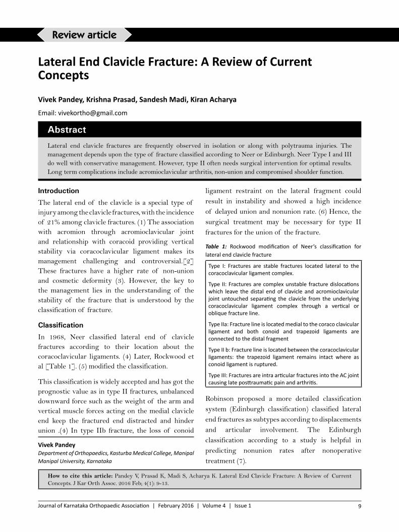
Table 1: Rockwood modification of Neer’s classification for lateral end clavicle fracture
Type I: Fractures are stable fractures located lateral to the coracoclavicular ligament complex.
Type II: Fractures are complex unstable fracture dislocations which leave the distal end of clavicle and acromioclavicular joint untouched separating the clavicle from the underlying coracoclavicular ligament complex through a vertical or oblique fracture line.
Type IIa: Fracture line is located medial to the coraco clavicular ligament and both conoid and trapezoid ligaments are connected to the distal fragment
Type II b: Fracture line is located between the coracoclavicular ligaments: the trapezoid ligament remains intact where as conoid ligament is ruptured.
Type III: Fractures are intra articular fractures into the AC joint causing late posttraumatic pain and arthritis.
Robinson proposed a more detailed classification system (Edinburgh classification) classified lateral end fractures as subtypes according to displacements and articular involvement. The Edinburgh classification according to a study is helpful in predicting nonunion rates after nonoperative treatment (7).

Vivek Pandey, et al: Lateral End Clavicle Fracture: A Review of Current Concepts
10 Journal of Karnataka Orthopaedic Association | February 2016 | Volume 4 | Issue 1
Clinical and Radiological assessment Unless undisplaced, lateral end clavicular fractures clinically produce and obvious painful deformity at the acromioclavicular joint, clinically indistinguishable to that of acromioclavicular joint dislocation. Radiologically, stress views are rarely recommended to know the integrity of coracoclavicular ligaments in association with lateral end clavicle fractures.(8) Careful radiographic survey is mandatory to exclude associated chest injury, such as pneumothorax or haemothorax and ipsilateral rib fractures. (9)
Treatment Type of fracture will dictate the Treatment of choice for Lateral end clavicle fractures.
A) Undisplaced lateral end fractures (Neer type I, Edinburgh Type 3A)
In this type, the intact periosteum and the conoid and trapezoid ligaments bind the fracture fragments together preventing the displacement of the fracture fragments. (4) Non-operative management is the treatment of choice.[10, 11] A simple arm sling for four to six weeks suffices. Analgesic and local cold pack for a few days help in relieving pain and swelling.
Fractures extending into acromioclavicular joint may be associated with pain due to step defect. It may end up into late arthritis of AC joint that might need excision of the lateral end of the clavicle (open or arthroscopic). Late excision of distal fragments either by open or arthroscopic is the treatment when the fracture fragment is small.[4, 10]
B) Displaced lateral end fractures (Neer Type II, Edinburgh type 3B)
Non-operative treatment: High rates of nonunion with pain and limitation of shoulder function after non-operative treatment for this variant were reported in some retrospective studies. (4,7,10) This led to a recommendation for operative intervention for this type of fractures. However, unless planned accurately there are high rates of complications too reported after operative fixation.
The majority of these injuries occur in middle age and elderly population who easily tolerate the cosmetic and functional disabilities. However,
informing regarding the complications like nonunion, acromioclavicular arthritis is worthwhile and can be overcome with excision of the lateral end of the clavicle among symptomatic individuals. (4,10,12)
Operative treatment:
The indications for operative management of Neer Type II fracture can be divided into mainly two categories: Early, and Late. Early indications include a compromise on soft tissue envelope, double disruption of the ipsilateral shoulder suspensory complex, a fracture in active young individual, athlete, a manual labourer who wants to get back to his/her work quickly. Late indications are Symptomatic non-union or malunion, acromioclavicular arthritis.
Methods of fixation: Most fixation methods utilize the fracture reduction to stabilize the coracoclavicular joint too. Once the fracture heals, it stabilizes the joint also.
1. Plate fixation for lateral end fracture: Adequate plate fixation is possible if the lateral fragment accepts two to three locking bicortical screw. Currently, there are pre-contoured plates available, which have an advantage of hold on the lateral fragments with more number of locking screws for the better purchase of smaller fragments. This in turn increases the stability of the construct [Figure 1]. (13) A minimum of three screw purchase in lateral fragment is acceptable. (14) However, it still carries a chance of screw cutout in early postoperative period. The fragments that appear to be intact might be comminuted, and porotic is leading to screw backout. However, it works well with the large intact lateral fragment.
Figure 1: Preoperative and postoperative images of type II Lateral end clavicle fracture managed by a precontoured locking plate
2. Hook-plate fixation: Clavicular hook plate was devised for the treatment of lateral clavicle

Vivek Pandey, et al: Lateral End Clavicle Fracture: A Review of Current Concepts
Journal of Karnataka Orthopaedic Association | February 2016 | Volume 4 | Issue 1 11
fracture where the distal fragments were too small, comminuted, or osteoporotic to allow conventional locking plate fixation. This small distal fragment (comminuted and porotic) will be poorly held by conventional plates, and may lead to cut out of the screw from the fragment. Hook plate works by reducing the medial end of the fracture to the lateral fragment by taking a fulcrum under the acromion. It need not to have any screw purchase in the lateral fragment [Figure 2]. The use of Hook plate with good results is reported in many series. However, some series have reported shoulder stiffness after using the hook plate. (15,16) Most surgeons consider removal of the plate after three to six months. This makes the patient undergo a second surgery. Early removal of the plate can lead to non-union of the fracture, and late removal of the plate also has its share of complications of shoulder stiffness and fracture medial to the plate respectively. (17) The authors recommend plate removal after six to nine months. An early mobilization of the shoulder can prevent stiffness. However, osteolysis around the tip of the hook is noted when the movement around the shoulder increases but this has no clinical significance. (18)
Figure 2: Preoperative and postoperative images of type II Lateral end clavicle fracture managed by a Hook plate
3. Suture and sling techniques: Coracoclavicular sling and Dacron graft ligaments have also been described to reconstruct the coracoclavicular ligaments. In this, the Dacron graft either looped around or drilled through the coracoid and over the clavicle fragment to form a sling. Dual endobutton (Smith Nephew, Andover, USA), Tightrope (Arthrex, USA) and Dog button (Arthrex, Naples, FL, USA) has also been used successfully for type II Neer’s fracture [Figure 3]. The latter can be performed arthroscopically. The advantage of this technique
is that it acts as both; a primary stabilizer and a reinforcement of other fixation techniques with good functional results(14-19,21)
Figure 3: Postoperative images of type II Lateral end clavicle fracture managed by a precontoured plate and Dog Button (blue arrow)
4. Coracoclavicular screw: This is the method explained in acromioclavicular joint dislocation or subluxation. (22) This method was widely employed in the past even though it was technically demanding for the reason the space available for the screw purchase in coracoid was narrow and complications such as screw cut out and loosening were reported. The other drawbacks of this method are a need for the screw removal and the limitation of shoulder movement. (2,23) However, currently, this method is out of flavour to be used for the fixation due to the problems said above.
5. Kirschner wire fixation: This technique was first popularized by Neer (24) and remained a preferred technique of lateral end fracture fixation for long. However, it is no more a recommended method of fixation due to high incidence of complications like implant breakage, migration, high rates of non-union and infection. (14-16)
6. Other techniques: Many other techniques like tension band wiring with Kirschner wire, cannulated cancellous screw fixation have been described with limited success.
C) Intraarticular lateral end fractures (Neer type III, Edinburgh types 3 A2, 3B2)
These fractures are rare and treated according to their degree of displacement, commonly as extra-
Vivek Pandey, et al: Lateral End Clavicle Fracture: A Review of Current Concepts
12 Journal of Karnataka Orthopaedic Association | February 2016 | Volume 4 | Issue 1
articular fractures. These fractures are prone to acromioclavicular arthritis that may need treatment in future.
Authors’ preferred method of treatment:Neer Type I and III: Conservative
Neer Type II:
A) Small/porotic/comminuted lateral fragment: Hook-plate fixation or dog button fixation
B) Large lateral fragment: Pre-contoured plate fixation
Complications1. Nonunion: Nonunion rate of non-operative
treatment of the lateral end of clavicle vary from 11-40% (25) and the factors influencing are an older age group, displacement, and comminution of fracture. The disabilities include pain around the shoulder girdle and loss of shoulder function (26-28). Often, it is asymptomatic in elderly patients or patients with low functional demand. In established symptomatic non-union, excision of the lateral end of the clavicle is the most recommended treatment when the fracture fragment is small and coracoclavicular ligament is intact, the fixation of fracture recommended with or without bone grafting when the fractured fragment is large and has good bone stock.
2. Osteoarthritis of acromioclavicular joints:
The incidence of ACJ arthritis is 6% (29) and this complication is most commonly observed in intraarticular fractures but extra-articular fractures are not exceptions. Diagnosis of acromioclavicular arthritis may be difficult because of mimicking symptoms from rotator cuff impingement and chronic regional pain syndrome. Relief of symptoms after ultrasound guided intraarticular local anesthetic is the most recommended diagnostic sign. Symptomatic acromioclavicular osteoarthritis can be treated with open or arthroscopic excision of the lateral fragment. (4,10,12)
Conclusions
Lateral end clavicle fractures are typed based on the articular involvement, displacement of fragment and size of the fracture fragment and with a custom made approach combining the factors like age,
occupation, activity levels, the appropriate operative technique will result in a good functional outcome.
References1. Nordqvist A, Petersson C. The incidence of
fractures of the clavicle. Clin Orthop Relat Res. 1994:127-132.
2. Ballmer FT, Gerber C. Coracoclavicular screw fixation for unstable fractures of the distal clavicle. A report of five cases. J Bone Joint Surg Br. 1991;73:291-294.
3. Robinson CM, Court-Brown CM, McQueen MM, Wakefield AE. Estimating the risk of nonunion following nonoperative treatment of a clavicular fracture. J Bone Joint Surg Am. 2004; 86-A:1359-1365.
4. Neer CS, 2nd. Fractures of the distal third of the clavicle. Clin Orthop Relat Res. 1968;58:43-50.
5. Jr. RC. Fractures of the outer clavicle in children and adults. J Bone Joint Surg Br. 1982;64 B:642.
6. Edwards DJ, Kavanagh TG, Flannery MC. Fractures of the distal clavicle: a case for fixation. Injury. 1992;23:44-46.
7. Robinson CM. Fractures of the clavicle in the adult. Epidemiology and classification. J Bone Joint Surg Br. 1998;80:476-484.
8. Rowe CR. An atlas of anatomy and treatment of midclavicular fractures. Clin Orthop Relat Res. 1968;58:29-42.
9. Dugdale TW, Fulkerson JP. Pneumothorax complicating a closed fracture of the clavicle. A case report. Clin Orthop Relat Res. 1987:212-214.
10. Nordqvist A, Petersson C, Redlund-Johnell I. The natural course of lateral clavicle fracture. 15 (11-21) year follow-up of 110 cases. Acta Orthop Scand. 1993;64:87-91.
11. Post M. Current concepts in the treatment of fractures of the clavicle. Clin Orthop Relat Res. 1989:89-101.
12. Petersson CJ. Resection of the lateral end of the clavicle. A 3 to 30-year follow-up. Acta Orthop Scand. 1983;54:904-907.
13. Kalamaras M, Cutbush K, Robinson M. A method for internal fixation of unstable distal clavicle fractures: early observations using a new
Vivek Pandey, et al: Lateral End Clavicle Fracture: A Review of Current Concepts
Journal of Karnataka Orthopaedic Association | February 2016 | Volume 4 | Issue 1 13
technique. J Shoulder Elbow Surg. 2008;17:60-62.
14. Hessmann M, Kirchner R, Baumgaertel F, Gehling H, Gotzen L. Treatment of unstable distal clavicular fractures with and without lesions of the acromioclavicular joint. Injury. 1996;27:47-52.
15. Bezer M, Aydin N, Guven O. The treatment of distal clavicle fractures with coracoclavicular ligament disruption: a report of 10 cases. J Orthop Trauma. 2005;19:524-528.
16. Flinkkila T, Ristiniemi J, Hyvonen P, Hamalainen M. Surgical treatment of unstable fractures of the distal clavicle: a comparative study of Kirschner wire and clavicular hook plate fixation. Acta Orthop Scand. 2002;73:50-53.
17. Nadarajah R, Mahaluxmivala J, Amin A, Goodier DW. Clavicular hook-plate: complications of retaining the implant. Injury. 2005;36:681-683.
18. Mizue F, Shirai Y, Ito H. Surgical treatment of comminuted fractures of the distal clavicle using Wolter clavicular plates. J Nippon Med Sch. 2000;67:32-34.
19. Goldberg JA, Bruce WJ, Sonnabend DH, Walsh WR. Type 2 fractures of the distal clavicle: a new surgical technique. J Shoulder Elbow Surg. 1997;6:380-382.
20. Levy O. Simple, minimally invasive surgical technique for treatment of type 2 fractures of the distal clavicle. J Shoulder Elbow Surg. 2003;12:24-28.
21. Webber MC, Haines JF. The treatment of lateral clavicle fractures. Injury. 2000;31:175-179.
22. Bosworth BM. Acromioclavicular separation. Surg Gynecol Obstet. 1941;73:866-871.
23. Yamaguchi H, Arakawa H, Kobayashi M. Results of the Bosworth method for unstable fractures of the distal clavicle. Int Orthop. 1998;22:366-368.
24. Neer CS, 2nd. Fracture of the distal clavicle with detachment of the coracoclavicular ligaments in adults. J Trauma. 1963;3:99-110.
25. Khan LA, Bradnock TJ, Scott C, Robinson CM. Fractures of the clavicle. J Bone Joint Surg Am. 2009;91:447-460.
26. Eskola A, Vainionpaa S, Patiala H, Rokkanen P. Outcome of operative treatment in fresh lateral clavicular fracture. Ann Chir Gynaecol. 1987;76:167-169.
27. Neviaser JS. The treatment of fractures of the clavicle. Surg Clin North Am. 1963;43:1555-1563.
28. Neviaser RJ, Neviaser JS, Neviaser TJ, Neviaser JS. A simple technique for internal fixation of the clavicle. A long term evaluation. Clin Orthop Relat Res. 1975:103-107.
29. Robinson CM, Cairns DA. Primary nonoperative treatment of displaced lateral fractures of the clavicle. J Bone Joint Surg Am. 2004;86-A:778-782.

Monappa A Naik, et al: Management of high energy tibial plateau fractures
14 Journal of Karnataka Orthopaedic Association | February 2016 | Volume 4 | Issue 1
AbstractIt is challenging to treat high energy proximal tibial fractures because of the peculiar fracture personality. These fractures are almost always associated with soft tissue injuries ranging from simple to limb threatening compartment syndromes. These injuries always comprise multi-fragmented multi-planar fracture components. Medial or posteromedial condylar fractures are radiologically simplest of them but are associated with multiple soft tissue and vascular injuries. These factors make them ideally to be named as proximal tibial injuries instead proximal tibial fractures. These high energy injuries need immediate soft tissue stabilization by knee joint spanning external fixators. Fracture geometry needs to be well understood with proper x-rays, traction views and CT with 3D reconstruction scanning. Proper planning regarding the timing, approach, implant selection, intra-operative decision making, post-operative joint mobilization can reduce incidences of associated complications. Better understanding of patho-mechanics and soft tissue injury, availability of better imaging technology and implants, continuous development of surgical approaches and techniques, makes the surgeon to improve the outcome of treating these injuries. These injuries demand multiple surgical approaches for fragment specific osteosynthesis at appropriate time to avoid postoperative soft tissue complications, knee stiffness and articular cartilage damage. With all these development in care and planning, it is challenging to restore the knee function with respect to alignment, stability, and mobility.
Keywords: proximal tibia fractures spanning external fixator, Schatzker classification.
Monappa NaikAssociate Professor, Department of Orthopaedics,Kasturba Medical College, Manipal- 576104
Management of high energy tibial plateau fractures
Monappa A Naik, Sharath K Rao
Email: [email protected]
How to cite this article: Naik MA, Rao SK. Management of high energy tibial plateau fractures. J Kar Orth Assoc. 2016 Feb; 4(1): 14-20.
Review article
Introduction
High energy tibial plateau fractures are common in young patients, seen often as a result of blunt trauma. By definition, “High energy” or ‘Complex tibial plateau fractures” are essentially open or extensively internal degloving (closed or internally open) injures of proximal part of leg with fractures of tibial condyles with: a large degree of articular depression, multiple condylar fracture lines that are displaced and metaphyseal-diaphyseal comminution (1).These are multi-fragmentary intraarticular fractures with significant soft tissue and vascular injuries. Schatzker’s type IV, V and VI fractures fall into this category. Unique challenge in these fractures is management of soft tissue damage and assessing
of evolving nature of the associated soft tissue injury (internal degloving). Hemorrhagic blistering in so called closed type of high energy fractures can occur as later as 3 or more days. Disastrous outcomes are associated if soft tissue damage is not recognized initially. The goal of treatment in closed high energy tibial plateau fractures is: minimization of further damage to already injured soft tissue envelope, anatomic reconstruction of proximal tibial articular surface, restoration of limb axis by restoring the meta-diaphyseal dissociation and restoration of functional range of movements in the stable knee joint. This is achieved by two stage reconstructions: knee joint spanning external fixation for soft tissue recovery and reconstruction of articular surface and meta-diaphyseal dissociation.
Relevant anatomyThe proximal part of tibia has muscle cover only on posterior and lateral surfaces. The subcutaneous

Monappa A Naik, et al: Management of high energy tibial plateau fractures
Journal of Karnataka Orthopaedic Association | February 2016 | Volume 4 | Issue 1 15
nature of anteromedial surface makes this area susceptible for open injuries as well as degloving of the bone thereby compromising the blood supply to the bone. The proximal tibia is triangular in cross section with its proximal surface entirely articular, which is covered by menisci. The metaphyseal area has a thin cortical shell which is often prone for comminution and can lead to compromised fixation methods due to poor implant holding. Cruciate ligaments (anterior and posterior), patellar tendon, medial collateral ligaments are attached to the proximal part of tibia which give stability to the knee joint. Injury to the insertion site of these structures can lead to instability of the knee joint. The proximal tibiofibular syndesmosis is an articulation of fibula with the proximal tibia. The intact fibula and proximal tibiofibular joint can lead to varus angulation of proximal tibial fractures. The posterior tibial artery which is tethered at Hunter’s canal is intimately related to the posterior aspect of the proximal tibia. Displacement of the fracture fragments can lead to the injury of this vessel. The common peroneal nerve is susceptible to injury because of its subcutaneous placement across the fibular neck. The mechanism of direct injury in high energy proximal tibia fractures often cause significant comminution of proximal metaphysis as well as soft tissue injury. Bleeding from these kind of injuries can develop the compartment syndrome of leg and haemarthrosis of the knee joint. Proximal tibia has thickest articular cartilage, probably a factor due to which it can tolerate a higher amount of articular step and incongruence.
Initial local assessment
Soft tissue disruptions should be assessed for the extent, amount of contamination and planned for standard management of open fractures. Soft tissue injury caused by the direct force, displaced fragment or by the internal degloving will be less obvious. This “skin at risk” will evolve and eventually breakdown. A high level of suspicion must be maintained for the compartment syndrome in the fractures of high energy proximal tibia fractures. Bleeding from the fractured proximal tibial metaphysis and associated soft tissue injury can cause swelling into
the osteofascial compartments of the leg. Bleeding from the intraarticular fracture fragment surfaces can develop tight haemarthrosis of knee joint. In all these situations it is important to avoid further injury, either surgical (urgent surgery) or mechanical (compression bandages with or without splinting) which may eventually breakdown the skin. Skin blisters represent a more severe soft tissue damage caused by shearing of skin and injuring dermal-epidermal junction (2).
Fracture assessment needs standard radiographic evaluation including anteroposterior and lateral views. Schatzker’s type IV, V, VI (medial condyle and bicondyar fractures) injuries should be considered as high energy injuries (3). Because of the inherent limitation of Schatzker’s classification, it is obvious to assess the fracture fragmentation by CT scan. CT angiogram is indicated in all fractures with suspected vascular injuries, especially in medial condyle fractures.
Initial management:
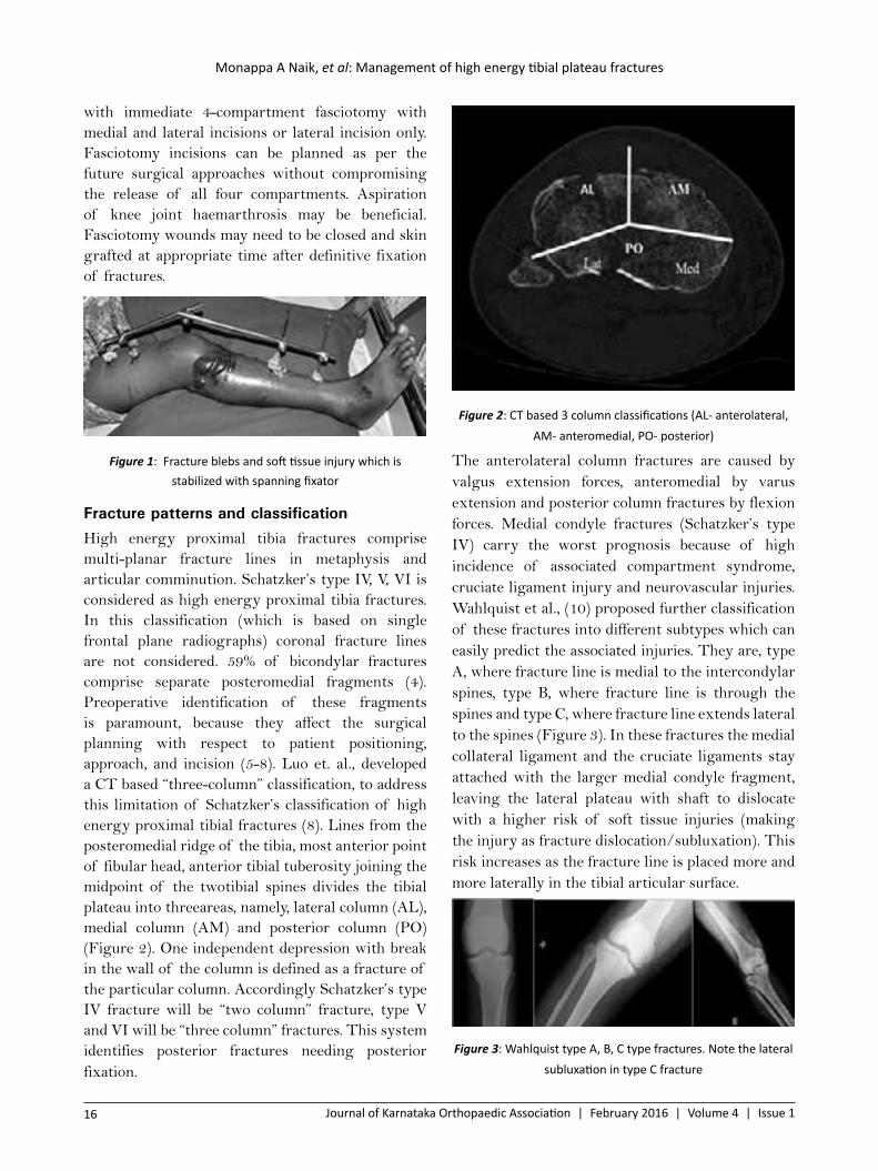
Tibial condyle fractures with impending or established compartment syndrome, associated vascular injury, internal degloving of the skin with swelling and/or skin blisters dictates the immediate intervention. Knee spanning external fixator should be used as a first stage of management in these high energy fractures as “local damage control”. Anteriorly placed distal femur (proximal to knee joint capsule) and distal tibial half pins (away from future area of definitive fixation) are used for temporary external fixator application. This spanning fixator is useful forresting and stabilizing the soft tissue, reduction of articular fragments by ligamentotaxis, access to the soft tissue and compartment monitoring and easier reduction of fragments during definitive fixation (Figure 1). Spanning fixators should be continued till the soft tissue settles down and definitive surgery is planned. The radiological assessment of this spanned fracture will provide traction views of the fracture, which makes better understanding of the fracture geometry. With the spanned fixator, CT scan will provide a better planning of definitive fixation. Associated impending or established compartment syndrome of leg should be managed
Monappa A Naik, et al: Management of high energy tibial plateau fractures
16 Journal of Karnataka Orthopaedic Association | February 2016 | Volume 4 | Issue 1
with immediate 4-compartment fasciotomy with medial and lateral incisions or lateral incision only. Fasciotomy incisions can be planned as per the future surgical approaches without compromising the release of all four compartments. Aspiration of knee joint haemarthrosis may be beneficial. Fasciotomy wounds may need to be closed and skin grafted at appropriate time after definitive fixation of fractures.
Figure 1: Fracture blebs and soft tissue injury which is stabilized with spanning fixator
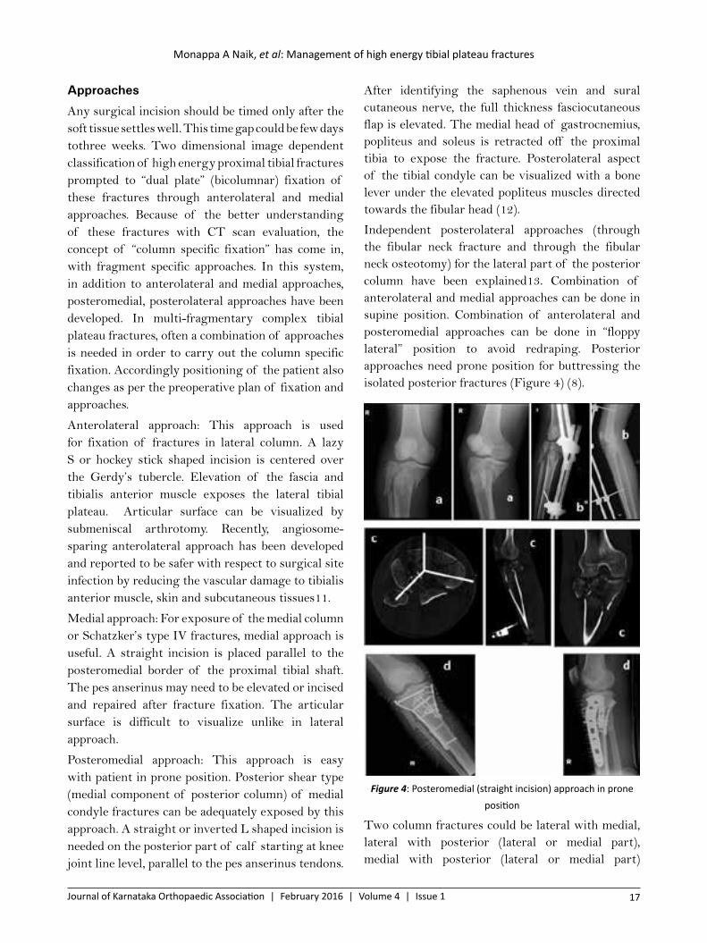
Fracture patterns and classificationHigh energy proximal tibia fractures comprise multi-planar fracture lines in metaphysis and articular comminution. Schatzker’s type IV, V, VI is considered as high energy proximal tibia fractures. In this classification (which is based on single frontal plane radiographs) coronal fracture lines are not considered. 59% of bicondylar fractures comprise separate posteromedial fragments (4). Preoperative identification of these fragments is paramount, because they affect the surgical planning with respect to patient positioning, approach, and incision (5-8). Luo et. al., developed a CT based “three-column” classification, to address this limitation of Schatzker’s classification of high energy proximal tibial fractures (8). Lines from the posteromedial ridge of the tibia, most anterior point of fibular head, anterior tibial tuberosity joining the midpoint of the twotibial spines divides the tibial plateau into threeareas, namely, lateral column (AL), medial column (AM) and posterior column (PO) (Figure 2). One independent depression with break in the wall of the column is defined as a fracture of the particular column. Accordingly Schatzker’s type IV fracture will be “two column” fracture, type V and VI will be “three column” fractures. This system identifies posterior fractures needing posterior fixation.
Figure 2: CT based 3 column classifications (AL- anterolateral, AM- anteromedial, PO- posterior)
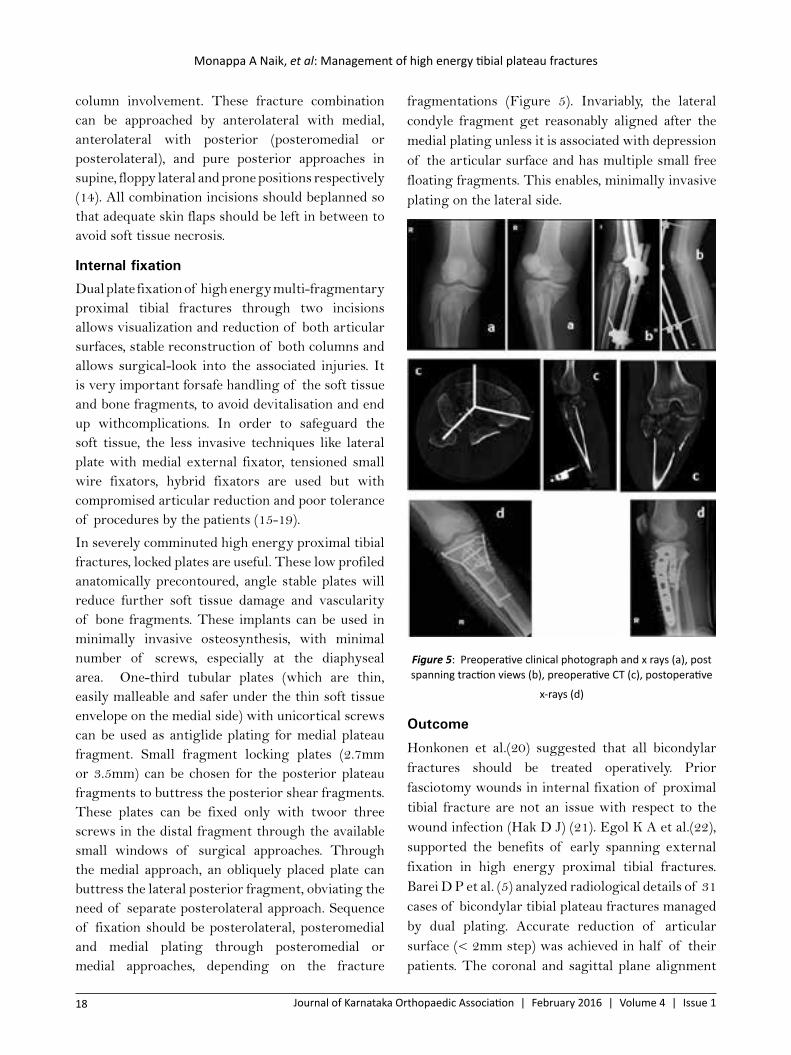
The anterolateral column fractures are caused by valgus extension forces, anteromedial by varus extension and posterior column fractures by flexion forces. Medial condyle fractures (Schatzker’s type IV) carry the worst prognosis because of high incidence of associated compartment syndrome, cruciate ligament injury and neurovascular injuries.Wahlquist et al., (10) proposed further classification of these fractures into different subtypes which can easily predict the associated injuries. They are, type A, where fracture line is medial to the intercondylar spines, type B, where fracture line is through the spines and type C, where fracture line extends lateral to the spines (Figure 3). In these fractures the medial collateral ligament and the cruciate ligaments stay attached with the larger medial condyle fragment, leaving the lateral plateau with shaft to dislocate with a higher risk of soft tissue injuries (making the injury as fracture dislocation/subluxation). This risk increases as the fracture line is placed more and more laterally in the tibial articular surface.
Figure 3: Wahlquist type A, B, C type fractures. Note the lateral subluxation in type C fracture
Monappa A Naik, et al: Management of high energy tibial plateau fractures
Journal of Karnataka Orthopaedic Association | February 2016 | Volume 4 | Issue 1 17
Approaches
Any surgical incision should be timed only after the soft tissue settles well. This time gap could be few days tothree weeks. Two dimensional image dependent classification of high energy proximal tibial fractures prompted to “dual plate” (bicolumnar) fixation of these fractures through anterolateral and medial approaches. Because of the better understanding of these fractures with CT scan evaluation, the concept of “column specific fixation” has come in, with fragment specific approaches. In this system, in addition to anterolateral and medial approaches, posteromedial, posterolateral approaches have been developed. In multi-fragmentary complex tibial plateau fractures, often a combination of approaches is needed in order to carry out the column specific fixation. Accordingly positioning of the patient also changes as per the preoperative plan of fixation and approaches.
Anterolateral approach: This approach is used for fixation of fractures in lateral column. A lazy S or hockey stick shaped incision is centered over the Gerdy’s tubercle. Elevation of the fascia and tibialis anterior muscle exposes the lateral tibial plateau. Articular surface can be visualized by submeniscal arthrotomy. Recently, angiosome-sparing anterolateral approach has been developed and reported to be safer with respect to surgical site infection by reducing the vascular damage to tibialis anterior muscle, skin and subcutaneous tissues11.
Medial approach: For exposure of the medial column or Schatzker’s type IV fractures, medial approach is useful. A straight incision is placed parallel to the posteromedial border of the proximal tibial shaft. The pes anserinus may need to be elevated or incised and repaired after fracture fixation. The articular surface is difficult to visualize unlike in lateral approach.
Posteromedial approach: This approach is easy with patient in prone position. Posterior shear type (medial component of posterior column) of medial condyle fractures can be adequately exposed by this approach. A straight or inverted L shaped incision is needed on the posterior part of calf starting at knee joint line level, parallel to the pes anserinus tendons.
After identifying the saphenous vein and sural cutaneous nerve, the full thickness fasciocutaneous flap is elevated. The medial head of gastrocnemius, popliteus and soleus is retracted off the proximal tibia to expose the fracture. Posterolateral aspect of the tibial condyle can be visualized with a bone lever under the elevated popliteus muscles directed towards the fibular head (12).
Independent posterolateral approaches (through the fibular neck fracture and through the fibular neck osteotomy) for the lateral part of the posterior column have been explained13. Combination of anterolateral and medial approaches can be done in supine position. Combination of anterolateral and posteromedial approaches can be done in “floppy lateral” position to avoid redraping. Posterior approaches need prone position for buttressing the isolated posterior fractures (Figure 4) (8).
Figure 4: Posteromedial (straight incision) approach in prone position
Two column fractures could be lateral with medial, lateral with posterior (lateral or medial part), medial with posterior (lateral or medial part)
Monappa A Naik, et al: Management of high energy tibial plateau fractures
18 Journal of Karnataka Orthopaedic Association | February 2016 | Volume 4 | Issue 1
column involvement. These fracture combination can be approached by anterolateral with medial, anterolateral with posterior (posteromedial or posterolateral), and pure posterior approaches in supine, floppy lateral and prone positions respectively (14). All combination incisions should beplanned so that adequate skin flaps should be left in between to avoid soft tissue necrosis.
Internal fixation
Dual plate fixation of high energy multi-fragmentary proximal tibial fractures through two incisions allows visualization and reduction of both articular surfaces, stable reconstruction of both columns and allows surgical-look into the associated injuries. It is very important forsafe handling of the soft tissue and bone fragments, to avoid devitalisation and end up withcomplications. In order to safeguard the soft tissue, the less invasive techniques like lateral plate with medial external fixator, tensioned small wire fixators, hybrid fixators are used but with compromised articular reduction and poor tolerance of procedures by the patients (15-19).
In severely comminuted high energy proximal tibial fractures, locked plates are useful. These low profiled anatomically precontoured, angle stable plates will reduce further soft tissue damage and vascularity of bone fragments. These implants can be used in minimally invasive osteosynthesis, with minimal number of screws, especially at the diaphyseal area. One-third tubular plates (which are thin, easily malleable and safer under the thin soft tissue envelope on the medial side) with unicortical screws can be used as antiglide plating for medial plateau fragment. Small fragment locking plates (2.7mm or 3.5mm) can be chosen for the posterior plateau fragments to buttress the posterior shear fragments. These plates can be fixed only with twoor three screws in the distal fragment through the available small windows of surgical approaches. Through the medial approach, an obliquely placed plate can buttress the lateral posterior fragment, obviating the need of separate posterolateral approach. Sequence of fixation should be posterolateral, posteromedial and medial plating through posteromedial or medial approaches, depending on the fracture
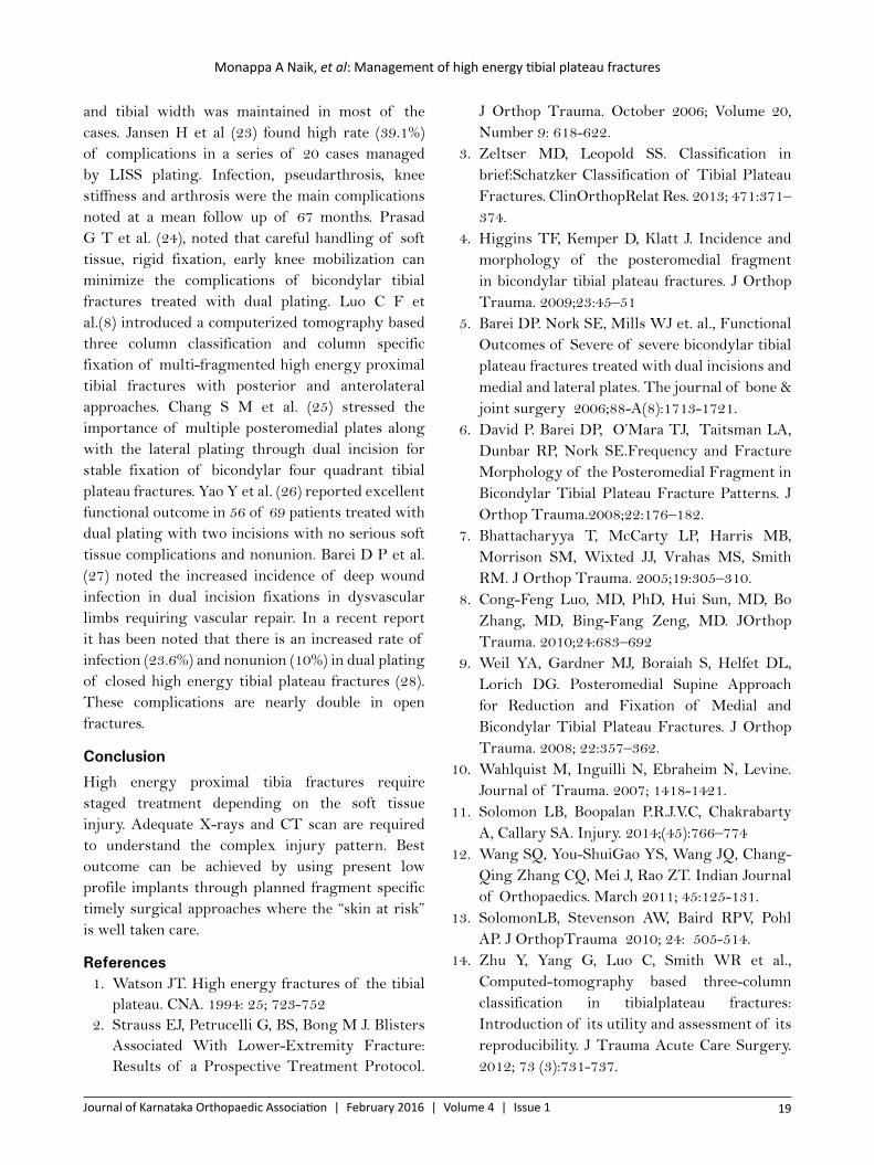
fragmentations (Figure 5). Invariably, the lateral condyle fragment get reasonably aligned after the medial plating unless it is associated with depression of the articular surface and has multiple small free floating fragments. This enables, minimally invasive plating on the lateral side.
Figure 5: Preoperative clinical photograph and x rays (a), post spanning traction views (b), preoperative CT (c), postoperative
x-rays (d)
Outcome
Honkonen et al.(20) suggested that all bicondylar fractures should be treated operatively. Prior fasciotomy wounds in internal fixation of proximal tibial fracture are not an issue with respect to the wound infection (Hak D J) (21). Egol K A et al.(22), supported the benefits of early spanning external fixation in high energy proximal tibial fractures. Barei D P et al. (5) analyzed radiological details of 31 cases of bicondylar tibial plateau fractures managed by dual plating. Accurate reduction of articular surface (< 2mm step) was achieved in half of their patients. The coronal and sagittal plane alignment
Monappa A Naik, et al: Management of high energy tibial plateau fractures
Journal of Karnataka Orthopaedic Association | February 2016 | Volume 4 | Issue 1 19
and tibial width was maintained in most of the cases. Jansen H et al (23) found high rate (39.1%) of complications in a series of 20 cases managed by LISS plating. Infection, pseudarthrosis, knee stiffness and arthrosis were the main complications noted at a mean follow up of 67 months. Prasad G T et al. (24), noted that careful handling of soft tissue, rigid fixation, early knee mobilization can minimize the complications of bicondylar tibial fractures treated with dual plating. Luo C F et al.(8) introduced a computerized tomography based three column classification and column specific fixation of multi-fragmented high energy proximal tibial fractures with posterior and anterolateral approaches. Chang S M et al. (25) stressed the importance of multiple posteromedial plates along with the lateral plating through dual incision for stable fixation of bicondylar four quadrant tibial plateau fractures. Yao Y et al. (26) reported excellent functional outcome in 56 of 69 patients treated with dual plating with two incisions with no serious soft tissue complications and nonunion. Barei D P et al. (27) noted the increased incidence of deep wound infection in dual incision fixations in dysvascular limbs requiring vascular repair. In a recent report it has been noted that there is an increased rate of infection (23.6%) and nonunion (10%) in dual plating of closed high energy tibial plateau fractures (28). These complications are nearly double in open fractures.
Conclusion
High energy proximal tibia fractures require staged treatment depending on the soft tissue injury. Adequate X-rays and CT scan are required to understand the complex injury pattern. Best outcome can be achieved by using present low profile implants through planned fragment specific timely surgical approaches where the “skin at risk” is well taken care.
References1. Watson JT. High energy fractures of the tibial
plateau. CNA. 1994: 25; 723-7522. Strauss EJ, Petrucelli G, BS, Bong M J. Blisters
Associated With Lower-Extremity Fracture: Results of a Prospective Treatment Protocol.
J Orthop Trauma. October 2006; Volume 20, Number 9: 618-622.
3. Zeltser MD, Leopold SS. Classification in brief:Schatzker Classification of Tibial Plateau Fractures. ClinOrthopRelat Res. 2013; 471:371–374.
4. Higgins TF, Kemper D, Klatt J. Incidence and morphology of the posteromedial fragment in bicondylar tibial plateau fractures. J Orthop Trauma. 2009;23:45–51
5. Barei DP. Nork SE, Mills WJ et. al., Functional Outcomes of Severe of severe bicondylar tibial plateau fractures treated with dual incisions and medial and lateral plates. The journal of bone & joint surgery 2006;88-A(8):1713-1721.
6. David P. Barei DP, O’Mara TJ, Taitsman LA, Dunbar RP, Nork SE.Frequency and Fracture Morphology of the Posteromedial Fragment in Bicondylar Tibial Plateau Fracture Patterns. J Orthop Trauma.2008;22:176–182.
7. Bhattacharyya T, McCarty LP, Harris MB, Morrison SM, Wixted JJ, Vrahas MS, Smith RM. J Orthop Trauma. 2005;19:305–310.
8. Cong-Feng Luo, MD, PhD, Hui Sun, MD, Bo Zhang, MD, Bing-Fang Zeng, MD. JOrthop Trauma. 2010;24:683–692
9. Weil YA, Gardner MJ, Boraiah S, Helfet DL, Lorich DG. Posteromedial Supine Approach for Reduction and Fixation of Medial and Bicondylar Tibial Plateau Fractures. J Orthop Trauma. 2008; 22:357–362.
10. Wahlquist M, Inguilli N, Ebraheim N, Levine. Journal of Trauma. 2007; 1418-1421.
11. Solomon LB, Boopalan P.R.J.V.C, Chakrabarty A, Callary SA. Injury. 2014;(45):766–774
12. Wang SQ, You-ShuiGao YS, Wang JQ, Chang-Qing Zhang CQ, Mei J, Rao ZT. Indian Journal of Orthopaedics. March 2011; 45:125-131.
13. SolomonLB, Stevenson AW, Baird RPV, Pohl AP. J OrthopTrauma 2010; 24: 505-514.
14. Zhu Y, Yang G, Luo C, Smith WR et al., Computed-tomography based three-column classification in tibialplateau fractures: Introduction of its utility and assessment of its reproducibility. J Trauma Acute Care Surgery. 2012; 73 (3):731-737.

Monappa A Naik, et al: Management of high energy tibial plateau fractures
20 Journal of Karnataka Orthopaedic Association | February 2016 | Volume 4 | Issue 1
15. Marsh JL, Smith ST, Do TT. External fixation and limited internal fixation for complex fractures of the tibial plateau. J Bone Joint Surg Am. 1995;77: 661–673.
16. Kumar A, Whittle AP. Treatment of complex (Schatzker type VI) fractures of the tibial plateau with circular wire external fixation: retrospective case review. J Orthop Trauma. 2000;14:339–344.
17. Babis GC, Evangelopoulos DS, Kontovazenitis P, et al., High energy tibial plateau fractures treated with hybrid external fixation. J Orthop Surg Res. 2011;6:35.
18. Spagnolo R, Pace F. Management of Schatzker VI fractures with lateral locked screw plating. Musculoskelet Surg. 2012;96:75–80.
19. Katsenis D, Vasilis A, Panayiotis M, et al., Minimal internal fixation augmented by small wire transfixion frames for high-energy tibial plateaufractures. J Orthop Trauma. 2005;19:241–248.
20. Honkonen SE. Indictions for surgical treatment of tibial condyle fractures.CORR. 1994; 302; 199-205
21. Hak DJ, Lee M, Gotham DR, Influence of prior fasciotomy on infection after open reduction and internal fixation of tibial plateau fractures. J Trauma. 2010;69:886–888.
22. Egol KA, Tejwani NC, Capla EL et. al., Staged management of high energy proximal tibia fractures (OTA types 41A).KAJ Orthop Trauma. 2005;19:448–455
23. Jansen H, Frey SP. Doht S, et. al., Medium term results after complex intra-articular fractures of the tibial plateau. J Orthop Sci.2013; 18:569–577.
24. Prasad GT, Kumar TS, Kumar RK, Murthy GK, Sundaram N. Indian Journal of Orthopaedics. 2013;47 (2):188-194
25. Chang SM, Hu SJ, Zhang YQ et. al., A surgical protocol for bicondylar four-quadrant tibial plateau fractures. International Orthopaedics (SICOT). 2014; 38:2559–2564
26. Yao Y, HaoLv, Zan J, Li J, Zhu N, Jing J.Injury. 2014);45:1980–1984
27. Barei DP, Nork SE, Mills WJ et. al.,Complications Associated With Internal Fixation of High-Energy Bicondylar Tibial Plateau Fractures Utilizing a Two-Incision Technique. J Orthop Trauma.2004;18:649–657.
28. Ruffolo MR,Gettys FK,Montijo HE, Seymour RB,Karunakar MA. Complications of High-Energy Bicondylar Tibial PlateauFractures Treated With Dual Plating Through 2 Incisions. J Orthop Trauma. 2015;29(2):85-90.
Ashwath M Acharya, et al: Adult Brachial Plexus Injuries – An Appraisal on its Primary Management
Journal of Karnataka Orthopaedic Association | February 2016 | Volume 4 | Issue 1 21
AbstractBrachial Plexus injuries are catastrophic injuries affecting the earning young adult in the majority of cases. Most of the time the treatment is prolonged as it involves restoration of sensorimotor function, but also relief from pain and subsequent rehabilitation. From an era of gloom where amputation was often suggested, we now have reached a stage of hopeful return to the useful function. With the recent developments in anatomy, pathophysiology, investigations and treatment with microsurgical techniques much can be offered for our patients today. We present an appraisal of the current scenario in the primary management of this difficult clinical problem.
Keywords: Brachial plexus, electrodiagnostic tests, nerve transfers, nerve grafting.
Dr. Anil K. Bhat,Head of DepartmentDivision of Hand and Microsurgery, Department of Orthopaedics, Kasturba Medical College, Manipal University, Manipal - 576104.
Adult Brachial Plexus Injuries – An Appraisal on its Primary Management
Ashwath M Acharya, Anil K Bhat, Mahesh Kulkarni, KN Jayakrishnan
Email: [email protected]
How to cite this article: Acharya AM, Bhat AK, Kulkarni M, Jayakrishnan KN. Adult Brachial Plexus Injuries – An Appraisal on its Primary Management. J Kar Orth Assoc. 2016 Feb; 4(1): 21-30.
Review article
Introduction
Brachial Plexus injuries represent one of the most devastating injuries in our clinical practice and its surgery in the first half of twentieth century was regarded with pessimism because of poor results (1,2). Most of the victims are young adults and disability that accompanies these injuries results in significant loss of work force in the society. With pioneering contributions of Jacobson, Millesi, Narakas in the 60s and 70s and further advances in the clinical, radiological and technological field in the last 30 years, there is much to offer to these patients (3-9).
Etiology
Based on his lifetime works, and often quoted frequently, Narakas gave the rule of seven seventies, which gives information on the etiology and the severity of the injury (10). According to his study, 70% of all brachial plexus injuries were due to motor-vehicle accidents, out of which, 70% were
due to two wheeler accidents, among whom, 70% had multiple injuries.70% of these patients had supraclavicular injury out of which,70% had at least a root avulsion injury and out of these70% involved C7, C8, T1. In this group 70% had chronic pain (10).
Epidemiological studies in our country show an even higher frequency of 94% attributed to motor vehicle accidents; out of which, 90% are due to two-wheeler accidents (11). 54% of these injuries are a part of multitrauma injuries (11).
Pathomechanism And Classifications
Most of the adult brachial plexus injuries are caused by closed trauma, 95 % of which is due to traction (13). Following a traction injury, the nerves may beavulsed from the spinal cord, or significantly stretched, but remain intact or rupture (Figure 1). Other causes may be directly due to gunshot injury, sharp or blunt penetrative trauma or compression due to neoplasm or hematoma. Each patient has a different and unique set of presentation with respect to the events surrounding the injury, associated injuries, and recovery pattern. Hence, the treatment has to be individualized to offer the best possible

Ashwath M Acharya, et al: Adult Brachial Plexus Injuries – An Appraisal on its Primary Management
22 Journal of Karnataka Orthopaedic Association | February 2016 | Volume 4 | Issue 1
outcome. Classification of level and type of injury helps in guiding the prognosis and type of treatment.
Figure 1: a) normal nerve, b) avulsion injuries, c) ruptures, d) neuroma in continuity
Chuang has proposed a classification, which is simple and closest to the anatomical terminology (1). Level 1 injuries were pre-ganglionic root injuries representing avulsion from spinal cord. Level 2 injuries involved post-ganglionic spinal nerve (extra-foraminal root). Level 3 included preclavicular and retroclavicular affecting the trunks and divisions, and level 4 suggested infraclavicular cord injuries. While Level 1 injury carries a poor prognosis and will require nerve transfers, the remaining three are stretch injuries or ruptures requiring nerve grafting or transfers. Once the level is determined, the type of injury needs to be defined. In this respect,the well-known Seddon’s and Sunderland’s classification continues to guide us in the management of nerve injuries (14). Neuropraxia and Sunderland’s type 1 and 2 will recover with regular hand therapy.Usually avulsion injuries, complete ruptures (Neurotmesis, Sunderland’s type 5) will not recover without surgical intervention and the decision to offer early surgery in these patients is never in doubt. The dilemma always comes in the management of Sunderland’s type 3, 4 and Neuroma in continuity (type 6) where an experienced clinical judgement needs to be made in the presence of ambiguous investigation reports.
Millesi has given a classification of nerve fibrosis for such situations in which surgical exploration is necessary (15).
1. Type A fibrosis represents involvement of the epifascicular epineurium (the outermost covering) where the whole nerve is compressed
like a tight stocking.They will need epifascicular epineurotomy (15).
2. Type B fibrosis, additionally involves fibrosis of interfascicular epineurium (mesoneurium – tissue in between the fascicles). They are likely to recover with only epifascicular epineurectomy (15).
3. Type C fibrosis is the most severe where the content of the fascicles of the endoneurium also become completely fibrotic. They are candidates for resection and nerve grafting (15).
Clinical Approach And Evaluation
Since Brachial plexus injuries occur along with multitrauma in 50-70% of cases, they can often be missed in the emergency setting (10,11). A high index of suspicion should be looked for in the presence of significant shoulder girdle injury, first rib injuries, or axillary arterial injuries (16). During the secondary survey, a detailed sensorimotor examination should be carried out. The first priority should be to look for tell-tale signs of Pre-ganglionic injuries (Table 1).17
Table 1: Signs and physical findings that suggest preganglionic injury (Modified from Limthongthang et al.)17
Pre-ganglionic injuries Post-ganglionic injuriesGlobal palsy Partial injuries affecting
the shoulder and elbow, hand intrinsic function preserved
Horner syndromeMiosis, Ptosis and Enophthalmos
Usually absent
Winging of Scapula Function well preservedAtrophy of RhomboidsCervical paraspinal muscle weakness and loss of posterior neck sensationHemidiaphragm paralysisAbsence of Tinel sign in neck area Present. Suggests level
and prognosis of injuryProfound pain out of proportion to signs and symptoms
Pain is observed but not as severe as in avulsion injuries
A simple evaluation of each muscle for strength is done and graded on a scale of 0–5 as per the British Medical Research Council muscle grading system (18). Examining select few muscles supplied by terminal branches at various levels is the key to find the site and type of injury (Table 2).
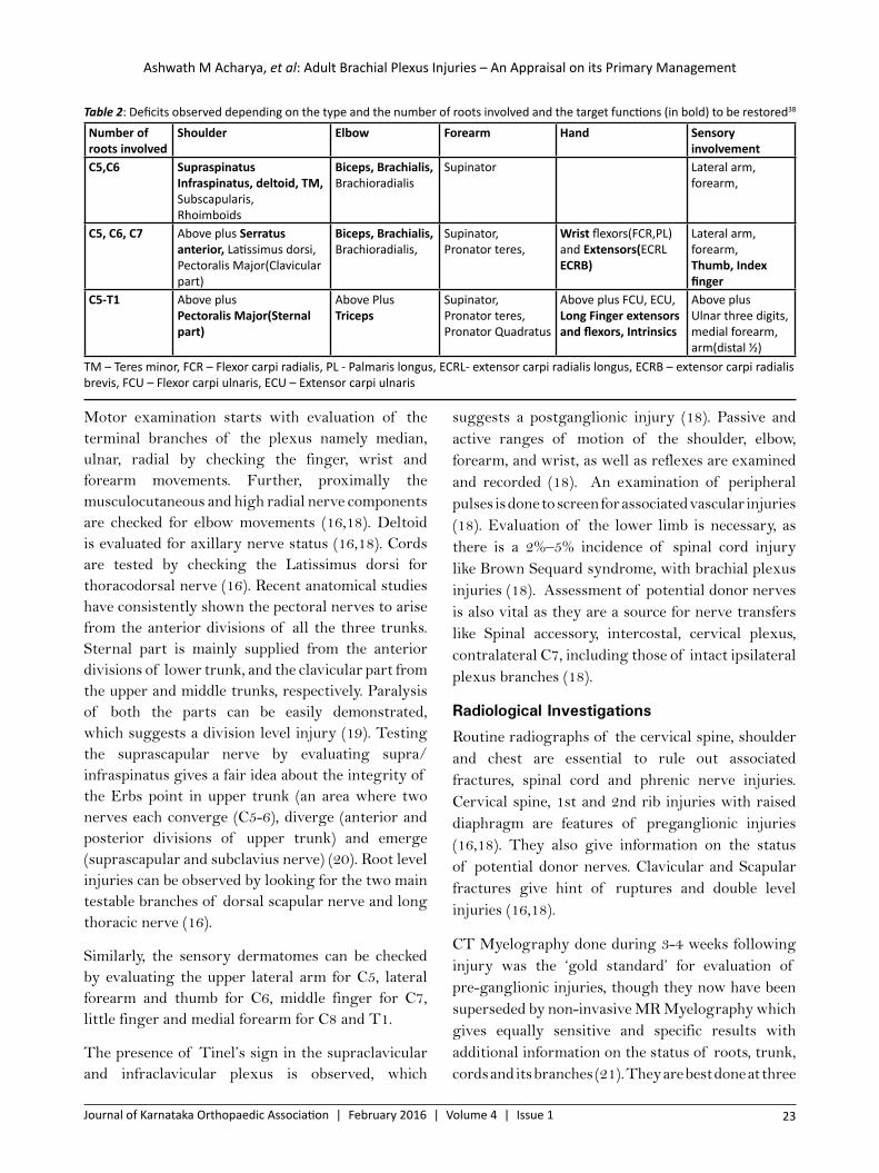
Ashwath M Acharya, et al: Adult Brachial Plexus Injuries – An Appraisal on its Primary Management
Journal of Karnataka Orthopaedic Association | February 2016 | Volume 4 | Issue 1 23
Motor examination starts with evaluation of the terminal branches of the plexus namely median, ulnar, radial by checking the finger, wrist and forearm movements. Further, proximally the musculocutaneous and high radial nerve components are checked for elbow movements (16,18). Deltoid is evaluated for axillary nerve status (16,18). Cords are tested by checking the Latissimus dorsi for thoracodorsal nerve (16). Recent anatomical studies have consistently shown the pectoral nerves to arise from the anterior divisions of all the three trunks. Sternal part is mainly supplied from the anterior divisions of lower trunk, and the clavicular part from the upper and middle trunks, respectively. Paralysis of both the parts can be easily demonstrated, which suggests a division level injury (19). Testing the suprascapular nerve by evaluating supra/infraspinatus gives a fair idea about the integrity of the Erbs point in upper trunk (an area where two nerves each converge (C5-6), diverge (anterior and posterior divisions of upper trunk) and emerge (suprascapular and subclavius nerve) (20). Root level injuries can be observed by looking for the two main testable branches of dorsal scapular nerve and long thoracic nerve (16).
Similarly, the sensory dermatomes can be checked by evaluating the upper lateral arm for C5, lateral forearm and thumb for C6, middle finger for C7, little finger and medial forearm for C8 and T1.
The presence of Tinel’s sign in the supraclavicular and infraclavicular plexus is observed, which
suggests a postganglionic injury (18). Passive and active ranges of motion of the shoulder, elbow, forearm, and wrist, as well as reflexes are examined and recorded (18). An examination of peripheral pulses is done to screen for associated vascular injuries (18). Evaluation of the lower limb is necessary, as there is a 2%–5% incidence of spinal cord injury like Brown Sequard syndrome, with brachial plexus injuries (18). Assessment of potential donor nerves is also vital as they are a source for nerve transfers like Spinal accessory, intercostal, cervical plexus, contralateral C7, including those of intact ipsilateral plexus branches (18).
Radiological Investigations
Routine radiographs of the cervical spine, shoulder and chest are essential to rule out associated fractures, spinal cord and phrenic nerve injuries. Cervical spine, 1st and 2nd rib injuries with raised diaphragm are features of preganglionic injuries (16,18). They also give information on the status of potential donor nerves. Clavicular and Scapular fractures give hint of ruptures and double level injuries (16,18).
CT Myelography done during 3-4 weeks following injury was the ‘gold standard’ for evaluation of pre-ganglionic injuries, though they now have been superseded by non-invasive MR Myelography which gives equally sensitive and specific results with additional information on the status of roots, trunk, cords and its branches (21). They are best done at three
Table 2: Deficits observed depending on the type and the number of roots involved and the target functions (in bold) to be restored38
Number of roots involved
Shoulder Elbow Forearm Hand Sensory involvement
C5,C6 Supraspinatus Infraspinatus, deltoid, TM, Subscapularis,Rhoimboids
Biceps, Brachialis, Brachioradialis
Supinator Lateral arm, forearm,
C5, C6, C7 Above plus Serratus anterior, Latissimus dorsi, Pectoralis Major(Clavicular part)
Biceps, Brachialis, Brachioradialis,
Supinator,Pronator teres,
Wrist flexors(FCR,PL) and Extensors(ECRL ECRB)
Lateral arm, forearm,Thumb, Index finger
C5-T1 Above plus Pectoralis Major(Sternal part)
Above PlusTriceps
Supinator,Pronator teres,Pronator Quadratus
Above plus FCU, ECU, Long Finger extensors and flexors, Intrinsics
Above plusUlnar three digits, medial forearm, arm(distal ½)
TM – Teres minor, FCR – Flexor carpi radialis, PL - Palmaris longus, ECRL- extensor carpi radialis longus, ECRB – extensor carpi radialis brevis, FCU – Flexor carpi ulnaris, ECU – Extensor carpi ulnaris

Ashwath M Acharya, et al: Adult Brachial Plexus Injuries – An Appraisal on its Primary Management
24 Journal of Karnataka Orthopaedic Association | February 2016 | Volume 4 | Issue 1
to four weeks during which the edema, hematoma and other artefacts settle, and pseudomeningocoele develops in avulsion injuries. Conventional MRI gives information of the extra-foraminal and infra clavicular plexus, with several reports proving its value (21). In the absence of root avulsions, a normal MRI of the supraclavicular plexus effectively excludes significant post-ganglionic injury (21). A T2 weighted hyperintensity may suggest either a reversible endoneurial edema (neuropraxia), or a reversible or irreversible Wallerien degeneration (21). Hence, pathophysiological status cannot be assessed by MRI, except when the nerve is divided (21). The combination of a signal abnormality and increased nerve diameter suggests a poorer prognosis (21). Therefore, except in obvious total avulsions, MRI on its own doesn’t help in clinching a diagnosis, and needs to be correlated with repeated
clinical examination and electrophysiological studies to determine the surgical strategy, with or without per-operative stimulation (21).
Electrophysiological Studies
Electrophysiological studies are essential to localize and assess the severity of injury, identify those nerves likely or unlikely to recover, as well as available donor nerves for transfer (18,22). In addition, they provide a useful guide to post-injury prognosis helpingin rehabilitation, and if necessary, additional surgical intervention (18,22). They are usually done at three weeks; by which time, the Wallerian degeneration is established. It includes nerve conduction study (NCS) and Electromyography (EMG) that evaluates motor, sensory nerves and the muscles respectively (22). To get the most useful interpretation, they must be done serially every 6 weeks in the presence of careful clinical judgment (Table 3) (14,16,18,22,23).
Table 3: Patterns of presentation in electrodiagnostic studies14
Type of injury Nerve conduction studies ElectromyographyNeuropraxia • CMAP/SNAP reduced in size,
• Nerve conduction velocity slow • Prolonged latency• Conduction distally can be demonstrated• Distal response amplitude normal
• Recruitment pattern with a decreased number of normal motor units firing at increased rates.
• Spontaneous activity absent
Axonotmesis • CMAP/SNAP small/absent• Nerve conduction velocity slow • Prolonged latency• Conduction distally cannot be seen• Distal response amplitude decreased
• Spontaneous activity like positive sharp waves, fibrillations • Motor unit recruitment is abnormalRecovery signs • Collateral sprouting results in tri/diphasic potentials with
polyphasic tails.• Axonal regeneration causes nascent low amplitude
polyphasic potentialsNeurotmesis • CMAP/SNAP absent
• No conduction in distal segment• Positive sharp waves• Fibrillations are profuse and• No signs of recovery
CMAP – compound motor action potential, SNAP – sensory nerve action potential
Perhaps the most important information, which can be derived from electrodiagnostic studies is that of differentiation between pre-ganglionic from the post-ganglionic injuries (16,18,22,23). This is demonstrated by the findings of triad of root avulsion: 1) The NCS evaluation shows a normal Sensory Nerve Action Potential (SNAP) and preserved histamine response in presence of complete anaesthesia at the tested site, 2) EMG reveals spontaneous activity, fibrillation and absent voluntary motor units in muscles innervated by the root and in particular those of cervical paraspinal
muscles, 3) Evokes potential findings of an absent Sensory Evoked Potential (SEP) (23).
Since the dorsal root ganglion is intact in avulsion injuries, the SNAP will be intact in the presence of profound anesthesia. This is absent in post-ganglionic ruptures (16,18,22).
Per-operative evaluation using the Nerve Action Potential helps in planning the type of procedure in neuroma-in continuity. If conduction is observed, it suggests the viability of a significant volume of axons, which can be further freed by neurolysis as suggested by Millesi (15,18,22). If there is no
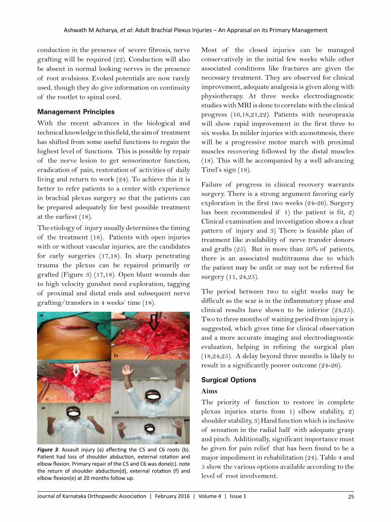
Ashwath M Acharya, et al: Adult Brachial Plexus Injuries – An Appraisal on its Primary Management
Journal of Karnataka Orthopaedic Association | February 2016 | Volume 4 | Issue 1 25
conduction in the presence of severe fibrosis, nerve grafting will be required (22). Conduction will also be absent in normal looking nerves in the presence of root avulsions. Evoked potentials are now rarely used, though they do give information on continuity of the rootlet to spinal cord.
Management Principles
With the recent advances in the biological and technical knowledge in this field, the aim of treatment has shifted from some useful functions to regain the highest level of functions. This is possible by repair of the nerve lesion to get sensorimotor function, eradication of pain, restoration of activities of daily living and return to work (24). To achieve this it is better to refer patients to a center with experience in brachial plexus surgery so that the patients can be prepared adequately for best possible treatment at the earliest (18).
The etiology of injury usually determines the timing of the treatment (18). Patients with open injuries with or without vascular injuries, are the candidates for early surgeries (17,18). In sharp penetrating trauma the plexus can be repaired primarily or grafted (Figure 3) (17,18). Open blunt wounds due to high velocity gunshot need exploration, tagging of proximal and distal ends and subsequent nerve grafting/transfers in 4 weeks’ time (18).
Figure 3: Assault injury (a) affecting the C5 and C6 roots (b). Patient had loss of shoulder abduction, external rotation and elbow flexion. Primary repair of the C5 and C6 was done(c). note the return of shoulder abduction(d), external rotation (f) and elbow flexion(e) at 20 months follow up.
Most of the closed injuries can be managed conservatively in the initial few weeks while other associated conditions like fractures are given the necessary treatment. They are observed for clinical improvement, adequate analgesia is given along with physiotherapy. At three weeks electrodiagnostic studies with MRI is done to correlate with the clinical progress (16,18,21,22). Patients with neuropraxia will show rapid improvement in the first three to six weeks. In milder injuries with axonotmesis, there will be a progressive motor march with proximal muscles recovering followed by the distal muscles (18). This will be accompanied by a well advancing Tinel’s sign (18).
Failure of progress in clinical recovery warrants surgery. There is a strong argument favoring early exploration in the first two weeks (24-26). Surgery has been recommended if 1) the patient is fit, 2) Clinical examination and investigation shows a clear pattern of injury and 3) There is feasible plan of treatment like availability of nerve transfer donors and grafts (25). But in more than 50% of patients, there is an associated multitrauma due to which the patient may be unfit or may not be referred for surgery (11, 24,25).
The period between two to eight weeks may be difficult as the scar is in the inflammatory phase and clinical results have shown to be inferior (24,25). Two to three months of waiting period from injury is suggested, which gives time for clinical observation and a more accurate imaging and electrodiagnostic evaluation, helping in refining the surgical plan (18,24,25). A delay beyond three months is likely to result in a significantly poorer outcome (24-26).
Surgical Options Aims
The priority of function to restore in complete plexus injuries starts from 1) elbow stability, 2) shoulder stability, 3) Hand function which is inclusive of sensation in the radial half with adequate grasp and pinch. Additionally, significant importance must be given for pain relief that has been found to be a major impediment in rehabilitation (24). Table 4 and 5 show the various options available according to the level of root involvement.
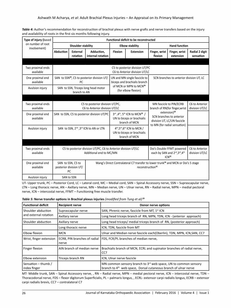
Ashwath M Acharya, et al: Adult Brachial Plexus Injuries – An Appraisal on its Primary Management
26 Journal of Karnataka Orthopaedic Association | February 2016 | Volume 4 | Issue 1
Table 4: Author’s recommendation for reconstruction of brachial plexus with nerve grafts and nerve transfers based on the injury and availability of roots in the first six months following injury.
Type of injury (based on number of root
involvement)
Functional deficit to be reconstructed
Shoulder stability Elbow stability Hand function
Abduction External rotation
Adduction, internal rotation
Flexion Extension Finger, wrist flexion
Finger, wrist extension
Radial 2 digit sensation
C5-6
Two proximal ends available
C5 to posterior division UT/PCC6 to Anterior division UT/LC
One proximal end available
SAN to SSN29, C5 to posterior division UT/PC
UN and MN single fascicle to biceps and brachialis branch
of MCN or MPN to MCN29
(for elbow flexion)
SCN branches to anterior division UT, LC
Avulsion injury SAN to SSN, Triceps long head motor branch to AN
C5-7
Two proximal ends available
C5 to posterior division UT/PC, C6 to Anterior division UT/LC
MN fascicle to PIN/ECRB branch of RN(for finger,wrist
extension)29
SCN branches to anterior division UT, LC/UN fascicle
to MN (for radial sensation)
C6 to Anterior division UT/LC
One proximal end available
SAN to SSN, C5 to posterior division UT/PC 3rd ,4th, 5th ICN to MCN29 / UN to biceps or brachialis
branch of MCN
Avulsion injury SAN to SSN, 2nd ,3rd ICN to AN or LTN 4th,5th,6th ICN to MCN / UN to biceps or brachialis
branch of MCN
Global Palsy
Two proximal ends available
C5 to posterior division UT/PC, C6 to Anterior division UT/LCAdditional end to MC/MN
Doi’s Double FFMT powered each by SAN and 2nd,3rd,4th
ICN32
C6 to Anterior division UT/LC
One proximal end available
SAN to SSN, C5 to posterior division UT/
PC
Wang’s Direct Contralateral C7 transfer to lower trunk34 and MCN or Doi’s 5 stage reconstruction32
Avulsion injury SAN to SSN
UT- Upper trunk, PC – Posterior Cord, LC – Lateral cord, MC – Medial cord, SAN – Spinal Accessory nerve, SSN – Suprascapular nerve, LTN – Long thoracic nerve, AN – Axillary nerve, MN – Median nerve, UN – Ulnar nerve, RN – Radial nerve, MPN – medial pectoral nerve, ICN – intercostal nerve, FFMT – Functioning free muscle transfer.
Table 5: Nerve transfer options in Brachial plexus injuries (modified from Tung et al) 29
Functional deficit Recipient nerve Donor nerve options
Shoulder abduction and external rotation
Suprascapular nerve SAN, Phrenic nerve, fascicle from MT, 1st ICN
Axillary nerve Long head triceps branch of RN, MPN, TDN, ICN - (anterior approach)
Shoulder abduction Axillary nerve Long head triceps/ medial triceps branch of RN, (posterior approach)
Long thoracic nerve ICN, TDN, fascicle from MT
Elbow flexion MCN Ulnar and Median nerve fascicle each(Oberlin), TDN, MPN, ICN,SAN, CC7
Wrist, finger extension ECRB, PIN branches of radial nerve
FDS, FCR/PL branches of median nerve,
Finger flexion AIN branch of median nerve Brachialis branch of MCN, ECRL and supinator branches of radial nerve, CC7
Elbow extension Triceps branch RN ICN, Ulnar nerve fascicle
Sensation – thumb /index finger
MN common sensory branch to 3rd web space, UN to common sensory branch to 4th web space, Dorsal cutaneous branch of ulnar nerve
MT- Middle trunk, SAN – Spinal Accessory nerve, , RN – Radial nerve, MPN – medial pectoral nerve, ICN – intercostal nerve, TDN – Thoracodorsal nerve, FDS – flexor digitorum Superficialis, PL – palmaris longus, , ECRL- extensor carpi radialis longus, ECRB – extensor carpi radialis brevis, CC7 – contralateral C7

Ashwath M Acharya, et al: Adult Brachial Plexus Injuries – An Appraisal on its Primary Management
Journal of Karnataka Orthopaedic Association | February 2016 | Volume 4 | Issue 1 27
Options
Neurolysis: Millesi has stressed the importance of Neurolysis in his classification of nerve fibrosis (15). Neurolysis alone is in the absence of normal conduction across the nerve injury is inappropriate and must always be accompanied by a combination of resection with nerve grafts and/or nerve transfers.15
Nerve grafting: Interfascicular nerve grafting remains the workhorse in rupture injuries (Figure 4). Pioneering work by Millesi in the refinement of this technique has been an important factor in improving results in these injuries (27). Sural nerve, medial cutaneous nerve of arm and forearm, radial sensory nerve and the saphenous nerve has been the common source of harvest for nerve grafting.
Nerve transfers: Nerve transfer is a procedure of intentional division and transfer of a physiologically active nerve with low morbidity to a more important but irreparable damaged nerve (Figure 5) (1).The major benefits of a nerve transfer are (28):
1. It is the only option available in avulsion injuries.2. It decreases the regeneration distance especially
in very proximal injuries, as the procedure can be done at a region close to the myoneural junction.
3. It is versatile as a pure motor or sensory nerve can be used for transfer, specifically to a target motor or sensory nerve.
4. Since it involves only a single repair site when compared to nerve grafts, the inflammatory barrier for axons to cross is halved.
Figure 4: infraclavicular level of injury showing axillary neuroma in continuity(a), rupture of musculocutaneous nerve (b). this was resected and the gap bridged by 8cm interfascicular sural nerve grafts (c). the results after 18 months follow up showing elbow flexion and shoulder abduction(d).
Figure 5: Post ganglionic spinal nerve level injury of C5,C6 where the proximal end was not found to be healthy for grafting(a).nerve transfers Accessory to Suprascapular(b), Oberlin’s ulnar and median nerve fascicle to biceps and brachialis branch of musculocutaneous nerve(c), long head triceps branch of radial nerve to axillary(d). At 30 months follow up showing near normal abduction, external rotation of shoulder and elbow flexion(e).
Chuang has classified nerve transfers into four types: 1) extraplexus, 2) intraplexus, 3) close-target, and 4) endto-side neurorrhaphy (1).
As the name suggests, extraplexal transfers refers to the nerves outside the brachial plexus, which can be used for restoring a part of its function. Examples include, spinal accessory, intercostal, cervical plexus and contralateral C7 transfer. Intraplexal sources are mainly the ulnar, median and radial nerve, which are intact in incomplete injuries. Close target represents the transfers thatare done very close to the affected muscle, which decreases the distance for the axons to reach the myoneural junction, thus achieving a faster recovery. They are helpful in delayed cases. A good example is the coaptation of ulnar and median nerve fascilcle to the biceps and brachialis branch of musculocutaneous nerve (Oberlin).1In end to side transfers, the affected nerve is divided and its distal end is sutured to the side of an intact nerve after making an epineural window.(1,29)
Functioning free muscle transfer:Perhaps one of the most difficult functions to restore in complete palsies is that of prehensile hand function. Grafting of C8 and T1 is not an option, as avulsion injuries are more likely to have occurred (30). A prolonged reinnervation time for distal flexor and intrinsic muscles also rules out such an option unless in paediatric age group. (29) Grafting at best will restore some useful sensation (30).
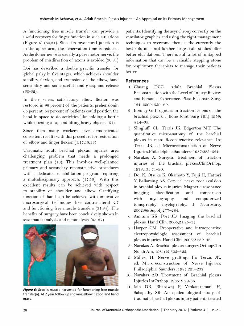
Ashwath M Acharya, et al: Adult Brachial Plexus Injuries – An Appraisal on its Primary Management
28 Journal of Karnataka Orthopaedic Association | February 2016 | Volume 4 | Issue 1
A functioning free muscle transfer can provide a useful recovery for finger function in such situations (Figure 6) (30,31). Since its myoneural junction is in the upper arm, the denervation time is reduced.Asthe donor nerve is usually a pure motor nerve, the problem of misdirection of axons is avoided.(30,31)
Doi has described a double gracilis transfer for global palsy in five stages, which achieves shoulder stability, flexion, and extension of the elbow, hand sensibility, and some useful hand grasp and release (30-32).
In their series, satisfactory elbow flexion was restored in 96 percent of the patients, prehensionin 65 percent. 54 percent of patients could position the hand in space to do activities like holding a bottle while opening a cap and lifting heavy objects. (31)
Since then many workers have demonstrated consistent results with this procedure for restoration of elbow and finger flexion (1,17,18,33)
Traumatic adult brachial plexus injuries area challenging problem that needs a prolonged treatment plan (18). This involves well-planned primary and secondary reconstructive procedures with a dedicated rehabilitation program requiring a multidisciplinary approach. (17,18). With this excellent results can be achieved with respect to stability of shoulder and elbow. Gratifying function of hand can be achieved with innovative microsurgical techniques like contra-lateral C7 and functioning free muscle transfers (31,34). The benefits of surgery have been conclusively shown in systematic analysis and metanalysis. (35-37)
Figure 6: Gracilis muscle harvested for functioning free muscle transfer(a). At 2 year follow up showing elbow flexion and hand grasp.
patients. Identifying the asynchrony correctly on the ventilator graphics and using the right management techniques to overcome them is the currently the best solution until further large scale studies offer better elucidations. There is still a lot of untapped information that can be a valuable stepping stone for respiratory therapists to manage their patients better.
References1. Chuang DCC. Adult Brachial Plexus
Reconstruction with the Level of Injury: Review and Personal Experience. Plast.Reconstr. Surg. 124: 2009: 359- 69.
2. Bonney G. Prognosis in traction lesions of the brachial plexus. J Bone Joint Surg (Br.) 1959; 41:4–35.
3. Slingluff CL, Terzis JK, Edgerton MT. The quantitative microanatomy of the brachial plexus in man: Reconstructive relevance. In: Terzis JK, ed. Microreconstruction of Nerve Injuries.Philadelphia: Saunders; 1987:285–324.
4. Narakas A. Surgical treatment of traction injuries of the brachial plexus.ClinOrthop. 1978;133:71–90.
5. Doi K, Otsuka K, Okamoto Y, Fujii H, Hattori Y, Baliarsing AS. Cervical nerve root avulsion in brachial plexus injuries: Magnetic resonance imaging classification and comparison with myelography and computerized tomography myelography. J Neurosurg. 2002;96(Suppl):277–284.
6. Amrami KK, Port JD. Imaging the brachial plexus. Hand Clin. 2005;21:25–37.
7. Harper CM. Preoperative and intraoperative electrophysiologic assessment of brachial plexus injuries. Hand Clin. 2005;21:39–46.
8. Narakas A. Brachial plexus surgery.OrthopClin North Am. 1981;12:303–323.
9. Millesi H. Nerve grafting. In: Terzis JK, ed. Microreconstruction of Nerve Injuries. Philadelphia: Saunders; 1987:223–237.
10. Narakas AO. Treatment of Brachial plexus Injuries.IntOrthop. 1985: 9.29-36.
11. Jain DK, Bhardwaj P, Venkataramani H, Sabapathy SR. An epidemiological study of traumatic brachial plexus injury patients treated
Ashwath M Acharya, et al: Adult Brachial Plexus Injuries – An Appraisal on its Primary Management
Journal of Karnataka Orthopaedic Association | February 2016 | Volume 4 | Issue 1 29
at an Indian centre. Indian J Plast Surg. 2012 Sep;45(3):498-503.
12. Songcharoen P, Shin AY. Brachial plexus injury: acute diagnosis and treatment. In: Berger RA, Weiss APC, editors. Hand surgery. Philadelphia: Lippincott, Williams & Wilkins; 2004. p. 1005–25.
13. Songcharoen P, Shin AY. Brachial plexus injury: Acute diagnosis and treatment. In: Berger RA, Weis APC, editors. Hand surgery. Philadelphia: Lippincott, Williams & Wilkins; 2004. p. 1005–25.
14. Mackinnon SE ,Dvali LT. Basic Pathology of the Hand, Wrist, and Forearm: Nerve.In: Berger RA, Weis APC, editors. Hand surgery. Philadelphia: Lippincott, Williams & Wilkins; 2004. p. 37-48
15. Mazal PR, Millesi H. Neurolysis: is it beneficial or harmful? ActaNeurochir Suppl. 2005;92: 3-6.
16. Moran SL, Steinmann SP, Shin AY. Adult Brachial Plexus Injuries: Mechanism, Patterns of Injury, and Physical Diagnosis. Hand Clin 21 (2005) 13–24.
17. Limthongthang R, Bachoura A, Songcharoen P, Lee Osterman A. Adult Brachial Plexus Injury Evaluation and Management. OrthopClin North Am. 2013 Oct;44(4):591-603.
18. Giuffre JL, Kakar S, Bishop AT, Spinner RJ, Shin AY. Current concepts of the treatment of adult brachial plexus injuries. J Hand Surg Am. 2010 Apr;35(4):678-88;
19. Aszmann OC, Rab M, Kamolz L, Frey M. The anatomy of the pectoral nerves and their significance in brachial plexus reconstruction. J Hand Surg Am. 2000 Sep;25(5):942-7
20. Rajasabapathy S. personal communication. 21. Post-traumatic brachial plexus MRI in practice.
Silbermann-Hoffman O, Teboul F. DiagnInterv Imaging. 2013 Oct;94(10):925-43.
22. O’Shea K, Feinberg JH, Wolfe SW. Imaging and electrodiagnostic work-up of acute adult brachial plexus injuries. J Hand Surg Eur Vol. 2011 Nov;36(9):747-59.
23. Smith SJ. The role of neurophysiological investigation in traumatic brachial plexus
lesions in adults and children. J Hand Surg Br. 1996 Apr;21(2):145-7.
24. Birch R. Timing of surgical reconstruction for closed traumatic injury to the supraclavicular brachial plexus. J Hand SurgEur Vol. 2015 Jul;40(6):562-7.
25. Hems TE. Timing of surgical reconstruction for closed traumatic injury to the supraclavicular brachial plexus. J Hand SurgEur Vol. 2015 Jul;40(6):568-72
26. Jivan S, Kumar N, Wiberg M, Kay S. The influence of pre-surgical delay on functional outcome after reconstruction of brachial plexus injuries .J PlastReconstrAesthet Surg. 2009 Apr;62(4):472-9.
27. Millesi H.Techniques for nerve grafting. Hand Clin. 2000 Feb;16(1):73-91
28. Wood MB, Murray PM.Heterotopic nerve transfers: recent trends with expanding indication.J Hand Surg Am. 2007 Mar;32(3):397-408.
29. Tung TH, Mackinnon SE.Nerve transfers: indications, techniques, and outcomes. J Hand Surg Am. 2010 Feb;35(2):332-41
30. Doi K. Brachial Plexus: Free Composite Tissue Transfers. In: Berger RA, Weiss APC, editors. Hand surgery. Philadelphia: Lippincott, Williams & Wilkins; 2004. p.1041-54.
31. Doi K, Muramatsu K, Hattori Y, Otsuka K, Tan SH, Nanda V, Watanabe M. Restoration of prehension with the double free muscle technique following complete avulsion of the brachial plexus. Indications and long-term results. J Bone Joint Surg Am. 2000 May;82(5):652-66.
32. Doi K. Management of Total Paralysis of the Brachial Plexus by the Double Free-muscle Transfer Technique. J Hand Surg Eur Vol 2008; 33; 240-51
33. Bishop AT (2005). Functioning free-muscle transfer for brachial plexus injury. Hand Clinic, 21: 91–102.
34. Wang SF1, Li PC, Xue YH, Yiu HW, Li YC, Wang HH. Contralateral C7 nerve transfer with direct coaptation to restore lower trunk function after traumatic brachial plexus avulsion. J Bone Joint Surg Am. 2013 May 1;95(9):821-7

Ashwath M Acharya, et al: Adult Brachial Plexus Injuries – An Appraisal on its Primary Management
30 Journal of Karnataka Orthopaedic Association | February 2016 | Volume 4 | Issue 1
35. Dy CJ, Garg R, Lee SK, Tow P, Mancuso CA, Wolfe SW. A systematic review of outcomes reporting for brachial plexus reconstruction. J Hand Surg Am. 2015 Feb;40(2):308-13
36. Merrell GA, Barrie KA, Katz DL, Wolfe SW. Results of nerve transfer techniques for restoration of shoulder and elbow function in the context of a meta-analysis of the English literature. J Hand Surg Am. 2001 Mar; 26(2):303-14.
37. Yang LJ, Chang KW, Chung KC.A systematic review of nerve transfer and nerve repair for the treatment of adult upper brachial plexus injury. Neurosurgery. 2012 Aug;71(2):417-29
38. Bertelli JA, Ghizoni MF. Clinical findings in C5-C6 and C5-C7 root palsies with brachial plexus traction lesions. J Hand Surg Eur Vol 2013 38: 237- 41

Journal of Karnataka Orthopaedic Association | February 2016 | Volume 4 | Issue 1 31
M Ajith Kumar, et al: Modified Stoppa approach for acetabular fractures: “Lighting up the tunnel”
Modified Stoppa approach for acetabular fractures: “Lighting up the tunnel”
M Ajith Kumar, Shailesh Pai
E-mail:[email protected]
How to cite this article: Ajith Kumar M, Pai S. Modified Stoppa approach for acetabular fractures:“Lighting up the tunnel”. J Kar Orth Assoc. 2016 Feb; 4(1): 31-34.
Review article
Introduction
The current understanding of the acetabular fractures and management is the result of extensive work on the subject by Judet and Letournel. 1-3They described the classification, surgical approaches and treatment for these fractureswhich still holds fort till this day (1-3). Most of the other approaches (ilioinguinal, Kocher-Langenbeck etc.) are modifications of the same approach.
Stoppa described a midline approach for the treatment of complicated groin and incisional hernias with the support of Dacron tulle prosthesis in 1989 (4). Cole and Bolhofner in 1994 modified this approach for internal fixation of pelvi-acetabular injuries and mentioned it as Anterior Intrapelvic (AIP) extraperitoneal approach which was later described in detail as “Modified Stoppa’s Approach” by Hirvensalo in 2007(5,6). This approach has gained popularity in the recent past and is often used for treating acetabular fractures in many centres. This paper describes the preoperative assessment, surgical approach, advantages, disadvantages and our experience of the same approach.
Preoperative Assessment
The fractures of anterior column along with quadrilateral plate can be visualised via modified
Stoppa’s approach (Figure 1). For fractures exiting into the iliac crest, the lateral window of the ilioinguinal approach needs to be added on so that the entire iliac wing up to the sacroiliac joints is exposed. All the anterior column fractures can then be appropriately dealt with.
Figure 1: a) Exposure region of Modified Stoppa approachb) Exposure region of “lateral window” 7
Surgical Approach
Positioning: Patient is positioned supine on the table. Foley’s catheter is inserted into the urinary bladder to prevent intraoperative bladder injuries. The fractured limb is draped free and a pillow or a sandbag is placed below the knee to relax iliopsoas and to aid in traction. The surgeon stands on the opposite side of the fractured acetabulum as he works his way from midline into the “tunnel” visualising the pelvic brim.
Dr. M Ajith KumarConsultant Orthopaedic surgeonTejasvini Hospital & SSIOT, Mangalore,Tejaswini Building, Kadri Road, Mallikatte, Kadri, Mangalore - 575002, Karnataka
Keywords: Acetabular fractures, Modified Stoppa approach, Kocher-Langenbeckapproach Ventilation.
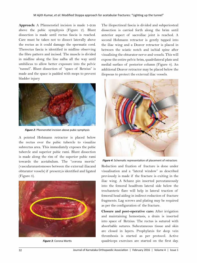
M Ajith Kumar, et al: Modified Stoppa approach for acetabular fractures: “Lighting up the tunnel”
32 Journal of Karnataka Orthopaedic Association | February 2016 | Volume 4 | Issue 1
Approach: A Pfannenstiel incision is made 1-2cm above the pubic symphysis (Figure 2). Blunt dissection is made until rectus fascia is reached. Care must be taken not to dissect laterally above the rectus as it could damage the spermatic cord. Therectus fascia is identified in midline observing the fibre pattern and incised. The muscle is divided in midline along the line aalba all the way until umbilicus to allow better exposure into the pelvic “tunnel”. Blunt dissection of “space of Retzius” is made and the space is padded with mops to prevent bladder injury
Figure 2: Pfannenstiel incision above pubic symphysis
A pointed Hohmann retractor is placed below the rectus over the pubic tubercle to visualize subrectus area. This immediately exposes the pubic tubercle and superior pubic rami. Blunt dissection is made along the rim of the superior pubic rami towards the acetabulum. The “corona mortis” (vascularanastomoses between the external iliacand obturator vessels) if present,is identified and ligated (Figure 3).
Figure 3: Corona Mortis
The iliopectineal fascia is divided and subperiosteal dissection is carried forth along the brim until anterior aspect of sacroiliac joint is reached. A second Hohmann retractor is gently tapped into the iliac wing and a Deaver retractor is placed in between the sciatic notch and ischial spine after visualising the obturator nerve and vessels. This will expose the entire pelvic brim, quadrilateral plate and medial surface of posterior column (Figure 4). An additional Deaver retractor may be placed below the iliopsoas to protect the external iliac vessels.
Figure 4: Schematic representation of placement of retractors
Reduction and fixation of fracture is done under visualisation and a “lateral window” as described previously is made if the fracture is exiting in the iliac wing. A Schanz pin inserted percutaneously into the femoral headfrom lateral side below the trochanteric flare will help in lateral traction of femoral head aiding in indirect reduction of fracture fragments. Lag screws and plating may be required as per the configuration of the fracture.
Closure and post-operative care: After irrigation and maintaining hemostasis, a drain is inserted into space of Retzius. The rectus is sutured with absorbable sutures. Subcutaneous tissue and skin are closed in layers. Prophylaxis for deep vein thrombosis is started as per protocol. Active quadriceps exercises are started on the first day.
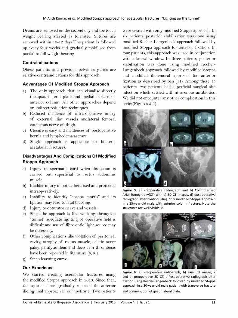
Journal of Karnataka Orthopaedic Association | February 2016 | Volume 4 | Issue 1 33
M Ajith Kumar, et al: Modified Stoppa approach for acetabular fractures: “Lighting up the tunnel”
were treated with only modified Stoppa approach. In six patients, posterior stabilisation was done using modified Kocher-Langenbeck approach followed by modified Stoppa approach for anterior fixation. In four patients, this approach was used in conjunction with a lateral window. In three patients, posterior stabilisation was done using modified Kocher-Langenbeck approach followed by modified Stoppa and modified iliofemoral approach for anterior fixation as described by Sen (11). Among these 15 patients, two patients had superficial surgical site infection which settled withintravenous antibiotics. We did not encounter any other complication in this series(Figures 5-7).
Figure 5: a) Preoperative radiograph and b) Computerised Axial Tomography(CT) with c) 3D CT images, d) post-operative radiograph after fixation using only modified Stoppa approach in a 25-year-old male with anterior column fracture. Note the structures are well visible .8
Figure 6: a) Preoperative radiograph, b) axial CT image, c and d) preoperative 3D CT, a)Post-operative radiograph after fixation using Kocher-Langenbeck followed by modified Stoppa approach in a 30-year-old male patient with transverse fracture
and comminution of quadrilateral plate.
Drains are removed on the second day and toe touch weight bearing started as tolerated. Sutures are removed within 10-12 days.The patient is followed up every four weeks and gradually mobilised from partial to full weight bearing
ContraindicationsObese patients and previous pelvic surgeries are relative contraindications for this approach.
Advantages Of Modified Stoppa Approacha) The only approach that can visualise directly
the quadrilateral plate and medial surface of anterior column. All other approaches depend on indirect reduction techniques.
b) Reduced incidence of intra-operative injury of external iliac vessels andlateral femoral cutaneous nerve of thigh.
c) Closure is easy and incidences of postoperative hernia and lymphedema arerare.
d) Single approach is applicable for bilateral acetabular fractures.
Disadvantages And Complications Of Modified Stoppa Approacha) Injury to spermatic cord when dissection is
carried out superficial to rectus abdominis muscle.
b) Bladder injury if not catheterised and protected intraoperatively.
c) Inability to identify “corona mortis” and its ligation may lead to fatal bleeding.
d) Injury to obturator nerve and vessels.e) Since the approach is like working through a
“tunnel” adequate lighting of operative field is difficult and use of fibre optic light source may be necessary.
f) Other complications like violation of peritoneal cavity, atrophy of rectus muscle, sciatic nerve palsy, paralytic ileus and deep vein thrombosis have been reported in literature (9,10).
g) Steep learning curve.
Our ExperienceWe started treating acetabular fractures using the modified Stoppa approach in 2013. Since then, this approach has gradually replaced the anterior ilioinguinal approach in our institute. Two patients
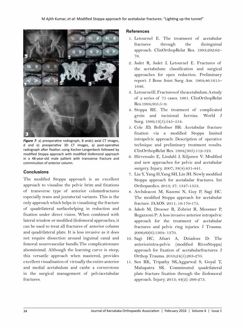
M Ajith Kumar, et al: Modified Stoppa approach for acetabular fractures: “Lighting up the tunnel”
34 Journal of Karnataka Orthopaedic Association | February 2016 | Volume 4 | Issue 1
References
1. Letournel E. The treatment of acetabular fractures through the ilioinguinal approach. ClinOrthopRelat Res. 1993;292:62–76.
2. Judet R, Judet J, Letournel E. Fractures of the acetabulum: classification and surgical approaches for open reduction. Preliminary report. J Bone Joint Surg Am. 1964;46:1615–1646.
3. Letournel E. Fractures of the acetabulum. A study of a series of 75 cases. 1961. ClinOrthopRelat Res.1994;305:5–9.
4. Stoppa RE. The treatment of complicated groin and incisional hernias. World J Surg. 1989;13(5):545–554.
5. Cole JD, Bolhofner BR: Acetabular fracture fixation via a modified Stoppa limited intrapelvic approach: Description of operative technique and preliminary treatment results. ClinOrthopRelat Res. 1994;(305):112-123.
6. Hirvensalo E, Lindahl J, Kiljunen V: Modified and new approaches for pelvic and acetabular surgery. Injury. 2007; 38(4):431-441.
7. Liu Y, Yang H,Yang SH, Lin JH. Newly modified Stoppa approach for acetabular fractures. Int Orthopaedics. 2013; 37: 1347-1353.
8. Archdeacon M, Kazemi N, Guy P, Sagi HC. The modified Stoppa approach for acetabular fracture. JAAOS. 2011; 19:170-175.
9. Jakob M, Droeser R, Zobrist R, Messmer P, Regazzoni P: A less invasive anterior intrapelvic approach for the treatment of acetabular fractures and pelvic ring injuries. J Trauma. 2006;60(6):1364- 1370.
10. Sagi HC, Afsari A, Dziadosz D: The anteriorintra-pelvic (modified RivesStoppa) approach for fixation of acetabularfractures. J Orthop Trauma. 2010;24(5):263-270.
11. Sen RK, Tripathy SK,Aggarwal S, Goyal T, Mahapatra SK. Comminuted quadrilateral plate fracture fixation through the iliofemoral approach. Injury. 2013; 44(2): 266-273.
Figure 7: a) preoperative radiograph, B andc) axial CT images, d and e) preoperative 3D CT images, a) post-operative radiograph after fixation using Kocher-Langenbeck followed by modified Stoppa approach with modified iliofemoral approach in a 48-year-old male patient with transverse fracture and comminution of anterior column.
Conclusions
The modified Stoppa approach is an excellent approach to visualise the pelvic brim and fixations of transverse type of anterior columnfractures especially trans and juxtatectal variants. This is the only approach which helps in visualising the fracture of quadrilateral surfacehelping in reduction and fixation under direct vision. When combined with lateral window or modified iliofemoral approaches, it can be used to treat all fractures of anterior column and quadrilateral plate. It is less invasive as it does not require dissection around inguinal canal and femoral neurovascular bundle.The complicationsare alsominimal. Although the learning curve is steep, this versatile approach when mastered, provides excellent visualisation of virtually the entire anterior and medial acetabulum and canbe a cornerstone in the surgical management of pelviacetabular fractures.

Muthukumar Balaji S, et al: Management of open traumatic extruded talus
Journal of Karnataka Orthopaedic Association | February 2016 | Volume 4 | Issue 1 35
AbstractIntroduction: This is an extremely rare injury involving high energy violence. It is often associated with fractures of the malleoli or Talar body/neck. Complete extrusion called “missing talus” is extremely rare. The incidence is 0.06% of all dislocations and 2% of all talar injuries. There is no established treatment protocol due to the rarity of the injury. We describe our management of this rare injury and the functional and radiological outcome of talar extrusion. Materials and method: The study included patients who had partial or complete extrusion of the talus following RTA and treated at the institute of orthopaedic research and accident surgery between 2011 june and 2013 june. Patients with missing talus and those who did not undergo salvage of the talus were excluded from the study. We describe two such cases with more than two years follow up in this case report. Results: Both our patients had good functional result using AOFAS SCORES. Both patients were able to walk full weight bearing without crutches. Conclusion: Extruded talus represent an extremely rare high energy injury. Functional outcome can be good if the proper treatment on patient basis is selected. The timely administration of antibiotics, staged open fracture protocol, early stabilization, and close follow up leads to a favorable outcome
Key words: Partial or completely Extruded Talus, Re-implantation of Talus, functional outcome.
Management of open traumatic extruded talus
Muthukumar Balaji S , Rohit R K , S Devadoss , A Devadoss
Email: [email protected]
How to cite this article: Balaji MS , Rohit R K , S Devadoss , A Devadoss. Management of open traumatic extruded talus. J Kar Orth Assoc. 2016 Feb; 4(1): 35-38.
Original article
Introduction
Complete extrusion of the talus is a rare injury caused by high energy accidents. It accounts for 0.06% of all the dislocations and 2% of all the talar injuries (1). It was first reported by the German surgeon Fabricius Hildanus in 1680 (2). The complete extrusion of the Talus is known as “Missing Talus” and is an even rarer form of the injury. It is usually associated with a fracture of either the medial malleolus or the body/neck of Talus (3). The injury is very rare and only case reports or small series can be found in literature. There is no established treatment protocol.
Patients and Methods
During the preceding three years, we have come across four cases of extruded talus of which two cases underwent re-implantation of the same. The cases where a re-implantation was not done were excluded from the study.Asynchronous episodes occur when the patient’s breathing pattern does not match the ventilator’s efforts.
Case 1
A 48 year old lady presented with a history of RTA while travelling on a two wheeler and sustained an injury to the left foot and ankle. She presented 6 hours after the injury with a wound on the foot and ankle with exposed bone (Figure 1a). She had no medical co morbidities.
Her vitals and systemic examination was essentially normal. Head to toe examination revealed a swelling and tenderness around the shoulder which was
Muthukumar Balaji SDepartment of Orthopaedic Surgery, Devadoss Multi Specialty
Hospital
75/1, Alakarkovil Main Road, Surveyor Colony, Madurai, Tamil
Nadu 625007, India

Muthukumar Balaji S, et al: Management of open traumatic extruded talus
36 Journal of Karnataka Orthopaedic Association | February 2016 | Volume 4 | Issue 1
confirmed to be a fracture of the clavicle and was treated surgically.
Figure 1: a) pre-operative picture b) after surgery
She had a lacerated wound on the dorsum of foot with loss of soft tissue and cut extensor tendons. The Talus was dislocated and lying outside with minimal soft tissue attachment on the lateral aspect. Vascularity and sensation of foot & toes was intact. Posterior tibial artery was palpable. The patient was evaluated and surgery was planned.
Wound debridement, reduction of Talus and trans-articular Steinman pin fixation with ankle spanning AO ex fix was done. K wire fixation for 5th MT & LisFrancs fracture was done. Clavicle fracture was fixed. A Free Latissimus Dorsi flap and SSG was performed three days later (Figure 1b).
Figure 2: a) after immediate fixation, b) at 3 months, c) at 6 months, d) at 1 year, e) at 2 years follow up
The patient was put on non weight bearing crutch walking for 4 weeks followed by partial weight bearing until 3 months with a PTB top ankle foot orthosis. The patient was regularly followed up over
two years for AVN or infection and to follow the functional recovery( Figure 2a-e). At the last follow up, the patient had minimal pain while walking, movements at the ankle was about 5 degrees of dorsiflexion and 20 degrees of plantar flexion with grossly restricted subtalar movements (Figure 3). Radiographic evaluation showed a narrowing of the joint space, minimal collapse and the final ankle & hindfoot score was 69 out of 100. Revascularisation confirmed with MRI
Figure 3: clinical picture showing range of movement
Case 2
A 45 year old male patient met with a RTA and sustained an injury to the left foot and ankle. He presented three hours after injury with a wound on the dorsum of the foot and ankle with a loss of bone which had been brought to the hospital stored in a plastic bag surrounded by ice (Figure 4). His vitals and systemic examination were essentially normal. Head to toe examination revealed no other injuries. He was a known smoker and an occasional alchoholic.
Figure 4: open ankle injury with extruded talus and pre-operative radiographs
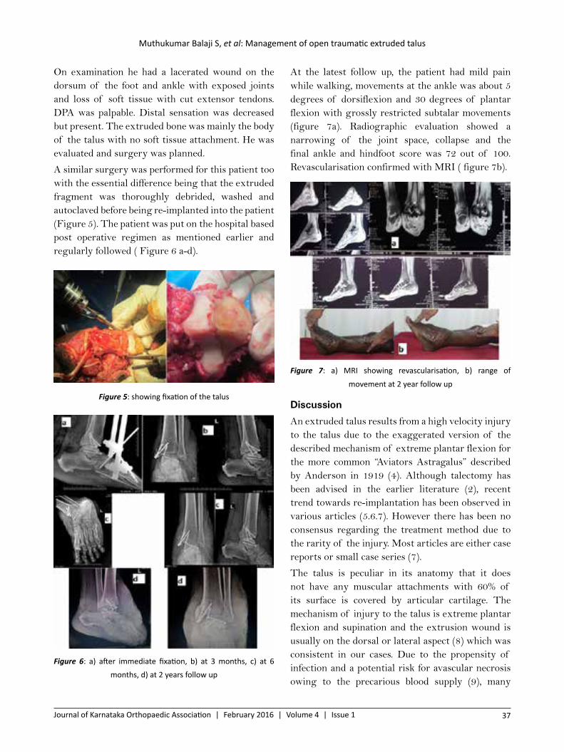
Muthukumar Balaji S, et al: Management of open traumatic extruded talus
Journal of Karnataka Orthopaedic Association | February 2016 | Volume 4 | Issue 1 37
At the latest follow up, the patient had mild pain while walking, movements at the ankle was about 5 degrees of dorsiflexion and 30 degrees of plantar flexion with grossly restricted subtalar movements (figure 7a). Radiographic evaluation showed a narrowing of the joint space, collapse and the final ankle and hindfoot score was 72 out of 100. Revascularisation confirmed with MRI ( figure 7b).
Figure 7: a) MRI showing revascularisation, b) range of
movement at 2 year follow up
Discussion
An extruded talus results from a high velocity injury to the talus due to the exaggerated version of the described mechanism of extreme plantar flexion for the more common “Aviators Astragalus” described by Anderson in 1919 (4). Although talectomy has been advised in the earlier literature (2), recent trend towards re-implantation has been observed in various articles (5.6.7). However there has been no consensus regarding the treatment method due to the rarity of the injury. Most articles are either case reports or small case series (7).
The talus is peculiar in its anatomy that it does not have any muscular attachments with 60% of its surface is covered by articular cartilage. The mechanism of injury to the talus is extreme plantar flexion and supination and the extrusion wound is usually on the dorsal or lateral aspect (8) which was consistent in our cases. Due to the propensity of infection and a potential risk for avascular necrosis owing to the precarious blood supply (9), many
On examination he had a lacerated wound on the dorsum of the foot and ankle with exposed joints and loss of soft tissue with cut extensor tendons. DPA was palpable. Distal sensation was decreased but present. The extruded bone was mainly the body of the talus with no soft tissue attachment. He was evaluated and surgery was planned.
A similar surgery was performed for this patient too with the essential difference being that the extruded fragment was thoroughly debrided, washed and autoclaved before being re-implanted into the patient (Figure 5). The patient was put on the hospital based post operative regimen as mentioned earlier and regularly followed ( Figure 6 a-d).
Figure 5: showing fixation of the talus
Figure 6: a) after immediate fixation, b) at 3 months, c) at 6 months, d) at 2 years follow up
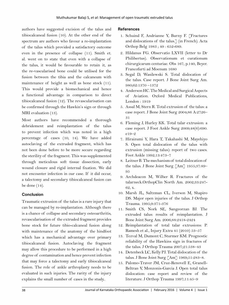
Muthukumar Balaji S, et al: Management of open traumatic extruded talus
38 Journal of Karnataka Orthopaedic Association | February 2016 | Volume 4 | Issue 1
authors have suggested excision of the talus and tibiocalcaneal fusion (10). At the other end of the spectrum are authors who favour a re-implantation of the talus which provided a satisfactory outcome even in the presence of collapse (11). Smith et. al. went on to state that even with a collapse of the talus, it would be favourable to retain it, as the re-vascularised bone could be utilised for the fusion between the tibia and the calcaneum with maintenance of height as well as bone stock (11). This would provide a biomechanical and hence a functional advantage in comparison to direct tibiocalcaneal fusion (12). The revascularisation can be confirmed through the Hawkin’s sign or through MRI evaluation (13).
Most authors have recommended a thorough debridement and reimplantation of the talus to prevent infection which was noted in a high percentage of cases (10, 14). We have added autoclaving of the extruded fragment, which has not been done before to be more secure regarding the sterility of the fragment. This was supplemented through meticulous soft tissue dissection, early wound closure and rigid internal fixation. We did not encounter infection in our case. If it did occur, a talectomy and secondary tibiocalcaneal fusion can be done (14).
Conclusion
Traumatic extrusion of the talus is a rare injury that can be managed by re-implantation. Although there is a chance of collapse and secondary osteoarthritis, revascularisation of the extruded fragment provides bone stock for future tibio-calcaneal fusion along with maintenance of the anatomy of the hindfoot which has a mechanical advantage over primary tibiocalcaneal fusion. Autoclaving the fragment may allow this procedure to be performed in a high degree of contamination and hence prevent infection that may force a talectomy and early tibiocalcaneal fusion. The role of ankle arthroplasty needs to be evaluated in such injuries. The rarity of the injury explains the small number of cases in the study..
References1. Schuind F, Andrianne Y, Burny F. [Fractures
and dislocations of the talus.] (in French). Acta Orthop Belg 1983 ; 49 : 652-689.
2. Hildanus FG. Observatio LXVII (letter to Dr Philibertus). Observationum et curationum chirurgicarum centuriae. Obs 167, p.140, Beyer. Francofurti ad Moenum 1680
3. Segal D, Wasilewski S. Total dislocation of the talus. Case report. J Bone Joint Surg Am. 980;62:1370 –1372
4. Anderson HC. The Medical and Surgical Aspects of Aviation. Oxford Medical Publications, London : 1919
5. Assal M, Stern R. Total extrusion of the talus: a case report. J Bone Joint Surg 2004;86 A:2726–31
6. Fleming J, Hurley KK. Total talar extrusion: a case report. J Foot Ankle Surg 2009;48(6):690.e19–2
7. Hiraizumi Y, Hara T, Takahashi M, Mayehiyo S. Open total dislocation of the talus with extrusion (missing talus): report of two cases. Foot Ankle 1992;13:473–7
8. Leitner B. The mechanism of total dislocation of the talus. J Bone Joint Surg [Am] 1955;37:89–95
9. Archdeacon M, Wilber R. Fractures of the talarneck.OrthopClin North Am. 2002;33:247-62, x.
10. Marsh JL, Saltzman CL, Iverson M, Shapiro DS. Major open injuries of the talus. J Orthop Trauma. 1995;9:371-376
11. Smith CS, Nork SE, Sangeorzan BJ. The extruded talus: results of reimplantation. J Bone Joint Surg Am. 2006;88:2418-2424
12. Reimplantation of total talar extrusions: P. Ramesh et al., Injury Extra 41 (2010) 53–57
13. Tezval M, Dumont C, Sturmer KM. Prognostic reliability of the Hawkins sign in fractures of the talus. J Orthop Trauma 2007;21:538–43
14. Detenbeck LC, Kelly PJ. Total dislocation of the talus. J Bone Joint Surg [Am] 1969;51:283–8.
15. Palomo-Traver JM, Cruz-Renovell E, Granell-Beltran V, Monzonis-Garcia J. Open total talus dislocation: case report and review of the literature. J Orthop Trauma 1997;11:45–9
Patil NVP, et al: Intramedullary interlocking nailing in type II and type III open fractures of tibia – a clinical study
Journal of Karnataka Orthopaedic Association | February 2016 | Volume 4 | Issue 1 39
AbstractPurpose: Controversies exist between primary nailing and external fixator application as treatment of choice for severely open fractures. This study was conducted to assess the outcome of Intramedullary nailing in compound tibia fracture and evaluate its complications. Methods: In the years 2006 to 2013, a total of 86 cases of open tibial shaft fractures were classified according to Gustilo and Anderson classification. These were grouped in to three groups. There were 56 type II and 20 type IIIA and 10 type IIIB open fractures which were grouped A, B and C respectively. All cases were operated with primary Intramedullary interlocking nail. Wound debridement was done with care taken to give adequate soft tissue coverage for wound. Results: The patients were followed for period of 16-36 months and were evaluated according to Katenjians criteria. The results ranged from good to excellent in 81% of cases (70 cases out of total 86 cases). Poor results found in 11% of group A, 30% of group B and 40% of group C fracture. Major complications included deep infection and nonunion in 6 cases, limb shortening in 5 cases, delayed union in 8 cases. Conclusion: primary interlocking Intramedullary nailing can be used as treatment with minimum complications in open tibia fracture fractures when done with proper soft tissue management and wound care
Keywords: Open fractures, Gustilo-Anderson classification, Intramedullary nailing, interlocking nailing.
Neelanagowda V P Patil,Associate ProfessorMMCRI, Mysore.
Intramedullary interlocking nailing in type II and type III open fractures of tibia – a clinical study
Patil NVP, Bellad S H, Anand S R
Email: [email protected]
How to cite this article: Patil NVP, Bellad SH, Anand SR. Intramedullary interlocking nailing in type II and type III open fractures of tibia – a clinical study. J Kar Orth Assoc. 2016 Feb; 4(1): 39-43.
Original article
IntroductionManagement of open tibial fractures continues to be a major therapeutic problem and challenge to orthopaedic surgeons. Three goals must be met for the successful treatment of open fractures of tibia; the prevention of infection, the achievement of bone unions and the restoration of function.Immobilization in a plaster cast has been used most commonly in the past but it does not always maintain the length of the tibia and it leaves the wound relatively inaccessible (1). Open reduction and internal fixation with plates and screws has yielded unacceptably high rates of infection (2, 3).
External fixation, considered as treatment of choice by many Orthopaedic surgeons has the disadvantage of the bulky frames and frequent pin tract infection, non-union and malunion (4-6). With improvement
in soft tissue care and wound coverage technique number of patients going for limb amputations has reduced (7-9). The Intramedullary nailing locked or unlocked has become an attractive option since image intensifier has made closed Intramedullary nailing possible. Nail is a load sharing device and is stiff for both axial and torsion forces. Closed nailing involves minimal disturbance of softtissues, fracture hematoma and better natural process of bone healing as compared to other forms of internal fixation (10-16). The locking of Intramedullary nails to major proximal and distal fragments decreases the prevalence of malunion and comminuted fracture. The rate of infection after treatment of open tibial fractures with interlocking IL nailing has been relatively high. Most investigators are not in favour of using this technique for grade II and grade III open fracture (17-22).With availability of new generation antibiotics and improved wound care infection rate is reduced. A study was designed
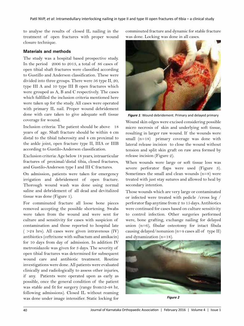
Patil NVP, et al: Intramedullary interlocking nailing in type II and type III open fractures of tibia – a clinical study
40 Journal of Karnataka Orthopaedic Association | February 2016 | Volume 4 | Issue 1
to analyse the results of closed IL nailing in the treatment of open fractures with proper wound closure technique.
Materials and methodsThe study was a hospital based prospective study. In the period 2006 to 2013, a total of 86 cases of open tibial shaft fractures were classified according to Gustillo and Anderson classification. These were divided into three groups. There were 56 type II, 20, type III A and 10 type III B open fractures which were grouped as A, B and C respectively. The cases which fulfilled the inclusion criteria mentioned here were taken up for the study. All cases were operated with primary IL nail. Proper wound debridement done with care taken to give adequate soft tissue coverage for wound.
Inclusion criteria: The patient should be above 18 years of age. Shaft fracture should be within 4 cm distal to the tibial tuberosity and 4 cm proximal to the ankle joint, open fracture type II, IIIA or IIIB according to Gustilo-Anderson classification.
Exclusion criteria: Age below 18 years, intraarticular fractures of proximal/distal tibia, closed fractures, and Gustilo-Anderson type I and III C fractures.
On admission, patients were taken for emergency irrigation and debridement of open fracture. Thorough wound wash was done using normal saline and debridement of all dead and devitalized tissue was done (Figure 1).
For comminuted fracture all loose bone pieces removed accepting the possible shortening. Swabs were taken from the wound and were sent for culture and sensitivity for cases with suspicion of contamination and those reported to hospital late ( >24 hrs). All cases were given intravenous (IV) antibiotics (ceftrixone with sulbactum and amikacin) for 10 days from day of admission. In addition IV metronidazole was given for 3 days. The severity of open tibial fractures was determined for subsequent wound care and antibiotic treatment. Routine investigations were done. All patients were evaluated clinically and radiologically to assess other injuries, if any. Patients were operated upon as early as possible, once the general condition of the patient was stable and fit for surgery (range from12-48 hr, following admissions). Closed IL without reaming was done under image intensifier. Static locking for
comminuted fracture and dynamic for stable fracture was done. Locking was done in all cases.
Figure 1: Wound debridement. Primary and delayed primary
Wound skin edges were excised considering possible micro necrosis of skin and underlying soft tissue, resulting in larger raw wound. If the wounds were small (n=18) primary coverage was done with lateral release incision to close the wound without tension and split skin graft on raw area formed by release incision (Figure 2).
When wounds were large or soft tissue loss was severe perforator flaps were used (Figure 3). Sometimes the small and clean wounds (n=8) were treated with just stay sutures and allowed to heal by secondary intention.
Those wounds which are very large or contaminated or infected were treated with pedicle /cross leg /perforator flap anytime from 2 to 15 days. Antibiotics were continued for cases based on culture sensitivity to control infection. Other surgeries performed were, bone grafting, exchange nailing for delayed union (n=6), fibular osteotomy for intact fibula causing delayed/nonunion (n=4 cases all of type II) and dynamization (n=18).
Figure 2

Patil NVP, et al: Intramedullary interlocking nailing in type II and type III open fractures of tibia – a clinical study
Journal of Karnataka Orthopaedic Association | February 2016 | Volume 4 | Issue 1 41
Figure 3
Most of the patients were mobilized with non-weight bearing active knee movements on the day after the operation . Partial weight bearing was started between 11 to 20 days post operatively. In three cases, partial weight bearing was delayed for more than 30 days (7.5%). Most of the patients were given protective full weight bearing activity in 8 to 12 weeks after the operation (75%). Regular clinical and radiographic assessment was done to assess the fracture union and any other complications. (Figure 4, 5)
Figure 4: pre and follow up radiographs type II fracture
Figure 5

Patil NVP, et al: Intramedullary interlocking nailing in type II and type III open fractures of tibia – a clinical study
42 Journal of Karnataka Orthopaedic Association | February 2016 | Volume 4 | Issue 1
Results:
This study includes 40 cases of open fractures of tibial shaft surgically treated with closed interlocking nailing done between years 2006 to 2013.
In our study majority of patient were from age group of 20-35 (n=52) and 32 patients were male
Major cause of fracture in our study was road traffic accident. Most of fractures were comminuted. Right tibial fracture was found in 48 cases and bilateral in 2 cases. The fracture patterns were comminuted (n=42), segmental (n=6), spiral and oblique (n=38) fractures.
The patients were followed for period of 16 to 36 months and were evaluated according to Katenjians criteria (23). The results ranged from good to excellent in 81% of cases (70 cases out of total 86 cases). Poor results were found in 11% of group A, 30% of group B and 40% of group C fracture. Major complications included deep infection and nonunion in 6 cases, limb shortening in 5 cases, mal union in 4 cases and delayed union in 8 cases superficial infection 4 cases ,and anterior knee pain/knee stiffness 3 cases.
Discussion:
The management of compound tibia fracture still remains to be controversial as there is an uncertainty in the type of implant to be used, timing of surgery and fracture type. Triad of complexity of fracture, soft tissue loss/damage due to high energy injury and infection make management more difficult.
There are many studies done which recommends Intramedullary nailing as a better option for the treatment of open tibia fracture, as it gives better control of length, angulations and rotation when locked. Many of these studies have given more importance to timing of surgery and soft tissue coverage. The studies have shown good result in type I and II, fracture but controversy exists for type III fracture due to high rate of infected nonunion.
In our study we had few complications with type II fractures treated with IL nailing but we also had good functional outcome. Our results are comparable with other similar studies and have similar results
with respect to type III A and type III B fractures. We had relatively better results in type II fracture.
Poor results in type II fracture are attributed to fracture pattern (segmental fracture and comminuted fracture) in 4 cases which went for delayed union nonunion which were treated with bone grafting. Though there were 2 cases with deep infection we had satisfactory union because of continued weight bearing in presence of stable fixation, control of infection with higher new generation proper antibiotics and repeated wound debridement. Infection was eradicated in these cases after bony union and nail removal.
In case of type III fracture, poor results were due to deep infection with nonunion (n=4) and in cases with delayed wound coverage (3 - 8days from time of injury), failure of local transposition flap coverage (n=2) and limb shortening (n=4) with nonunion. All the cases with poor results in type III fracture needed removal of nail and 1 - 2 secondary procedure like cross leg flap, bone transport procedure, bone grafting and external fixator as required. One case where shortening was more than 8 cm with nonunion which is considered as poor result was later treated with vascularised fibular graft by plastic surgeon.
Conclusion
In presence of 76% good results for type II fracture (50 of 56 cases) authors feel IL nailing is better choice of treatment irrespective of timing of surgery for type II compound tibia fracture. If thorough debridement and wound care is done, IL nailing can be used as treatment for type IIIA fracture. Similarly good results can be achieved with type IIIB open fractures with proper soft tissue coverage of the wound along with control of infection with proper and effective antibiotics. The limitation in our study was less number of type IIIB fracture for study to comment and compare with other study.
References1. Gustilo RB, Merkow RL, Templeman B.Curent
concept review: The management of open fracture. JBJS 1990; 72 A: 299-303
2. Ruidi T, Webb JK, and Allgoer M; “Experience with the Dynamic Compression Plate (DCP)
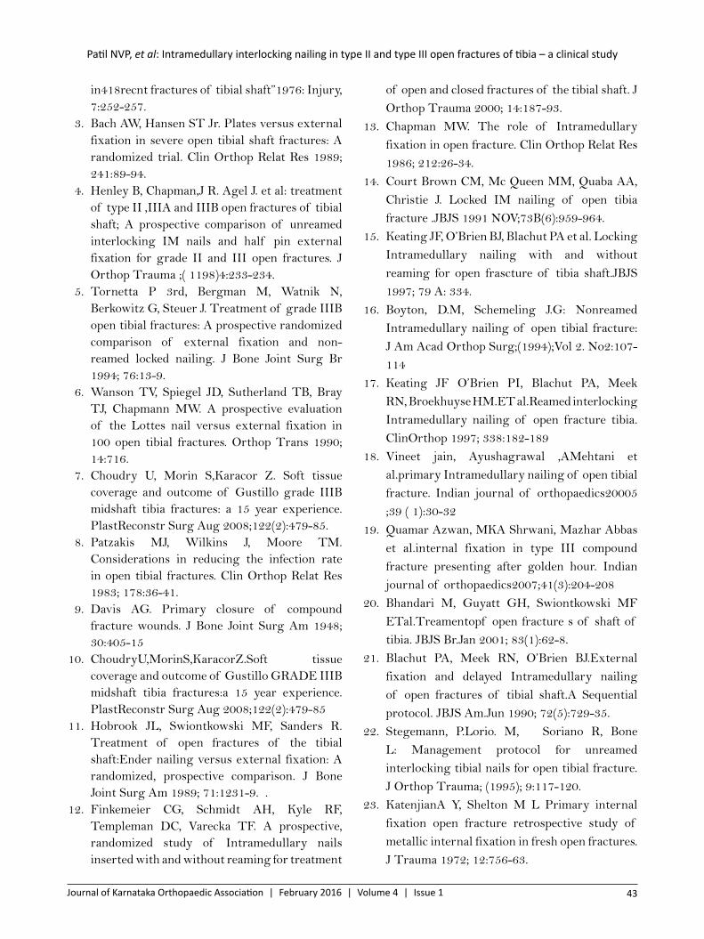
Patil NVP, et al: Intramedullary interlocking nailing in type II and type III open fractures of tibia – a clinical study
Journal of Karnataka Orthopaedic Association | February 2016 | Volume 4 | Issue 1 43
in418recnt fractures of tibial shaft”1976: Injury, 7:252-257.
3. Bach AW, Hansen ST Jr. Plates versus external fixation in severe open tibial shaft fractures: A randomized trial. Clin Orthop Relat Res 1989; 241:89-94.
4. Henley B, Chapman,J R. Agel J. et al: treatment of type II ,IIIA and IIIB open fractures of tibial shaft; A prospective comparison of unreamed interlocking IM nails and half pin external fixation for grade II and III open fractures. J Orthop Trauma ;( 1198)4:233-234.
5. Tornetta P 3rd, Bergman M, Watnik N, Berkowitz G, Steuer J. Treatment of grade IIIB open tibial fractures: A prospective randomized comparison of external fixation and non-reamed locked nailing. J Bone Joint Surg Br 1994; 76:13-9.
6. Wanson TV, Spiegel JD, Sutherland TB, Bray TJ, Chapmann MW. A prospective evaluation of the Lottes nail versus external fixation in 100 open tibial fractures. Orthop Trans 1990; 14:716.
7. Choudry U, Morin S,Karacor Z. Soft tissue coverage and outcome of Gustillo grade IIIB midshaft tibia fractures: a 15 year experience. PlastReconstr Surg Aug 2008;122(2):479-85.
8. Patzakis MJ, Wilkins J, Moore TM. Considerations in reducing the infection rate in open tibial fractures. Clin Orthop Relat Res 1983; 178:36-41.
9. Davis AG. Primary closure of compound fracture wounds. J Bone Joint Surg Am 1948; 30:405-15
10. ChoudryU,MorinS,KaracorZ.Soft tissue coverage and outcome of Gustillo GRADE IIIB midshaft tibia fractures:a 15 year experience. PlastReconstr Surg Aug 2008;122(2):479-85
11. Hobrook JL, Swiontkowski MF, Sanders R. Treatment of open fractures of the tibial shaft:Ender nailing versus external fixation: A randomized, prospective comparison. J Bone Joint Surg Am 1989; 71:1231-9. .
12. Finkemeier CG, Schmidt AH, Kyle RF, Templeman DC, Varecka TF. A prospective, randomized study of Intramedullary nails inserted with and without reaming for treatment
of open and closed fractures of the tibial shaft. J Orthop Trauma 2000; 14:187-93.
13. Chapman MW. The role of Intramedullary fixation in open fracture. Clin Orthop Relat Res 1986; 212:26-34.
14. Court Brown CM, Mc Queen MM, Quaba AA, Christie J. Locked IM nailing of open tibia fracture .JBJS 1991 NOV;73B(6):959-964.
15. Keating JF, O’Brien BJ, Blachut PA et al. Locking Intramedullary nailing with and without reaming for open frascture of tibia shaft.JBJS 1997; 79 A: 334.
16. Boyton, D.M, Schemeling J.G: Nonreamed Intramedullary nailing of open tibial fracture: J Am Acad Orthop Surg;(1994);Vol 2. No2:107-114
17. Keating JF O’Brien PI, Blachut PA, Meek RN, Broekhuyse HM.ET al.Reamed interlocking Intramedullary nailing of open fracture tibia.ClinOrthop 1997; 338:182-189
18. Vineet jain, Ayushagrawal ,AMehtani et al.primary Intramedullary nailing of open tibial fracture. Indian journal of orthopaedics20005 ;39 ( 1):30-32
19. Quamar Azwan, MKA Shrwani, Mazhar Abbas et al.internal fixation in type III compound fracture presenting after golden hour. Indian journal of orthopaedics2007;41(3):204-208
20. Bhandari M, Guyatt GH, Swiontkowski MF ETal.Treamentopf open fracture s of shaft of tibia. JBJS Br.Jan 2001; 83(1):62-8.
21. Blachut PA, Meek RN, O’Brien BJ.External fixation and delayed Intramedullary nailing of open fractures of tibial shaft.A Sequential protocol. JBJS Am.Jun 1990; 72(5):729-35.
22. Stegemann, P.Lorio. M, Soriano R, Bone L: Management protocol for unreamed interlocking tibial nails for open tibial fracture. J Orthop Trauma; (1995); 9:117-120.
23. KatenjianA Y, Shelton M L Primary internal fixation open fracture retrospective study of metallic internal fixation in fresh open fractures. J Trauma 1972; 12:756-63.

Vivek Pandey, et al: Lateral End Clavicle Fracture: A Review of Current Concepts
44 Journal of Karnataka Orthopaedic Association | February 2016 | Volume 4 | Issue 1
AbstractBackground: Supracondylar fracture of humerus among children is one of the most common fractures seen in pediatric orthopedic clinic setting worldwide. Closed reduction and percutaneous K-wire fixation is a preferred method of treatment and should be attempted at the earliest possible time. In this study, displaced extension type supracondylar fractures among children were managed with closed reduction and percutaneous K-wire fixation using image intensifier to study age, sex incidence, and side preponderance, assess clinical and radiological outcomes. It is also to compare clinical outcomes in two types of pin configurations (crossed and triple) and early and late complications associated with these procedures in this study. Materials And Methods: This study was conducted between June 2013 and June 2015 in the Department of Orthopaedics in Karnataka Institute of Medical Sciences, Hubballi. The mean age of the patients was 8.73 years (range 6–13 years). The male/female ratio was 3:1 and the left side was involved in 53.8 % whereas the right side had 42.2% injuries. All the 26 admitted patients had extension type injury with 96.15% fractures being Gartland type III and 3.85 % were type II. Posteromedial displacement was noted in 57.7 % whereas 42.3 % fractures were posterolaterally displaced. In 13 cases, crossed K-wires were used whereas, in 13 cases, two lateral and one medial Kwire were used. Kwires were removed at the fourth week postoperatively and follow-up was done at the eighth, 12th week, and the final follow-up at sixth month when they were evaluated according to the criteria described by Flynn. Chisquare test and Fischer –Exact test was used as a statistical tool to compare results among different variables. Results: All 26 patients had satisfactory outcomes (100%) as per Flynn’s criteria. Although triple pinning had shown better results but there was no statistical difference in the outcomes and complication rates among patients treated by either crossed or triple pinning technique due to small sample size. Younger age patients were found to have better final clinical outcomes to older children. Patients who had Baumann angle within four degrees compared to normal side had better outcomes and it can be used as a reliable radiologic parameter to assess adequacy of reduction. Conclusion: Closed reduction and percutaneous fixation using K-wires gives excellent results and is the most accepted modality for treating displaced supracondylar fractures of humerus among children. Triple pinning has shown encouraging results in comparison to Crossed pinning, but larger sample size is needed to give statistical significance to the comparative study.
Keywords: Percutaneous pinning, supracondylar fracture humerus, triple pinning, crossed pinning.
C V MudgalAssociate Professor, Department of Orthopaedics, Karnataka Institute of Medical Sciences, Hubballi
Surgical Management of Unstable Supracondylar Fractures of Humerus in Children by Closed Reduction and Percutaneous K Wire Fixation by Crossed Versus Triple Pin Configuration-A Comparative Randomized Study
C V Mudgal, S F Kammar, Viresh Murgodi
Email: [email protected]
How to cite this article: Mudgal CV, Kammar SF, Murgodi V. Surgical Management of Unstable Supracondylar Fractures of Humerus in Children by Closed Reduction and Percutaneous K Wire Fixation by Crossed Versus Triple Pin Configuration-A Comparative Randomized Study: A Review of Current Concepts. J Kar Orth Assoc. 2016 Feb; 4(1): 44-51.
Original article
IntroductionSupracondylar fracture of humerus among children is one of the most common fractures seen in paediatric orthopaedic clinic setting worldwide. It accounts for 65.4% of upper extremity fractures in five to10 year age group among children1 .Various methods have
been used for management of displaced fractures as closed reduction and cast application, skin traction, overhead skeletal traction, closed reduction or open reduction and percutaneous K-wire fixation.2
Closed reduction and percutaneous pinning under fluoroscopic guidance is the procedure of choice for the treatment of these fractures whenever possible and the original Swenson technique of cross pinning continues to be used today with excellent results and
Vivek Pandey, et al: Lateral End Clavicle Fracture: A Review of Current Concepts
Journal of Karnataka Orthopaedic Association | February 2016 | Volume 4 | Issue 1 45
low morbidity. In the developing world, proportion of delayed presentation is much higher because of poorly developed health delivery system and patients reaching the tertiary care centers from long distance.3 The goal of treatment in children is to achieve and maintain a close to anatomic reduction until the fracture is properly healed minimizing the risks of complications. Closed reduction and percutaneous pinning have become a standard method of treatment although controversy persists concerning the optimal pin configuration.4
The purpose of this study is to evaluate the age, sex, and side incidence, radiological and functional outcome in closed reduction and percutaneous K-wire fixation and comparative study of clinical outcomes, post-operative complications in crossed and triple pinning.
Materials And Methods
Twenty-six displaced, closed extension type of supracondylar fractures (Gartland’s typeII and III) of humerus among children were treated by closed reduction and internal fixation by percutaneous K-wires between July 2013 and October 2015. Two pin configurations crossed pinning (referred to as group C) and triple pinning (referred to as Group T) for percutaneous K-wire fixation were done and the functional and radiological outcomes among the two treatment groups were compared. This study was conducted in Karnataka Institute of Medical Sciences, Hubballi. Permission was taken from Ethical committee prior to the study.
Gartland’s type II and III fractures (Unstable fractures) were included for the patients, age less than 13 years, presenting and operated within 72 hours of the injury. Open fractures and patients presenting with neurovascular complications, patients presenting after 72 hours of injury and patients with associated fractures around the elbow and ipsilateral upper limb fractures were excluded. Plain radiograph of elbow in A-P and Lateral views affected and normal side were taken. Based on the radiographs all cases were classified according to Gartland’s classification5 and Type II and III were selected. Type II fractures were managed by
selective pinning i.e., the fractures where satisfactory reduction was not obtained or after five - six days post reduction radiograph showed loss of reduction were managed by pinning. Routine laboratory investigations like complete blood counts, RBS, Renal function tests and HIV and HBsAg and Chest X ray PA view were done and immediately taken up for surgery. One dose of Inj ceftriaxone as per body weight was given 30 min before the start of procedure.
Operative Technique: Patients were given general anesthesia and placed supine on the operating table, with the C-arm screen, used as an operating table. Closed reduction was performed under image intensification. Initially sustained longitudinal traction was given with elbow in extension and forearm in supination and counter traction applied by assistant. This maneuver corrected the impaction. Next the medio-lateral displacement was corrected by valgus or varus force at fracture site. The posterior displacement of the distal fragment was corrected next by applying an anterior force over olecranon process with gradual flexion of elbow and pronation of forearm, triceps muscle acts as an internal splint.
Anatomic reduction (Baumann angle within four degree of normal side) is confirmed with the image intensifier before pinning. If reduction is satisfactory, parts are painted and draped. The lateral pin is always inserted first. The insertion site is made at an angle of 30 degree so that the pin will traverse the lateral portion of the ossified capitellum, cross the physis, proceed up the lateral column, and always engage the opposite medial cortex proximally. Using a Kirschner wire (1.5 or 1.8 mm), the position for inserting the pin is documented on AP and lateral views. A small incision is made in the skin. The pin is placed using a power drill and a sharp K-wire. Provisional stability is achieved with the first pin. In case triple pinning a lateral pin is inserted, then the second lateral pin is inserted next in divergent manner followed by medial pin with elbow in extension to avoid ulnar nerve.
In case of crossed pinning, a lateral pin is drilled and then second pin is placed medially. The position of the K-wire from the medial side is confirmed

Vivek Pandey, et al: Lateral End Clavicle Fracture: A Review of Current Concepts
46 Journal of Karnataka Orthopaedic Association | February 2016 | Volume 4 | Issue 1
with fluoroscopy. The medial pin is generally more horizontal than the lateral pin and should traverse the medial column and engage the opposite lateral cortex. 6
Pins were bent to prevent pin migration and 1 cm length was left behind for the removal after 4 weeks without anesthesia. Post-operative radiographs of elbow-AP and lateral views were taken. Post-operatively all patients were given well-padded high above elbow POP slab with 90 degree elbow flexion and strict limb elevation. Patient was encouraged to do active finger movements. Appropriate antibiotics and analgesics were initiated and regular monitoring of neurovascular status was done. Patients were discharged after four - five days in absence of any complications. At fourth week follow up, check x ray was done and K-wire were removed and active range of motion exercises were initiated. Patient relatives were advised not to massage or passively stretch elbow joint as it may lead to myositis ossificans. Patient was advised regular follow up at fourth, eighth week respectively and final follow up at the sixth month. At the final follow up the patient was examined clinically for loss of flexion and extension and cosmetically by loss of carrying angle. Radiological parameters like Baumann’s angle, ulno-humeral angle were noted. The results were graded as excellent, good, fair, and poor using Flynn’s criteria.1
RESULTS
Age distribution was 6- 13 years. Most of the patients were in 6-10 years’ group (88.5%). Both the study groups were similar in terms of age (P value=0.180). Mean age of patients was 8.31±1.84 yrs. Out of 23 patients in six-10 yrs. of age, 16 patients showed excellent results and seven had good results. In 11-15 yrs age group, two had good results and one had fair result as per Flynn criteria. Moderate significance could be given to younger patients having better outcomes than the older children do (P=0.014, significant, Fisher Exact test). Overall 76.9% were males compared to 23.1% females. Out of the total 26 patients,14 patients had fracture on left side (53.8%) and 12 patients on right side (46.2%). Out of total 26 patients, one patient
(3.8%) had type II fracture,15 patients (57.7%) had type IIIA and 10 patients (38.5%) had type IIIB. In our study mean injury to surgery time delay was 2.46 days. Patients 7.7% were operated within a day, 38.5% patients within two days, and 53.8% within three days. Mean time for union was seven weeks and all patients’ signs of union at the fourth week’s follow-up.
Out of 21 patients who had loss of Baumann angle within five degree compared to normal side (n=21), 14(66.66%) patients showed excellent and 7(33.3%) showed good outcomes. In comparison if loss of Baumann angle is more than five degree, then excellent outcome was seen in only 40% patients and 40% showed good outcome and 10% showed fair outcome showing positive association between maintenance of Baumann angle and final clinical outcome ( P=0.141, positive association, Fisher Exact test).
In our study, 16 patients (61.5%) had excellent, nine (34.6%) and one patients (3.8%) had fair outcome as per Flynn criteria. None of the patient had poor outcome. Statistically no significance could be attached to the difference in the outcome between the two study groups (P=0.688, Not significant, Fisher Exact test).
Out of total 26 patients’ post-operative complications. Four patients (15.4%) had superficial pin tract infection. Two patients (7.7%) had pin migration during post-operative period. One patient had post-operative varus deformity. No case of iatrogenic nerve injury was noted. Complication rates were similar between the two groups (P=0.378, Not significant, Fisher Exact test).
CASE 1- Crossed pinning group
Figure 1A-Pre-operative radiograph showing type III Gartland’s supracondylar fracture

Vivek Pandey, et al: Lateral End Clavicle Fracture: A Review of Current Concepts
Journal of Karnataka Orthopaedic Association | February 2016 | Volume 4 | Issue 1 47
Figure 1B- Immediate post-operative radiograph with two K-wires insitu, 1 medial and 1 lateral
Figure 1C-Radiograph at fourth week showing callous formation.
Figure 1D-Radiograph at sixth month’s follow-up
Figure 1E-Clinical photograph at sixth month’s-Flexion and Extension
CASE 2-Triple pinning group
Figure 2A-Pre-operative radiograph showing type III Gartland supracondylar fracture
Figure 2B-Shows immediate post-op radiograph with Triple pin configuration
Figure 2C-Shows radiograph at fourth week’s post-op
Figure 2D-Shows radiograph at sixth month’s follow-up
Figure 2E-Shows clinical photographs at sixth month’s follow-up- Flexion and Extension
Discussion
The main aim of management of supracondylar fractures of humerus among children is to achieve satisfactory results in terms of functional and cosmetic point of view with minimal post-operative complications and minimal hospital stay leading to low treatment costs to patient. Closed reduction and

Vivek Pandey, et al: Lateral End Clavicle Fracture: A Review of Current Concepts
48 Journal of Karnataka Orthopaedic Association | February 2016 | Volume 4 | Issue 1
percutaneous pinning under fluoroscopic guidance is now the accepted standard treatment for displaced supracondylar fractures of the Humerus.7 Recent studies have shown excellent long term functional results among patients with 12-72 hours delay in presentation treated with closed reduction and percutaneous fixation. Hence, patients presenting within 72 hours were selected for our study. 3
Olcay et al compared the fixation techniques used in the supracondylar fractures of the humerus anatomically and biomechanically by using the torsional strength in an adult cadaver model. It was found out that the most severe resistance against the rotation strengths was obtained by fixation via a third cross K-wire from the medial in addition to the two K-wires parallel from the lateral side; while the least resistance was through the fixation with two K-wires from the lateral side. Finally, the fixation with a cross K-wire each from the medial and lateral sides was less resistant than the fixation with the three K-wires.8
The present study was conducted to assess the results of closed reduction and percutaneous fixation with K-wires and comparative study between crossed pinning and triple pinning (two lateral and one medial pin) in terms of clinical outcome and complications in displaced extension type supracondylar humerus fractures.
In our study patient with average age was 8.73+1.59 yrs. age, 88.5% patients were between six to 10 yrs. and 11.5% in 11-15 yrs. group. In a study conducted by Mazda et al8 the mean age at presentation of 5.7 years was noted among patients, Sharma A et al9 noted the mean age at 6.7 yrs. in their study; Edmonds et al10 found average age to be 5.8 yrs. The reason behind the variation in age incidence in our study compared to previous study maybe due to incorrect details given by patients due to poor educational status of patients. All patients in age group 6-10 yrs (n=23) had satisfactory outcome (excellent to good) compared to 10-15 yrs. age group (n=3) where two patients had good and one had fair outcome suggesting better results among younger patients.
Table 1-Comparison of final clinical outcome among different age groups
Edmonds et al10 reported 45.3% patients to be males, 54.7% were females. Sharma et al9 reported 83.33% patients to be males and 16.67% females, Wilkins et al11 reported 62.8% were males and 37.2% were females. In our study, 76.9% were males and 33.1%were females, suggesting males are predisposed to these fractures possibly due to increased outdoor activities and aggressive nature among boys. In our study, 53.8% patients had fractures affecting left side while 46.2% patients had right side affection. Mazda et al8 reported 56% fractures on left side and 44% right side affection in 116 cases studied. Wilkins et al11 reported 60.8% left side and 39.2% right side affection. The reason for left sided preponderance could not be explained.
Sharma et al9 reported 73.3% fractures being Gartland’s type III(Posteromedial displacement-70% ,posterolateral displacement-30%) and 26.7% were type II.Karapinar et al6 reported posterolateral displacement in 59.32% and posteromedial displacement in 40.68% of their patients. In our study, 25 patients (96.15%) out of 26 patients were type III Gartland’s fracture out of which 57.7% had posteromedial displacement, 38.5% had posterolateral displacement, and one patient (3.85%) was type II. Type II fractures were managed by selective pinning in case closed reduction and cast immobilization fails at 5th day follow up, hence the incidence of type II fractures were less in our study.
In our study, mean injury to surgery time delay was 2.46 days. Patients 7.7% were operated within a day, 38.5% patients within two days and 53.8% within three days. Average injury to surgery time delay noted by Skagg’s et al12 was 1.4 days. Dua et al3 showed excellent long term functional results
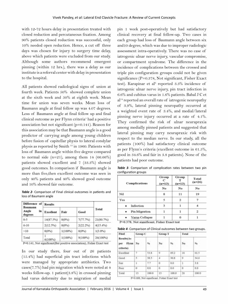
Vivek Pandey, et al: Lateral End Clavicle Fracture: A Review of Current Concepts
Journal of Karnataka Orthopaedic Association | February 2016 | Volume 4 | Issue 1 49
with 12-72 hours delay in presentation treated with closed reduction and percutaneous fixation. Among 90% patients closed reduction was successful, only 10% needed open reduction. Hence, a cut off three days was chosen for injury to surgery time delay, above which patients were excluded from our study. Although some authors recommend emergent pinning (within 12 hrs.), there was a delay as our institute is a referral center with delay in presentation to the hospital.
All patients showed radiological signs of union at fourth week. Patients 50% showed complete union at the sixth week and 50% at eighth week. Mean time for union was seven weeks. Mean loss of Baumann angle at final follow up was 4.07 degrees. Loss of Baumann angle at final follow up and final clinical outcome as per Flynn criteria1 had a positive association but not significant (p=0.141). Reason for this association may be that Baumann angle is a good predictor of carrying angle among young children before fusion of capitellar physis to lateral condylar physis as reported by Smith 13 in 1960. Patients with loss of Baumann angle within five degree compared to normal side (n=21), among them 14 (66.66%) patients showed excellent and 7 (33.3%) showed good outcomes. In comparison if Baumann angle is more than five,then excellent outcome was seen in only 40% patients and 40% showed good outcome and 10% showed fair outcome.
Table 2- Comparison of Final clinical outcomes in patients and loss of Baumann angle
In our study there, four out of 26 patients (15.4%) had superficial pin tract infections which were managed by appropriate antibiotics. Two cases(7.7%) had pin migration which were noted at 4 weeks follow-up. 1 patient(3.8%) in crossed pinning had varus deformity due to migration of medial
pin 1 week post-operatively but had satisfactory clinical recovery at final follow-up. Two cases in each group had loss of Baumann angle between six and10 degree, which was due to improper radiologic assessment intra-operatively. There was no case of iatrogenic ulnar nerve injury, vascular compromise, or compartment syndrome. The difference in the incidence of complications between the crossed and triple pin configuration groups could not be given significance (P=0.378, Not significant, Fisher Exact test). Karapinar et al6 reported 3.3% incidence of iatrogenic ulnar nerve injury, pin tract infection in 6.6% and cubitus varus in 1.6% patients. Babal J C et al14 reported an overall rate of iatrogenic neuropathy of 3.9%, lateral pinning neuropathy occurred at a weighted event rate of 3.4%, and medial/lateral pinning nerve injury occurred at a rate of 4.1%. They confirmed the risk of ulnar neurapraxia among medially pinned patients and suggested that lateral pinning may carry neurapraxic risk with respect to the median nerve. In our study, all the patients (100%) had satisfactory clinical outcome as per Flynn’s criteria (excellent outcome in 61.5%, good in 34.6% and fair in 3.8 patients). None of the patients had poor outcome.
Table 3- Comparison of complication rates between two pin configuration groups
Table 4- Comparison of Clinical outcomes between two groups.

Vivek Pandey, et al: Lateral End Clavicle Fracture: A Review of Current Concepts
50 Journal of Karnataka Orthopaedic Association | February 2016 | Volume 4 | Issue 1
The results were evaluated using Flynn’s criteria1 and all patients had satisfactory outcome (excellent in 16, good in nine and fair result in one patient), none had poor outcome. Aronson et al15 in their study on management of unstable supracondylar fractures of humerus among 26 patients by closed reduction and percutaneous pinning reported excellent results among 16 patients, good among nine and fair result in one patient as per Flynn criteria. Karapinar et al6 in their study on 61 patients of unstable supracondylar fractures using triple pinning reported excellent outcomes among 49 patients, good among seven and fair in two patients. Sharma et al9 reported excellent outcome among 12 patients, 54 showed good and 15 patients fair.
Only one patient had five degrees of varus deformity and four cases of superficial pin tract infection successfully managed with antibiotics and two cases of pin migration. Most of the patients were discharged by fourth day except in superficial pin infection. After comparing our results with other similar studies, we conclude that closed reduction and percutaneous K-wire fixation is an excellent modality for treating displaced supracondylar fractures of humerus among children. The Triple pinning group shows better results than Crossed pinning group but no statistical significance could be attached, reason being very small sample size in both groups. More number of cases needs to be studied to attach any kind of statistical significance to the results of the comparative study.
Conclusion
From our study we conclude that closed reduction and percutaneous pin fixation is a safe and excellent method for managing displaced supracondylar fractures of the humerus among children. Supracondylar fractures are common among six-10 yrs age, more common among males and has left side predisposition. Posteromedial displacement is more common than posterolateral displacement. The clinical and cosmetic outcomes are better, fewer complications, minimal morbidity and lesser duration of hospital stay compared to other methods like closed reduction and casting, tractions and open reduction and internal fixation. This method
allows elbow to be placed in more than 90 degrees of extension, thus allowing venous drainage and prevention of vascular compromise. The ideal pinning configuration is still controversial, triple pin configuration (two lateral and one medial) shows promise as an ideal pin configuration but statistical significance could not be attached to our observation due to small sample size. More cases need to be evaluated to come to statistical significance to compare crossed and triple pin configuration groups.
Bibliography
1. Piggot J, Graham HK, McCoy GF. Supracondylar fracture of humerus in children. Treatment by straight lateral traction. J Bone J Surg.1986 Aug; 68B:238-242.
2. Pirone AM et al. Management of displaced extension type supracondylar fractures of humerus in children. J Bone & Joint Surg1998; 70A; 641-650.
3. Dua A, Eachempati KK, Malhotra R, Sharma L, Gidaganti M. Closed reduction and percutaneous pinning of displaced supracondylar fractures of humerus in children with delayed presentation. Chinese Journal of Traumatology.14 .2011; 14(1):14-19
4. Novais EN, Andrade MAP, Gomes DC. Joystick Pin in Supracondylar Humerus Fracture, J Pediatr Orthop Volume 33, Number 1, January/February 2013.
5. Gartland JJ. Management of supracondylar fractures of the humerus in children. Surg Gynecol Obstet 1959 109(2):145-154.
6. Karapinar L, Ozturk H, Altay T, Kose B. Closed reduction, and percutaneous pinning with three Kirschner wires in children with type III displaced supracondylar fractures of the humerus. Acta Orthop Traumatol Turc 2005;39(1):23-29
7. Skaggs DL, Cluck MW, Mostofi A, et al. Lateral-entry pin fixation in the management of supracondylar fractures in children. J Bone Joint Surg (Am) 2004 86A(4):702-707
8. Mazda K, Boggione C, Fitoussi F, Penneçot GF. Systematic pinning of displaced extension-type supracondylar fractures of the humerus in children-a prospective study of 116 consecutive

Vivek Pandey, et al: Lateral End Clavicle Fracture: A Review of Current Concepts
Journal of Karnataka Orthopaedic Association | February 2016 | Volume 4 | Issue 1 51
patients. J Bone Joint Surg [Br] 2001; 83-B: 888-93.
9. Sharma A, Walia J P S, Brar BS, Sethi S. Early results of displaced supracondylar fractures of humerus in children treated by closed reduction and percutaneous pinning. Indian Journal of Orthopaedics; September 2015 Vol. 49 Issue 5
10. Edmonds EW, Roocroft JH, Mubarak SJ. Treatment of displaced paediatric supracondylar humerus fracture patterns requiring medial fixation: A reliable and safer cross-pinning technique. J Pediatr Orthop2012,32:346-351
11. Wilkins KE.The operative management of supracondylar fracture in children. Ortho Clin North Am,1990;21(20);269-289
12. Skaggs DL et al. Operative treatment of supracondylar fractures of the humerus in children. J Bone Joint Surg [Br] 2001;83-A(5);735-740
13. Smith L. Deformity following supracondylar fractures of the humerus. Am J Orthop 1960 42A:235-252
14. Babal JC, Charles T., Klein MG. Nerve injuries associated with pediatric supracondylar humeral fractures: A meta-analysis. J Pediatr Orthop 2010; 30:253–263.
15. Aronson DD, Prager BI. Supracondylar fractures of the humerus in children-A modified technique for closed pinning. Clin Orthop.1987;219;174-183

Vigneshwara B: A Study of Functional Outcome of Surgical Management of Floating Knee Among Adults
52 Journal of Karnataka Orthopaedic Association | February 2016 | Volume 4 | Issue 1
AbstractBackground: ‘Floating knee injury’ is a term used to denote ipsilateral femoral and tibial metaphyseal injuries. However, recent literature has expanded this term to include most ipsilateral fractures of the femur and tibia. There are no specific guidelines for the management. The implant choice needs to be determined depending on nature of fracture and soft tissue injuries. A specific pattern of management can often not be determined. This study is about the Functional Outcome of Surgical Managements of Floating Knee. For this study 17 consecutive patients with ipsilateral femur and tibia fractures were presented to KIMS casualty from Nov 2013- Oct2015. Follow up study was done for six weeks, 12 weeks, six months and one year. Serial x-rays and functional assessment were carried out at each visit in outpatient clinic itself using the Karlstorm and Oleruds criteria. As per Karlstrom and Oleraud criteria 23.5% had excellent outcome, 29.4% had good outcome, 29.4% had acceptable outcome and 17.6% had poor outcome. Patients who underwent primary nailing had Excellent or Good results. Patients who underwent MIPPO had good result. The most important factors, which determine the functional outcomes, were the type of fractures (open or closed), nature of comminution including intra-articular extensions, timing of fixations and postoperative infections.
Keywords: Floating knee; primary nailing; intra-articular extension; comminution; metaphysis
A Study of Functional Outcome of Surgical Management of Floating Knee Among Adults
Suryakanth Kalluraya, V K Bhasme, Vigneshwara B
Email: [email protected]
How to cite this article: Kalluraya S, Bhasme VK, Vigneshwara B. A Study of Functional Outcome of Surgical Management of Floating Knee Among Adults. J Kar Orth Assoc. 2016 Feb; 4(1): 52-59.
Original article
Introduction
Floating knee injury is a term used to denote ipsilateral femoral and tibial metaphyseal injuries. However, recent literature has expanded this term to include most ipsilateral fractures of the femur and tibia.
These are extremely heterogeneous groups of injuries. They usually occur due to very high-energy trauma. These are relatively uncommon injuries and always associated with high morbidity. Most of these injuries result in some permanent disability.
There are no specific guidelines for the management. The implant choice needs to be determined depending on nature of fracture and soft tissue injuries. A specific pattern of management can often not be determined.
The incidence of floating knee injuries was reported as 2.6 % of all fractures by Letts et al in 1986.1 These injuries were associated with life threatening injuries such as head injury, chest injury, and abdominal injuries as shown by Veith.2
Other skeletal injuries were also seen in these patients. The injuries were often a combination of different fracture patterns. There was extensive soft tissue damage of the limb as well. The soft tissue injuries can also be variable from minor abrasions to grade III open injuries. The injuries to the neurovascular structures add a treacherous component to the whole picture. This often perplexes even the most experienced clinicians in the choice of management.
The established principles of treatment are1) Early and thorough debridement of the wound
in case of open fractures.2) Accurate reduction of intra-articular fractures
and reduction of dislocations.
Dr Vigneshwara BJunior Resident, Department of Orthopaedics, KIMS, Hubli -580022
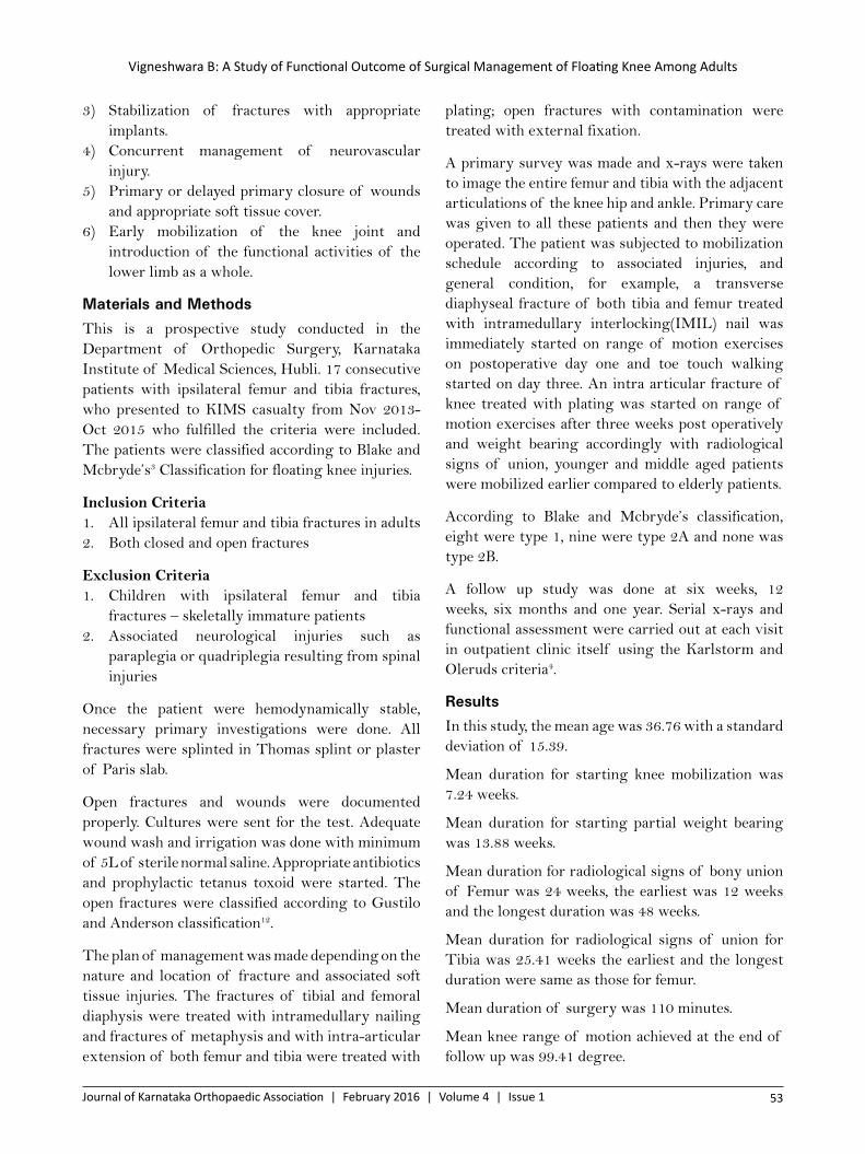
Vigneshwara B: A Study of Functional Outcome of Surgical Management of Floating Knee Among Adults
Journal of Karnataka Orthopaedic Association | February 2016 | Volume 4 | Issue 1 53
3) Stabilization of fractures with appropriate implants.
4) Concurrent management of neurovascular injury.
5) Primary or delayed primary closure of wounds and appropriate soft tissue cover.
6) Early mobilization of the knee joint and introduction of the functional activities of the lower limb as a whole.
Materials and MethodsThis is a prospective study conducted in the Department of Orthopedic Surgery, Karnataka Institute of Medical Sciences, Hubli. 17 consecutive patients with ipsilateral femur and tibia fractures, who presented to KIMS casualty from Nov 2013-Oct 2015 who fulfilled the criteria were included. The patients were classified according to Blake and Mcbryde’s3 Classification for floating knee injuries.
Inclusion Criteria1. All ipsilateral femur and tibia fractures in adults2. Both closed and open fractures
Exclusion Criteria1. Children with ipsilateral femur and tibia
fractures – skeletally immature patients2. Associated neurological injuries such as
paraplegia or quadriplegia resulting from spinal injuries
Once the patient were hemodynamically stable, necessary primary investigations were done. All fractures were splinted in Thomas splint or plaster of Paris slab.
Open fractures and wounds were documented properly. Cultures were sent for the test. Adequate wound wash and irrigation was done with minimum of 5L of sterile normal saline. Appropriate antibiotics and prophylactic tetanus toxoid were started. The open fractures were classified according to Gustilo and Anderson classification12.
The plan of management was made depending on the nature and location of fracture and associated soft tissue injuries. The fractures of tibial and femoral diaphysis were treated with intramedullary nailing and fractures of metaphysis and with intra-articular extension of both femur and tibia were treated with
plating; open fractures with contamination were treated with external fixation.
A primary survey was made and x-rays were taken to image the entire femur and tibia with the adjacent articulations of the knee hip and ankle. Primary care was given to all these patients and then they were operated. The patient was subjected to mobilization schedule according to associated injuries, and general condition, for example, a transverse diaphyseal fracture of both tibia and femur treated with intramedullary interlocking(IMIL) nail was immediately started on range of motion exercises on postoperative day one and toe touch walking started on day three. An intra articular fracture of knee treated with plating was started on range of motion exercises after three weeks post operatively and weight bearing accordingly with radiological signs of union, younger and middle aged patients were mobilized earlier compared to elderly patients.
According to Blake and Mcbryde’s classification, eight were type 1, nine were type 2A and none was type 2B.
A follow up study was done at six weeks, 12 weeks, six months and one year. Serial x-rays and functional assessment were carried out at each visit in outpatient clinic itself using the Karlstorm and Oleruds criteria4.
ResultsIn this study, the mean age was 36.76 with a standard deviation of 15.39.
Mean duration for starting knee mobilization was 7.24 weeks.
Mean duration for starting partial weight bearing was 13.88 weeks.
Mean duration for radiological signs of bony union of Femur was 24 weeks, the earliest was 12 weeks and the longest duration was 48 weeks.
Mean duration for radiological signs of union for Tibia was 25.41 weeks the earliest and the longest duration were same as those for femur.
Mean duration of surgery was 110 minutes.
Mean knee range of motion achieved at the end of follow up was 99.41 degree.
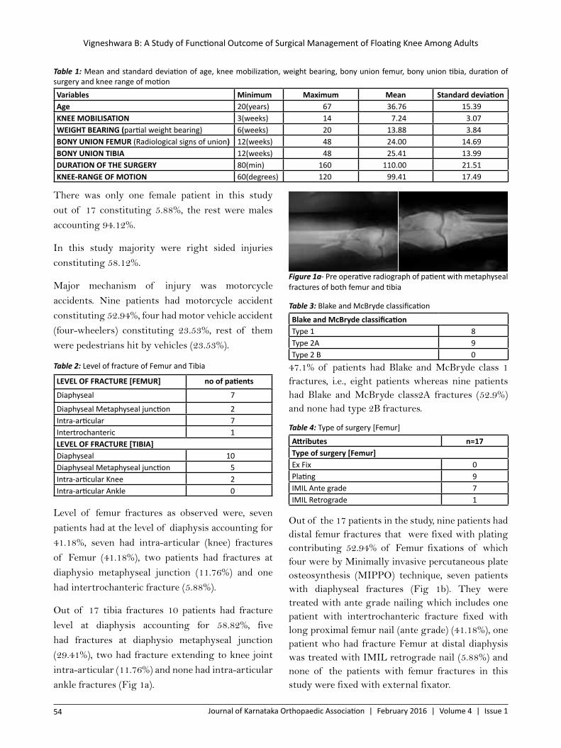
Vigneshwara B: A Study of Functional Outcome of Surgical Management of Floating Knee Among Adults
54 Journal of Karnataka Orthopaedic Association | February 2016 | Volume 4 | Issue 1
There was only one female patient in this study out of 17 constituting 5.88%, the rest were males accounting 94.12%.
In this study majority were right sided injuries constituting 58.12%.
Major mechanism of injury was motorcycle accidents. Nine patients had motorcycle accident constituting 52.94%, four had motor vehicle accident (four-wheelers) constituting 23.53%, rest of them were pedestrians hit by vehicles (23.53%).
Table 2: Level of fracture of Femur and Tibia
LEVEL OF FRACTURE [FEMUR] no of patients
Diaphyseal 7
Diaphyseal Metaphyseal junction 2Intra-articular 7Intertrochanteric 1LEVEL OF FRACTURE [TIBIA]Diaphyseal 10Diaphyseal Metaphyseal junction 5Intra-articular Knee 2Intra-articular Ankle 0
Level of femur fractures as observed were, seven patients had at the level of diaphysis accounting for 41.18%, seven had intra-articular (knee) fractures of Femur (41.18%), two patients had fractures at diaphysio metaphyseal junction (11.76%) and one had intertrochanteric fracture (5.88%).
Out of 17 tibia fractures 10 patients had fracture level at diaphysis accounting for 58.82%, five had fractures at diaphysio metaphyseal junction (29.41%), two had fracture extending to knee joint intra-articular (11.76%) and none had intra-articular ankle fractures (Fig 1a).
Figure 1a- Pre operative radiograph of patient with metaphyseal fractures of both femur and tibia
Table 3: Blake and McBryde classification
Blake and McBryde classificationType 1 8Type 2A 9Type 2 B 0
47.1% of patients had Blake and McBryde class 1 fractures, i.e., eight patients whereas nine patients had Blake and McBryde class2A fractures (52.9%) and none had type 2B fractures.
Table 4: Type of surgery [Femur]
Attributes n=17Type of surgery [Femur]Ex Fix 0Plating 9IMIL Ante grade 7IMIL Retrograde 1
Out of the 17 patients in the study, nine patients had distal femur fractures that were fixed with plating contributing 52.94% of Femur fixations of which four were by Minimally invasive percutaneous plate osteosynthesis (MIPPO) technique, seven patients with diaphyseal fractures (Fig 1b). They were treated with ante grade nailing which includes one patient with intertrochanteric fracture fixed with long proximal femur nail (ante grade) (41.18%), one patient who had fracture Femur at distal diaphysis was treated with IMIL retrograde nail (5.88%) and none of the patients with femur fractures in this study were fixed with external fixator.
Table 1: Mean and standard deviation of age, knee mobilization, weight bearing, bony union femur, bony union tibia, duration of surgery and knee range of motionVariables Minimum Maximum Mean Standard deviationAge 20(years) 67 36.76 15.39KNEE MOBILISATION 3(weeks) 14 7.24 3.07WEIGHT BEARING (partial weight bearing) 6(weeks) 20 13.88 3.84BONY UNION FEMUR (Radiological signs of union) 12(weeks) 48 24.00 14.69BONY UNION TIBIA 12(weeks) 48 25.41 13.99DURATION OF THE SURGERY 80(min) 160 110.00 21.51KNEE-RANGE OF MOTION 60(degrees) 120 99.41 17.49
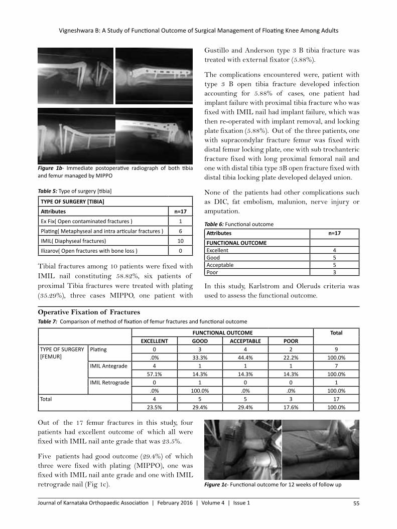
Vigneshwara B: A Study of Functional Outcome of Surgical Management of Floating Knee Among Adults
Journal of Karnataka Orthopaedic Association | February 2016 | Volume 4 | Issue 1 55
Figure 1b- Immediate postoperative radiograph of both tibia and femur managed by MIPPO
Table 5: Type of surgery [tibia]
TYPE OF SURGERY [TIBIA]
Attributes n=17
Ex Fix( Open contaminated fractures ) 1
Plating( Metaphyseal and intra articular fractures ) 6
IMIL( Diaphyseal fractures) 10
Ilizarov( Open fractures with bone loss ) 0
Tibial fractures among 10 patients were fixed with IMIL nail constituting 58.82%, six patients of proximal Tibia fractures were treated with plating (35.29%), three cases MIPPO, one patient with
Gustillo and Anderson type 3 B tibia fracture was treated with external fixator (5.88%).
The complications encountered were, patient with type 3 B open tibia fracture developed infection accounting for 5.88% of cases, one patient had implant failure with proximal tibia fracture who was fixed with IMIL nail had implant failure, which was then re-operated with implant removal, and locking plate fixation (5.88%). Out of the three patients, one with supracondylar fracture femur was fixed with distal femur locking plate, one with sub trochanteric fracture fixed with long proximal femoral nail and one with distal tibia type 3B open fracture fixed with distal tibia locking plate developed delayed union.
None of the patients had other complications such as DIC, fat embolism, malunion, nerve injury or amputation.
Table 6: Functional outcomeAttributes n=17
FUNCTIONAL OUTCOMEExcellent 4Good 5Acceptable 5Poor 3
In this study, Karlstrom and Oleruds criteria was used to assess the functional outcome.
Operative Fixation of FracturesTable 7: Comparison of method of fixation of femur fractures and functional outcome
FUNCTIONAL OUTCOME TotalEXCELLENT GOOD ACCEPTABLE POOR
TYPE OF SURGERY [FEMUR]
Plating 0 3 4 2 9.0% 33.3% 44.4% 22.2% 100.0%
IMIL Antegrade 4 1 1 1 757.1% 14.3% 14.3% 14.3% 100.0%
IMIL Retrograde 0 1 0 0 1.0% 100.0% .0% .0% 100.0%
Total 4 5 5 3 1723.5% 29.4% 29.4% 17.6% 100.0%
Out of the 17 femur fractures in this study, four patients had excellent outcome of which all were fixed with IMIL nail ante grade that was 23.5%.
Five patients had good outcome (29.4%) of which three were fixed with plating (MIPPO), one was fixed with IMIL nail ante grade and one with IMIL retrograde nail (Fig 1c). Figure 1c- Functional outcome for 12 weeks of follow up
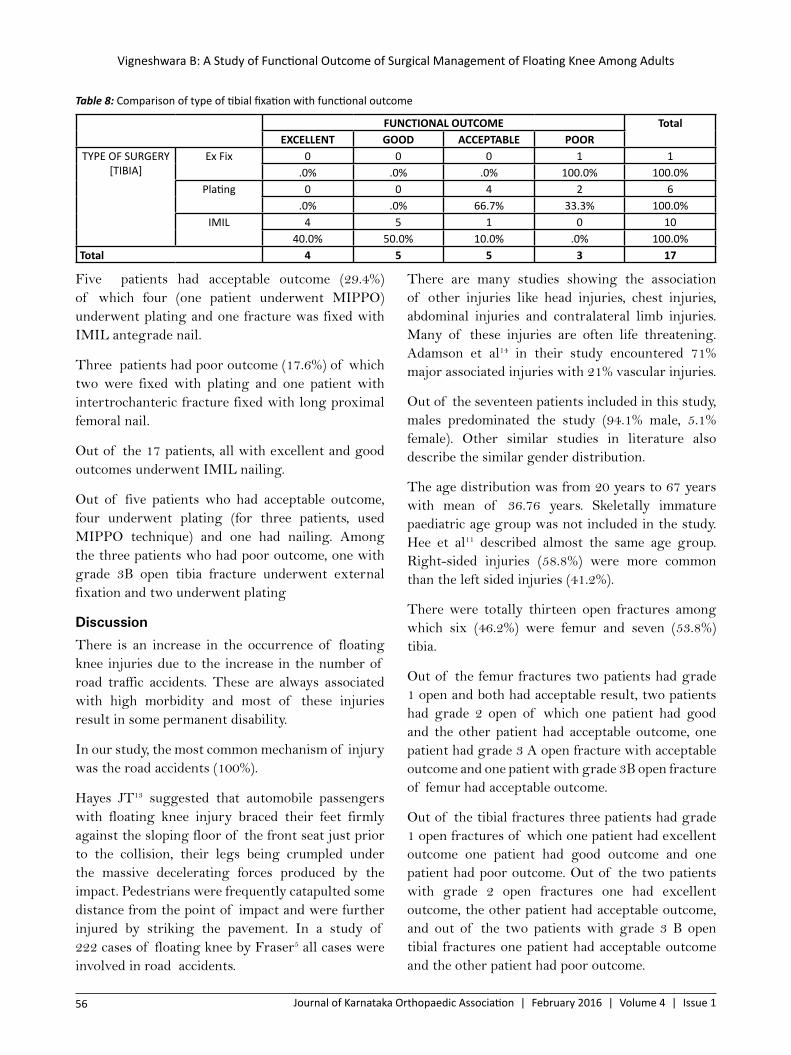
Vigneshwara B: A Study of Functional Outcome of Surgical Management of Floating Knee Among Adults
56 Journal of Karnataka Orthopaedic Association | February 2016 | Volume 4 | Issue 1
Five patients had acceptable outcome (29.4%) of which four (one patient underwent MIPPO) underwent plating and one fracture was fixed with IMIL antegrade nail.
Three patients had poor outcome (17.6%) of which two were fixed with plating and one patient with intertrochanteric fracture fixed with long proximal femoral nail.
Out of the 17 patients, all with excellent and good outcomes underwent IMIL nailing.
Out of five patients who had acceptable outcome, four underwent plating (for three patients, used MIPPO technique) and one had nailing. Among the three patients who had poor outcome, one with grade 3B open tibia fracture underwent external fixation and two underwent plating
DiscussionThere is an increase in the occurrence of floating knee injuries due to the increase in the number of road traffic accidents. These are always associated with high morbidity and most of these injuries result in some permanent disability.
In our study, the most common mechanism of injury was the road accidents (100%).
Hayes JT13 suggested that automobile passengers with floating knee injury braced their feet firmly against the sloping floor of the front seat just prior to the collision, their legs being crumpled under the massive decelerating forces produced by the impact. Pedestrians were frequently catapulted some distance from the point of impact and were further injured by striking the pavement. In a study of 222 cases of floating knee by Fraser5 all cases were involved in road accidents.
There are many studies showing the association of other injuries like head injuries, chest injuries, abdominal injuries and contralateral limb injuries. Many of these injuries are often life threatening. Adamson et al14 in their study encountered 71% major associated injuries with 21% vascular injuries.
Out of the seventeen patients included in this study, males predominated the study (94.1% male, 5.1% female). Other similar studies in literature also describe the similar gender distribution.
The age distribution was from 20 years to 67 years with mean of 36.76 years. Skeletally immature paediatric age group was not included in the study. Hee et al11 described almost the same age group. Right-sided injuries (58.8%) were more common than the left sided injuries (41.2%).
There were totally thirteen open fractures among which six (46.2%) were femur and seven (53.8%) tibia.
Out of the femur fractures two patients had grade 1 open and both had acceptable result, two patients had grade 2 open of which one patient had good and the other patient had acceptable outcome, one patient had grade 3 A open fracture with acceptable outcome and one patient with grade 3B open fracture of femur had acceptable outcome.
Out of the tibial fractures three patients had grade 1 open fractures of which one patient had excellent outcome one patient had good outcome and one patient had poor outcome. Out of the two patients with grade 2 open fractures one had excellent outcome, the other patient had acceptable outcome, and out of the two patients with grade 3 B open tibial fractures one patient had acceptable outcome and the other patient had poor outcome.
Table 8: Comparison of type of tibial fixation with functional outcome
FUNCTIONAL OUTCOME TotalEXCELLENT GOOD ACCEPTABLE POOR
TYPE OF SURGERY [TIBIA]
Ex Fix 0 0 0 1 1.0% .0% .0% 100.0% 100.0%
Plating 0 0 4 2 6.0% .0% 66.7% 33.3% 100.0%
IMIL 4 5 1 0 1040.0% 50.0% 10.0% .0% 100.0%
Total 4 5 5 3 17
Vigneshwara B: A Study of Functional Outcome of Surgical Management of Floating Knee Among Adults
Journal of Karnataka Orthopaedic Association | February 2016 | Volume 4 | Issue 1 57
Out of the overall six open fractures of femur in this study, 83.3% of the subjects had acceptable outcome and 16.7% had good outcome.
Out of the seven patients with open tibial fractures 57.2% of the subjects had poorer outcome (acceptable and poor outcomes) 28.6% of patients had excellent outcome and 14.2% of patients with open tibial fractures had good outcome.
According to Hegazy open fractures are the poor prognostic factors shown by the above data of this study. 15
Four had excellent outcome accounting for 66.7% of transverse femur fractures in this study of six transverse femur fractures. One had good outcome (16.7%), one had acceptable outcome (16.7%) and none had poor outcome.
Out of 11 comminuted fractures in this study four had good outcome 36.4% among all comminuted fractures of this study, four had acceptable outcome (36.4%), three had poor outcome (17.6%) and none of the comminuted fractures had excellent outcome.
Out of the eight transverse fractures of tibia of this study, four had excellent outcome, which was 50% of transverse fractures of tibia of this study and four had good outcome.
Out of the nine comminuted fracture of tibia in this study, none had excellent outcome, one patient had good outcome (11.1%), five had acceptable outcome (55.6%) and three patients had poor outcome (55.3%).
Hee et al11 described in their study that comminuted and segmental fractures were poor predictors of functional outcome.
Out of the 17 patients in this study, eight patients had Blake and McBryde class 1 of which four had excellent outcome accounting for 50% of patients with class 1 Blake and McBryde, two patients had good outcome (25%), one patient had acceptable outcome (12.5%) and one patient had poor outcome (12.5%).
Out of the nine patients who had Blake and McBryde class 2 A, none had excellent outcome, three patients had good outcome which was 33.3% of all class 2
A Blake and McBryde, four patients had acceptable outcome (44.4%) and two patients had poor outcome (22.2%).
Blake and McBryde3 were one of the pioneers to classify the floating knee injuries. After an extensive study, they had classified floating knee injuries as Type-I fractures involving both shafts, Type-2 A- fractures involving the knee joint, Type- IIB- fractures involving the hip or ankle joints.
Fraser et al5 in 1978, studied 222 cases with ipsilateral fractures of the femur and tibia. They found that poor function outcome was seen in intra-articular fractures. Similar results were shown by Bansal et al6.
Comparision of mode of fixation with other studies
There are only few studies in literature, which show specific treatment for floating knee injuries. Initially non-operative management was described by Ul-Haque et al16 in 1983. With the advancement of various operative techniques the management changed. In 1984 Katada et al7 described that both femoral and tibial fractures, must be fixed rigidly. In 1986 Letts et al1 described that at least one fracture should be rigidly fixed either internally or externally, usually the femur. Femoral fixation and non-operative management for associated ipsilateral tibial fractures by plaster of paris was described by Bansal et al6 in 1984. Flexible intramedullary nails were described by Behr et al8 in 1987. Soft tissue sparing surgery like percutaneous plating was described by Lobenhoffer9 1996. Single incision nailing for both tibia and femur was described by Rethnam et al10 in 2006. Dwyer et al 200517 described that the preferred method of fixation in both femoral and tibial diaphyseal fracture was intra-medullary nailing.
Out of the 17 femur fractures in this study, four patients had excellent outcome fixed with IMIL ante grade nail, which was 23.5%.
Out of the five patients who had good outcome (29.4%) three were fixed with plating ( by MIPPO technique), one was fixed with IMIL nail ante grade and one with IMIL retrograde nail.

Vigneshwara B: A Study of Functional Outcome of Surgical Management of Floating Knee Among Adults
58 Journal of Karnataka Orthopaedic Association | February 2016 | Volume 4 | Issue 1
Among the five patients who had acceptable outcome (29.4%), four underwent plating and one fracture was fixed with IMIL ante grade nail.
Among the three patients who had poor outcome (17.6%), two were fixed with plating and one with intertrochanteric fracture fixed with long proximal femoral nail. Hence, among the femoral fixations, ante grade Intra-meduallary nailing has a better prognosis.
Out of the 17 patients in the study, four patients had excellent outcome and all underwent IMIL nailing as the modality of fixation of tibia, which was 40% of all IMIL nailing of this study.
Five patients had good outcome and all of them underwent IMIL nailing as modality of fixation (50%).
Out of the five patients who had acceptable outcome, four patients had undergone plating (three were by MIPPO) as a definitive fixation for tibia (66.7% of all plating of tibia in this study) and one patient had nailing as modality of fixation (10% of all IMIL nailing of tibia in this study).
Out of the three patients who had poor outcome, one patient with grade 3B open tibia fracture underwent external fixation as modality of treatment (100% of tibial external fixation in this study) and two patients underwent plating as the modality of fixation (33.3% of tibial plating in this study). The complications were found to be more in open fractures and comminuted fractures.
Post operative complication
One patient each had infection and implant failure, and three patients had delayed union. None of the patients had DIC, fat embolism, malunion, nerve injury or amputation.
One patient with grade 3 B open tibia fracture was fixed with external fixator developed implant infection and had poor functional outcome.
One patient had proximal tibia fracture fixed with high-bend tibia nail had implant failure and was re-operated with implant removal and plating.
Three patients had developed delayed union One with distal femur fracture fixed with distal femur locking plate had poor functional outcome. Another with sub trochanteric fracture femur fixed with long proximal femoral nail developed delayed union. The same patient also had grade 3B open tibia fracture which was managed with external fixator had developed implant infection and had poor outcome. One patient with distal tibia fracture, grade 3 B open for whom split skin grafting was done as a wound coverage procedure and fracture fixed with distal tibia locking plate developed delayed union and had acceptable functional outcome.
In a study done by Hee et al11 encountered post operative complications pertaining to operative fixation of 89 fractures included wound infection (16 cases), osteomyelitis (four cases) and fat embolism six cases and also he noted 60 patients had delayed union and of these 28 cases went on to non union.
In a study done by Alaa15 also encountered complications like knee stiffness in two patients, delayed union of tibia in two patients and superficial infection in one patient
Table 9: Range of motion kneeDegree No of patients Percentage0-120 5 29.40-110 2 11.80-100 3 17.60-90 4 23.50-80 2 11.80-60 1 5.9Total 17 100
The knee range of motion was an important criterion for the functional outcome.
The maximum achieved range was 0-120 and the minimum was 0-60.
The average range of motion was 0-99 degree.
ConclusionFloating knee injuries are due to high velocity trauma. Road accidents particularly two-wheeler accidents are the commonest of the causes. The most important factors that determine the functional outcomes were the type of fractures (open or closed), nature of comminution including intra-articular
Vigneshwara B: A Study of Functional Outcome of Surgical Management of Floating Knee Among Adults
Journal of Karnataka Orthopaedic Association | February 2016 | Volume 4 | Issue 1 59
extensions, timing of fixations and postoperative infections. Patients who undergo primary nailing will have Excellent or Good results.
Post-operative complications like, infection, delayed union and implant failure were observed. Poor outcomes were mainly due to open fractures, comminution and intra-articular extensions.
Karlstorm and Olerud criterion is an effective scoring system to grade the functional outcome of floating knee injuries.
References1. Letts M, Vincent N, Gouw G. The “floating
knee” in children. J Bone Joint Surg Br. 1986 May; 68(3):442-46
2. Veith RG, Winquist RA, Hansen ST Jr. Ipsilateral fractures of the femur and tibia. A report of fifty-seven consecutive cases. J Bone Joint Surg Am.1984 Sep; 66(7): 991-1002
3. Blake R, McBryde A Jr. The floating knee: Ipsilateral fractures of the tibia and femur. South Med J.1975 Jan; 68(1):13-6.
4. Karlstrom G, Olerud S. Ipsilateral fracture of the femur and tibia. J Bone Joint Surg Am. 1977 Mar; 59(2):240-43.
5. Fraser RD, Hunter GA, Waddell JP. Ipsilateral fracture of the femur and tibia. J Bone Joint Surg Br.1978 Nov; 60-B (4):510-15.
6. Bansal VP, Singhal V, Mam MK, Gill SS. The floating knee. 40 cases of ipsilateral fractures of the femur and the tibia. Int Orthop. 1984; 8(3):183-87.
7. Katada S, Ando K, Nakagawa K, Yamada T, Sasamoto H, Kawabe N et al. Floating knee fracture (ipsilateral fracture of the femur and tibia)--treatment by closed Ender nailing. Nippon Seikeigeka Gakkai Zasshi.1984 May; 58(5):475-83.
8. Behr JT, Apel DM, Pinzur MS, Dobozi WR, Behr MJ. Flexible intramedullary nails for ipsilateral femoral and tibial fractures. J Trauma.1987 Dec; 27(12):1354-57.
9. Lobenhoffer P, Krettek C, Tscherne H. [Complex knee joint trauma] Orthopade. 1997 Dec; 26(12):1037-45.
10. Rethnam U. Single incision nailing of the floating knee-do we ignore the knee ligaments? Int Orthop. 2006 Aug; 30(4):311.
11. Hee HT, Wong HP, Low YP, Myers L. Predictors of outcome of floating knee injuries in adults: 89 patients followed for 2-12 years. Acta Orthop Scand. 2001 Aug; 72(4):385-94.
12. Gustilo RB, Anderson JT. Prevention of infection in the treatment of one thousand and twenty-five open fractures of long bones: retrospective and prospective analyses. J Bone Joint Surg Am. 1976 Jun; 58 (4):453-58.
13. Hayes JT: Multiple fractures in same extremity: Some problems in their management. Surgical Clinics of North America. 1961; 41: 1379-88.
14. Adamson GJ, Wiss DA, Lowery GL, Peters CL. Type II floating knee: ipsilateral femoral and tibial fractures with intra-articular extension into the knee joint. J Orthop Trauma. 1992; 6(3):333-39.
15. Hegazy AM. Surgical management of ipsilateral fracture of the femur and tibia in adults (the floating knee): Postoperative clinical, radiological, and functional outcome. Clin Orthop Surg. 2011 Jun; 3 (2):133-9.
16. Ul-Haque I. A floating knee treated conservatively: a case report. Injury. 1983 May; 14(6):554-57.
17. Dwyer AJ, Paul R, Mam MK, Kumar A, Gosselin RA. Floating knee injuries: long-term results of four treatment methods. Int Orthop 2005 Oct; 29(5):314-18.
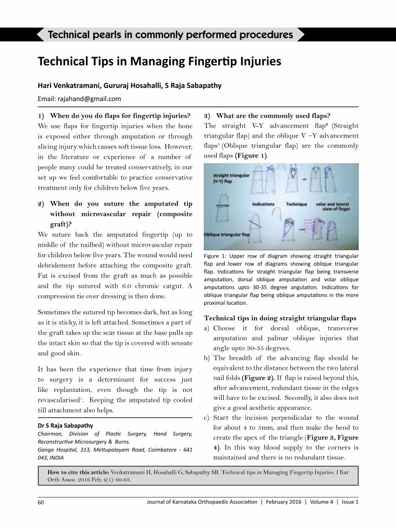
Sabapathy SR: Technical tips in Managing Fingertip Injuries
60 Journal of Karnataka Orthopaedic Association | February 2016 | Volume 4 | Issue 1
Dr S Raja SabapathyChairman, Division of Plastic Surgery, Hand Surgery, Reconstructive Microsurgery & Burns.Ganga Hospital, 313, Mettupalayam Road, Coimbatore - 641 043, INDIA
Technical Tips in Managing Fingertip Injuries
Hari Venkatramani, Gururaj Hosahalli, S Raja Sabapathy
Email: [email protected]
How to cite this article: Venkatramani H, Hosahalli G, Sabapathy SR. Technical tips in Managing Fingertip Injuries. J Kar Orth Assoc. 2016 Feb; 4(1): 60-63.
Technical pearls in commonly performed procedures
1) When do you do flaps for fingertip injuries?We use flaps for fingertip injuries when the bone is exposed either through amputation or through slicing injury which causes soft tissue loss. However, in the literature or experience of a number of people many could be treated conservatively, in our set up we feel comfortable to practice conservative treatment only for children below five years.
2) When do you suture the amputated tip without microvascular repair (composite graft)?
We suture back the amputated fingertip (up to middle of the nailbed) without microvascular repair for children below five years. The wound would need debridement before attaching the composite graft. Fat is excised from the graft as much as possible and the tip sutured with 6.0 chromic catgut. A compression tie over dressing is then done.
Sometimes the sutured tip becomes dark, but as long as it is sticky, it is left attached. Sometimes a part of the graft takes up the scar tissue at the base pulls up the intact skin so that the tip is covered with sensate and good skin.
It has been the experience that time from injury to surgery is a determinant for success just like replantation, even though the tip is not revascularised1. Keeping the amputated tip cooled till attachment also helps.
3) What are the commonly used flaps?The straight V-Y advancement flap2 (Straight triangular flap) and the oblique V –Y advancement flaps3 (Oblique triangular flap) are the commonly used flaps (Figure 1).
Figure 1: Upper row of diagram showing straight triangular flap and lower row of diagrams showing oblique triangular flap. Indications for straight triangular flap being transverse amputation, dorsal oblique amputation and volar oblique amputations upto 30-35 degree angulation. Indications for oblique triangular flap being oblique amputations in the more proximal location.
Technical tips in doing straight triangular flapsa) Choose it for dorsal oblique, transverse
amputation and palmar oblique injuries that angle upto 30-35 degrees.
b) The breadth of the advancing flap should be equivalent to the distance between the two lateral nail folds (Figure 2). If flap is raised beyond this, after advancement, redundant tissue in the edges will have to be excised. Secondly, it also does not give a good aesthetic appearance.
c) Start the incision perpendicular to the wound for about 4 to 5mm, and then make the bend to create the apex of the triangle (Figure 3, Figure 4). In this way blood supply to the corners is maintained and there is no redundant tissue.

Sabapathy SR: Technical tips in Managing Fingertip Injuries
Journal of Karnataka Orthopaedic Association | February 2016 | Volume 4 | Issue 1 61
Figure 2: Technique of straight triangular flap is to mark distance ab, the width of the nail plate equal to cd, which is the advancing edge of the flap e, is the apex of the straight triangle that ends at the DIP joint crease. cde is the flap which is advanced as shown.
Figure 3: Skin incision for straight triangular flap: wrong and right method.
Figure 4: Patient who came with transverse amputation of right thumb at distal nail bed level (a and b) with immediate post-operative views (c and d)
d) After making an incision, the flap is pulled distally and a sharp 15 knife is pressed into the wound to divide the fibrous septa. The most important constraint to advancement is situated in the apex of the triangle. As these fibrous septa get divided the flap advances well.
e) In addition, distal few millimeters of attachment of the flap to the base is freed so that suturing is easy.
f) The common cause of the problem is inadequate mobilization and tight suturing of the flap to the nail bed or the skin. Tight suturing also causes hook nail deformity.
Technical tips for doing a good oblique triangular flapa. In the oblique triangular flap, one limb of the
triangle is in the mid lateral line and the other limb starts at a distance equivalent to the distance between the lateral nail folds.
b. The key to success is adequate mobilization. The oblique triangular flap is based on the digital neurovascular bundle. The side mobilization is performed by dividing the Cleland ligament, which is nicely seen if the wound is kept in stretched position (Fig 1, Fig 5, Fig 6, and Fig 7).
c. Mobilization of the other limb is done by keeping the flap stretched and dividing the tight fibrous septa with the knife. One to 1.5 cm of mobilization can easily be achieved by this technique.
Figure 5: Anatomy for oblique triangular flap showing Cleland’s Ligament (shown with large black arrow) and the need to cut it for mobilizing the flap. FDS: Flexor Digitorum Superficialis FDP: Flexor Digitorum Profundus.
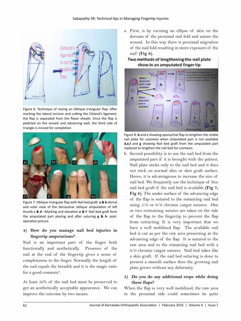
Sabapathy SR: Technical tips in Managing Fingertip Injuries
62 Journal of Karnataka Orthopaedic Association | February 2016 | Volume 4 | Issue 1
Figure 6: Technique of raising an Oblique triangular flap: After marking the lateral incision and cutting the Cleland’s ligament, the flap is separated from the flexor sheath. Once the flap is pedicled on the vessels and advancing well, the third side of triangle is incised for completion
Figure 7: Oblique triangular flap with Nail bed graft: a & b-dorsal and volar view of the dorsoulnar oblique amputation of left thumb c & d –Marking and elevation e & f- Nail bed graft form the amputated part placing and after suturing g & h- post-operative picture.
4) How do you manage nail bed injuries in fingertip amputations?
Nail is an important part of the finger both functionally and aesthetically. Presence of the nail at the end of the fingertip gives a sense of completeness to the finger. Normally the length of the nail equals the breadth and it is the magic ratio for a good cosmesis4.
At least 50% of the nail bed must be preserved to get an aesthetically acceptable appearance. We can improve the outcome by two means:
a. First, is by excising an ellipse of skin on the dorsum of the proximal nail fold and suture the wound. In this way there is proximal migration of the nail fold resulting in more exposure of the nail5 (Fig 8).
Figure 8: b and c showing eponychial flap to lengthen the visible nail plate for cosmesis when amputated part is not available d,e,f and g showing Nail bed graft from the amputated part replaced to lengthen the nail bed for cosmesis.b. Second possibility is to use the nail bed from the
amputated part if it is brought with the patient. Nail plate sticks only to the nail bed and it does not stick on normal skin or skin graft surface. Hence, it is advantageous to increase the size of nail bed. We frequently use the technique of free nail bed graft if the nail bed is available (Fig 7, Fig 8). The under surface of the advancing edge of the flap is sutured to the remaining nail bed using 5/0 or 6/0 chromic catgut sutures. One or two restraining sutures are taken on the side of the flap to the fingertip to prevent the flap from retracting. It is very important that we have a well mobilized flap. The available nail bed is cut as per the raw area presenting at the advancing edge of the flap. It is sutured to the raw area and to the remaining nail bed with a 6/0 chromic catgut sutures. Nail bed takes like a skin graft. If the nail bed suturing is done to present a smooth surface then the growing nail plate grows without any deformity.
5) Do you do any additional steps while doing these flaps?
When the flap is very well mobilized, the raw area in the proximal side could sometimes be quite

Sabapathy SR: Technical tips in Managing Fingertip Injuries
Journal of Karnataka Orthopaedic Association | February 2016 | Volume 4 | Issue 1 63
significant. If the patient has brought the amputated fingertip, the skin is extensively defatted to leave only the dermis. It is cut to the size of the raw area and sutured to the raw area. In this way, the wound heals much better and there is less amount of pain while changing the dressing. If the amputated fingertip is not available a small graft could be shaved from the hypothenar eminence and used.
References:1. Elliot D, Moiemen N. Composite graft
replacement of digital tips1. Before 1850 and after 1950. The Journal of Hand Surgery: Journal of the British Society for Surgery of the Hand. 1997 Jun; 22(3):341–345.
2. Atasoy E, Lokimidis E, Kasdan ML, Kutz JE, Kleinert HE. Reconstruction of the amputated fingertip with a triangular volar flap-a new surgical procedure. Plastic and Reconstructive Surgery. 1971; 48(5):215.
3. Venkataswami R, Subramanian N. Oblique triangular flap. Plastic and Reconstructive Surgery. 1980; 66(2):296–300.
4. Baran R, Maibach HI, editors. Textbook of cosmetic dermatology. 4th ed. New York: Informa Healthcare; 2010. 247 p. ISBN: 9781841847009.
5. Bakhach J. Eponychial flap. Ann Chir Plast Esthet. 1998 Jun; 43(3):259-63

Shekar.M, et al: Solitary Osteochondroma Of Scaphoid: A Case Report
64 Journal of Karnataka Orthopaedic Association | February 2016 | Volume 4 | Issue 1
AbstractWe present a case of 22 year male cricket player, who camewith pain in the wrist, which increased on bowling, to an extent that he had to stop playing cricket. Plain radiograph of wrist suggested an osseous mass in the distal pole of scaphoid. CT scan of wrist revealed the presence of an osteocartilagenous lesion in the distal pole of the scaphoid. This was resected and histology showed it to be an osteochondroma.
Keywords: scaphoid, osteochondroma
Dr.Shekar. M, DNB(Ortho)No-776, 7th main, Brindavannagar, Bengaluru-19
Solitary Osteochondroma of Scaphoid: A Case Report
Shekar M, Mahadev Jatti, Bharath Kadadi
Email: [email protected]
How to cite this article: Shekar M, Jatti M, Kadadi B. Solitary Osteochondroma Of Scaphoid: A Case Report. J Kar Orth Assoc. 2016 Feb; 4(1): 64-66.
Case Report
Introduction
Osteochondromasare more likely to involve the phalanx or metacarpal bones when it occurs in the hand (1,2). Involvement of the scaphoid is uncommon (1). The lesion may cause localised pain if it is extensive, but, in most cases the tumor is discovered incidentally (3). We describe a rare occurrence of osteochondroma in the scaphoid. The patient and his family were informed that the data from the case would be submitted for publication and their consent taken.
Case Report
A 22 year old male IPL (Indian Premiere League) cricket player came with pain in the dominant right wrist of three month duration in May 2011. Pain increased in intensity on extension and radial deviation of wrist, which was the position while bowling. This led to impairment in his sports activity. On examination the patienthad a tender bony mass of 0.5cm × 0.5cm in the anatomical snuff box. He had wrist extension of 400 flexion 500 ulnar deviation of 200 and radial deviation of 50. Pinch and grip strengths were normal. No previous event of trauma was noted. Plain x-ray of wrist with hand AP and scaphoid view showed abnormal
bony protrusion on the dorsoradial aspect of distal pole of scaphoid (Figure 1).
Figure 1: Radiographs of right wrist
CT scan with 3D reconstruction image revealed “mushroom” shaped osteocartilagenous mass arising from distal pole of scaphoid (Fig 2 and 3).
A tentative diagnosis of tumor mass was made and surgical excision was planned. The scaphoid was exposed through a dorso-radial incision (1). The projecting bony lesion which was impinging on the distal articular surface of radius on wrist extension and radial deviation was demonstrated on table (Fig 4). The bony mass measuring 1cm ×

Shekar.M, et al: Solitary Osteochondroma Of Scaphoid: A Case Report
Journal of Karnataka Orthopaedic Association | February 2016 | Volume 4 | Issue 1 65
0.5cm was completely excised (Fig 5). Histological examination showed proliferation of cartilage and bone consistent with osteochondroma. At follow up, threemonths after this operation, the patient had gained full range of movements at the wrist joint and went back to playing cricket. At one year follow up, he did not have any symptoms.
Figure 2: CT scan of wrist
Figure 3: 3D CTreconstruction of the scaphoid
Figure 4: intraoperative finding
Figure 5: Osteochondroma after excision
DiscussionOsteochondromais one of the most common benign skeletal tumorscharacterised by a bony projection capped by cartilage (1). It consists of a bony prominence covered by a cartilaginous cap. Solitary osteochondroma are rare in the hand and are more likely to be found in the phalanx or metacarpal (1, 4-5). Osteochondromas were brought to the attention of the patient and physician. These may have occurred due to the injury as they do not occur due to trauma. In such cases, patients usually come with a palpable painless mass. Symptoms occur due to compression of the overlying soft tissue and subsequent restriction of motion. Malignant transformation is observed in 1% of solitary tumors beingmore commonin multiple presentations (1,2)
Osteochondroma usually originates from the epiphyses of longitudinally growing bones. The carpal bones develop from centrifugally expanding centres of ossification and hence origin from these bones may have a different etiology (2). Malhotra et al suggests that in such cases they represents a true neoplasm rather than a developmental defect (2,6).
Differential diagnosis such cases include scapholunate dissociation (2,7-8) carpal coalition (4, 9), scaphoid non-union, radiocarpal arthritis and Dequervain’s disease.
Medlar RC et al and Shalom Stahl et al both suggest excision of these lesions when they restrict joint motion (1,2).
ConclusionOsteochondroma of scaphoid is a rare entity and hardly reported in Indian literature. It usually startswith a localised pain and the diagnosis is sometimes missed. It is not caused by trauma, but often coincides with the history of trauma. Symptoms

Shekar.M, et al: Solitary Osteochondroma Of Scaphoid: A Case Report
66 Journal of Karnataka Orthopaedic Association | February 2016 | Volume 4 | Issue 1
occur due to compression of the neighbouring tissues.CT scan with 3D reconstruction is an effective tool of investigation. It is cured by surgical excision and rarely transforms into malignancy.
References1. Medlar RC, Sprague HH.Osteochondroma of
the carpal scaphoid.. J Hand Surg Am. 1979 Mar;4(2):150-1
2. Stahl S, Rayek S. Scaphoid osteochondroma with scapholunate dissociation- a case report. Hand Surg.2000 Jul;5(1):73-5
3. Uchida K, Kobayashi S, Takamura T, Yayama T, Inukai T, Baba H.Osteochondroma arising from the scaphoid. J Orthop Sci.2007 Jul;12(4):381-4
4. Rice J, Stephens M, Colville JJ.Scaphoid osteochondroma mimicking carpal coalition. Hand Surg Br 1996 Dec;21(6):779-80
5. Curtis RM, Wigis E F S,Moore J R.Osteocartilaginous lesions of the digits in children: An experience with ten cases. J Hand Surg 1983;8:309-315
6. Malhotra R, Maheshwari J, Dinda AK.A solitary osteochondroma of the capitate bone. J Hand Surgery 1992;17A:1082-1083
7. Van Alphen JC, te Slaa RL, Eulderink F, Obermann WR. Solitary osteochondroma of the scaphoid: a case report. J Hand Surg Am.1996 May;21(3):423-5
8. De Smet L, Degreef I. Bilateral osteochondroma of the scaphoid causing scapholunate dissociation: a case report. Chir Main.2007 Jun;26(3):141-2
9. Barfred T J. Scaphoid osteochondroma. Hand Surg Br. 1997 Dec;22(6):825-6
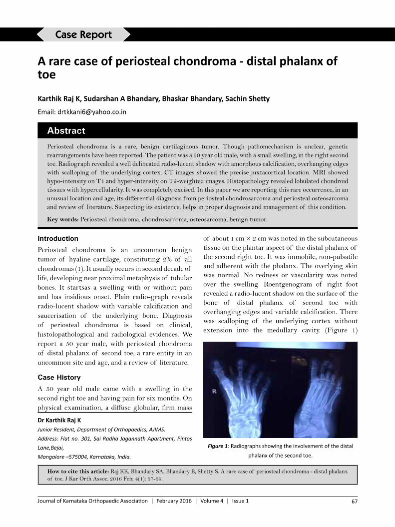
Karthik Raj K, et al: A rare case of periosteal chondroma - distal phalanx of toe
Journal of Karnataka Orthopaedic Association | February 2016 | Volume 4 | Issue 1 67
AbstractPeriosteal chondroma is a rare, benign cartilaginous tumor. Though pathomechanism is unclear, genetic rearrangements have been reported. The patient was a 50 year old male, with a small swelling, in the right second toe. Radiograph revealed a well delineated radio-lucent shadow with amorphous calcification, overhanging edges with scalloping of the underlying cortex. CT images showed the precise juxtacortical location. MRI showed hypo-intensity on T1 and hyper-intensity on T2-weighted images. Histopathology revealed lobulated chondroid tissues with hypercellularity. It was completely excised. In this paper we are reporting this rare occurrence, in an unusual location and age, its differential diagnosis from periosteal chondrosarcoma and periosteal osteosarcoma and review of literature. Suspecting its existence, helps in proper diagnosis and management of this condition.
Key words: Periosteal chondroma, chondrosarcoma, osteosarcoma, benign tumor.
A rare case of periosteal chondroma - distal phalanx of toe
Karthik Raj K, Sudarshan A Bhandary, Bhaskar Bhandary, Sachin Shetty
Email: [email protected]
How to cite this article: Raj KK, Bhandary SA, Bhandary B, Shetty S. A rare case of periosteal chondroma - distal phalanx of toe. J Kar Orth Assoc. 2016 Feb; 4(1): 67-69.
Case Report
Introduction
Periosteal chondroma is an uncommon benign tumor of hyaline cartilage, constituting 2% of all chondromas (1). It usually occurs in second decade of life, developing near proximal metaphysis of tubular bones. It startsas a swelling with or without pain and has insidious onset. Plain radio-graph reveals radio-lucent shadow with variable calcification and saucerisation of the underlying bone. Diagnosis of periosteal chondroma is based on clinical, histolopathological and radiological evidences. We report a 50 year male, with periosteal chondroma of distal phalanx of second toe, a rare entity in an uncommon site and age, and a review of literature.
Case History
A 50 year old male came with a swelling in the second right toe and having pain for six months. On physical examination, a diffuse globular, firm mass
of about 1 cm × 2 cm was noted in the subcutaneous tissue on the plantar aspect of the distal phalanx of the second right toe. It was immobile, non-pulsatile and adherent with the phalanx. The overlying skin was normal. No redness or vascularity was noted over the swelling. Roentgenogram of right foot revealed a radio-lucent shadow on the surface of the bone of distal phalanx of second toe with overhanging edges and variable calcification. There was scalloping of the underlying cortex without extension into the medullary cavity. (Figure 1)
Figure 1: Radiographs showing the involvement of the distal phalanx of the second toe.
Dr Karthik Raj KJunior Resident, Department of Orthopaedics, AJIMS.Address: Flat no. 301, Sai Radha Jagannath Apartment, Pintos Lane,Bejai,Mangalore –575004, Karnataka, India.

Karthik Raj K, et al: A rare case of periosteal chondroma - distal phalanx of toe
68 Journal of Karnataka Orthopaedic Association | February 2016 | Volume 4 | Issue 1
osteosarcoma shows perpendicular spicules and a peripheral Codman’s triangle (6). Laboratory tests ruled out systemic conditions like infections. CT images showed a precise juxtacortical location differentiating from osteochondroma, osteoid osteoma and myositis ossificans, but not from periosteal osteosarcoma. MRI showed hypo-intensity on T1 and hyper-intensity on T2-weighted images reflecting cartilaginous origin (7). Extra-osseous soft tissue oedema was seen adjacent to the lesion on T2-weighted images. Intravenous gadolinium administration may demonstrate peripheral enhancement though not done in our case. Isotope scans show enhanced uptake for periosteal chondrosarcomas than for periosteal chondromas. Lorento et al (1,7) noted that CT and MRI provide better means for diagnosis.Histopathology, usually shows lobulated chondroid tissues covered by periosteum, with hypocellularity, sometimes hyper-cellular with nuclear pleomorphism, bi-nucleation, multi-nucleation, atypia and focal myxoid degeneration is seen, and can be misdiagnosed as chondrosarcoma (Figure 3) (8).
Figure 3: Histopathology showinglobulated chondroid tissues
covered by periosteum, with hypocellularity
For asymptomatic cases only observation is necessary. Treatment includes intralesional, marginal and en bloc excision. Lewis reported a recurrence of 3.6% and Jerome 5.5 % and was mainly due to incomplete excision (9,10). To avoid recurrences, marginal excision and curettage of the underlying cortical bone has been suggested.
Sonography showed an ill-defined hypo-echoic soft tissue lesion of about 1.4 cm × 0.6 cm, adjacent to the distal phalanx of right second toe with cortical irregularity of the distal phalanx. CT images showed the precise juxtacortical location. MRI showed hypo-intensity on T1 and hyper-intensity on T2-weighted images. There was no evidence of increased vascularity involving the lesion. Pre-operative laboratory tests were normal. Excision biopsy was done under regional anaesthesia.
Gross specimen was dull white in color.(Figure 2)
Figure 2: Showing the dull white gross specimen.
Histopathology revealed irregular well defined lobules of mature cartilaginous tissue with abundant pale hyaline matrix along with small groups of chondrocytes and thin fibrovascular stroma. Based on clinical, radiological and histopathological findings, a diagnosis of periosteal chondroma was made. Postoperative period was uneventful.
Discussion:
Surface type of bone tumors usually originate from the periosteum. These are periosteal chondroma, periosteal chondrosarcoma, periosteal osteosarcoma, and surface high-grade osteosarcoma. These should be differentiated from primary skin and subcutaneous tumors. Periosteal chondroma arising from the periosteum accounts for only 2.2% of benign tumors and 0.5% of all tumors in the Mayo clinic series. Brien et al (2-4) reported them in 1.3% of all cartilage tumors. Isolated cases were described by McWhorter in 1922 and by Keiller (2-4). In 1925. Keiller called it as subperiosteal

Karthik Raj K, et al: A rare case of periosteal chondroma - distal phalanx of toe
Journal of Karnataka Orthopaedic Association | February 2016 | Volume 4 | Issue 1 69
Conclusion
This case demonstrates the clinical, radiological and histological features of periosteal chondroma, a rare tumor, in an unusual site and age. Knowledge of its characteristic features would lead us to accurate diagnosis, thereby avoiding unnecessary radical surgery.
References1. Parodi KK, Farrett W, Paden MH, Stone PA. A
report of a rare phalangeal periosteal chondroma of the foot. J Foot Ankle Surg. 2011;50:122-5.
2. Lichtenstein L, Hall J. Periosteal chondroma: a distinctive benign cartilage tumour. J Bone Joint Surg Am 1952; 34-A: 691-7.
3. Luevitoonvechkij S, Arphornchayanon O, Leerapun T, Khunsree S. Periosteal chondroma of the Proximal Humerus: A Case Report and Review of the Literature. J Med Assoc Thai 2006; 89:1970-5.
4. Al-Qudah AS, Abu-Ali HM, Al-Hussaini MA, Massad IM. Periosteal chondroma of the clavicle: Case report and review of the literature. International Journal of Surgery 2009;7:140–141.
5. Hagiwara Y, Hatori M, Abe A, Tanaka K, Kokubun S. Periosteal chondroma of the fifth toe – A case report. Upsala J Med Sci 2004;109: 65–70.
6. Zheng K, Yu X, Xu S, Xu M. Periosteal chondroma of the femur: A case report and review of the literature. Oncology letters 2015; 9: 1637-1640.
7. Yamamoto T,Nagira K, Kurosaka M. Periosteal chondroma presenting as a subcutaneous mass in the thumb. Journal of Clinical Imaging 2001;25: 432–434.
8. Akansu B, Atik E, Altintas S, Kalaci A, Canda S. Periosteal chondroma of the ischium; An unusual location. Turk PatolojiDerg 2012;28:172-174.
9. Lewis MM, Kenan S, Yabut SM, Norman A, Steiner G. Periosteal chondroma. A report of ten cases and review of the literature. ClinOrthopRelat Res 1990; 185-92.
10. Nosanchuk JS, Kaufer H. Recurrent periosteal chondroma. Report of two cases and a review of the literature. J Bone Joint Surg Am 1969; 51: 375-80.

70 Journal of Karnataka Orthopaedic Association | February 2016 | Volume 4 | Issue 1
Master class for Postgraduates
History � Pain:
A) Site-Cervical region, dorsal region, in the dorsolumbar region or lumbar region, in the lumbosacral region or in the sacral region.
B) Mode of onset-Whether the pain started immediately after trauma or lifting weight or during strenuous exercise as seen in prolapsed intervertebral disc
C) Nature of pain-Stabbing –PIVD Continuous and throbbing type in osteomyelitis Intermittent pain –spondylolisthesis Dull ache in Pott’s disease
D) RadiationE) Aggravating factorsF) Relieving factors-restG) Positional varianceH) Rest pain
• Deformity- localized/diffuse ,onset progression
• Swelling – site ,onset,1st noticed, duration, progression
• stiffness of the back• Weakness :
- Duration progression- Unilateral/bilateral- Motor /sensory- Sudden /insidious- Bowel/bladder involvement –early/late
• Restriction of ROM• Abnormality in walking/ gait
disturbances• Disability• Immunization history• Developmental history• Family history• Past exposure to tuberculosis
General examination• Height , weight, gait and posture
Local examinationFrom behind• Head and neck tilt• Level of shoulders• Alignment of spine• Level of iliac crest• Any swelling or features of cold abscess• Any visible step• Any features of Spina bifida( tuft of hair,
neuro-cutaneous markers) From side:
• Exaggerated lumbar lordosis• Shape of the chest wall• Rib humpFrom front:• Level of nipples• Level of ASIS
Palpation:• Tenderness over spinous process• Point tenderness• Twist tenderness• Thrust tenderness• Feel for any step along the spine, cold
abscess
Movements:• All movements of cervical spine, dorsal
spine and lumbar spine• Schober’s test
(* movements to be avoided in patients with neurological deficits or in unstable spine)
Measurements • Chest expansion• Limb length discrepancy
Special tests• SLRT/Lasegue test for lumbar spine• Spurling test for cervical spine• Adson/ Roos test for thoracic outlet
syndrome
Examination of Spine CaseDr M Shantaram ShettyPro Chancellor, NITTE UniversityChairman, Tejasvini Hospital & SSIOT, Mangalore

Journal of Karnataka Orthopaedic Association | February 2016 | Volume 4 | Issue 1 71
Neurological Examination
Higher mental functions
Cranial nerves
Motor system: • Bulk• Tone• Power( MRC Grading)
Sensory system-• Fine touch,• Crude touch• Vibration• Temperature• Joint position sense• 2-point discrimination• Stereo gnosis
Reflexes
Having given in detail the examination of the Spine as the first part which every Postgraduate should know.
As a part of the master class, I would dwell through what are the usual questions asked in a long case of Tuberculosis of Spine at the thoraco lumbar level in a 44 year old male.
History: Have you taken these relevant history?
Symptoms: • Insidious or sudden• Are there any history of night cries and why do
night cries occur? • Character of pain • Does it radiate • Any swelling in the back or inguinal region or
Gluteal region - to rule out cold abscess• What is the neurological status of the patient?
In case patient complains of altered neurology was it gradual or sudden?
Inspection: Have you noted the following points?
Gait: A cautious gait almost on the toes denote a painful spine condition like tuberculosis of spine
Attitude: The patient almost bending the knee and spine denotes a point for tuberculosis
Note the deformity:• A knuckle denotes involvement of a single
vertebra • An angular kyphosis where more than 2 but less
than 4 vertebra are involved • Kyphus deformity is more common in children
as most vertebrae are mostly cartilaginous and destruction occurs anteriorly with normal growth of the posterior segment deformity tends to increase
• Different sites where tuberculosis abscess from T12 – L1 can tracka) Renal Angleb) Petit’s trianglec) Psoas abscessd) Gluteal abscesse) Femoral trianglef) along the vessel s and nerves – related to the diseased vertebra.
Palpation: It is important to know various landmarks in the spine to correlate it with the level of disease.• Most prominent vertebrae in cervical region- C7
• Spine of scapula – corresponds to T3
• Inferior border of scapula- corresponds to T7
• Highest point of iliac crest - Space between L3 - L4
• Dimple on either side- S2 and S1 joint• What are the common levels of involvement of
spinal tuberculosis?Dorsal spine- 35-40%Dorsolumbar spine -25-30%Lumbosacral spine-25-30%Cervical spine- 4-5%
• What are the sites of lesion in the vertebraeParadiscalAnteriorPosteriorCentral
Movements: If it is painful as in tuberculosis, don’t do it but should know all the movements of spine as was described in the examination of spine.
Most important part of examination is the assessment of neurological status.
One should know to differentiate between UMN/LMN lesions

72 Journal of Karnataka Orthopaedic Association | February 2016 | Volume 4 | Issue 1
• What the causes of early onset paraplegia?AbscessInflammatory edemaGranulation tissueSequestrumThrombosis of anterior spinal artery
• What are the causes of late onset paraplegia?Stretching of spinal cord over the internal gibbus
Questions on Management1. What are the investigations in a case of spinal
tuberculosis?Complete blood picture, chest X-ray, Mantoux test, x ray of spine, MRI of spine, Open/ closed biopsy, PCR studies
2. What are the radiological signs of tuberculosis?Osteopenia, disc space reduction, collapse of vertebral body, bird nest and v shaped abscess, widening of psoas shadow.
3. What is the advantage of MRI in tuberculosis of spine?
- Better delineation of soft tissues, abscess, skip lesions, spinal tumour syndrome
4. What are the drugs used in Tuberculosis of spine and how long? Complications of each of the drug.Isoniazid, rifampicin, pyrazinamide and ethambutol for 3 months- intensive phaseIsoniazid, rifampicin and pyrazinamide- 4 months- extension phaseIsoniazid and rifampicin- 11 months- continuation phaseIsoniazid-hepatotoxicity, neuropsychiatricRifampicin- hepatotoxicity, flu like syndromeStreptomycin- ototoxicityPyrazinamide- hyperuricaemiaEthambutol- optic neuritis
5. What is MDR?Multi drug resistance- resistance to isoniazid and rifampicin
6. What are indications for surgery in middle path regimen?- Not responding to TT after 4-8 weeks of
treatment - Progressive neurological deficits developing
while on treatment- Worsening of neurological deficits during
treatment- Kyphosis of more than 30 degree- Spinal tumour syndrome
7. What are the different types of surgery?Costo transversectomyAnterolateral decompressionRadical debridement and arthrodesisHong Kong operation
8. What is costo transversectomy? Posterior ends of ribs and corresponding transverse process excised
9. Anterolateral decompression?In addition to costo transversectomy pedicles and vertebral body excised
10. What are the different approaches? Dorsal spine: anterolateral extra pleural, Trans pleural anteriorCervical spine: anterior Smith Robinson approachAtlantoaxial: Trans oral and Trans thyroidCervico-dorsal: Tran’s pleural thoracotomy, anterior approachThoracolumbar and lumbar- posterior
11. What are the surgical options for treating Dorsolumbar tuberculosis?Anterior decompression and fusionAnterolateral lateral decompressionCosto transversectomyTranspedicular decompression and posterior fusion

JKOAJournal of Karnataka Orthopaedic Association
EditorialWhy and how should we index our journal?Dr. Anil K. Bhat ........................................................1
Review articlesFractures of Lateral Humeral Condyle in ChildrenEdward L Nazareth .................................................3
Lateral End Clavicle Fracture: A Review of Current ConceptsVivek Pandey, Krishna Prasad, Sandesh Madi, Kiran Acharya .........................................................9
Management of High Energy Tibial Plateau FracturesMonappa A Naik, Sharath K Rao ..........................14
Adult Brachial Plexus Injuries – An Appraisal on its Primary ManagementAshwath M Acharya, Anil K Bhat, Mahesh Kulkarni, KN Jayakrishnan ......................21
Modified Stoppa approach for Acetabular Fractures: “Lighting up the tunnel”M Ajith Kumar, Shailesh Pai ..................................31
Original articlesManagement of Open Traumatic Extruded TalusMuthukumar Balaji S, Rohit R K, S Devadoss, A Devadoss .......................................35
Intramedullary Interlocking Nailing in Type II and Type III Open Fractures of Tibia – A Clinical StudyPatil NVP, Bellad S H, Anand S R ...........................39
Surgical Management of Unstable Supracondylar Fractures of Humerus in Children by Closed Reduction and Percutaneous K Wire Fixation by Crossed Versus Triple Pin Configuration-A Comparative Randomized Study
C V Mudgal, S F Kammar, Viresh Murgodi ............44
A Study of Functional Outcome of Surgical Management of Floating Knee Among Adults
Suryakanth Kalluraya, V K Bhasme, Vigneshwara B ......................................................52
Technical pearls in commonly performed procedures
Technical Tips in Managing Fingertip Injuries
Hari Venkatramani, Gururaj Hosahalli, S Raja Sabapathy ..................................................60
Case reports
Solitary Osteochondroma of Scaphoid: A Case Report
Shekar M, Mahadev Jatti, Bharath Kadadi ...........64
A Rare Case of Periosteal Chondroma - Distal Phalanx of Toe
Karthik Raj K, Sudarshan A Bhandary, Bhaskar Bhandary, Sachin Shetty .......................................67
Master class for Postgraduates
Examination of Spine Case
M Shantaram Shetty .............................................70
Volume 4, Issue 1, February 2016