iterative decomposition of water and fat with echo...
TRANSCRIPT
Magnetic Resonance Insights 2008:1 1–6 1
METHODOLOGY
Correspondence: Kenneth L. Weiss, M.D., Associate Professor Radiology and Psychiatry, University of Cincinnati College of, Medicine 234 Goodman Street, PO Box 670762, Cincinnati, OH 45267-0762. Tel: (513) 584-0606 or 584-1584; Fax: (513) 584-9100; Email: [email protected]
Copyright in this article, its metadata, and any supplementary data is held by its author or authors. It is published under the Creative Commons Attribution By licence. For further information go to: http://creativecommons.org/licenses/by/3.0/.
Iterative Decomposition of Water and Fat with Echo Asymmetric and Least—Squares Estimation (IDEAL) (Reeder et al. 2005) Automated Spine Survey Iterative Scan Technique (ASSIST) (Weiss et al. 2006)
Kenneth L. Weiss1, Dongmei Sun1,2, Rebecca S. Cornelius1 and Jane L. Weiss3
1University of Cincinnati, Department of Radiology, Cincinnati, Ohio, U.S.A. 2Beijing Jiaotong University, Institute of Information Science, Beijing, P.R. China. 3WestImage, Division of Research, Cincinnati, Ohio, U.S.A.
AbstractBackground and Purpose: Multi-parametric MRI of the entire spine is technologist-dependent, time consuming, and often limited by inhomogeneous fat suppression. We tested a technique to provide rapid automated total spine MRI screening with improved tissue contrast through optimized fat-water separation.
Methods: The entire spine was auto-imaged in two contiguous 35 cm fi eld of view (FOV) sagittal stations, utilizing out-of-phase fast gradient echo (FGRE) and T1 and/or T2 weighted fast spin echo (FSE) IDEAL (Iterative Decomposition of Water and Fat with Echo Asymmetric and Least-squares Estimation) sequences. 18 subjects were studied, one twice at 3.0T (pre and post contrast) and one at both 1.5 T and 3.0T for a total of 20 spine examinations (8 at 1.5 T and 12 at 3.0T). Images were independently evaluated by two neuroradiologists and run through Automated Spine Survey Iterative Scan Technique (ASSIST) analysis software for automated vertebral numbering.
Results: In all 20 total spine studies, neuroradiologist and computer ASSIST labeling were concordant. In all cases, IDEAL provided uniform fat and water separation throughout the entire 70 cm FOV imaged. Two subjects demonstrated breast metastases and one had a large presumptive schwannoma. 14 subjects demonstrated degenerative disc disease with associated Modic Type I or II changes at one or more levels. FGRE ASSIST afforded subminute submillimeter in-plane resolution of the entire spine with high contrast between discs and vertebrae at both 1.5 and 3.0T. Marrow signal abnormalities could be particularly well characterized with IDEAL derived images and parametric maps.
Conclusion: IDEAL ASSIST is a promising MRI technique affording a rapid automated high resolution, high contrast survey of the entire spine with optimized tissue characterization.
IntroductionCurrently, MRI spine exams are technologist-dependent and lack standardization. They may also be degraded by motion/susceptibility artifact and provide suboptimal tissue characterization, limiting reproducibility, sensitivity and specifi city. Moreover, MRI has been too time intensive and costly to justify as a routine screening instrument or adjunct for interventional spine procedures. A conventional multi-planar, multi-sequence MRI spine study to include separate coverage of the cervical, thoracic, and lumbar-sacral regions may take 1–2 hrs to complete and cost several thousand dollars, particularly if a contrast agent is to be administered.
Routine MRI T1 and T2 sequences may be somewhat limited in the detection and characterization of vertebral marrow pathology to include metastatic disease, myeloma, and Modic Type I-III changes. T1 sequences typically provide good soft tissue contrast between normal bone marrow and most malignancies due to the presence of fat (short T1) in the former but not in the latter. Unfortunately, normal bone marrow may have heterogeneous or diminished lipid content, confounding interpretation. Moreover, the composition of bone marrow changes with age. Fat fraction increases linearly with age, ranging from 20.5% in the second and third decades of life to 49.4% in the eight or ninth decades.(Schellinger et al. 2001)
2
Weiss
Magnetic Resonance Insights 2008:1
The presence of lipid can limit the sensitivity of conventional T2 sequences for the detection of pathology such as metastases as both neoplasm and lipid demonstrate relative hyperintensity with T2 imaging. Routine chemical shift and short tau inversion recovery (STIR) fat saturation techniques may provide some help but have several important limitations. Conventional chemical shift techniques often fail when applied to large fi eld-of-view sag-ittal spine imaging, consequent to magnetic fi eld inhomogeneity. STIR sequences, while insensitive to such fi eld inhomogeneity, suffers from reduced signal-to-noise ratio (SNR) and mixed contrast that is dependent on T1.(Reeder et al. 2005)
Recently, Reeder et al. described a novel modifi cation of the Dixon technique (Dixon, 1984), termed IDEAL (Iterative Decomposition of Water and Fat with Echo Asymmetric and Least-squares Estimation). Rather than obtaining the classic in-phase and out-of-phase images to create fat and water images, their technique utilizes three equally spaced intermediate phase echoes to optimize S/N and allow accurate fat and water quantifi cation. While this technique has demonstrated promise in several areas (liver, breast, heart, knee, ankle, brachial plexus and cervical spine)(Reeder et al. 2007; Reeder et al. 2006), to our knowledge, total spine imaging with IDEAL has not been previously reported.
Weiss et al. developed and tested an Automated Spine Survey Iterative Scan Technique (ASSIST) to rapidly survey the entire spine, enabling accu-rate numbering of all intervertebral discs and vertebrae.(Weiss, 2005; Weiss et al. 2006) Their technique surveys the entire spine with sub-millimeter in-plane resolution in less than one minute in two contiguous auto-prescribed sagittal sequences. Post-processing software identifi es and labels the intervertebral discs and vertebral bodies. The MRI sequences require a total of 42 seconds and provide coverage of all vertebrae through the sacrum with discs prominently hyperintense in contrast to vertebral bodies. ASSIST automated disc detection and numbering was concordant with neuroradiologist assignments in 50/50 cases (100%) after mid-study parameter refi nement related to a case of vertebral planus.(Weiss et al. 2006)
However, as initially described, ASSIST provided only 3.5 cm of sagittal coverage (7 slices at 4 mm skip 1mm) potentially limiting evaluation of scoliotic or mal-positioned patients. Moreover, while highly accurate in their select population, generalization to other populations and MR platforms was
indeterminate as post-processing software parameters had been optimized for their adult out-patient popu-lation utilizing a single 1.5T MRI scanner and pulse sequence.(Weiss et al. 2006)
To rectify the aforementioned potential short-comings, we modifi ed ASSIST(Weiss et al. 2006) and integrated experimental FSE IDEAL pulse sequences(Zhiquang et al. 2006; Reeder et al. 2005), the goal being an automated multi-parametric sagittal survey of the entire spine in less than twenty minutes.
Materials and Methods
SubjectsIRB approval and informed consent was obtained. 18 subjects were studied from 04-15-06 through 04-17-07, (9 women and 9 men; 17–77 yrs old, mean age 39 +/− 17 yrs) to include 9 patients scheduled for a concurrent clinical MRI spine exam and 9 volunteers not scheduled for a clinical exam. Ethnicity included African American, White, South Pacifi c, and Asian.
MR Imaging20 examinations were performed utilizing commercial 8-element spine-array coils and three GE Excite MRI systems (GE Healthcare, Milwaukee, WI); 8 exams at 1.5T and 12 at 3T. One subject was studied at both 1.5 and 3.0T and one twice at 3.0T. The latter patient was examined first without contrast administration and on a subsequent day, post-contrast administration.
Fast gradient-echo (FGRE) ASSIST (Weiss et al. 2006) and fast spin-echo (FSE) IDEAL were integrated to provide rapid assessment of both vertebral morphology and marrow composition throughout the entire spine. The spine, from skull base to sacrum, was imaged in the sagittal plane (4 mm skip 1 mm) in two automated contiguous 35 cm FOV stations providing 70 cm coverage. All subjects received out-of-phase FGRE sequences (512 × 352, TR 57–62 msec, TE 2.2 msec at 1.5 T and TE 1.4 msec or 3.2 msec at 3.0 T) and experimental FSE IDEAL (GE Healthcare, Milwaukee, WI) sequences, the latter with T1 and/or T2-weighting. For T1-weighting, TR ranged from 350–750 msec, TE 14–33 msec, ET 1–4, frequency encoding 320–512 and phase encoding 224–256. For T2-weighting, TR ranged from 1867–2600 msec, TE 20–70 msec, ET 8–10,
3
Ideal assist
Magnetic Resonance Insights 2008:1
frequency 320–512 and phase 224–352. Dependent on software/hardware constraints 7, 9, or 11 sections were obtained in a single acquisition. Acquisition time for the FGRE sequences was 21–22 sec to accommodate breath-holding and IDEAL sequences were limited to less than fi ve minutes. FGRE sequences were routinely surface-coil intensity corrected (SCIC) on-line with com-mercial software. The experimental IDEAL software version tested did not provide SCIC.
Image Processing and Computer Automated NumberingFor IDEAL sequences, decomposed fat (F) and water (W) images, composite (F + W) chemical shift corrected images, and difference (W-F) images were generated on-line in the host com-puter. Digital Imaging and Communications in Medicine (DICOM) images were transferred to e-fi lm( Merge Healthcare, Milwaukee, WI) and thence to Matlab 7 (Mathworks, Natick, MA) where Water % [(W/(F + W) × 100%] maps were generated and pseudocolor displayed.
FGRE ASSIST DICOM images were similarly transferred to e-fi lm (Merge Healthcare, Milwaukee, WI), thence to Matlab 7(Mathworks, Natick, MA) and run through the modifi ed ASSIST analysis software for automated disc and vertebral numbering. Modifi -cation to the initially reported algorithm (Weiss et al. 2006) include: accommodation for variable number of sections; integration of age, gender and weight information available in DICOM header with an expanded database including pediatric population to constrain search; comparing candidate disc properties with estimated values derived from this database; additional sequential search algorithm constraining subsequent discs based on previous (more cephalad) disc characteristics; reviewing intensity statistics of candidate discs; and scoring multi-parametric features holistically rather than as separate inclusion criteria.
Image AnalysisImages were independently evaluated on the e-fi lm workstation by two neuroradiologists with subspe-cialty CAQs (R.S.C. and K.L.W.). Studies were compared to conventional imaging sequences where available. For each study, the two neuroradiologists numbered all vertebrae using the two-station FGRE sequences and rated IDEAL fat water decomposition over the entire 70 cm FOV imaged as either success-ful/homogeneous or unsuccessful (inhomogeneous).
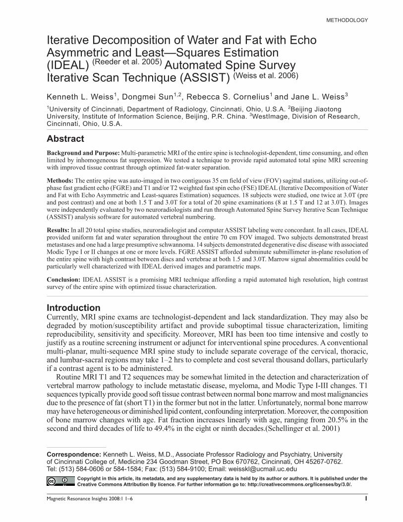
Figure 1. 52 year old woman with metastatic breast cancer involving T11 and more subtly L3 vertebral bodies, (closed arrows); T8–9 HNP with anterior cord impingement, (open arrow); and lower lumbar spondylosis including anterior Modic II changes at L3–4 (arrowheads). MRI performed at 1.5T without contrast.a) From left to right: ASSIST auto-labeled FGRE (TR 61, TE 2.2 msec); T2-weighted FSE IDEAL (TR 2017, TE 61 msec, ET 10) water, fat, composite (W + F), and W% map.
The various pulse sequences and magnet platforms (1.5T or 3.0T) were holistically rated in terms of S/N, image contrast, coverage, spatial resolution, and acquisition time; with the highest rated combination chosen for a subsequent American Cancer Society (ACS) sponsored study, now ongoing.
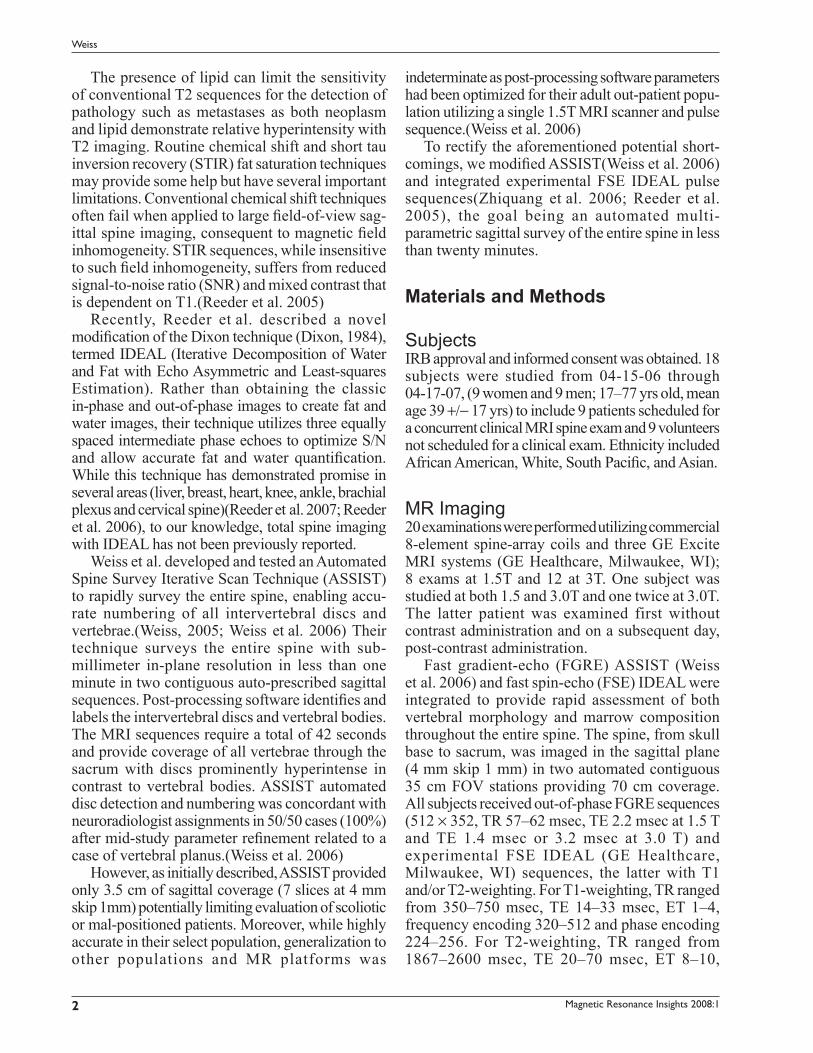
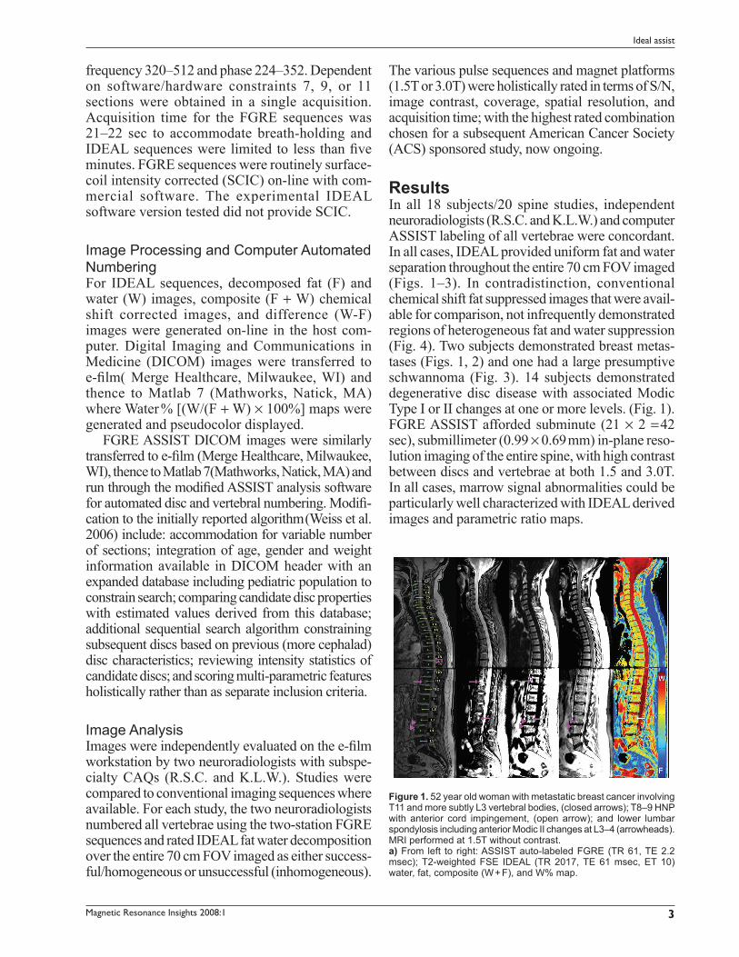
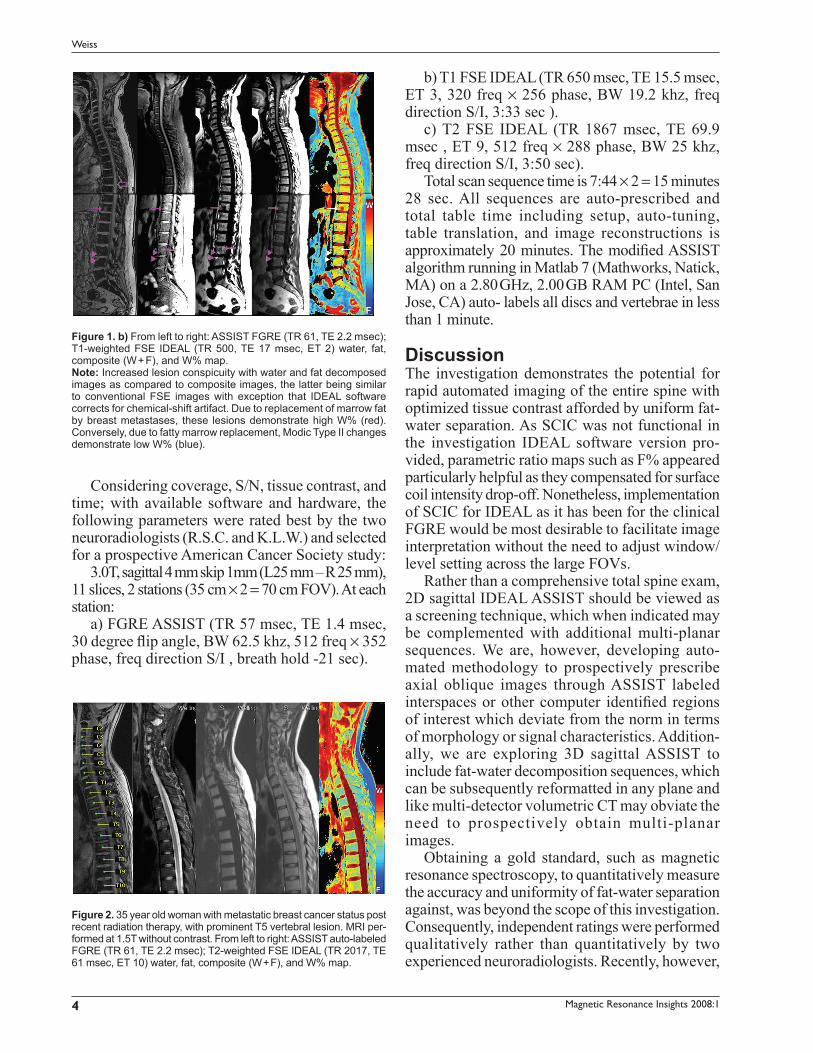
ResultsIn all 18 subjects/20 spine studies, independent neuroradiologists (R.S.C. and K.L.W.) and computer ASSIST labeling of all vertebrae were concordant. In all cases, IDEAL provided uniform fat and water separation throughout the entire 70 cm FOV imaged (Figs. 1–3). In contradistinction, conventional chemical shift fat suppressed images that were avail-able for comparison, not infrequently demonstrated regions of heterogeneous fat and water suppression (Fig. 4). Two subjects demonstrated breast metas-tases (Figs. 1, 2) and one had a large presumptive schwannoma (Fig. 3). 14 subjects demonstrated degenerative disc disease with associated Modic Type I or II changes at one or more levels. (Fig. 1). FGRE ASSIST afforded subminute (21 × 2 = 42 sec), submillimeter (0.99 × 0.69 mm) in-plane reso-lution imaging of the entire spine, with high contrast between discs and vertebrae at both 1.5 and 3.0T. In all cases, marrow signal abnormalities could be particularly well characterized with IDEAL derived images and parametric ratio maps.
4
Weiss
Magnetic Resonance Insights 2008:1
Considering coverage, S/N, tissue contrast, and time; with available software and hardware, the following parameters were rated best by the two neuroradiologists (R.S.C. and K.L.W.) and selected for a prospective American Cancer Society study:
3.0T, sagittal 4 mm skip 1mm (L25 mm – R 25 mm), 11 slices, 2 stations (35 cm × 2 = 70 cm FOV). At each station:
a) FGRE ASSIST (TR 57 msec, TE 1.4 msec, 30 degree fl ip angle, BW 62.5 khz, 512 freq × 352 phase, freq direction S/I , breath hold -21 sec).
b) T1 FSE IDEAL (TR 650 msec, TE 15.5 msec, ET 3, 320 freq × 256 phase, BW 19.2 khz, freq direction S/I, 3:33 sec ).
c) T2 FSE IDEAL (TR 1867 msec, TE 69.9 msec , ET 9, 512 freq × 288 phase, BW 25 khz, freq direction S/I, 3:50 sec).
Total scan sequence time is 7:44 × 2 = 15 minutes 28 sec. All sequences are auto-prescribed and total table time including setup, auto-tuning, table translation, and image reconstructions is approximately 20 minutes. The modifi ed ASSIST algorithm running in Matlab 7 (Mathworks, Natick, MA) on a 2.80 GHz, 2.00 GB RAM PC (Intel, San Jose, CA) auto- labels all discs and vertebrae in less than 1 minute.
DiscussionThe investigation demonstrates the potential for rapid automated imaging of the entire spine with optimized tissue contrast afforded by uniform fat-water separation. As SCIC was not functional in the investigation IDEAL software version pro-vided, parametric ratio maps such as F% appeared particularly helpful as they compensated for surface coil intensity drop-off. Nonetheless, implementation of SCIC for IDEAL as it has been for the clinical FGRE would be most desirable to facilitate image interpretation without the need to adjust window/level setting across the large FOVs.
Rather than a comprehensive total spine exam, 2D sagittal IDEAL ASSIST should be viewed as a screening technique, which when indicated may be complemented with additional multi-planar sequences. We are, however, developing auto-mated methodology to prospectively prescribe axial oblique images through ASSIST labeled interspaces or other computer identifi ed regions of interest which deviate from the norm in terms of morphology or signal characteristics. Addition-ally, we are exploring 3D sagittal ASSIST to include fat-water decomposition sequences, which can be subsequently reformatted in any plane and like multi-detector volumetric CT may obviate the need to prospectively obtain multi-planar images.
Obtaining a gold standard, such as magnetic resonance spectroscopy, to quantitatively measure the accuracy and uniformity of fat-water separation against, was beyond the scope of this investigation. Consequently, independent ratings were performed qualitatively rather than quantitatively by two experienced neuroradiologists. Recently, however,
Figure 1. b) From left to right: ASSIST FGRE (TR 61, TE 2.2 msec); T1-weighted FSE IDEAL (TR 500, TE 17 msec, ET 2) water, fat, composite (W + F), and W% map.Note: Increased lesion conspicuity with water and fat decomposed images as compared to composite images, the latter being similar to conventional FSE images with exception that IDEAL software corrects for chemical-shift artifact. Due to replacement of marrow fat by breast metastases, these lesions demonstrate high W% (red). Conversely, due to fatty marrow replacement, Modic Type II changes demonstrate low W% (blue).
Figure 2. 35 year old woman with metastatic breast cancer status post recent radiation therapy, with prominent T5 vertebral lesion. MRI per-formed at 1.5T without contrast. From left to right: ASSIST auto-labeled FGRE (TR 61, TE 2.2 msec); T2-weighted FSE IDEAL (TR 2017, TE 61 msec, ET 10) water, fat, composite (W + F), and W% map.
5
Ideal assist
Magnetic Resonance Insights 2008:1
others have quantitatively demonstrated the accuracy of the IDEAL technique, to include deter-mining fat%.(Bernard et al. 2007)
As previously advocated,(Weiss et al. 2006) we also recommend FGRE ASSIST localizers for all thoracic or lumbar spine exams to assure correct numbering and facilitate subsequent scan prescrip-tions. We have successfully implemented a similar protocol on other magnet systems to include a 1.5T Siemens unit (Siemens Medical Systems, Erlander, Germany) and have achieved similar success with the modifi ed ASSIST automated numbering algo-rithm. Children as young as 9 years of age have also been successfully studied without change in protocol or computer algorithm. Depending on magnet homogeneity, however, smaller children might be more effi ciently imaged with a single
large fi eld of view acquisition. If contrast is to be administered in conjunction with IDEAL ASSIST, we currently recommend pre-contrast breath-hold FGRE and T2 FSE IDEAL sequences followed by post-contrast breath-hold FGRE and T1 FSE IDEAL.
More study is required as well as FDA/EU approval before FSE IDEAL ASSIST sequences can be recommended for routine clinical examina-tion. Other versions of IDEAL, such as RADIAL GRASE IDEAL(Altbach et al. 2007) need to be tested as well in conjunction with the ASSIST protocol and compared to determine optimal sequences over a large spectrum of pathologies. Additionally, IDEAL sequences should be com-pared to the rapid dual gradient echo Dixon sequences and fat-water separation technique
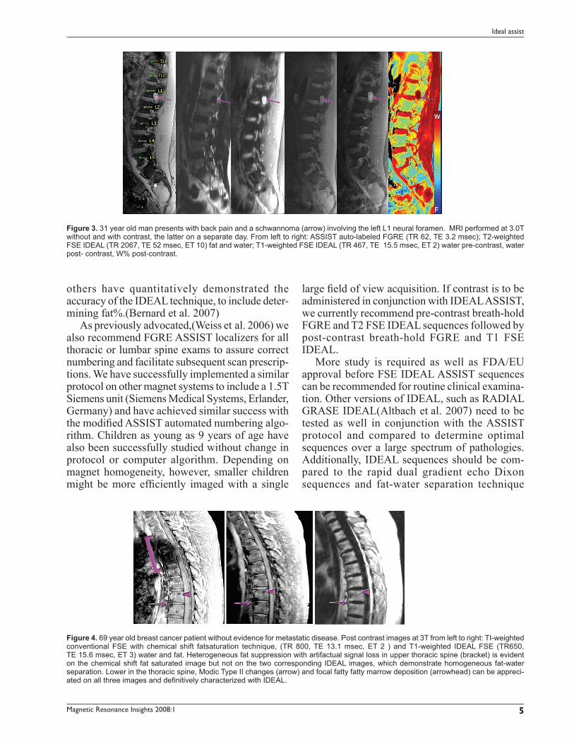
Figure 3. 31 year old man presents with back pain and a schwannoma (arrow) involving the left L1 neural foramen. MRI performed at 3.0T without and with contrast, the latter on a separate day. From left to right: ASSIST auto-labeled FGRE (TR 62, TE 3.2 msec); T2-weighted FSE IDEAL (TR 2067, TE 52 msec, ET 10) fat and water; T1-weighted FSE IDEAL (TR 467, TE 15.5 msec, ET 2) water pre-contrast, water post- contrast, W% post-contrast.
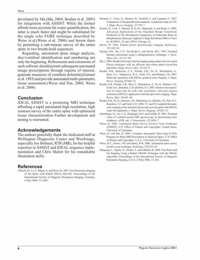
Figure 4. 69 year old breast cancer patient without evidence for metastatic disease. Post contrast images at 3T from left to right: TI-weighted conventional FSE with chemical shift fatsaturation technique, (TR 800, TE 13.1 msec, ET 2 ) and T1-weighted IDEAL FSE (TR650, TE 15.6 msec, ET 3) water and fat. Heterogeneous fat suppression with artifactual signal loss in upper thoracic spine (bracket) is evident on the chemical shift fat saturated image but not on the two corresponding IDEAL images, which demonstrate homogeneous fat-water separation. Lower in the thoracic spine, Modic Type II changes (arrow) and focal fatty fatty marrow deposition (arrowhead) can be appreci-ated on all three images and defi nitively characterized with IDEAL.
6
Weiss
Magnetic Resonance Insights 2008:1
developed by Ma.(Ma, 2004; Boehm et al. 2005) for integration with ASSIST. While the former affords more accurate fat-water quantifi cation, the latter is much faster and might be substituted for the single echo FGRE technique described by Weiss et al.(Weiss et al. 2006) and herein there by permitting a sub-minute survey of the entire spine in two breath-hold sequences.
Regarding, automated spine image analysis, disc/vertebral identifi cation and numbering are only the beginning. Refi nements and extensions of such software should permit subsequent automated image prescriptions through regions of interest, generate measures of vertebral deformity(Genant et al. 1993) and provide automated multi-parametric tissue assessment.(Weiss and Pan, 2004; Weiss et al. 2006)
ConclusionIDEAL ASSIST is a promising MRI technique affording a rapid automated high resolution, high contrast survey of the entire spine with optimized tissue characterization. Further development and testing is warranted.
AcknowledgementsThe authors gratefully thank the dedicated staff at Wellington Diagnostic Center and WestImage, especially Joy Bohnert, RTR (MR), for her helpful expertise in ASSIST and IDEAL sequence imple-mentation and Chris Malott for his remarkable illustration skills.
ReferencesAltbach, M., Li, Z., Bilgin, A. and Weiss, K. 2007. Fast Parametric Imaging
of the Spine with Radial IDEAL-GRASE. Proceedings of the International Society of Magnetic Resonance Imaging, Germany, 6 May 2006, 15, 2685.
Bernard, C., Liney, G., Manton, D., Turnbull, L. and Lamgton, C. 2007. Comparison of fat quantifi cation methods: A phantom study at 3.0T. J. Magn. Reson. Imaging., 27:192–7.
Boehm, H., Link, T., Monetti, R, D, M., Majumdar, S. and Raeth, C. 2005. Advanced Application of the Standard Hough Transform: Prediction of the Mechanical Competence of Trabecular Bone by Morphometric Measures Applied to High Resolution MRI in Vitro. In: HUSSEY., D. (ed.) RSNA. Chicago, IL.
Dixon, W. 1984. Simple proton spectroscopic imaging. Radiology, 153:189–94.
Genant, H.K., Wu, C.Y., Van Kuijk, C. and Nevitt, M.C. 1993. Vertebral fracture assessment using a semiquantitative technique. J. Bone.Miner. Res., 8:1137–48.
Ma, J. 2004. Breath-hold water and fat imaging using a dual-echo two-point Dixon technique with an efficient and robust phase-correction algorithm. Magn. Reson. Med., 52:415–9.
Reeder, S.B., Mckenzie, C.A., Pineda, A.R., Yu, H., Shimakawa, A., Brau, A.C., Hargreaves, B.A., Gold, G.E. and Brittain, J.H. 2007. Water-fat separation with IDEAL gradient-echo imaging. J. Magn. Reson. Imaging, 25:644–52.
Reeder, S.B., Pineda, A.R., Wen, Z., Shimakawa, A., Yu, H., Brittain, J.H., Gold, G.E., Beaulieu, C.H. and Pelc, N.J. 2005. Iterative decomposi-tion of water and fat with echo asymmetry and least-squares estimation (IDEAL): application with fast spin-echo imaging. Magn. Reson. Med., 54:636–44.
Reeder, S.B., Yu, H., Johnson, J.W., Shimakawa, A., Brittain, J.H., Pelc, N.J., Beaulieu, C.F. and Gold, G.E. 2006. T1- and T2-weighted fast spin-echo imaging of the brachial plexus and cervical spine with IDEAL water-fat separation. J. Magn. Reson. Imaging., 24:825–32.
Schellinger, D., Lin, C.S., Hatipoglu, H.G. and Fertikh, D. 2001. Potential value of vertebral proton MR spectroscopy in determining bone weakness. AJNR. Am. J. Neuroradiol., 22:1620–7.
Weiss, K. 2005. Automated Spine Survey Iterative Scan Technique (ASSIST). U.S. Offi ce of Patents and Copyrights. United States, University of Cincinnati.
Weiss, K. and Pan, H. 2004. Computer Automated Three-Step (CATS) Program for Brain MRI Prescription in Talairach Space. U.S. Offi ce of Patents and Copyrights. U.S.A., University of Cincinnati.
Weiss, K.L., Storrs, J.M. and Banto, R.B. 2006. Automated spine survey iterative scan technique. Radiology, 239:255–62.
Zhiquang, L., Squire, S., Gmitro, A. and Altbach, M. 2006. Fast Water and Fat Imaging Using a Radial GRASE Technique with the IDEAL Algorithm. Proceedings of the International Society of Magnetic Resonance Imaging, U.S.A., 6 May 2006, 15, 625.