molecular and developmental analyses of thyroid hormone...
TRANSCRIPT
Review
Molecular and developmental analyses of thyroid hormone receptorfunction in Xenopus laevis, the African clawed frog
Daniel R. Buchholz *, Bindu D. Paul, Liezhen Fu, Yun-Bo Shi
Section on Molecular Morphogenesis, Laboratory of Gene Regulation and Development, NICHD/NIH, Building 18T, Room 106, Bethesda,MD 20892-5431, USA
Received 27 January 2005; revised 23 June 2005; accepted 1 July 2005Available online 2 November 2005
Abstract
The current review focuses on the molecular mechanisms and developmental roles of thyroid hormone receptors (TRs) in gene reg-ulation and metamorphosis in Xenopus laevis and discusses implications for TR function in vertebrate development and diversity. Ques-tions addressed are: (1) what are the molecular mechanisms of gene regulation by TR, (2) what are the developmental roles of TR inmediating the thyroid hormone (TH) signal, (3) what are the roles of the di!erent TR isoforms, and (4) how do changes in these molec-ular and developmental mechanisms a!ect evolution? Even though detailed knowledge of molecular mechanisms of TR-mediated generegulation is available from in vitro studies, relatively little is known about how TR functions in development in vivo. Studies on TRfunction during frog metamorphosis are leading the way toward bridging the gap between in vitro and in vivo studies. In particular,a dual function model for the role of TR in metamorphosis has been proposed and investigated. In this model, TRs repress genes allow-ing tadpole growth in the absence of TH during premetamorphosis and activate genes important for metamorphosis when TH is present.Despite the lack of metamorphosis in most other vertebrates, TR has important functions in development across vertebrates. The under-lying molecular mechanisms of TR in gene regulation are conserved through evolution, so other mechanisms involving TH-target genesand TH tissue-sensitivity and dependence underlie di!erences in role of TR across vertebrates. Continued analysis of molecular anddevelopmental roles of TR in X. laevis will provide the basis for understanding how TR functions in gene regulation in vivo across ver-tebrates and how TR is involved in the generation of evolutionary diversity.Published by Elsevier Inc.
Keywords: Thyroid hormone receptor; Frog metamorphosis; Xenopus laevis; Developmental endocrinology
1. Hormonal context of TR function in frog development
Metamorphosis in frogs is a post-embryonic develop-mental process (Tata, 1999) that transforms aquatic, her-bivorous tadpoles into terrestrial (usually), carnivorousjuveniles (Dodd and Dodd, 1976). Many review articlesand books have been written on various aspects of this sub-ject, including hormonal control, molecular and develop-mental mechanisms, and biochemical and histologicalmetamorphosis of skin, brain, intestine, blood, immunesystem, liver, and other organs (Allen, 1938; Atkinson,
1994; Balcells, 1955; Brown et al., 1996; Dent, 1988; Den-ver et al., 2002; Dodd and Dodd, 1976; Etkin, 1964; Gal-ton, 1983; Gilbert and Frieden, 1981; Gilbert et al., 1996;Hourdry, 1993; Kikuyama et al., 1993; Kollros, 1961;Rose, 2005; Sachs et al., 2000; Shi, 1999; Tata, 1996; Waka-hara and Yamaguchi, 2001). The intent of this review is tohighlight recent molecular studies on the developmentalroles of thyroid hormone receptor (TR) in the frog Xenopuslaevis and how this research informs comparative studies invertebrate diversity.
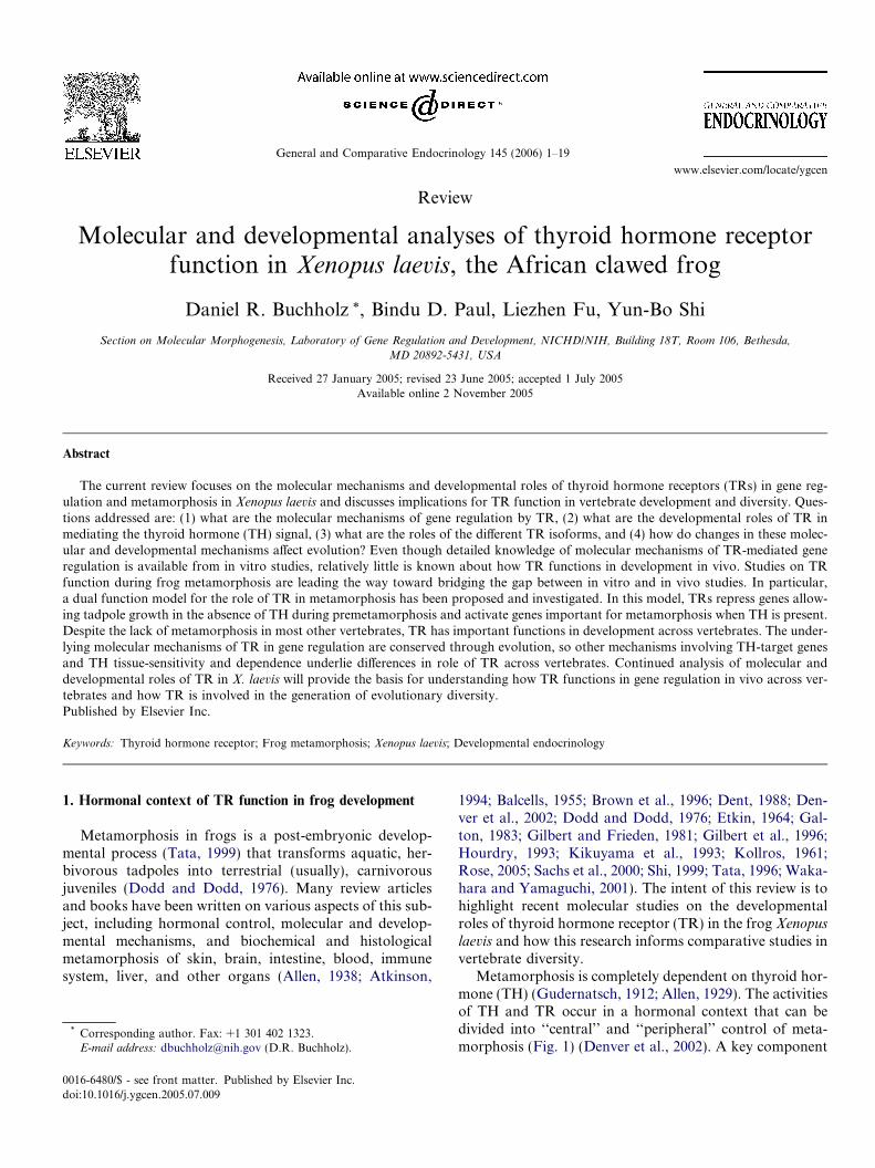
Metamorphosis is completely dependent on thyroid hor-mone (TH) (Gudernatsch, 1912; Allen, 1929). The activitiesof TH and TR occur in a hormonal context that can bedivided into ‘‘central’’ and ‘‘peripheral’’ control of meta-morphosis (Fig. 1) (Denver et al., 2002). A key component
0016-6480/$ - see front matter. Published by Elsevier Inc.doi:10.1016/j.ygcen.2005.07.009
* Corresponding author. Fax: +1 301 402 1323.E-mail address: [email protected] (D.R. Buchholz).
www.elsevier.com/locate/ygcen
General and Comparative Endocrinology 145 (2006) 1–19
of metamorphosis, the timing of the peak in TH plasmalevels during development, is under central control by thehypothalamus–pituitary–thyroid gland axis (Figs. 1A andB). The neurosecretory hypothalamus, which coordinatesenvironmental and nutritional signals, secretes corticotrop-ic releasing factor into the median eminence, a vascular tis-sue that supplies the pituitary (Denver, 1999). TH isthought to control the functional development of the medi-an eminence, whose integrity is important for metamor-phosis (Etkin, 1965). Corticotropic releasing factorstimulates pituitary thyrotrope secretion of thyroid stimu-lating hormone (TSH) (Okada et al., 2004), which, in turn,stimulates thyroid follicle cell proliferation and productionof TH by the thyroid gland (Kaye, 1961; Sakai et al., 1991).The thyroid gland secretes into the blood mostly thyroxine(T4) and very little of the more active form triiodothyro-nine (T3) (White and Nicoll, 1981), both forms collectivelycalled TH. TH negatively feeds back on pituitary secretionof TSH (Kaye, 1961; Denver, 1996; Manzon and Denver,2004). Changes in gene expression of TRs, deiodinases,and receptors for hypothalamic and pituitary hormoneslikely influence the e!ectiveness of this feedback (Huanget al., 2001; Manzon and Denver, 2004).
TH target organs outside the central axis, includinglimbs, intestine, and tail, are collectively known as the ‘‘pe-riphery’’ and respond to T4 and T3 circulating in the bloodin a tissue-specific manner (Fig. 1C) (Dodd and Dodd,1976; Shi, 1999). Some organs develop de novo, such asthe limbs, whereas others are completely resorbed, suchas the tail and gills (Nakajima et al., 2005). However, mostorgans, such as intestine and skin, are remodeled from the
larval form to the adult version (Fox, 1981; McAvoy andDixon, 1977; Shi and Ishizuya-Oka, 2001; Suzuki et al.,2002). The only developmental event not known to bea!ected by TH physiology is primary gonad di!erentiation(Hoskins and Hoskins, 1919; Gruca and Michalowski,1961; Ogielska and Kotusz, 2004; Rot-Nikcevic and Was-sersug, 2004). Physiological changes accompany the mor-phological changes, e.g., the transition fromammonotelism to ureotelism, from larval to adult hemo-globins, and from larval to adult immune systems (Gilbertet al., 1996). These tissue-specific developmental eventsoccur asynchronously, e.g., intestine transforms after thelimbs and before the tail, and this asynchrony is thoughtto be due to tissue-specific control of e!ective intracellularT3 levels (Shi et al., 1996). T4 to T3 conversion by the deio-dinase DII in target tissues is one such peripheral mecha-nism that increases tissue and organ sensitivity to TH(Becker et al., 1997; Cai and Brown, 2004). In addition,T4 and T3 degradation by the deiodinase DIII present intarget tissues reduces cellular levels of T3 (Becker et al.,1997). Indeed, transgenic overexpression of DIII blocksTH-induced metamorphic events (Huang et al., 1999;Marsh-Armstrong et al., 1999). Additional cellular proteinscontrolling intracellular T3 levels and thereby tissue sensi-tivity to circulating TH, include cytosolic TH binding pro-tein (Shi et al., 1994; Yamauchi and Tata, 1994), TRa (Shiet al., 1996), and TH transporters (Ritchie et al., 2003).TRa expression levels may a!ect how sensitive cells areto circulating TH. The levels of cytosolic TH binding pro-teins likely a!ect free TH within the cells available to bindTR. Furthermore, higher TH transporter expression levels
Fig. 1. Endocrine context of TR action and levels of control of metamorphosis. (A) Production of TH by the thyroid gland and tissues responses to THduring metamorphosis constitute the endocrine context of TR. TH is produced by the thyroid gland and secreted into the blood in response to thyroidstimulating hormone (TSH) from the pituitary, which is stimulated by corticotropin releasing factor (CRF) from the hypothalamus. Central control ofmetamorphosis is the regulation of the levels of circulating TH across development by the hypothalamus, pituitary, and thyroid gland in response to acombination of environmental and nutritional signals and hormonal feedback. Peripheral control of metamorphosis is the tissue-specific responses to THof tissues outside the hypothalamic–pituitary–thyroid gland axis. (B) The blood levels of TH across development, derived from the thyroid gland andtissue metabolism of T4 to T3, are first detectable in the blood at the point when tadpoles begin to metamorphose at NF stage 55, i.e., when the limbs beginto grow out, and then rise to a peak in the middle of metamorphosis when the morphological transition is most dramatic (Leloup and Buscaglia, 1977). (C)Tissue-specific responses to TH include predominance of cell proliferation in developing limbs (NF stages 51 and 56 are shown). Intestinal remodelinginvolves both cell proliferation and death in the same tissue (NF stages 54 and 66 are shown in cross-section just posterior to bile duct entry, stained withmethyl green pyronine Y). Cell death is predominant during tail resorption (NF stages 57 and 66 are shown).
2 D.R. Buchholz et al. / General and Comparative Endocrinology 145 (2006) 1–19
correlate with increased ability of cells to carry out TR-mediated transcription.
Amphibian metamorphosis o!ers a unique opportunityto study the molecular mechanisms of TR and interactingcofactors in regulating gene expression in vivo duringdevelopment. Such in vivo studies have been di"cult inmammalian systems because of lack of knowledge aboutTH-regulated genes during development and the di"cultyof obtaining samples from embryos in utero. An additionaldi"culty in mammalian systems is the inability to studyreceptor function in plus or minus hormonal states withoutpathologically disrupting normal development because THis continuously present, either from the mother or from thefetus. In contrast to mammals, tadpoles are large andaccessible throughout their development. In addition, pre-metamorphosis (NF stages 45–53) (Nieuwkoop and Faber,1994) is characterized by the absence of TH (Etkin, 1932,1935; Leloup and Buscaglia, 1977), indicating that all ofthe receptors in vivo are in the unliganded condition. Pro-metamorphosis (NF stages 54–58) and metamorphic cli-max (NF stages 59–66) have increasing amounts ofendogenous TH (Etkin, 1932, 1935; Leloup and Buscaglia,1977), so that addition of exogenous TH enables precisetiming of change to the liganded state that can mimic nat-ural metamorphosis. This ability to control the THresponse is a key advantage for using tadpoles, first exploit-ed by showing a role for TH in development and later byisolating TH-responsive genes using subtractive hybridiza-tion (Shi, 1999). Recent work has continued to take advan-tage of frog development as a valuable model of vertebratedevelopmental endocrinology to address the molecular anddevelopmental roles of TR in post-embryonicdevelopment.
2. Molecular mechanisms of TR in gene regulation
2.1. DNA binding, heterodimerization, and role of ligand
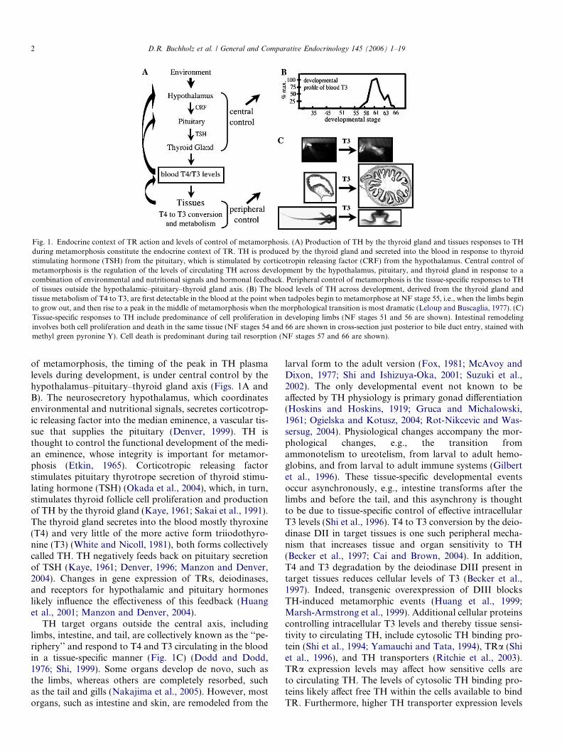
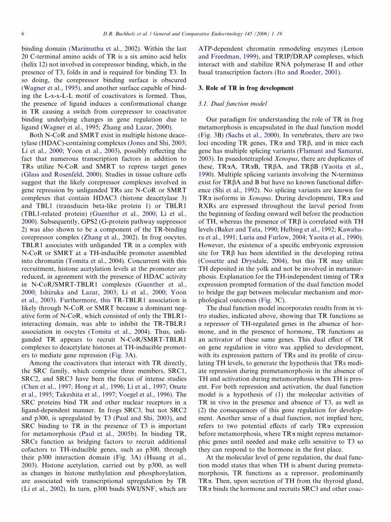
Classical in vitro and oocyte studies from a decade agorevealed three critical components for how TR regulatesgene expression: DNA binding, heterodimerization, androle of TH (Ranjan et al., 1994; Wong and Shi, 1995;Yen, 2001). TRs are modular proteins consisting of severaldomains that function largely independently, includingDNA and ligand binding domains (Fig. 2A) (Zhang andLazar, 2000). The 100 amino-acid N-terminal DNA bind-ing domain is responsible for recognizing thyroid hormoneresponse elements (TREs) in promoters or enhancers ofTH-regulated genes, while the C-terminal half of the pro-tein binds to the heterodimerization partner (9-cis-retinoicacid receptor, RXR), TH, and transcriptional cofactors.TR e!ects gene transcription by binding to the TREs, themost common of which are direct repeats of AGGTCAseparated by four nucleotides, the DR4 type of TREs (Lau-det and Gronemeyer, 2002). The TREs in five TH-regulat-ed promoters in Xenopus have been characterized to date,TRb (Machuca et al., 1995; Ranjan et al., 1994), TH-in-
duced basic leucine zipper transcription factor (Furlowand Brown, 1999), basic transcription element binding pro-tein (Furlow and Kanamori, 2002), collagenase or MMP1(Oofusa and Yoshizato, 1991) and stromelysin-3 (Fu,
Fig. 2. Role of ligand and receptors in TH-response gene regulation. Agood model to study molecular mechanisms of gene regulation is theXenopus oocyte transcription assay because oocytes have large stores oftranscription and translation machinery and reporter plasmids becomechromatinized when injected into oocytes. This assay involves microin-jection of a reporter plasmid (luciferase driven by a TH-induciblepromoter as an example here) and transcription factor mRNA intoXenopus oocytes followed by luciferase assay to quantitate levels oftranscriptional activity. (A) Diagrams of wild type and mutant TRsshowing functional domains. Wild type TRa has a DNA binding domain(DBD) and a ligand binding domain (LBD), which is also the site of co-factor binding and heterodimerization with RXR (9-cis-retinoic acidreceptor). A nuclear localization signal is located in the hinge regionbetween the DBD and LBD. Dominant negative TRa (dnTRa) cannotbind ligand because of a short C-terminal deletion (dashed portion).Dominant positive or constitutively active TRa (dpTRa) has a viraltransactivation domain (VP16) fused to the N-terminus that stronglyrecruits coactivators. The chimeric receptor illustrated here (TRa-VDR)contains the N-terminal portion of TRa, which includes the the DNAbinding domain of TRa, fused to the ligand binding domain of the vitaminD receptor (VDR). (B) Because of lack of detectable TR and TH in theoocytes, microinjection of reporter plasmid, containing a TH-responsivepromoter controlling the transcription of luciferase, alone into the largeoocyte nucleus has a basal level of transcription, as reflected by luciferaseactivity (lane 1). Basal levels of transcription are not changed uponadditional microinjection of TRa mRNA alone into the cytoplasm, withor without T3 (lanes 2 and 3), indicating both the lack of RXR in theoocytes and importance of TRa/RXR heterodimerization in gene regu-lation by TR. If reporter plasmids and TRa/RXR mRNAs are microin-jected into the oocytes, the level of transcription and luciferase activitydepends upon the presence of T3, such that in the absence of T3,transcription levels are repressed compared to basal levels, and in thepresence of T3, transcription levels are induced above basal levels (lanes 4and 5). Mutant TRs a!ect transcription di!erently with respect to the roleof ligand. The dnTRa represses transcription from basal levels, even in thepresence of T3 (lanes 6 and 7), because it constitutively binds corepressorsdue to inability to bind ligand and undergo conformational change.Dominant positive TRa (dpTRa) induces transcription above basal levelseven in the absence of hormone (lanes 8 and 9) because of constitutiverecruitment of coactivators. This coactivator recruitment even overcomesthe possible recruitment of corepressors at the LBD. The TRa-VDR bindsto TH-regulated promoters and represses them in the absence andpresence of T3 (lanes 10 and 11). Ligand-induced upregulation occurs inthe presence of vitamin D rather than T3 (lane 12).
D.R. Buchholz et al. / General and Comparative Endocrinology 145 (2006) 1–19 3
unpubl.) (Table 1). In addition, TRs are able to activatetranscription through non-DR4 types of TREs (Desvergne,1994; Williams and Brent, 1995), e.g, in the intestinal alka-line phosphatase gene (Malo et al., 2004) and HIV long ter-minal repeat (Hsia and Shi, 2002).
The critical roles of heterodimerization of TR with RXRand of ligand can be shown using the frog oocyte transcrip-tion assay, where exogenous TR, RXR, and reporter DNAcan be introduced into oocytes under conditions mimickingthose in somatic cells (Wong et al., 1995). In the absence ofT3 and TR, TH-inducible promoters have a basal level oftranscriptional activation (Fig. 2B, lane 1). This basal levelof transcription is not altered in the presence of TR or T3(Fig. 2B, lanes 2 and 3), a result consistent with gel mobil-ity shift assays where TRs alone do not bind TREs well inpromoters in the absence of heterodimerization (Tsai andO!Malley, 1994; Wong and Shi, 1995). In the presence ofTR and RXR, transcription from TH-inducible genes isrepressed, and upon addition of T3, transcription is acti-vated (Fig. 2B, lanes 4 and 5).
Mutant TRs with a small truncation or mutations in theC-terminus function as dominant negative TRs becausethey cannot bind ligand but bind TREs and corepressorsnormally, thereby repressing TH-inducible genes both inthe absence and presence of T3 (Fig. 2B, lanes 6 and 7)(Ulisse et al., 1996). On the other hand, TRs can be con-verted into dominant positive TRs to activate transcriptionindependent of TH by fusing a strong viral transactivationdomain to the N-terminus (Fig. 2B, lanes 8 and 9) (Buch-holz et al., 2004). In addition, chimeric receptors, whereDNA binding domains and ligand binding domains fromdi!erent hormone receptors have been swapped, havealtered hormone requirements for activation. For example,a chimeric receptor (TRa-VDR) made of the TRa DNAbinding domain and the vitamin D receptor ligand binding
domain binds TREs but does not respond to TH. However,it activates the TH-regulated genes upon addition of vita-min D (Fig. 2B, lanes 10–12) (Buchholz et al., submitted),indicating that the DNA-binding and hormone-bindingdomains function largely independently.
Two basic types of transcription response to TH areknown, one using positive TREs and the other using neg-ative TREs (Yen, 2001). Positive TREs exhibit the classi-cal case of repression in the absence of TH and activationin its presence, whereas negative TREs behave in theopposite manner. The majority of TH-response genescloned by subtractive hybridization during frog metamor-phosis from intestine (Shi and Brown, 1993), tail (Wangand Brown, 1993), limb (Buckbinder and Brown, 1992),and brain (Denver et al., 1997) are genes upregulated byTH, though a few are downregulated in skin (Furlowet al., 1997) and intestine (Ishizuya-Oka et al., 1994).Many of them appear to be regulated by TH at the tran-scriptional level and thus likely have positive TREs. Onthe other hand, TH-response genes analyzed by micro-array in mice show a more complicated picture. In liver(Yen et al., 2003) and cerebellum (Miller et al., 2004),most genes are induced by TH. However, in the heartand white adipose tissue, greater than 50% of the genesare downregulated. In the mouse liver microarray study,five modes of TH regulation were observed, classicalupregulation and downregulation, derepression only, acti-vation only, and repression only (Yen et al., 2003). Fur-ther studies are needed to determine whether thesedi!erent TH response genes are regulated directly at thetranscriptional level through TR or are downstream genesindirectly regulated by T3. Nonetheless, the use of micro-array studies in metamorphic tissues are revealing similarcomplexities in TH-response gene regulation (Veldhoenet al., 2002; Helbing et al., 2003).
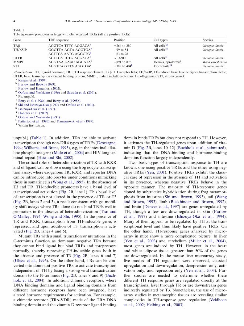
Table 1TH-responsive promoters in frogs with characterized TREs (all are positive TREs)
Gene TRE sequence Position Cell types Species
TRb AGGTCA TTTC AGGACAa +264 to 280 All cellsf,g Xenopus laevisTH/bZIP GGGTTA AGTA AGGTGAb !99 to 84 All cellsf,h Xenopus laevis
AGTTCA AATG AGGCTGb !63 to 78BTEB AGTTCA TCTG AGGACAc "!6500 All cellsf,i Xenopus laevisMMP1 AGGTAA GAAC AGGATAd !891 to 876 Dermis, epi-dermisj Rana catesbeianaST3 AGGTCA GTTA AGGTGAe +389 to 404l Fibroblastsf,k Xenopus laevis
Abbreviations: TH, thyroid hormone; TRE, TH response element; TRb, TH receptor beta; TH/bZIP, TH-induced basic leucine zipper transcription factor;BTEB, basic transcription element binding protein; MMP1, matrix metalloproteinase 1 (collagenase); ST3, stromelysin-3.a Ranjan et al. (1994).b Furlow and Brown (1999).c Furlow and Kanamori (2002).d Oofusa and Yoshizato (1996) and Sawada et al. (2001).e Fu, unpubl.f Berry et al. (1998a) and Berry et al. (1998b).g Shi and Ishizuya-Oka (1997) and Oofusa et al. (2001).h Ishizuya-Oka et al. (1997).i Hoopfer et al. (2002).j Oofusa and Yoshizato (1991).k Patterton et al. (1995) and Damjanovski et al. (1999).l Within first intron.
4 D.R. Buchholz et al. / General and Comparative Endocrinology 145 (2006) 1–19
2.2. Transcriptional cofactors interacting with TR
The molecular mechanisms of TR in regulating TH-re-sponse genes involves cofactors that interact directly orindirectly with TR (Fig. 3A) (Chen and Li, 1998; Ito andRoeder, 2001; McKenna et al., 1999; Xu et al., 1999; Zhangand Lazar, 2000). In the absence of TH, TRs bind core-pressors, a number of which have been identified (Burkeand Baniahmad, 2000). The best studied among them arethe highly related corepressors N-CoR (nuclear receptorcorepressor) and SMRT (silencing mediator for retinoidand thyroid hormone receptors) (Chen and Evans, 1995;
Horlein et al., 1995; Privalsky, 2004). When T3 is present,the corepressors are replaced by coactivators including thep160 or steroid receptor coactivator (SRC) family of pro-teins, p300/CBP (Chakravarti et al., 1996), the DRIP/TRAP/ARC complex (Yuan et al., 1998; Rachez andFreedman, 2001), and chromatin remodeling factors(Huang et al., 2003).
The ligand TH plays an essential role in gene regulationby TR. By means of a CoRNR box (a corepressor/nuclearreceptor) consensus sequence, I/L-x-x-I/V-I, (Cohen et al.,2001; Hu and Lazar, 1999), N-CoR or SMRT bind a TRsurface formed by helices 1–11 of the C-terminal ligand
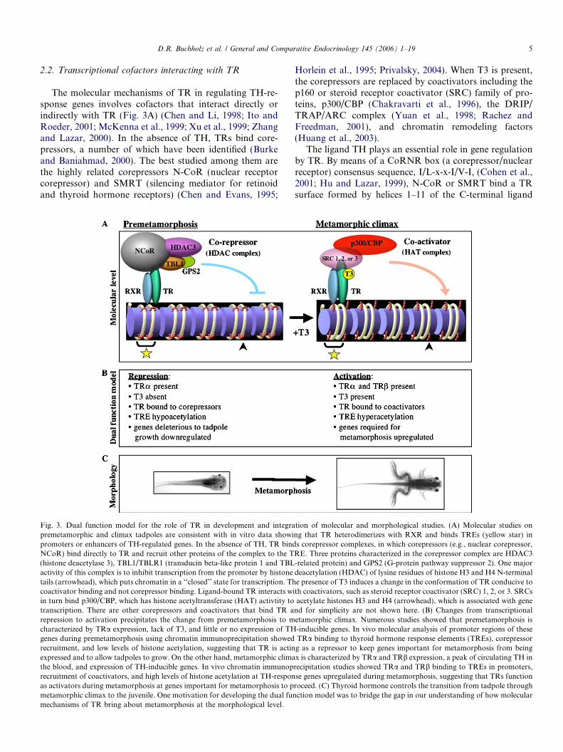
Fig. 3. Dual function model for the role of TR in development and integration of molecular and morphological studies. (A) Molecular studies onpremetamorphic and climax tadpoles are consistent with in vitro data showing that TR heterodimerizes with RXR and binds TREs (yellow star) inpromoters or enhancers of TH-regulated genes. In the absence of TH, TR binds corepressor complexes, in which corepressors (e.g., nuclear corepressor,NCoR) bind directly to TR and recruit other proteins of the complex to the TRE. Three proteins characterized in the corepressor complex are HDAC3(histone deacetylase 3), TBL1/TBLR1 (transducin beta-like protein 1 and TBL-related protein) and GPS2 (G-protein pathway suppressor 2). One majoractivity of this complex is to inhibit transcription from the promoter by histone deacetylation (HDAC) of lysine residues of histone H3 and H4 N-terminaltails (arrowhead), which puts chromatin in a ‘‘closed’’ state for transcription. The presence of T3 induces a change in the conformation of TR conducive tocoactivator binding and not corepressor binding. Ligand-bound TR interacts with coactivators, such as steroid receptor coactivator (SRC) 1, 2, or 3. SRCsin turn bind p300/CBP, which has histone acetyltransferase (HAT) activtity to acetylate histones H3 and H4 (arrowhead), which is associated with genetranscription. There are other corepressors and coactivators that bind TR and for simplicity are not shown here. (B) Changes from transcriptionalrepression to activation precipitates the change from premetamorphosis to metamorphic climax. Numerous studies showed that premetamorphosis ischaracterized by TRa expression, lack of T3, and little or no expression of TH-inducible genes. In vivo molecular analysis of promoter regions of thesegenes during premetamorphosis using chromatin immunoprecipitation showed TRa binding to thyroid hormone response elements (TREs), corepressorrecruitment, and low levels of histone acetylation, suggesting that TR is acting as a repressor to keep genes important for metamorphosis from beingexpressed and to allow tadpoles to grow. On the other hand, metamorphic climax is characterized by TRa and TRb expression, a peak of circulating TH inthe blood, and expression of TH-inducible genes. In vivo chromatin immunoprecipitation studies showed TRa and TRb binding to TREs in promoters,recruitment of coactivators, and high levels of histone acetylation at TH-response genes upregulated during metamorphosis, suggesting that TRs functionas activators during metamorphosis at genes important for metamorphosis to proceed. (C) Thyroid hormone controls the transition from tadpole throughmetamorphic climax to the juvenile. One motivation for developing the dual function model was to bridge the gap in our understanding of how molecularmechanisms of TR bring about metamorphosis at the morphological level.
D.R. Buchholz et al. / General and Comparative Endocrinology 145 (2006) 1–19 5
binding domain (Marimuthu et al., 2002). Within the last20 C-terminal amino acids of TR is a six amino acid helix(helix 12) not involved in corepressor binding, which, in thepresence of T3, folds in and is required for binding T3. Inso doing, the corepressor binding surface is obscured(Wagner et al., 1995), and another surface capable of bind-ing the L-x-x-L-L motif of coactivators is formed. Thus,the presence of ligand induces a conformational changein TR causing a switch from corepressor to coactivatorbinding underlying changes in gene regulation due toligand (Wagner et al., 1995; Zhang and Lazar, 2000).
Both N-CoR and SMRT exist in multiple histone deace-tylase (HDAC)-containing complexes (Jones and Shi, 2003;Li et al., 2000; Yoon et al., 2003), possibly reflecting thefact that numerous transcription factors in addition toTRs utilize N-CoR and SMRT to repress target genes(Glass and Rosenfeld, 2000). Studies in tissue culture cellssuggest that the likely corepressor complexes involved ingene repression by unliganded TRs are N-CoR or SMRTcomplexes that contain HDAC3 (histone deacetylase 3)and TBL1 (transducin beta-like protein 1) or TBLR1(TBL1-related protein) (Guenther et al., 2000; Li et al.,2000). Subsequently, GPS2 (G-protein pathway suppressor2) was also shown to be a component of the TR-bindingcorepressor complex (Zhang et al., 2002). In frog oocytes,TBLR1 associates with unliganded TR in a complex withN-CoR or SMRT at a TH-inducible promoter assembledinto chromatin (Tomita et al., 2004). Concurrent with thisrecruitment, histone acetylation levels at the promoter arereduced, in agreement with the presence of HDAC activityin N-CoR/SMRT-TBLR1 complexes (Guenther et al.,2000; Ishizuka and Lazar, 2003; Li et al., 2000; Yoonet al., 2003). Furthermore, this TR-TBLR1 association islikely through N-CoR or SMRT because a dominant neg-ative form of N-CoR, which consisted of only the TBLR1-interacting domain, was able to inhibit the TR-TBLR1association in oocytes (Tomita et al., 2004). Thus, unli-ganded TR appears to recruit N-CoR/SMRT-TBLR1complexes to deacetylate histones at TH-inducible promot-ers to mediate gene repression (Fig. 3A).
Among the coactivators that interact with TR directly,the SRC family, which comprise three members, SRC1,SRC2, and SRC3 have been the focus of intense studies(Chen et al., 1997; Hong et al., 1996; Li et al., 1997; Onateet al., 1995; Takeshita et al., 1997; Voegel et al., 1996). TheSRC proteins bind TR and other nuclear receptors in aligand-dependent manner. In frogs SRC3, but not SRC2and p300, is upregulated by T3 (Paul and Shi, 2003), andSRC binding to TR in the presence of T3 is importantfor metamorphosis (Paul et al., 2005b). In binding TR,SRCs function as bridging factors to recruit additionalcofactors to TH-inducible genes, such as p300, throughtheir p300 interaction domain (Fig. 3A) (Huang et al.,2003). Histone acetylation, carried out by p300, as wellas changes in histone methylation and phosphorylation,are associated with transcriptional upregulation by TR(Li et al., 2002). In turn, p300 binds SWI/SNF, which are
ATP-dependent chromatin remodeling enzymes (Lemonand Freedman, 1999), and TRIP/DRAP complexes, whichinteract with and stabilize RNA polymerase II and otherbasal transcription factors (Ito and Roeder, 2001).
3. Role of TR in frog development
3.1. Dual function model
Our paradigm for understanding the role of TR in frogmetamorphosis is encapsulated in the dual function model(Fig. 3B) (Sachs et al., 2000). In vertebrates, there are twoloci encoding TR genes, TRa and TRb, and in mice eachgene has multiple splicing variants (Flamant and Samarut,2003). In psuedotetraploid Xenopus, there are duplicates ofthese, TRaA, TRaB, TRbA, and TRbB (Yaoita et al.,1990). Multiple splicing variants involving the N-terminusexist for TRbA and B but have no known functional di!er-ence (Shi et al., 1992). No splicing variants are known forTRa isoforms in Xenopus. During development, TRa andRXRs are expressed throughout the larval period fromthe beginning of feeding onward well before the productionof TH, whereas the presence of TRb is correlated with THlevels (Baker and Tata, 1990; Helbing et al., 1992; Kawaha-ra et al., 1991; Luria and Furlow, 2004; Yaoita et al., 1990).However, the existence of a specific embryonic expressionsite for TRb has been identified in the developing retina(Cossette and Drysdale, 2004), but this TR may utilizeTH deposited in the yolk and not be involved in metamor-phosis. Explanation for the TH-independent timing of TRaexpression prompted formation of the dual function modelto bridge the gap between molecular mechanism and mor-phological outcomes (Fig. 3C).
The dual function model incorporates results from in vi-tro studies, indicated above, showing that TR functions asa repressor of TH-regulated genes in the absence of hor-mone, and in the presence of hormone, TR functions asan activator of these same genes. This dual e!ect of TRon gene regulation in vitro was applied to development,with its expression pattern of TRs and its profile of circu-lating TH levels, to generate the hypothesis that TRs medi-ate repression during premetamorphosis in the absence ofTH and activation during metamorphosis when TH is pres-ent. For both repression and activation, the dual functionmodel is a hypothesis of (1) the molecular activities ofTR in vivo in the presence and absence of T3, as well as(2) the consequences of this gene regulation for develop-ment. Another sense of a dual function, not implied here,refers to two potential e!ects of early TRa expressionbefore metamorphosis, where TRamight repress metamor-phic genes until needed and make cells sensitive to T3 sothey can respond to the hormone in the first place.
At the molecular level of gene regulation, the dual func-tion model states that when TH is absent during premeta-morphosis, TR functions as a repressor, predominantlyTRa. Then, upon secretion of TH from the thyroid gland,TRa binds the hormone and recruits SRC3 and other coac-
6 D.R. Buchholz et al. / General and Comparative Endocrinology 145 (2006) 1–19
tivators, thereby becoming a transcriptional activator thatinduces target gene expression. The activation of TRainduces high expression of TRb, which itself interacts withcoactivators due to the presence of TH. Thus, the predom-inant role of TRb in gene regulation is in activation, notbecause it cannot interact with corepressors, but becauseTRb is expressed for the most part only when T3 is present.The switch from corepressor to coactivator binding resultsin changes in histone acetylation and chromatin remodel-ing favoring gene activation. The molecular aspects of thismodel apply to the mechanisms of TR in regulating directresponse genes, i.e., genes where TR binds TREs in thepromoter, as opposed to downstream or late genes in theTH-induced gene regulation cascade (Shi, 1994).
The dual function model at the developmental level sug-gests that TR-mediated repression is important to keepmetamorphic genes turned o! to allow tadpole growthand prevent metamorphosis from occurring too early. Thisincludes several genes expressed during embryogenesis thatare also involved in metamorphosis, such as stromelysin-3and sonic hedgehog (Ishizuya-Oka et al., 2000, 2001; Sto-low and Shi, 1995). Thus, early TR expression may beimportant to turn o! these embryonic genes until theyare again required for metamorphosis. Furthermore, geneactivation in the presence of T3 is important to initiatethe morphological and physiological changes of metamor-phosis. The developmental aspects of the model apply tothe consequences for development of TR-regulated genesexpression.
Two considerations are important when discussing theapplicability of the dual function model. First, consequenc-es of the conformational change induced by TH causingTR to switch from a repressor to an activator may dependon the promoter. Given the variety of gene regulation pat-terns directly mediated by TR including TH-induced geneactivation or repression (Yen et al., 2003), the dual func-tion model may not represent all activities of gene regula-tion mediated by TR. The dual function model wasoriginally conceived to apply to positive TREs. For thegenes that are only activated or only repressed by TR,other models of gene regulation are needed to explainhow a TH-response gene can be, for example, activatedand not repressed by TR. For direct response genes thatare downregulated, such as TSH in the pituitary (Manzonand Denver, 2004), the dual function model may apply butjust be opposite in sign. However, the molecular mecha-nisms are not understood in vitro for TR-mediated regula-tion of genes other than those with positive TREs, so amodel for the developmental role of TR for regulatinggenes with other types of TREs must await biochemicalcharacterization.
Second, the dual function model may also have to beunderstood in the context of tissue-specific gene regulation.For some genes that are up regulated in all cells, such asTRb, TH-induced basic leucine zipper transcription factor,and basic transcription element binding protein (Table 1),we suggest the dual function model applies similarly to
these genes in all cells. Other genes are upregulated in abroad organ distribution but in particular cell types, suchas stromelysin-3 in fibroblasts (Table 1), or expressed in aspecific tissue and organ, such as sonic hedgehog in intesti-nal epithelium (Patterton et al., 1995; Stolow and Shi,1995). For these genes, the dual function model appliesonly in the cell-type in which they are expressed. In addi-tion to tissue-specificity are temporal considerations. Meta-morphic events occur asynchronously, indicating that thetransition from repression to activation of the dual func-tion model occurs at di!erent times in di!erent tissues dur-ing development. For example, the hind limbs developbefore the tail resorbs, suggesting that TR is an activatorin the hind limbs at the same time it is a repressor in thetail, which may have to do with intracellular levels ofligand owing to deiodinase expression.
3.2. In vivo support for molecular aspects of the dual functionmodel
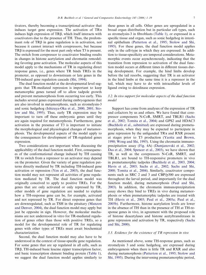
Support has come from analyses of the expression of TRand cofactors by us and others. We have found that core-pressor components N-CoR, SMRT, and TBLR1 (Sachset al., 2002; Tomita et al., 2004) and GPS2 and HDAC3(Buchholz et al., submitted) are expressed during premeta-morphosis, when they may be expected to participate ingene repression by the unliganded TRa and RXR presentat stages prior to T3 production (Yaoita and Brown,1990; Wong and Shi, 1995). Using the chromatin immuno-precipitation assay (Fig. 4A) (Damjanovski et al., 2002;Das et al., 2004; Spencer et al., 2003), we have shown thatTR, as well as the corepressors N-CoR, SMRT, andTBLR1, are bound to TH-responsive promoters in vivoin premetamorphic tadpoles (Buchholz et al., 2003, 2004;Havis et al., 2003; Sachs et al., 2002; Sachs and Shi,2000; Tomita et al., 2004). Similarly, coactivator compo-nents such as SRC 2 and 3 and CBP/p300 are expressedthroughout the larval period, and importantly for the dualfunction model, during metamorphosis (Paul and Shi,2003). In addition, the chromatin immunoprecipitationassay shows they bind to TREs in vivo during metamor-phosis or when premetamorphic tadpoles are treated withTH (Havis et al., 2003; Paul et al., 2005a; Paul et al.,2005b). Furthermore, histone acetylation levels are lowerin the absence of TH than in the presence of TH at TH-re-sponse genes in vivo, in agreement with the proposed roleof histone deacetylases and histone acetyltransferases ingene repression and activation by TR, respectively (Sachsand Shi, 2000).
3.3. Evidence for the existence of TR repression in vivo
As mentioned above, some TH-response genes, such asstromelysin 3 and sonic hedgehog, are expressed duringembryogenesis when there is little TR and RXR and alsoduring metamorphosis (Patterton et al., 1995; Stolow andShi, 1995). During the intervening premetamorphic period,
D.R. Buchholz et al. / General and Comparative Endocrinology 145 (2006) 1–19 7
i.e., after tadpole feeding begins until the onset of meta-morphosis, these genes are expressed at lower levels. Thisinterval between the expression peaks of these genes corre-sponds with the expression of TRa and the absence of T3,suggesting that TRa may repress these genes. Similarly, thedownregulation of the transgene GFP under control of theTRbA promoter correlated with the expression of TRa inthe absence of T3 (Oofusa et al., 2001). Embryo injectionexperiments have provided further evidence to show thatTR is capable of repressing or activating gene expressionin developing Xenopus embryos (Puzianowska-Kuznickaet al., 1997). Overexpression of TR and RXR together,but not either one alone, by microinjecting their mRNAsinto fertilized eggs was shown to repress endogenous THresponse genes while the addition of TH led to the reversalof the repression and further activation of these genes, sug-gesting that subsequent expression of endogenous TRmight repress these genes. Similarly, endogenous TR andcofactors are capable of repressing reporter genes, as sug-gested when dominant negative N-CoR peptides causedupregulation of a co-injected reporter gene in tadpole tailcells in vivo (Sachs et al., 2002). According to the develop-mental aspects of the dual function model, we expect TR/
corepressor complexes to repress genes that, if expressed,would cause metamorphic defects. In support of this, func-tional studies show deleterious consequences of precociousexpression of the TH-regulated gene stromelysin 3 in trans-genic animals where normal intestine morphology is com-promised in the absence of T3 (Ishizuya-Oka et al., 2000;Fu et al., 2005).
Correlations between TR expression and TH-responsegene expression, as well as embryo and tail injections stud-ies, need to be augmented by direct experimental evidencein order to establish endogenous TR is responsible forrepression of endogenous genes, for example, by blockingcorepressor function at the TR with overexpressed trans-genic dominant negative corepressors. Two pieces of indi-rect evidence suggest that TR-mediated repression mayplay only a minor role in Xenopus post-embryonic develop-ment. First, we used a dominant negative form of XenopusSRC3 (F-dnSRC3) overexpressed in transgenic animals toinvestigate the function of coactivators during metamor-phosis, and we showed that this overexpression was su"-cient to inhibit or delay both TH-induced and naturalmetamorphosis (Paul et al., 2005b). Despite the corepressorrelease in these animals treated with T3, little derepression
Fig. 4. Techniques important for in vivo molecular analysis of the role of TR during development. (A) The chromatin immunoprecitation assay directlymeasures protein–DNA interactions in cells and takes advantage of the specificity of antibodies and the sensitivity of PCR. For in vivo analysis, tadpoletissues are harvested and the nuclei are isolated. The nuclei are then lysed with detergent, and the intact genomic DNA is sonicated to produce small piecesof chromatin ("200–1000 bp in length). Antibodies specific for a protein of interest, e.g., anti-TR antibodies or antibodies against a cofactor that interactswith TR, are added to the chromatin to immunoprecipitate (IP) fragments of DNA that may be associating with the protein of interest. Then, theimmunoprecipitated DNA is purified and PCR is carried out with primers specific for a TH-responsive promoter to detect whether or not the protein ofinterest was associated with that promoter region. The chromatin immunoprecipitation assay always involves two chromatin samples from, for example,premetamorphic tadpoles treated with or without T3. In this example, anti-coactivator antibodies would not yield a PCR product in the untreated tadpolesbecause in the absence of T3, TR does not bind coactivators. However, in the presence of T3, corepressors are dissociated from the TR and replaced bycoactivators so that the chromatin from the T3-treated tadpoles would give a strong PCR product with the anti-coactivator antibodies. (B) The restrictionenzyme-mediated integration method produces transgenic frogs with transgenic DNA inserted randomly, yet stably, into the genome throughoutdevelopment from the first cell stage on. Sperm nuclei prepared from the testis are mixed with linearized plasmid containing a promoter controllingexpression of the transgene. Then restriction enzyme and egg extract are added to swell the nuclei and aid in plasmid integration. These treated nuclei arethen injected into freshly ovulated eggs to fertilize them. Surviving transgenic frogs can be detected by PCR typing or by use of a cassette of crystallinpromoter driving green fluorescent protein in the eyes included in the transgenesis plasmid that results in transgenic tadpoles with green eyes (Fu et al.,2002).
8 D.R. Buchholz et al. / General and Comparative Endocrinology 145 (2006) 1–19
and tissue transformation were observed, suggesting thatTR/corepressor complexes were not the major factorsresponsible for repressing these genes in premetamorpho-sis. Second, in vivo TR binding to the TH/bZIP promoteris low or not detectable in the absence of T3 but increasesdramatically in the presence of hormone, whereas high TRbinding to the TRb promoter was constitutive (Buchholzet al., submitted). These results suggest that at least forsome genes, su"cient amounts of TR may not be presentat the promoters to mediate gene repression so that unli-ganded TR binding and repression during premetamor-phosis is likely gene-specific.
3.4. Evidence for a developmental role of activation in vivo
The transformations induced by TH during metamor-phosis are believed to be mostly, if not completely, medi-ated by TR, although non-genomic e!ects of TH areknown to exist (Davis and Davis, 1996). Several types ofexperiments strongly support the model that TR-mediatedactivation is necessary and su"cient to initiate metamor-phic events. Indirect evidence for the role of TR in theTH response comes from the correlation of TR expressionwith metamorphosis, where TR expression is highest at cli-max when TH levels are highest (Yaoita and Brown, 1990).Also, exogenous TH added before TR expression does notinduce metamorphic transformation suggesting the impor-tance of TR (Tata, 1968). In addition, chromatin immuno-precipitation experiments show TR binding directly topromoters of TH-regulated genes during metamorphosis
(Buchholz et al., 2003; Sachs and Shi, 2000). Furthermore,overexpression of TR and RXR in developing embryosthrough microinjection of mRNAs into fertilized eggs dis-rupts development in a TH-dependent manner (Puz-ianowska-Kuznicka et al., 1997), indicating thatinappropriate regulation of TH-response genes has delete-rious e!ects.
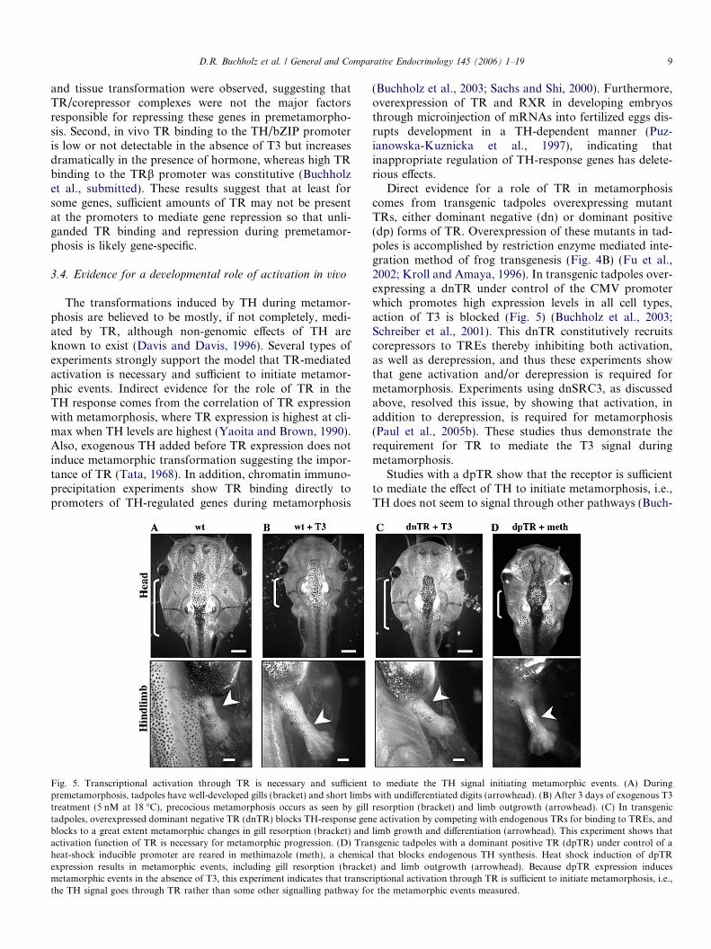
Direct evidence for a role of TR in metamorphosiscomes from transgenic tadpoles overexpressing mutantTRs, either dominant negative (dn) or dominant positive(dp) forms of TR. Overexpression of these mutants in tad-poles is accomplished by restriction enzyme mediated inte-gration method of frog transgenesis (Fig. 4B) (Fu et al.,2002; Kroll and Amaya, 1996). In transgenic tadpoles over-expressing a dnTR under control of the CMV promoterwhich promotes high expression levels in all cell types,action of T3 is blocked (Fig. 5) (Buchholz et al., 2003;Schreiber et al., 2001). This dnTR constitutively recruitscorepressors to TREs thereby inhibiting both activation,as well as derepression, and thus these experiments showthat gene activation and/or derepression is required formetamorphosis. Experiments using dnSRC3, as discussedabove, resolved this issue, by showing that activation, inaddition to derepression, is required for metamorphosis(Paul et al., 2005b). These studies thus demonstrate therequirement for TR to mediate the T3 signal duringmetamorphosis.
Studies with a dpTR show that the receptor is su"cientto mediate the e!ect of TH to initiate metamorphosis, i.e.,TH does not seem to signal through other pathways (Buch-
Fig. 5. Transcriptional activation through TR is necessary and su"cient to mediate the TH signal initiating metamorphic events. (A) Duringpremetamorphosis, tadpoles have well-developed gills (bracket) and short limbs with undi!erentiated digits (arrowhead). (B) After 3 days of exogenous T3treatment (5 nM at 18 !C), precocious metamorphosis occurs as seen by gill resorption (bracket) and limb outgrowth (arrowhead). (C) In transgenictadpoles, overexpressed dominant negative TR (dnTR) blocks TH-response gene activation by competing with endogenous TRs for binding to TREs, andblocks to a great extent metamorphic changes in gill resorption (bracket) and limb growth and di!erentiation (arrowhead). This experiment shows thatactivation function of TR is necessary for metamorphic progression. (D) Transgenic tadpoles with a dominant positive TR (dpTR) under control of aheat-shock inducible promoter are reared in methimazole (meth), a chemical that blocks endogenous TH synthesis. Heat shock induction of dpTRexpression results in metamorphic events, including gill resorption (bracket) and limb outgrowth (arrowhead). Because dpTR expression inducesmetamorphic events in the absence of T3, this experiment indicates that transcriptional activation through TR is su"cient to initiate metamorphosis, i.e.,the TH signal goes through TR rather than some other signalling pathway for the metamorphic events measured.
D.R. Buchholz et al. / General and Comparative Endocrinology 145 (2006) 1–19 9
holz et al., 2004). Because early embryogenesis may bea!ected by inappropriate TR function (Puzianowska-Kuznicka et al., 1997; Wong and Shi, 1995), dpTR wasput under control of a heat shock-inducible promoter tocontrol the timing of transgene expression (Fu et al.,2002; Wheeler et al., 2000). In addition, to rule out poten-tial e!ects from endogenous TH, transgenic tadpoles werereared in methimazole, a chemical that blocks endogenousTH synthesis (Buckbinder and Brown, 1993). Upon heatshock to induce transgene expression, dpTR transgenictadpoles at premetamorphic stages underwent metamor-phosis in the absence of hormone like the wild type siblingstreated with T3 (Fig. 5). It is important to note that dpTRexpression and exogenous T3 share similar phenotypes, inthat, unlike natural metamorphosis, induced metamorpho-sis is characterized by simultaneous transformation of TH-responsive tissues and a lack of normal developmentalasynchrony. Furthermore, dpTR was shown to bindendogenous TH target promoters, and all TH-responsegenes, both early and late, were found to be regulated asin tadpoles treated with TH. These studies indicate thatTR is su"cient to mediate the e!ects of exogenous T3 onall metamorphic parameters that were measured.
3.5. Molecular and developmental roles of TR isoforms
The Xenopus TRa and TRb have no known functionaldi!erences in transcription regulation in vitro or in vivoat the molecular level (Puzianowska-Kuznicka et al.,1997; Wong and Shi, 1995). Oocyte assays using eitherTRa or TRb show no di!erence in transcription from theTRb promoter (Wong and Shi, 1995). In vivo, both TRaand TRb bind T3-regulated promoters, and TRa can acti-vate all tested direct response genes, based on TRa trans-genic receptors (Buchholz et al., 2004). In addition,Scatchard analysis indicates no di!erence in a"nitybetween TRa and TRb for binding to TREs of TRb andTH/bZIP promoters, though both TRs bind the TRb pro-moter with 4-fold higher a"nity than the TH/bZIP pro-moter (Buchholz et al., submitted). However, experimentsfrom mammalian TRa and TRb domain swapping studiessuggest di!erences in gene regulation from a negative TRE,though no di!erences were found on positive TREs (Guis-souma et al., 2002). Thus, di!erences in molecular mecha-nisms of TRa and TRb in gene regulation at di!erentpromoters through the divergent N-terminal domains ofthe TRs cannot be ruled out completely, although theobserved di!erences between the TRs may be due to in vivopost-translational modification di!erences between TRaand TRb leading to di!erent levels of the receptors underthe assay conditions of the mammalian study (Guissoumaet al., 2002). Even though, for the most part, TRa and TRbseem to have similar molecular roles at TH-response pro-moters, they likely have di!erent roles in developmentbecause of di!erences in the TH inducibility of their expres-sion and other unknown aspects of the TR promoters con-trolling their tissue-specific di!erential expression.
As detailed above, the temporal expression profiles ofTRa and TRb led to the dual function model. TRa proteinlevels are 2–3-fold more than TRb in the head or tail at pre-metamorphosis and do not change significantly as meta-morphosis proceeds, whereas TRb protein expressiondramatically increases through metamorphic climax (Elice-iri and Brown, 1994). This major di!erence between theTRs may be explained by the presence of a TRE in theTRb promoter, which is induced by the T3 present duringmetamorphsis (Machuca et al., 1995; Ranjan et al., 1994).Consequently, at metamorphic climax, TRb protein levelsexceed TRa levels in both head and tail (Eliceiri andBrown, 1994). Thus, TRa may serve predominantly asthe repression arm of the dual function model, whereasthe feed forward autoregulation of TRb expression (Tata,1994; Yaoita and Brown, 1990) may ensure that TRb playsa critical role in carrying out activation duringmetamorphosis.
The increase in TRb levels, or autoregulation, is corre-lated with metamorphosis (Shi and Ishizuya-Oka, 1997),though direct experimental support for the importance ofautoregulation is lacking. Indirect evidence comes fromthe near background levels of TR binding to the TH/bZIPpromoter in premetamorphosis due to a lower a"nity TREcompared to the TRE in the TRb promoter (Buchholzet al., submitted). Only an increase in TR expression levelsby autoregulation will allow lower a"nity TREs to beoccupied enabling their upregulation duringmetamorphosis.
Another di!erence between receptors is tissue di!erenc-es, i.e., di!erent tissues may have di!erent relative amountsof TRa vs. TRb not explained by the TRb promoter TRE.For example, in the hind limb, high TRb mRNA expres-sion is localized to a subset of cells during transformation,whereas TRa mRNA is spread throughout (Faircloughand Tata, 1997; Rabelo et al., 1994). Also, in the tadpolebrain, proliferating cells in the subventricular zones expresshigh levels of TRa mRNA, whereas these cells do notexpress TRb (R.J. Denver, pers. comm.) Cells distal tothe ventricles that are destined to di!erentiate or undergoapoptosis express TRb. Use of an isoform-specific agonist,GC-1, showed developmental disregulation di!erent fromthat observed upon T3 treatment (Furlow et al., 2004). Invitro, GC-1 preferentially activates TRb over TRa with a10-fold di!erence in selectivity. Treatment of tadpoles withGC-1 leads to tail and gill resorption with lesser e!ects onhind limb outgrowth. These organ-specific responses can beexplained by tissue-specific TRa expression levels in pre-metamorphosis and/or tissue-specific ability for TRbautoinduction. The tail resorbs and limbs do not in GC-1-treated tadpoles either because the high TRa in the limbs(Kawahara et al., 1991; Cai and Brown, 2004) acts as adominant negative against the small amount of TRb thatis induced by GC-1in the limb or because the limbs donot strongly induce TRb. In the tail, the small amount ofTRb upregulated by GC-1 can continue autoregulationwithout much competitive inhibition from low endogenous
10 D.R. Buchholz et al. / General and Comparative Endocrinology 145 (2006) 1–19
levels of TRa and therefore carry out the GC-1-inducedtransformation. These examples suggest the two TR iso-forms have di!erent developmental roles, in that TRa isassociated with proliferating cells and TRb with cell di!er-entiation, though high levels of TRb are correlated withboth larval epithelial cell death and adult epithelial cell pro-liferation in the intestine (Shi and Ishizuya-Oka, 1997).Thus, the similarities in molecular action of TRa andTRb at the level of gene regulation contrast with the diver-gent roles of the TR isoforms in development due to di!er-ential control of the expression levels in and across tissues.On the other hand, because it seems necessary to have pro-liferative events precede resorptive or di!erentiative eventsin metamorphosis, the TR isoforms, with their di!erentialexpression temporally and spatially due to tissue-specificlevels of TRa expression and TRb autoinduction, mayact, not divergently, but coordinately as another mecha-nism to control developmental timing of metamorphicevents (Shi et al., 1996).
3.6. Tissue-specific consequences of TR activation
Di!erent cell types have very di!erent responses to THactivation, e.g., cell death in the tail and cell proliferationin limbs and both death and proliferation in remodeledorgans (Dodd and Dodd, 1976). These di!erent cell fatessuggest tissue-specific gene regulation cascades inducedby TR. Many TH-regulated genes have been isolated bysubtractive hybridization from intestine (Shi and Brown,1993), tail (Wang and Brown, 1993), limb (Buckbinderand Brown, 1992), and brain (Denver et al., 1997), andmany more are now being identified by microarray technol-ogy (Helbing et al., 2003; Veldhoen et al., 2002). Eventhough many of these genes are similarly upregulated inall tissues, e.g., TRb and TH-responsive basic leucine zip-per transcription factor, unique cell expression patternsamong other TH-response genes, such as sonic hedgehogin the intestinal epithelium, may underlie the di!ering cellfates among tissues. Indeed, in situ hybridization analysisof TH-induced genes in the tail (Berry et al., 1998b) andhead (Berry et al., 1998a), reveal cell type-specific expres-sion patterns of many induced genes within these bodyregions.
The extent of cell autonomy of the tissue-specificresponses to T3 has been probed using transgenicapproaches to block the action of T3 in a particular celltype by overexpressing dominant negative TRs or DIIIwith tissue-specific promoters. Both cell autonomousevents and events requiring TH-activation of other celltypes have been identified (Nakajima et al., 2005). Overex-pression of a dominant negative TR under control of a lar-val keratin promoter directing expression specifically inlarval epidermis, inhibited the death of these cells inresponse to T3 without a!ecting adjacent fibroblasts or for-mation of adult skin (Schreiber and Brown, 2003). Using amuscle-specific promoter expressing dominant negativeTR, early TH-induced death in muscle cells in the tail
was delayed until the tail shortened later in metamorphosisby inducing non-cell autonomous muscle cell death (Daset al., 2002; Yaoita and Nakajima, 2003). Also, muscle cellsof the limbs were very poorly developed, even thoughbones, nerves, and skin of the limb was normal. Theseexperiments revealed the cell autonomous nature of earlytail muscle death and limb muscle growth, which is inde-pendent of TH action on other cell types. In addition,non-autonomous development was observed in that tailmuscles eventually died, presumably by breakdown of con-nective tissue around the muscle cells caused by TH-acti-vated fibroblasts. Similarly, dominant negative TRoverexpressed in neurons showed cell autonomousdevelopment of spinal cord cells innervating the limbs(Marsh-Armstrong et al., 2004). Further evidence for theimportance of both cell autonomy and cell–cell interactionin TH-induced transformation came from in vitro tissuerecombination studies of the intestine where the impor-tance of intercellular signals in both directions between epi-thelium and connective tissue were observed (Ishizuya-Okaand Shimozawa, 1992, 1994).
4. Implications of TR studies in X. laevis
To what extent can we use our knowledge of TR func-tion during metamorphosis of X. laevis to help understandvertebrate development and the evolution of vertebratediversity? Among vertebrates, metamorphosis, character-ized by a peak in TH levels and a sudden larval to juveniletransition, seems to be found in three distantly relatedgroups from each other, namely, amphibians, Pleuronecti-formes (flatfish), and Elopimorpha (eels and relatives)(Norris, 1983; Tagawa et al., 1990; Youson, 1988). Lam-preys undergo TH-dependent metamorphosis, but the lar-val to juvenile transition is precipitated by a drop in THlevels, rather than a peak (Youson, 2003). Many groupsof teleosts have larvae with an extended transition periodwhere larval traits are gradually transformed to adult ver-sions (Urho, 2002; Youson, 1988), in some ways resem-bling the development in utero or in ovo of mammals,birds, and reptiles.
Despite lack of a larval period and metamorphosis inmost vertebrates, all vertebrates are believed to requireTH for normal post-embryonic development. In fact, apeak in plasma TH concentration associated with thyroidhormone-dependent development occurs across verte-brates, such that developmental endocrinology studies inXenopus may contribute knowledge about developmentof all vertebrates. In mice, the period of weaning character-ized by a change in diet from milk to adult food is associ-ated with a peak in thyroid hormone (Hadj-Sahraoui et al.,2000; Henning, 1987), and the accompanying intestinalremodeling at weaning has been shown by hormone manip-ulation studies and TR gene knock mice to be dependenton TH (Plateroti et al., 1999). A peak in TH also occursin humans at birth (Fisher, 2002) and in some fish, suchas salmon, during smoltification (Norris, 1996), a transi-
D.R. Buchholz et al. / General and Comparative Endocrinology 145 (2006) 1–19 11
tion period in physiology from freshwater parrs to saltwa-ter smolts. However, the role of TH physiology duringthese periods in humans and fish is not clear. An importantfeature of detailed knowledge of TR function in Xenopuslies in the ability to identify possible changes in molecularmechanisms and developmental roles of TH and TR thatmay have contributed to the large diversity of morphologyand physiology across vertebrates (for extended reviews ofthese ideas applied to salamander larval diversity, see Rose,1996, 1999). First, we discuss whether the dual functionmodel applies to vertebrates without metamorphosis, andthen we suggest potential changes in the developmentalrole of TR, including changes in tissue sensitivity, response,and dependence, that may underlie evolutionary diversity.
4.1. Dual function of TR during development in othervertebrates
Comparisons between molecular mechanisms character-ized in frogs and mammalian cell culture studies, such ashistone acetylation and cofactor recruitment, reveal anemerging picture of similarity across vertebrates at the levelof molecular mechanisms controlling gene regulation at thepromoter (see detailed examples above). The molecularcomponents that interact with TR in frogs are homologousto those in Hela cells and mice, i.e., similarities are the rulefor TRE binding, cofactor recruitment, chromatin remod-eling, and role of ligand. Does this similarity in molecularmechanisms of TR in gene regulation extend to a dualfunction model for development applicable across verte-brates, where TR functions as a repressor to downregulategenes important for development, which are subsequentlyactivated via TR upon the presence of T3? Indirect evi-dence from mouse knockout studies suggests the dual func-tion model does apply. TR double knockouts have a lesssevere phenotype than hypothyroid mice, suggesting thatuncontrolled, TR-mediated repression has deleteriousdevelopmental e!ects (Flamant and Samarut, 2003). Also,overexpression of a dominant negative N-CoR in liverparenchyma upregulated liver TH response genes, indicat-ing that TR mediates both repression and activation in theliver (Feng et al., 2001; Yen et al., 2003). More important-ly, during normal mouse development, unliganded TRarepresses heart rate in prenatal embryos, accompanyingthe repression of TRb and several genes encoding ion chan-nels involved in cardiac contractile activity, and at or afterbirth when the T3 levels are high, liganded TRa turns onthe expression of some of these same genes concomitantlywith heart rate increase (Mai et al., 2004). These resultsindicate that dual functions of TR are important for nor-mal mouse development as proposed for frog development.
4.2. Tissue sensitivity
Because molecular mechanisms of TR-mediated generegulation are shared across vertebrates, it is unlikely thatexplanations for developmental di!erences between species
will be found in molecular di!erences in TR function.Rather, developmental aspects of TR will contribute tovertebrate diversity. Di!erential timing of developmentalevents with respect to each other within Xenopus is wellknown, where the hind limbs develop before intestine,and this is believed be due, at least in part, to di!erentialtissue sensitivity to TH, i.e., tissue-specific control of theswitch from TR repression to TR activation as a result oftissue-specific levels of TRa, cytosolic TH binding protein,type II deiodinases, and/or type III deiodinases (Beckeret al., 1997; Shi et al., 1996). Di!erential expression of theseproteins that control developmental timing within speciesmay have changed during evolution to underlie di!erencesin timing of developmental events across species, for exam-ple, the late transformation of the extended oral disc usedto adhere to rocks in torrent-adapted tadpoles (Fig. 6A)(Nodzenski and Inger, 1990). The late transforming oraldisc may have lower sensitivity to TH due to high levelsof type III deiodinases or cytosolic TH binding proteinsor low TRa or type II expression levels.
Changes in tissue sensitivity may also underlie changesin sexual di!erentiation in desert frogs. New World spade-foot toads breed in extremely ephemeral desert pools, andrapid metamorphosis has evolved to allow tadpoles toachieve tail resorption before drying (Buchholz and Hayes,2002). Changes in TH physiology underlie their ability tometamorphose faster than their non-desert-adapted closestrelatives, where New World spadefoot toads have highertissue content of, and faster response to, TH (Buchholzand Hayes, 2005). This endocrine evolution had conse-quences for the developmental timing of gonad di!erentia-tion, which is not under control of TH (Ogielska andKotusz, 2004). Whereas gonad di!erentiation occurs dur-ing the larval period in spadefoot species with long larvalperiods, sexual di!erentiation occurs after metamorphosisin species with rapid metamorphosis likely because somaticmetamorphosis has been accelerated relative to gonaddevelopment by increased activity of TH physiology(Fig. 6B).
4.3. Tissue response
Another means of TH-mediated evolutionary diver-gence is that di!erent species may have di!erent tissueresponses to TH. Within a species, the downstream conse-quences of the switch from TR repression to activation isdrastically di!erent in di!erent tissues, in that, for example,cell death and cell proliferation are simultaneously inducedby TH in a tissue-dependent manner, e.g., cell death occursin the tail, whereas cell proliferation occurs in the limbs.Presumably, tissue-specific gene regulatory cascades under-lie tissue-specific responses to TH, such that di!erent genesare induced by TH in di!erent cell types (Berry et al.,1998a). The basis of tissue-specific responses within Xeno-pus may provide potential mechanisms for di!erences inthe same tissue between species. A colorful example of anevolutionary change in TH-induced gene regulation cas-
12 D.R. Buchholz et al. / General and Comparative Endocrinology 145 (2006) 1–19
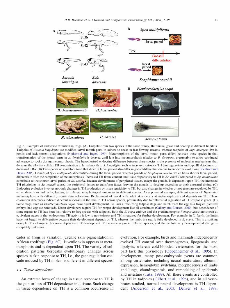
cades in frogs is variation juvenile skin pigmentation inAfrican reedfrogs (Fig. 6C). Juvenile skin appears at meta-morphosis and is dependent upon TH. The variety of col-oration patterns bespeaks evolutionary changes acrossspecies in skin response to TH, i.e., the gene regulation cas-cade induced by TH in skin is di!erent in di!erent species.
4.4. Tissue dependence
An extreme form of change in tissue response to TH isthe gain or loss of TH dependence in a tissue. Such changein tissue dependence on TH is a common occurrence in
evolution. For example, birds and mammals independentlyevolved TH control over thermogenesis, lipogenesis, andlipolysis, whereas cold-blooded vertebrates for the mostpart lack this physiology (Oppenheimer et al., 1995). Indevelopment, many post-embryonic events are commonamong vertebrates, including neural maturation, albuminexpression, hemoglobin switching, morphogenesis of limbsand lungs, chondrogenesis, and remodeling of epidermisand intestine (Tata, 1999). All these events are controlledby TH in tadpoles (Gilbert et al., 1996), and in all verte-brates studied, normal neural development is TH-depen-dent (Anderson et al., 2003; Denver et al., 1997;
Fig. 6. Examples of endocrine evolution in frogs. (A) Tadpoles from two species in the same family, Bufonidae, grow and develop in di!erent habitats.Tadpoles of Ansonia longidigita use modified larval mouth parts to adhere to rocks in fast-flowing streams, whereas tadpoles of Bufo divergens live inponds and lack torrent adaptations (Nodzenski and Inger, 1990). Metamorphosis of the larval mouth parts di!ers between these species in thattransformation of the mouth parts in A. longidigita is delayed until late into metamorphosis relative to B. divergens, presumably to allow continuedadherence to rocks during metamorphosis. The hypothesized endocrine di!erence between these species is the presence of molecular mechanisms thatdecrease the e!ective cellular TH concentration in larval mouth in A. longidigita, such as increased cytosolic TH binding protein and type III deiodinase ordecreased TRa. (B) Two species of spadefoot toad that di!er in larval period also di!er in gonad di!erentiation due to endocrine evolution (Buchholz andHayes, 2005). Gonads of Spea multiplicata di!erentiate during the larval period, whereas gonads of Scaphiopus couchii, which has a shorter larval period,di!erentiate after the completion of metamorphosis. Increased TH tissue content and tissue responsivity to TH in Sc. couchii compared to Sp. multiplicatacontribute to the shorter larval period of Sc. couchii. Because development of peripheral tissues, except the gonads, is dependent upon TH, the increasedTH physiology in Sc. couchii caused the peripheral tissues to transform faster, leaving the gonads to develop according to their ancestral timing. (C)Endocrine evolution involves not only changes in TH production or tissue sensitivity to TH, but also changes in whether or not genes are regulated by TH,either directly or indirectly, leading to di!erent morphological outcomes in di!erent species. As a potential example, di!erent species of Hyperoliusmetamorphose with di!erent juvenile skin coloration. Replacement of larval with adult skin occurs at metamorphosis and depends on TH. Thesecoloration di!erences indicate di!erent responses in the skin to TH across species, presumably due to di!erential regulation of TH-response genes. (D)Some frogs, such as Eleutherodactylus coqui, have direct development, i.e. lack a free-living tadpole stage and hatch from the egg as a froglet (picturedembryo had egg sac removed). Direct developers require TH for proper development like all vertebrates (Callery and Elinson, 2000), but dependence ofsome organs to TH has been lost relative to frog species with tadpoles. Both the E. coqui embryo and the premetamorphic Xenopus laevis are shown atequivalent stages in that endogenous TH activity is low to non-existent and TH is required for further development. For example, in X. laevis, the limbshave not begun to di!erentiate because their development depends on TH, whereas the limbs are nearly fully developed in E. coqui. This is a strikingexample of a change in hormone dependence of development of the same organ in di!erent species, and the evolutionary developmental change iscompletely unknown.
D.R. Buchholz et al. / General and Comparative Endocrinology 145 (2006) 1–19 13
Morreale de Escobar et al., 2004). However, the role of THand TR in the other post-embryonic events has not beenconsistently conserved. Based on TR knockout mice, TRis involved in intestine development at weaning (Platerotiet al., 1999), whereas no such role for TR is expected toexist in human intestinal remodeling (Menard, 2004). Inzebrafish, pectoral fin morphogenesis, chondrogenesis,and intestine development are TH dependent (Brown,1997; Liu and Chan, 2002), though skin developmentapparently is not.
A striking example of changes in tissue dependence onTH within frogs is found in direct developers, where thereis no free-living tadpole stage and froglets hatch out of theegg (Fig. 6D) (Elinson, 2001; Jennings and Hanken, 1998).As in Xenopus, adult skin formation, Meckel!s cartilageproliferation, and other events are TH-regulated in Eleut-herodactylus (Callery and Elinson, 2000). On the otherhand, a long, coiled intestine is lacking in Eleutherodacty-lus, and intestine development seems to be independentof TH because hatched froglets lack a di!erentiated intes-tine which forms subsequent to hatching (Lynn, 1942).Thus, changes in tissue response to, as well as changes independence on, TH during development contribute to dif-ferences between direct developers and species with larvaldevelopment.
The mechanisms for how tissues become dependent orindependent of hormonal control are not clear. A potentialclass of mechanisms to change gene regulation by TRacross species is through changes in the existence of aTRE in promoters of TH-regulated genes. Few such com-parative studies have been done, but TH regulation of itsown receptor is an exception. For example, TRb is TH-reg-ulated in Xenopus (Yaoita and Brown, 1990), Rana cates-beiana (Helbing et al., 1992; Schneider and Galton, 1991),zebrafish (Liu and Chan, 2002), and chicken (Forrestet al., 1990). Similarly, in rats, TRb increases in the brainaround birth coinciding with TH peak (Strait et al.,1990). In contrast, TRa, and not TRb, is TH-regulated inturbot fish (Marchand et al., 2003). In conger eel (Kawaka-mi et al., 2003), TRa isoforms are upregulated transientlyduring metamorphosis, and TRb isoforms are upregulatedat metamorphosis and remain highly expressed into adult-hood. In the neotenic salamanders, TRs are not regulatedby TH in the axolotl (Safi et al., 2004), and in Necturus,TRa but not TRb is expressed in the intestine, liver, andgills (Safi et al., 1997). In mice (Sadow et al., 2003), TH reg-ulation of TR is tissue-specific, where TRa is upregulatedby T3 in the heart and downregulated in the liver, whereasthe opposite is true for TRb. The molecular mechanismsunderlying these di!erences in TR regulation await charac-terization and comparison of TR promoters across di!er-ent species.
5. Conclusion
TH plays important roles in post-embryonic develop-ment across vertebrates. Studies on amphibian metamor-
phosis have elucidated critical in vivo mechanismsgoverning the gene expression pathways regulated by TH.In particular, a dual function model for the role of TR indevelopment has been proposed and supported by a num-ber of in vivo studies first in X. laevis and then in mouse.Conservation across divergent species from fish to frog tohuman in the molecular mechanisms of TH signaling mech-anisms suggest that changes in gene- or tissue-specific sen-sitivity and/or response to TH may be a significant factorin evolutionary diversity. The key to this issue is to under-stand mechanisms of tissue-specificity, i.e., how does thetail degenerate and the intestine remodel in response tothe same TH signal. Molecularly, it is critical to understandtissue-specific gene regulation by TH within an organism,because then, we can hypothesize what is required torelieve such development of its TH dependence in the sametissue of di!erent organisms. The first step towards deter-mining mechanisms of TH-dependent evolutionary chang-es is to characterize the TH-induced gene regulationcascade in di!erent tissues, most easily done in a modelorganism with a sequenced genome where hormonal con-trol of development can be easily studied as in Xenopus,and then applying this knowledge towards understandingtissue-dependence on hormones in vertebrate evolution.
Acknowledgments
We greatly appreciate comments on the manuscript byR. Denver. This work also benefited from a National Re-search Service Award from National Institutes of Healthto DRB.
References
Allen, B.M., 1929. The influence of the thyroid gland and hypophosisupon growth and development of amphibian larvae. Q. Rev. Biol. 4,325–352.
Allen, B.M., 1938. The endocrine control of amphibian metamorphosis.Biol. Rev. 13, 1–19.
Anderson, G.W., Schoonover, C.M., Jones, S.A., 2003. Control of thyroidhormone action in the developing rat brain. Thyroid 13, 1039–1056.
Atkinson, B.G., 1994. Metamorphosis: model systems for studying geneexpression in postembryonic development. Dev. Genet. 15, 313–319.
Baker, B.S., Tata, J.R., 1990. Accumulation of proto-oncogene c-erb-Arelated transcripts during Xenopus development: association with earlyacquisition of response to thyroid hormone and estrogen. EMBO J. 9,879–885.
Balcells, E.R., 1955. Contributions to the study of the life cycle of Spanishamphibians. Br. J. Herpetol. 2, 1–6.
Becker, K.B., Stephens, K.C., Davey, J.C., Schneider, M.J., Galton, V.A.,1997. The type 2 and type 3 iodothyronine deiodinases play importantroles in coordinating development in Rana catesbeiana tadpoles.Endocrinology 138, 2989–2997.
Berry, D.L., Rose, C.S., Remo, B.F., Brown, D.D., 1998a. The expressionpattern of thyroid hormone response genes in remodeling tadpoletissues defines distinct growth and resorption gene expression pro-grams. Dev. Biol. 203, 24–35.
Berry, D.L., Schwartzman, R.A., Brown, D.D., 1998b. The expressionpattern of thyroid hormone response genes in the tadpole tail identifiesmultiple resorption programs. Dev. Biol. 203, 12–23.
14 D.R. Buchholz et al. / General and Comparative Endocrinology 145 (2006) 1–19
Brown, D.D., 1997. The role of thyroid hormone in zebrafish and axolotldevelopment. Proc. Natl. Acad. Sci. 94, 13011–13016.
Brown, D.D., Wang, Z., Furlow, J.D., Kanamori, A., Schwartzman,R.A., Remo, B., Pinder, A., 1996. The thyroid hormone-induced tailresorption program during Xenopus laevis. Proc. Natl. Acad. Sci. 93,1924–1929.
Buchholz, D.R., Hayes, T.B., 2002. Evolutionary patterns of diversity inspadefoot toad metamorphosis (Anura: Pelobatidae). Copeia 2002,180–189.
Buchholz, D.R., Hayes, T.B., 2005. Variation in thyroid hormone actionand tissue content underlies species di!erences in the timing ofmetamorphosis in desert frogs. Evol. Dev. 7 (5), 458–467.
Buchholz, D.R., Hsia, S.-C.V., Fu, L., Shi, Y.-B., 2003. A dominantnegative thyroid hormone receptor blocks amphibian metamorphosisby retaining corepressors at target genes. Mol. Cell. Biol. 23, 6750–6758.
Buchholz, D.R., Paul, B.P., Shi, Y.-B. Gene-specific changes in promoteroccupancy by thyroid hormone receptor during frog metamorphosis:implications for developmental gene regulation. J. Biol. Chem.(submitted).
Buchholz, D.R., Tomita, A., Fu, L., Paul, B.D., Shi, Y.-B., 2004.Transgenic analysis reveals that thyroid hormone receptor is su"cientto mediate the thyroid hormone signal in frog metamorphosis. Mol.Cell. Biol. 24, 9026–9037.
Buckbinder, L., Brown, D.D., 1992. Thyroid hormone-induced geneexpression changes in the developing frog limb. J. Biol. Chem. 267,25786–25791.
Buckbinder, L., Brown, D.D., 1993. Expression of the Xenopus laevisprolactin and thyrotropin genes during metamorphosis. Proc. Natl.Acad. Sci. 90, 3820–3824.
Burke, L.J., Baniahmad, A., 2000. Co-repressors. FASEB J. 14, 1876–1888.
Cai, L., Brown, D.D., 2004. Expression of type II iodothyroninedeiodinase marks the time that a tissue responds to thyroid hor-mone-induced metamorphosis in Xenopus laevis. Dev. Biol. 266, 87–95.
Callery, E.M., Elinson, R.P., 2000. Thyroid hormone-dependent meta-morphosis in a direct developing frog. Proc. Natl. Acad. Sci. 97, 2615–2620.
Chakravarti, D., LaMorte, V.J., Nelson, M.C., Nakajima, T., Schulman,I.G., Juguilon, H., Montminy, M., Evans, R.M., 1996. Role of CBP/P300 in nuclear receptor signalling. Nature 383, 99–103.
Chen, J.D., Evans, R.M., 1995. A transcriptional co-repressor thatinteracts with nuclear hormone receptors. Nature 377, 454–457.
Chen, J.D., Li, H., 1998. Coactivation and corepression in transcriptionalregulation by steroid/nuclear hormone receptors. Crit. Rev. Eukaryot.Gene Expr. 8, 169–190.
Chen, H., Lin, R.J., Schiltz, R.L., Chakravarti, D., Nash, A., Nagy, L.,Privalsky, M.L., Nakatani, Y., Evans, R.M., 1997. Nuclear receptorcoactivator ACTR is a novel histone acetyltransferase and forms amultimeric activation complex with P/CAF and CBP/p300. Cell 90,569–580.
Cohen, R.N., Brzostek, S., Kim, B., Chorev, M., Wondisford, F.D.,Hollenberg, A.N., 2001. The specificity of interactions between nuclearhormone receptors and corepressors is mediated by distinct amino acidsequences within the interacting domains. Mol. Endocrinol. 15, 1049–1061.
Cossette, S.M.M., Drysdale, T.A., 2004. Early expression of thyroidhormone receptor beta and retinoid X receptor gamma i the Xenopusembryo. Di!erentiation 72, 239–249.
Damjanovski, S., Ishizuya-Oka, A., Shi, Y.-B., 1999. Spatial and temporalregulation of collagenases-3, -4, and stromelysin-3 implicates distinctfunctions in apoptosis and tissue remodeling during frog metamor-phosis. Cell Res. 9, 91–105.
Damjanovski, S., Sachs, L.M., Shi, Y.-B., 2002. Function of thyroidhormone receptors during amphibian metamorphosis. Methods Mol.Biol. 202, 153–176.
Das, B., Schreiber, A.M., Huang, H., Brown, D.D., 2002. Multiplethyroid hormone-induced muscle growth and death programs during
metamorphosis in Xenopus laevis. Proc. Natl. Acad. Sci. 99, 12230–12235.
Das, P.M., Ramachandran, K., vanWert, J., Singal, R., 2004. Chromatinimmunoprecipitation assay. Biotechniques 37, 961–969.
Davis, P.J., Davis, F.B., 1996. Nongenomic actions of thyroid hormone.Thyroid 6, 497–504.
Dent, J.N., 1988. Hormonal interaction in amphibian metamorphosis.Am. Zool. 28, 297–308.
Denver, R.J., 1996. Neuroendocrine control of amphibian metamorpho-sis. In: Gilbert, L.I., Tata, J.R., Atkinson, B.G. (Eds.), Metamorpho-sis: Postembryonic Reprogramming of Gene Expression in Amphibianand Insect Cells. Academic Press, San Diego, pp. 433–464.
Denver, R.J., 1999. Evolution of the corticotropin-releasing hormonesignaling system and its role in stress-induced phenotypic plasticity.Ann. N. Y. Acad. Sci. 897, 46–53.
Denver, R.J., Glennemeier, K.A., Boorse, G.C., 2002. Endocrinology ofComplex Life Cycles: Amphibians. In: Pfa!, D.W., Arnold, A.P.,Etgen, A.M., Fahrbach, S.E., Rubin, R.T. (Eds.), Hormones, Brainand Behavior, Vol. 2. Academic Press, New York, pp. 469–513.
Denver, R.J., Pavgi, S., Shi, Y.-B., 1997. Thyroid hormone-dependentgene expression program for Xenopus neural development. J. Biol.Chem. 272, 8179–8188.
Desvergne, B., 1994. How do thyroid hormone receptors bind tostructurally diverse response elements? Mol. Cell. Endocrinol. 100,125–131.
Dodd, M.H.I., Dodd, J.M., 1976. The biology of metamorphosis. In:Lofts, B. (Ed.), Physiology of Amphibia, Vol. 3. Academic Press, NewYork, USA, pp. 467–599.
Eliceiri, B.P., Brown, D.D., 1994. Quantitation of endogenous thyroidhormone receptors a and b during embryogenesis and metamorphosisin Xenopus laevis. J. Biol. Chem. 269, 24459–24465.
Elinson, R.P., 2001. Direct development: an alternative way to make afrog. Genesis 29, 91–95.
Etkin, W., 1932. Growth and resorption phenomena in anuran metamor-phosis. Physiol. Zool. 5, 275–300.
Etkin, W., 1935. The mechanisms of anuran metamorphosis 1. Thyroxineconcentration and the metamorphic pattern. J. Exp. Zool. 71, 312–340.
Etkin, W., 1964. Metamorphosis. In: Moore, J.A. (Ed.), Physiology of theAmphibia. Academic Press, New York, pp. 427–468.
Etkin, W., 1965. The phenomena of amphibian metamorphosis. IV. Thedevelopment of the median eminence. J. Morphol. 116, 371–378.
Fairclough, L., Tata, J.R., 1997. An immunocytochemical analysis of theexpression of thyroid hormone receptor alpha and beta proteins duringnatural and thyroid hormone-induced metamorphosis in Xenopus.Dev. Growth Di!er. 39, 273–283.
Feng, X., Jiang, Y., Meltzer, P., Yen, P.M., 2001. Transgenic targeting ofa dominant negative corepressor to liver blocks basal repression bythyroid hormone receptor and increases cell proliferation. J. Biol.Chem. 276, 15066–15072.
Fisher, D.A., 2002. Chapter 6: Fetal-perinatal thyroid physiology. In:Eugster, E.A., Pescovitz, O.H. (Eds.), Contemporary Endocrinology:Developmental Endocrinology: From research to clinical practice.Humana Press, Totowa, NJ, pp. 135–149.
Flamant, F., Samarut, J., 2003. Thyroid hormone receptors: lessons fromknockout and knock-in mutant mice. Trends Endocrinol. Metab. 14,85–90.
Forrest, D., Sjoberg, M., Vennstrom, B., 1990. Contrasting developmentaland tissue-specific expression of alpha and beta thyroid hormonereceptor genes. EMBO J. 9, 1519–1528.
Fox, H., 1981. Cytological and morphological changes during amphibianmetamorphosis. In: Gilbert, L.I., Frieden, E. (Eds.), Metamorphosis,A Problem in Developmental Biology. Plenum Press, New York, pp.327–362.
Fu, L., Buchholz, D.R., Shi, Y.-B., 2002. Novel double promoterapproach for identification of transgenic animals: a tool for in vivoanalysis of gene function and development of gene-based therapies.Mol. Reprod. Dev. 62, 470–476.
D.R. Buchholz et al. / General and Comparative Endocrinology 145 (2006) 1–19 15

Fu, L., Ishizuya-Oka, A., Buchholz, D.R., Amano, T., Matsuda, H., Shi,Y.-B., 2005. A causative role of stromelysin-3 in extracellular matrixremodeling and epithelial apoptosis during intestinal metamorphosisin Xenopus laevis. J. Biol. Chem. 280 (30), 27856–27865.
Furlow, J.D., Berry, D.L., Wang, Z., Brown, D.D., 1997. A set of noveltadpole specific genes expressed only in the epidermis are down-regulated by thyroid hormone during Xenopus laevis metamorphosis.Dev. Biol. 182, 284–298.
Furlow, J.D., Brown, D.D., 1999. In vitro and in vivo analysis of theregulation of a transcription factor gene by thyroid hormone duringXenopus laevis metamorphosis. Mol. Endocrinol. 13, 2076–2089.
Furlow, J.D., Kanamori, A., 2002. The transcription factor basictranscription element-binding protein 1 is a direct thyroid hormoneresponse gene in the frog Xenopus laevis. Endocrinology 143, 3295–3305.
Furlow, J.D., Yang, H.Y., Hsu, M., Lim, W., Ermio, D.J., Chiellini, G.,Scanlan, T.S., 2004. Induction of larval tissue resorption in Xenopuslaevis tadpoles by the thyroid hormone receptor agonist GC-1. J. Biol.Chem. 279, 26555–26562.
Galton, V.A., 1983. Thyroid hormone action in amphibian metamorpho-sis. In: Oppenheimer, J.H., Samuels, H.H. (Eds.), Molecular Basis ofThyroid Hormone Action. Academic Press, New York, pp. 445–483.
Gilbert, L.I., Frieden, E., 1981. Metamorphosis: A Problem in Develop-mental Biology. Plenum Press, New York, pp. 1-578.
Gilbert, L.I., Tata, J.R., Atkinson, B.G., 1996. Metamorphosis: Postem-bryonic Reprogramming of Gene Expression in Amphibian and InsectCells. Academic Press, San Diego, pp. 1–687.
Glass, C.K., Rosenfeld, M.G., 2000. The coregulator exchange intranscriptional functions of nuclear receptors. Genes Dev. 14, 121–141.
Gruca, S., Michalowski, J., 1961. The rate of development of gonadsagainst the background of the development of the whole organism inthe tadpole of Xenopus laevis Daud. Acta Biol. Cracov. IV, 191–197.
Gudernatsch, J.F., 1912. Feeding experiments on tadpoles. I. Theinfluence of specific organs given as food on growht and di!erentia-tion: a contribution to the knowledge of organs with internal secretion.Arch. Entwicklungsmech. Org. 35, 457–483.
Guenther, M.G., Lane, W.S., Fischle, W., Verdin, E., Lazar, M.A.,Shiekhattar, R., 2000. A core SMRT corepressor complex containingHDAC3 and TBL1, a WD40-repeat protein linked to deafness. GenesDev. 14, 1048–1057.
Guissouma, H., Dupre, S.M., Becker, N., Jeannin, E., Seugnet, I.,Desvergne, B., Demeneix, B.A., 2002. Feedback on hypothalamicTRH transcription is dependent on thyroid hormone receptor Nterminus. Mol. Endocrinol. 16, 1652–1666.
Hadj-Sahraoui, N., Seugnet, I., Ghorbel, M.T., Demeneix, B.A., 2000.Hypothyroidism prolongs mitotic activity in the post-natal mousebrain. Neurosci. Lett. 280, 79–82.
Havis, E., Sachs, L.M., Demeneix, B.A., 2003. Metamorphic T3-responsegenes have specific co-regulator requirements. EMBO Rep. 4, 883–888.
Helbing, C.C., Gergely, G., Atkinson, B.G., 1992. Sequential up-regula-tion of thyroid hormone b receptor, ornthine transcarbamylase, andcarbamyl phosphate synthetase mRNA in the liver of Rana catesbeianatadpoles during spontaneous and thyroid hormone-induced metamor-phosis. Dev. Genet. 13, 289–301.
Helbing, C.C., Werry, K., Crump, D., Domanski, D., Veldhoen, N.,Bailey, C.M., 2003. Expression profiles of novel thyroid hormone-responsive genes and proteins in the tail of Xenopus laevis tadpolesundergoing precocious metamorphosis. Mol. Endocrinol. 17, 1395–1409.
Henning, S.J., 1987. Chapter 9: Functional development of the gastroin-testinal tract. In: Johnson, L.R. (Ed.), Physiology of the Gastrointes-tinal Tract, Vol. 9. Raven Press, New York, pp. 285–300.
Hong, H., Kohli, K., Trivedi, A., Johnson, D.L., Stallcup, M.R., 1996.GRIP1, a novel mouse protein that serves as a transcriptionalcoactivator in yeast for the hormone binding domains of steroidreceptors. Proc. Natl. Acad. Sci. USA 93, 4948–4952.
Hoopfer, E.D., Huang, L., Denver, R.J., 2002. Basic transcription elementbinding protein is a thyroid hormone-regulated transcription factor
expressed during metamorphosis in Xenopus laevis. Dev. GrowthDi!er. 44, 365–381.
Horlein, A.J., Naar, A.M., Heinzel, T., Torchia, J., Gloss, B., Kurokawa,R., Ryan, A., Kamei, Y., Soderstrom, M., Glass, C.K., et al., 1995.Ligand-independent repression by the thyroid hormone receptormediated by a nuclear receptor co-repressor. Nature 377, 397–404.
Hoskins, E.R., Hoskins, M.M., 1919. Growth and development ofamphibia as a!ected by thyroidectomy. J. Exp. Zool. 29, 1–69.
Hourdry, J., 1993. Passage to the terrestrial life in amphibians II.Endocrine determinism. Zool. Sci. 10, 887–902.
Hsia, S.-C.V., Shi, Y.-B., 2002. Chromatin disruption and histoneacetylation in regulation of the human immunodeficiency virus type1 long terminal repeat by thyroid hormone receptor. Mol. Cell. Biol.22, 4043–4052.
Hu, X., Lazar, M.A., 1999. The CoRNR motif controls the recruitment ofcorepressors by nuclear hormone receptors. Nature 402, 93–96.
Huang, H., Marsh-Armstrong, N., Brown, D.D., 1999. Metamorphosis isinhibited in transgenic Xenopus laevis tadpoles that overexpress typeIII deiodinase. Proc. Natl. Acad. Sci. USA 96, 962–967.
Huang, H., Cai, L., Remo, B., Brown, D.D., 2001. Timing of metamor-phosis and the onset of the negative feedback loop between the thyroidgland and the pituitary is controlled by type II iodothyroninedeiodinase in Xenopus laevis. Proc. Natl. Acad. Sci. 98, 7348–7353.
Huang, Z.-Q., Li, J., Sachs, L.M., Cole, P.A., Wong, J.M., 2003. A rolefor cofactor–cofactor and cofactor–histone interactions in targetingp300, SWI/SNF and mediator for transcription. EMBO J. 22, 2146–2155.
Ishizuka, T., Lazar, M.A., 2003. The N-CoR/histone deacetylase 3complex is required for repression by thyroid hormone receptor. Mol.Cell. Biol. 23, 5122–5131.
Ishizuya-Oka, A., Shimozawa, A., 1992. Connective tissue is involved inadult epithelial development of the small intestine during anuranmetamorphosis in vitro. Roux!s Arch. Dev. Biol. 201, 322–329.
Ishizuya-Oka, A., Shimozawa, A., 1994. Inductive action of epithelium ondi!erentiation of intestinal connective tissue of Xenopus laevis tadpolesduring metamorphosis in vitro. Cell Tissue Res. 277, 427–436.
Ishizuya-Oka, A., Shimozawa, A., Takeda, H., Shi, Y.-B., 1994. Cell-specific and spatio-temporal expression of intestinal fatty acid-bindingprotein gene during amphibian metomorphosis. Roux!s Arch. Dev.Biol. 204, 150–155.
Ishizuya-Oka, A., Stolow, M., Ueda, S., Shi, Y.-B., 1997. Temporal andspatial expression of an intestinal Na#=PO3
4- cotransporter correlateswith epithelial transformatin during thyroid hormone-dependent frogmetamorphosis. Dev. Genet. 20, 53–66.
Ishizuya-Oka, A., Li, Q., Amano, T., Damjanovski, S., Ueda, S., Shi, Y.-B., 2000. Requirement for matrix metalloproteinase stromelysin-3 incell migratin and apoptosis during tissue remodeling in Xenopus laevis.J. Biol. Chem. 150, 1177–1188.
Ishizuya-Oka, A., Ueda, S., Inokuchi, T., Amano, T., Damjanovski, S.,Stolow, M., Shi, Y.-B., 2001. Thyroid hormone-induced expression ofsonic hedgehog correlates with adult epithelial development duringremodeling of the Xenopus stomach and intestine. Di!erentiation 69,27–37.
Ito, M., Roeder, R.G., 2001. The TRAP/SMCC/Mediator complex andthyroid hormone receptor function. Trends Endocrinol. Metab. 12,127–134.
Jennings, D.H., Hanken, J., 1998. Mechanistic basis of life historyevolution in anuran amphibians: thyroid gland development in thedirect-developing frog, Eleutherodactylus coqui. Gen. Comp. Endocri-nol. 111, 225–232.
Jones, P.L., Shi, Y.-B., 2003. N-CoR-HDAC corepressor complexes: rolesin transcriptional regulation by nuclear hormone receptors. In:Workman, J.L. (Ed.), Current Topics in Microbiology and Immunol-ogy: Protein Complexes that Modify Chromatin, Vol. 274. Springer,Berlin, pp. 237–268.
Kawahara, A., Baker, B.S., Tata, J.R., 1991. Developmental and regionalexpression of thyroid hormone receptor genes during Xenopus meta-morphosis. Development 111, 933–943.
16 D.R. Buchholz et al. / General and Comparative Endocrinology 145 (2006) 1–19
Kawakami, Y., Tanda, M., Adachi, S., Yamauchi, K., 2003. Character-ization of thyroid hormone receptor a and b in the metamorphosingJapanese conger eel, Conger myriaster. Gen. Comp. Endocrinol. 132,321–332.
Kaye, N.W., 1961. Interrelationships of the thyroid and pituitary inembryonic and premetamorphic stages of the frog, Rana pipiens. Gen.Comp. Endocrinol. 1, 1–19.
Kikuyama, S., Kawamura, K., Tanaka, S., Yamamoto, K., 1993. Aspectsof amphibian metamorphosis: hormonal control. Int. Rev. Cytol. 145,105–148.
Kollros, J.J., 1961. Mechanisms of amphibian metamorphosis: hormones.Am. Zool. 1, 107–114.
Kroll, K., Amaya, E., 1996. Transgenic Xenopus embryos from spermnuclear transplantations reveal FGF signaling requirements duringgastrulation. Development 122, 3173–3183.
Laudet, V., Gronemeyer, H., 2002. The Nuclear Receptor Facts Book.Academic Press, San Diego, pp. 1–462.
Leloup, J., Buscaglia, M., 1977. La triiodothyronine, hormone de lametamorphose des Amphibiens. C. R. Acad. Sci. Paris, Ser. D 284,2261–2263.
Lemon, B.D., Freedman, L.P., 1999. Nuclear receoptor cofactors aschromatin remodelers. Curr. Opin. Genet. Dev. 9, 499–504.
Li, H., Gomes, P.J., Chen, J.D., 1997. RAC3, a steroid/nuclear receptor-associated coactivator that is related to SRC-1 and TIF2. Proc. Natl.Acad. Sci. USA 94, 8479–8484.
Li, J., Lin, Q., Yoon, H.-G., Huang, Z.-Q., Strahl, B.D., Allis, C.D.,Wong, J., 2002. Involvement of histone methylation and phosphory-lation in regulation of transcription by thyroid hormone receptor. Mol.Cell. Biol. 22, 5688–5697.
Li, J., Wang, J., Wang, J., Nawaz, Z., Liu, J.M., Qin, J., Wong, J., 2000.Both corepressor proteins SMRT and N-CoR exist in large proteincomplexes containing HDAC3. EMBO J. 19, 4342–4350.
Liu, Y.-W., Chan, W.-K., 2002. Thyroid hormones are important forembyronic to larval transitory phase in zebrafish. Di!erentiation 70,36–45.
Luria, A., Furlow, J.D., 2004. Spatiotemporal retinoid-X receptoractivation detected in live vertebrate embryos. Proc. Natl. Acad. Sci.101, 8987–8992.
Lynn, W.G., 1942. The embryology of Eleutherodactylus nubicola, ananuran which has no tadpole stage. Contrib. Embryol. Carnegie Inst.Washington 190, 27–62.
Machuca, I., Esslemont, G., Fairclough, L., Tata, J.R., 1995. Analysis ofstructure and expression of the Xenopus thyroid hormone receptor-beta gene to explain its autoinduction. Mol. Endocrinol. 9, 96–107.
Mai, W., Janier, M.F., Allioli, N., Quignodon, L., Chuzel, T., Flamant,F., Samarut, J., 2004. Thyroid hormone receptor alpha is a molecularswitch of cardiac function between fetal and postnatal life. Proc. Natl.Acad. Sci. 101, 10332–10337.
Malo, M.S., Zhang, W., Alkhoury, F., Pushpakaran, P., Abedrapo, M.A.,Mozumder, M., Fleming, E., Siddique, A., Henderson, J.W., Hodin,R.A., 2004. Thyroid hormone positively regulates the enterocytedi!erentiation marker intestinal alkaline phosphatase gene via anatypical response element. Mol. Endocrinol. 18, 1941–1962.
Manzon, R.G., Denver, R.J., 2004. Regulation of pituitary thyroitropingene expression during Xenopus metamorphosis: negative feedback isfunctional throughout metamorphosis. J. Endocrinol. 182, 273–285.
Marchand, O., Du!raisse, M., Triqueneaux, G., Safi, R., Laudet, V., 2003.Molecular cloning and developmental expression patterns of thyroidhormone receptors and T3 target genes in the turbot (Scophtalmusmaximus) during post-embryonic development. Gen. Comp. Endocri-nol. 135, 345–357.
Marimuthu, A., Feng, W., Tagami, T., Nguyen, H., Jameson, J.L.,Fletterick, R.J., Baxter, J.D., West, B.L., 2002. TR surfaces andconformations required to bind nuclear receptor corepressor. Mol.Endocrinol. 16, 271–286.
Marsh-Armstrong, N., Cai, L., Brown, D.D., 2004. Thyroid hormonecontrols the development of connections between the spinal cord and
limbs during Xenopus laevis metamorphosis. Proc. Natl. Acad. Sci.101, 165–170.
Marsh-Armstrong, N., Huang, H., Remo, B.F., Liu, T.T., Brown, D.D.,1999. Asymmetric growth and development of the Xenopus laevisretina during metamorphosis is controlled by Type III deiodinases.Neuron 24, 871–878.
McAvoy, J.W., Dixon, K.E., 1977. Cell proliferation and renewal in thesmall intestine epithelium of metamorphosing and adult Xenopuslaevis. J. Exp. Zool. 202, 129–138.
McKenna, N.J., Lanz, R.B., O!Malley, B.W., 1999. Nuclear receptorcoregulators: cellular and molecular biology. Endocr. Rev. 20, 321–344.
Menard, D., 2004. Functional development of the human gastrointestinaltract: hormone- and growth factor-mediated regulatory mechanisms.Can. J. Gastroenterol. 18, 39–44.
Miller, L.D., McPhie, P., Suzuki, H., Kato, Y., Liu, E.T., Cheng, S.-y.,2004. Multitissue gene-expression analysis in a mouse model of thyroidhormone resistance. Genome Biol. 5, R17–R31.1.
Morreale de Escobar, G., Obregon, M.J., Escobar del Rey, F., 2004.Maternal thyroid hormones early in pregnancy and fetal braindevelopment. Best Pract. Res. Clin. Endocrinol. Metab. 18, 225–248.
Nakajima, K., Fujimoto, K., Yaoita, Y., 2005. Programmed cell deathduring amphibian metamorphosis. Semin. Cell Dev. Biol. 16, 271–280.
Nieuwkoop, P.D., Faber, J., 1994. Normal table of Xenopus laevis(Daudin). Garland Publishing Inc., New York, pp. 252.
Nodzenski, E., Inger, R.F., 1990. Uncoupling of related structural changesin metamorphosing torrent-dwelling tadpoles. Copeia 1990, 1047–1054.
Norris, D.O., 1983. Evolution of endocrine regulation of metamorphosisin lower vertebrates. Am. Zool. 23, 709–718.
Norris, D.O., 1996. Vertebrate Endocrinology. Academic Press, SanDiego, pp. 1–634.
Ogielska, M., Kotusz, A., 2004. Pattern and rate of ovary di!erentiationwith reference to somatic development in anuran amphibians. J.Morphol. 259, 41–54.
Okada, R., Yamamoto, K., Koda, A., Ito, Y., Hayashi, H., Tanaka, S.,Hanaoka, Y., Kikuyama, S., 2004. Development of radioimmunoassayfor bullfrog thyroid-stimulating hormone (TSH): e!ects of hypotha-lamic releasing hormones on the release of TSH from the pituitaryin vitro. Gen. Comp. Endocrinol. 135, 42–50.
Onate, S.A., Tsai, S.Y., Tsai, M.J., O!Malley, B.W., 1995. Sequence andcharacterization of a coactivator for the steroid hormone receptorsuperfamily. Science 270, 1354–1357.
Oofusa, K., Yoshizato, K., 1991. Biochemical and immunologicalcharacterization of collagenase in tissues of metamorphosing bullfrogtadoles. Dev. Growth Di!er. 33, 329–339.
Oofusa, K., Yoshizato, K., 1996. Thyroid hormone-dependent expressionof bullfrog tadpole collagenase gene. Roux!s Arch. Dev. Biol. 205,243–251.
Oofusa, K., Tooi, O., Kashiwagi, A., Kashiwagi, K., Kondo, Y.,Watanabe, Y., Sawada, T., Fujikawa, K., Yoshizato, K., 2001.Expression of thyroid hormone receptor bA gene assayed bytransgenic Xenopus laevis carrying its promoter sequences. Mol. Cell.Endocrinol. 181, 97–110.
Oppenheimer, J.H., Schwartz, H.L., Strait, K.A., 1995. Chapter 17: anintegrated view of thyroid hormone actions in vivo. In: Weintraub,B.D. (Ed.), Molecular Endocrinology: Basic Concepts and ClinicalCorrelations. Raven Press, New York, pp. 249–267.
Patterton, D., Hayes, W.P., Shi, Y.-B., 1995. Transcriptional activation ofthe matrix metalloproteinase gene stromelysin-3 coincides with thyroidhormone-induced cell death during frog metamorphosis. Dev. Biol.167, 252–262.
Paul, B.D., Buchholz, D.R., Fu, L., Shi, Y.-B., 2005a. Tissue- and gene-specific recruitment of steroid receptor coactivator-3 by thyroidhormone receptor during development. J. Biol. Chem. 280 (29),27165–27172.
D.R. Buchholz et al. / General and Comparative Endocrinology 145 (2006) 1–19 17
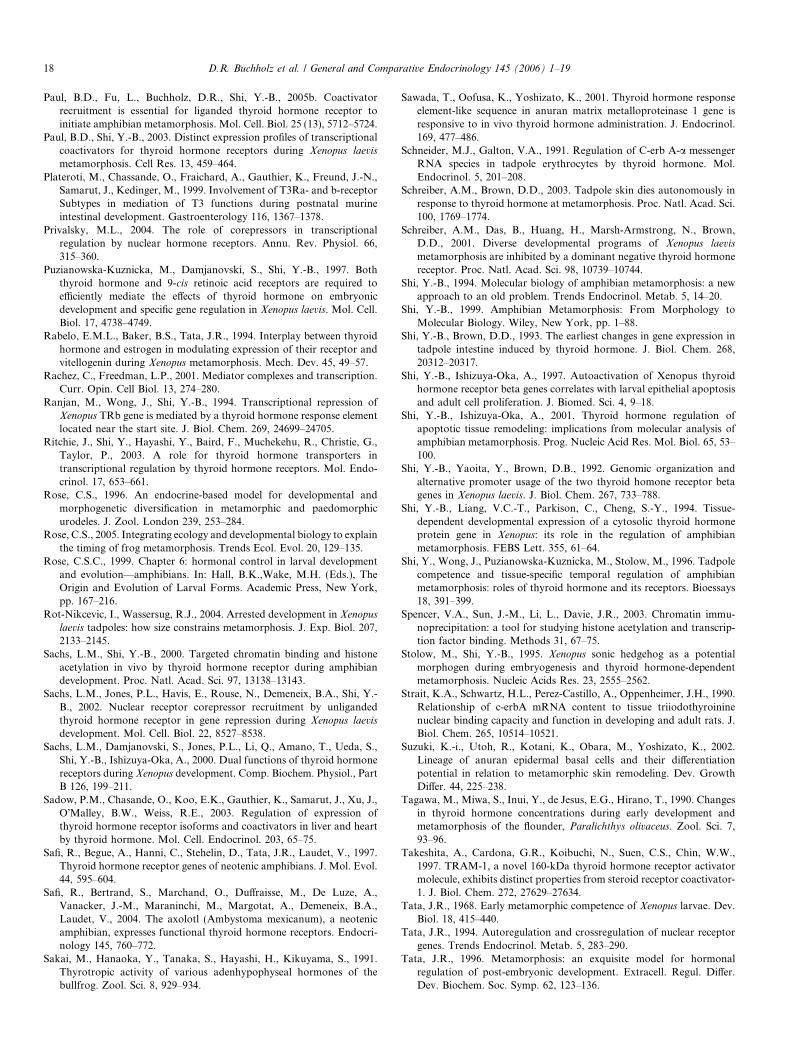
Paul, B.D., Fu, L., Buchholz, D.R., Shi, Y.-B., 2005b. Coactivatorrecruitment is essential for liganded thyroid hormone receptor toinitiate amphibian metamorphosis. Mol. Cell. Biol. 25 (13), 5712–5724.
Paul, B.D., Shi, Y.-B., 2003. Distinct expression profiles of transcriptionalcoactivators for thyroid hormone receptors during Xenopus laevismetamorphosis. Cell Res. 13, 459–464.
Plateroti, M., Chassande, O., Fraichard, A., Gauthier, K., Freund, J.-N.,Samarut, J., Kedinger, M., 1999. Involvement of T3Ra- and b-receptorSubtypes in mediation of T3 functions during postnatal murineintestinal development. Gastroenterology 116, 1367–1378.
Privalsky, M.L., 2004. The role of corepressors in transcriptionalregulation by nuclear hormone receptors. Annu. Rev. Physiol. 66,315–360.
Puzianowska-Kuznicka, M., Damjanovski, S., Shi, Y.-B., 1997. Boththyroid hormone and 9-cis retinoic acid receptors are required toe"ciently mediate the e!ects of thyroid hormone on embryonicdevelopment and specific gene regulation in Xenopus laevis. Mol. Cell.Biol. 17, 4738–4749.
Rabelo, E.M.L., Baker, B.S., Tata, J.R., 1994. Interplay between thyroidhormone and estrogen in modulating expression of their receptor andvitellogenin during Xenopus metamorphosis. Mech. Dev. 45, 49–57.
Rachez, C., Freedman, L.P., 2001. Mediator complexes and transcription.Curr. Opin. Cell Biol. 13, 274–280.
Ranjan, M., Wong, J., Shi, Y.-B., 1994. Transcriptional repression ofXenopus TRb gene is mediated by a thyroid hormone response elementlocated near the start site. J. Biol. Chem. 269, 24699–24705.
Ritchie, J., Shi, Y., Hayashi, Y., Baird, F., Muchekehu, R., Christie, G.,Taylor, P., 2003. A role for thyroid hormone transporters intranscriptional regulation by thyroid hormone receptors. Mol. Endo-crinol. 17, 653–661.
Rose, C.S., 1996. An endocrine-based model for developmental andmorphogenetic diversification in metamorphic and paedomorphicurodeles. J. Zool. London 239, 253–284.
Rose, C.S., 2005. Integrating ecology and developmental biology to explainthe timing of frog metamorphosis. Trends Ecol. Evol. 20, 129–135.
Rose, C.S.C., 1999. Chapter 6: hormonal control in larval developmentand evolution—amphibians. In: Hall, B.K.,Wake, M.H. (Eds.), TheOrigin and Evolution of Larval Forms. Academic Press, New York,pp. 167–216.
Rot-Nikcevic, I., Wassersug, R.J., 2004. Arrested development in Xenopuslaevis tadpoles: how size constrains metamorphosis. J. Exp. Biol. 207,2133–2145.
Sachs, L.M., Shi, Y.-B., 2000. Targeted chromatin binding and histoneacetylation in vivo by thyroid hormone receptor during amphibiandevelopment. Proc. Natl. Acad. Sci. 97, 13138–13143.
Sachs, L.M., Jones, P.L., Havis, E., Rouse, N., Demeneix, B.A., Shi, Y.-B., 2002. Nuclear receptor corepressor recruitment by unligandedthyroid hormone receptor in gene repression during Xenopus laevisdevelopment. Mol. Cell. Biol. 22, 8527–8538.
Sachs, L.M., Damjanovski, S., Jones, P.L., Li, Q., Amano, T., Ueda, S.,Shi, Y.-B., Ishizuya-Oka, A., 2000. Dual functions of thyroid hormonereceptors during Xenopus development. Comp. Biochem. Physiol., PartB 126, 199–211.
Sadow, P.M., Chasande, O., Koo, E.K., Gauthier, K., Samarut, J., Xu, J.,O!Malley, B.W., Weiss, R.E., 2003. Regulation of expression ofthyroid hormone receptor isoforms and coactivators in liver and heartby thyroid hormone. Mol. Cell. Endocrinol. 203, 65–75.
Safi, R., Begue, A., Hanni, C., Stehelin, D., Tata, J.R., Laudet, V., 1997.Thyroid hormone receptor genes of neotenic amphibians. J. Mol. Evol.44, 595–604.
Safi, R., Bertrand, S., Marchand, O., Du!raisse, M., De Luze, A.,Vanacker, J.-M., Maraninchi, M., Margotat, A., Demeneix, B.A.,Laudet, V., 2004. The axolotl (Ambystoma mexicanum), a neotenicamphibian, expresses functional thyroid hormone receptors. Endocri-nology 145, 760–772.
Sakai, M., Hanaoka, Y., Tanaka, S., Hayashi, H., Kikuyama, S., 1991.Thyrotropic activity of various adenhypophyseal hormones of thebullfrog. Zool. Sci. 8, 929–934.
Sawada, T., Oofusa, K., Yoshizato, K., 2001. Thyroid hormone responseelement-like sequence in anuran matrix metalloproteinase 1 gene isresponsive to in vivo thyroid hormone administration. J. Endocrinol.169, 477–486.
Schneider, M.J., Galton, V.A., 1991. Regulation of C-erb A-a messengerRNA species in tadpole erythrocytes by thyroid hormone. Mol.Endocrinol. 5, 201–208.
Schreiber, A.M., Brown, D.D., 2003. Tadpole skin dies autonomously inresponse to thyroid hormone at metamorphosis. Proc. Natl. Acad. Sci.100, 1769–1774.
Schreiber, A.M., Das, B., Huang, H., Marsh-Armstrong, N., Brown,D.D., 2001. Diverse developmental programs of Xenopus laevismetamorphosis are inhibited by a dominant negative thyroid hormonereceptor. Proc. Natl. Acad. Sci. 98, 10739–10744.
Shi, Y.-B., 1994. Molecular biology of amphibian metamorphosis: a newapproach to an old problem. Trends Endocrinol. Metab. 5, 14–20.
Shi, Y.-B., 1999. Amphibian Metamorphosis: From Morphology toMolecular Biology. Wiley, New York, pp. 1–88.
Shi, Y.-B., Brown, D.D., 1993. The earliest changes in gene expression intadpole intestine induced by thyroid hormone. J. Biol. Chem. 268,20312–20317.
Shi, Y.-B., Ishizuya-Oka, A., 1997. Autoactivation of Xenopus thyroidhormone receptor beta genes correlates with larval epithelial apoptosisand adult cell proliferation. J. Biomed. Sci. 4, 9–18.
Shi, Y.-B., Ishizuya-Oka, A., 2001. Thyroid hormone regulation ofapoptotic tissue remodeling: implications from molecular analysis ofamphibian metamorphosis. Prog. Nucleic Acid Res. Mol. Biol. 65, 53–100.
Shi, Y.-B., Yaoita, Y., Brown, D.B., 1992. Genomic organization andalternative promoter usage of the two thyroid homone receptor betagenes in Xenopus laevis. J. Biol. Chem. 267, 733–788.
Shi, Y.-B., Liang, V.C.-T., Parkison, C., Cheng, S.-Y., 1994. Tissue-dependent developmental expression of a cytosolic thyroid hormoneprotein gene in Xenopus: its role in the regulation of amphibianmetamorphosis. FEBS Lett. 355, 61–64.
Shi, Y., Wong, J., Puzianowska-Kuznicka, M., Stolow, M., 1996. Tadpolecompetence and tissue-specific temporal regulation of amphibianmetamorphosis: roles of thyroid hormone and its receptors. Bioessays18, 391–399.
Spencer, V.A., Sun, J.-M., Li, L., Davie, J.R., 2003. Chromatin immu-noprecipitation: a tool for studying histone acetylation and transcrip-tion factor binding. Methods 31, 67–75.
Stolow, M., Shi, Y.-B., 1995. Xenopus sonic hedgehog as a potentialmorphogen during embryogenesis and thyroid hormone-dependentmetamorphosis. Nucleic Acids Res. 23, 2555–2562.
Strait, K.A., Schwartz, H.L., Perez-Castillo, A., Oppenheimer, J.H., 1990.Relationship of c-erbA mRNA content to tissue triiodothyroininenuclear binding capacity and function in developing and adult rats. J.Biol. Chem. 265, 10514–10521.
Suzuki, K.-i., Utoh, R., Kotani, K., Obara, M., Yoshizato, K., 2002.Lineage of anuran epidermal basal cells and their di!erentiationpotential in relation to metamorphic skin remodeling. Dev. GrowthDi!er. 44, 225–238.
Tagawa, M., Miwa, S., Inui, Y., de Jesus, E.G., Hirano, T., 1990. Changesin thyroid hormone concentrations during early development andmetamorphosis of the flounder, Paralichthys olivaceus. Zool. Sci. 7,93–96.
Takeshita, A., Cardona, G.R., Koibuchi, N., Suen, C.S., Chin, W.W.,1997. TRAM-1, a novel 160-kDa thyroid hormone receptor activatormolecule, exhibits distinct properties from steroid receptor coactivator-1. J. Biol. Chem. 272, 27629–27634.
Tata, J.R., 1968. Early metamorphic competence of Xenopus larvae. Dev.Biol. 18, 415–440.
Tata, J.R., 1994. Autoregulation and crossregulation of nuclear receptorgenes. Trends Endocrinol. Metab. 5, 283–290.
Tata, J.R., 1996. Metamorphosis: an exquisite model for hormonalregulation of post-embryonic development. Extracell. Regul. Di!er.Dev. Biochem. Soc. Symp. 62, 123–136.
18 D.R. Buchholz et al. / General and Comparative Endocrinology 145 (2006) 1–19
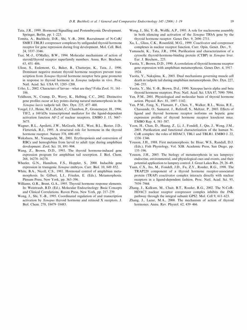
Tata, J.R., 1999. Hormonal Signalling and Postembryonic Development.Springer, Berlin, pp. 1–223.
Tomita, A., Buchholz, D.R., Shi, Y.-B., 2004. Recruitment of N-CoR/SMRT-TBLR1 corepressor complexes by unliganded thyroid hormonereceptor for gene repression during frog development. Mol. Cell. Biol.24, 3337–3346.
Tsai, M.-J., O!Malley, B.W., 1994. Molecular mechanisms of action ofsteroid/thyroid receptor superfamily members. Annu. Rev. Biochem.63, 451–486.
Ulisse, S., Esslemont, G., Baker, B., Chatterjee, K., Tata, J., 1996.Dominant negative mutant thyroid hormone receptors prevent tran-scription from Xenopus thyroid hormone receptor beta gene promoterin response to thyroid hormone in Xenopus tadpoles in vivo. Proc.Natl. Acad. Sci. USA 93, 1205–1208.
Urho, L., 2002. Characters of larvae—what are they? Folia Zool. 51, 161–186.
Veldhoen, N., Crump, D., Werry, K., Helbing, C.C., 2002. Distinctivegene profiles occur at key points during natural metamorphosis in theXenopus laevis tadpole tail. Dev. Dyn. 225, 457–468.
Voegel, J.J., Heine, M.J., Zechel, C., Chambon, P., Gronemeyer, H., 1996.TIF2, a 160 kDa transcriptional mediator for the ligand-dependentactivation function AF-2 of nuclear receptors. EMBO J. 15, 3667–3675.
Wagner, R.L., Apriletti, J.W., McGrath, M.E., West, B.L., Baxter, J.D.,Fletterick, R.J., 1995. A structural role for hormone in the thyroidhormone receptor. Nature 378, 690–697.
Wakahara, M., Yamaguchi, M., 2001. Erythropoiesis and conversion ofRBCs and hemoglobins from larval to adult type during amphibiandevelopment. Zool. Sci. 18, 891–904.
Wang, Z., Brown, D.D., 1993. The thyroid hormone-induced geneexpression program for amphibian tail resorption. J. Biol. Chem.268, 16270–16278.
Wheeler, G.N., Hamilton, F.S., Hoppler, S., 2000. Inducible geneexpression in transgenic Xenopus embryos. Curr. Biol. 10, 849–852.
White, B.A., Nicoll, C.S., 1981. Hormonal control of amphibian meta-morphosis. In: Gilbert, L.I., Frieden, E. (Eds.), Metamorphosis.Plenum Press, New York, pp. 363–396.
Williams, G.R., Brent, G.A., 1995. Thyroid hormone response elements.In: Weintraub, B.D. (Ed.), Molecular Endocrinology: Basic Conceptsand Clinical Correlations. Raven Press, New York, pp. 217–239.
Wong, J., Shi, Y.-B., 1995. Coordinated regulation of and transcriptionactivation by Xenopus thyroid hormone and retinoid X receptors. J.Biol. Chem. 270, 18479–18483.
Wong, J., Shi, Y.-B., Wol!e, A.P., 1995. A role for nucleosome assemblyin both silencing and activation of the Xenopus TRbA gene by thethyroid hormone receptor. Genes Dev. 9, 2696–2711.
Xu, L., Glass, C.K., Rosenfeld, M.G., 1999. Coactivator and corepressorcomplexes in nuclear receptor function. Curr. Opin. Genet. Dev., 9.
Yamauchi, K., Tata, J.R., 1994. Purification and characterization of acytosolic thyroid-hormone-binding protein (CTBP) in Xenopus liver.Eur. J. Biochem., 225.
Yaoita, Y., Brown, D.D., 1990. A correlation of thyroid hormone receptorgene expression with amphibian metamorphosis. Genes Dev. 4, 1917–1924.
Yaoita, Y., Nakajima, K., 2003. Dual mechanisms governing muscle celldeath in tadpole tail during amphibian metamorphosis. Dev. Dyn. 227,246–255.
Yaoita, Y., Shi, Y.-B., Brown, D.d., 1990. Xenopus laevis alpha and betathyroid hormone receptors. Proc. Natl. Acad. Sci. USA 87, 7090–7094.
Yen, P.M., 2001. Physiological and molecular basis of thyroid hormoneaction. Physiol. Rev. 81, 1097–1142.
Yen, P.M., Feng, X., Flamant, F., Chen, Y., Walker, R.L., Weiss, R.E.,Chassande, O., Samarut, J., Refeto!, S., Meltzer, P., 2003. E!ects ofligand and thyroid hormone receptor isoforms on hepatic geneexpression profiles of thyroid hormone receptor knockout mice.EMBO Rep. 4, 581–587.
Yoon, H., Chan, D., Huang, Z., Li, J., Fondell, J., Qin, J., Wong, J.M.,2003. Purification and functional characterization of the human N-CoR complex: the roles of HDAC3, TBL1 and TBLR1. EMBO J. 22,1336–1346.
Youson, J.H., 1988. First metamorphosis. In: Hoar, W.S., Randall, D.J.(Eds.), Fish Physiology, Vol. XIB. Academic Press, San Diego, pp.135–196.
Youson, J.H., 2003. The biology of metamorphosis in sea lampreys:endocrine, environmental, and physiological cues and events, and theirpotential application to lamprey control. J. Great Lakes Res. 29, 26–49.
Yuan, C.X., Ito, M., Fondell, J.D., Fu, Z.Y., Roeder, R.G., 1998. TheTRAP220 component of a thyroid hormone receptor-associatedprotein (TRAP) coactivator complex interacts directly with nuclearreceptors in a ligand-dependent fashion. Proc. Natl. Acad. Sci. 95,7939–7944.
Zhang, J., Kalkum, M., Chait, B.T., Roeder, R.G., 2002. The N-CoR-HDAC3 nuclear receptor corepressor complex inhibits the JNKpathway through the integral subunit GPS2. Mol. Cell 9, 611–623.
Zhang, J., Lazar, M.A., 2000. The mechanism of action of thyroidhormones. Annu. Rev. Physiol. 62, 439–466.
D.R. Buchholz et al. / General and Comparative Endocrinology 145 (2006) 1–19 19