introduction to mass spectrometry
DESCRIPTION
mass spectroscopy detailsTRANSCRIPT

Introduction to Mass Spectrometry
Training course on GC/MS
Nha Trang 13-18/10 2008
Charlotta Rylander

Outline General principles of mass spectrometry
Ionization techniques
Mass analyzers for GC/MS Detectors
Scan and SIM mode
Tuning
Common problems with GC-MS systems
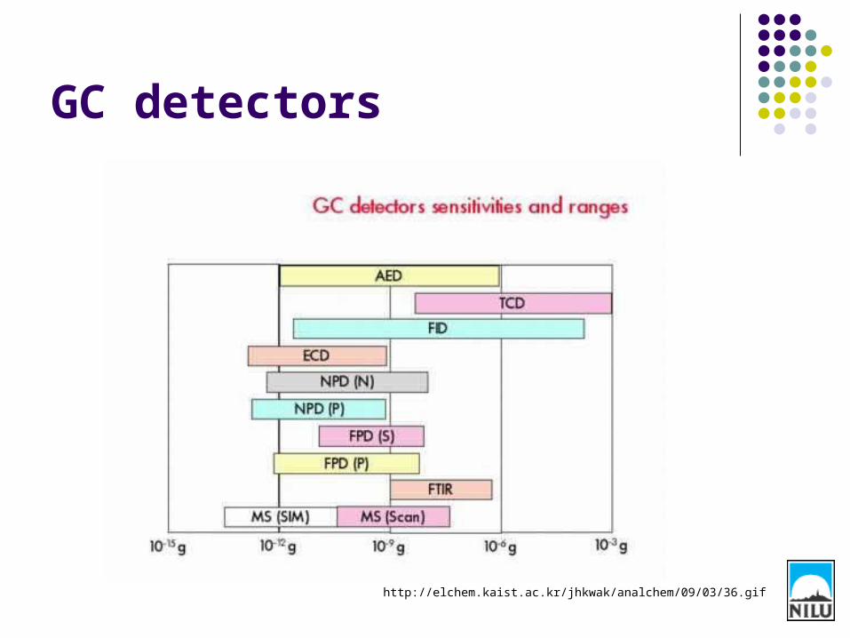
GC detectors
http://elchem.kaist.ac.kr/jhkwak/analchem/09/03/36.gif

Some useful definitions
Analytes = The compounds we would like to to analyze.
Isotopes = Atoms with the same number of electrons and protons, but different number of neutrons. Isotopes have the same chemical properties but differ in molecular mass.

5
Analysis by GC-MS
Mixture Separation Identification
GC MS
A + B + C B A C
B
A
C
m/z
m/z
m/z

General principles on mass spectrometry
Sample introduction
Ionization of sample in the ion source
Separation of ions by mass analyzer
Detector
Computer system
Resulting chromatogram

Ionization techniques
• The ionization takes place in the ion source.• Samples from GC interface are in gas-phase when introduced into the
MS. The MS is operated in vacuum to make sure analytes are evaporized and to avoid collisions between analytes and other compounds.
• Two different ionization methods• Electron Impact Ionization (EI)• Chemical Ionization (CI)
Sample introduction
Ionization of sample in the ion source
Separation of ions by mass analyzer
Detector
Computer system

Electron Impact Ionization (EI)
• Most widely used method• Analytes are bombared
with high-energy electrons (usually 70eV)
• As a result of collision, an electron is removed from the analytes (M), generating a molecular ion M+ (radical cation)
M + e- M+ + 2e-

Electron Impact Ionization (EI)
• Due to excess internal energy, fragmentation of the molecular ion will occur.
• The fragmentation is reproducible and characteristic of the compund.
• It is also possible to predict the fragmentation on the basis of chemical structures which is why MS is good tool for structure elucidation of unknown compounds

Drawbacks with EI• Sometimes the fragmentation is
too great (depending on the stability of the sample molecules) and the molecular ion will not show up in the mass spectra. It is then possible to reduce the ionization voltage, but the disadvantage is that the fragmentation pattern will change and the obtained spectra cannot be compared to ”standard” litterature spectra.
• Another solution is to use chemical ionization (CI)!
Watson, Introduction to Mass Spectrometry, 4th ed

Chemical Ionization (CI)• Softer ionization technique Less fragmentation Easier to find
molecular ions.
• Two different modes: Negative chemical ionization (NCI) and Positive Chemical Ionization (PCI).
• NCI is used for analytes that are able to form stable negative ions, for example samples containing acidic groups or halogens. NCI is often used to analyze pesticides (contains Cl or Br).
• PCI is used for samples that can form positive ions (most compounds).

Chemical Ionization (CI)• The principle for NCI and PCI is similar:
• Reagent gas (usually methane, isobutane or ammonia) is introduced into the source where it is ionized:
PCI (simplified): CH4 + e- CH4+ +2e-
CH4 + CH4+
CH3 + C H5+
NCI: CH4 + e- CH4-
• The ionized gas collide with the sample molecules generating a [M+H]+ or [M+H]- ion that is detected:
PCI: CH5+
+ M [M+H]+ + C H4
NCI: CH4- + M [M+H]-
+ C H4

EI spectra/ PCI Spectra
Harris. Quantitative Chemical Analysis, 6th ed.
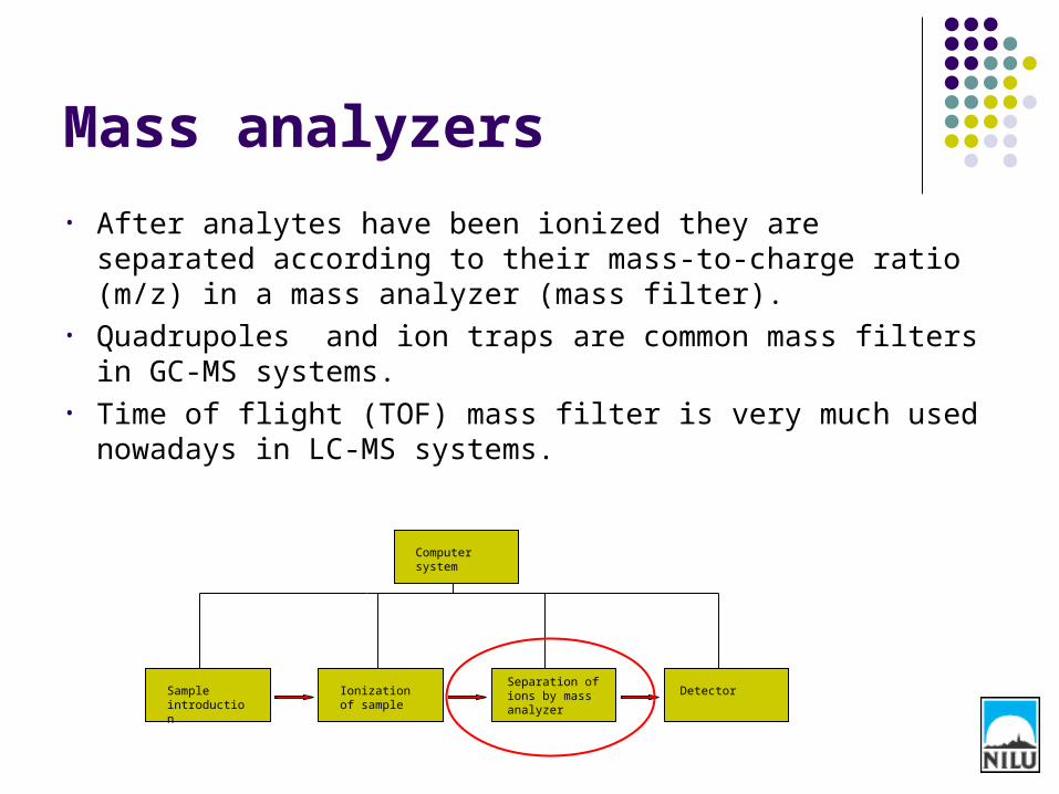
Mass analyzers
• After analytes have been ionized they are separated according to their mass-to-charge ratio (m/z) in a mass analyzer (mass filter).
• Quadrupoles and ion traps are common mass filters in GC-MS systems.
• Time of flight (TOF) mass filter is very much used nowadays in LC-MS systems.
Sample introduction
Ionization of sample
Separation of ions by mass analyzer
Detector
Computer system

Transport of ions to the mass filter
• The ionization takes place in the ion source.• Ions are then transported to the mass filter by
focusing lenses. These have a voltage running through them and by either attracting or repelling the ions they guide them into the mass filter.

High and low resolution MS
• Resolution = difference in m/z values of ions that can be separate from another.
• Quadrupoles and ion traps have constant resolution, meaning that ions that differ with 1 m/z unit will have the same separation at m/z 150 an 151 as they do at m/z 1000 and 1001.
• High resolution instruments do not have constant resolution, but constant resolving power.

Resolving power (R)
Low resolution MS instruments have a resolving power of 1000-2000. Quadrupoles and ion traps has constant resolution (ΔM=1) and R will then vary with m/z. On the other hand TOF has contant resolving power, meaning that at lower m/z the resolution will increase.
200019992000
2000
lowR
Mn=2000Mm=1999
mn
n
MMM
R
Mn=highest massMm=lowest mass
Mn=250,1807Mm=250,193319857
1807,2501933,2501933,250
highR
High resolution MS ha a resolving power of up to 20 000, making it possible to distinguish between very similar masses.
http://www.ivv.fraunhofer.de/ms/resolve10.gif

Quadrupole mass spectrometers
http://www.bris.ac.uk/nerclsmsf/images/quadrupole.gif
• A quadrupole consists of four cylindrical rods, all parallel to each other.
• Ions are introduced into the tunnel in between the four rods.
• A direct current field is applied to two rods in the quadrupole and a radio frequency to the others. The rods generate an electric field which ions can travel through.
• For a given DC/RF potential only ions with a specific m/z value are able to pass through the quadropole and reach the detector.
• All other ions will either collide with the rods or travel outside the qudrupole. Therefore they will never reach the detector.

Quadrupole
http://ael.gsfc.nasa.gov/images/saturn/quadrupole.jpg

The quadrupole instrument
Vacuum
DC and AC Voltage

Ion trap mass spectrometer
• The ion trap uses three electrodes to trap ions in small volumes.• Various voltages are applied to the ring electrodes as well as to
the entrance and exit endcap electrodes. A cavity is created were the ions are trapped.
• Depending on different voltage settings, ions at a specific m/z is ejected and detected.

Time of flight (TOF)
• High resolution MS.• Separates ion with the same kinetic energy
but different m/z, because heavier ions require more time to travel a fixed distance.
Harris. Quantitative Chemical Analysis, 6th ed.

MS detectors
• Many different types available• Electron multipliers (EM) are often used
• Continuous –Dynode Version mainly in GC-MS
Sample introduction
Ionization of sample
Separation of ions by mass analyzer
Detector
Computer system

Continious dynode EM
The EM multiplies incident charges, thereby amplifying the signal. The current is measured that is proportional to the amount of analyte in the sample.
EMs have limited lifetime which is dependent on the number of ions that hits the device, i.e., the amount of samples introduced and number of samples analyzed.
+Fast response+ High sensitivity
Watson, Introduction to Mass Spectrometry, 4th ed

Scan mode
• The MS can be operated in scan mode or in single ion monitoring (SIM).
• Scan mode means that the mass filter is set to pass a range of masses. A spectra is obtained that is used for interpretation or mass library search.
• Scan mode is less sensitive since most ions strike the quadropole rods during the scan and never reaches the detector.

EI Mass spectra
Acetone
Elemental composition: C3H6O
Nominal mass: 58
Propionaldehyde
Elemental composition: C3H6O
Nominal mass: 58
Harris. Quantitative Chemical Analysis, 6th ed.

Isotope peak pattern Many POPs contains Cl or Br atoms. Cl has two isotopes; 35Cl
and 37Cl. 76% of all chlorine atoms are 35Cl and 24% are 37Cl. The mass spectra we obtain from scan mode show the isotope
composition of fragments. If we have a molecule containing four Chlorine atoms, there are
five possible combinations of chlorine isotopes: 4 x 35Cl 3 x 35Cl + 1 x 37Cl 2 x 35Cl + 2 x 37Cl 1 x 35Cl + 3 x 37Cl 4 x 37Cl
Since every four chlorine atom in nature occur as 37Cl, most of the molecules will have the isotope composition 3 x 35Cl + 1 x 37Cl. Therefore, the most intensive peak in the mass spectra will occur at that m/z.

Isotope peak patterns

Single ion monitoring (SIM) In SIM mode the mass filter is set to pass some specific m/z ratios.
Therefore it is possible to monitor only a few compounds at specific retention time windows.
In SIM mode, two masses are monitored for each compound. These are usually the two most intensive isotope peaks within the fragment.
Window 1: Monitor only PCB 28 & 52
Window 2: Monitor PCB 99, 101,105
Window 3: Monitor PCB 138, 153,156
Window 4: Monitor PCB 170, 180, 194
1 3 42

SIM
• SIM increases sensitivity and is used for quantitative analysis.
• When using SIM mode one needs to know in advance which masses that should be monitored.
• When starting to analyze new analytes, always start by running scan mode to select appropriate ions for SIM.

Scan and SIM chromatograms
Scan mode SIM mode

Tuning
• Tuning of the MS instrument means optimization of the technical settings so you will have good senisitivity when the analysis starts.
• Tuning can be performed manually or automatically.• The MS needs to be tuned when it has been
opened.• Frequency of tuning highly instrument dependent.
(Source)

Common problems in GC-MS systems
PROBLEM SUGGESTED SOLUTION
Poor sensitivity in the MS Tune the MS, if no improvement, the ion source may be dirty and needs to be cleaned.
No filament current The filament is broken and needs to be replaced.
Poor resolution between peaks May depend on a dirty GC-column. Run the GC-oven isothermal for 3 h at a temperature close to maximum temparature for the column (se specifications from producer). If no improvement, cut a piece of the column at the inlet (30-50 cm). If this doesn’t help, one may need to change the whole column.

GC/MS-MS
MS1 Collision cell MS2
• Taking a ”Mass spectra of a mass spectra”
• MS 1 selects an ion cuurent at a specific m/z for entry into the collision cell.
• In the collision cell, the selected ion becomes energized by collisions.
• Some of the selected ions will dissociate in the collision cell and form fragment ions that are moniored in MS 2.

Use of MS/MS
Quantitative analysis; high specificity and senistivity.
Structure determination of unknown compounds.
Mapping fragmentation pathways.