induction of cd14 expression and differentiation to...
TRANSCRIPT
Advanced Pharmaceutical Bulletin, 2013, 3(2), 329-332
doi: http://dx.doi.org/10.5681/apb.2013.053
http://apb.tbzmed.ac.ir/
*Corresponding author: Behzad Baradaran, Immunology Research Center, Faculty of Medicine, Tabriz University of Medical Sciences, Tabriz,
Iran. Tel: +98 (411) 3364665, Fax: +98 (411) 3364665, Email: [email protected]
Copyright © 2013 by Tabriz University of Medical Sciences
Induction of CD14 Expression and Differentiation to Monocytes or
Mature Macrophages in Promyelocytic Cell Lines: New Approach
Fatemeh Zamani1,2
, Fatemeh Zare Shahneh1,2
, Leili Aghebati-Maleki1,2
, Behzad Baradaran1,2
* 1 Immunology Research Center, Tabriz University of Medical Sciences, Tabriz, Iran.
2 Department of Immunology, Faculty of Medicine, Tabriz University of Medical Sciences, Tabriz, Iran.
Introduction
Cluster of differentiation 14 (CD14) was described as
monocyte/ macrophage differentiation antigen on the
surface of myeloid lineage, such as monocytes,
macrophages and dendritic cells (DCs).1 This protein
plays a crucial role in the immune recognition and
reactivation in microbial cell wall components from
Gram-positive and Gram-negative bacteria.2 Recently,
CD14 role is known in phagocytic clearance of
apoptotic cells.3 CD14 isoforms, 52-55 kDa expressed
on the surfaces of monocytes and neutrophils are
attached to the cell surface by a glycosyl
phosphatidylinositol (GPI) anchor, membrane protein
(mCD14) and the serum soluble 48–56 kDa (sCD14,
an acute phase protein).4 Soluble CD14 found in
human serum has been attributed to the shedding of
mCD14 from monocytes, macrophages and PMN.
Membrane CD14 as a receptor for lipopolysaccharide
(LPS) on the membrane of the mononuclear
phagocyte (MPS) binds to LPS-binding protein (LBP)
in plasma and transfers to the cell surface receptor
CD14.5 It has been commonly used in normal tissue
or blood and in leukemia as a marker for myeloid
cells. LPS stimulates the human monocytes activation
via several intracellular signaling pathways that
involves the proinflammatory factors.6
On the cell surface, CD14 associates with Toll-like
receptor 4 (TLR4). Binding of the LPS and LPS-
binding protein complex to CD14 induces signal
transduction through TLR4, which then triggers the
synthesis and release of proinflammatory chemokine
(IP10), and cytokines (TNF-α, IL-6 and IL-1).7,8
CD14 Expression on myeloid cell line can be induced.
Among several myeloid cell lines (HL60, THP-1,
Mononomac- 1, and U937), U937 cells and HL-60
cells are the most frequently used as valid model for
investigating monocytic differentiation and
consequent biological functions of differentiated cells
in vitro. U937 cells of histocytic lymphoma basis are
arrested in a more advanced step of differentiation
(promonocyte/monocyte).9 HL-60 human leukemia
cells can be induced to differentiation into monocyte,
macrophage and granulocytes by inducing factors.
Various stimuli [LPS (lipopolysaccharide), Dimethyl
Sulfoxide (DMSO), 1, 25-dihydroxyvitamin D3
[1,25(OH)2D3], either alone or in combination, have
been recognized that have an effect on the level of
CD14 expression in the human HL-60 and human
A R T I C L E I N F O A B S T R A C T
Article Type:
Research Article
Article History:
Received: 22 February 2013
Revised: 18 March 2013
Accepted: 6 April 2013
ePublished: 20 August 2013
Keywords:
CD14
1, 25-D3
LPS
DMSO
Monocyte
Purpose: CD14, one of the main differentiation markers on the surface of myeloid
lineage cells, acts as a key role in activation of LPS-induced monocytes. LPS
(lipopolysaccharide) binds to LPS-binding protein in plasma and are delivered to the
cell surface receptor CD14. In this study, Various stimuli [Dimethyl Sulfoxide
(DMSO), active 1,25-dihydroxyvitamin D3 [1,25(OH)2D3] and LPS], either alone or
in combination, have been recognized that have an effect on the level of CD14
expression in the human HL-60 and U937 promonocytic cell lines and therefore induce
their terminal differentiation into monocytes or mature macrophages. Methods: U937
and HL-60 cells were cultured in RPMI 1640 supplemented with 10% FBS. For each
cell line, 1×10⁶ cells were seeded for 72 hours with DMSO, 14 days with LPS and 18
days with 1, 25-D3 in each well plate; then ELISA method was used to study their
responses to the factors by means of anti-CD14. Results: ELISA assay demonstrated
that U937 and HL-60 cells were induced by both [1,25(OH)2D3] and DMSO to obtain
characteristics of adherent cells and express CD14 protein; moreover, LPS at a low
dose increased CD14 expression on surface of this cells. Conclusion: According to the
our results, it is speculated that CD14 gene expression may be induced in human U937
and HL-60 cell lines by different factors including 1,25-D3, DMSO and LPS.
330 |
Zamani et al.
Advanced Pharmaceutical Bulletin, 2013, 3(2), 329-332 Copyright © 2013 by Tabriz University of Medical Sciences
U937 promonocytic cell lines and therefore induce
their terminal differentiation into monocytes or
mature macrophages.10 Upon differentiation, U937
cells gain a large range of macrophage function
through the concerted expression of several genes.
Differentiated U937 cells can be further stimulated
with LPS to mimic inflammatory response of
activated macrophages.11 In this study, U937 and HL-
60 model system was employed to express monocyte-
macrophage differentiation patterns. LPS 1, 25-
Dihydroxyvitamin D3 [1,25(OH)2D3] and DMSO
were used for inducing monocytic-macrophage
differentiation of the U937 and HL-60 leukemic cell
lines in a dose- and time dependent manner. It have
been investigated that this maturation mimics the in
vivo monocytic or myeloid differentiation.
Material and Methods
Cell Preparation and Seed
U937 and HL-60 cell lines were purchased from
Pasteur Institute of Iran (cell Bank). The cells were
grown and maintained in a humidified incubator at
37°C and in 5% CO2 atmosphere. RPMI-1640
medium (SIGMA) was supplemented with 15% heat
inactivated Fetal Bovine Serum (FBS), 100 units/mL
penicillin, 2.5 ml amphotericin B, 5× 10 M 2-
mercaptoethanol and 2.5 mM L-glutamine, and 100
𝜇g/mL streptomycin (all from Invitrogen Gibco) were
used for cell cultures. Upon reaching appropriate
confluence, the cells were passaged every 2-3 days
and seeding was at in initial concentration of 1× 10⁶
cell/ml. LPS (Escherichia coli O111: B4) and 1,25-D3
were purchased from sigma chemical company. Five
well of 12-well flat-bottom culture plates (Nunc,
Denmark) were seeded per cell line. 1×10⁶ cells per
well was incubated and treated with DMSO + LPS for
14 days, 0.1 µmol 1,25-D3 , 1000 ng/ ml LPS for 18
days, and with LPS + Vitamin D3 for 18 day,
respectively.
Microscopic Analysis
Morphological changes after 5-6 days incubation,
maturation and morphology differentiation into
macrophage-monocyte was detected by light
microscopic analysis.
ELISA Assay
CD14 protein expression on cell membrane was
analyzed by ELISA assay. Briefly, after incubation
period, 1 × 10⁶ cell was seeded in 12-well flat-bottom
ELISA plate. Glotaraldehid 25% was used to fix the
suspended cells into bottom of ELISA plate wells for
12-16 hour in 4°C. After the overnight incubation, the
plates were washed 3 times with PBS containing
0.05% (v/v) Tween 20 (PBS-T). Each well was
blocked with 200 μL of PBS containing 1% (w/v)
BSA (Sigma) for 1 h at room temperature and washed
3 times with PBS-T. Subsequently 100 µl of first
antibody (mouse anti-human CD14 /monoclonal
antibodies: 1/1000 diluted in PBS buffer), was added
and incubated for 2 hour in 37°C, and washed 3 times
with PBS-T. Secondary antibody (rabbit anti-mouse
IgG conjugated to horseradish peroxidase 1/3000
diluted in PBS buffer) was added to each well and
incubated for 1 hour at 37 °C. 100 µL of 0.01% (w/v)
3, 3’, 5, 5’ tetramethylbenzidine (TMB)
chromogenic/substrate solution (Sigma) was added to
each well. The reaction was stopped by adding 50 μL
of 2 N sulfuric acid, yielding a yellow color. The
optical density (OD) colored solution was quantified
at 450 nm wavelengths by using an enzyme linked
immunoabsorbent assay reader (ELISA Reader, Bio-
Rad).
Results
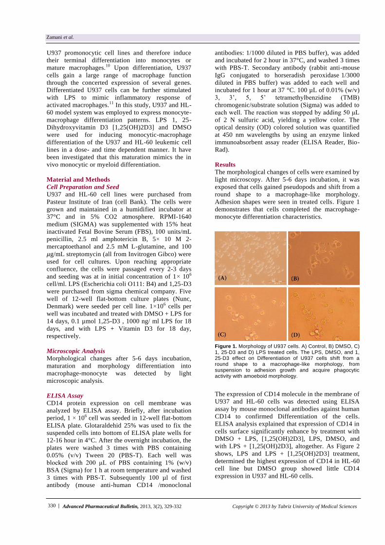
The morphological changes of cells were examined by
light microscopy. After 5-6 days incubation, it was
exposed that cells gained pseudopods and shift from a
round shape to a macrophage-like morphology.
Adhesion shapes were seen in treated cells. Figure 1
demonstrates that cells completed the macrophage-
monocyte differentiation characteristics.
Figure 1. Morphology of U937 cells. A) Control, B) DMSO, C) 1, 25-D3 and D) LPS treated cells. The LPS, DMSO, and 1, 25-D3 effect on Differentiation of U937 cells shift from a round shape to a macrophage-like morphology, from suspension to adhesion growth and acquire phagocytic activity with amoeboid morphology.
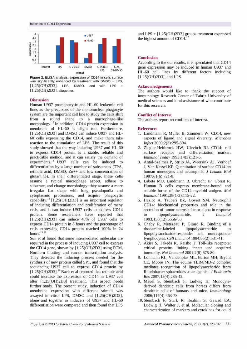
The expression of CD14 molecule in the membrane of
U937 and HL-60 cells was detected using ELISA
assay by mouse monoclonal antibodies against human
CD14 to confirmed Differentiation of the cells.
ELISA analysis explained that expression of CD14 in
cells surface significantly enhance by treatment with
DMSO + LPS, [1,25(OH)2D3], LPS, DMSO, and
with LPS + [1,25(OH)2D3], altogether. As Figure 2
shows, LPS and LPS + [1,25(OH)2D3] treatment,
determined the highest expression of CD14 in HL-60
cell line but DMSO group showed little CD14
expression in U937 and HL-60 cells.
| 331 Advanced Pharmaceutical Bulletin, 2013, 3(2), 329-332 Copyright © 2013 by Tabriz University of Medical Sciences
Induction of CD14 Expression
Figure 2. ELISA analysis, expression of CD14 in cells surface was significantly enhanced by treatment with DMSO + LPS,
[1,25(OH)2D3], LPS, DMSO, and with LPS +
[1,25(OH)2D3], altogether.
Discussion
Human U937 promonocytic and HL-60 leukemic cell
lines as the precursors of the mononuclear phagocyte
system are the important cell line to study the cells shift
from a round shape to a macrophage-like
morphology.12 In addition, CD14 protein expression in
membrane of HL-60 is slight too. Furthermore,
[1,25(OH)2D3] and DMSO can induce U937 and HL-
60 cells expressing the CD14, and make them take
reaction to the stimulation of LPS. The result of this
study showed that the way inducing U937 and HL-60
to express CD14 protein is a stable, reliable and
practicable method, and it can satisfy the demand of
experiments.13 U937 cells can be induced to
differentiation by a large number of substances (TPA,
retinoic acid, DMSO, Zn++ and low concentration of
glutamine). In their differentiated stage, these cells
assume a typical macrophage aspect, adhere to
substrate, and change morphology: they assume a more
irregular flat shape with long pseudopodia and
cytoplasmic protrusions, and acquire phagocytic
capability.14 [1,25(OH)2D3] is an important regulator
of inducing differentiation and proliferation of many
cells, and it can induce U937 cells to express CD14
protein. Some researchers have reported that
[1,25(OH)2D3] can induce 40% of U937 cells to
express CD14 protein in 8 hours and the percentage of
cells expressing CD14 protein reached 100% in 24
hours.15,16
Sun et al found that some intermediated molecular are
required in the process of inducing U937 cell to express
the CD14 gene, shown by [1,25(OH)2D3] using FCM,
Northern blotting and run-on transcription analysis.
They detected the inducing process needed for the
synthesis of new protein called SP1, and found that the
sequencing U937 cell to express CD14 protein by
[1,25(OH)2D3].16 Baek et al reported that retinoic acid
could increase the expression of CD14 in U937 cell
after [1,25(OH)2D3] treatment. This aspect needs
further study. The present study, induction of CD14
membrane expression with different stimuli was
assayed in vitro. LPS, DMSO and [1,25(OH)2D3],
alone and together as inducers of U937 and HL-60
differentiation were compared and then found that LPS
and LPS + [1,25(OH)2D3] groups treatment expressed
the highest amount of CD14.17
Conclusion
According to the our results, it is speculated that CD14
gene expression may be induced in human U937 and
HL-60 cell lines by different factors including
[1,25(OH)2D3], and LPS.
Acknowledgements
The authors would like to thank the support of
immunology Research Center of Tabriz University of
medical sciences and kind assistance of who contribute
for this research.
Conflict of Interest
The authors report no conflicts of interest.
References
1. Landmann R, Muller B, Zimmerli W. CD14, new
aspects of ligand and signal diversity. Microbes
Infect 2000;2(3):295-304.
2. Ziegler-Heitbrock HW, Ulevitch RJ. CD14: cell
surface receptor and differentiation marker.
Immunol Today 1993;14(3):121-5.
3. Antal-Szalmas P, Strijp JA, Weersink AJ, Verhoef
J, Van Kessel KP. Quantitation of surface CD14 on
human monocytes and neutrophils. J Leukoc Biol
1997;61(6):721-8.
4. Labeta MO, Landmann R, Obrecht JP, Obrist R.
Human B cells express membrane-bound and
soluble forms of the CD14 myeloid antigen. Mol
Immunol 1991;28(1-2):115-22.
5. Haziot A, Tsuberi BZ, Goyert SM. Neutrophil
CD14: biochemical properties and role in the
secretion of tumor necrosis factor-alpha in response
to lipopolysaccharide. J Immunol
1993;150(12):5556-65.
6. Chaby R, Metezeau P, Girard R. Binding of a
rhodamine-labeled lipopolysaccharide to
lipopolysaccharide-responder and nonresponder
lymphocytes. Cell Immunol 1984;85(2):531-41.
7. Akira S, Takeda K, Kaisho T. Toll-like receptors:
critical proteins linking innate and acquired
immunity. Nat Immunol 2001;2(8):675-80.
8. Lohmann KL, Vandenplas ML, Barton MH, Bryant
CE, Moore JN. The equine TLR4/MD-2 complex
mediates recognition of lipopolysaccharide from
Rhodobacter sphaeroides as an agonist. J Endotoxin
Res 2007;13(4):235-42.
9. Mauel S, Steinbach F, Ludwig H. Monocyte-
derived dendritic cells from horses differs from
dendritic cells of humans and mice. Immunology
2006;117(4):463-73.
10. Steinbach F, Stark R, Ibrahim S, Gawad EA,
Ludwig H, Walter J, et al. Molecular cloning and
characterization of markers and cytokines for equid
332 |
Zamani et al.
Advanced Pharmaceutical Bulletin, 2013, 3(2), 329-332 Copyright © 2013 by Tabriz University of Medical Sciences
myeloid cells. Vet Immunol Immunopathol
2005;108(1-2):227-36.
11. Werners AH, Bull S, Vendrig JC, Smyth T, Bosch
RR, Fink-Gremmels J, et al. Genotyping of Toll-
like receptor 4, myeloid differentiation factor 2 and
CD-14 in the horse: an investigation into the
influence of genetic polymorphisms on the LPS
induced TNF-alpha response in equine whole
blood. Vet Immunol Immunopathol 2006;111(3-
4):165-73.
12. Pagliara P, Lanubile R, Dwikat M, Abbro L, Dini L.
Differentiation of monocytic U937 cells under static
magnetic field exposure. Eur J Histochem
2005;49(1):75-86.
13. Hosaya H, Maranouchi T. Differentiation and
dedifferentiation of the human monocyte leukemia
cell line, U937. Cell Struct Funct 1992;17(5):263-9.
14. Jarvinen M, Ylanne J, Virtanen I. The effect of
differentiation on the integrin expression K562
erythroleukemia cells. Cell Biol Int 1993;17(4):399-
407.
15. Brackman D, Lund-Johansen F, Aarskog D.
Expression of cell surface antigens during the
differentiation of HL-60 cells induced by 1,25-
ihydroxyvitamin D3, retinoic acid and DMSO. Leuk
Res 1995;19(1):57-64.
16. Sun H, Wu CX, Gong JP, Liu HZ, Li XH, You HB,
et al. Expression of CD14 protein in U937 cells
induced by vitamin D3. Xi Bao Yu Fen Zi Mian Yi
Xue Za Zhi 2005;21(2):155-8.
17. Baek YS, Haas S, Hackstein H, Bein G, Hernandez-
Santana M, Lehrach H, et al. Identification of novel
transcriptional regulators involved in macrophage
differentiation and activation in U937 cells. BMC
Immunol 2009;10:18.