i s t o m a t o lo g jis Ë the v-th pan-albanian dental...
TRANSCRIPT

SShhooqqaattaa ee SSttoommaattoollooggëëvvee ttëë KKoossoovvëëss
DDeennttaall AAssssoocciiaattiioonn ooff KKoossoovvoo
KKOONNGGRREESSII II VV--ttëë MMBBAARRËËKKOOMMBBËËTTAARR
II SSTTOOMMAATTOOLLOOGGJJIISSËË
TTHHEE VV--tthh PPAANN--AALLBBAANNIIAANN
DDEENNTTAALL CCOONNGGRREESSSS
2277 –– 2299 TTeettoorr 22001177,,
OOccttoobbeerr 2277tthh –– 2299tthh,, 22001177
SSwwiissss DDiiaammoonndd HHootteell,, PPrriisshhttiinnëë..

KONGRESI I V-TË MBARËKOMBËTAR I STOMATOLOGJISË, 27-29 TETOR, 2017, PRISHTINË, KOSOVË
2
Në bashkëpunim me Shoqatën Dentare Shqiptare dhe Shoqërinë Stomatologjike Shqiptare, me
mbështetjen e Odës së Stomatologëve të Kosovës, Qendrën Klinike Stomatologjike Universitare të
Kosovës dhe Fakultetin e Mjekësisë – Dega e Stomatologjisë
In cooperation with the Albanian Dental Society and Albanians’ Stomatological Society,
with support from Dental Chamber of Kosovo, University Dentistry Clinical Center of Kosovo and Faculty of Medicine, Dentistry School.

KONGRESI I V-TË MBARËKOMBËTAR I STOMATOLOGJISË, 27-29 TETOR, 2017, PRISHTINË, KOSOVË
3
Këshilli organizativ – Organizing Committee
Prof. Dr. Blerim Kamberi – Kryetar, President
Prof. Asoc. Dorjan Hysi - zv. Kryetar, Vice-president
Mr.sc. Nedim Kasami - zv. Kryetar, Vice-president
Dr. sc. Donika Bajrami
Ass. Dr. Tringa Kelmendi
Dr. Visar Bunjaku
Ass. Dr. Zana Sejfija
Ass. Dr. Nora Aliu
Këshilli shkencor – Scientific Committee
Prof. Ass. Fatmir Dragidella – Kryetar, President
Prof. Asoc. Edit Xhajanka
Prof. Dr. Veton Hoxha
Prof. Asoc. Milaim Sejdini
Prof. Asoc. Agim Begzati
Prof. Asoc. Gloria Staka
Dr. sc. Ali Gashi
Dr. sc. Shefqet Mrasori
Dr. Fisnik Kasapi
Dr. sc. Kastriot Meqa
Dr. sc. Miranda Stavileci-Veliu
Këshilli i nderit – The Council of Honor
Prof. Dr. Osman Sejfija
Prof. Dr. Fejzi Keraj
Prof. Dr. Agim Islami
Prof. Dr. Adil Raka
Prof. Dr. Hasan Mehmeti
KONGRESI I V-TË MBARËKOMBËTAR I STOMATOLOGJISË, 27-29 TETOR, 2017, PRISHTINË, KOSOVË
4
PËRMBAJTJA
CONTENT
SHQIP 5-56
ENGLISH 58-108

KONGRESI I V-TË MBARËKOMBËTAR I STOMATOLOGJISË, 27-29 TETOR, 2017, PRISHTINË, KOSOVË
5
Fjala e kryetarit
Me kënaqësinë më të madhe ju ftojmë në Kongresin e V-të Mbarëkombëtar të Stomatologjisë nga
data 27-29 Tetor 2017 në Prishtinë, Kosovë.
Ky kongres që bashkon tri shoqatat profesionale mbarëkombëtare do të jetë një shprehje e
vullnetit dhe dëshirës së stomatologëve drejt edukimit të vazhdueshëm profesional të tyre, të
kombinuar me shkëmbim idesh, njohuri dhe praktika të ndërsjella ndërmjet ekspertëve të
disiplinave të ndryshme stomatologjike. Shpresojmë që ju do të përfitoni nga kjo mundësi dhe do
të bashkoheni me ne në këtë ngjarje të jashtëzakonshme. Është një mënyrë e shkëlqyer për të
ndërtuar marrëdhënie me kolegët, për të fituar njohuri, përvojë dhe mbështetje brenda këtij
kongresi.
Qëllimi kryesor i Kongresit të V-të Mbarëkombëtar të Stomatologjisë është promovimi i
hulumtimeve dhe lidhshmëria në të gjitha fushat e shkencave stomatologjike, për të inkurajuar
përmirësime në metodat për parandalimin dhe trajtimin e sëmundjeve orale dhe dentare, për të
përmirësuar shëndetin publik oral përmes hulumtimit dhe për të lehtësuar bashkëpunimin midis
hulumtuesve të rinj.
Kongresi në programin e tij shkencor tërheq ekspertë të huaj e vendorë në cilësinë e ligjëruesit të
ftuar, prezantuesit oral dhe poster prezantuesit që përfaqësojnë shkolla të ndryshme dhe praktikat
më të mira të vendeve të tyre.
Me dëshirat e mia më të mira përshëndes dhe uroj sukses për të gjithë pjesëmarrësit e Kongresit!
Prof. Dr. Blerim Kamberi
Kryetar i Shoqatës së Stomatologëve të Kosovës
Kryetar i Këshillit Organizativ

KONGRESI I V-TË MBARËKOMBËTAR I STOMATOLOGJISË, 27-29 TETOR, 2017, PRISHTINË, KOSOVË
6
Programi i Kongresit
27.10.2017 – E Premte
15.00 – 17.00 REGJISTRIMI DHE MARRJA E MATERIALIT
17.00 – 19.00 HAPJA SOLEMNE – Salla DYAR
- FJALIMET PËRSHËNDETËSE
- NDARJA E MIRËNJOHJEVE
- PROMOVIMI I MONOGRAFISË DHËMBËT E IMPAKTUAR , DR.SC. ALI GASHI
- PIKË MUZIKORE
- KOKTEL
28.10.2017 – E Shtunë
08.30 – 12.00 REGJISTRIMI DHE MARRJA E MATERIALIT
09.00 – 18.00 LIGJËRATAT PLENARE – Salla DYAR
Seanca I
Moderatorët: Prof. dr. Veton Hoxha, Prof. asoc. Edit Xhajanka, Dr. sc. Ferjale Perjuci
09.00 – 09.30 LP1 - Ender Kazazoğlu – Ngarkimi i menjëhershëm: Pse, kur dhe si?
09.45 – 10.15 LP2 - Violeta Ukmata-Vula – Obturimi i kanalit të rrënjës dhe mikrodepërtueshmëria apikale
10.30 – 11.00 LP3 - Uğur Meriç – Kocka autogjene Standardi i artë në praktikën e përditshme
11.15 – 11.45 Pauza për kafe
Seanca II
Moderatorët: Prof. asoc. Ferit Koçani, Prof. asoc. Agim Begzati, Mr. sc. Flurije Asllani-Hoxha
11.45 – 12.15 LP4 - Jean-Philippe Mallet – Trajtimi endodontik i sigurt dhe efikas me një sistem inovativ të thjeshtë

KONGRESI I V-TË MBARËKOMBËTAR I STOMATOLOGJISË, 27-29 TETOR, 2017, PRISHTINË, KOSOVË
7
12.30 – 13.00 LP5 - Dorjan Hysi – Solucionet shpëlarëse në Endodonti, Irrigantët
13.15 – 14.30 Pauza për drekë
Seanca III
Moderatorët: Prof. ass. Resmije Ademi, Dr. sc. Xhevdet Aliu, Mr. sc. Merita Kuçi
14.30 – 15.00 LP6 - Dubravka Knezović-Zlatarić – Estetika dhe përshtatja e nuancave
15.15 – 15.45 LP7 - Maja Žagar – VITA Assist, një softuer – mijëra mundësi
16.00 – 16.30 Pauza për kafe
Seanca IV
Moderatorët: Dr. sc. Ali Gashi, Mr.sc. Nedim Kasami, Dr. sc. Zana Sllamniku-Dalipi
16.30 – 17.00 LP8 – Irina Filipović-Zore – Përdorimi i procedurës PRF në implantologjinë dentare
17.15 – 17.45 LP9 – Kristina Mitić – Shëndeti oral dhe goja e thatë
28.10.2017 – E Shtunë
10.00 – 15.15 PREZENTIMET ORALE – Salla DIAMOND
Seanca 1
Moderatorët: Dr. Sc. Donika Bajrami, Dr. sc. Kastriot Meqa, Dr. sc. Teuta Ademaj
10.00 – 10.10 PO1 – Almira Isufi - Një klasifikim i ri i kaviteteve endodontike
10.15 – 10.25 PO2 – Kaltrina Beqiri – Shpërndarja e elementeve zëvendësuese te urat metal-qeramike laterale
10.30 – 10.40 PO3 – Brunilda Koçi – Translucenca e koronave të zirkoniumit, të shtresëzuara vs. monolitike
10.45 – 10.55 PO4 – Shevale Alili – Hipomineralizimi i molarëve dhe incizivëve (HMI)
11.00 – 11.10 PO5 – Amet Demiri – Vlerësimi i rezultateve pas apikotomive me mbushje retrograde të kanaleve të rrënjëve
11.15 – 11.45 Pauza për kafe

KONGRESI I V-TË MBARËKOMBËTAR I STOMATOLOGJISË, 27-29 TETOR, 2017, PRISHTINË, KOSOVË
8
Seanca 2
Moderatorët:, Dr. sc. Jehona Ahmedi, Dr. sc. Aida Namani, Ass. Dr. Tringa Kelmendi
11.45 – 11.55 PO6 – Kreshnik Keraj – Vlerësimi i protezave totale me rezinë që polimerizohen me zierje
12.00 – 12.10 PO7 – Zana Sejfija – Citokinet inflamatore në monocitet e ekspozuara ndaj zink oksidit nano partikulare - me flow citometri
12.15 – 12.25 PO8 – Besian Abazi – Periodontiti agresiv dhe antibiotikët në Shqipëri: korrelacioni dhe karakteristikat
12.30 – 12.40 PO9 – Ilma Robo – Luhatjet e përbërësve sasiorë dhe cilësorë të salivës, si indikatorë të diabetit në kavitetin oral
12.45 -12.55 PO10 – Fisnik Mekaj – Udhëzuesi kirurgjik dhe ngarkimi i menjëhershëm i implanteve dentare
13.15 – 14.30 Pauza për drekë
Seanca 3
Moderatorët: Dr. sc. Zana Agani, Dr. sc. Nexhmije Ajeti, Dr. sc. Teuta Bicaj
14.30 – 14.40 PO11 – Sherif Shaqiri – Struktura profesionale e pacientëve tanë të trajtuar në pikëpamje protetikore
14.45 – 14.55 PO12 – Miranda Stavileci-Veliu – Efikasiteti i materialeve të ndryshme në trajtimin e perforimeve jatrogjene – Studim in vitro
15.00 – 15.10 PO13 – Aldo Vangjeli – Implementimi i mjekësisë rigjeneruese në stomatologji

KONGRESI I V-TË MBARËKOMBËTAR I STOMATOLOGJISË, 27-29 TETOR, 2017, PRISHTINË, KOSOVË
9
29.10.2017 – E Diel
09.00 – 10.30 LIGJËRATAT PLENARE – Salla DYAR
Seanca V
Moderatorët: Prof. asoc. Milaim Sejdini, Dr. sc. Shefqet Mrasori, Dr. sc. Mërgime Prekazi
09.00 – 09.30 LP10 – Višnja Katić - Rritja e fëmiut dhe problemet ortodontike
09.45 – 10.15 LP11 – Nedim Kasami - Tumoret beninje të gjëndrës parotide - diagnoza dhe trajtimi kirurgjik
10.30 – 11.30 PREZENTIMET ORALE – Salla DYAR
Seanca 4
Moderatorët: Dr. sc. Agron Bytyqi, Ass. Dr. Ariana Kameri, Ass. Dr. Blerim Mehmeti
10.30 – 10.40 PO14 – Astrit Kuçi – SAF sistemi në endodoncion
10.45 – 10.55 PO15 – Donika Iljazi-Shahiqi – Indeksi për përcaktimin e nevojës për trajtim ortodontik – Përdorimi i tij në përcaktimin e shkallës së malokluzionit
11.00 – 11.10 PO16 – Jeta Kelmendi – Rëndësia e përcaktimit të moshës në ortodonti
11.15 – 11.25 PO17 – Tefik Ismaili - Prevalenca e padhëmbësisë në rajonin e Gostivarit
09.00 – 11.00 PREZENTIMET ME POSTER (PP1-PP8) – Salla DIAMOND
Komisioni vlerësues
Dr. sc. Miranda Stavileci-Veliu, Dr. sc. Linda Dula, Dr. sc. Blerta Xhemajli
27 - 29.10.2017 Ekspozita dentare – Salla ULPIANA

KONGRESI I V-TË MBARËKOMBËTAR I STOMATOLOGJISË, 27-29 TETOR, 2017, PRISHTINË, KOSOVË
10

KONGRESI I V-TË MBARËKOMBËTAR I STOMATOLOGJISË, 27-29 TETOR, 2017, PRISHTINË, KOSOVË
11
LIGJËRATAT PLENARE

KONGRESI I V-TË MBARËKOMBËTAR I STOMATOLOGJISË, 27-29 TETOR, 2017, PRISHTINË, KOSOVË
12

KONGRESI I V-TË MBARËKOMBËTAR I STOMATOLOGJISË, 27-29 TETOR, 2017, PRISHTINË, KOSOVË
13
Prof. Dr. Ender Kazazoğlu
Fakulteti i Stomatologjisë, Universiteti Yeditepe
Stamboll, Turqi
Dr. Ender Kazazoğlu u diplomua në vitin në Universitetin e Marmaras, Fakulteti i Stomatologjisë, Stamboll, Turqi. Ai mori
titullin doktor shkence në Kolegjin Mjekësor të Spitalit të Londrës
në Britani të Madhe në vitin 1991. Pastaj, punoi në Spitalin e
Londrës si asistent hulumtues (1991-1992). Në fillim të vitit 1993, ai filloi të punonte si
pedagog në fushën e protetikës në Universitetin e Marmaras. Aktualisht është duke
punuar në Universitetin e Yeditepes, në Fakultetin e Stomatologjisë. Punon si redaktor
kryesor në pjesën turke të revistës Journal of Balkan Stomatology, redaktor në zhurnalin
e Stomatologjisë, anëtar i këshillit redaktues të J. Yeditepe Clinic, kryeredaktor i J.
Esthetic & Implantology, anëtar i këshillit redaktues të International J. Prosthodontics.
Dr. Kazazoglu ka mbikëqyr gjashtë teza të magjistraturës, si dhe 17 teza të doktoratës.
Ai gjithashtu ka botuar 64 artikuj në revista ndërkombëtare dhe 20 artikuj në revista
kombëtare; ka pasur 62 prezentime ndërkombëtare dhe 31 kombëtare; si dhe ka
ligjëruar 42 ligjërata kombëtare dhe 7 ndërkombëtare.
LP1 – Ngarkimi i menjëhershëm: pse, kur dhe si?
Është bërë një arritje e madhe në trajtimin e pacientëve pa dhëmbë me implante, sipas
protokollit të Prof. Branemark (1974). Ne po jetojmë në një botë që gjithçka po shkon aq
shpejt. Prandaj, si stomatologë ne gjithashtu ndryshojmë protokollet tona të trajtimit
pothuajse në çdo fushë duke i përcjellë trendet e reja. Sot, ne jemi të inkurajuar nga
fabrikat dhe vet pacientët për të vendosur menjëherë restaurimet pas kirurgjisë së
implanteve. Megjithatë, ky lloj i trajtimeve kërkon njohuri dhe përvojë. Por fundja,
stomatologët nuk janë të arsimuar mirë as në shkollat dentare, as në kurset e edukimit
të vazhdueshëm. Unë do të diskutoj për të gjetur përgjigjen e pyetjeve të mëposhtme në
këtë leksion: A është i aplikueshëm ngarkimi i menjëhershëm, përfshirë këtu edhe
personat e moshuar; cili është protokolli për ngarkim të menjëhershëm dhe cilat janë
pikat strategjike për trajtim afatgjatë të ngarkimit të menjëhershëm. Unë gjithashtu do të
hap një diskutim se si mund të modifikohet ngarkimi i menjëhershëm për të moshuarit.

KONGRESI I V-TË MBARËKOMBËTAR I STOMATOLOGJISË, 27-29 TETOR, 2017, PRISHTINË, KOSOVË
14
Prof.ass. Violeta Ukmata-Vula
Dega e Stomatologjisë, Fakulteti i Mjekësisë,
Universiteti i Prishtinës, Kosovë
Dr. Violeta Ukmata-Vula, aktualisht punon si Profesor asistent nё Katedrën e Sëmundjeve tё Dhëmbit me Endodoncion. Studimet universitare i ka përfunduar në vitin .Nё vitin ka përfunduar specializimin nga lёmia e Sёmundjeve tё Dhёmbit dhe
Gojёs. Nё vitin , përfundoi magjistraturёn, ndërsa nё vitin mbrojti temёn e doktoraturёs nё Universitetin e Prishtinës, Fakulteti i Mjekësisë, Dega e Stomatologjisё. Ёshtё anёtare e Shoqatёs sё Stomatologёve tё Kosovёs dhe Shoqatёs Europiane tё Endodontёve. Aktualisht është shefe e Klinikës së Sëmundjeve të Dhëmbit me Endodoncion.
LP2 - Obturimi i kanalit të rrënjës dhe mikrodepërtueshmëria apikale
Obturimi i kanalit pas instrumentimit finalizon ndërhyrjen endodontike. Obturimi
definohet si mbushje tredimensionale e brendësisë sё sistemit kanalikular, mundësisht me mbyllje tё kufirit cement-dentinё, me sasi minimale tё pastёs endodontike, biokompatibile, e aplikuar nё kanal dhe nё ngushticën apikale nё prani tё koneve,qё tё fitohet mbyllje adekuate .
Arsyeja kryesore pёr obturimin e plotё tё hapёsirёs sё kanalit ёshtё pengimi i mikrodepёrtueshmёrisё apikale – fenomen ky i cili krijohet nё mes tё materialit pёr obturim dhe mureve tё kanalit, si dhe nё mes tё gutaperkёs dhe materialit pёr obturim, nё funksion tё kohёs. Pёr kёtё problematikё do tё diskutohet edhe nё kёtё ligjëratё.
KONGRESI I V-TË MBARËKOMBËTAR I STOMATOLOGJISË, 27-29 TETOR, 2017, PRISHTINË, KOSOVË
15
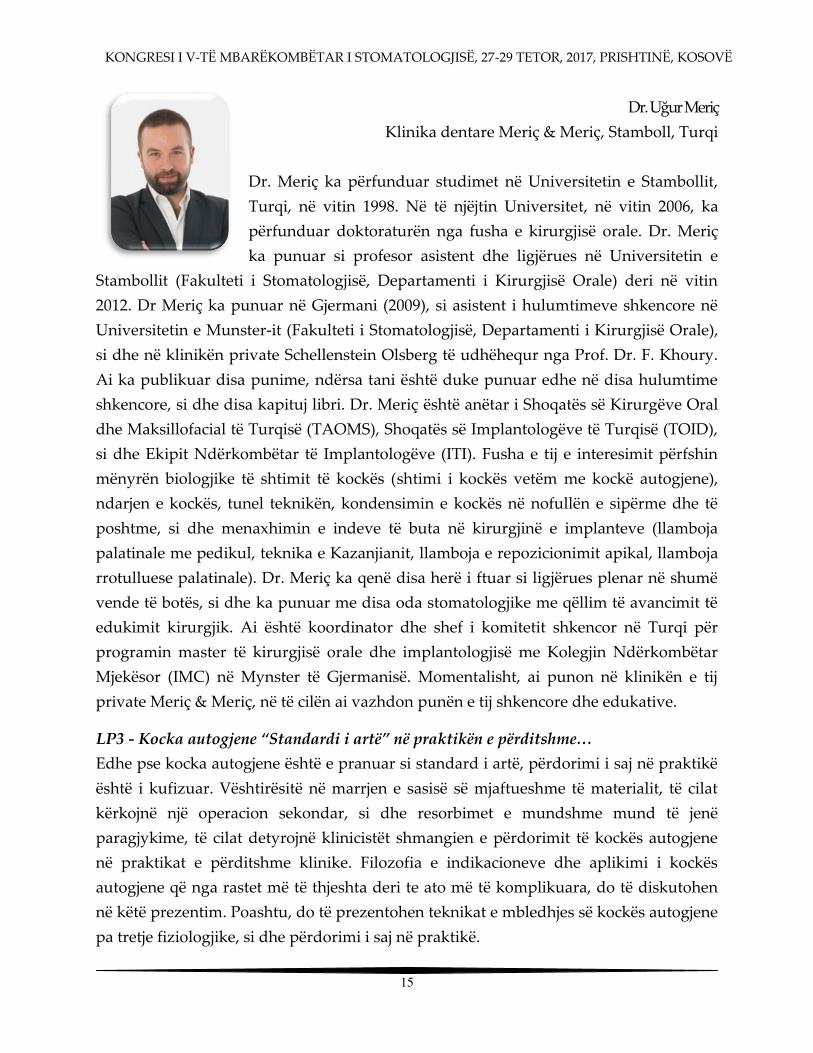
Dr. Uğur Meriç Klinika dentare Meriç & Meriç, Stamboll, Turqi
Dr. Meriç ka përfunduar studimet në Universitetin e Stambollit,
Turqi, në vitin 1998. Në të njëjtin Universitet, në vitin 2006, ka
përfunduar doktoraturën nga fusha e kirurgjisë orale. Dr. Meriç
ka punuar si profesor asistent dhe ligjërues në Universitetin e
Stambollit (Fakulteti i Stomatologjisë, Departamenti i Kirurgjisë Orale) deri në vitin
2012. Dr Meriç ka punuar në Gjermani (2009), si asistent i hulumtimeve shkencore në
Universitetin e Munster-it (Fakulteti i Stomatologjisë, Departamenti i Kirurgjisë Orale),
si dhe në klinikën private Schellenstein Olsberg të udhëhequr nga Prof. Dr. F. Khoury.
Ai ka publikuar disa punime, ndërsa tani është duke punuar edhe në disa hulumtime
shkencore, si dhe disa kapituj libri. Dr. Meriç është anëtar i Shoqatës së Kirurgëve Oral
dhe Maksillofacial të Turqisë (TAOMS), Shoqatës së Implantologëve të Turqisë (TOID),
si dhe Ekipit Ndërkombëtar të Implantologëve (ITI). Fusha e tij e interesimit përfshin
mënyrën biologjike të shtimit të kockës (shtimi i kockës vetëm me kockë autogjene),
ndarjen e kockës, tunel teknikën, kondensimin e kockës në nofullën e sipërme dhe të
poshtme, si dhe menaxhimin e indeve të buta në kirurgjinë e implanteve (llamboja
palatinale me pedikul, teknika e Kazanjianit, llamboja e repozicionimit apikal, llamboja
rrotulluese palatinale). Dr. Meriç ka qenë disa herë i ftuar si ligjërues plenar në shumë
vende të botës, si dhe ka punuar me disa oda stomatologjike me qëllim të avancimit të
edukimit kirurgjik. Ai është koordinator dhe shef i komitetit shkencor në Turqi për
programin master të kirurgjisë orale dhe implantologjisë me Kolegjin Ndërkombëtar
Mjekësor (IMC) në Mynster të Gjermanisë. Momentalisht, ai punon në klinikën e tij
private Meriç & Meriç, në të cilën ai vazhdon punën e tij shkencore dhe edukative.
LP3 - Kocka autogjene Standardi i artë në praktikën e përditshme…
Edhe pse kocka autogjene është e pranuar si standard i artë, përdorimi i saj në praktikë
është i kufizuar. Vështirësitë në marrjen e sasisë së mjaftueshme të materialit, të cilat
kërkojnë një operacion sekondar, si dhe resorbimet e mundshme mund të jenë
paragjykime, të cilat detyrojnë klinicistët shmangien e përdorimit të kockës autogjene
në praktikat e përditshme klinike. Filozofia e indikacioneve dhe aplikimi i kockës
autogjene që nga rastet më të thjeshta deri te ato më të komplikuara, do të diskutohen
në këtë prezentim. Poashtu, do të prezentohen teknikat e mbledhjes së kockës autogjene
pa tretje fiziologjike, si dhe përdorimi i saj në praktikë.

KONGRESI I V-TË MBARËKOMBËTAR I STOMATOLOGJISË, 27-29 TETOR, 2017, PRISHTINË, KOSOVË
16
Dr. Jean Philippe Mallet D.D.S., MSc.
Universiteti Paul Sabatier, Tuluz, Francë
Praktika stomatologjike e kufizuar në endodoncion (Paris, Francë,
1987); I diplomuar në Universitetin e Paris-it V – René Descartes
(Paris, Francë, 1980). Ish Profesor Ass. në Universitetin e Paris-it V, (1983-1987).
Momentalisht: Prof.asoc. në Universitetin Paul Sabatier (Tuluz, Francë), (2000-).
Ish- nënkryetar i Shoqatës së Endodontëve të Francës (2007-2010).
Ish- kryetar i Shoqatës së Endodontëve të Francës (2010-2013).
Ish- kryetar i Shoqatës së Endodontëve të Francës (2013-2015).
Ish- reprezentues i shtetit në FSE (2013-2015).
I ngarkuar për botim dhe komunikim në FSE (2016-2017).
Anëtar i shoqatave shkencore
Shoqata Frankofone e Mikrostomatologjisë ( themelues dhe ish-kryetar)
Shoqata e Endodontëve të Francës, FSE ( anëtar dhe anëtar i bordit)
Shoqata Europiane e Endodontëve ( anëtar)
Shoqata e Endodontëve Amerikan (anëtar i asociuar)
IFEA - ligjërues
LP4 - Trajtimi endodontik i sigurtë dhe efikas me një sistem inovativ të thjeshtë
Pastrimi dhe përpunimi i kanalit janë faza të rëndësishme për një trajtim të suksesshëm
endodontik.
Për më shumë se 25 vite instrumentet e Ni-Ti përdoren për përpunimin e hapësirës
endodontike. Vazhdimisht kompanitë mundohen të avancojnë instrumentet
endodontike, me qëllim të përpunimit më adekuat të kanalit të rrënjës, si dhe
përmirësimin e efikasitetit të pastrimit dhe dezinfektimit të kanalit. Inovacionet
fokusohen në dizajnin e instrumenteve, si p.sh. prerja tërthore, koniciteti, këndet e
spiraleve dhe specifikat tjera që dallojnë identitetin e një instrumenti të veçantë. Këto
inovacione në mënyrë signifikante reduktojnë thyerjen e e instrumenteve dhe i detyron

KONGRESI I V-TË MBARËKOMBËTAR I STOMATOLOGJISË, 27-29 TETOR, 2017, PRISHTINË, KOSOVË
17
hulumtuesit në drejtime të reja. Në të njëjtën kohë stomatologët kanë mundësi arritjen e
një produktiviteti më të lartë dhe me ndjesi më të lehtë.
Trajtimi termik-mekanik i pjesës punuese të instrumenteve endodontike para, gjatë ose
pas procesit të përpunimit ka rritur fleksibilitetin e këtyre instrumenteve. Këto
procedura inovative e kanë mundësuar hapjen e një epoke të re të legurave të Ni-Ti me
përbërje specifike mikrostrukturale atomike. Kapitalizimi i tyre në instrumente të
suksesshme, siç janë RevoS®, si dhe kombinimi i njohurive lidhur me dizajnin e
instrumenteve dhe procedurave të nxehjes, kanë rezultuar në prodhimin e
instrumenteve 2Shape® nga Micro Mega. Sistemi i ri i instrumenteve përfshin vetëm dy
sosh, të cilat me rrotullim të vazhdueshëm mund të përpunojnë shumicën e
konfiguracioneve të kanalit të rrënjës.
Për largimin e kufizimeve, si dhe për avancimin optimal të trajtimit inicial, duke
përfshirë këtu edhe anatominë, janë prodhuar instrumente specifike opcionale. Dizajni i
tyre ka përparësi, sepse ka përfituar në saje të avancimeve teknologjike, sa i përket
asimetrisë, sekcionit dhe trajtimit të nxehjes, si dhe janë të përshtatshme për arritjen e
rrugës rrëshqitëse, ose përpunimin deri te zona apikale.
Në këtë prezentim do të përshkruhet forca e këtij sistemi, si dhe gama e gjerë e
gjendjeve klinike, në të cilat ky sistem mund të përdoret shpejt dhe sigurtë.
Në fund të prezentimit, pjesëmarrësit mund të:
- bëjnë klasifikimin e llojeve të ndryshme të sistemeve, varësisht prej kritereve të reja
- të identifikojnë sistemin 2Shape®, si instrumente opsionale dhe asete unike
- të përfitojnë nga karakteristikat e tyre për parashikimin e një trajtimi endodontik.

KONGRESI I V-TË MBARËKOMBËTAR I STOMATOLOGJISË, 27-29 TETOR, 2017, PRISHTINË, KOSOVË
18
Prof. Asoc. Dorjan Hysi Kryetari i Shoqatës Dentare Shqiptare, Tiranë, Shqipëri
Drejtor i Klinikës Stomatologjike Universitare (Korrik 2012- Korrik
2014). Shef i Departamentit të Terapisë Stomatologjike, Universiteti
Mjekësor i Tiranës, Fakulteti i Mjekësisë Dentare. Ka kryer studimet
për stomatologji, si dhe ato pasuniversitare në Fakultetin e
Mjekësisë, Departamentin e Stomatologjisë, në Universitetin e
Kansasit dhe Universitetin e Texas Health Science Center, Dental Branch në Houston,
USA me bursën Fulbright. Ka mbrojtur gradën shkencore Doktor në vitin 2012 në UT,
Fakulteti i Mjekësisë dhe në tetor 2014 ka marrë titullin Profesor i Asocuar. Përveç
eksperiencës akademike, ka punuar për një periudhë 6 vjeçare në Ministrinë e
Shëndetësisë në sektorin Stomatologjik dhe të liçensimit të aktivitetit privat. Është
anëtar i disa shoqatave profesionale Evropiane dhe Botërore si: EADPH, FDI, CECDO,
BASS, etj. Ka një veprimtari të gjerë botuese, referuese brenda dhe jashtë vendit në
revista të indeksuara. Është pjesëmarrës aktiv në takime, konferenca, kongrese,
workshop-e në nivel evropian dhe më gjerë.
LP5 - Solucionet shpëlarëse në Endodonti, Irrigantët
Suksesi i trajtimit endodontik varet nga eliminimi i ngacmuesve / mikroorganizmave në sistemin kompleks të kanaleve dhe parandalimi i ri-infektimit të tyre. Ngacmuesit e shumtë që gjenden në kanalet e rrënjëve duhet të eliminohen mekanikisht dhe kimikisht. Eliminimi mekanik kryhet nëpërmjet instrumenteve manuale dhe rotatore të përpunimit të kanalit. Për shkak të kompleksitetit të kanaleve të rrënjës një pjesë e mirë e mbeturinave dhe mikroorganizmave mbeten të pa prekura në muret e kanalit dhe për eliminimin e tyre përdoren shpërlarësit irrigantët . Ajo çfarë mund të bëjnë irrigantët është një dezinfektim dhe largim i mbetjeve organike dhe inorganike të kanalit. Instrumentimi ose përpunimi mekanik i kanalit lehtëson irrigimin dhe efektin e tij dhe e kundërta irrigimi ndihmon instrumentimin. Në tubulat dentinare në një kanal të infektuar gjenden baktere dhe dezinfektimi i tubulave dentinare do të kryhet pas largimit të smear shtresës me ndihmën e irrigantëve. Irrigantët më të përdorshëm në endodonti janë: Hipokloriti i Natriumit (NaOCL), Acidi Etilendiamintetracetik (EDTA) dhe Klorheksidina (CHX). Është shumë e rëndësishme që mjeku stomatolog të njohë faktorët që ndikojnë në irrigim, efektet pozitive dhe negative të këtyre irrigantëve, indikacionin dhe mënyrën e përdorimit të tyre. Në këtë mënyrë do të shmangen efektet e padëshiruara dhe do të maksimalizohej efekti dhe rezultati i trajtimit endodontik. Megjithëse Irrigimi është një tematikë e gjerë, në këtë prezantim do të mund t u japim përgjigje çështjeve kryesore të përshkruara më sipër.

KONGRESI I V-TË MBARËKOMBËTAR I STOMATOLOGJISË, 27-29 TETOR, 2017, PRISHTINË, KOSOVË
19
Prof. Assoc. Dubravka Knezović-Zlatarić
Fakulteti i Stomatologjisë, Universiteti i Zagrebit, Kroaci
Dr. Dubravka Knezovic-Zlataric është profesoreshë në
Departamentin e Protetikës, Fakulteti i Stomatologjisë, Universiteti
i Zagrebit (Kroaci),si dhe njëherit është këshilltare shkencore. Si
studente, ajo dy herë u shpërblye për punimet e saj shkencore, si
dhe ishte bursiste e Universitetit dhe CEEPUS-it. Ajo diplomoi në
vitin 1995, magjistroi në vitin 2000, doktoroi në vitin 2001, ndërsa specializimin nga
lëmia e protetikës e kreu në vitin 2004. Për studimin shkencor më të mire të prezentuar,
ajo fitoi çmimin Dentsply Shield Award , si dhe për poster prezentimin më të mire të
hulumtuesve të rinj në Konferencën Europiane të Protetikës, fitoi çmimin Rowland
Fereday Award . Në vitin 2004, ajo ishte fituese e çmimit në fushën e shkencave
biomjekësore, në kuadër të hulumtuesve të rinj. Në vitin 2007, ajo zuri vendin e dytë në
poster prezentimin në kuadër të Shoqatës Ndërkombëtare të Proteticientëve në
Fukuoka të Japonisë.
Në vitin 2010, ajo u bë udhëheqëse e kurseve: Baza e stomatologjisë estetike , ndërsa,
lidhur me këtë qëllim, ajo publikoi librin universitar. Në vitin 2011 dhe 2014, ajo u bë
hulumtuese kryesore e projekteve shkencore të financuara nga Ministria e Shkencës,
përkatësisht gjatë viteve 2014, 2015, 2016 dhe 2017 nga Universiteti i Zagrebit.
LP6 - Estetika dhe përshtatja e nuancave
Ngjyra natyrale e dhëmbit është rezultat i kombinimit të reflektimit të dritës nga
sipërfaqja e smaltit, si dhe shpërndarjes dhe reflektimit të dritës nga smalti dhe dentina.
Tre faktorë ndikojnë në perceptimin e ngjyrës së dhëmbit: burimi i dritës, objekti që
shikohet dhe vëzhguesi që shikon objektin. Ekzistojnë dy mënyra të vlerësimit të
ngjyrave. Njëra është subjektive dhe përdor udhëzues të ndryshëm të hijeve
(nuancave), ndërsa tjetra është objektive dhe përdor pajisje të ndryshme digjitale të
përputhjes së nuancave. Përdorimi i udhëzuesve për të përcaktuar nuancat në
stomatologji është një proces subjektiv dhe shumë faktorë mund të ndikojnë në arritjen
e rezultateve. Për të reduktuar jo përsosmëritë dhe mospërputhjet tradicionale të
nuancave, në treg janë futur instrumente të ndryshme. Në këtë leksion do të diskutohen
çasjet e ndryshme për vlerësimin e ngjyrës, si dhe instrumentet që përdoren për
përputhjen e nuancave dentale.

KONGRESI I V-TË MBARËKOMBËTAR I STOMATOLOGJISË, 27-29 TETOR, 2017, PRISHTINË, KOSOVË
20
Prof. Ass. Maja Žagar
Fakulteti i Stomatologjisë, Universiteti i Zagrebit, Kroaci
Dr. Maja Žagar është Prof. Ass. në Departamentin e Protetikës, në Fakultetin e Stomatologjisë të Universitetit të Zagrebit
(Kroaci). Ka diplomuar në vitin 2003, ka doktoruar në vitin 2011,
ndërsa specializimin e ka përfunduar në vitin 2010. Gjatë vitit 2011, si dhe 2014, ajo
bëhet anëtare e projekteve shkencore të financuara nga Ministria e Shkencës, ndërsa,
gjatë viteve 2014, 2015, 2016 dhe 2017 anëtare e projekteve shkencore të financuara nga
Universiteti i Zagrebit. Është anëtare e Odës së Stomatologëve të Kroacisë, si dhe
anëtare e Shoqatës Ndërkombëtare të Proteticientëve.
LP7 - VITA Assist, një softuer – mijëra mundësi
Zakonisht, për një periudhë të gjatë kohore, stomatologjia restorative është bazuar në
largimin e kariesit dhe restaurimin funksional të dhëmbëve të dëmtuar, por në dekadat
e fundit ajo ka ndryshuar shumë. Sot, pacientët kërkojnë jo vetëm restaurim funksional,
por edhe estetik, e posaçërisht në zonën estetike (dhëmbët e përparmë maksillarë). Për
këtë arsye, është e rëndësishme përdorimi i materialeve estetike, procedurave të
avancuara stomatologjike, si dhe teknologjisë digjitale me qëllim ofrimi të restaurimeve
estetike dentare të pranueshme për pacientët në njërën anë, si dhe stomatologëve në
anën tjetër.
Softueri VITA Assist paraqet një modul interaktiv të konsultimit që u lejon pacientëve
të përfshihen në planifikimin dhe dizajnimin e pamjes së tyre të ardhshme sa i përket
estetikës dentare, e gjithashtu paraqet një ndërlidhje të përsosur ndërmjet klinikës dhe
laboratorit.

KONGRESI I V-TË MBARËKOMBËTAR I STOMATOLOGJISË, 27-29 TETOR, 2017, PRISHTINË, KOSOVË
21
Prof. Dr. Irina Filipović Zore
Fakulteti i Mjekësisë Dentare, Universiteti i Zagrebit, Kroaci
Prof. Dr. Irina Filipović Zore është profesoreshë e rregullt në Departamentin e Kirurgjisë Orale dhe Specialiste e Kirurgjisë Orale, mësimdhënëse e kirurgjisë orale dhe
implantologjisë për specializantët e kësaj lëmie, studentët e studimeve themelore, etj. Po
ashtu jep mësim në studimet pasdiplomike shkencore dhe specialistike në Fakultetin e
Mjekësisë Dentare të Universitetit të Zagrebit. Trajton pacientët me osteonekrozë dhe
nevoja të posaçme.
LP8 - Përdorimi i procedurës PRF në implantologjinë dentare
Ekziston numër i madh i studimeve sa i përket faktorëve të rritjes nga qelizat e kuqe të
gjakut (trombociteve) dhe plazma e gjakut. Është vërejtur se faktori i rritjes nga alfa-
granulat e trombociteve të fituara nga zbërthimi i trombociteve ka një efekt regjenerues
dhe nxitës në shumë qeliza të indit lidhor (fibroblastet, angiolobastet, osteoblastet). Ata
faktorë (rritja e përqëndrimit të trombociteve nga të cilat lirohen faktorët e rritjes)
merren nga centrifugimi i gjakut të pacientëve, i cili mundëson një përqendrim pesë
herë më të madh të trombociteve dhe faktorëve të rritjes në plazmë, se sa në gjak.
Teknika PRF u zhvillua nga Choukroun me bp. në vitin 2001. PRF përbëhet nga matrica
fibrine në të cilën zbatohen sasi të mëdha të trombociteve, të cilat lëshojnë në mënyrë
progresive faktorët e rritjes, ndërkohë që rrjeti i fibrinës është zbërthyer (për 7-10 ditë).
Hulumtimet in vitro kanë treguar se PRF liron gjashtë faktorë të rritjes me një
përqendrim konstant, gjë kjo e cila e bënë rrjetën e fibrinës një burim të çmuar në lidhje
me vetitë regjeneruese të PRF-së. Hulumtimi in vitro ka treguar gjithashtu se në mostrat
e PRF-së ekziston një përqendrim i qelizave të padiferencuara deri në 2%.
Zhvillimi i modaliteteve të ndryshme centrifugale PRF; a-PRF (PRF i avancuar) dhe i-
PRF (PRF e injektueshme), të cilat janë në përdorim që nga viti 2013, e ka bërë atë një
procedurë rutinore në kirurgjinë ortopedike, mjekësi sportive, dermatologji dhe mjekësi
dentare, si dhe një kombinim me biomaterialet.

KONGRESI I V-TË MBARËKOMBËTAR I STOMATOLOGJISË, 27-29 TETOR, 2017, PRISHTINË, KOSOVË
22
Fillimisht PRF-ja është përdor në disa degë sportive dhe ortopedi mjekësore, si një mjet
për rinovim në dermatologji dhe kirurgji plastike të fytyrës (riparimi dhe shërimi i
tendineve, ligamenteve, rinovimit të fytyrës).
Pozicionet e PRF nga gjaku kanë gjetur një përdorim të gjerë në rigjenerim, pothuajse në
çdo procedurë të lidhur me kirurgjinë orale dhe pjesën kirurgjikale të implantologjisë
dentare (shtojca e shtimit me një teknikë të hapur të sinusit, ruajtjen e alveolës pas
heqjes së dhëmbëve, shtojcave për materialet e rigjenerimit të kockave, vetë-
rigjenerimin e indeve të buta në kirurgjinë muko-gingivale). Përdorimi i a-PRF dhe i-
PRF është bërë një procedurë rutinore që rrit shërimin dhe integrimin e biomaterialeve
në procesin e rimodelimit të kockave dhe osteointegrimit. PRF-ja ka mundësuar
gjithashtu procesin e osteointegrimit të implanteve dentare në pacientët me rrezik të
lartë (mungesa e vitaminës D, osteoporoza dhe osteopenia, pacientët me kimioterapi,
etj). Faktorët e rritjes konsiderohen me vlerë të lartë në trajtimin e patologjive peri-
implantuese.
Kjo ligjeratë do të ofrojë një pasqyrë të hulumtimeve shkencore dhe arritjeve, si dhe një
pasqyrë të metodologjisë së përdorur nga Fakulteti i Stomatologjisë në Zagreb dhe në
Klinikën e Stomatologjisë në Spitalin e Zagrebit.

KONGRESI I V-TË MBARËKOMBËTAR I STOMATOLOGJISË, 27-29 TETOR, 2017, PRISHTINË, KOSOVË
23
Prof. Assoc. Kristina Mitić
Fakulteti i Stomatologjisë, Shkup, Maqedoni Prof. Assoc. dr. Kristina Mitić është specialiste e Mjekësisë orale dhe Parodontologjisë, si dhe profesoreshë e asocuar në Katedrën e Parodontologjisë dhe Mjekësisë Orale në Qendrën Klinike Universitare, në Fakultetin e Stomatologjisë në Shkup. Prof. Assoc. dr. Kristina Mitić ka të publikuara shumë artikuj, si dhe ka ligjëruar në disa konferenca profesionale dhe shkencore kombëtare dhe ndërkombëtare. Po ashtu, ka marrë pjesë në disa
punëtori të kirurgjisë parodontale dhe për mirëmbajtjen e higjienës orale. Fusha e interesimit të saj përfshin rritjen e jashtëzakonshme të gingivës dhe apoptozën, probiotikët dhe punën me pacientët me transplante. Ajo është bashkëpunëtore në projektin Raporti mes infeksionit oral dhe atij gjenital HPV shkaktuar nga koha e vaksinimit: po, ose jo? (Ministria e edukimit dhe shkencës, Republika e Maqedonisë). Vazhdimisht, ajo është e inkuadruar në trajnime dhe edukime nga lëmia e Parodontologjisë dhe Mjekësisë Orale.
LP9 - Shëndeti oral dhe goja e thatë
Kserostomia ose goja e thatë, është një gjendje e zakonshme, por nganjëherë e neglizhuar, e cila lidhet me hipofunksionin e gjëndrave të pështymës, por mund të jetë simptomë e gjendjeve të ndryshme mjekësore, efekteve anësore të ilaçeve, toksicitetit të kimioterapisë dhe / ose terapia rrezatuese e kokës dhe qafës, sëmundjeve autoimune, sëmundjeve tjera kronike dhe dëmtimeve nervore. Rrjedhja e reduktuar e pështymës mund të shkaktojë vështirësi në shije, përtypje, gëlltitje dhe të folur; ajo gjithashtu mund të rrisë mundësinë e zhvillimit të prishjes së dhëmbëve, demineralizimit të dhëmbëve, ndjeshmërisë së dhëmbëve dhe / ose infeksioneve orale. Kserostomia nuk është një sëmundje, por një gjendje që ndikon në cilësinë e jetës së një individi. Me rritjen e vazhdueshme të numrit të pacientëve të infektuar me HIV, HCV, Hepatit C, me sëmundje malinje dhe terapi të reja të drogës, ka gjasë që numri i pacientëve që kanë këtë simptomë të rritet gjithashtu. Edhe pse shpesh nuk trajtohen seriozisht për shkaqe të padefinuara, kserostomia mund të jetë një simptomë e çrregullimeve të padiagnostifikuara sistemike. Prandaj, mjekët duhet të diagnostikojnë gjendjen dhe të sigurojnë trajtime preventive dhe intervenuese për përmirësimin e shëndetit oral. Qëllimet e trajtimit të kserostomisë përfshijnë identifikimin e shkaktarit, lehtësimin e shqetësimeve dhe parandalimin e komplikimeve. Menaxhimi i kserostomisë me masa të ndryshme paliative dhe parandaluese, duke përfshirë trajtimin farmakologjik me stimulues të pështymës, fluoride, zëvendësues të pështymës dhe përdorimin e produkteve pa sheqer, lehtëson simptomet e tharjes së gojës dhe përmirëson cilësinë e jetës së pacientit.

KONGRESI I V-TË MBARËKOMBËTAR I STOMATOLOGJISË, 27-29 TETOR, 2017, PRISHTINË, KOSOVË
24
Dr. sc. Višnja Katić
Universiteti i Rijekës, Kroaci
Dr. sc. Višnja Katić është një hulumtuese e asocijuar në
Departamentin e Ortodoncisë, në Shkollën e Mjekësisë të
Universitetit të Rijekës, Kroaci. Ajo poashtu është doktoreshë në Qendrën Spitalore
Klinike të Rijekës. Ajo ka një eksperiencë të pasur klinike dhe shkencore në stomatologji
dhe ortodonci. Gjatë edukimit të vazhdueshëm dhe hulumtimeve të saj, ajo ka qëndruar
disa muaj në Universitetin Federico IIat të Napolit në Itali, si dhe në Universitetin e
Cardiffit në Mbretërinë e Bashkuar (nëpërmjet bursës Erasmus). Hulumtimet dhe temat
klinike të saj janë në kuadër të ortodoncisë, çrregullimet temporomandibulare,
biomaterialet dhe diagnostifikimi 3D.
LP10 - Rritja e fëmiut dhe problemet ortodontike
Kjo ligjëratë do të përshkruajë zhvillimin paralel të fëmijës, si dhe problemet përkatëse
ortodontike. Cilat probleme ortodontike mund të shmangen dhe cila do të ishte koha e
duhur për trajtimin e tyre? Cilat probleme mund të menaxhohen nga stomatologu i
përgjithshëm, si dhe cilat duhet t'i referohen ortodontit? Çfarë probleme shfaqen gjatë
trajtimit ortodontik dhe kush duhet t'i trajtojë këto probleme? Ligjërata do të përfshijë
shembuj nga ambienti klinik, do të parashtrojë shumë pyetje dhe do të jep disa
përgjigje…

KONGRESI I V-TË MBARËKOMBËTAR I STOMATOLOGJISË, 27-29 TETOR, 2017, PRISHTINË, KOSOVË
25
Mr.sc. Nedim Kasami, Vladimir Popovski
Qendra Klinike Nënë Tereza , Shkup, Maqedoni
Nedim Kasami lindi në vitin 1969 në Gostivar. Në Fakultetin
Stomatologjik pranë Universitetit të Zagrebit u regjistrua në
vitin 1990 dhe më 1996 diplomoi me notë mesatare të shkëlqyer
në klasën e mentorit prof. dr. Ivica Anic, me temë diplome të punuar në Institutin
hulumtues Rudjer Boshkovic në Zagreb, në repartin për mikroskopim elektronik ku
bëri hulumtime të materialeve për mbushje retrograde të kanalit të rrënjës. Nga viti
2005 deri më tani punon në Klinikën për Kirurgji maksilofaciale në Qendrën Klinike të
Shkupit, ku në vitin 2010 fillon specializimin, të cilën e kreu në vitin 2014. Në vitin 2013
mbron tezën e magjistraturës me titull: Evaluimi kirurgjik dhe klinik i cistave
kongjenitale të qafës dhe fiton titullin magjistër i shkencave në kirurgji maksilofaciale.
Interesi hulumtues janë tumorët e gjëndrës parotide dhe në vitin 2013 fiton çmimin
Helen Matras nga EACMFS, si dhe realizon qëndrim në Spitalin e përgjithshëm të
Vjenës në Klinikën për Kirurgji maksilofaciale Hans Pichler në Vjenë. Është duke
punuar në temën e doktoraturës së tij në klinikën për kirurgji maksilofaciale në
Qendrën Klinike në Shkup, ku dhe aktualisht është në marrëdhënie pune të rregullt. Ka
publikuar punime shkencore në Acta Stomatologica Croatica dhe ka marrë pjesë në
shumë simpoziume dhe kongrese ndërkombëtare, ku ka prezentuar punimet e tij
burimore shkencore nga lëmia e kirurgjisë maksilofaciale. Ka qenë president i kongresit
me karakter ndërkombëtar të mbajtur në Strugë në maj të vitit 2015 me titull Managing
Challenges in Dentistry . Aktualisht është kryetar i Shoqërisë Stomatologjike Shqiptare
në Republikën e Maqedonisë.

KONGRESI I V-TË MBARËKOMBËTAR I STOMATOLOGJISË, 27-29 TETOR, 2017, PRISHTINË, KOSOVË
26
LP11 - Tumoret beninje të gjëndrës parotide- diagnoza dhe trajtimi kirurgjik
Tumoret e gjëndrave pështymore janë relativisht të rrallë dhe përfaqësojnë vetëm 2-5%
të tumoreve të regjionit cerviko-facial. Ato mund të hasen në gjëndrat e mëdha dhe të
vogla pështymore, por më shpesh hasen në gjëndrën parotide dhe atë 10 herë më
shpesh në krahasim me gjëndrat tjera pështymore. Këto tumore më shpesh paraqiten në
moshën 40-50 vjeçare, por mund të hasen dhe në mosha të tjera. Adenoma pleomorfe
(tumor mixtus) është tumori më i shpeshtë i gjëndrës parotide dhe mund të jetë i
lokalizuar në lobusin sipërfaqësor, ose të thellë të gjëndrës që e vështirëson çasjen
kirurgjike ndaj tumorit. Tumori i Warthinit (papilar cystadenoma – termi i vjetër) është i
dyti sa i përket frekuencës dhe paraqet tumorin me karakteristika cistike në brendinë e
tij, prej nga rrjedhë edhe emri i tij. CT-ja dhe MRI-ja janë metodat standarde
diagnostifikuese për planifikimin e qasjes operative, meqë ato mundësojnë lokalizimin e
saktë të tumorit dhe relacionin e tij me indet përreth, e veçanërisht me nervin e 7 –të
kranial, respektivisht nervin facial. Të dy tumoret nuk e lëndojnë nervin facial dhe
zakonisht nuk shkaktojnë dhimbje, ose parezë të nervit facial. Parotidektomia
superficiale dhe ruajtja e nervit facial është e domosdoshme kur pritet rishfaqja e
mundshme e tumorit. Kjo nënkupton se duhet të bëhet detektimi i tumorit, preparimi
minimal i nervit facial dhe degëve të tij nga trungu kah periferia, ose në raste të qasjes
centripetale- prej periferisë kah qendra, metodë kjo e cila kontestohet nga shumë autorë.
Vetëm heqja e tumorit nuk mjafton si terapi protokollare, pasi që shkalla e recidivit
është shumë e lartë, ndërsa me aplikimin e parotidektomisë superficiale reduktohet
dukshëm rishfaqja e tumorit, pasi që hiqet lobi sipërfaqësor dhe mundësohet ruajtja e
nervit.

KONGRESI I V-TË MBARËKOMBËTAR I STOMATOLOGJISË, 27-29 TETOR, 2017, PRISHTINË, KOSOVË
27
PREZENTIMET ORALE

KONGRESI I V-TË MBARËKOMBËTAR I STOMATOLOGJISË, 27-29 TETOR, 2017, PRISHTINË, KOSOVË
28

KONGRESI I V-TË MBARËKOMBËTAR I STOMATOLOGJISË, 27-29 TETOR, 2017, PRISHTINË, KOSOVË
29
PO1 - NJË KLASIFIKIM I RI I KAVITETEVE ENDODONTIKE
*ALMIRA ISUFI1,2 DDS, MSC, PHD, GIANLUCA PLOTINO1 DDS, PHD,
NICOLA MARIA GRANDE3 DDS, PHD, GIANLUCA GAMBARINI1 MD, DDS 1 Departamenti i Endodontisë, Sapienza Università di Roma, Rome, Itali 2 Fakulteti i Mjekësisë Dentare, Universiteti i Mjekësisë Tiranë, Shqipëri 3 Università Cattolica del Sacro Cuore, Rome, Itali
Qëllimi:Ekzistojnë shumë studime për kavitetet endodontike, por shumë pak informacion
për diferencat e volumeve, duke shkaktuar konfuzion ndërmjet kërkuesve dhe
klinicistëve. Qëlllimi i këtij studimi është të përcaktojë dhe të krahasojë diferencat e
volumeve të dentinës së eliminuar gjatë përgatitjes së kaviteteve hyrëse tradicionale dhe
dy kaviteteve minimalisht invasive (konservative e ultra-konservative) dhe të propozojë një
klasifikim të ri.
Materiali dhe metodat: 30 molarë maksilarë dhe 30 molarë mandibularë u përzgjodhën
bazuar në përmasat e tyre të ngjashme, u skanuan fillimisht me anë të CBCT (I cat) dhe më pas
u ndanë në dy grupe secili (n=20) që përmbanin 10 molarë maksilarë dhe 10 molarë
mandibularë. Në Grupin A, u përgatit një kavitet hyrës endodontik konservativ minimalisht
invaziv (CMI) sipas përshkrimeve të Clark Khademi. Në Grupin B, u përgatit një kavitet hyrës
endodontik ultra-konservativ minimalisht invaziv (UCMI), duke ruajtur pjesën me të madhe
të tavanit të dhomës pulpare. Në Grupin C, u përgatit një kavitet hyrës endodontik tradicional
(TAC), duke eliminuar të gjitha interferencat e dentinës e smaltit. Pasi u përgatiten kavitetet
për secilin grup, kampionet u skanuan përsëri me anë të CBCT. Të dhënat u eksportuan si file
DICOM dhe u importuan në sistemin kompjuterik MeVisLab për segmentim e matje të
volumeve. Pas segmentimit, u maten volumet e dentinës koronale dhe smaltit dhe u llogarit
diferenca në përqindje e volumit të eliminuar gjatë përgatitjes së kaviteteve hyrëse
endodontike. Të dhënat iu nënshtruan analizës statistike të variancës.
Përfundimi:
1. U propozua një klasifikim i ri i kaviteteve endodontike, duke i ndarë ato në tri kategori.
2. Ky klasifikim ndihmoi në kryerjen e një studimi që kishte si qëllim vlerësimin e impaktit
klinik të kaviteteve të ndryshme endodontike në rezistencën ndaj frakturës të dhëmbëve të
trajtuar endodontikisht.

KONGRESI I V-TË MBARËKOMBËTAR I STOMATOLOGJISË, 27-29 TETOR, 2017, PRISHTINË, KOSOVË
30
PO2 - SHPËRNDARJA E ELEMENTEVE ZËVENDËSUESE TE URAT METAL-QERAMIKE LATERALE
KALTRINA BEQIRI1*, SHERIF SHAQIRI1,2
1 KLINIKA SPECIALISTIKE PËR PROTETIKË PROTETIKA AG , TETOVË 2 PROGRAMI STUDIMOR STOMATOLOGJI, FAKULTETI I SHKENCAVE MJEKËSORE, UNIVERSITETI
I TETOVËS
Qëllimi i studimit tonë është që të:
- analizohet frekuenca e mungesës së dhëmbëve në segmentin anësor sipas gjinisë dhe
nofullave,
- analizohet shpërndarja e elementeve zëvendësuese në segmentin anësor dhe raporti në mes
tyre;
- analizohet korelacioni ndërmjet mungesës së dhëmbëve dhe elementeve zëvendësuese.
Materiali dhe metodat: Materialin klinik e përbëjnë 455 anëtarë të urave anësore metal-
qeramike nga 151 pacientë të dy gjinive, të moshës prej 26 gjer më 70 vjeç në të dyja nofullat të
punuara në klinikën specialistike për protetikë Protetika AG në Tetovë gjatë viteve 2015-2016.
Të dhënat e fituara i kemi paraqitur me anë të grafikoneve dhe tabelave, ndërsa ata të rëndësisë
së veçantë me anë të T-testit dhe koeficientit të probabilitetit (p).
Rezultatet: Te gjinia mashkullore, në maksillë dhëmbi më i zëvendësuar ishte molari i parë me
38.85% të rasteve, ndërsa në mandibullë, dhëmbi i zëvendësuar më shpesh ishte, poashtu
molari i parë me 42.33% të rasteve.Te gjinia femërore, në maksillë rezultatet e fituara tregojnë
për një zëvendësim me nga 37.62% të rasteve për secilin paramolar veç e veç, ndërsa në
mandibullë, molari i pare ishte në 41.55% të rasteve.
Përfundimi: 1. Frekuenca e punimeve protetike fikse metal-qeramike si dhe frekuenca e
elementeve që mungojnë dhe kurorave në urat dentare anësore është dukshëm më e madhe te
gjinia mashkullore, krahasuar me gjininë femërore.
2. Numri i elementeve që mungojnë te rezultatet tona tregon raporte të përshtatshme statike,
pasi që për çdo dy elemente zëvendësuese, si kurora shtyllë paraqiten dy.
3. Në bazë të T-testit (t=6,75) dhe koeficientit të probabilitetit (p<0.01), shihet qartë se te
rezultatet tona sinjifikanca statistikore është e rëndësishme dhe jo e rastit.
Fjalë kyçe: Elementet zëvendësuese, urat metal-qeramike, shpërndarja, sektori lateral

KONGRESI I V-TË MBARËKOMBËTAR I STOMATOLOGJISË, 27-29 TETOR, 2017, PRISHTINË, KOSOVË
31
PO3 - TRANSLUCENCA E KORONAVE TË ZIRKONIUMIT
TË SHTRESËZUARA VS. MONOLITIKE
DR.SHK.MJ. BRUNILDA KOÇI1, DR. ERVIN KOÇI2, DR. ENKELIDA BEQARI2,
PROF.ASOC. NAZMI KOÇI2
1 Universiteti Aldent, Fakulteti i Mjekësisë Dentare, Tiranë ,Shqipëri 2 Klinika Dentare Evident, Tiranë, Shqipëri
Qëllimi: Koronat monolitike pretendohet se kanë rezistencë dhe përqindje më të lartë suksesi se
koronat e shtresëzuara. Kur porcelani i zirkoniumit përdoret për të bërë një koronë totalisht prej
zirkoniumi, translucenca mbetet një problem pasi zirkoniumi është materiali më pak
translucent ndër materialet e porcelaneve dentare. Një zirkonium i përmirësuar nga ana optike
është hedhur në treg, LAVA Plus 3 M ESPE dhe qëllimi i këtij studimi është të krahasojë
translucencën e koronave monolitike me koronat e shtresëzuara të realizuara prej këtij materiali
të zirkoniumit.
Materiali dhe metodat: U përzgjodhën dy dhëmbë të ekstraktuar: një inciziv i sipërm dhe një
kanin dhe u preparuan për korona LAVA Plus ngjyrë A2. Për secilin prej dhëmbëve u
përgatiten 6 korona monolitike dhe 6 korona të shtresëzuara duke përdorur teknologjinë CAD
CAM dhe duke përdorur në rastin e dytë teknikën e presimit me shtresë IPS e.max ZirPress A2.
Drita e reflektuar prej koronave u mat me radiometër në dy kushte të ndryshme, kur korona
vendosej mbi dhëmb shtylle natyrale dhe dhëmb shtylle të rikostruktuar me shtift. Raporti i
dritës së reflektuar prej koronave në të dy gjendjet e ndryshme të dhëmbëve shtyllë, i quajtur
raporti i kontrastit, përbën dhe njësinë matëse të translucencës.
Rezultatet: Koronat monolitike rezultuan më pak translucente se koronat tradicionale, por ky
ndryshim nuk ishte statistikisht sinijifikativ si për incizivin {p_0.098}, ashtu edhe për kaninin
{p_0.0340}. Raporti i kontrastit ishte respektivisht 0.978 vs 0.956 and 0.941 vs 0.929 .
Përfundimi: Translucenca e koronave monolitike të realizuara nga blloqet e reja Y-TZP nuk
ishte e ndryshme krahasuar me koronat tradicionale të shtresëzuara nën strukurë me 0.5 mm
trashësi. Duke marrë në konsideratë cilësitë më të mira fizike, koronat monolitike LAVa Plus
duken më me avantazh për t u përdorur në krahasim me ato tradicionale, edhe për sa i përket
translucencës.

KONGRESI I V-TË MBARËKOMBËTAR I STOMATOLOGJISË, 27-29 TETOR, 2017, PRISHTINË, KOSOVË
32
PO4 - HIPOMINERALIZIMI I MOLARËVE DHE INCIZIVËVE (HMI)
SHEVALE ALILI*, E. ZABOKOVA-BILBILOVA, D. GLIGOROVA
QPSH Qendra Klinike Stomatologjike Universitare Sh. Kirili dhe Metodij Shkup, Maqedoni
Departamenti i Pedodoncisë dhe Stomatologjisë Preventive
Hipomineralizimi i molarëve dhe incizivëve (HMI) është një gjendje relativisht e zakonshme
zhvillimore e karakterizuar nga defekte të hipomineralizimit të zmaltit në molarët dhe incizivët
e përhershëm. Përhapja e HMI-së është në rritje dhe menaxhimi i fëmijëve të prekur paraqet një
problem të zakonshëm për pedodontët.
Qëllimi i këtij punimi ishte të përshkruante diagnozën, prevalencën, faktorët etiologjik të
supozuar dhe tiparet e zmaltit të hipomineralizuar në hipomineralizimin e molarëve dhe
incizivëve dhe të paraqesë një çasje të vazhdueshme të menaxhimit.
Kujdesi kompleks i përfshirë duhet të adresojë sjelljen dhe anksiozitetin e fëmijës, me qëllim që
të sigurojë restaurime të qëndrueshme nën kushte pa dhimbje. Restaurimi i sipërfaqeve me
përfshirje të kufizuara me përbërës rrëshire rekomandohet si më poshtë:
1. heqja e gjithë zmaltit të diskoloruar të hipomineralizuar;
2. vendosja e marginave të kavitetit në zmaltin me pamje normale; dhe
3. ngjitja me një self-etching primer adhesive.
Molarët e prekur në masë të madhe mund të kërkojnë restaurime ekstrakoronale ose nxjerrje.
Hulumtimi është i nevojshëm për të sqaruar faktorët etiologjik dhe për të përmirësuar
qëndrueshmërinë e restaurimeve në dhëmbët e prekur.

KONGRESI I V-TË MBARËKOMBËTAR I STOMATOLOGJISË, 27-29 TETOR, 2017, PRISHTINË, KOSOVË
33
PO5 - VLERËSIMI I REZULTATEVE PAS APIKOTOMIVE ME MBUSHJE
RETROGRADE TË KANALEVE TË RRËNJËVE
AMET DEMIRI1,2, SEHA MUSTAFAI3
1 KLINIKA PËR KIRURGJI ORALE ESTETSKA ORALNA HIRURGIJA TETOVË
2 PROGRAMI STUDIMOR STOMATOLOGJI, FAKULTETI I SHKENCAVE MJEKËSORE, UNIVERSITETI
I TETOVËS
3 KLINIKA DENTARE VIVADENT
Qëllimi i këtij studimi është vlerësimi i efikasitetit të ritrajtimeve me mbushje retrograde me
amalgam dhe jodoform cement fosfat te dhëmbët me ndryshime periapikale (paraprakisht të
trajtuar në mënyrë endodontiko-kirurgjike).
Materiali dhe metodat: Për këtë qëllim është kryer ekzaminimi klinik dhe ai rentgenologjik i 86
dhëmbëve të trajtuar në mënyrë kirurgjike, në periudhën kohore 2010- 2014, në klinikën
specialistike të Kirurgjisë Orale- Tetovë. Në 86 pacientë, realizuam apiko-ektomi me mbushje
retrograde pas reseksionit apikal duke aplikuar material vulosës: te grupi i parë (43 pacientë)-
amalgam; si dhe te grupi i dytë (43 pacientë)-jodoform cement fosfat.
Rezultatet: Në kontrollin e bërë pas 24 muajve, te grupi i përbërë prej 43 dhëmbëve të ritrajtuar
me mbushje retrograde me amalgam, fituam rezultate plotësisht të suksesshme në 32 raste
(74.42%), kurse rezultate jo komplete hasëm në 6 raste (13.95%), si dhe të dështuara ishin 5 raste
(11.63%). Te grupi i përbërë nga 43 dhëmbë të ritrajtuar me mbushje retrograde me jodoform
cement fosfat, rezultate plotësisht të suksesshme fituam te 34 raste (79.09%), kurse rezultate jo
komplete hasëm në 6 raste (13.95%), ndërsa të dështuara ishin 3 raste (6,98%).
Përfundimi:
•Mbushja retrograde e kanaleve të dhëmbëve është një metodë premtuese duke dhënë një
shërim përafërsisht në 80% të rasteve të ekzaminuara, dhe atë 24 muaj pas trajtimit operativ.
•Një përqindje e lartë e rrënjëve me mbushje retrograde amalgamike u shëruan me sukses,
në krahasim me rrënjët e mbushura me jodoform cement fosfat, mirëpo diferenca nuk kishte
sinjifikancë statistikore (P>0.05).
Fjalë kyçe: Mbushja retrograde, trajtimi kirurgjik, proceset periapikale.

KONGRESI I V-TË MBARËKOMBËTAR I STOMATOLOGJISË, 27-29 TETOR, 2017, PRISHTINË, KOSOVË
34
PO6 - VLERËSIMI I PROTEZAVE TOTALE ME REZINË
QË POLIMERIZOHEN ME ZIERJE
DR.SH. KRESHNIK KERAJ*, PROF.DR. FEJZI KERAJ, BORA KERAJ, LUNAREDA KERAJ
Fakulteti i Mjekësisë Dentare, Universiteti i Mjekësisë Tiranë, Shqipëri
Qëllimi i studimit tonë është të vlerësojmë ndryshimet që pëson rezina akrilike gjatë
polimerizimit me zierje.
Materiali dhe metodat: Kemi marrë në studim 63 proteza të plota në nofullën e sipërme. Këto
proteza janë studiuar nga viti 2012-2017 të realizuara në klinikën universitare, klinikën private
dhe klinika të tjera. Metodika e ndjekur ka synuar të evidentohen shkaqet që çojnë në
ndryshime që pëson proteza me rezinë akrilike.
Përfundimi: Nga studimi ynë vlerësojmë që protezat e plota të polimerizuara me zierje pësojnë
ndryshime dimensionale. Këto ndryshime vijnë si rezultat i moszbatimit të rregullave bazë të
polimerizimit.
Gjithashtu, kërkesën tonë për minimizimin e mangësive në strukturën e rezinës e kemi zgjidhur
nëpërmjet granimit në 1/3 fundore palatinale deri në linjën AH.

KONGRESI I V-TË MBARËKOMBËTAR I STOMATOLOGJISË, 27-29 TETOR, 2017, PRISHTINË, KOSOVË
35
PO7 - CITOKINET INFLAMATORE NË MONOCITET E EKSPOZUARA NDAJ ZINK OKSIDIT NANO PARTIKULAVE – ME FLOW CITOMETRI
*ZANA SEJFIJA1,2, LORNA PROUDFOOT2 1 Fakulteti i Mjekësisë-Stomatologji, Universiteti i Prishtinës Hasan Prishtina 2 Edinburgh Napier University, UK
Numri i nanomaterialeve të prodhuara me vetitë e tyre unike është në rritje nga dita në ditë. Më
parë është vërtetuar se zinku në formën nano ka efekte të ndryshme në qeliza, krahasuar me atë
në formën themelore/shkallë më të lartë. Megjithë mungesës së konsiderueshme të të dhënave
toksikologjike, nanopartikulat e zink oksidit (ZnO NPs) përdoren gjerësisht në një numër të
madh disciplinash. Meqë këto grimca janë aplikuar në shumë fusha si: kimi organike,
nanomjekësi dhe stomatologji edhe fokusi i këtij studimi ka qenë hulumtimi i ndikimit të ZnO
NPs në qelizat e sistemit imunitar, në mënyrë që të sigurohet se funksioni i sistemit imunitar
nuk kompromitohet.
Qëllimi i studimit: Qëllimi i këtij studimi ka qenë identifikimi i përgjigjes së citokinës dhe
markerëve sipërfaqësorë të qelizave THP-1 të stimuluara nga një dozë subletale e NP ZnO para,
gjatë, ose pas ekspozimit ndaj LPS. Përveç kësaj, qëndrueshmëria dhe ndryshimet morfologjike
u ekzaminuan me Flow Citometër, me qëllim të interpretimit më të mirë të të dhënave të marra.
Materiali dhe metodat: Studimi është hulumtim laboratorik dhe të gjitha kemikalet e përdorura
për përgatitjen e kulturave të mediumeve janë siguruar nga InVitrogen, Britani e Madhe. Si
model in vitro është përdorur linja qelizore THP-1, model i monociteve primare. Teknika ELISA
është realizuar duke përdorur supernatant të mbledhur nga THP-1 qelizat e ekspozuara pas 18
orësh. Hulumtimi është realizuar në nano laboratorin e Universitetit të Edinburgut, Britani e
Madhe.
Flow Citometria është përdorur për të analizuar efektin e ZnO NP në qëndrueshmërinë dhe
markerët imunologjik sipërfaqësor të THP-1 qelizave, të cilat kanë qenë ose nuk kanë qenë të
ekspozuara ndaj LPS në intervale të ndryshme kohore.
Rezultatet: Rezultatet nga ky studim sugjerojnë se ZnO NG në koncentrimet prej 25, 12.5, 6.2
dhe . μg/ml mund të lirojnë nivele të ndryshme të IL-8. Megjithatë, niveli i ndërmjetësuesve
pro-inflamacionit të liruar nga qelizat e trajtuara në të njëjtën kohë nga nanogrimcat dhe LPS
kanë rezultuar me nivel më të lartë të IL-8.
Përfundimi: Lirimi i IL-8 është i varur nga koncentrimi. Shtimi i citokineve pas ekspozimit të
qelizave ndaj ZnO, NG dhe LPS sugjeron efekt sinergjik. Prandaj përmes këtij studimi është
synuar zgjerimi i të kuptuarit i efektit të ZnO NG në relacion me inflamacionin in vitro.

KONGRESI I V-TË MBARËKOMBËTAR I STOMATOLOGJISË, 27-29 TETOR, 2017, PRISHTINË, KOSOVË
36
PO8 - PERIODONTITI AGRESIV DHE ANTIBIOTIKËT NË SHQIPËRI:
KORRELACIONI DHE KARAKTERISTIKAT
BESIAN ABAZI1, JOANA MIHANI2, EDIT XHAJANKA1
1 Universiteti i Mjekësisë Tiranë, Fakulteti i Mjekësisë Dentare
2 Universiteti i Mjekësisë Tiranë, Departamenti i Farmacisë
Hyrje: Periodontiti agresiv përbën një sfidë për stomatologët në tërësinë e patologjive orale.
Ndër mundësitë e trajtimit, antibiotikët luajnë një rol shumë të rëndësishëm. Deri më sot, nuk
ka të dhëna mbi përshkrimin e antibiotikëve për periodontitet në vendin tonë.
Qëllimi: Identifikimi i karakteristikave lidhur me përshkrimin e antibiotikëve sistemik për
periodontitin agresiv në Shqipëri.
Materiali dhe metodat: Të dhënat janë mbledhur nga dymbëdhjetë farmaci të zgjedhura në
mënyrë rastësore, duke u limituar në receta të përshkruara vetëm nga stomatologët. Në studim
u përfshinë vetëm përshkrimet me diagnozë periodontit me të paktën një antibiotik të
përshkruar. Analizimi i të dhënave u krye me SPSS20.
Rezultatet: Ndër 674 receta fillestare, vetëm 214 përmbushën kriteret e caktuara. Mosha
mesatare e pacientëve ishte 35.11 ± 12.41 vjeç. Kohëzgjatja mesatare e terapive ishte 5.08 ± 1.5
ditë. Forma më e zakontë e përshkrimit ishte një antibiotik me spektër të gjerë (78.6%),
antibiotikë të spektrave të gjerë dhe të ngushtë të kombinuar (17.8%) dhe antibiotikë me
spektër të ngushtë (3.6%). Terapia e kombinuar përfshin përdorimin e Metronidazolit me
Amoksicilinën (10.6%) dhe Metronidazolit me Spiramicinën (7.2%).
Përfundimi: Terapitë e kombinuara dhe antibiotikët e tjerë përshkruhen më shumë me rritjen e
moshës së pacientëve, ndërsa metronidazoli përgjithësisht përshkruhej në moshat e reja. Përpos
faktit se metronidazoli ka një tolerancë të mirë dhe përbën një medikament shumë të dobishëm
në periodontitin e avancuar, ky kërkim tregon se përdoret rrallë nga stomatologët për
sëmundjet periodontale.
Fjalët kyçe: antibiotikë; periodontiti agresiv; korrelacion
KONGRESI I V-TË MBARËKOMBËTAR I STOMATOLOGJISË, 27-29 TETOR, 2017, PRISHTINË, KOSOVË
37
PO9 - LUHATJET E PËRBËRËSVE SASIORË DHE CILËSORË TË SALIVËS SI INDIKATORË TË DIABETIT NË KAVITETIN ORAL
M.D. ILMA ROBO,* PROF. AS. LUAN MAVRIQI, HABIBE BABASI,
DR. SHK. MED. SAIMIR HETA, DR. SHK. MED. NEVILA ALLIU
Pedagog në Albanian University, Katedra e Periodontologjisë, Tiranë, Shqipëri. Pedagog në Albanian University, Katedra e Periodontologjisë, Tiranë, Shqipëri. Klinikë prívate dentare, Tiranë, Shqipëri.
Kirurg Pediatër, Shërbimi i Kirurgjisë Pediatrike, QSUT, Tiranë, Shqipëri. Qendra Spitalore Universitare, Laboratori Biokimik, Tiranë, Shqipëri.
Hyrja: Saliva me mekanizmat mbrojtës imunitar, për t iu përshtatur sasisë minimale të salivës
së prodhuar dhe pranisë së lartë të glukozës, elemente këto që funksionojnë në kohë, të gjithë së
bashku japin karakteristikat tipike të diabetit, si shkaktar i lezioneve të indeve të buta në
kavitetin oral.
Qëllimi: Studimi ynë synon tё gjejё lidhjen e mundshme midis lezioneve orale dhe ndryshimit në përbërje dhe sasi tё salivës së prodhuar, tek pacientët diabetikë.
Materiali dhe metodat: Studimi u realizua me 37 pacientë prej të cilëve, 22 ishin diabetikë
pavarësisht tipit të diabetit, apo kontrollit glicemik, si dhe ishin pacientë të shëndetshëm. Nё studim ёshtё pёrzgjedhur matja në sasi e salivës së stimuluar dhe e salivës normë, për të bërë krahasimin në vlera mesatare të këtyre sasive tek grupi i pacientëve diabetikë dhe tek grupi i
pacientëve të shëndetshëm.
Rezultatet: Studimi rezultoi në 17 pacientë me kserostomi: 5 pacientë me kserostomi ishin nga
grupi i pacientëve jodiabetikë dhe 12 nga grupi i diabetikëve; 12 pacientët me hiposalivacion
ishin nga grupi i diabetikëve. Jonet K ishin me diferenca pak më të mëdha, por gjithsesi jo
sinjifikative, respektivisht 17 dhe 24, ndërsa jonet e Cl kishin diferencë të konsiderueshme
respektivisht 26 dhe 36. Vihet re diferencë sinjifikante midis pH salivare tek pacientët diabetik,
në drejtim më shumë acidike, respektivisht pH=5.9, në krahasim me pacientët jo diabetik, ku pH
rezultoi bazike, respektivisht pH=7. Niveli i glukozës në salivë flet për diferenca të mëdha,
respektivisht nivelet e glukozës në salivë tek pacientët diabetikë, ishin 4.6 dhe tek pacientët jo
diabetikë 1.7.
Përfundimi: Stimuli i salivës ndjehet dukshëm si tek pacientët diabetikë dhe tek pacientët e
grupit të kontrollit. Tek pacientët diabetikë si saliva normë dhe saliva e stimuluar janë në sasi
ndjeshëm më të reduktuar. Sasitë e mbledhura të salivës tregojnë korrelacion të dukshëm me
moshën e pacientit dhe me gjininë e tij. Luhatjet në jone, në glukozë, në pH-shin e salivës
drejtojnë në shfaqjen e lezioneve orale tipike të pacientëve diabetikë.
Fjalët kyçe: diabeti, saliva, jonet, glukoza

KONGRESI I V-TË MBARËKOMBËTAR I STOMATOLOGJISË, 27-29 TETOR, 2017, PRISHTINË, KOSOVË
38
PO10 - UDHËZUESI KIRURGJIK DHE NGARKIMI I MENJËHERSHËM
I IMPLANTEVE DENTARE
FISNIK MEKAJ KIRURG ORAL , ENI TUTULAKU STOMATOLOGE ,
YLL ABDYLI STOMATOLOG ,CDT. CEM NIS TEKNIK DENTAR
Klinika Dentare Advanced Dental Center , Tiranë
Hyrje: Ditët e sotme kirurgjia pa llambo për vendosjen e implanteve po bëhet gjithmonë e më
popullore ndër stomatologët dhe pacientët. Përparësitë e kësaj metode kirurgjike janë: natyra
joinvazive, saktësia e vendosjes së implantit, shpejtësia, parashikueshmëria, rezultatet më të
kënaqshme protetikore, komoditeti për pacientin i përcjellur me më pak probleme pas kirurgjisë
etj.
Qëllimi: Analizimi i rezultateve të pacientëve me edentuli parciale të cilët u trajtuan me
vendosje të implanteve pa llambo me ndihmën e udhëzuesit kirurgjik.
Materiali dhe metodat: Planifikimi digjital i implanteve me anë të software-it Galileo nga
Sirona na lejon të planifikojmë paraprakisht edhe rindërtimin protetik duke i siguruar pacientit
funksionin dhe estetikën. Me anë të Inlab CAM bëjmë planifikimin e udhëzuesit kirurgjik dhe
më pas prerjen e diskut të PMMA guide me inLab MC X5.
Përfundimi: Përdorimi i udhëzuesit kirurgjik në kirurgjinë pa llambo e bën më të
parashikueshme vendosjen e implanteve dhe procesi është më komod për pacientin.
Fjalë kyç: udhëzues kirurgjik, implantologji, radiografia 3D

KONGRESI I V-TË MBARËKOMBËTAR I STOMATOLOGJISË, 27-29 TETOR, 2017, PRISHTINË, KOSOVË
39
PO11 - STRUKTURA PROFESIONALE E PACIENTËVE TANË
TË TRAJTUAR NË PIKËPAMJE PROTETIKORE
SHERIF SHAQIRI1,2*, KALTRINA BEQIRI1
1 KLINIKA DENTARE SPECIALISTIKE PËR PROTETIKË PROTETIKA AG , TETOVË
2 PROGRAMI STUDIMOR STOMATOLOGJI, FAKULTETI I SHKENCAVE MJEKËSORE, UNIVERSITETI
I TETOVËS
Qëllimi: Qëllimi i studimit tonë është që të përcaktohet:
- frekuenca e pacientëve të trajtuar sipas gjinisë,
- frekuenca e pacientëve të trajtuar sipas profesionit, dhe
- korelacioni mes gjinive të pacientëve të trajtuar sipas profesionit.
Materiali dhe metodat: Materialin klinik e përbëjnë 1785 pacientë të trajtuar në klinikën dentare
specialistike për protetikë Protetika AG Tetovë, në periudhën kohore 2014-2016. Mosha e të
ekzaminuarve ishte prej 13 gjer 82 vjeç, me moshë mesatare 48.2 vjeç.
Të dhënat e fituara janë futur në kartela të pacientit duke përdorur formën e modifikuar të
vlerësimit të shëndetit oral sipas OBSH-së, të përshtatur dhe modifikuar me natyrën e studimit
tonë. Rezultatet e fituara janë treguar me anë të grafikoneve dhe tabelave, ndërsa ata të
rëndësisë së veçantë me anë të X2-testit, koeficientit të probabilitetit (p) dhe koeficientit të
korelacionit.
Rezultatet: Nga totali i pacientëve të trajtuar në përqindje më të lartë janë amviset me 31.93%,
ndërsa më pak të trajtuar kemi inxhinierë me vetëm 0.84%.
Përfundimi: Sa i përket profesionit te pacientët tanë të ekzaminuar, shihet qartë përfaqësimi
ekstrem i pacientëve sipas gjinisë te profesionet e caktuara.
1. Te profesioni i burrave (zejtarët, tregtarët dhe bujqit), ata në 100% të rasteve i takojnë gjinisë
mashkullore,
2. Te profesioni i grave (amviset), ata në 100% të rasteve i takojnë gjinisë femërore,
3. Është brengosës fakti se kemi një numër të konsiderueshëm të studentëve 135 (7.56%) dhe të
nxënësve 105 (5.88%) që kanë patologji të sistemit stomatognatik dhe probleme të shëndetit oral.
Fjalë kyçe: Pacientët, profesioni, frekuenca, trajtimi protetik

KONGRESI I V-TË MBARËKOMBËTAR I STOMATOLOGJISË, 27-29 TETOR, 2017, PRISHTINË, KOSOVË
40
PO12 - EFIKASITETI I MATERIALEVE TË NDRYSHME NË TRAJTIMIN
E PERFORIMEVE JATROGJENE - STUDIM IN VITRO
MIRANDA STAVILECI-VELIU*, VETON HOXHA, DONIKA BAJRAMI,
ASTRIT KUÇI, AGIME DRAGIDELLA
Universiteti i Prishtinës, Fakulteti i Mjekësisë, Dega e Stomatologjisë, Katedra e Sëmundjeve të Dhëmbit
me Endodoncion.
Hyrje: Gabimet procedurale gjatë trajtimit endodontik ndikojnë në prognozën e terapisë
endodontike. Njëra ndër to është edhe perforimi jatrogjen endodontik, për trajtimin e të cilit
aktualisht përdoren disa materiale.
Qëllimi: Qëllimi i këtij studimi in vitro ka qenë vlerësimi i efikasitetit të biodentinës, mineral
treoksid agregat cementit, xham jonomer cementit, si dhe amalgamit në trajtimin e perforimit të
rrënjës.
Materiali dhe metodat: Për këtë studim janë testuar 32 molarë të ekstrahuar të nofullës së
poshtme. Pas trepanimit dhe largimit të pulpës koronale dhe debrisit janë bërë perforimet në
mes të dyshemesë dhe pastaj dhëmbët janë ndarë në grupe eksperimentale dhe kontrolluese.
Perforimet e rrënjëve të 7 dhëmbëve (grupi i parë) janë mbushur me biodentinë, shtatë dhëmbët
e grupit të dytë me mineral treoksid agregat cement, shtatë dhëmbët e grupit të tretë me xham
jonomer cement, si dhe shtatë dhëmbët e grupit të katërt me amalgam. Perforimet e rrënjëve të
dy dhëmbëve nuk janë mbushur fare dhe kanë shërbyer si grup kontrollues pozitiv, ndërsa dy
dhëmbë të tjerë intakt kanë shërbyer si grup kontrollues negativ. Mostrat janë zhytur në metilen
blu 2% dhe pas shpërlarjes janë prerë dhe ekzaminuar në stereomikroskop për vlerësimin e
depërtimit të ngjyrës.
Rezultatet: Të dhënat janë përpunuar në mënyrë statistikore me analizën e variancës (ANOVA)
për nivelin e sinjifikancës p=0.05. Perforimet e rrënjëve të mbushura me biodentinë, në mënyrë
sinjifikante kanë treguar më pak depërtim të ngjyrës, krahasuar me MTA-në, amalgamin dhe
xham jonomer cementin (p<0.05).
Përfundimi: Te të gjitha materialet është vërejtur depërtimi i ngjyrës. Biodentina ka treguar
mikrodepërtueshmërinë më të vogël.
Fjalët kyçe: perforimi, biodentina, MTA, xham jonomer cementi, amalgami

KONGRESI I V-TË MBARËKOMBËTAR I STOMATOLOGJISË, 27-29 TETOR, 2017, PRISHTINË, KOSOVË
41
PO13 - IMPLEMENTIMI I MJEKËSISË RIGJENERUESE NË STOMATOLOGJI
D.SH.M ALDO VANGJELI*1, ARJANA SAKO2, LINDITA HYSI3, OLESIA DUKA4, EDA JEMINI5
1 Fakulteti i Mjekësisë Dentare, Tiranë, 2 Klinika Dentare AS, 3 Klinika Dentare Linea,
4 Klinika Dentare Shendident, 5 Klinika Dentare Eda
Hyrje: Mjekësia rigjeneruese dhe veçanërisht implementimi i saj në stomatologji mund të jenë
veçanërisht të dobishme në kirurgjinë orale dhe atë implantare si trend në rritje.
Futja dhe shfrytëzimi i përbërësve organik të gjakut të njëriut dhe jo vetëm,aktivizimi i
inxhinierisë indirekte si koncept përbëjnë në vetvete një mekanizëm realisht të komplikuar në
dukje,por me një begraund shumë të pasur teorik gjë që të lejon mundësinë e interpretimit
shkencor .
Qëllimi: Është analiza teorike e procesit të realizimit të rigjenerimit të drejtuar të indeve të forta
e të buta. Ҫfarë përfaqëson në të vërtetë faktori i rritjes dhe si mundemi ne të arrijmë të
ndërlidhemi me të për të shfrytëzuar në maksimum cilësitë pozitive të këtij reaksioni. Paraqitja
me raste klinike e implementimit të këtyre metodave është pjesë natyrale e këtij materiali.
Qëllimi derivon në analizën dhe krahasimin e mënyrave të aplikimit.
Membrana Atelo-Colagene ose dhe kolagene bovine.Ajo përfaqëson 99.9% kolagen pa
telopeptide,jo e ndërlidhur por e pastruar dhe e modifikuar ndaj si pasojë është
apirogene,tërësisht sterile absolutisht absorbabël me efekt hemostatik të përsosur pas aplikimit
të membranës realizohet rigjenerimi i indeve të forta e të buta Guided Tissue Regeneration’
(GTR at GBR). Si pasojë e zhvillimit të faktorëve të rritjes (TGF,PDGF) rrjedhojë kjo e
përqëndrimit të lartë të trombociteve.
Fibrina e pasur me platelet (PRF) përfitohet nga gjaku pa shtuar antikoagulantë. A-PRF-ja
ndikon në rigjenerimin e kockës dhe indeve të buta. nëpërmjet prezencës së
monociteve/makrofagëve dhe faktorëve të tyre të rritjes.
Përfundimi: Aplikimi i grafteve bovine ose jo është një mundësi më shumë për rigjenerimin e
përshpejtuar.Të përfituara këto nga gjaku i vetë pacientit e aplikimi i tyre e vendos kirurgun
padyshim në avangard të shfrytëzimit të shkencës dhe teknologjisë.
KONGRESI I V-TË MBARËKOMBËTAR I STOMATOLOGJISË, 27-29 TETOR, 2017, PRISHTINË, KOSOVË
42
PO14 - SAF SISTEMI NË ENDODONCION
ASTRIT KUÇI1, BLERIM KAMBERI1, SHEFQET MRASORI1, TAYFUN ALAÇAM2
1 Universiteti i Prishtinës, Fakulteti i Mjekësisë, Departamenti i Sëmundjeve të Dhëmbit me Endodoncion
2 Universiteti Gazi, Departamenti i Endodoncionit, Ankara, Turqi.
Qëllimi i studimit
Vlerësimi real i aftësive pastruese të SAF sistemit, respektivisht aftësia e këtij sistemi për të
larguar dentinë në mënyrë uniforme nga të gjitha pjesët e murit të kanalit të rrënjës.
Materiali dhe metodat
Për këtë studim i kemi përzgjedhur paramolarët e parë të mandibullës që janë ekstrahuar për
shkaqe parodontologjike dhe të cilët janë ruajtur në formalinë 10%. Është përdorur Saf sistemi
me setin e gjilpërave, kokën speciale të këtij sistemi rdt-3 për kundërkënd, si dhe sistemin
irigues Vatea. Për metodologjinë e obturimit të kanalit kemi përdorur system B dhe obtura II
sistemin, pastën AH 26 dhe MTA fillapex, si dhe leximi është bërë me mikroskop konfokal
laserik.
Rezultatet
Në bazë të mostrave të rrënjëve të përdorura dhe observimit të analizave, kemi ardhur në
përfundim se SAF sistemi e ka bërë pastrimin e të gjitha mureve të kanalit të rrënjës së dhëmbit.
Përfundimi
Në kuadër të kushteve ex vivo që është bërë ky hulumtim, mund të themi se përpunimi i
kanaleve, respektivisht i tërë mureve të tij në të gjitha segmentet e rrënjës së dhëmbit me SAF
sistem është plotësisht i mundshëm dhe i realizueshëm në aspektin klinik.
Fjalët kyçe: SAF sistemi, RDT-3,Vatea sistemi, mikroskopi konfokal laserik.
KONGRESI I V-TË MBARËKOMBËTAR I STOMATOLOGJISË, 27-29 TETOR, 2017, PRISHTINË, KOSOVË
43
PO15 - INDEKSI PËR PËRCAKTIMIN E NEVOJËS PËR TRAJTIM ORTODONTIK-
PËRDORIMI I TIJ NË PËRCAKTIMIN E SHKALLËS SË MALOKLUZIONIT
DONIKA ILIJAZI-SHAHIQI1, BLERIM KAMBERI2, SANDRA ANIC1, JETA KELMENDI1
1 Universiteti i Zagrebit, Fakulteti i Stomatologjisë, Departamenti i Ortodoncisë.
2 Universiteti i Prishtinës, Fakulteti i Mjekësisë, Departamenti i Sëmundjeve të Dhëmbit me Endodoncion.
Malokluzioni është njëri nga problemet më të shpeshta dentare, ku sipas studimeve radhitet i
dyti në listën e problemeve të hasura në shëndetin oral. Në bazë të të dhënave nga studime të
shumta lidhur me prevalencën e malokluzionit, ai varion nga 26% deri në 86%. Para se te bëhet
planifikimi terapeutik, vlerësimi i shkallës së malokluzionit është njëri nga hapat e parë që
duhet bërë. Duke pasur parasysh koston e lartë të trajtimit ortodonik, selektimi i rasteve për
trajtim duhet të bëhet me kujdes, në mënyrë që rastet me shkallë më të lartë të malokluzionit të
kenë prioritet. Në përcaktimin e shkallës së malokluzionit dhe vlerësimin e nevojës për trajtim
ortodonik, në shërbim të ortodontëve gjenden indeksa të ndryshëm. Njëri nga indeksat që ka
përdorim më të gjerë është indeksi për përcaktimin e nevojës për trajtim ortodonik - IOTN
(Index of orthodontic treatment need).
Qëllimi i këtij prezentimi është informimi lidhur me IOTN indeksin, ku më gjerësisht do të
diskutohet mbi metodën e aplikimit, përparësitë, si dhe kufizimet, apo mangësitë e hasura gjatë
përdorimit të tij. Përdorimi i këtij indeksi në qendra klinike na ndihmon në unifikimin e
diagnozave, alokim më të mirë të mjeteve, uljen e rrezikut nga ndonjë trajtim i panevojshëm
ortodontik dhe një çasje të njëjtë trajtimi të të gjithë pacientëve.

KONGRESI I V-TË MBARËKOMBËTAR I STOMATOLOGJISË, 27-29 TETOR, 2017, PRISHTINË, KOSOVË
44
PO16 - RËNDËSIA E PËRCAKTIMIT TË MOSHËS NË ORTODONCI
JETA KELMENDI1,3, F.KOÇANI2, B.KAMBERI2, M.VODANOVIC3, D.SHAHIQI-ILIJAZI1,3,
O.ALKU-LATIFI 4, A.RESHITAJ1
1 Qendra Klinike Stomatologjike Universitare e Kosovës, Klinika e Ortodoncisë
2 Fakulteti i Mjekësisë, Dega e Stomatologjisë, Departamenti i Sëmundjeve të Dhëmbit me Endodoncion
3 Universiteti i Zagrebit, Shkolla e Mjekësisë Dentare
4 Qendra Klinike Stomatologjike Universitare e Kosovës, Klinika e Mjekësisë Orale me Parodontologji
Njëra nga objektivat për trajtimin e diskrepancës skeletore është përfitimi nga vrulli i rritjes së
pacientit, që ndihmon në arritjen e rezultateve optimale brenda një periudhe të shkurtër kohore.
Vlerësimi i orës biologjike individuale dhe identifikimi i periudhës së rritjes së përshpejtuar
është thelbësore për vendimet klinike dhe atë në lidhje me procedurat e modulimit të rritjes për
diskrepancën skeletore, opsionet e trajtimit me apo pa ekstraksion, planifikimi për kirurgji
ortognatike dhe përdorimi i forcave shtesë ortopedike ekstraorale. Mosha kronologjike shpesh
nuk është e mjaftueshme për të vlerësuar fazën e zhvillimit dhe maturinë somatike të pacientit,
në mënyrë që të përcaktohet mosha biologjike. Mosha biologjike përcaktohet nga mosha
skeletore (oseale), dentare dhe morfologjike dhe fillimi i pubertetit. Mosha kronologjike e
pacientit përcaktohet si periudha kohore nga lindja deri në datën aktuale. Si rezultat, shumë
hulumtues janë përpjekur të parashikojnë kohëzgjatjen, madhësinë, drejtimin dhe kohën e
ndryshimeve të rritjes adoleshente. Statusi i zhvillimit të fëmijës zakonisht vlerësohet në lidhje
me ngjarjet që ndodhin gjatë zhvillimit dhe rritjes. Mosha kronologjike, karakteristikat
maturuese seksuale, zhvillimi i dhëmbëve , matjet e lartësisë dhe peshës, si dhe mosha skeletore
janë disa nga indikatorët biologjikë që janë përdorur për të identifikuar fazat e rritjes.
Konkluzioni: Zhvillimi i pjekurisë somatike mishëron progresin biologjik gjatë jetës. Gjatë
viteve të rritjes, indikatorët e nivelit të zhvillimit të pjekurisë të individit japin mjete më të mira
për përcaktimin e moshës biologjike. Zhvillimi i pjekurisë somatike mund të konsiderohet me
ndihmën e të gjithë indikatorëve të lartpërmendur. Megjithatë, duhet të kihet parasysh se çdo
fëmijë demonstron një model unik të ngjarjeve. Asnjë fëmijë nuk është i njëjtë me tjetrin.
Indikatorët skeletor të maturimit janë provuar të jenë më të besueshmit. Kombinimi i
indikatorëve skeletor dhe dentar tentojnë të japin pamje shumë të saktë për statusin zhvillimor
të secilit fëmijë. Së fundmi duhet të merret parasysh se në praktikën ortodontike mund të jetë
më i rëndësishëm vlerësimi i zhvillimi të pacientit në lidhje me potencialin e tij të rritjes për të
vlerësuar nëse kulmi i shpejtësisë së rritjes është i afërt, i pranishëm apo i përfunduar. Zgjedhja
e indikatorëve që përdoren varet nga preferencat e ortodontit.

KONGRESI I V-TË MBARËKOMBËTAR I STOMATOLOGJISË, 27-29 TETOR, 2017, PRISHTINË, KOSOVË
45
PO17 - PREVALENCA E PADHËMBËSISË NË RAJONIN E GOSTIVARIT
TEFIK ISMAILI1*, KUJTIM SHALA2, LINDIHANA EMINI1 1 Universiteti i Tetovës, Fakulteti i Stomatologjisë 2 Universiteti i Prishtinës, Fakulteti i Mjekësisë, Dega e Stomatologjisë
Hyrje: Edentulismi është humbje e një numri të vogël të dhëmbëve apo mungesë totale e tyre.
Ai është proces biokimik, për shkak se dhëmbët, nofullat dhe mukoza orale nuk janë objekte
statike; ato janë dinamike (ndryshojnë me kalimin e kohës).
Qëllimi i këtij studimi ishte që të hulumtohej shkalla e prevalencës së protezave mobile, të
bëhet klasifikimi i padhëmbësisë parciale, i moshës mesatare të popullsisë, të cilët kanë një lloj
proteze të lëvizshme, si dhe të përcaktohet qëndrueshmëria e dhëmbëve në nofull.
Materiali dhe metodat: Të dhënat janë marrë në institutin e fondit shëndetësor të Republikës së
Maqedonisë, e cila funksionon në bazë të programit My mouth , si dhe nga arkivi i Spitalit
Shtetëror Ferid Murad në qytetin e Gostivarit. Seleksionimi i tyre është bërë në bazë të llojit
të protezës së lëvizshme, faktorit demografik, moshës dhe gjinisë. Kurse, klasifikimi i
padhëmbësisë së pjesëshme është bërë në bazë të klasifikimit të Dr.Kennedy.
Rezultatet: Në hulumtim është vërejtur një dallueshmëri e madhe në mes pacientëve me
proteza totale, të cilët përfaqësonin 90 %, në krahasim me ato me proteza parciale, të cilët ishin
10 %. Mosha mesatare për një protezë të pjesëshme ishte 50.6 vjeç, kurse për protezat totale
ishte 61.3 vjeç. Në bazë të klasifikimit të Kenedy, klasa e parë e këtij klasifikimi ishte më e
shpeshta me 64 % të rasteve. Nuk ekzistonte dallim shumë i madh në bazë të raportit
demografik dhe atij gjinor.
Përfundimi:Sipas studimit tone, ne mund të konkludojmë se gjinia nuk ndikon në edentulisëm.
Pacientët e rajoneve rurale janë më pak të prekur nga edentulismi. Ekzistojnë dallime të
konsiderueshme mes pacientëve me proteza totale dhe atyre parciale. Gjithashtu, vërejmë se
individi më shumë mundësi ka t i humb dhëmbët maksillar, krahasuar me dhëmbët
mandibullar që qëndrojnë më gjatë në kavitetin oral.
Fjalë kyqe:Republika e Maqedonisë, proteza mobile, proteza parciale, Dr.Kennedy, Gostivar.

KONGRESI I V-TË MBARËKOMBËTAR I STOMATOLOGJISË, 27-29 TETOR, 2017, PRISHTINË, KOSOVË
46
KONGRESI I V-TË MBARËKOMBËTAR I STOMATOLOGJISË, 27-29 TETOR, 2017, PRISHTINË, KOSOVË
47
PREZENTIMET ME POSTER

KONGRESI I V-TË MBARËKOMBËTAR I STOMATOLOGJISË, 27-29 TETOR, 2017, PRISHTINË, KOSOVË
48

KONGRESI I V-TË MBARËKOMBËTAR I STOMATOLOGJISË, 27-29 TETOR, 2017, PRISHTINË, KOSOVË
49
PP1 - MORFOLOGJIA E SISTEMIT KANALAR NË DHËMBËT PREMOLARË
MAKSILARË
XHANINA GAVAZI*, ROZARKA BUDINA
Fakulteti i Mjekësisë Dentare,Tiranë
Hyrje: Një njohuri e plotë e morfologjisë së dhëmbëve, interpretimi i kujdesshëm i radiografisë
me kënde të ndryshme, përgatitja e duhur e kavitetit hyrës dhe një eksplorim i detajuar i
brendësisë së dhëmbit janë parakushte thelbësore për një rezultat të suksesshëm të trajtimit.
Qëllimi: Qëllimi i këtij studimi është të dallojmë karakteristikat anatomike të dhomës pulpare
dhe të sistemit kanalar në dhëmbët premolarë maksilar në popullatën tonë dhe të krahasojmë
rezultatet me studimet në vende të tjera.
Materiali dhe metodat: Janë mbledhur në mënyrë rastësore 140 dhëmbë premolarë maksilar të
hequr në Shërbimin e Kirurgjisë, pranë Klinikës Stomatologjike Universitare, Tiranë. Pas heqjes
së indit pulpar, dhëmbët u dekalcifikuan me 5% acid nitrik dhe u dehidruan me alkool me
përqendrime të përshkallëzuara. U injektua ngjyra dhe morfologjia e kanalit të rrënjës u
ekzaminua me mikroskop.
Rezultatet: Pjesa më e madhe e premolarëve të parë ka dy rrënjë më shumë se 70% të rasteve,
por ka dhe raste me një dhe tre rrënjë. Në shumicën e rasteve kanalet janë të drejtë dhe
divergjojnë. Premolari i dytë rezultoi më shumë se 90.3% të rasteve me një rrënjë, dy rrënjë të
ndara në 2 % të rasteve dhe dy rrënjë në dukje të bashkuara në 7.7% të rasteve. Kanë kanal të
ngushtë dhe paraqesin vështirësi për t u gjendur dhe për t u përpunuar.
Përfundimi: Brenda kufijve të këtij studimi, u arrit në përfundimin se në popullatën tonë,ka një
prirje të rritur për tipin 4,1,2 dhe 5 të konfigurimit të morfologjisë kanalare në premolarin e parë
maksilar dhe tipin 1, 2 dhe 4 për premolarin e dytë maksilar.
Fjalët kyçe: Sistemi kanalar; foramina apikale; indi pulpar; komunikimi interkanalar.
KONGRESI I V-TË MBARËKOMBËTAR I STOMATOLOGJISË, 27-29 TETOR, 2017, PRISHTINË, KOSOVË
50
PP2 - PERIODONTITI KRONIK NË DENTICIONIN PRIMAR -
ALTERNATIVA TË TRAJTIMIT
MANOLA KELMENDI1, SHIRLI KELMENDI2, ENIDA PETRO3, ENO GACE4
1 Phd Manola Kelmendi - Klinika Stomatologjike Universitare
2 MSc Shirli Kelmendi - Klinikë Private
3 PhD Enida Petro - Fakulteti i Mjekësisë Dentare Tiranë
4 PhD Eno Gace - Shërbimi publik AShR
Hyrje: Në denticionin primar, proçeset inflamatore të pulpës janë të shpeshta si pasojë e kariesit
të pa trajtuar. Inflamacioni akut i pulpës kalon në atë kronik në një kohë të shkurtër , kjo dhe
nga vetë ndërtimi morfologjik i dhëmbëve primarë. Shpesh para mjekëve shtrohet pyetja nëse
një dhëmb primar me periodontit kronik do të trajtohet apo do të ekstraktohet.
Qëllimi: Trajtimi i proçeseve inflamatore të pulpës në dhëmbët primarë ka rëndësi për të
siguruar prezencën e dhëmbit në gojë deri në periudhën e ndërrimit fiziologjik për një zhvillim
normal të aparatit dhëmbor dhe shëndetit të fëmijës në përgjithësi.
Materiali dhe metodat: U morën në studim 30 fëmijë të paraqitur me periodontit kronik në
Klinikë Private. 19 dhëmbë u trajtuan me pastën ZnOE dhe 15 dhëmbë u trajtuan me pastën
Endoflas (40.6% trijodmetan, 63.5% ZnOE dhe 1.07% Ca(OH)2. U trajtuan gjithsej 38 dhëmbë
primarë. Metoda e trajtimit ishte PULPEKTOMIA - heqja tërësore e mbeturinave putride të
pulpës nga dhoma pulpare deri në fund të kanalit të rrënjës për të siguruar një mbyllje
hermetike të kanalit.
Përfundimi: Trajtimi i periodontiteve kronike në dhëmbët primarë me medikamente
bashkëkohore që eliminojnë efektet anësore të ZnOE siguron një jetëgjatësi më të madhe të
dhëmbit në gojë. Nga 19 dhëmbë të trajtuar me Endoflas, vetëm 1 përfundoi në ekstraksion.
Nga 19 dhëmbë të trajtuar me ZnOE, 6 u ekstraktuan.
Fjalët kyçe: periodontit kronik; dhëmb primar; ekstraksion; ZnOE; Endoflas.

KONGRESI I V-TË MBARËKOMBËTAR I STOMATOLOGJISË, 27-29 TETOR, 2017, PRISHTINË, KOSOVË
51
PP3 - QËNDRUESHMËRIA DIMENSIONALE PAS POLIMERIZIMIT E PROTEZAVE
TË LËVIZSHME, TË PËRBËRA NGA DY LLOJE TË NDRYSHME MATERIALESH
PROF. ASOC. EDIT XHAJANKA, DR. NEADA HYSENAJ*
Fakulteti i Mjekësisë Dentare, Klinika Stomatologjike Universitare, Tiranë, Shqipëri
Hyrje: Pjesa më e madhe e protezave të lëvizshme përbëhen nga rezina akrilike. Përdorimi i
rezinës akrilike ka disavantazhe, një ndër të cilat është tkurrja gjatë polimerizimit.
Qëllimi: Qëllimi i këtij studimi është krahasimi i qëndrueshmërisë dimensionale ndërmjet
protezave të përbëra nga rezina akrilike e zakonshme dhe protezave të konstruktuara nga
rezina akrilike dhe POSS (Polyhedral Oligomeric Silsequioxane) 1%.
Materiali dhe metodat: Nga e njëjta masë silikoni u krijuan 20 modele maksilare edentuloze
identike. Në këto modele u konstruktuan 10 proteza tradicionale, me rezinë të zakonshme
polimetil-metakrilat dhe 10 proteza me rezinë akrilike të përforcuar me POSS. U konstruktuan
20 proteza prej dylli me dhëmbë rezine dhe u modeluan me një trashësi 2mm në pjesën e
palatumit. Protezat prej dylli u konstruktuan në mënyrë identike nga lartësia, trashësia dhe
konturet, meqenëse u përdor i njëjti model. Pas polimerizimit të rezinës me metodën e
zakonshme, nëpërmjet mikroskopit u studiua ndryshimi i pozicionit të dhëmbëve menjëherë
pas polimerizimit, pas 7 ditësh, si dhe pas 4 javësh që u ruajtën në pështymë artificiale. Në
dhëmbë u shënuan 3 pika referimi në mënyrë që të llogaritej ndryshimi i pozicionit të tyre para
dhe pas polimerizimit.
Rezultatet: Menjëherë pas polimerizimit, në protezat e përgatitura prej rezinës akrilike, u vu re
një ndryshim statistikisht i rëndësishëm i pozicionit të dhëmbëve (p<0,01). Nuk u vunë re
ndryshime lineare statistikisht të rëndësishme pas 1 dhe 4 javësh. Ndërkohë që në protezat prej
rezinës akrilike dhe POSS nuk u vunë re ndryshime statistikisht të rëndësishme të pozicionit të
dhëmbëve menjëherë pas polimerizimit, pas 1 dhe pas 4 javësh.
Përfundimi: Në këtë studim, protezat e përbëra nga rezina akrilike e përforcuar me POSS 1%
treguan një qëndrueshmëri dimensionale më të mirë, krahasuar me protezat prej rezinës
akrilike të zakonshme.
Fjalët kyçe: Proteza tradicionale; proteza të përforcuara me POSS; qëndrueshmëri
dimensionale.

KONGRESI I V-TË MBARËKOMBËTAR I STOMATOLOGJISË, 27-29 TETOR, 2017, PRISHTINË, KOSOVË
52
PP4 - NDIKIMI I USHQYERJES SË ZGJATUR ME GJI NË KARIESIN E FËMIJËRISË
SË HERSHME TEK FEMIJËT E MOSHËS PARASHKOLLORE TË TIRANËS ENIDA PETRO1, ETLEVA DROBONIKU1, MANOLA KELMENDI2,
DORJAN HYSI1, SHIRLI KELMENDI3
1 Fakulteti i Mjekësisë Dentare, Tiranë, Shqipëri
2 Klinika Universitare Stomatologjike, Tiranë, Shqipëri
3 Klinike dentare private, Tiranë, Shqipëri
Hyrje: Kariesi i fëmijërisë së hershme(ECC) prek denticionin e përkohshëm dhe zakonisht
lidhet me mënyrën e gabuar të ushqyerjes. Megjithëse qumështi i gjirit është shumë i
rëndësishëm për foshnjat, ushqyerja e zgjatur me gji mund të ndikojë në shfaqjen e ECC.
Ushqyerja e shpeshtë me gji sa herë e kërkon fëmija sidomos gjatë natës në kohën kur fëmijës i
kanë dalë dhëmbët është një faktor riskant për ECC (AAPD 2008).
Qëllimi: Të përcaktohet prevalenca dhe ashpërsia e ECC në fëmijët 3-5 vjeç si dhe të vlerësohet
lidhja midis ushqyerjes së zgjatur me gji dhe ECC.
Materiali dhe metodat: Në këtë studim epidemiologjik, transversal 6 nga 42 kopshtet publike të
Tiranës u selektuan rastësisht. Në këtë studim u përfshinë 904 fëmijë 3-5 vjeç. Informacioni u
mblodh nëpërmjet pyetësorëve dhe ekzaminimit intraoral. Ekzaminimi, diagnostikimi dhe
regjistrimi u realizuan sipas kritereve të OBSH(1997).Forma e ECC u përcaktua bazuar në
kriteret e klasifikimit të AAPD (2008). Të dhënat u analizuan në programin SPSS. 18 nëpërmjet
testeve përshkruese dhe Hi square test. Nivel sinjifikativ u konsiderua p ≤ . .
Rezultatet: Prevalenca e ECC në fëmijët 3-5 vjeç në kopshtet publike të Tiranës ishte 91%
(823/904). Pjesa më e madhe e fëmijëve (62.1%) vuanin ECC e rëndë. Vlera mesatare e indeksit
deft ishte 6.45 (SD±4.25). Rezultatet treguan lidhje statistikisht sinjifikante midis ECC dhe
ushqyerjes së zgjatur me gji pas moshës 1 vjeç.
Përfundimi: Prevalenca e ECC dhe indeksi deft janë në vlera shumë të larta në fëmijët e moshës
parashkollore të Tiranës. Rezultatet e këtij studimi konfirmuan se ushqyerja e zgjatur me gji
është një faktor riskant sinifikativ në shfaqjen e ECC tek fëmijët e ekzaminuar në këtë studim.
Fjalët kyçe: kariesi i fëmijërisë së hershme; ushqyerja e zgjatur me gji; mosha parashkollore;
kopshtet publike të Tiranës.

KONGRESI I V-TË MBARËKOMBËTAR I STOMATOLOGJISË, 27-29 TETOR, 2017, PRISHTINË, KOSOVË
53
PP5 - VLERËSIMI I STRESIT TË TRI VIDAVE ORTODONTIKE TË NDRYSHME,
TË DIZENJUARA SË FUNDMI - STUDIM I ANALIZËS SË ELEMENTEVE PËRFUNDIMTARE
PROF. DR. SEVIL AKKAYA, DR. AMIR MAMUSHA
UNIVERSITETI GAZI, DEPARTAMENTI I ORTODONCISË
Hyrje: Historia e biomaterialeve metalike për aplikim medikal daton që në shekullin XIX;
respektivisht nga Revolucioni Industrial. Karakteristika më kryesore e biomaterialeve është
kontakti direkt që ato kanë me trupin e njeriut. Është shumë e rëndësishme që biomaterialet të
mos shkaktojnë asnjë efekt anësor. Biomateriali më biokompatibil është Titaniumi (Ti). Në
literature ortodontike metali më i përdorur për prodhimin e vidave ortodontike (VO) është Ti
6Al4V. Biomaterialet e aplikuara në trupin e njeriut lëshojnë në mënyrë të pashmangshme jone
jo/biokompatibile. Materiali i Ti lëshon Aluminium (Al) dhe Vanadium (V). Për këtë arsye
ekziston një tendencë për një material alternative të Ti 6Al4V.
Materiali dhe metodat: Në këtë studim janë vlerësuar VO të prodhuara nga tri biomateriale të
ndryshme: a) Ti 6Al4V, b) Titanium-Zirkonium (Ti-Zr) dhe c) Zirkonium (Zr). Dimensionet e
vidave ortodontike janë: 8 mm gjatësi dhe 1,6 mm diametër. VO janë aplikuar në nofullën e
sipërme; në një rast ekstraktimi. Premolari i parë u ekstraktua për qëllim ortodontik dhe
retraktimi i kaninëve me një mekanikë të vazhdueshme me mbështetjen e VO (të vendosura
midis premolarit të dytë dhe molarit të parë). Një sustë e mbyllur është varur nga braketa e
kaninit në VO. Dimensioni i braketave është 0,18 slot. Forca e aplikuar është1 N. Modeli i
nofullës së sipërme konsiston në 414,300 elemente dhe 83371 noda. Nodat janë në formë
tetrahedrale. Modeli i VO konsiston në 47,444 elemente dhe 10,893 noda. Modelet janë
konstruktuar me Altair Hypermesh (Michigan, SHBA) ndërsa për vlerësimin biomekanik është
përdorur Dassault System Abaqus (2014, SHBA).
Rezultatet:Bazuar në rezultatet e studimit tonë, stresi më i ulët është nga Ti-Zr, e ndjekur nga Zr
dhe nga Ti 6Al4V.
Përfundimi: Sipas njohurive tona, ky është studimi i parë që krahason në aspektin biomekanik
tre tipe të ndryshme të materialeve ortodontike. Megjithatë, ndihet nevoja për studime të
mëtejshme klinike.
Fjalët kyçe:ortodonti; vida ortodontike; analizë e elementeve përfundimtare.

KONGRESI I V-TË MBARËKOMBËTAR I STOMATOLOGJISË, 27-29 TETOR, 2017, PRISHTINË, KOSOVË
54
PP6 - HIPNODONCIA
DR. ILIR HOTI
Ordinancë Stomatologjike Hollywood Smile
Hipnoza është një formë e pavetëdijes që ngjason me gjumin, në të cilën subjekti i
hipnotizuar është plotësisht i zgjuar por ka ulje të ndërgjegjësimit periferik. Hipnoza që
përdoret në dentistri për largimin e fobive të ndryshme apo dhimbjeve të pacienteve quhet
hipnodonci. Hipnodoncia është promovuar nga Dr. Aaron A. Moss, si dhe e pranuar nga
Shoqata Amerikane Dentare për afër 60 vite më parë.
Hipnodoncia bën të mundur trajtimin e pacientëve me fobi dhe probleme të ndryshme pa
përdorimin e analgjetikëve të ndryshëm. Edhe pse koha e trajtimit të këtyre pacientëve është më
e gjatë, rezultatet pozitive asnjëherë nuk mungojnë. Në këtë punim do të paraqesim mënyrën e
hipnozës tek pacientët, avantazhet e hipnodoncisë dhe disavantazhet e saj.
Në ditët e sotme, zhvillimi marramendës i teknologjisë si dhe dinamika e jetës bën që njerëzit të
kenë lloje të ndryshme të fobisë, për këtë arsye do të ishte e mirëseardhur që mjekët e lëmive të
ndryshme të trajnohen në përdorimin e hipnoterapisë, e në të ardhmen, pacientëve të tyre t u
ofrojnë trajtim sa më të mirë.
Fjalët kyçe: Hipnoza, hipnodoncia, fobia.

KONGRESI I V-TË MBARËKOMBËTAR I STOMATOLOGJISË, 27-29 TETOR, 2017, PRISHTINË, KOSOVË
55
PP7 - EVALUIMI KLINIK I BIODENTINËS NË MBULIMIN DIREKT TË PULPËS
SHEVALE ALILI, E. ZABOKOVA-BILBILOVA, D. GLIGOROVA
QPSH Qendra Klinike Stomatologjike Univerzitare "Sh. Kirili dhe Metodij" Shkup, Maqedoni
Departamenti i Pedodoncisë dhe Stomatologjisë preventive
Biodentina është një çimento e re bioaktive me veti mekanike të dentinit, që mund të përdoret si
zëvendësues i dentinit në kurora dhe rrënjë. Ajo ka një efekt pozitiv në qelizat vitale të pulpës
dhe stimulon formimin e dentinit terciar. Në kontakt të drejtpërdrejtë me indet vitale të pulpës
vitale ajo gjithashtu nxit formimin e dentinit reparativ.
Qëllimi i këtij studimi ishte që të vlerësohet klinikisht përdorimi i Biodentines në mbulimin
direkt të pulpës tek dhëmbët e përhershëm.
Janë marrë shënime klinike të pacientëve të trajtuar me teknikën MDP. Shkalla e suksesit (në
bazë të simptomëve, testëve të ndjeshmërisë dhe analizës radiografike) dhe ngjarjeve të
padëshiruara janë analizuar.
Pacientët u shqyrtuan në 1, 3, 6 dhe 12 muaj pas trajtimit. Ekzaminimi klinik dhe radiologjik pas
trajtimit të mbulimit direkt të pulpës tek dhëmbët e përhershëm ka rezultuar në vitalitetin e
duhur të pulpës dhe imazhin radiografik normal të dhëmbit.
Rastet tona tregojnë efektivitetin e Biodentinës në trajtimin biologjik të pulpës tek dhëmbët e
përhershëm. Biodentina mund të jetë një alternativë e shkëlqyer në trajtimin e mbulimit të
pulpës.

KONGRESI I V-TË MBARËKOMBËTAR I STOMATOLOGJISË, 27-29 TETOR, 2017, PRISHTINË, KOSOVË
56
PP8 - EROZIONI I DHËMBËVE DHE PARANDALIMI PRIMAR
DHE SEKONDAR I TYRE – PREZENTIM RASTI
GLIGOROVA DRAGICA, ALILI SHEVALE, KOKOCEVA-IVANOVSKA OLGA,
AMBARKOVA VESNA
QPSH Qendra Klinike Stomatologjike Universitare "Sh. Kirili dhe Metodij" Shkup, Maqedoni
Departamenti i Pedodoncisë dhe Stomatologjisë preventive
Erozioni i dhëmbëve është humbje patologjike e indeve të forta të dhëmbit përmes rrugës
kimike ose elektrolike. Është sëmundje multikazuale. Është një indicidencë e rritur e
sëmundjeve jo-kariogjenike dhe humbje e indeve të forta të dhëmbit pa influencë të
mikroorganizmave.
Qëllimi i këtij punimi është të jepet një raport rasti të erozionit te vazhdueshëm të dhëmbëve te
fëmijët me refluks gastrointestinal, të cilët provokojnë humbjen e indeve të forta të dhëmbëve,
humbjen e estetikës si dhe ndryshimin e okluzionit.Gjithashtu, ne paraqesim një prevencion
sekondar të erozionit me përdorimin e kompoziteve dhe mbushjeve me glass jonomer.
Duke u bazuar në përvojën tonë të mëhershme, të konfirmuar me këtë rast, një moment shumë i
rëndësishëm në parandalimin e erozionit është diagnoza e hershme, e cila mundëson
preventivën primare, siç janë: higjiena e gojës, medikamentet e fluorit, ushqimi i rregullt etj, si
dhe preventiva sekondare në kohë.

KONGRESI I V-TË MBARËKOMBËTAR I STOMATOLOGJISË, 27-29 TETOR, 2017, PRISHTINË, KOSOVË
57

KONGRESI I V-TË MBARËKOMBËTAR I STOMATOLOGJISË, 27-29 TETOR, 2017, PRISHTINË, KOSOVË
58
The message of the President
It is our greatest pleasure to invite you to the fifth Pan-Albanian Dental Congress on October
27th - 29th, 2017 in Prishtina, Kosovo.
This Congress, that unites three Albanian professional associations, will be an expression of good
will and desire of dentists towards their continuing professional education, combined with
mutual exchange of ideas, knowledge and practice between dental professionals of various
disciplines. We sincerely hope that you will take advantage of this opportunity and join us in
this extraordinary event. It's an excellent opportunity to build relationships with the colleagues,
to gain new knowledge, experience and support within this Congress.
The main goal of the Fifth Pan-Albanian Dental Congress is the promotion of the research and
interconnection of all dental disciplines, to encourage improvements of methods for oral and
dental diseases' prevention and treatment, to improve the oral public health using research, and
to facilitate the cooperation amongst young researchers.
The Congress in its scientific program has attracted international and national experts as invited
speakers, oral presenters, as well as poster presenters, who represent different schools and best
practices of their countries.
With my all best wishes, I send you my greetings and wish success for all the Congress
participants!
Prof. Dr. Blerim Kamberi
President of the Dental Association of Kosovo
President of the Organizing Committee

KONGRESI I V-TË MBARËKOMBËTAR I STOMATOLOGJISË, 27-29 TETOR, 2017, PRISHTINË, KOSOVË
59
Congress Program
27.10.2017 – Friday
15.00 – 17.00 REGISTRATION AND CONGRESS MATERIALS
17.00 – 19.00 OPENING CEREMONY – DYAR Hall
- GREETING SPEECHES
- ACKNOWLEDGMENTS
- PROMOTION OF THE MONOGRAPH IMPACTED TEETH , DR.SC. ALI GASHI
- MUSICAL MINUTES
- COCKTAIL
28.10.2017 – Saturday
08.30 – 12.00 REGISTRATION AND CONGRESS MATERIALS
09.00 – 18.00 PLENARY LECTURES – DYAR Hall
Session I
Moderators: Prof. dr. Veton Hoxha, Prof. asoc. Edit Xhajanka, Dr. sc. Ferjale Perjuci
09.00 – 09.30 PL1 - Ender Kazazoğlu – Immediate loading: Why, When and How?
09.45 – 10.15 PL2 - Violeta Ukmata-Vula – Root canal obturation and apical micro-leakage
10.30 – 11.00 PL3 – Uğur Meriç – Autogenous Bone Gold Standard back in daily practice…
11.15 – 11.45 Coffee break
Session II
Moderators: Prof. asoc. Ferit Koçani, Prof. asoc. Agim Begzati, Mr. sc. Flurije Asllani-Hoxha
11.45 – 12.15 PL4 – Jean-Philippe Mallet – Safe and efficient endodontics with simple innovative file system

KONGRESI I V-TË MBARËKOMBËTAR I STOMATOLOGJISË, 27-29 TETOR, 2017, PRISHTINË, KOSOVË
60
12.30 – 13.00 PL5 – Dorjan Hysi – Rinse solutions in Endodontics, "Irrigants"
13.15 – 14.30 Lunch break
Session III
Moderators: Prof. ass. Resmije Ademi, Dr. sc. Xhevdet Aliu, Mr. sc. Merita Kuçi
14.30 – 15.00 PL6 – Dubravka Knezović-Zlatarić – Esthetics and shade communication
15.15 – 15.45 PL7 – Maja Žagar – VITA Assist, one software – thousand possibilities
16.00 – 16.30 Coffee break
Session IV
Moderators: Dr. sc. Ali Gashi, Mr.sc. Nedim Kasami, Dr. sc. Zana Sllamniku-Dalipi
16.30 – 17.00 PL8 – Irina Filipović-Zore – The use of PRF procedure in dental implantology
17.15 – 17.45 PL9 – Kristina Mitić – Oral health and dry mouth
28.10.2017 – Saturday
10.00 – 15.15 ORAL PRESENTATIONS – DIAMOND Hall
Session 1
Moderators: Dr. Sc. Donika Bajrami, Dr. sc. Kastriot Meqa, Dr. sc. Teuta Ademaj
10.00 – 10.10 OP1 – Almira Isufi – New classification of endodontic access cavities
10.15 – 10.25 OP2 – Kaltrina Beqiri – The distribution of substitute elements by metal-ceramic lateral bridges
10.30 – 10.40 OP3 – Brunilda Koçi – Translucency of layered vs. monolithic zirconia crowns
10.45 – 10.55 OP4 – Shevale Alili – Molar incisor hypomineralization (MIH)
11.00 – 11.10 OP5 – Amet Demiri – The results from follow up after apicoectomy with retrograde filling of root canals with two different materials
11.15 – 11.45 Coffee break

KONGRESI I V-TË MBARËKOMBËTAR I STOMATOLOGJISË, 27-29 TETOR, 2017, PRISHTINË, KOSOVË
61
Session 2
Moderators: Dr. sc. Jehona Ahmedi, Dr. sc. Aida Namani, Ass.Dr. Tringa Kelmendi
11.45 – 11.55 OP6 – Kreshnik Keraj – Evaluation of total resin prosthesis polymerized by boiling
12.00 – 12.10 OP7 – Zana Sejfija – Inflammatory cytokines in monocytes exposed to zinc oxide (ZNO) nanoparticles
12.15 – 12.25 OP8 – Besian Abazi – Aggressive periodontitis and antibiotics in Albania: correlation and characteristics
12.30 – 12.40 OP9 – Ilma Robo – Fluctuations of quantitative and qualitative salivary components, as indicators of diabetes in oral cavity
12.45 – 12.55 OP10 – Fisnik Mekaj – Surgical guide and immediate loading of dental implants
12.45 – 14.30 Lunch break
Session 3
Moderators: Dr. sc. Zana Agani, Dr. sc. Nexhmije Ajeti, Dr. sc. Teuta Bicaj
14.30 – 14.40 OP11 – Sherif Shaqiri – Professional structure of our prosthodontic treated patients
14.45 – 14.55 OP12 – Miranda Stavileci-Veliu – Efficacy of different materials in treatment of iatrogenic perforations -in vitro study
15.00 – 15.10 OP13 – Aldo Vangjeli – Implementation of regenerative medicine in stomatology

KONGRESI I V-TË MBARËKOMBËTAR I STOMATOLOGJISË, 27-29 TETOR, 2017, PRISHTINË, KOSOVË
62
29.10.2017 – Sunday
09.00 – 10.30 PLENARY LECTURES – DYAR Hall
Session V
Moderators: Prof. asoc. Milaim Sejdini, Dr. sc. Shefqet Mrasori, Dr. sc. Mërgime Prekazi
09.00 – 09.30 LP10 – Višnja Katić - Growing child and orthodontic problems through time
09.45 – 10.15 LP11 – Nedim Kasami - Benign tumors of the parotid gland - diagnosis and surgical treatment
10.30 – 11.30 ORAL PRESENTATIONS – Salla DYAR
Seanca 4
Moderatorët: Dr. sc. Agron Bytyqi, Ass. Dr. Ariana Kameri, Ass. Dr. Blerim Mehmeti
10.30 – 10.40 OP14 – Astrit Kuçi – SAF system in endodontics
10.45 – 10.55 OP15 – Donika Iljazi-Shahiqi – Index of orthodontic treatment need- its usage in assessment of maloclusion severity
11.00 – 11.10 OP16 – Jeta Kelmendi – The importance of age estimation in orthodontics
11.15 – 11.25 OP17 – Tefik Ismaili - The prevalence of edentulism in the Gostivar region
09.00 – 11.00 POSTER PRESENTATIONS (PP1-PP8) – DIAMOND Hall
Evaluation commission:
Dr. sc. Miranda Stavileci-Veliu, Dr. sc. Linda Dula, Dr. sc. Blerta Xhemajli
27 - 29.10.2017 Dental exhibition – ULPIANA Hall

KONGRESI I V-TË MBARËKOMBËTAR I STOMATOLOGJISË, 27-29 TETOR, 2017, PRISHTINË, KOSOVË
63
PLENARY LECTURES

KONGRESI I V-TË MBARËKOMBËTAR I STOMATOLOGJISË, 27-29 TETOR, 2017, PRISHTINË, KOSOVË
64

KONGRESI I V-TË MBARËKOMBËTAR I STOMATOLOGJISË, 27-29 TETOR, 2017, PRISHTINË, KOSOVË
65
Prof. Dr. Ender Kazazoğlu
University of Yeditepe Dental School
Istanbul, Turkey
Dr. Ender Kazazoğlu graduated in from the University of Marmara Dental School, Istanbul, Turkey. He received his Ph.D.
from The London Hospital Medical College in U.K. 1991. Then
he worked at the London Hospital as a research assistant (1991-1992). Beginning of
1993, he started to work as a lecturer in the field of Prosthetics at the University of
Marmara Dental School. Currently he is working at the University of Yeditepe Dental
School. Working as a chief editor for Turkish part of the Journal of Balkan Stomatology,
Editor for J. Dishekimligi, Member of editorial board of J. Yeditepe Clinic, Chief Editor
of J. Esthetic & Implantology, Member of editorial board of International J.
Prosthodontics. Dr. Kazazoglu supervised 6 MSc and 17 PhD thesis. He also has;
published 64 articles in international and 20 articles in national journals; 62 international
and 31 national presentations; 42 national and 7 international lecturers.
PL1 - Immediate loading: Why, When and How?
Indeed, we have a great achievement for implant treatments of fully or partially
edentate patients using Prof. Branemark protocol (1974). We are living a world that
everything is going so fast. Therefore, as a dentist we also change our treatment
protocols almost in every fields according these n new trends. Such as today, we are
encouraged to replace the restorations immediately after the implant surgery by
implant manufacturers and patients. However, this type of treatments need certain type
of experience and knowledge. But after all, dentists are not well educated neither in
dental schools nor at the courses of continuous educations. I am going to discuss to find
out answer of the following questions in this lecture: Immediate loading is it applicable
treatment for everyone including elderly people, what is the protocol for immediate
loading and what is the strategic points for long lasting treatment of immediate loading.
I also open a discussion on how treatment of immediate loading modified for the
elderly people.

KONGRESI I V-TË MBARËKOMBËTAR I STOMATOLOGJISË, 27-29 TETOR, 2017, PRISHTINË, KOSOVË
66
Prof.Ass.Violeta Ukmata-Vula
Dentistry School, Medical Faculty,
University of Prishtina, Kosovo
Dr. Violeta Ukmata-Vula currently works as Assistant Professor at
Department of Dental pathology and Endodontics. She completed
the university studies in 1988. She became a specialist of Dental
pathology and Mouth disease in 2000. In 2002, she obtained her master's degree and in
2012 she obtained PhD in the University of Prishtina, Medical Faculty, Dental school.
She is a member of Dental Association of Republic of Kosovo and of the European
Society of Endodontics. She is currently the head of Department of Dental pathology
and Endodontics.
PL2 - Root canal obturation and apical microleakage
Root canal obturation finalizes the endodontic treatment. Root canal obturation is
defined as "three-dimensional filling of root canal system, possibly with cement-
dentinal junction closure, with minimal amount of endodontic paste, biocompatible,
applied in the canal and apical constriction in the presence of the cones, in order to
obtain adequate closure."
Root canal obturation is the main reason to avoid apical microleakage - a phenomenon
which is created between obturation material and root canal walls, as well as between
the cones and obturation material, in function of time. This issue will also be discussed
in this lecture.

KONGRESI I V-TË MBARËKOMBËTAR I STOMATOLOGJISË, 27-29 TETOR, 2017, PRISHTINË, KOSOVË
67
Dr. Uğur Meriç
Meriç & Meriç Dentistry Clinic, Istanbul, Turkey
Dr. Meriç received his DDS at the University of Istanbul, Turkey
in 1998. From the same University, he received his PhD (2006) in
the field of Oral Surgery. Dr Meriç worked as assistant professor
and lecturer in the University of Istanbul, Faculty of Dentistry
Department of Oral Surgery until 2012. Dr. Meriç worked in
Germany (2009) as a guest research-assistant at the University of
Munster, School of Dentistry Department of Oral Surgery and Private Clinic
Schellenstein Olsberg (Chairman: Prof. Dr. F. Khoury). He has published several
articles, abstracts and currently working on several researches and book chapters. Dr.
Meriç is member of Turkish Oral and Maxillofacial Surgeons Association (TAOMS),
Turkish Implantology Association (TOID), International Team of Implantology (ITI).
His field of interest involves biological way of bone augmentation (bone augmentation
with just autogenous bone), bone splitting, tunnel technique, bone condensing in upper
and lower jaw and soft tissue management in implant surgery (palatal pediculed flap,
Kazanjian technique, apical reposition flap, palatal roll flap). Dr. Meriç has been invited
several lectures worldwide and working with several chambers of dentistry nationally
for advanced surgery education. He is the coordinator and head of scientific comity in
Turkey for the master program for oral surgery and implantology with International
Medical College (IMC), Muenster Germany. Dr. Meriç is currently working in his
private clinic Meriç & Meriç where he continues his scientific and educational studies.
PL3 – Autogenous Bone Gold Standard back in daily practice…
Even if the Autogenous Bone is accepted as a Gold Standard , usage in our daily
practices is very limited. Difficulties in acquiring sufficient quantity of the material,
needs a secondary surgery and probable resorptions possibilities are the prejudices for
the clinicians avoiding autogenous bone in their daily practices.
The philosophy indications and application of autogenous bone from simple to complex
cases will be discussed in this presentation. Techniques of collecting autogenous bone
by drilling without saline. Usage of Autogenous Bone in practice.

KONGRESI I V-TË MBARËKOMBËTAR I STOMATOLOGJISË, 27-29 TETOR, 2017, PRISHTINË, KOSOVË
68
Dr. Jean Philippe MALLET D.D.S., MSc.
University Paul Sabatier, Toulouse, France
Dental practice limited to Endodontics (Paris, France) (1987)
Graduated from the University Paris V – René Descartes (Paris,
France). (1980), Ex Assistant Professor at University Paris V. (1983-
1987)
At present: Teaching Associate at the University Paul Sabatier, Toulouse, France, (2000-)
Ex Vice President of the French Society of Endodontics (2007-2010)
Ex-President of the French Society of Endodontics (2010-2013)
Ex Past President of the French Society of Endodontics (2013-2015)
Ex Country representative of the FSE (2013-2015)
In charge of publication and communication of FSE (2016-2017)
Scientific Society Member
Société Francophone de Microdentisterie (Founder and Past-Président)
Société Francaise d'Endodontie / FSE (Member and Board)
European Society of Endodontics (Member)
American Association of Endodontist (Associate member)
IFEA Speaker
PL4 - Safe and efficient endodontics with simple innovative file system
Cleaning and shaping are the paradigm of success of endodontic treatment.
For more than 25 years NiTi files have been used to shape root canal space. Ever since
companies have been trying to improve endodontic files in order to achieve a more
predictable outcome of the shaping procedures and enhance the cleaning efficiency of
endodontic irrigants. Innovations focused on the designs of the files such as cross
sections, taper, helical angles and many other specifications that distinguish the identity
of a specific instrument. These evolutions reduced significantly file breakage that
nevertheless was still haunting NiTi users and pushed forward the researches into new

KONGRESI I V-TË MBARËKOMBËTAR I STOMATOLOGJISË, 27-29 TETOR, 2017, PRISHTINË, KOSOVË
69
directions. At the same time General Practitioners aspire to an efficiency with soft
sensations.
Thermo-mechanical treatment of endodontic wires prior, during or post machining of
endodontic files enhanced the flexibility of these files. These innovative treatment
procedures opened the way for a totally new area of NiTi alloy with specific
microstructural atomic composition. Capitalizing on successful files such as RevoS®
and combining the cumulative knowledge on file design and new heat treatment
procedures, Micro Mega introduces the 2Shape®. The new file system comprises only
two files in continuous rotation that can address most canal configurations.
To remove the constraints on the files and optimize the initial treatment, according to
the anatomy, specific optional files have been also developed. Their designs benefits
from technological advances in terms of asymmetry, section and heat treatment, and are
available for the pre-flaring, the glide-path or the apical zone finishing.
In this presentation, we will describe the system, showcase its strength, and pinpoint
the wide range of clinical situation that could be addressed swiftly and safely.
By the end of this presentation attendees will be able to:
- Classify the variety of file systems according to some new criteria
- Identify the 2Shape® and optional files and their unique assets
- Take advantage of its features to achieve predictable endodontics

KONGRESI I V-TË MBARËKOMBËTAR I STOMATOLOGJISË, 27-29 TETOR, 2017, PRISHTINË, KOSOVË
70
Prof. Assoc. Dorjan Hysi
President of the Albanian Dental Association, Tirana Albania
Director of the University Dental Clinic (July 2012- July 2014). Head
of Department of Dental Therapy, Tirana Medical University,
Faculty of Dental Medicine. He completed his dentistry and
postgraduate studies at the Faculty of Medicine, Department of
Dentistry, Kansas University and Texas Health Science Center at the Dental Branch in
Houston, USA with the Fulbright scholarship. He obtained his doctoral degree "Doctor"
in 2012 at UT, Faculty of Medicine and in October 2014 received the title Associate
Professor. In addition to the academic experience, he worked for a period of 6 years at
the Ministry of Health in Dental Sector and licensing of private activity.
He is a member of several European and World professional associations such as
EADPH, FDI, CECDO, BASS, etc. There is a broad publishing, referral activity at home
and abroad in indexed journals. He is an active participant in meetings, conferences,
congresses, workshops at European level and beyond.
PL5 - Rinse solutions in Endodontics, "Irrigants"
The success of endodontic treatment depends on the elimination of irritants /
microorganisms from root canal system and the prevention of their re-infection. The
many irritants found in the root canals should be mechanically and chemically
eliminated. Mechanical elimination is performed through manual and rotary
manipulation instruments. Because of the complexity of the root canals, a good part of
the debris and microorganisms remain untouched in the canal walls and their
elimination is done by the "irrigants". What irrigants can do is disinfecting and
removing the organic and inorganic debris from root canal. Mechanical preparation of
root canal easies its irrigation, and also irrigation can help instrumentation. In dentinal
tubulesof infected root canal there are bacteria and the disinfection of them will be
performed after the removal of the "smear layer" with help of irrigants. The most useful
endodontic irrigants are: Sodium Hypochlorite (NaOCL), Ethylenediaminetetracetate
(EDTA) and Chlorhexidine (CHX). It is very important for the dentist to recognize the
factors that affect the irrigation, the positive and negative effects of these irrigants, the
indication and the way of their use. This will avoid undesirable effects and maximize
the effect and outcome of endodontic treatment. Although "Irrigation" is a broad topic,
in this presentation we will be able to answer the key issues outlined above.

KONGRESI I V-TË MBARËKOMBËTAR I STOMATOLOGJISË, 27-29 TETOR, 2017, PRISHTINË, KOSOVË
71
Prof.Assoc. Dubravka Knezović-Zlatarić
School of Dental Medicine, University of Zagreb, Croatia
Dr. Dubravka Knezović-Zlatarić is currently professor at the Department of Removable Prosthodontics, School of Dental
Medicine, University of Zagreb (Croatia) and scientific advisor. As
a student she was rewarded twice for her student s scientific
papers and she received both University and CEEPUS
scholarships. She received degree in dentistry in 1995, obtained M.S.D. in 2000, Ph.D. in
2001 and specialized in prosthodontics in 2004. She gained Dentsply Shield Award
for the best presented scientific study and Rowland Fereday Award for the young
investigators in the fields of prosthodontics as well as best poster presentation award at
the European Prosthodontic Association Conferences. In 2004, she was the winner of
the Croatian scientific award for the young investigators in the fields of biomedicine. In
2007, she was the winner of the second place for outstanding poster presentation at the
meeting of the International College of Prosthodontists in Fukuoka, Japan.
In 2010, she became course lead of newly developed course Basics of esthetic
dentistry and published university book for that purpose. In 2011 and 2014, she
became the principle investigator of scientific projects financed by Ministry of science
and in 2014, 2015, 2016 and 2017 of scientific projects financed by University of Zagreb.
PL6 - Esthetics and shade communication
The colour of natural tooth is the result of the combination of light reflected from the
enamel surface and light scattered and reflected from both enamel and dentine. Three
factors can influence the perception of tooth colour – the light source, the object being
viewed and the observer viewing the object. There are two ways of assessing colour,
one subjective, using different shade guides, and other objective, using different digital
shade-matching devices. The use of shade guides to measure shades in dentistry is a
subjective process and many variables may affect the results. Dental shade matching
instruments have been brought to market to reduce or overcome imperfections and
inconsistencies of traditional shade matching. Different approaches in color assessing
procedure, as well as the usage of different dental shade guides and dental shade
matching instruments is going to be discussed in this lecture.

KONGRESI I V-TË MBARËKOMBËTAR I STOMATOLOGJISË, 27-29 TETOR, 2017, PRISHTINË, KOSOVË
72
Prof.Ass. Maja Žagar
School of Dental Medicine, University of Zagreb, Croatia
Dr. Maja Žagar is currently assistant professor at the Department of Removable Prosthodontics, School of Dental Medicine,
University of Zagreb (Croatia). She received degree in dentistry in 2003, obtained Ph.D.
in 2011 and specialized in prosthodontics in 2010. In 2011 and 2014, she became the
member of scientific projects financed by Ministry of science and in 2014, 2015, 2016 and
2017 of scientific projects financed by University of Zagreb. She is a member of Croatian
Dental Chamber and International College of Prosthodontists.
PL7 - VITA Assist, one software – thousand possibilities
Traditionally, over a long period of time, restorative dentistry has been based on decay
removal and functional restoration of the damaged teeth, but in recent decades it has
changed dramatically. The patients today require not only functional, but also esthetic
restorations of their teeth, especially in the esthetic zone (maxillary anterior teeth).
Therefore, it is important to use esthetic materials as well as some new and improved
dental procedures and modern digital technologies in order to create dental restorations
with an acceptable esthetic appearance for both patients and dentists.
VITA Assist software presents interactive consultation module allowing patients to be
involved in planning and designing their own future esthetic dental appearance, as well
as perfect interface between the practice and laboratory.

KONGRESI I V-TË MBARËKOMBËTAR I STOMATOLOGJISË, 27-29 TETOR, 2017, PRISHTINË, KOSOVË
73
Prof. Dr. Irina Filipović Zore
School of Dental Medicine, University of Zagreb, Croatia
Prof Dr Irina Filipović Zore is full professor at the Department of Oral Surgery and Specialist of Oral Surgery, a teacher of oral surgery, and implantology to candidates
during training for a specialist of oral surgery degree, to undergraduate students, etc.
Also teaching in postgraduate specialistic and scientific studies at the School of Dental
Medicine University of Zagreb. Treating patients with osseonecrosis and special
requirements
PL8 - The use of PRF procedure in dental implantology
There is a plethora of research on growth factors from red blood cells (trombocites) and
blood plasma. It has been observed that the growth factor from alpha-granules of
trombocites released by trombocite decomposition have an regenerative and stimulative
effect on many cells of connective tissue (fibroblasts, anigoblasts, osteoblasts). Those
factors (increased trombocite concentration from which growth factors are released) are
obtained by centrifuging of the patient s blood, which enables a five times greater
concentration of trombocites and growth factors in the plasma than in the blood. The
PRF technique was developed by Choukroun et al. in 2001. PRF is made up of fibrine
matrix in which large quantities of trombocites are implemented which progressively
release growth factors while the fibrine net is decomposing (7-10 days). In vitro research
have shown that the PRF releases six growth factors in an unaltered concentration,
which makes the fibrin net an invaluable resource in regards to PRF's regenerative
attributes. In vitro research has also shown that in i-PRF samples a stem cell
concentration of up to 2% is present.

KONGRESI I V-TË MBARËKOMBËTAR I STOMATOLOGJISË, 27-29 TETOR, 2017, PRISHTINË, KOSOVË
74
The development of different PRF centrifuge modalities; a-PRF (advanced PRF) and i-
PRF (injectable PRF), which have been in use since 2013, has made it a routine
procedure in orthopedic surgery, sports medicine, dermatology and dental medicine,
for it enables direct tissue application, as well as a combination with biomaterials.
Initially PRF was used in parts of sports and orthopedic medicine as a rejuvenation
mean, in dermatology and facial plastic surgery (repair and healing of tendons,
ligaments, cartilage and face rejuvenation).
PRF potions from one's blood have found a wide use for regeneration in almost every
oral surgery related procedure and in the surgical part of dental implantology
(supplement of the augment with an open technique of sinus lifting, for the
preservation of the alveolus after a tooth removal, supplement for other bone
regeneration materials, self-regeneration of soft tissue in mucogingival surgery). The
use of a-PRF and i-PRF has become a routine procedure which increases healing and the
integration of biomaterials in the process of bone remodelation and oseointegration.
PRF has also enabled the process of oseointegraion of dental implants in high risk
patients (vitamin D deficiency, osteoporosis and osteopenia, patients which received
chemotherapy, etc.). Growth factors are considered a high value mean in the treatment
of peri-implant pathology.
The lecture will provide an insight into scientific research and accomplishments as well
as an overview of the methology used by the Faculty of Dental Medicine in Zagreb and
the Dental Clinical Hospital Zagreb.

KONGRESI I V-TË MBARËKOMBËTAR I STOMATOLOGJISË, 27-29 TETOR, 2017, PRISHTINË, KOSOVË
75
Prof. Assoc. Kristina Mitić
Faculty of Dentistry, Skopje, Macedonia
Assoc. Prof. dr. Kristina Mitić, is Specialist Dentist in Periodontology and Oral Medicine, and Associate Professor at the Department of Periodontology and Oral Medicine, University Dental Clinical Centre, Faculty of Dental Medicine, in Skopje. Assoc. Prof. dr. Kristina Mitić has published many peer reviewed papers and has given lectures at several national and
international scientific and professional events. She participated on several workshops at periodontal surgery, and iTOP individual approach in maintenance of oral hygiene. Main field of interests include gingival overgrowth and apoptosis, probiotics and work with transplant patients. She is collaborator in project Association between oral and genital HPV infection-the causes of timely vaccination: yes or no? (Ministry of Education and Science, Republic of Macedonia). In addition, she has actively involved in training and education in Periodontology and Oral Medicine.
PL9 - Oral health and dry mouth
Xerostomia or dry mouth, is a common, but sometimes overlooked, condition that is
typically associated with salivary gland hypofunction, but it may be a symptom of
various medical conditions, adverse effects of medication, toxicity of chemotherapy
and/or radiation therapy of the head and neck, autoimmune disease, other chronic
disease, and nerve damage. Reduced salivary flow can cause difficulties in tasting,
chewing, swallowing, and speaking; it can also increase the chance of developing dental
decay, demineralization of teeth, tooth sensitivity, and/or oral infections. Xerostomia is
not a disease but a condition that affects the quality of life of an individual. As the
number of patients infected with HIV, HCV, Hepatitis-C, malignancies and new drug
therapies continually develop, it is likely that the number of patients who have this
symptom will also increase. Although often unseriously treated, in case of undefined
reasons, xerostomia may be a symptom of undiagnosed systemic disorders. Therefore,
clinicians should diagnose the condition and ensure preventive and interventional
treatments, aimed at improving oral health. The goals of treating xerostomia include
identifying the possible cause, relieving discomfort, and preventing complications.
Management of xerostomia with various palliative and preventive measures, including
pharmacologic treatment with salivary stimulants, topical fluoride, saliva substitutes,
and use of sugar-free products, may alleviate some symptoms of dry mouth and may
improve patient s quality of life.
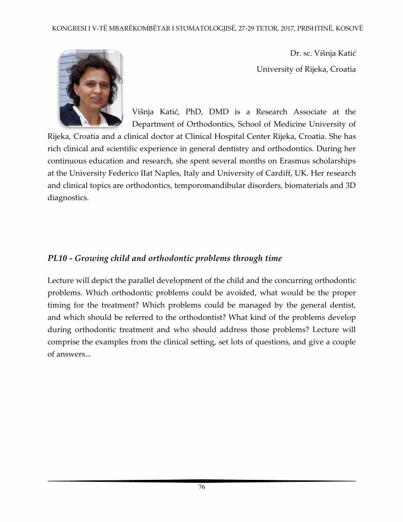
KONGRESI I V-TË MBARËKOMBËTAR I STOMATOLOGJISË, 27-29 TETOR, 2017, PRISHTINË, KOSOVË
76
Dr. sc. Višnja Katić
University of Rijeka, Croatia
Višnja Katić, PhD, DMD is a Research Associate at the Department of Orthodontics, School of Medicine University of
Rijeka, Croatia and a clinical doctor at Clinical Hospital Center Rijeka, Croatia. She has
rich clinical and scientific experience in general dentistry and orthodontics. During her
continuous education and research, she spent several months on Erasmus scholarships
at the University Federico IIat Naples, Italy and University of Cardiff, UK. Her research
and clinical topics are orthodontics, temporomandibular disorders, biomaterials and 3D
diagnostics.
PL10 - Growing child and orthodontic problems through time
Lecture will depict the parallel development of the child and the concurring orthodontic
problems. Which orthodontic problems could be avoided, what would be the proper
timing for the treatment? Which problems could be managed by the general dentist,
and which should be referred to the orthodontist? What kind of the problems develop
during orthodontic treatment and who should address those problems? Lecture will
comprise the examples from the clinical setting, set lots of questions, and give a couple
of answers...

KONGRESI I V-TË MBARËKOMBËTAR I STOMATOLOGJISË, 27-29 TETOR, 2017, PRISHTINË, KOSOVË
77
Mr.sc. Nedim Kasami, Vladimir Popovski
Clinical Center Mother Theresa , Skopje, Macedonia
Dr. Nedim Kasami was born in Gostivar 1969. School of Dentistry University of Zagreb
finished in 1996 at the class of Professor Ivica Anic with average mark of exams 9.25.
From 2005 up to present works at University Clinic for Maxillofacial Surgery and 2010
becomes trainee at the same Clinic and finished in 2014. In 2013 graduates in
postdiplomic studies with thesis Clinical and surgical evaluation of Congenital cysts of
the neck and becomes Master of Science in maxillofacial surgery. His research interests
are the tumors of parotid gland and in 2013 earns Helen Matras Schollarship from
EACMFS and realised stay in General Hospital of Wienna at the Hans Pichler Clinic for
maxillofacial Surgery. Currently works in his PhD thesis at the same Clinic for
Maxillofacuial surgery in the Clinical Center Mother Theresa in Skopje, Macedonia
where he actually works. He has publicated in international scientific papers like Acta
Stomatologica Croatica and has participated in international simposiums and
congresses where he has presented his genuine scientific work in the field of
maxillofacial surgery. Recently in 2015 he was the president of the Congress with
international character in Struga called Managing Challenges in Dentistry . Dr. Nedim
Kasami is current president of Albanian Dental Society in the Republic of Macedonia.

KONGRESI I V-TË MBARËKOMBËTAR I STOMATOLOGJISË, 27-29 TETOR, 2017, PRISHTINË, KOSOVË
78
PL11 - Benign tumors of the parotid gland - diagnosis and surgical treatment
Sputum gland tumors are relatively rare and represent only 2-5% of the cervical-facial
regions. They can be found in large and small salivary glands, but are most commonly
encountered in the parotid gland 10 times more often than other sputum glands. These
tumors are most commonly present at age 40-50, but can be encountered at other ages.
Pleomorphic adenoma (tumor mixtus) is the most common tumor of the parotid gland
and may be located in the superficial or deep gland of the gland that complicates the
tumor access. Warthter tumor (papillary cystadenoma - the old term) is the second in
terms of frequency and represents the tumor with cystic characteristics in its inward,
from its mainstream and its name. CT and MRI are the standard diagnostic methods for
operational access planning, as they provide accurate tumor location and its relation to
the surrounding tissues, particularly the 7th cranial nerve, respectively the facial nerve.
Both tumors do not hurt the facial nerve and usually do not cause pain or facial nerve
paresis. Surface parotidectomy and the preservation of the nerve is necessary when the
possible reoccurrence of the tumor is expected. This means that the tumor detection, the
minimal preparation of the facial nerve and its branches from the trunk to the
periphery, or in the case of centripetal approaches from the periphery to the center,
should be done, which is contested by many authors. Only tumor removal is not
enough as a protocol therapy since the rate of recurrence is very high, whereas with the
application of superficial parotidectomy, the tumor reoccurrence is noticeably reduced
after removing the surface lobe and providing nerve preservation.

KONGRESI I V-TË MBARËKOMBËTAR I STOMATOLOGJISË, 27-29 TETOR, 2017, PRISHTINË, KOSOVË
79
ORAL PRESENTATIONS

KONGRESI I V-TË MBARËKOMBËTAR I STOMATOLOGJISË, 27-29 TETOR, 2017, PRISHTINË, KOSOVË
80

KONGRESI I V-TË MBARËKOMBËTAR I STOMATOLOGJISË, 27-29 TETOR, 2017, PRISHTINË, KOSOVË
81
OP1 - NEW CLASSIFICATION OF ENDODONTIC ACCESS CAVITIES
*ALMIRA ISUFI1,2 DDS, MSC, PHD, GIANLUCA PLOTINO1 DDS, PHD,
NICOLA MARIA GRANDE3 DDS, PHD, GIANLUCA GAMBARINI1 MD, DDS 1 Department of Endodontics, Sapienza Università di Roma, Rome, Itali 2 Faculty of Dental Medicine, Medical University, Tiranë, Albania 3 Università Cattolica del Sacro Cuore, Rome,
Aim: There are many studies about cavities but very few information about volumetric
differences, thus generating confusion among researchers and practitioners. The aim of the
present study was to determine and compare the difference of dentine volume removed
when preparing traditional and two different minimally invasive (conservative and ultra-
conservative) access cavities, and propose a new classification.
Materials and Methods: 30 maxillary and 30 mandibular molars were selected based on
similar dimensions, initially scanned with CBCT (I cat) and then divided in two groups
each one (n=20) containing 10 upper and 10 lower molars. In Group A, a conservative
minimally invasive endodontic access cavity preparation (CMI) was performed as
described by Clark and Khademi. In Group B, an ultra-conservative minimally invasive
(UCMI) endodontic access cavity was performed, preserving most part of the pulp
chamber roof. In Group C, a traditional access cavity (TAC) was performed, eliminating all
dentinal and enamel interferences. After cavities were prepared for each group, specimens
were scanned again with CBCT. The sliced image data were exported as DICOM-file and
imported into the MeVisLab framework system for segmentation and volume
measurement. After segmentation, volumes of coronal dentine and enamel were measured
and the difference in percentage of the volume removed when preparing endodontic access
cavities was calculated. The data were subjected to statistical analyses of variance.
Conclusions:
1. A new classification for endodontic cavities is proposed, dividing them into three
categories. 2. This classification was helpful to complete a study aiming at evaluating the
clinical impact of different endodontic cavities on the fracture resistance of endodontically
treated teeth.

KONGRESI I V-TË MBARËKOMBËTAR I STOMATOLOGJISË, 27-29 TETOR, 2017, PRISHTINË, KOSOVË
82
OP2 - THE DISTRIBUTION OF SUBSTITUTE ELEMENTS BY METAL-CERAMIC
LATERAL DENTAL BRIDGES KALTRINA BEQIRI1*, SHERIF SHAQIRI1,2
1 SPECIALISED CLINIC FOR PROSTHODONTICS PROTETIKA AG , TETOVA 2 STUDY PROGRAME OF DENTISTRY, FACULTY OF MEDICAL SCINECES, UNIVERSITY OF
TETOVA
The aim of our study was to:
- analyze the frequency of missed teeth in lateral segment according gender and jaws,
- analyze the distribution of substitute elements in lateral segment and their correlation,
- analyze the correlation between teeth absence and substitute elements.
Material and methods: The clinic material is compound from 455 substitute elements of lateral
metal-ceramic bridges from 151 patients from both sex, with age from 26 to 70 years old in both
jaws made in specialized clinic for prosthodontics Protetika AG in Tetova in a period 2015-
2016 year.
The getting data are showed with graphics and tables, meanwhile those with special
importance with T-test and coefficient of probability (p).
Results: By males in maxille the most substitute tooth is the first molar with 38.85% of cases,
and in mandible the most substitute tooth is also the first molar with 42.33% of cases.
By females in maxilla both premolars separately are most subtituted with 37.62% of cases, and
in mandible is the first molar with 41.55% of cases.
Conclusions:
1. The frequency of fixed metal-ceramic prosthetic appliances, and the frequency of substitute
elements and crowns in lateral dental bridges is evidently higher by males opposite females.
2. The number of substitute elements by our results showed proper statistically proportion,
because for two substitute elements like retainer crowns are two.
3. According the T-test (t=6,75) and the coefficient of probability (p<0.01), is cleared that by
our results the statistically significance is important, not by chance.
Key words: Substitute elements, metal-ceramic bridges, distribution, lateral sector

KONGRESI I V-TË MBARËKOMBËTAR I STOMATOLOGJISË, 27-29 TETOR, 2017, PRISHTINË, KOSOVË
83
OP3 - TRANSLUCENCY OF LAYERED VS. MONOLITHIC ZIRCONIA CROWNS
DR. SHK. MJ .BRUNILDA KOÇI1, DR. ERVIN KOÇI2, DR. ENKELIDA BEQARI2,
PROF. ASOC. NAZMI KOÇI2
1 Aldent University, Faculty of Dentistry, Tirana, Albania
2 Evident Dental Clinic, Tirana, Albania
Objectives: Monolithic, full contour, ceramic crowns are claimed te have higher resistance and
success rate than layred ones. When zirconia ceramic is employed to make full contour
restoration, translucency still remains an issue since zirconia is the least translucent among
dental dental ceramic materials. An optical improved zirconia has been recently introduces
{LAVA Plus, 3M ESPE}, the aim of this study is to compare the translucency of monolithic and
layered crowns obtained with this new zirconia ceramic.
Material and Methods: One upper incisor and one canine were prepared to receive LAVA Plus
crowns, A2 VITA shade. For each kind of tooth, 6 monolithic and 6 layered crowns were
replicated using the CAD CAM technology and the pressed-on layering technique, IPS e.max
ZirPress A2. The light reflected by the crowns was measured with radiometer in two different
conditions, natural and metal plated natural abutment tooth. The ratio between light reflected
by the crown in the two conditions gave a measure of translucency, contrast ratio.
Results: Monolithic crowns were not significantly less translucent than layered ones in both
incisor{p_0.098} and canine {p_0.0340}, contrast ratio 0.978 vs 0.956 and 0.941 vs 0.929
respectively
Conclusion: The translucency of monolithic crowns made using a new Y-TZP zirconia ceramic
was not different from layered ones {0.5 mm thick coping} considering their superior strength,
monolithic LAVA Plus zirconia crowns appear as an advantageous alternative to layered ones
as far as translucency is concerned.

KONGRESI I V-TË MBARËKOMBËTAR I STOMATOLOGJISË, 27-29 TETOR, 2017, PRISHTINË, KOSOVË
84
OP4 - MOLAR INCISOR HYPOMINERALIZATION (MIH)
SHEVALE ALILI*, E. ZABOKOVA-BILBILOVA, D. GLIGOROVA
PHC University Dental Clinical Center St. Pantejlemon Skopje, Macedonia
Department of Pedodontics and Preventive Dentistry
Molar Incisor Hypomineralisation (MIH) is a relatively common, developmental condition
characterized by hypomineralisation defects of the enamel on the first permanent molars and
the permanent incisors. The prevalence of MIH appears to be increasing, and managing affected
children is now a common problem for pediatric dentists.
The purpose of this paper was to describe the diagnosis, prevalence, putative etiological factors,
and features of hypomineralized enamel in molar incisor hypomineralization and to present a
sequential approach to management.
The complex care involved must address the child s behavior and anxiety aiming to provide
durable restorations under pain-free conditions. Restoration of surfaces with limited
involvement with resin composite is recommended following:
1. removal of all discolored hypomineralized enamel;
2. placement of cavity margins on apparently normal enamel; and
3. bonding with a self-etching primer adhesive.
Extensively affected molars may require extracoronal restorations or extraction. Research is
needed to clarify etiological factors and improve the durability of restorations in affected teeth.

KONGRESI I V-TË MBARËKOMBËTAR I STOMATOLOGJISË, 27-29 TETOR, 2017, PRISHTINË, KOSOVË
85
OP5 - THE RESULTS FROM FOLLOW UP AFTER APICOECTOMY WITH
RETROGRADE FILLING OF ROOT CANALS
WITH TWO DIFFERENT MATERIALS
AMET DEMIRI1,2, SEHA MUSTAFAI3
1 CLINIC FOR ORAL SURGERY ESTETSKA ORALNA HIRURGIJA TETOVA
2 STUDY ROGRAMME DENTISTRY, FACULTY OF MEDICAL SCIENCES, UNIVERSITY OF TETOVA
3 DENTAL CLINIC VIVADENT
The aim of this study is to evaluate the efficacity of retreatments with retrograde fillings with
amalgam and iodoform phosphate of failed teeth with periapical lesions, which were treated
earlier with endodontic-surgery method.
Material and methods: For this study, were done clinical examination and radiographic
examination of 86 teeth surgically treated in a period from 2010-2014, in the clinic of oral
surgery in Tetova. At 86 pacients was realised the apico-ectomy with retrograde filling after
apical resection using amalgam filling at first group (43 patients) and iodoform-phosphate at
second group (43 patients).
Results: After 24 months, at first group with 43 treated teeth with retrograde filling material-
amalgam, successful results we have got in 32 cases (74.42%), meanhwile incomplete results we
have got in 6 cases (13.95%) and failure results in 5cases (11.63%). At the second group, from 43
teeth retreated with retrograde filling-iodoform-phosphate, successful results we have got in 34
cases (79.09%), meanhwile incomplete results in 6cases (13.95%) and failure results in 3cases
(6,98%).
Conclusions:
•The retrograde filling of teeth canals is a promising method, giving healing approximately
in 80% of examined cases, 24 months after surgery treatment.
•A high per cent of roots with amalgame filling in comparation with roots filled with
iodoform-phosphate were succesfully healed, but the difference was without statistically
significance (P>0.05).
Key words: Retrograde filling, surgical treatment, periapical lesions

KONGRESI I V-TË MBARËKOMBËTAR I STOMATOLOGJISË, 27-29 TETOR, 2017, PRISHTINË, KOSOVË
86
OP6 - EVALUATION OF TOTAL RESIN PROTHESIS
POLYMERIZED BY BOILING.
DR.SH. KRESHNIK KERAJ*, PROF.DR. F. KERAJ, BORA KERAJ, LUNAREDA KERAJ
Faculty of Dental Medicine, Medical University, Tirana, Albania
Purpose of our study is to evaluate the changes of the acrylic resin during polymerization by
boiling process.
Material and Methods: we have studied 63 Total Prothesis in the upper jaw. These prothesis
have been studied since "2012-2017" in University's Clinic, Private Clinic etc. The Methodology
followed has The aim to identify the causes that lead to those changes in prothesis by acrylic
resin.
Conclusion: From our study, we evaluate that totally polymerized prothesis by boiling lead to
dimensional changes.
These changes came as a result of non application rules of the basic polymerization.
Our request is to minimize those gaps in the resin structure, we solve it by grinding 1/3 of
palatal end till AH line.
KONGRESI I V-TË MBARËKOMBËTAR I STOMATOLOGJISË, 27-29 TETOR, 2017, PRISHTINË, KOSOVË
87
OP7 - INFLAMATORY CYTOKINES IN MONOCYTES EXPOSED TO ZINC OXIDE (ZNO) NANOPARTICLES – WITH FLOW CITOMETRY
*ZANA SEJFIJA1,2, LORNA PROUDFOOT2 1 Medical Faculty – Dentistry, University of Prishtina Hasan Prishtina 2 Edinburgh Napier University, UK
Number of engineered nanomaterials with unique properties is increasing day by day. It has
been previously shown that zinc in nano formulation has different effects on cells than that of
its bulk counterpart. Despite the significant lack of toxicological data, zinc oxide nanoparticles
(ZnO NPs) are used extensively in a large number of disciplines. As these particles are applied
in many fields, such as organic chemistry, nanomedicine and dentistry. In this study focus was
given to researching the impact of ZnO NPs on cells of the immune system, in order to ensure
that the function of immune system is not left compromised.
The aim of the study: This study attempted to investigate the cytokine responses and surface
markers of THP-1 cells stimulated by a sublethal dose of ZnO NPs prior to, during or after
exposure to LPS. In addition, viability and morphological changes were examined with Flow
cytometry to allow better interpretation of the data obtained.
Methods and Materials: Study was a laboratory research and chemicals used in the preparations
of culture media were purchased from InVitrogen, U.K.THP-1 cell line is used as in vitro model
of primary monocytes. IL-8 ELISA was performed using supernatants collected from the THP-1
cells exposed to different treatments for 18 hours. Research was conducted in the Nano
laboratory of the University of Edinburgh, United Kingdom. Flow cytometry was performed to
analyze the effects of ZnO NPs on viability and immune surface markers of THP-1 cells which
were or were not exposed to LPS at different time points.
Results: The results from this study suggest that NG ZnO in concentrations of 25, 12.5, 6.2 and
3.1 g / ml may relieve various levels of IL-8. However, the level of pro-inflammatory mediators
released from the treated cells at the same time from the nanoparticles and LPS resulted in high
leve of IL-8.
Conclusion: Release of IL-8 is dependent on the concentration. The addition of cytokines
superiority exposure of cells to: ZnO NG AND LPS suggests synergistic effect. Therefore, this
study is aimed at expanding the understanding of the effect of ZnO in relation NG
inflammation in vitro.

KONGRESI I V-TË MBARËKOMBËTAR I STOMATOLOGJISË, 27-29 TETOR, 2017, PRISHTINË, KOSOVË
88
OP8 - AGGRESSIVE PERIODONTITIS AND ANTIBIOTICS IN ALBANIA:
CORRELATION AND CHARACTERISTICS
BESIAN ABAZI1, JOANA MIHANI2, EDIT XHAJANKA1
1 University of Medicine Tirana, Faculty of Dental Medicine
2 University of Medicine Tirana, Department of Pharmacy
Introduction: Aggressive periodontitis poses a challenge for the dentist in the entirety of oral
pathologies. Amongst treatment options, antibiotics play a crucial role. To date, there is no data
regarding the prescription of antibiotics for periodontitis in our country.
Aim: Identification of characteristics related to the pattern of prescription of systemic antibiotics
for aggressive periodontitis in our country.
Materials and Methods: Data were collected from twelve pharmacies randomly selected, on
prescriptions only from dental practitioners. Only prescription containing a diagnosis of
periodontitis with at least one antibiotic given were included in the study. Data analysis was
done with SPSS 20.
Results: Out of 674 initial prescriptions, only 214 met the selection criteria. The average age of
patients was 35.11 ± 12.41 years. Mean duration of therapies was 5.08 ± 1.5 days. The most
common form of prescription was broad spectrum antibiotic (78.6%), combined spectrum
antibiotics (17.8%) and narrow spectrum antibiotic (3.6%). Combined antibiotics involved the
use of Metronidazole with Amoxicillin (10.6%) and Metronidazole with Spiramycin (7.2%).
Conclusions: Combined therapy and other antibiotics were prescribed with the increasing of
patient s age, instead metronidazole was usually prescribed in younger patients. Despite the
fact that metronidazole has a good tolerance and is an effective drug in advanced periodontitis,
this study shows that it s infrequently used among dentists for periodontal disease.
Key words: antibiotics; aggressive periodontitis; correlation
KONGRESI I V-TË MBARËKOMBËTAR I STOMATOLOGJISË, 27-29 TETOR, 2017, PRISHTINË, KOSOVË
89
OP9 - FLUCTUATIONS OF QUANTITATIVE AND QUALITATIVE SALIVARY COMPONENTS, AS INDICATORS OF DIABETES, IN THE ORAL CAVITY
M. D. ILMA ROBO,* PROF. AS. LUAN MAVRIQI, HABIBE BABASI,
DR. SHK. MED. SAIMIR HETA, DR. SHK. MED. NEVILA ALLIU
Professor at Albanian University, Department of Periodontology, Tirana, Albania Professor at Albanian University, Department of Periodontology, Tirana, Albania Dental Clinic, Tirana, Albania
Pediatric Surgeon, Pediatric Surgery Service, QSUT, Tirana, Albania University Hospital Center, Biochemical Laboratory, Tirana, Albania
Introduction: Saliva with immune defense mechanisms to fit the minimum amount of saliva
produced and high glucose presence, these elements that function in time, all together give the
typical characteristics of diabetes as a cause of tissue lesions in oral cavity.
Purpose: Our study aims to find the possible link between oral lesions and changes in
composition and amount of saliva produced in diabetic patients.
Material and method: The study was conducted in 37 patients, of whom 22 were diabetic
regardless of type of diabetes or glycemic control, and 15 were healthy patients. In the study,
the measurement of stimulated saliva and saliva rate was selected to compare the median
values of these amounts to the diabetic patient group and to the healthy patient group.
Results: The study resulted in 17 patients with xerostomia: 5 patients with xerostomia were
from the group of non-diabetic patients and 12 from the group of diabetics; 12 patients with
hypo-salvation were from the group of diabetics. K+ ions were slightly larger, but not
significant, respectively 17 and 24, while Cl- ions had significant differences respectively 26 and
36. Significant difference between salivary pH in diabetic patients was observed in the direction
of many acids, respectively, pH = 5.9, compared to non-diabetic patients, where the pH resulted
basically pH = 7. The level of glucose in the saliva speaks for large differences, respectively the
glucose levels in saliva in diabetic patients were 4.6 and in non-diabetic patients 1.7.
Conclusion: Saliva stimulus is noticeable both in diabetic patients and patients in the control
group. In diabetic patients as saliva norm and stimulated saliva are in significantly reduced
amounts. Collected saliva quantities show significant correlation with the age of the patient and
his gender. Fluctuations in ion, in glucose, in salivary pH lead to the appearance of oral lesions
typical of diabetic patients.
Key words: diabetes, saliva, ion, glucose

KONGRESI I V-TË MBARËKOMBËTAR I STOMATOLOGJISË, 27-29 TETOR, 2017, PRISHTINË, KOSOVË
90
OP10 - SURGICAL GUIDE AND IMMEDIATE LOADING OF DENTAL IMPLANTS
FISNIK MEKAJ ORAL SURGEON , ENI TUTULAKU DENTIST ,
YLL ABDYLI DENTIST , CDT. CEM NIS DENTAL TECHNICIAN
Dental Clinic Advanced Dental Center , Tirana
Summary: Nowadays flapless surgery for implant placement is gaining more and more
popularity among dentists and patients. The advantages of this form of surgery are: its
noninvasive nature, accurate, expeditious and predictable implant placement, satisfactory
prosthodontic outcome, less postsurgical disturbances for the patient, etc.
Purpose: The study of the results from partially edentulous patients treated with flapless
guided implant surgery.
Material and methods: Digital implant planning using the Sirona Galileo software allows for
prior prosthodontic suprastructure planning, offering better function and esthetics for the
patient. Surgical guide planning is done by employing Inlab CAM followed by cutting of the
PMMA guide disc using inLab MC X5.
Conclusions: Application of flapless guided implant surgery provides a more predictable
implant placement with the procedure causing less disturbance to the patient.
Keywords: surgical guide, implantology, 3D radiography
KONGRESI I V-TË MBARËKOMBËTAR I STOMATOLOGJISË, 27-29 TETOR, 2017, PRISHTINË, KOSOVË
91
OP11 - PROFESSIONAL STRUCTURE OF OUR
PROSTHODONTIC-TREATED PATIENTS
SHERIF SHAQIRI1,2*, KALTRINA BEQIRI1
1 SPECIALISED DENTAL CLINIC FOR PROSTHODONTICS PROTETIKA AG , TETOVA
2 STUDY PROGRAMME FOR DENTISTRY, FACULTY OF MEDICAL SCIENCES, UNIVERSITY OF
TETOVA
Aim: The aim of our study is to determine:
- the frequency of treated patients according to gender,
- the frequency of treated patients according to profession,
- the correlation between gender of treated patients according to profession.
Material and methods: The clinical material is compound from 1785 patients treated in
prosthodontic clinic Protetika AG Tetova, in a period from 2014-2016. The age of examined
patients was from 13 to 82-year old, with average age from 48.2 year. The getting data are
evidenced in patient s chart using the modified form of oral health evaluation according WHO,
adapted and modificated with the nature of our study. The results are showed in graphics and
tables, meanhwile those with speciale importance are showed with X2-test, coeficient of
probability (p) and coeficient of correlation.
Results: From the total of treated patients, in a high per cent were housweifes with 31.93%, and
less treated were engineers with only 0.84%.
Conclusion: Concerning the profession by our treated patients, there is evidently that the
extreme representation of patients according genders by destinated professions.
1. By profession for men (artisans, merchants, farmers), those in 100% of cases belong to
males,
2. By profession for women (houswifes), those in 100% of cases belong to females,
3. Is distressed the fact that we have a considerable number of students 135 (7.56%) and
schoolboys 105 (5.88%) with pathology in their stomatognatic system and problems concerning
the oral health.
Key words: Patients, profession, frequency, prosthodontic treatment

KONGRESI I V-TË MBARËKOMBËTAR I STOMATOLOGJISË, 27-29 TETOR, 2017, PRISHTINË, KOSOVË
92
OP12 - EFFICACY OF DIFFERENT MATERIALS IN TREATMENT
OF IATROGENIC PERFORATIONS - IN VITRO STUDY
MIRANDA STAVILECI-VELIU*, VETON HOXHA, DONIKA BAJRAMI,
ASTRIT KUÇI, AGIME DRAGIDELLA
University of Prishtina, Medical Faculty, Dental School, Department of Dental Pathology and
Endodontics.
Introduction: Procedural errors during endodontic treatment affect the prognosis of endodontic
therapy. One of them is the endodontic iatrogenic perforation for the treatment of which
currently some materials are used.
Aim: The aim of this study was effectiveness evaluation of biodentine, mineral treoxid
aggregate cement, glass ionomer cement and amalgam as root perforation repaired materials.
Material and methods: Thirty-two extracted human lower molars were used for this study.
After access cavity preparation and pulp tissue and debris cleaning, root perforations were
made on the center of the floor and teeth were divided into experimental and control groups.
Root perforation of seven teeth (first group) were repaired with biodentine, root perforation of
other seven teeth (second group) were repaired with MTA, root perforation of seven teeth (third
group) with glass ionomer cement and root perforation of last seven teeth (fourth group) with
amalgam. Two teeth were not repaired and served as positive control group. Two intact teeth
served as negative control group. The samples were immersed in 2% methylene blue and after
rinse were sectioned and examined under stereo-microscope for dye penetration evaluation.
Results: Data were statistically analyzed using analysis of variance (ANOVA) at level of
significance p=0.05. Root perforation repaired with Biodentine showed significantly less dye
penetration, compared with MTA, amalgam and glass ionomer cement (P<0.05).
Conclusions: All repaired materials showed some degree of dye penetration. Biodentine
showed least microleakage.
Key words: perforation, biodentine, MTA, glass ionomer, amalgam
KONGRESI I V-TË MBARËKOMBËTAR I STOMATOLOGJISË, 27-29 TETOR, 2017, PRISHTINË, KOSOVË
93
OP13 - IMPLEMENTATION OF REGENERATIVE MEDICINE IN STOMATOLOGY
D.SH.M ALDO VANGJELI * 1, ARJANA SAKO2, LINDITA HYSI3, OLESIA DUKA4, EDA JEMINI5
1 Faculty of Dental Medicine, Tirana, 2 Dental Clinic AS, 3 Dental Clinic Linea,
4 Dental Clinic Shendident, 5 Dental Clinic Eda
Introduction: Regenerative medicine and especially its implementation in dentistry can be
particularly useful in oral and implant surgery as a growing trend. The introduction and
utilization of organic components of human blood and not only the activation of indirect
engineering as a concept is in itself a really complicated seeming mechanism, but with a very
rich theoretical background which allows the possibility of scientific interpretation.
Purpose: It is the theoretical analysis of the process of realization of the directed regeneration of
strong and soft tissues. What does actually represent the growth factor and how can we get in
touch with it to maximize the positive qualities of this reaction. The clinical presentation of the
implementation of these methods is a natural part of this purpose material derives from the
analysis and comparison of application modes.
Atelo-Colagene'ose membrane and bovine collagen. It represents 99.9% non-telopeptide-free,
non-related, but purified and modified collagen, as a consequence of the apirogenic, totally
sterile absorbent with perfect hemostatic effect after application of the membrane, the
regeneration of soft tissue 'Guided Tissue Regeneration' (GTR at GBR). As a consequence of the
development of growth factors (TGF, PDGF), this results in high platelet concentration.
Platelet-rich fibrin (PRF) is obtained from blood without adding anticoagulants. A-PRF affects
bone and soft tissue regeneration through the presence of monocytes / macrophages and their
growth factors.
Conclusion: The application of bovine grafts or not is a further opportunity for accelerated
regeneration. Benefiting from the patient's own blood, their application places the surgeon
undoubtedly into avant-garde use of science and technology.

KONGRESI I V-TË MBARËKOMBËTAR I STOMATOLOGJISË, 27-29 TETOR, 2017, PRISHTINË, KOSOVË
94
OP14 - SAF SYSTEM IN ENDODONTIC
ASTRIT KUÇI1, BLERIM KAMBERI1, SHEFQET MRASORI1, TAYFUN ALAÇAM2
1University of Prishtina, Faculty of Medicine, Department of Dental Pathology and Endodontics
2 Department of Endodontics, Faculty of Dentistry, Gazi University, Ankara, Turkey
Purpose of the study
Realistic assessment of SAF system capabilities, respectively the ability of this system to remove
dentine uniformly from all parts of the root canal wall.
Materials and methodology
For this study, we have selected the first mandible premolar that have been extruded for
paradontic reasons and are stored in 10% formalin. The Saf system with the needle set was
used, the special head of this rdt-3 system for the counterpart and the irrigation system Vatea .
For the channel obturation methodology we used the system B and obtura II system, the AH 26
and MTA fillapex pastes and the reading was done with a laser confocal microscope.
Results
Based on the samples of used roots and observation of the analysis we came to the conclusion
that the SAF system had clean all the tooth root walls of the tooth.
Conclusions
Within the ex vivo conditions that this research has been conducted, it can be said that the
preparation of channels of its walls in all segments of the tooth root with SAF system is fully
feasible and clinically feasible.
Keywords: SAF systems, RDT-3, Vatea system, Laser confocal microscopes.

KONGRESI I V-TË MBARËKOMBËTAR I STOMATOLOGJISË, 27-29 TETOR, 2017, PRISHTINË, KOSOVË
95
PO15 - INDEX OF ORTHODONTIC TREATMENT NEED-ITS USAGE IN
ASSESSMENT OF MALOCCLUSION SEVERITY
DONIKA ILIJAZI-SHAHIQI1, BLERIM KAMBERI2, SANDRA ANIC1, JETA KELMENDI1
1University of Zagreb, Dental School, Department of Orthodontics
2University of Prishtina, Faculty of Medicine, Department of Dental Pathology and Endodontics
Malocclusion is one of the most common dental problem and according to studies it is ranked
second in the list of problems encountered in the oral health. Based on data from multiple
studies related to the prevalence of malocclusion it ranges from 26% to 86%. Prior to the
therapeutic planning, one of the first step to be taken is assessment of the degree or severity of
malocclusion. Given the high cost of orthodontic treatment, the selection of cases for treatment
should be done with care, in order that cases with higher degree of malocclusion have priority.
In determining the degree of malocclusion different indexes are available in the service of
orthodontics. Among the indices most commonly used is the Index of Orthodontic Treatment
Need IOTN.
The purpose of this presentation is to inform about the IOTN index, where it will be more
widely discussed about the method of application, the advantages and the limitations
encountered during its use. The use of this index in clinical centers helps us in unification of
diagnosis,better allocation of the recourses, reducing the risk of unnecessary orthodontic
treatment and the same approach to treatment for all patients.
KONGRESI I V-TË MBARËKOMBËTAR I STOMATOLOGJISË, 27-29 TETOR, 2017, PRISHTINË, KOSOVË
96
PO16 - THE IMPORTANCE OF AGE ESTIMATION IN ORTHODONTICS
JETA KELMENDI1,3, F. KOÇANI2, B. KAMBERI2, M. VODANOVIC3, D. SHAHIQI-ILIJAZI1,3,
O. ALKU-LATIFI 4, A. RESHITAJ1
1 University Dentistry Clinical Center of Kosova, Orthodontic Department
2University of Prishtina, Faculty of Medicine, Department of Dental Pathology & Endodontics
3 University of Zagreb, School of Dental Medicine
4University Dentistry Clinical Center of Kosova, Oral Medicine and Periodontology department
One of the goals for treating skeletal discrepancies is to gain benefits from the patient s growth
spurt, which help to reach optimal results, within the short period of time. Identification of
period of accelerated growth and evaluation of individual biologic time table is essential for
clinical decisions, concerning growth modulation procedures for skeletal discrepancies,
extraction versus non extraction options, planning for orthognathic surgery and use of extra
oral orthopaedic forces. Chronological age is often not sufficient for assessing the
developmental stage and somatic maturity of the patient, so that the biological age has to be
determined. The biological age is determined from the skeletal, dental and morphologic age and
onset of puberty. Patient chronological age is defined as the time period from the birth to till
date. As a results many investigators have attempted, to predict the duration, magnitude,
direction and timing of the adolescent growth changes. The developmental status of a child is
usually assessed in relation to events that take place during progress of growth. Thus
chronological age, sexual maturational characteristics, dental development, height and weight
measurements and skeletal age are some of the biological indicators that have been used to
identify stages of growth. Conclusion: Maturational development embodies the biologic
progression through life. In the growing years, indicators of the level of maturational
development of the individual provide the best means for evaluating biologic age. Maturational
development can be assed with the help of all the indicators previously described. However, it
must be kept in mind that every child demonstrates a unique sequential pattern of events. No
child is the same as the other. Skeletal indicators of maturation have been proved to be the most
reliable. A combination of skeletal and dental indicators tends to give a very accurate picture of
each child s developmental status. Finally it must be kept in mind that in orthodontic practice it
may be more relevant to evaluate the development of the patient in relation to his own growth
potential in order to assess whether peak velocity growth is imminent, present or completed.
The choice of indicators to be used finally depends upon an orthodontist s preference.

KONGRESI I V-TË MBARËKOMBËTAR I STOMATOLOGJISË, 27-29 TETOR, 2017, PRISHTINË, KOSOVË
97
PO17- THE PREVALENCE OF EDENTULISM IN THE GOSTIVAR REGION TEFIK ISMAILI1*, KUJTIM SHALA2, LINDIHANA EMINI1
1 University of Tetova, Dentistry School. 2 University of Prishtina, Faculty of Medicine, Dental School
Introduction: Edentulism is the loss of a small number of teeth or their total absence; it is a
biochemical process, because the teeth, jaws and oral mucous membranes are not static objects;
They are dynamic (they change over time)
Aim of this study was to investigate the extent of the prevalence of the prosthetic dentures, the
classification of the partial edentulism, the average age of the population which have a kind of
prosthetic denture as well as tooth stability in the jaw.
Materials and Methods: The data was taken at the institute of the health fund of the Republic of
Macedonia, which operates under the "My mouth" program, and archive records of the State
Hospital "Ferid Murad" in the city of Gostivar. Their selection was made based on the type of
moving dentures, demographic factor, age and sex. While the classification of partial
edentulism is made based on the classification of Dr. Kennedy.
Results: In the research we found a great difference between patients with total dentures who
represented 90% compared to those with partial prosthesis who were 10%. The average age for
a partial prosthesis was 50.6 years, whereas for total prosthesis was 61.3 years. According to the
classification of Kenedy, the first class of this classification was most frequent with 64% of cases.
Conclusion: According to our study we can conclude that sex does not affect edentulism.
Patients in rural areas are less affected by edentulism. There is considerable difference between
total and partial prostate patients. We also note that the individual most likely missed the
maxillary teeth as compared to the mandible teeth stay longer in the oral cavity
Key words: Republic of Macedonia, mobile dentures, partial dentures, Dr. Kennedy, Gostivar

KONGRESI I V-TË MBARËKOMBËTAR I STOMATOLOGJISË, 27-29 TETOR, 2017, PRISHTINË, KOSOVË
98
KONGRESI I V-TË MBARËKOMBËTAR I STOMATOLOGJISË, 27-29 TETOR, 2017, PRISHTINË, KOSOVË
99
POSTER PRESENTATIONS

KONGRESI I V-TË MBARËKOMBËTAR I STOMATOLOGJISË, 27-29 TETOR, 2017, PRISHTINË, KOSOVË
100

KONGRESI I V-TË MBARËKOMBËTAR I STOMATOLOGJISË, 27-29 TETOR, 2017, PRISHTINË, KOSOVË
101
PP1 - MORPHOLOGY OF ROOT CANAL SYSTEM IN MAXILLARY PREMOLARS
XHANINA GAVAZI, ROZARKA BUDINA
Faculty of Dental Medicine, Tirana
Introduction: A thorough knowledge of tooth morphology, careful interpretation of different
planes radiography, proper cavity preparation and detailed exploration of inner tooth anatomy
are essential prerequisites for a successful treatment outcome.
Purpose: The purpose of this study is to distinguish the anatomical characteristics of the pulp
chamber and the canal system in the maxillary premolars teeth in our population and compare
the results with studies in other countries.
Material and Method: 140 maxillary premolars teeth extracted at the Department of Surgery at
the University Dental Clinic, Tirana. After pulp tissue removal, the teeth were decalcified with
5% nitric acid and dehydrated with alcohol with escalated concentrations. After the staining the
teeth morphology were observed under a microscope.
Results: Most of the first premolars have two roots in more than 70% of cases, but there are
cases with one and three roots. In most cases there are straight and diverge canals. In more than
90.3% of cases the second premolars show to own a single root, in 2% of cases with two separate
roots and two attached roots in 7.7% of cases respectively. They have tight channels and are
difficult to find and to process.
Conclusion: Within the limits of this study, it was concluded that in our population, there is an
increased percentage for the types 4, 1, 2 and 5 of the configuration of the canal morphology in
the first maxillary premolar and 1, 2 and 4 for the second maxillary premolar.
Key words: Root canal system; apical foramen; pulp tissue; inter-canal communication.

KONGRESI I V-TË MBARËKOMBËTAR I STOMATOLOGJISË, 27-29 TETOR, 2017, PRISHTINË, KOSOVË
102
PP2 - CHRONIC PERIODONTITIS IN PRIMARY DENTITION,
ALTERNATIVES OF TREATMENT
MANOLA KELMENDI1, SHIRLI KELMENDI2, ENIDA PETRO3, ENO GACE4
1. Phd Manola Kelmendi – University Dentistry Clinic
2. MSc Shirli Kelmendi – Private clinic
3. PhD Enida Petro – Faculty of Dental Medicine Tirana
4. PhD Eno Gace – Public Service RHA
Background: In primary dentition, inflammatory processes of the pulp are frequent due to
untreated caries. Acute pulp inflammation can pass to chronic in a short time due to the
morphological construction of the primary teeth. There exists the question whether a primary
tooth with chronic periodontitis must be treated or extracted.
Purpose: Treatment of inflammatory processes of the pulp in the primary teeth is important to
ensure the presence of the tooth in the mouth until the period of physiological change for a
normal development of the dental system and the health of the child in general.
Material and methods: There were studied 30 children presented with chronic periodontitis at
the private dental clinic. 19 teeth were treated with ZnOE paste and 19 teeth were treated with
Endoflas paste (40.6% triiodomethane, 63.5% ZnOE and 1.07% Ca (OH)2. In total there were
treated 38 primary teeth. The method of treatment was the method of PULPECTOMY - the
complete removal of putrid wastes from the pulp chamber to the bottom of the root canal to
ensure an airtight sealing of the canal.
Conclusion: The treatment of chronic periodontitis in primary teeth with contemporary
medications that eliminate the side effects of ZnOE, provides a greater longevity of the tooth in
the mouth. Out of 19 that were treated with Endoflas, only 1 ended up in extraction. Out of 19
that were treated with ZnOE, 6 were extracted.
Key words: chronic periodontitis; primary teeth; extraction; ZnOE; Endoflas.

KONGRESI I V-TË MBARËKOMBËTAR I STOMATOLOGJISË, 27-29 TETOR, 2017, PRISHTINË, KOSOVË
103
PP3 -THE DIMENSIONAL STABILITY OF DENTURES
MADE OF TWO DIFFERENT MATERIALS
PROF. ASSOC. EDIT XHAJANKA, DR. NEADA HYSENAJ*
The Faculty of Dental Medicine, University Dental Clinic, Tirana, Albania.
Introduction: Acrylic resin is most commonly used for denture bases. However, acrylic resin
has week points of volumetric shrinkage during polymerization that reduces denture fit.
Purpose: The purpose of this study was to compare the dimensional stability in the
conventional acrylic resin base, POSS-containing acrylic resin base.
Materials and methods: Twenty maxillary edentulous casts were constructed from the same
preliminary silicone impression. Twenty dentures of two different bases were fabricated. Ten
conventional dentures and ten POSS containing acrylic resin dentures were constructed. Wax
dentures were identically constructed. They had the same width and height of wax in every
part of the dentures and 2mm thickness in the palatum. Tooth movement and tissue contour
change of denture after processing (resin curing, deflasking, decasting and finishing without
polishing) and immersion in artificial saliva at 37℃ for 1 week and 4 weeks were measured.
Results: The results were as follows: The conventional resin group showed significant (p<0.01)
dimensional change throughout the procedure (processing and immersion in artificial saliva).
After processing, the POSS resin group showed lower linear and 3-dimensional change than
conventional resin group (p<0.01). There was no statistically significant linear and 3-
dimensional change after immersion for 1 week and 4 weeks in POSS resin group.
Conclusions: In this study, a reinforced acrylic-based resin with POSS showed better
dimensional stability compared to conventional acrylic-based resin dentures.
Key words: Conventional dentures; POSS resin dentures; dimensional stability.

KONGRESI I V-TË MBARËKOMBËTAR I STOMATOLOGJISË, 27-29 TETOR, 2017, PRISHTINË, KOSOVË
104
PP4 - INFLUENCE OF PROLONGED BREASTFEEDING ON EARLY CHILDHOOD CARIES IN PRESCHOOL CHILDREN OF TIRANA
ENIDA PETRO1, ETLEVA DROBONIKU1, MANOLA KELMENDI2,
DORJAN HYSI1, SHIRLI KELMENDI3
1 Faculty of Dental Medicine, Tirana, Albania 2 University Dental Clinic, Tirana, Albania 3 Private dental clinic, Tirana, Albania
Introduction: Early Childhood Caries (ECC) is a form of dental caries occurring in the primary
dentition and it is commonly associated with incorrect feeding habits. Although breastfeeding is
the best possible nutrition to infants in the first year of life, prolonged breastfeeding may be
implicated in contributing to the development ECC. Frequent breast feedings during the day on
demand and especially during the night after eruption of teeth are at great risk for ECC (AAPD
2008).
Aim: This study aimed to determine the prevalence and severity of ECC among 3-5 years old
children and to evaluate possible association between prolonged breastfeeding and ECC.
Methods: In this cross-sectional study, 6 out of 42 public kindergartens in Tirana city, were
randomly selected. The representative sample was 904 children, 3-5 years old. Information was
obtained through structured questionnaires that were completed by parents and intraoral
examination of children. Screening, diagnosis and recording of the subjects in this study were
obtained according to WHO (1997) criteria. ECC form was marked for each child based on the
classification criteria of AAPD (2008). The data were analyzed by SPSS.18 Software. Descriptive
Statistics and Hi square test were used for a level of significance as p ≤ . .
Results: The prevalence of ECC in children 3-5 years old in public kindergartens in Tirana city
was 91% (823/904). 62.1% (561/904) of children suffered severe ECC and the mean deft was 6.45
(SD±4.25). There were significant association between ECC and prolonged breastfeeding
(p<0.05).
Conclusions: The prevalence of ECC and the mean deft are very high in preschool children of
Tirana. Most of the children suffered severe ECC. Prolonged breastfeeding is identified as a
significant caries risk factor of the examined children.
Key words: early childhood caries; prolonged breastfeeding; preschool children; public
kindergartens in Tirana.

KONGRESI I V-TË MBARËKOMBËTAR I STOMATOLOGJISË, 27-29 TETOR, 2017, PRISHTINË, KOSOVË
105
PP5 -STRESS EVALUATION OF RECENTLY DESIGNED THREE DIFFERENT
ORTHODONTIC MINI SCREW (OMS) - A FINITE ELEMENT ANALYSE STUDY
PROF DR SEVIL AKKAY, DR AMIR MAMUSHA
GAZI UNIVERSİTY, DEPARTMENT OF ORTHODONTICS
Introduction: The history of metallic biomaterials for medical application goes back to 19th
century; respectively to the Industrial Revolution. The basic characteristic of the biomaterials is
that they are directly in contact with the living body. It is very important that biomaterials must
no cause any side effect. The most biocompatible biomaterial is Titanium (Ti) alloy. In
orthodontic literature, the most used alloy for production of the OMS is Ti6Al4V alloy.
Biomaterials applied to the living body unavoidably release biocompatible or non-
biocompatible ions. Titanium alloys release Aluminum (Al) and Vanadium (V). Therefore, there
is a tendency for alternative for Ti6Al4V alloy.
Material and method: 3 different biomaterial based OMS has been evaluated in this study: a)
Ti6Al4V; b) Titanium-Zirconium (Ti-Zr) and c) Zirconium based OMS. The dimension of the
OMS are: 8 mm in length and 1,6 mm diameter. The scenario is applied in an extraction case in
upper jaw. The first premolar is extracted with the orthodontic purpose and canine retraction
has been made on continuous arch wires (16x22SS) with support of OMS (placed between
second premolar and first molar). A closed coil spring hanged from the hook of the canine
bracket to OMS. The size of brackets is 0,18 slot. The applied force is 1N. Upper jaw model
consisted of 414,300 elements and 83371 nodes. The types of elements are 4 nodes tetrahedral
element. OMI model consisted of 47,444 elements and 10,893 nodes. Upper jaw and OMI model
was constructed with Altair Hypermesh (Michigan, USA) and for the biomechanical evaluation
has been used Dassault System Abaqus (2014, USA).
Results: Based on the results of our study, the least stress is distributed by TiZr, followed by Zr
and Ti6Al4V OMI.
Conclusion: To our knowledge this the first study to compare biomechanically three different
types of OMI materials. There is a need for clinical investigations.
Keywords: orthodontics; orthodontic mini screw; finite element analysis.

KONGRESI I V-TË MBARËKOMBËTAR I STOMATOLOGJISË, 27-29 TETOR, 2017, PRISHTINË, KOSOVË
106
PP6 - HIPNODONTICS
DR. ILIR HOTI
Dental Clinic Hollywood Smile
Hypnosis is a form of unconsciousness resembling sleep but in which the hypnotic subject is
fully awake but have decrease in peripheral awareness. Hypnosis used in dentistry for different
patient s phobia and as pain relief is called hypnodontics. Hypnodontics is promoted by Dr.
Aaron A. Moss, and accepted by American Dental Association for almost 60 years ago.
Hypnodontics make possible to treat of patients with different phobia and different problems
without using of different analgesics. Although the time of treatment for these patients is longer
the positive outcomes are never lacking. In this work we will present methodology of hypnosis
to patients, its advantages and disadvantages.
Nowadays, the daunting development of technology as well as the dynamics of life make
people have different types of phobia, therefore it would be wise that doctors in different areas
to be trained in the use of hypnotherapy, and in the future, to provide their patients with the
best treatment.
Key words: hypnosis, hypnodontics, phobia
KONGRESI I V-TË MBARËKOMBËTAR I STOMATOLOGJISË, 27-29 TETOR, 2017, PRISHTINË, KOSOVË
107
PP7 - CLINICAL EVALUATION OF BIODENTIN IN DIRECT PULP CAPPING
SHEVALE ALILI, E. ZABOKOVA-BILBILOVA, D. GLIGOROVA
PHC University Dental Clinical Center St. Pantejlemon Skopje, Macedonia
Departement of Pedodontics and Preventive Dentistry
Biodentine is a new bioactive cement with dentin-like mechanical properties, which can be used
as a dentin substitute on crowns and roots. It has a positive effect on vital pulp cells and
stimulates tertiary dentin formation. In direct contact with vital pulp tissue it also promotes the
formation of reparative dentin.
The aim of this study was to assess clinically the use of Biodentine in direct pulp capping in
permanent teeth.
Clinical records of patients treated with DPC technique were retrieved. Success rates (based on
symptoms, sensibility tests and radiographic analysis) and adverse events were analyzed.
The patients were reviewed at 1, 3, 6 and 12 months after treatment. Clinical and radiological
examination after treatment of direct pulp capping in adult resulted in proper pulp vitality and
normal radiographic image of the tooth.
Our cases demonstrates the effectiveness of Biodentine in the biological treatment of pulp in
permanent teeth in adults. Biodentine can be an excellent alternative to previously used
preparations in the treatment of pulp capping.

KONGRESI I V-TË MBARËKOMBËTAR I STOMATOLOGJISË, 27-29 TETOR, 2017, PRISHTINË, KOSOVË
108
PP8 - TEETH EROSION AND THEIR PRIMARY AND SECONDARY
PREVENTION - CASE REPORT
GLIGOROVA DRAGICA, ALILI SHEVALE, KOKOCEVA IVANOVSKA-OLGA,
AMBARKOVA VESNA
PHC University Dental Clinical Center St. Pantejlemon Skopje, Macedonia
Department of Pedodontics and Preventive Dentistry
Teeth erosion is pathological loss of hard dental tissues due to chemical or electrolitical path. It
is a multicausal disease. There is an increased incidence of non-cariogenic diseases and loss of
hard dental tissues without microorganisams influence.
The aim of our work is to give a case report of longterm erosion of teeth in children with
gastrointestinal reflux which provoced loss of hard dental tissues, lower esthetics and changed
occlusion. Also, we show a secondary prevention of erosion with the usage of composites and
glass ionomer fillings.
Based on our previous experience, confirmed with this solely case a very important moment in
erosion prevention is their early diagnosis which enables primary prevention, such as oral
hygiene, fluor medicaments, regular nutrition, etc as well as secondary prevention on time.