hepatitis e virus capsid c-terminal region is essential for the viral life
TRANSCRIPT

SHIOTA ET AL.
1 Novel role of HEV capsid C-terminal region
Hepatitis E virus capsid C-terminal region is essential 1
for the viral life-cycle: Implication in viral genome 2
encapsidation and particle stabilization 3
4
5
6
Running title: Novel role of HEV capsid C-terminal region 7
8
9
10
Keywords: 52 capsid C-terminal amino acids, infectious clone, amber mutant, revertant, 11
degradation 12
13
Tomoyuki Shiota, Tian-Cheng Li, Sayaka Yoshizaki, Takanobu Kato, Takaji Wakita, and 14
Koji Ishii* 15
16
Department of Virology II, National Institute of Infectious Diseases, Gakuen 4-7-1, 17
Musashi-murayama, Tokyo 208-0011, Japan 18
19
20
21
22
*Corresponding author: Koji Ishii, Department of Virology II, National Institute of 23
Infectious Diseases, Gakuen 4-7-1, Musashi-murayama, Tokyo 208-0011, Japan 24
E-mail: [email protected]; Phone: (+81)-42-561-0771; Fax: (+81)-42-561-4729 25
26
Abstract word count: 75 27
Text body word count: 1848 28
29
Copyright © 2013, American Society for Microbiology. All Rights Reserved.J. Virol. doi:10.1128/JVI.00444-13 JVI Accepts, published online ahead of print on 6 March 2013
on April 4, 2019 by guest
http://jvi.asm.org/
Dow
nloaded from

SHIOTA ET AL.
2 Novel role of HEV capsid C-terminal region
Abstract 30
Although The C-terminal 52 amino acids (C52aa) of hepatitis E virus (HEV) capsid 31
are not essential for the morphology, C52aa-encoding region is required for the 32
replication. Transfection of C52aa knockdown mutant showed transient growth, yielded 33
earliest population including a majority of noninfectious (possibly empty) and minority of 34
infectious particles with C-terminal capsid degradation, and finally the complete revertant 35
was generated reproducibly. C52aa is essential for the viral life-cycle, promoting 36
accurate encapsidation and stabilizing encapsidated particles. 37
38
on April 4, 2019 by guest
http://jvi.asm.org/
Dow
nloaded from

SHIOTA ET AL.
3 Novel role of HEV capsid C-terminal region
Text 39
Hepatitis E virus (HEV) is responsible for acute and enterically transmitted hepatitis in 40
the developing world (1). Before the establishment of high-efficiency HEV cell culture 41
systems (2), in vitro generation of HEV virus-like particles (HEV-VLPs) in insect cells or 42
in vivo propagation in nonhuman primates were the most useful models for the study of 43
HEV. Genetic deletions or cellular processing resulting in the loss of the N-terminal 111 44
or 13 amino acids (aa) and of the C-terminal 52 aa (C52aa) yielded capsid protein capable 45
of directing the formation of the HEV small (S) or large (L) VLPs (3-5). Particle 46
formation was required for C52aa abbreviation, limiting structural analysis of the 47
resulting particles (3, 4, 6-10). However, the contribution of the C52aa-encoding 48
sequence was confirmed by both in vivo (attenuated infectivity of the point mutant-virus 49
in nonhuman primates) and in vitro (reduced RNA synthesis by RdRp) assays (11-14). 50
Furthermore, the highly conserved nature of the C52aa sequence implies that the C52aa 51
domain itself is functionally important. In this study, we characterized the role of the 52
C52aa domain in the HEV life-cycle by using infectious clones. 53
We constructed infectious clones using the infectious virus G3-HEV83-2-27, 54
employing a procedure previously described by T.C.Li (2). Using a synthetic cDNA as 55
template, we PCR amplified 12 fragments covering the entire G3-HEV83-2-27 genome 56
by the designed primers in Table 1. These fragments were ligated together stepwise and 57
inserted into the EcoRI-HindIII site of pUC19, yielding a wild-type clone that we 58
designated wt. Site-directed mutagenesis of wt was used to generate clones that were 59
mutated to encode capsid protein lacking the C52aa domain, either by introduction of an 60
amber stop codon UAA (knockdown mutant, designated amut) or via deletion of the 61
corresponding segment of the ORF2 sequence (knockout mutant, designated dmut). We 62
performed experiments at three separate scales (normal, large, and huge, as described 63
below) in order to estimate virus progeny productivity, to clarify the growth kinetics, and 64
to analyze the process of encapsidation in the absence of revertants. 65
Normal scale: To estimate the virus progeny productivity of HEV without C52aa, the 66
transfection with amut and dmut was performed in comparison to that with wt. A 67
quantity of 50 μg RNA of each infectious clone was electroporated into 1×10⁷ cells of 68
PLC/PRF/5. ELISA analysis (using anti-G3-HEV-VLP rabbit polyclonal antibody (5)) 69
suggested that transient growth was observed with amut, in contrast to continuous growth 70
with wt (Fig. 1A) and non-growth with dmut (data not shown). Productivity (genome 71
copy number) of amut, measured by RNA real-time RT-PCR with a set of specific 72
primers (Table 1), was estimated as approximately 40-fold lower than that of wt (Fig. 1B 73
on April 4, 2019 by guest
http://jvi.asm.org/
Dow
nloaded from

SHIOTA ET AL.
4 Novel role of HEV capsid C-terminal region
and 1C; note the differences in scale). However, subsequent analysis demonstrated that 74
the amut-derived HEV actually harbored synonymous and non-synonymous reversion 75
mutations, suggesting that the actual productivity (of intact amut) was much lower than 76
suggested by real-time RT-PCR. To assess the progeny, sucrose density (10-60% 77
(wt/vol)) gradient analysis (SDGA) was performed. Subsequently, collected fractions 78
were separated by SDS-PAGE, and western blot analysis (WB) was performed with the 79
above polyclonal antibody (5). Chemiluminescence was recorded using an LAS-3000 80
luminescent image analyzer (Fujifilm, Tokyo, Japan). In the series of fractions obtained 81
from progeny derived from infection with wt, the presence of antigen was only confirmed 82
in Fraction 8 (F8) in Fig. 1C by WB (data not shown). The 72-kDa size of the 83
prominent band was in agreement with the size of the capsid protein predicted for the wt 84
clone. Quantification of HEV RNA genome copy number showed a trailing peak for the 85
progeny derived from infection with amut (Fig. 1B, F8 and 9) and a single peak for 86
progeny derived from infection with wt (Fig. 1C, F8). These peaks corresponded to 87
similar specific densities. Sequence analysis showed that while the progeny from 88
infection with wt carried the original sequence, progeny from infection with amut did not 89
contain the expected UAA (amber codon) at this position. Instead, the trailing peak of 90
this amut-derived sample corresponded to two distinct peaks (F8 and 9) harboring GUU 91
(Val-encoding) and GAC (Asp-encoding) codons, respectively. These changed RNA 92
sequences were predicted to encode full-length revertant capsid proteins. 93
Large scale: To clarify the precise growth kinetics of amut, a larger scale transfection 94
of amut RNA was performed. Specifically, the large-scale transfection was performed at 95
an approximately 30-fold larger scale than that described above, and culture supernatants 96
were collected periodically. This procedure permitted a time-course of quantification by 97
ELISA analysis, and showed that the peak of antigen accumulation occurred 25 days 98
post-transfection, while the number of viral genomes progressively declined during the 2 99
months of the study (except for small recoveries in copy number on Day 25 and at the 100
study end) (Fig. 2A). These data suggested the production of a low level of infectious 101
particles from amut transfection. However, the non-reverted amut antigens could not be 102
distinguished by WB in the normal- and large-scale experiments, suggesting that the amut 103
products were unstable, of low infectivity, and/or produced in small amounts. To 104
confirm the nature of the amut product, pooled supernatant was subjected to partial 105
purification and SDGA. WB of the resulting fractions detected a 72-kDa band in F7 106
(specific density 1.15 g/ml) (Fig. 2B). Quantification of the HEV RNA genome in the 107
fractions detected a single peak, primarily in F7 (Fig. 2C). Determination of the F7 108
on April 4, 2019 by guest
http://jvi.asm.org/
Dow
nloaded from

SHIOTA ET AL.
5 Novel role of HEV capsid C-terminal region
sequence revealed that the expected amber codon was instead GUC (complete reversion). 109
Additionally, infection assays demonstrated that F7 readily infected cells (Fig. 2D). 110
Based on our subsequent experiments, we suspect that the end product of the large-scale 111
experiment likely corresponded to a revertant to wt. 112
Huge scale: To clarify the apparent reversion of amut, transfection was performed 113
at an even larger scale (10-fold increased vs. large scale); culture supernatants were 114
collected periodically, and viral sequences from these samples were determined. The 115
results clearly showed a population shift from the originating amber codon of amut to the 116
complete revertant (GUC) via an intermediate mutant (GAC) (Table 2). Mutants were not 117
detected until 3 weeks post-transfection. The reproducible reversion of amut provides 118
evidence of the functional essentiality of the C52aa domain for the HEV life-cycle. 119
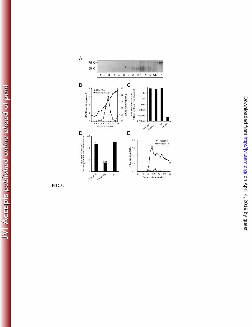
To permit analysis of the amut clone in the absence of revertants, culture supernatants 120
collected within the first 10 days were pooled and subjected to partial purification and 121
SDGA. WB detected multiple bands of approximately 55 kDa and smaller, starting in 122
F7; these bands formed a broad range, with peak accumulation detected in F10 (specific 123
density 1.21 g/ml) (Fig. 3A). In contrast, F8 (specific density 1.15 g/ml) had the largest 124
copy number of the genome (Fig. 3B). For subsequent analysis, F8 and 10 were 125
designated as the minor and major products (respectively, designated Mip and Map) 126
based on antigen levels. To determine the RNase sensitivity of the products, fractions 127
were treated with 20 μg/ml of RNase A for 30 min at 37°C. The RNase-resistance of 128
fractions was confirmed by the RT-PCR quantification analysis, indicating viral 129
encapsidation. Both products exhibited resistance to RNase treatment (Fig. 3C), 130
indicating the presence of encapsidated RNA. Neither the GAC nor GUC reversion 131
mutation was detected in these products by RT-PCR sequencing analysis, suggesting that 132
those specific alleles were largely absent from this population. 133
Further analysis of peak discrepancy between antigen level and genome copy number 134
revealed two points. First, the copy number in the Map fraction was approximately 15 135
times lower than that in the Mip fraction (Fig. 3B). Second, the constitution ratio 136
(genome/antigen) in the Map fraction was approximately 40-fold lower than that in the 137
Mip fraction from analysis by using Image Gauge Ver. 4.0 (Fujifilm, Tokyo, Japan); the 138
ratio in the Mip fraction was approximately equal to that of wt (Fig. 3D). On the other 139
hand, the RNA content of the Map fraction was extremely reduced, suggesting that these 140
products represented empty particles; this inference is consistent with the low 141
productivity of amut products in all scales. Specifically, we observed that the Map 142
fraction could not infect cells (Fig. 3E), while the Mip fraction was infectious for these 143
on April 4, 2019 by guest
http://jvi.asm.org/
Dow
nloaded from

SHIOTA ET AL.
6 Novel role of HEV capsid C-terminal region
cells (Fig. 3E) and yielded reverse mutants (GUC) during long-term observation (data not 144
shown). While amut viral reproduction was impaired, the Mip fraction could sustain 145
low levels of viral production, leading to the emergence of revertants as shown in the 146
large-scale experiment (Fig. 2A). 147
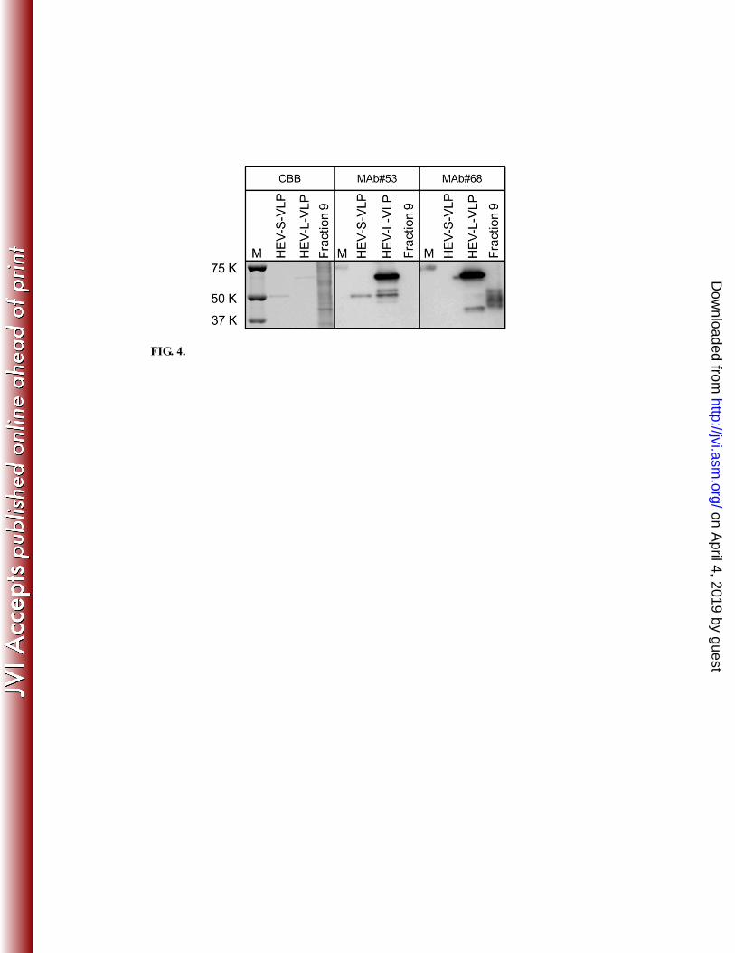
The observation, via WB (Fig. 3A), of a “smear” of antigen with maximum size 55 148
kDa was unexpected, given that capsid protein lacking C52aa (predicted size, 6 kDa) was 149
expected to migrate at 66 kDa (that is, 72 kDa less 6 kDa). The observed 11-kDa 150
decrease in size suggested further degradation of the capsid in the absence of the C52aa 151
domain. Mass spectroscopy followed by protein sequencing detected two fragments 152
with aa sequences corresponding to early N-terminal capsid sequences. The presence of 153
the capsid N-terminal domain was confirmed by detection with monoclonal antibody 154
(MAb) #68 (Fig. 4), a reagent that exhibits specificity for HEV-L-VLP (N-terminal 155
13-111aa-specific) (T.C. Li, unpublished observations). In contrast, the protein was not 156
detected using HEV-S-and-L-VLP-specific MAb #53 (Fig. 4), implying the absence of 157
S-and-L common region. Protein sequencing and reactivity with the HEV-VLP-specific 158
antibodies strongly suggested that the 55-KDa bands correspond to proteolytic products 159
generated by degradation from the C-terminus in viral surface, presumably via loss of the 160
P domain. Further degradation (to lower molecular weight species) probably occurred 161
after encapsidation, given that previous studies showed that this region was essential for 162
the dimerization and particle formation by the capsid (3, 15, 16). 163
HEV virions exhibit distinct buoyant densities in feces (1.26-1.27 g/ml) and in 164
circulating blood (1.15-1.16 g/ml), differences that might be associated with their cellular 165
membrane content (17). The density of the amut Map fraction was higher than that of 166
the Mip fraction. This result is inconsistent with the Map being an empty particle (18). 167
The amut Mip fraction had the specific density of membrane-associated virions, although 168
the ORF3 (egress-related) protein was not detected in these particles, in contrast to wt 169
particles (T. Shiota, unpublished observations) (19). We hypothesize that the correct 170
encapsidation of amut resulted in an enveloped particle lacking the ORF3 protein (Mip; 171
1.15 g/ml), whereas the incorrect encapsidation of amut resulted in an non-enveloped and 172
(usually) empty particle (Map; 1.21 g/ml), the density of which was intermediate between 173
that of the membrane-associated virion (1.15-1.16 g/ml) and the non-enveloped filled 174
virion (1.26-1.27 g/ml) (17). 175
In the present study, we showed that the C52aa domain of the HEV capsid was 176
essential for the HEV life-cycle, as confirmed by reproducible reversion at the amber 177
mutation that would otherwise truncate the C52aa domain. The presence of the C52aa 178
on April 4, 2019 by guest
http://jvi.asm.org/
Dow
nloaded from

SHIOTA ET AL.
7 Novel role of HEV capsid C-terminal region
domain promoted the accurate encapsidation of HEV and protected the particle from 179
further C-terminal degradation. To clarify the involvement of the C52aa domain in 180
neutralization, future studies (e.g., using MAb specific for this region) would be required. 181
182
on April 4, 2019 by guest
http://jvi.asm.org/
Dow
nloaded from

SHIOTA ET AL.
8 Novel role of HEV capsid C-terminal region
Acknowledgments 183
The authors would like to thank N. Sugiyama for excellent technical support and I. 184
Shiota for helpful discussions and critical reading. 185
This work was supported in part by grants-in-aid from the Ministry of Health, Labour, 186
and Welfare and the Ministry of Education, Culture, Sports, Science, and Technology, 187
Japan. 188
189
on April 4, 2019 by guest
http://jvi.asm.org/
Dow
nloaded from

SHIOTA ET AL.
9 Novel role of HEV capsid C-terminal region
References 190
191
1. Chandra V, Taneja S, Kalia M, Jameel S. 2008. Molecular biology and pathogenesis of 192
hepatitis E virus. J Biosci 33:451-464. 193
2. Tanaka T, Takahashi M, Kusano E, Okamoto H. 2007. Development and evaluation of an 194
efficient cell-culture system for Hepatitis E virus. J Gen Virol 88:903-911. 195
3. Li TC, Takeda N, Miyamura T, Matsuura Y, Wang JC, Engvall H, Hammar L, Xing L, 196
Cheng RH. 2005. Essential elements of the capsid protein for self-assembly into empty 197
virus-like particles of hepatitis E virus. J Virol 79:12999-13006. 198
4. Xing L, Li TC, Mayazaki N, Simon MN, Wall JS, Moore M, Wang CY, Takeda N, Wakita T, 199
Miyamura T, Cheng RH. 2010. Structure of hepatitis E virion-sized particle reveals an 200
RNA-dependent viral assembly pathway. J Biol Chem 285:33175-33183. 201
5. Li TC, Yamakawa Y, Suzuki K, Tatsumi M, Razak MA, Uchida T, Takeda N, Miyamura T. 202
1997. Expression and self-assembly of empty virus-like particles of hepatitis E virus. J Virol 203
71:7207-7213. 204
6. Mori Y, Matsuura Y. 2011. Structure of hepatitis E viral particle. Virus Res 161:59-64. 205
7. Xing L, Kato K, Li T, Takeda N, Miyamura T, Hammar L, Cheng RH. 1999. Recombinant 206
hepatitis E capsid protein self-assembles into a dual-domain T = 1 particle presenting native 207
virus epitopes. Virology 265:35-45. 208
8. Guu TS, Liu Z, Ye Q, Mata DA, Li K, Yin C, Zhang J, Tao YJ. 2009. Structure of the 209
hepatitis E virus-like particle suggests mechanisms for virus assembly and receptor binding. 210
Proc Natl Acad Sci U S A 106:12992-12997. 211
9. Yamashita T, Mori Y, Miyazaki N, Cheng RH, Yoshimura M, Unno H, Shima R, Moriishi K, 212
Tsukihara T, Li TC, Takeda N, Miyamura T, Matsuura Y. 2009. Biological and 213
immunological characteristics of hepatitis E virus-like particles based on the crystal 214
structure. Proc Natl Acad Sci U S A 106:12986-12991. 215
10. Xing L, Wang JC, Li TC, Yasutomi Y, Lara J, Khudyakov Y, Schofield D, Emerson SU, 216
Purcell RH, Takeda N, Miyamura T, Cheng RH. 2011. Spatial configuration of hepatitis E 217
virus antigenic domain. J Virol 85:1117-1124. 218
11. Agrawal S, Gupta D, Panda SK. 2001. The 3' end of hepatitis E virus (HEV) genome binds 219
specifically to the viral RNA-dependent RNA polymerase (RdRp). Virology 282:87-101. 220
12. Emerson SU, Zhang M, Meng XJ, Nguyen H, St Claire M, Govindarajan S, Huang YK, 221
Purcell RH. 2001. Recombinant hepatitis E virus genomes infectious for primates: 222
importance of capping and discovery of a cis-reactive element. Proc Natl Acad Sci U S A 223
98:15270-15275. 224
13. Graff J, Nguyen H, Kasorndorkbua C, Halbur PG, St Claire M, Purcell RH, Emerson SU. 225
2005. In vitro and in vivo mutational analysis of the 3'-terminal regions of hepatitis e virus 226
on April 4, 2019 by guest
http://jvi.asm.org/
Dow
nloaded from
SHIOTA ET AL.
10 Novel role of HEV capsid C-terminal region
genomes and replicons. J Virol 79:1017-1026. 227
14. Kumar A, Panda SK, Durgapal H, Acharya SK, Rehman S, Kar UK. 2010. Inhibition of 228
Hepatitis E virus replication using short hairpin RNA (shRNA). Antiviral Res 85:541-550. 229
15. Li SW, Zhang J, He ZQ, Gu Y, Liu RS, Lin J, Chen YX, Ng MH, Xia NS. 2005. Mutational 230
analysis of essential interactions involved in the assembly of hepatitis E virus capsid. J Biol 231
Chem 280:3400-3406. 232
16. Graff J, Zhou YH, Torian U, Nguyen H, St Claire M, Yu C, Purcell RH, Emerson SU. 2008. 233
Mutations within potential glycosylation sites in the capsid protein of hepatitis E virus 234
prevent the formation of infectious virus particles. J Virol 82:1185-1194. 235
17. Takahashi M, Tanaka T, Takahashi H, Hoshino Y, Nagashima S, Jirintai, Mizuo H, Yazaki Y, 236
Takagi T, Azuma M, Kusano E, Isoda N, Sugano K, Okamoto H. 2010. Hepatitis E Virus 237
(HEV) strains in serum samples can replicate efficiently in cultured cells despite the 238
coexistence of HEV antibodies: characterization of HEV virions in blood circulation. J Clin 239
Microbiol 48:1112-1125. 240
18. Jacobson MF, Baltimore D. 1968. Morphogenesis of poliovirus. I. Association of the viral 241
RNA with coat protein. J Mol Biol 33:369-378. 242
19. Tyagi S, Korkaya H, Zafrullah M, Jameel S, Lal SK. 2002. The phosphorylated form of the 243
ORF3 protein of hepatitis E virus interacts with its non-glycosylated form of the major 244
capsid protein, ORF2. J Biol Chem 277:22759-22767. 245
246
247 on April 4, 2019 by guest
http://jvi.asm.org/
Dow
nloaded from

SHIOTA ET AL.
11 Novel role of HEV capsid C-terminal region
FIG. 1. Initial characterization of amut and wt hepatitis E virus (HEV). (A) 248
Time-course of antigen production following transfection by amut or wt. HEV antigen 249
levels were measured by ELISA using anti-G3-HEV-VLP rabbit polyclonal antibody. 250
OD492: optical density at 492 nm. (B) Sedimentation analysis of amut product. (C) 251
Sedimentation analysis of wt product as a control. For (B) and (C), concentrated 252
supernatants derived from 50-mL cultures were sedimented on continuous sucrose 253
gradients (10%-60% (wt/vol) in phosphate-buffered saline); resulting fractions were 254
assessed for specific density and HEV RNA genome copy number (by real-time reverse 255
transcription PCR). Note distinct y-axis scales in panels (B) and (C). 256
257
on April 4, 2019 by guest
http://jvi.asm.org/
Dow
nloaded from

SHIOTA ET AL.
12 Novel role of HEV capsid C-terminal region
FIG. 2. Growth kinetics and character of amut. (A) Supernatant was collected 258
periodically during 2 months of culturing, and HEV antigen levels were measured by 259
ELISA using anti-G3-HEV-VLP rabbit polyclonal antibody; HEV RNA genome copy 260
number was determined by real-time reverse transcription PCR. OD492: optical density 261
at 492 nm. Supernatants from a pooled total of 3 liters of culture were concentrated and 262
sedimented. (B) Fractions were subjected to western blotting using anti-G3-HEV-VLP 263
rabbit polyclonal antibody. NC, negative control (untransfected cells). P, positive 264
control (HEV-L-VLPs). Symbols designate the position of the major band in the amut 265
supernatant (white arrowhead) and HEV-L-VLP (black arrowhead). (C) Fractions were 266
assessed for HEV RNA genome copy number and specific density. (D) Confirmation of 267
the infectivity of Fraction 7 by ELISA. 268
269
on April 4, 2019 by guest
http://jvi.asm.org/
Dow
nloaded from

SHIOTA ET AL.
13 Novel role of HEV capsid C-terminal region
FIG. 3. Encapsidation of amut genome and its characteristics. (A) Fractions were 270
subjected to western blotting using anti-G3-HEV-VLP rabbit polyclonal antibody. NC, 271
negative control (uninfected cells). Symbols designate the position of the major band in 272
the amut fraction (55 kDa; white arrowhead) and wt fraction as positive (P) control (72 273
kDa; black arrowhead). (B) Fractions were assessed for HEV RNA genome copy 274
number and specific density. (C) The RNase resistance of indicated fractions was 275
measured as the HEV RNA reduction ratio of RNase treated compared to untreated. wt 276
virions and extracted wt RNA were used as positive and negative controls, respectively. 277
(D) Constitution ratio (genome/antigen) was calculated by dividing the genome quantities 278
from (B) by the chemiluminescence intensities from (A). (E) To confirm infectivity of 279
the indicated fractions, the cells were inoculated and periodically analyzed by ELISA 280
using anti-G3-HEV-VLP rabbit polyclonal antibody. OD492: optical density at 492 nm. 281
282
on April 4, 2019 by guest
http://jvi.asm.org/
Dow
nloaded from

SHIOTA ET AL.
14 Novel role of HEV capsid C-terminal region
FIG. 4. Detection of amut degraded capsid termini. HEV small virus-like particles 283
(HEV-S-VLP), HEV large virus-like particles (HEV-L-VLP), and Fraction 9 (derived as 284
described in Fig. 3 legend) were stained with Coomassie brilliant blue (CBB) or 285
subjected to western blot analysis using an HEV-S-and-L-VLP-specific monoclonal 286
antibody (MAb#53) or an HEV-L-VLP-specific monoclonal antibody (MAb#68). M, 287
molecular weight markers. 288
on April 4, 2019 by guest
http://jvi.asm.org/
Dow
nloaded from

TABLE 1. Primers used for the construction of HEV infectious cDNA clone, C52aa deletion, and amber mutants, and HEV RNA real-time RT-PCR quantification and sequencing
Name Polari
tya Sequence (5'-3')
Position in
genomeb
Amplicon (Amplified region in
genome)c
ET7G2-F + GAATTCAATACGACTCACTATAGdGCAGACCACGTATGTGGTCGAT 2-23
155R-EV - AGTCTGCACGCGAGATAAAAACGGCCGGAC 126-155 Fragment 1-1 (2-155)
126F-EV + GTCCGGCCGTTTTTATCTCGCGTGCAGACT 126-155
1370R-EV - CACCCTGGGATCCAGATGGAAGCCCGCAG 1342-1370 Fragment 1-2 (126-1370)
1363F-EV + TCTGCGGGCTTCCATCTGGATCCCAGGGTG 1341-1370
1816R-EV - ACTGCTCAGGGCCGTTCGCCTCAAGATGAG 1765-1794 Fragment 2-1 (1341-1794)
1787F-EV + CTCATCTTGAGGCGAACGGCCCTGAGCAGT 1765-1794
2956R-EV - CGGCACAGGCACGGCCAACCTCTGTGGCAG 2905-2934 Fragment 2-2 (1765-2934)
2857F-EV + CCGATGCAGCCGGCACTCACAATAACGGAG 2835-2864
3216R-EV - AGCCCGCTGCATATGTAATAGCAGCAAGTG 3165-3194 Fragment 3-1 (2835-3194)
3187F-EV + CACTTGCTGCTATTACATATGCAGCGGGCT 3165-3194
3947R-EV - TCCGTAAGCTCAAAAACCAACACACTATCG 3896-3925 Fragment 3-2 (3165-3925)
3918F-EV + CGATAGTGTGTTGGTTTTTGAGCTTACGGA 3896-3925
4620R-EV - CTTCCAAAACCCCTTAAGGGATTCCTTAGG 4569-4598 Fragment 3-3 (3896-4598)
4591F-EV + CCTAAGGAATCCCTTAAGGGGTTTTGGAAG 4569-4598
5428R-EV - CTGTCGAGGGCGAGCTCCAGCCCCGGATTG 5377-5406 Fragment 4-1 (4569-5406)
5399F-EV + CAATCCGGGGCTGGAGCTCGCCCTCGACAG 5377-5406
5873R-EV - TGGAGTTCATGTCAACAGAAGTAGGGGTAG 5822-5851 Fragment 4-2 (5377-5851)
5844F-EV + CTACCCCTACTTCTGTTGACATGAACTCCA 5822-5851
6207R-EV - GTTCCATCGGCACCGCGGCGCAGCCGATG 6157-6185 Fragment 4-3 (5822-6185)
on April 4, 2019 by guest
http://jvi.asm.org/
Dow
nloaded from

6179F-EV + CATCGGCTGCGCCGCGGTGCCGATGGAAC 6157-6185
7101R-EV - AGTAGACTGGAAGGCGCAACCCTGC 7077-7101 Fragment 5-1 (6157-7101)
6981F-EV + CTGCGGTCGGTGTGTTAGCTCCACACTCGG 6959-6988
SmartIIA-Hin
d
- GCTCGAGCGGCCGCCAGTGTGATGGATATCTGCAGAATTCGGCTTAAGCAGTGGTATCAACGCAGAAAGCeTT
TTTTTTTTTTTTTTTTTTTTTTTTTT
7238-7266 Fragment 5-2 (6959-7266)
D81-F + ATGTGCCCTAGGGCTGTTCTGTTG 5173-5196
ORF2-52aa-P
ac-R - AATTAATTAATTAAfGCAAGGGCCGAGTGTGGAG 6977-6995
D81F/ORF2-52aa-Pac-R
(5173-6995)
ORF2-52aa-d
el-F + TCCACACTCGGCCCTTGCTTAAgCTTGAGGATACTATTGACTAT 6978-7020
ORF2-52aa-d
el-R - ATAGTCAATAGTATCCTCAAGTTAgAGCAAGGGCCGAGTGTGGA 6978-7020
D81-F/ORF2-52aa-del-R
(5173-7020)
7224R - AGGGAGCGCGAAAAGCAGAAAAGAAAAAT 7196-7224 ORF2-52aa-del-F/7224R
(6978-7224)
HEV-G3-AN
YF + ACCCCGGCAGTTGGTTTT 179-196
HEV-G3-AN
YR - CCCGCTGGATAGGATGATTCC 212-234
HEV-G3-ANYF/ANYR
(179-234)
HEV-G3-AN
YM1 + [FAM]CGCCCTGAGGTACTT[BHQ-1]h 198-212
on April 4, 2019 by guest
http://jvi.asm.org/
Dow
nloaded from

83-2-6564F + GCTTCGTGCTAATGATGTTCTGTG 6564-6587 83-2-6564F/3’-terminal end
(6564-7266)
83-2-6940F + CACCCAGGCTAGTGGTGTAGGTAGA 6940-6964 83-2-6940F/3’-terminal end
(6940-7266)
ORF2-R-pacI - GAGAATTAAGACTCCCGGGTTTTAC 7136-7160 83-2-6564F/ORF2-R-pacI
(6564-7160)
a Polarity of primers on the HEV genome. +, forward; -, reverse.
b Position of primers on the G3-HEV83-2-27 sequence GenBank ID AB740232.
c Position of amplicons on the G3-HEV83-2-27 sequence GenBank ID AB740232.
d Underlined sequence contains T7 promoter.
e Underlined sequence contains SmartIIA specific sequence and HindIII digestable sequence.
f Underlined sequence contains PacI digestable sequence.
g The mutated nucleotides are undrlined.
h A fluorophore 6-carboxyfluorescein (FAM) attached to the 5'-end of the probe and a quencher Black Hole Quencher-1(BHQ-1) at the 3'-end .
on April 4, 2019 by guest
http://jvi.asm.org/
Dow
nloaded from

TABLE 2. Time-course sequence of the codon mutated to amber codon for the supernatants of amut transfected cells
Days post-transfection 7 10 14 17 21 24 28 31 35
Amber mutant UAAa UAA UAA UAA UAA ND ND ND ND
Revertant Intermediate NDb ND ND ND ND GAC GAC ND ND
Complete ND ND ND ND ND GUC GUC GUC GUC a Sequence was determined for the first codon of the C52aa-encoding region of the ORF2 gene
b ND: not detected
on April 4, 2019 by guest
http://jvi.asm.org/
Dow
nloaded from