healthy offspring from freeze-dried mouse spermatozoa held ... · healthy offspring from...
TRANSCRIPT

Healthy offspring from freeze-dried mousespermatozoa held on the InternationalSpace Station for 9 monthsSayaka Wakayamaa,1, Yuko Kamadab, Kaori Yamanakac, Takashi Kohdad, Hiromi Suzukie, Toru Shimazue,Motoki N. Tadaf, Ikuko Osadaf, Aiko Nagamatsug, Satoshi Kamimurab, Hiroaki Nagatomoa,h, Eiji Mizutanib,Fumitoshi Ishinod, Sachiko Yanog, and Teruhiko Wakayamaa,b,1
aAdvanced Biotechnology Center, University of Yamanashi, Yamanashi 400-8510, Japan; bFaculty of Life and Environmental Sciences, University ofYamanashi, Yamanashi 400-8510, Japan; cDepartment of Experimental Oncology, Research Institute for Radiation Biology and Medicine, HiroshimaUniversity, Hiroshima 734-8553, Japan; dDepartment of Epigenetics, Medical Research Institute, Tokyo Medical and Dental University, Tokyo 113-8510, Japan;eDepartment of Science and Applications, Japan Space Forum, Tsukuba 305-8505, Japan; fJapanManned Space Systems Corporation, Tokyo 100-0004, Japan; gJapanAerospace Exploration Agency, Tsukuba 305-8505, Japan; and hCenter of Community Promotion Center, University of Yamanashi, Yamanashi 400-8510, Japan
Edited by George E. Seidel, Colorado State University, Fort Collins, CO, and approved April 20, 2017 (received for review January 26, 2017)
If humans ever start to live permanently in space, assisted repro-ductive technology using preserved spermatozoa will be impor-tant for producing offspring; however, radiation on the InternationalSpace Station (ISS) is more than 100 times stronger than that onEarth, and irradiation causes DNA damage in cells and gametes.Here we examined the effect of space radiation on freeze-driedmouse spermatozoa held on the ISS for 9 mo at –95 °C, with launchand recovery at room temperature. DNA damage to the sperma-tozoa and male pronuclei was slightly increased, but the fertiliza-tion and birth rates were similar to those of controls. Next-generation sequencing showed only minor genomic differencesbetween offspring derived from space-preserved spermatozoaand controls, and all offspring grew to adulthood and had normalfertility. Thus, we demonstrate that although space radiation candamage sperm DNA, it does not affect the production of viableoffspring after at least 9 mo of storage on the ISS.
International Space Station | preservation | freeze-dry | spermatozoa |fertilization
Since the “space dog” Laika (Лайка) was first placed intoorbit in 1957 (1), many humans and animals have been to
space or stayed on the International Space Station (ISS) formore than 6 mo. In the future, humans likely will live on large-scale space stations or in other space habitats for several years oreven over many generations. At that time, assisted reproductivetechnology (ART) likely will be used to produce humans in spacehabitats, given that the use of ART by infertile couples has in-creased year by year and that ART can be performed with cry-opreserved spermatozoa or embryos (2, 3). In a similar way,domestic animals likely will be generated by artificial insemina-tion (AI) in space, because many domestic animals are alreadyproduced by AI using long-term cryopreserved spermatozoa (4).In addition, genetic diversity is very important for maintaining aspecies, especially in small colonies, and this could be achievedby cryopreserving a diverse range of gamete cells. The environ-ment in space is very different from that on Earth, however,including high levels of space radiation and microgravity, and theeffects of these factors on mammalian reproduction are largelyunknown. Although with current technology, producing offspringin such an environment can be difficult or dangerous (5), thestudy of reproduction in space is a very important subject forour future.So far, the effects of microgravity on early development have
been studied using sea urchins, fish, amphibians, and birds (6–12). These studies have concluded that microgravity does notprevent animal reproduction. However, because of the difficultyin maintaining mammals and performing experiments in space,studies of mammal reproduction in space have not progressed as
well as in other animals, and only a few papers have been pub-lished (13–18). Those studies and our previous study (19) havesuggested that mammalian reproduction in space under condi-tions of microgravity cannot be easily compared with reproduc-tion in other species.Another difference between space and Earth is the high level
of radiation in space. The doses received inside the ISS dependon sunspot cycles and cosmic rays. The average dose rate mea-sured at the ISS is ∼0.5 mSv/day, roughly 100-fold higher thanthat measured on Earth (20). Ground-based studies have dem-onstrated the deleterious effects of radiation on living organisms,including the induction of mutations and tumor formation(21, 22). Humans living for several generations in space habitats ortraveling to Mars will encounter much higher cosmic radiation,possibly putting them at high risk for cancer (23). If space radia-tion also causes DNA damage to cryopreserved spermatozoa, itmight lead to serious problems, such as embryo death or abortion,or cause mutations in offspring and subsequent generations.Salamander and medaka fish eggs could be fertilized and de-
veloped normally during orbital space flight (6, 24), suggestingthat space radiation from brief space flights does not affect fer-tilization or later embryogenesis in these vertebrates. However,mammalian oocytes are known to have a strong potential for
Significance
Radiation on the International Space Station (ISS) is more than100 times stronger than at the Earth’s surface, and at levelsthat can cause DNA damage in somatic cell nuclei. The damageto offspring caused by this irradiation in germ cells has notbeen examined, however. Here we preserved mouse sperma-tozoa on the ISS for 9 mo. Although sperm DNA was slightlydamaged during space preservation, it could be repaired by theoocyte cytoplasm and did not impair the birth rate or normalityof the offspring. Our results demonstrate that generating hu-man or domestic animal offspring from space-preserved sper-matozoa is a possibility, which should be useful when the“space age” arrives.
Author contributions: S.W., T.K., H.S., T.S., F.I., S.Y., and T.W. designed research; S.W.,Y.K., K.Y., T.K., H.S., T.S., M.N.T., I.O., A.N., S.K., H.N., E.M., S.Y., and T.W. performedresearch; T.K., A.N., and F.I. analyzed data; and S.W. and T.W. wrote the paper.
The authors declare no conflict of interest.
This article is a PNAS Direct Submission.
Freely available online through the PNAS open access option.
Data deposition: Sequences have been deposited in the DNA Data Bank of Japan Se-quence Read Archive (DRA submission 005694).1To whom correspondence may be addressed: Email: [email protected] [email protected].
This article contains supporting information online at www.pnas.org/lookup/suppl/doi:10.1073/pnas.1701425114/-/DCSupplemental.
5988–5993 | PNAS | June 6, 2017 | vol. 114 | no. 23 www.pnas.org/cgi/doi/10.1073/pnas.1701425114
Dow
nloa
ded
by g
uest
on
May
1, 2
020

repairing damaged DNA (25–27), so when live animals are inspace for only short periods, the effect of space radiation mightbe masked by subsequent repair of the damaged DNA afterfertilization. In contrast, although cryopreserved cells are stillalive, they have stopped metabolizing, which means that theycannot repair DNA damage while still frozen (28, 29). There-fore, DNA damage might accumulate with increased duration inspace. If this is so, then the production of offspring from long-term cryopreserved spermatozoa in space will be compromisedbecause of increased embryo mortality or mutation rates in theoffspring. Therefore, it is very important to examine the effect ofspace radiation on spermatozoa preserved in space. In addition,the resistance of spermatozoa likely differs among species, be-cause the sperm structure differs (30). Therefore, to examine theinfluences of radiation in mammalian species, we must usemammalian species, and the mouse is a very convenient modelanimal for space study.For the present study, we decided to freeze-dry the mouse
spermatozoa (31) rather than use traditional cryopreservationmethods. When spermatozoa are freeze-dried or evaporativelydried, none of the sperm survive; however, mouse spermatozoacan maintain the ability to generate offspring when added towater and microinjected into fresh oocytes (31, 32), and this maypossible with human spermatozoa in the future (33, 34). Moreimportantly, such dried spermatozoa can be preserved at roomtemperature for up to 2 y (35, 36) and in a freezer almost in-definitely (37). In addition, the samples are very light and occupy
a small volume. Therefore, our samples could be launched to theISS without the need for a freezer, which greatly reduced the costof launching. The merits of this procedure, in terms of the ease oflaunching freeze-dried samples into space, are significant for en-abling the study of mammalian reproduction in space, even thoughthe production rate of offspring from freeze-dried spermatozoa islower than that from traditional cryopreservation methods.After exposure to space radiation for 9 mo at –95 °C, the
samples were returned to Earth. We evaluated these samples forsperm morphology and DNA damage, capacity for fertilizationby microinjection, in vitro developmental potential, and nor-mality of offspring derived from the spermatozoa.
ResultsCollection of Space Sperm Samples. The samples (Fig. 1 A and B)were launched to the ISS on August 4, 2013, and returned toground on May 19, 2014. Therefore, these space sperm sampleswere exposed to cosmic radiation for 288 d. The majority of theglass ampules sustained no damage during the launch or return.The ground control sperm samples from the same mice wereexposed to the same temperature changes at the same times andfor the same durations as the space sperm samples.
Total Doses of Space Radiation. The CR-39 plastic nuclear trackdetectors (PNTDs) in the Bio PADLES packages collected fromthe space-preserved cases (Fig. 1C) can detect nuclear tracks.Fig. 1D shows images of the etch pits corresponding to the tracks
γH2AX merge
H3K9me2
B6 BD BC Tg
B6 BD BC Tg0
0.5
1
1.5
2Ground Space
A B C
E F
G H I
JL
* * *
*
10μm10μm
K
DAPI
γH2AX merge
H3K9me2DAPI
Ground Space
Ground Space
10μm
D
Fig. 1. Preservation of spermatozoa on the ISSand assessment of DNA integrity after return toEarth. (A and B) Ampules of freeze-dried spermato-zoa were wrapped with polyimide film, and thenfour ampules from each donor mouse were wrappedtogether. Twelve groups of ampules derived from12 male mice were selected for this study. The smallwhite square (right side of B) represents the PADLESmonitor, used to detect the irradiation dose. (C) Allampules were inserted into a small case, and aPADLES radiation monitor was placed on top ofthe case. (D) The etch pits corresponding to thetracks of atomic nuclei produced during space flight.(E and F) Observation of ground control (E) andspace-preserved spermatozoa (F) by light micros-copy. (G–I) Comet DNA breakage assays of groundcontrol (G) and space-preserved spermatozoa (H).The lengths of DNA in the comet tails were stan-dardized against the mean lengths of ground controlsperm results for each mouse strain (I). The orangebars represent the mean lengths of ground controlsperm samples after standardization, and blue barsindicate the space sperm samples. The asterisk de-notes significant differences between samples (*P <0.001). (J and K) Immunostaining of zygotes derivedfrom ground control sperm samples (J) or spacesperm samples (K) by the anti–gamma-H2AX anti-body. Both male and female pronuclei were detectedby nuclear staining with DAPI (Upper Left, blue).Female pronuclei were detected by H3K9me2 immu-nostaining (Upper Right, green). The foci of gamma-H2AX signals show DNA double-strand breaks (LowerLeft, red), and merged images (Lower Right). (L) Thebrightness of each male pronucleus was plotted. Blackcircles indicate zygotes derived from ground controlsperm samples; white circles, zygotes derived fromspace sperm samples. The brightness of the malepronucleus in K was 1.3. In I and L, mouse strains:B6, C57BL/6N; BD, B6D2F1; BC, B6C3F1; Tg, 129B6F1expressing GFP. Asterisks indicate significant differ-ences (I, *P < 0.001; L, *P < 0.05).
Wakayama et al. PNAS | June 6, 2017 | vol. 114 | no. 23 | 5989
AGRICU
LTURA
LSC
IENCE
S
Dow
nloa
ded
by g
uest
on
May
1, 2
020

of atomic nuclei produced during space flight. Background dosesmeasured in the ground control sperm sample case were sub-tracted from the net doses measured during space flight. Fromthe results, the absorbed radiation dose rate was calculated as0.41 ± 0.01 mGy/day, or a dose-equivalent rate of 0.62 ± 0.03mSv/day, and the total absorbed dose was 117.24 ± 3.98 mGy inwater, or a total dose equivalent of 178.35 ± 7.27 mSv.
Morphology and DNA Damage in Spermatozoa. The morphology ofspace sperm samples could not be distinguished from that of theground control samples, at least at the light microscopy level(Figs. 1 E and F). Some spermatozoa showed breakage betweenthe head and tail or fragmented tails, but this is typical for freeze-dried spermatozoa. However, measurement of the comet DNAtails revealed significantly longer tails in the space sperm samplescompared with the ground control sperm samples in all mousestrains except the C57BL/6 strain (Fig. 1 G–I and Table S1).
In Vitro Fertilization and DNA Damage in Male Pronuclei. Whensamples were rehydrated, the spermatozoa were used for intra-cytoplasmic sperm injection (ICSI) of fresh oocytes within1 h. Most of the oocytes were fertilized and formed normal-appearing pronuclei irrespective of mouse strain, similar to theresults for the ground control sperm samples (Table 1). The two-cell rates in this study were slightly lower than those obtainedusing fresh spermatozoa (∼100% in our laboratory).We next examined the DNA damage in male pronuclei. When
zygotes fertilized with space sperm samples were immunostainedwith the anti–gamma-H2AX antibody, numerous foci were de-tected in male pronuclei (Fig. 1 J–L). Given the difficulty incounting the number of foci inside pronuclei, we measured thebrightness of the whole male pronucleus, which was then sub-tracted from the brightness of the zygote cytoplasm. As shown inFig. 1L and Table S2, the brightness of the male pronuclei variedamong zygotes. Although on average, male pronuclei derivedfrom space sperm samples were slightly brighter than those fromground control sperm samples, there were no statistically sig-nificant differences between them in any mouse strain exceptthe BCF1 strain.
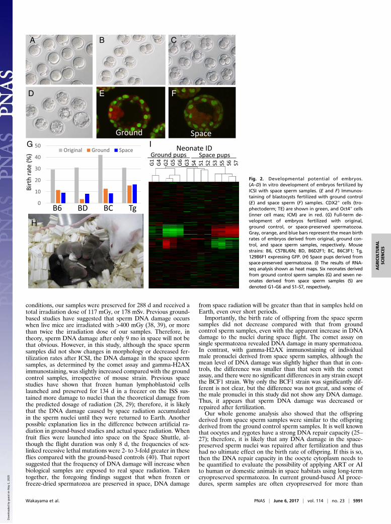
Developmental Potential in Vitro. To observe the developmentalpotential of zygotes fertilized with space spermatozoa, somezygotes were cultured in vitro until they developed to the blas-tocyst stage at 4.5 d after ICSI. Most of the injected oocytesfertilized normally, and some developed into blastocysts (Fig. 2A–D). Some of these blastocysts were immunostained to evaluatethe quality based on the cell number and allocation of inner cellmass (ICM) cells. The allocation of ICM cells did not show any
clear differences between ground control and space spermsamples (Fig. 2 E and F). On the other hand, although the meannumber of ICM cells was similar in the space and ground controlsamples (11 and 13, respectively), the mean number of tro-phectoderm (TE) cells was lower in the space samples (35 vs. 55).However, most of the embryos were used for embryo transfer, andthus only six blastocysts from the space samples and three blas-tocysts from the ground control samples were examined.
Full-Term Development and Normality of Offspring. The potentialfor development to full term is the strongest evidence of thenormality of the DNA of the space sperm samples. Therefore,when embryos reached the two-cell stage, some were transferredinto recipient females, and the birth rates were compared be-tween the space and ground control sperm samples, as well asbetween the freeze-dried spermatozoa and the original sperma-tozoa (before preservation). As shown in Table 1 and Fig. 2G,the mean birth rates of offspring derived from space spermsamples (“space pups”; Fig. 2H) and ground control spermsamples were almost the same (8–17% and 7–15%, respectively)but significantly lower than that with the original spermatozoa(28–39%). The sex ratio of space pups (53% male, 47% female)was within the normal range. After the pups grew to adulthood,two or three pairs from all strains were selected at random andmated with each other. All of these couples had offspring, clearlydemonstrating normal fertility.
Next-Generation Sequencing Analysis. Finally, we analyzed theglobal gene expression profiles of pups derived from space andground control sperm samples to assess for any nonphenotypicdifferences. Seven pups (four females, IDs S2, S4, S5, and S7, andthree males, IDs S1, S3, and S6) from space sperm samples andsix pups (one female, ID G1, and five males, IDs G2–G6) fromground control sperm samples derived from the same male mouse(C57BL/6N strain) were used, and freshly collected brains wereexamined. From the results of heat mapping (Fig. 2I), althoughthe two sets appeared slightly dissimilar, the differences were notstatistically significant. There was also no significant difference ingene expression profiles between male and female offspring.
DiscussionIn this study, freeze-dried mouse spermatozoa were held on theISS for 9 mo at –95 °C and then examined after exposure to spaceradiation.During the study period, irradiation from space to the sample
case was 0.4 mGy/day, or 0.6 mSv/day, which is typical for the ISSand ∼100 times higher than that on Earth (29). No special eventsoccurred in the local space during the study period. In these
Table 1. Production of offspring from ground or space-preserved spermatozoa injected into BDF1 oocytes
Mousestrain
Condition ofpreservation
No. ofoocytes
No. of oocytessurviving after ICSI
No. (%) offertilized embryos
No. (%) of two-cellembryos at 24 h
No. of transferred embryos(no. of recipients)
No. (%) ofoffspring
C57BL/6 Original 111 89 74 (83) 68 (76) 68 (4) 20 (29)*Ground 267 216 208 (96) 165 (76) 165 (8) 19 (12)**Space 452 367 359 (98) 262 (71) 262 (12) 24 (9)**
BDF1 Original 110 98 98 (100) 74 (76) 74 (4) 23 (31)*Ground 572 423 366 (87) 278 (66) 278 (14) 10 (4)**Space 345 282 256 (91) 182 (65) 182 (10) 15 (8)**
BCF1 Original 78 49 48 (98) 38 (78) 38 (3) 16 (42)*Ground 270 242 231 (95) 213 (88) 213 (10) 27 (13)**Space 400 341 327 (96) 279 (82) 279 (12) 24 (9)**
129B6F1-GFP Original 102 75 75 (100) 55 (73) 55 (3) 17 (31)*Ground 180 140 133 (95) 97 (69) 97 (4) 15 (15)**Space 120 86 75 (87) 61 (71) 61 (3) 10 (10)**
Original: When samples were prepared, some ampules were used to check the quality of each lot. These data were used as the original control. Ground: Atthe same times and for the same durations as when launching and storing the sample case, a control sample case was exposed to room temperature, thenfrozen at –95 °C and stored in a freezer at the Tsukuba Space Center in Japan. These are referred to as ground control sperm samples. Space: Samples werelaunched and stored on the ISS for 9 mo. (* vs. **: P < 0.05.)
5990 | www.pnas.org/cgi/doi/10.1073/pnas.1701425114 Wakayama et al.
Dow
nloa
ded
by g
uest
on
May
1, 2
020
conditions, our samples were preserved for 288 d and received atotal irradiation dose of 117 mGy, or 178 mSv. Previous ground-based studies have suggested that sperm DNA damage occurswhen live mice are irradiated with >400 mGy (38, 39), or morethan twice the irradiation dose of our samples. Therefore, intheory, sperm DNA damage after only 9 mo in space will not bethat obvious. However, in this study, although the space spermsamples did not show changes in morphology or decreased fer-tilization rates after ICSI, the DNA damage in the space spermsamples, as determined by the comet assay and gamma-H2AXimmunostaining, was slightly increased compared with the groundcontrol samples, irrespective of mouse strain. Previous spacestudies have shown that frozen human lymphoblastoid cellslaunched and preserved for 134 d in a freezer on the ISS sus-tained more damage to nuclei than the theoretical damage fromthe predicted dosage of radiation (28, 29); therefore, it is likelythat the DNA damage caused by space radiation accumulatedin the sperm nuclei until they were returned to Earth. Anotherpossible explanation lies in the difference between artificial ra-diation in ground-based studies and actual space radiation. Whenfruit flies were launched into space on the Space Shuttle, al-though the flight duration was only 8 d, the frequencies of sex-linked recessive lethal mutations were 2- to 3-fold greater in theseflies compared with the ground-based controls (40). That reportsuggested that the frequency of DNA damage will increase whenbiological samples are exposed to real space radiation. Takentogether, the foregoing findings suggest that when frozen orfreeze-dried spermatozoa are preserved in space, DNA damage
from space radiation will be greater than that in samples held onEarth, even over short periods.Importantly, the birth rate of offspring from the space sperm
samples did not decrease compared with that from groundcontrol sperm samples, even with the apparent increase in DNAdamage to the nuclei during space flight. The comet assay onsingle spermatozoa revealed DNA damage in many spermatozoa.In contrast, with gamma-H2AX immunostaining of individualmale pronuclei derived from space sperm samples, although themean level of DNA damage was slightly higher than that in con-trols, the difference was smaller than that seen with the cometassay, and there were no significant differences in any strain exceptthe BCF1 strain. Why only the BCF1 strain was significantly dif-ferent is not clear, but the difference was not great, and some ofthe male pronuclei in this study did not show any DNA damage.Thus, it appears that sperm DNA damage was decreased orrepaired after fertilization.Our whole genome analysis also showed that the offspring
derived from space sperm samples were similar to the offspringderived from the ground control sperm samples. It is well knownthat oocytes and zygotes have a strong DNA repair capacity (25–27); therefore, it is likely that any DNA damage in the space-preserved sperm nuclei was repaired after fertilization and thushad no ultimate effect on the birth rate of offspring. If this is so,then the DNA repair capacity in the oocyte cytoplasm needs tobe quantified to evaluate the possibility of applying ART or AIto human or domestic animals in space habitats using long-termcryopreserved spermatozoa. In current ground-based AI proce-dures, sperm samples are often cryopreserved for more than
G1 S1G2 G3G4 G5 G6 S2 S3S4 S5 S6 S7
Neonate IDGround pups Space pups
Birt
h ra
te (%
)
A B C
G
H
ISpace
D E
Ground
0
10
20
30
40
50Original Ground Space
B6 BD BC Tg
F
Fig. 2. Developmental potential of embryos.(A–D) In vitro development of embryos fertilized byICSI with space sperm samples. (E and F) Immunos-taining of blastocysts fertilized with ground control(E) and space sperm (F) samples. CDX2+ cells (tro-phectoderm; TE) are shown in green, and Oct4+ cells(inner cell mass; ICM) are in red. (G) Full-term de-velopment of embryos fertilized with original,ground control, or space-preserved spermatozoa.Gray, orange, and blue bars represent the mean birthrates of embryos derived from original, ground con-trol, and space sperm samples, respectively. Mousestrains: B6, C57BL/6N; BD, B6D2F1; BC, B6C3F1; Tg,129B6F1 expressing GFP. (H) Space pups derived fromspace-preserved spermatozoa. (I) The results of RNA-seq analysis shown as heat maps. Six neonates derivedfrom ground control sperm samples (G) and seven ne-onates derived from space sperm samples (S) aredenoted G1–G6 and S1–S7, respectively.
Wakayama et al. PNAS | June 6, 2017 | vol. 114 | no. 23 | 5991
AGRICU
LTURA
LSC
IENCE
S
Dow
nloa
ded
by g
uest
on
May
1, 2
020

10 y; however, we found increased DNA damage in spermatozoaafter only 9 mo in space. If sperm samples are to be preserved forlonger periods in space, then it is likely that DNA damage willincrease and exceed the limit of the oocyte’s capacity for repair.Testing this will require more extensive sperm preservation ex-periments in space. If the DNA damage occurring during long-term preservation is found to have a significant effect on offspring,we will need to develop methods to protect sperm samples againstspace radiation, such as with an ice shield, to enable future animalbreeding in this environment.In addition, sperm preservation in the event of disasters on
Earth will be an important tool for maintaining the genetic di-versity of mammalian species, much like plant seed preservationin the Svalbard Global Seed Vault. Although current spermdrying methods can maintain nuclear integrity for only a limitednumber of years at ambient temperature, this time eventually willbe extended. Reliability likely will be obtained before the be-ginning of the “Space Age,” given that preservation periods havealready been extended from a few months (31) to 2 y (36) byimproving the drying method. In addition, drying conditionsmight be better than traditional cryopreservation for long-termpreservation, because tardigrades can survive in extreme envi-ronments, such as space, only in a dehydrated state of dormancy(41). Once the reliability and integrity of space-preserved sper-matozoa can be demonstrated, underground storage on the Moon,such as in lava tubes (42), could be among the best places forprolonged or permanent sperm preservation because of their verylow temperatures, protection from space radiation by thick bedrocklayers, and complete isolation from any disasters on Earth.
MethodsAnimals. C56BL/6N, BDF1 (C57BL/6 × DBA/2), BCF1 (C57BL/6 × C3H/He), and129B6F1 male mice carrying the green fluorescent protein (GFP) gene (GFP-tg-129/Sv × GFP-tg-C57BL/6), aged 3 mo, were used to collect spermatozoa.The C57BL/6N, BDF1, and BCF1 mice were purchased from Shizuoka Labo-ratory Animal Center, and the 129B6F1 mice were bred in our mouse facility.The C57BL/6N and BDF1 mice, age 8–10 wk, were used to produce oocytes.The surrogate pseudopregnant females used as embryo transfer recipientswere ICR strain mice mated with vasectomized males of the same strain. Onthe day of the experiments, or after completion of all experiments, the micewere euthanized by CO2 inhalation or by cervical dislocation. All animalexperiments were performed in accordance with the National Institutes ofHealth’s Guide for the Care and Use of Laboratory Animals and were approvedby the Institutional Committee of Laboratory Animal Experimentation of theRIKEN Center for Developmental Biology and by the Institutional Committeeof Laboratory Animal Experimentation of the University of Yamanashi.
Preparation of Freeze-Dried Spermatozoa. Both epididymides were collectedfrommalemice, and the ducts were cut with sharp scissors. A few drops of thedense sperm mass were placed into a centrifuge tube with 2 mL of CZBmedium (43), followed by incubation for 30 min at 37 °C in 5% CO2. Aftersperm concentration measurements, 50-μL aliquots of the sperm suspensionwere divided into 30 glass ampules. These ampules were frozen using liquidnitrogen and then freeze-dried (EYELA FDU-2200; Tokyo Rikakikai andFreeZone; Labconco). The cock of the freeze-drying machine was opened forat least 3 h until the samples were completely dry. After drying, the ampuleswere sealed by melting the necks using a gas burner under vacuum condi-tions, then kept in a –30 °C freezer until use.
Sperm samples were prepared from 70 male mice of four different strains.The quality of each lotwas checked by examining its fertility (see below) usingone or two ampules from each mouse. From these results, we selected samplesfrom 12 mice: three from the C57BL/6 strain, four from the BDF1 strain, threefrom the BCF1 strain, and two from the 129B6F1 strain expressing GFP. Theseampules and their data are referred to as the “original sperm” samples. Eachampule was wrapped in polyimide film and then the ampules were furtherwrapped in groups of four (Fig. 1 A and B). All ampules from 12 male mice(total of 48 ampules) were placed into small cases for space preservation (Fig.1C) or as a ground control.
Launch to the ISS and Return to Earth. The sample cases, including the BioPADLES radiation monitors (Fig. 1C), were launched aboard the H-II TransferVehicle KOUNOTORI 4 on August 4, 2013, at room temperature. After arrivalat the ISS, astronauts stored them in a –95 °C freezer on August 10, 2013.
Nine months later (on May 19, 2014), the sample case was retrieved from thefreezer and then returned to Earth on the SpaceX-3 vehicle at ambienttemperature. Therefore, these space sperm samples were exposed to cosmicradiation for 288 d. On May 21, 2014, the sample case was delivered to NASAand placed into a –80 °C freezer. This was then transported to our labora-tory. These samples are designated the space sperm samples.
At the same times and for the same durations as the launching and storingof the sample case, a control sample case was exposed to room temperature,then frozen at –95 °C and stored in a freezer at the Tsukuba Space Center inJapan. These are designated the ground control sperm samples.
Space Radiation Dosimetry. Bio PADLES (TLD/CR39) monitoring devices(Fukuvi Chemical Industry) were used to measure radiation dosages. Theseinclude CR-39 PNTDs. The devices were placed inside the cases (Fig. 1C) ofspace and ground control sperm samples. After the sample cases were re-covered, the analysis was initiated on June 2, 2014. Pictures of radiation-etched pits reflecting the tracks of atomic nuclei accumulated during spaceflight (Fig. 1D) were used to calculate the total radiation dose (44).
Analysis and Scoring of Comet Slides. The single-cell gel electrophoresistechnique (i.e., the comet assay) measures DNA damage, including double-and single-strand breaks (45). Here comet assays to detect sperm DNA damagewere performed according to the manufacturer’s protocol (Trevigen). In brief,sperm specimens were collected from ampules immediately after opening andthen rehydrated in water. Space and ground control sperm samples derivedfrom the same male mouse were mounted on six slides, and 100–300 spermheads were analyzed following electrophoresis. In some cases, spermatozoahad aggregated and were difficult to measure. To standardize the resultsamong the different mouse strains, the length of each DNA comet “tail” wasdivided by the mean length of the ground control results in each strain.
Immunostaining of Zygotes. In histone H2AX, an H2A variants, the serine atposition 139 is rapidly phosphorylated within minutes after DNA damage.The phosphorylated form of H2AX, designated gamma-H2AX, forms foci atthe sites of DNA damage, which then serve as platforms to recruit variousrepair and cell cycle checkpoint proteins (46). Therefore, gamma-H2AX fociformation was used as a marker of DNA double-strand breaks in male andfemale pronuclei, and histone H3K9me2 signals were used to distinguishfemale pronuclei. The primary antibodies used were an anti–phospho-H2AX(Ser139) rabbit polyclonal antibody (1:500; Millipore Merck) and an anti-histoneH3 (dimethyl K9) mouse monoclonal antibody (1:500; Abcam). The secondaryantibodies used were Alexa Fluor 488-labeled goat anti-mouse IgG (1:500;Molecular Probes) and Alexa Fluor 568-labeled goat anti-rabbit IgG (1:500 di-lution; Molecular Probes). DNA was stained with DAPI (2 μg/mL; MolecularProbes). The brightness of whole male pronuclei was measured using ImageJand then subtracted from the brightness of the zygote cytoplasm.
ICSI and Embryo Transfer. To generate offspring from preserved spermatozoa,oocytes were collected from C57BL/6N or BDF1 female mice. Most experi-ments used BDF1 oocytes irrespective of the male mouse strain. Only whennext-generation sequencing was performed for genomic analysis wereC57BL/6 oocytes were used for C57BL/6 male mouse ICSI to generate pureinbred offspring. When ampules were opened, 50 μL of water was imme-diately added to rehydrate the sperm samples, and ICSI was initiated usingfour or five micromanipulators as described previously (47).
At 0.5 d postcoitum (dpc), embryos were transferred into the oviducts ofpseudopregnant ICR strain female mice that had been mated with a vasec-tomized male the night before transfer. At 18.5–19.5 dpc, the offspring weredelivered by caesarean section. Some embryos were cultured in 5% CO2 inair for 5 d in CZB medium at 37 °C to examine their in vitro developmentalpotential and the quality of blastocysts.
Immunostaining of Blastocysts. To evaluate the quality of blastocysts derivedfrom space sperm samples, immunofluorescence staining of blastocysts wasperformed as described previously (48). The primary antibodies used were ananti-CDX2 mouse monoclonal antibody (1:200; BioGenex) to detect TE cellsand an anti-Oct3/4 rabbit polyclonal antibody (1:500; MBL) to detect ICMcells. The secondary antibodies used were Alexa Fluor 488-labeled goat anti-mouse IgG (1:500; Molecular Probes) and Alexa Fluor 564-labeled goat anti-rabbit IgG (1:500; Molecular Probes). DNA was stained with DAPI (2 μg/mL;Molecular Probes).
RNA Isolation and RNA Sequencing. In this study, both space and groundcontrol sperm samples were obtained from the same male C57BL/6N mouse
5992 | www.pnas.org/cgi/doi/10.1073/pnas.1701425114 Wakayama et al.
Dow
nloa
ded
by g
uest
on
May
1, 2
020

strain, and fresh oocytes were collected fromC57BL/6N femalemice. Therefore,theoffspringwere of a pure C57BL/6N strain. At 18.5dpc, fetuseswere collectedby cesarean section. Several organs from seven pups (four females, neonate IDsS2, S3, S4, S7, and threemales, neonate IDs S1, S3, S6) from space sperm samplesand six pups (one female, neonate ID G1; and five males, neonate IDs G2–G5)from ground control sperm samples were collected, immediately frozen inliquid nitrogen, and kept at –30 °C until use. Brain tissues were used for furtheranalysis. Total RNA was purified using the AllPrep DNA/RNA Mini Kit (Qiagen)according to the manufacturer’s instructions. The library for RNA sequencing(RNA-seq) studies was prepared using the Kapa Stranded RNA/mRNA-Seq Kit(Illumina). The RNA-seq library was sequenced for 36-base single-end RNAsusing an Illumina GAIIx machine with a TruSeq SBS kit v5–GA kit (Illumina). Theresulting sequence data were mapped against the mouse reference genomesequence (GRCm38/mm10) using bowtie2 (49), followed by calculation of thereads per kilobase of exon per million mapped reads (RPKM) value for eachgene using the Bioconductor package DEGseq (50). Clustering analysis andprincipal component analysis were performed with Cluster 3.0 (51).
Statistical Analysis. The comet DNA breakage assay results were evaluatedusing the Wilcoxon–Mann–Whitney nonparametric test, the gamma-H2AXassay results were evaluated using the Student t test, and the birth rate wasevaluated using the χ2 test. RNA-seq analyses were evaluated using theWilcoxon–Mann–Whitney nonparametric test adjusted for a false discoveryrate, and differentially expressed genes between space and control pupswere statistically analyzed using the DESeq2 package (52) with the raw se-quence reads count. P < 0.05 (birth rate and gamma-H2AX staining) or P <0.01 (others) was considered statistically significant.
ACKNOWLEDGMENTS. We thank Dr. S. Kishigami, Dr. M. Shirakawa,Dr. T. Suzuki, C. Yamazaki, and K. Kishida for critical comments on the study.S.K. is supported by a postdoctoral fellowship from the Japan Society for thePromotion of Science (JSPS). This work was partially funded by the JSPS (Grant26506006, to S.W.) the Naito Foundation (S.W.), the Asada Science Foundation(T.W.), the Takeda Science Foundation (T.W.), and the Japan Aerospace Explo-ration Agency (T.W.).
1. West JB (2001) Historical aspects of the early Soviet/Russian manned space program.J Appl Physiol (1985) 91:1501–1511.
2. Kupka MS, et al.; European IVF-Monitoring Consortium, for the European Society ofHuman Reproduction and Embryology (2014) Assisted reproductive technology inEurope, 2010: Results generated from European registers by ESHRE. Hum Reprod 29:2099–2113.
3. Calhaz-Jorge C, et al.; European IVF-Monitoring Consortium (EIM) for the EuropeanSociety of Human Reproduction and Embryology (ESHRE) (2016) Assisted reproductivetechnology in Europe, 2012: Results generated from European registers by ESHRE.Hum Reprod 31:1638–1652.
4. Funk DA (2006) Major advances in globalization and consolidation of the artificialinsemination industry. J Dairy Sci 89:1362–1368.
5. Schuster H, Peck SL (2016) Mars ain’t the kind of place to raise your kid: Ethical im-plications of pregnancy on missions to colonize other planets. Life Sci Soc Policy 12:10.
6. Aimar C, et al. (2000) Microgravity and hypergravity effects on fertilization of thesalamander Pleurodeles waltl (urodele amphibian). Biol Reprod 63:551–558.
7. Ijiri K (2004) Ten years after medaka fish mated and laid eggs in space and furtherpreparation for the life-cycle experiment on ISS. Biol Sci Space 18:138–139.
8. Schatten H, et al. (1999) Effects of spaceflight conditions on fertilization and em-bryogenesis in the sea urchin Lytechinus pictus. Cell Biol Int 23:407–415.
9. Serova LV (1989) [Effect of weightlessness on the reproductive system of mammals].Kosm Biol Aviakosm Med 23:11–16. Russian.
10. Souza KA, Black SD, Wassersug RJ (1995) Amphibian development in the virtual ab-sence of gravity. Proc Natl Acad Sci USA 92:1975–1978.
11. Tash JS, Kim S, Schuber M, Seibt D, Kinsey WH (2001) Fertilization of sea urchin eggsand sperm motility are negatively impacted under low hypergravitational forcessignificant to space flight. Biol Reprod 65:1224–1231.
12. Ubbels GA, Berendsen W, Narraway J (1989) Fertilization of frog eggs on a SoundingRocket in space. Adv Space Res 9:187–197.
13. Amann RP, et al. (1992) Effects of microgravity or simulated launch on testicularfunction in rats. J Appl Physiol (1985) 73(2, Suppl):174S–185S.
14. Fedorova N (1967) Spermatogenesis of the dogs Ugolyok and Veterok after theirflight on board the satellite Kosmos 110. Kosm Biol Med 1:28.
15. Philpott DE, et al. (1985) Reduction of the spermatogonial population in rat testesflown on Space Lab-3. Physiologist 28(6, Suppl):S211–S212.
16. Sapp WJ, et al. (1990) Effects of spaceflight on the spermatogonial population of ratseminiferous epithelium. FASEB J 4:101–104.
17. Serova LV, Denisova LA (1982) The effect of weightlessness on the reproductivefunction of mammals. Physiologist 25:S9–S12.
18. Zhang S, et al. (2016) Simulated Microgravity Using a Rotary Culture System Compro-mises the In Vitro Development of Mouse Preantral Follicles. PLoS One 11:e0151062.
19. Wakayama S, et al. (2009) Detrimental effects of microgravity on mouse pre-implantation development in vitro. PLoS One 4:e6753.
20. Cucinotta FA, Kim MH, Willingham V, George KA (2008) Physical and biological organdosimetry analysis for international space station astronauts. Radiat Res 170:127–138.
21. Hada M, Georgakilas AG (2008) Formation of clustered DNA damage after high-LETirradiation: A review. J Radiat Res (Tokyo) 49:203–210.
22. Yatagai F, Ishioka N (2014) Are biological effects of space radiation really alteredunder the microgravity environment? Life Sciences in Space Research 3:46–89.
23. Sridharan DM, et al. (2016) Evaluating biomarkers to model cancer risk post- cosmicray exposure. Life Sci Space Res (Amst) 9:19–47.
24. Gualandris-Parisot L, et al. (2002) Effects of space environment on embryonic growthup to hatching of salamander eggs fertilized and developed during orbital flights.Biol Sci Space 16:3–11.
25. Brandriff B, Pedersen RA (1981) Repair of the ultraviolet-irradiated male genome infertilized mouse eggs. 211:1431–1433.
26. Matsuda Y, Tobari I (1989) Repair capacity of fertilized mouse eggs for X-ray damageinduced in sperm and mature oocytes. Mutat Res 210:35–47.
27. Marchetti F, Essers J, Kanaar R, Wyrobek AJ (2007) Disruption of maternal DNA repair in-creases sperm-derived chromosomal aberrations. Proc Natl Acad Sci USA 104:17725–17729.
28. Ohnishi T, et al. (2009) Detection of space radiation-induced double-strand breaks asa track in cell nucleus. Biochem Biophys Res Commun 390:485–488.
29. Yatagai F, et al. (2011) Frozen human cells can record radiation damage accumulatedduring space flight: Mutation induction and radioadaptation. Radiat Environ Biophys50:125–134.
30. Ausió J, González-Romero R, Woodcock CL (2014) Comparative structure of verte-brate sperm chromatin. J Struct Biol 188:142–155.
31. Wakayama T, Yanagimachi R (1998) Development of normal mice from oocytes in-jected with freeze-dried spermatozoa. Nat Biotechnol 16:639–641.
32. Liu J, Lee GY, Lawitts JA, Toner M, Biggers JD (2012) Preservation of mouse sperm byconvective drying and storing in 3-O-methyl-D-glucose. PLoS One 7:e29924.
33. Sánchez-Partida LG, Simerly CR, Ramalho-Santos J (2008) Freeze-dried primate spermretains early reproductive potential after intracytoplasmic sperm injection. Fertil Steril89:742–745.
34. Kusakabe H, Yanagimachi R, Kamiguchi Y (2008) Mouse and human spermatozoa canbe freeze-dried without damaging their chromosomes. Hum Reprod 23:233–239.
35. Kaneko T, Serikawa T (2012) Long-term preservation of freeze-dried mouse sper-matozoa. Cryobiology 64:211–214.
36. Liu J, Lee GY, Lawitts JA, Toner M, Biggers JD (2014) Live pups from evaporativelydried mouse sperm stored at ambient temperature for up to 2 years. PLoS One 9:e99809.
37. Kawase Y, Suzuki H (2011) A study on freeze-drying as a method of preserving mousesperm. J Reprod Dev 57:176–182.
38. Dubrova YE, Plumb M, Gutierrez B, Boulton E, Jeffreys AJ (2000) Transgenerationalmutation by radiation. Nature 405:37.
39. Barber R, Plumb MA, Boulton E, Roux I, Dubrova YE (2002) Elevated mutation rates inthe germ line of first- and second-generation offspring of irradiated male mice. ProcNatl Acad Sci USA 99:6877–6882.
40. Ikenaga M, et al. (1997) Mutations induced in Drosophila during space flight. Biol SciSpace 11:346–350.
41. Jönsson KI, Rabbow E, Schill RO, Harms-Ringdahl M, Rettberg P (2008) Tardigradessurvive exposure to space in low Earth orbit. Curr Biol 18:R729–R731.
42. Coombs CR, Hawke BR (1992) A search for intact lava tubes on the Moon: Possiblelunar base habitats. The Second Conference on Lunar Bases and Space Activities ofthe 21st Century. NASA Conferences Publication 3166, ed Mendell WW (NationalAeronautics and Space Administration, Washington, DC), Vol 1, pp 219–229.
43. Chatot CL, Lewis JL, Torres I, Ziomek CA (1990) Development of 1-cell embryos fromdifferent strains of mice in CZB medium. Biol Reprod 42:432–440.
44. Nagamatsu A, et al. (2013) Area radiation monitoring on ISS increments 17 to 22 us-ing PADLES in the Japanese Experiment Module Kibo. Radiat Meas 59:84–93.
45. Haines G, Marples B, Daniel P, Morris I (1998) DNA damage in human and mousespermatozoa after in vitro irradiation assessed by the comet assay. Adv Exp Med Biol444:79–91.
46. Fernandez-Capetillo O, Lee A, Nussenzweig M, Nussenzweig A (2004) H2AX: Thehistone guardian of the genome. DNA Repair (Amst) 3:959–967.
47. Kimura Y, Yanagimachi R (1995) Intracytoplasmic sperm injection in the mouse. BiolReprod 52:709–720.
48. Mizutani E, et al. (2016) Generation of cloned mice and nuclear transfer embryonicstem cell lines from urine-derived cells. Sci Rep 6:23808.
49. Langmead B, Trapnell C, Pop M, Salzberg SL (2009) Ultrafast and memory-efficientalignment of short DNA sequences to the human genome. Genome Biol 10:R25.
50. Wang L, Feng Z, Wang X, Wang X, Zhang X (2010) DEGseq: An R package for iden-tifying differentially expressed genes from RNA-seq data. Bioinformatics 26:136–138.
51. de Hoon MJ, Imoto S, Nolan J, Miyano S (2004) Open source clustering software.Bioinformatics 20:1453–1454.
52. Love MI, Huber W, Anders S (2014) Moderated estimation of fold change and dis-persion for RNA-seq data with DESeq2. Genome Biol 15:550.
Wakayama et al. PNAS | June 6, 2017 | vol. 114 | no. 23 | 5993
AGRICU
LTURA
LSC
IENCE
S
Dow
nloa
ded
by g
uest
on
May
1, 2
020