gold nanoparticle embedded, self-sustained chitosan films as substrates for surface-enhanced raman...
TRANSCRIPT
Gold Nanoparticle Embedded, Self-Sustained ChitosanFilms as Substrates for Surface-Enhanced Raman
Scattering
David S. dos Santos, Jr.,†,‡ Paul J. G. Goulet,† Nicholas P. W. Pieczonka,†Osvaldo N. Oliveira, Jr.,‡ and Ricardo F. Aroca*,†
Materials & Surface Science Group, School of Physical Sciences, University of Windsor,Windsor, ON, Canada, N9B 3P4, and Instituto de Fısica de Sao Carlos,
CP 369, 13560-970 Sao Carlos, SP, Brazil
Received July 5, 2004. In Final Form: August 27, 2004
In this work, self-sustained, biocompatible, biodegradable films containing gold nanostructures havebeen fabricated for potential application in nanobioscience and ultrasensitive chemical and biochemicalanalysis. We report a novel synthesis of gold nanoparticles mediated by the biopolymer chitosan. Self-supporting thin filmsare formed fromtheresultantgold-chitosannanocomposite solutionsandcharacterizedby UV-visible surface plasmon absorption, transmission electron microscopy, atomic force microscopy,infrared absorption, and Raman scattering measurements. Results demonstrate control over the size anddistribution of the nanoparticles produced, which is promising for several applications, including thedevelopment of biosensors. As a proof of principle, we demonstrate that gold-chitosan films can be employedin trace analysis using surface-enhanced Raman scattering.
IntroductionA wide range of materials, including those classified as
organic, inorganic, and biological, are now used in thesynthesis, fabrication, and processing of nanostructureswith unique physical properties.1 This multiplicity ofapproaches is illustrative of the expansion of nanoscience,driven by potential applications in fundamental researchand nanotechnology. It is also a result of the desire tocontrol the size, shape, structure, and morphology of thenanostructures produced. In a field experiencing suchrapid development, it may seem surprising that gold,which has been used since ancient times, plays such aprominent role.2 With their unique chemical and physicalproperties, gold nanoparticles have found widespread usein fundamental research,2 as well as in catalytic,3 biologi-cal,4 and sensing applications.5,6 They possess excellentbiocompatibility, low toxicity, and relatively low reactivity,thus providing benefits for use within living systems.Moreover, the unique optical properties that result fromsurface plasmon resonance in the visible range of theelectromagnetic spectrum make them particularly at-tractive for optical applications. The enhancements ofabsorption, fluorescence emission, and Raman scatteringfrom analytes positioned near the surface of appropriatenoble metallic nanoparticles are widely known.7 Surface-
enhanced Raman scattering (SERS) and surface-enhancedresonance Raman scattering (SERRS), as the mostsignificant of these phenomena, permit single moleculedetection at the surface of gold and silver nanoparticles,8-10
and can be employed as analytical tools for the investiga-tion of single living cells.11
Several physical and chemical methods have beenemployed to fabricate gold nanoparticles,12,13 producing awide variety of shapes including spheres, rods, prisms,14
and wires.15 Also, thin solid films containing gold nano-structures have also found wide application. Gold iselectropositive and can be reduced by various agents suchas borohydrate, amines, alcohols, and carboxylic acids.The most common methods, however, are based on sodiumcitrate, sodium borohydrate, and ascorbic acid. Typically,nanoparticles tend to aggregate during their synthesis,and stabilizers, such as small organic molecules orpolymers, must be used. The latter protect nanoparticlesafter their formation through steric hindrance, therebypreventing aggregation. Polymers also offer control overthe rate of the reduction process, and thus enable theproduction of nanoparticles of different shapes and sizes.16
In this report, we broaden the scope of materials usedin producing gold nanoparticles, by employing dilute aceticacid as a reducing agent in a reaction mediated by thebiopolymer chitosan to generate nanoparticles embeddedin self-sustained films. The use of chitosan as a stabilizerfor borohydrate-reduced Au nanoparticles in solution has
* To whom correspondence should be addressed. E-mail:[email protected].
† University of Windsor.‡ Instituto de Fısica de Sao Carlos.(1) Shipway, A. N.; Katz, E.; Willner, I. ChemPhysChem 2000, 1,
18-52.(2) Daniel, M.-C.; Astruc, D. Chem. Rev. 2004, 104, 293-346.(3) Mohr, C.; Hofmeister, H.; Radnik, J.; Claus, P. J. Am. Chem. Soc.
2003, 125, 1905-1911.(4) Hone, D. C.; Walker, P. I.; Evans-Gowing, R.; Fitzgerald, S.; Beeby,
A.; Chambrier, I.; Cook, M. J.; Russell, D. A. Langmuir 2002, 18, 2985-2987.
(5) Cao, Y. W. C.; Jin, R.; Mirkin, C. A. Science 2002, 297, 1536-1540.
(6) Natan, M. J.; Lyon, L. A. In Metal Nanoparticles: Synthesis,Characterization and Applications; Feldheim, D. L., Colby, A. F., Jr.,Eds.; Marcel Dekker: New York, 2002; pp 183-205.
(7) Moskovits, M. Rev. Mod. Phys. 1985, 57, 783-826.
(8) Nie, S.; Emory, S. R. Science 1997, 275, 1102-1106.(9) Kneipp, K.; Wang, Y.; Kneipp, H.; Perelman, L. T.; Itzkan, I.;
Dasari, R. R.; Feld, M. S. Phys. Rev. Lett. 1997, 78, 1667-1670.(10) Constantino, C. J. L.; Lemma, T.; Antunes, P. A.; Aroca, R. F.
Anal. Chem. 2001, 73, 3674-3678.(11) Kneipp, K.; Haka, A. S.; Kneipp, H.; Badizadegan, K.; Yoshizawa,
N.; Boone, C.; Shafer-Peltier, K. E.; Motz, J. T.; Dasari, R. R.; Feld, M.S. Appl. Spectrosc. 2002, 56, 150-154.
(12) Frens, G. Nat. Phys. Sci. 1973, 241, 20-22.(13) Bunge, S. D.; Boyle, T. J.; Headley, T. J. Nano Lett. 2003, 3,
901-905.(14) Liz-Marzan, L. M. Mater. Today 2004, 7, 26-31.(15) Hassenkam, T.; Norsgaard, K.; Iversen, L.; Kiely, C.; Brust, M.;
Bjornholm, T. Adv. Mater. 2002, 14, 1126-1130.(16) Toshima, N.; Yonezawa, T. New J. Chem. 1998, 22, 1179-1201.
10273Langmuir 2004, 20, 10273-10277
10.1021/la048328j CCC: $27.50 © 2004 American Chemical SocietyPublished on Web 10/16/2004

been recently reported in a study examining the catalyticactivity of Au.17 However, borohydrate is a very strongreducing agent, and thus requires precise control overexperimental conditions to avoid problems of irreproduc-ibility and clustering of nanoparticles. The present ap-proach, on the other hand, offers a much slower reduction,and thus a greater degree of control over the nanoparticlesproduced, along with higher reproducibility. Moreover, itis carried out in a mild, biofriendly medium that allowsfor the production of films, gels, and beads. In theliterature, there are reports of acetic acid being used asa reducing agent for the production of gold particles onthe microscale, either by the thermal decomposition ofgold acetate18 or by the optically induced reaction of goldsalts in acetic acid.19 In this work, however, we presentthe first report of Au nanoparticles being produced throughreduction by acetic acid.
Chitosan is the (â-1,4)-linked D-glucosamine derivativeof the polysaccharide chitin, the most abundant naturalpolysaccharide after cellulose, and has been extensivelystudied for more than 30 years. It is an inexpensive, re-newable material with applications in cosmetics, phar-maceuticals, food science, wastewater treatment, biotech-nology, and others.20,21 Analogous to other polysaccharides,chitosan has unique structural and physicochemicalcharacteristics that differ considerably from typical syn-thetic polymers. Chitosan’s structure is similar to that ofcellulose, but it has better processability due to thepresence of amino groups (pKa 6.2) in its chains. In fact,its chemistry is largely determined by its amino andhydroxyl groups that act as potential sites for chemicalenzyme immobilization or simply for altering the polymer’sfunctionalty.22 Also, chitosan is well-known as a strongchelating agent for metals and proteins,23 making itparticularly useful in sensor development24
Here, a report is made of a new class of high quality,self-supported thin films consisting of biopolymeric chi-tosanmatrices containingdispersionsofgoldnanoparticleswith a narrow size distribution. These films possessinteresting structural, chemical, and optical propertiesthat can be tailored for a wide variety of applicationsincluding biosensors. As a proof of principle, the filmswere adapted for use as substrates for surface-enhancedRaman scattering measurements using a quintessentialtest analyte, rhodamine 6G. Trace analysis, down to 10-6
M, was established without spectral interference from thechitosan host matrix.
Experimental Section
Chitosan, obtained from shrimp chitin (an N-acetylglu-cosamine polymer), was provided by Cyrbe do Brasil Corporation.It has an average molecular weight of 500 000 g/mol, asdetermined by viscometry, and a 75% degree of deacetylation,
as obtained from 1H NMR measurements.25 It was purified twiceby solubilization in a 1% aqueous acetic acid solution, filteredwith filter paper, precipitated with a 1% aqueous sodiumhydroxide solution, and washed extensively with deionized wateruntil neutral pH was reached. Hydrochloroauric acid waspurchased from Aldrich and used without further purification.All glasswarewas thoroughlycleanedusingdetergent,aquaregia,and copious rinsing with deionized water. Solutions wereprepared using deionized water. Films were prepared by castingmeasured volumes of the solutions onto glass slides at roomtemperature and allowing for evaporation of the solvent.Syntheses were carried out in a tri-necked round-bottom flaskequipped with a reflux condenser and involved the addition oftetrachloroauric(III) acid and chitosan in the proportions men-tioned in the text. Solutions were stirred until a temperature of85 °C was reached and were then allowed to react for an additional5 h.
UV-visible absorption spectroscopy was used to monitor theplasmon absorption of the gold nanoparticles produced, and allspectra were collected using a Cary UV-visible spectrometer.Atomic force microscopy (AFM) was recorded using a DigitalInstruments NanoScope IV, operating in non-contact tappingmode with a n+-silicon tip. A drive frequency of 214 kHz and ascan rate of 1 Hz were used for scan sizes of 5 µm2 with 256sample lines.Topographical (height), error (amplitude), andphaseimages were used for analysis of the surface morphology of thefilms. Transmission electron microscopy (TEM) images wereobtained with a Philips CM20 scanning transmission electronmicroscope operating with a 120 kV accelerating voltage. Infraredabsorption measurements were recorded with a Bruker Equinox55 Fourier transform infrared (FTIR) spectrometer, employinga nitrogen cooled mercury cadmium telluride (MCT) detector.Each spectrum was measured in transmission mode with 256scans and 4 cm-1 resolution. All Raman spectra were acquiredwith a Renishaw inVia Raman microscope system using a 633nm excitation line directed through a 50× objective with <1 mWof laser power at the sample. Spectra were recorded with 1 cm-1
resolution and 10 s accumulation times.
Results and Discussion
The production of gold nanoparticles using acetic acidalone as a reducing agent, while employing chitosan asa stabilizer and a mediator of the reaction, has beendemonstrated. The properties of the nanoparticles andfilms produced can be controlled by varying the experi-mental parameters, such as the relative and absoluteconcentrations of each of the reactants, temperature, ionicstrength, pH, and reaction time. To illustrate this controlcapability, we employ three sets of conditions for nano-particle synthesis, referred to here as syntheses S1, S2,and S3. In synthesis S1, a mixture was made with equalvolumes (15 mL) of a 1% chitosan solution in 1% aqueousacetic acid and a 0.01% aqueous tetrachloroauric(III) acid(HAuCl4) solution at 85 °C. The other syntheses wereperformed by decreasing the concentration of chitosan bya factor of 100 (synthesis S2) and by decreasing theconcentration of HAuCl4 to 0.0625% (synthesis S3), withthe remaining conditions being the same as those for S1.To prepare thin films from these solutions, they wereallowed to cool under ambient conditions and cast ontoglass substrates where they were left to dry for 24 h. Theywere then neutralized with a 1% aqueous solution of NaOHand rinsed with deionized water until a neutral pH wasreached. This was followed by removal from the glasssubstrates and resulted in self-sustained, stable, andflexible nanocomposite films.
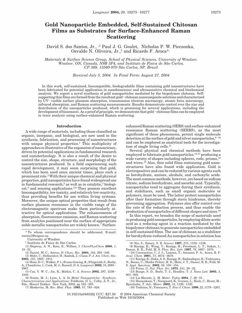
The production of Au nanoparticles in synthesis S1 isillustrated in Figure 1a, where the increase in theextinction of the 528 nm surface plasmon band withreaction time can be attributed to the formation and
(17) Esumi, K.; Takei, N.; Yoshimura, T. Colloids Surf., B 2003, 32,117-123.
(18) Warren, L. F., Jr.; Cunningham, P. H. United States Patent4,933,204, 1990.
(19) Krynetskii, A. B.; Rukhadze, A. A.; Fadeeva, S. S. Zh. Fiz. Khim.1999, 73, 576-579.
(20) Sandford, P. A. In Chitin and Chitosan: Sources, Chemistry,Biochemistry, Physical Properties and Applications; Skjak, G., An-thonsen, T., Sandford, P., Eds.; Elsevier: New York, 1988; pp 51-69.
(21) Rabea, E. I.; Badawy, E.-T.; Stevens, C. V.; Smagghe, G.;Steurbaut, W. Biomacromolecules 2003, 4, 1457-1465.
(22) Kurita, K. Prog. Polym. Sci. 2001, 26, 1921-1971.(23) Ligler, F. S.; Schauer, C. L.; Chen, M.-S.; Chatterley, M.;
Eisemann, K.; Welsh, E. R.; Price, R. R.; Schoen, P. E. Thin Solid Films2003, 434, 250-257.
(24) dos Santos, D. S., Jr.; Riul, A., Jr.; Malmegrin, R. R.; Fonseca,F. J.; Oliveira, O. N., Jr.; Mattoso, L. H. C. Macromol. Biosci. 2003, 3,591-595. (25) Signini, R.; Campana, S. P. Polym. Bull. 1999, 42, 159-166.
10274 Langmuir, Vol. 20, No. 23, 2004 dos Santos et al.
increasing population of Au nanoparticles. Visually, oneobserves a change in the color of the solution from yellowto dark pink as the reaction proceeds. In supplementaryexperiments, it was observed that nanoparticles are alsoformed at room temperature, with all other conditionsbeing the same as those employed in synthesis S1;however, the reaction requires 3 days to go to completion(results not shown).
Surface plasmon absorption, which has proven verysensitive for measuring differences in metallic nanopar-ticle size, shape, and spatial distribution,26,27 was employedin the study of the films as well. Figure 1b(i) indicates ashift in the absorption maximum from 528 nm for thenanocomposite solution (Figure 1a) to 538 nm for the filmobtained from synthesis S1. This confirms the expectedred shift caused by increasing the refractive index of themedia surrounding metallic nanoparticles.28 Control overthe structural and optical properties of the films andnanoparticles produced from these reactions, throughvariation of the experimental conditions used, can bedemonstrated by changes in the surface plasmon absorp-tion spectra observed, as shown in Figure 1b for filmsfrom syntheses S1, S2, and S3. For example, decreasingthe concentration of chitosan by a factor of 100 (synthesisS2), while maintaining all other variables, results in ared-shifted, broadened surface plasmon absorption ofdecreased intensity, as shown in Figure 1b(ii). The redshift seen in Figure 1b(iii), for the film from synthesis S3,is a result of the aggregation of Au nanoparticles, as willbe discussed later. To illustrate even further the largevariability of films that can be produced, a small group,from varied reactions, has been included in the opticalpicture presented in Figure 1c, which shows a wide rangeof colors from light pink to dark violet, in contrast withthe colorless pure chitosan film.
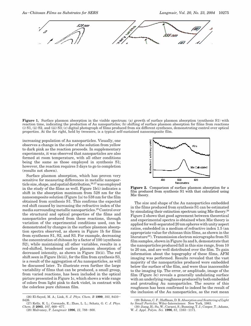
The size and shape of the Au nanoparticles embeddedin the films produced from synthesis S1 can be estimatedby simulating their extinction spectrum using Mie theory.29
Figure 2 shows that good agreement between theoreticaland experimental spectra is obtained when Mie theory isapplied for well-separated 20 nm spheres with unity aspectratios, embedded in a medium of refractive index 1.5 (anappropriate value for chitosan thin films, as shown in theliterature30). Transmission electron micrographs from S1film samples, shown in Figure 3a and b, demonstrate thatthe nanoparticles produced fall in this size range, from 10to 20 nm, and are well distributed over the film. To gaininformation about the topography of these films, AFMimaging was performed. Results revealed that the vastmajority of the nanoparticles produced were embeddedbelow the surface of the film, and were thus inaccessibleto the imaging tip. The error, or amplitude, image of thefilm (Figure 3c) reveals a generally undulating surfacewith an underlying roughness produced by both embeddedand protruding Au nanoparticles. The source of thisroughness has been confirmed to indeed be the result ofthe inclusion of the Au nanoparticles, as the root mean
(26) El-Sayed, M. A.; Link, S. J. Phys. Chem. B 1999, 103, 8410-8426.
(27) Kelly, K. L.; Coronado, E.; Zhao, L. L.; Schatz, G. C. J. Phys.Chem. B 2003, 107, 668-677.
(28) Mulvaney, P. Langmuir 1996, 12, 788-800.
(29) Bohren, C. F.; Huffman, D. R. Absorption and Scattering of Lightby Small Particles; Wiley-Interscience: New York, 1983.
(30) Jiang, H.; Su, W.; Caracci, S.; Bunning, T. J.; Cooper, T.; Adams,W. J. Appl. Polym. Sci. 1996, 61, 1163-1171.
Figure 1. Surface plasmon absorption in the visible spectrum: (a) growth of surface plasmon absorption (synthesis S1) withreaction time, indicating the production of Au nanoparticles; (b) shifting of surface plasmon absorption for films from reactions(i) S1, (ii) S2, and (iii) S3; (c) digital photograph of films produced from six different syntheses, demonstrating control over opticalproperties. At the far right, held by tweezers, is a typical self-sustained nanocomposite film.
Figure 2. Comparison of surface plasmon absorption for afilm produced from synthesis S1 with that calculated usingMie theory.
Au-Chitosan Films as Substrates for SERS Langmuir, Vol. 20, No. 23, 2004 10275
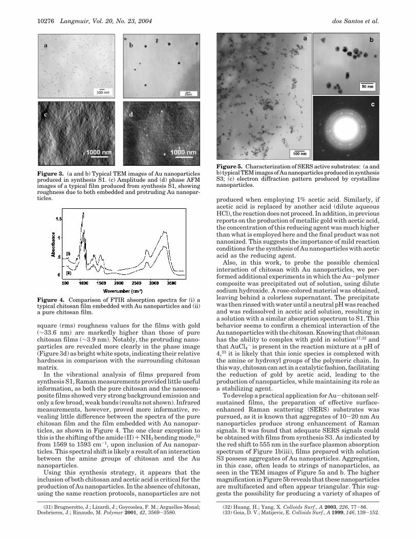
square (rms) roughness values for the films with gold(∼33.6 nm) are markedly higher than those of purechitosan films (∼3.9 nm). Notably, the protruding nano-particles are revealed more clearly in the phase image(Figure 3d) as bright white spots, indicating their relativehardness in comparison with the surrounding chitosanmatrix.
In the vibrational analysis of films prepared fromsynthesis S1, Raman measurements provided little usefulinformation, as both the pure chitosan and the nanocom-posite films showed very strong background emission andonly a few broad, weak bands (results not shown). Infraredmeasurements, however, proved more informative, re-vealing little difference between the spectra of the purechitosan film and the film embedded with Au nanopar-ticles, as shown in Figure 4. The one clear exception tothis is the shifting of the amide (II) + NH2 bending mode,31
from 1569 to 1593 cm-1, upon inclusion of Au nanopar-ticles. This spectral shift is likely a result of an interactionbetween the amine groups of chitosan and the Aunanoparticles.
Using this synthesis strategy, it appears that theinclusion of both chitosan and acetic acid is critical for theproduction of Au nanoparticles. In the absence of chitosan,using the same reaction protocols, nanoparticles are not
produced when employing 1% acetic acid. Similarly, ifacetic acid is replaced by another acid (dilute aqueousHCl), the reaction does not proceed. In addition, in previousreports on the production of metallic gold with acetic acid,the concentration of this reducing agent was much higherthan what is employed here and the final product was notnanosized. This suggests the importance of mild reactionconditions for the synthesis of Au nanoparticles with aceticacid as the reducing agent.
Also, in this work, to probe the possible chemicalinteraction of chitosan with Au nanoparticles, we per-formed additional experiments in which the Au-polymercomposite was precipitated out of solution, using dilutesodium hydroxide. A rose-colored material was obtained,leaving behind a colorless supernatant. The precipitatewas then rinsed with water until a neutral pH was reachedand was redissolved in acetic acid solution, resulting ina solution with a similar absorption spectrum to S1. Thisbehavior seems to confirm a chemical interaction of theAu nanoparticles with the chitosan. Knowing that chitosanhas the ability to complex with gold in solution17,32 andthat AuCl4
- is present in the reaction mixture at a pH of4,33 it is likely that this ionic species is complexed withthe amine or hydroxyl groups of the polymeric chain. Inthis way, chitosan can act in a catalytic fashion, facilitatingthe reduction of gold by acetic acid, leading to theproduction of nanoparticles, while maintaining its role asa stabilizing agent.
To develop a practical application for Au-chitosan self-sustained films, the preparation of effective surface-enhanced Raman scattering (SERS) substrates waspursued, as it is known that aggregates of 10-20 nm Aunanoparticles produce strong enhancement of Ramansignals. It was found that adequate SERS signals couldbe obtained with films from synthesis S3. As indicated bythe red shift to 555 nm in the surface plasmon absorptionspectrum of Figure 1b(iii), films prepared with solutionS3 possess aggregates of Au nanoparticles. Aggregation,in this case, often leads to strings of nanoparticles, asseen in the TEM images of Figure 5a and b. The highermagnification inFigure5breveals that thesenanoparticlesare multifaceted and often appear triangular. This sug-gests the possibility for producing a variety of shapes of
(31) Brugnerotto, J.; Lizardi, J.; Goycoolea, F. M.; Arguelles-Monal;Desbrieres, J.; Rinaudo, M. Polymer 2001, 42, 3569-3580.
(32) Huang, H.; Yang, X. Colloids Surf., A 2003, 226, 77-86.(33) Goia, D. V.; Matijevic, E. Colloids Surf., A 1999, 146, 139-152.
Figure 3. (a and b) Typical TEM images of Au nanoparticlesproduced in synthesis S1. (c) Amplitude and (d) phase AFMimages of a typical film produced from synthesis S1, showingroughness due to both embedded and protruding Au nanopar-ticles.
Figure 4. Comparison of FTIR absorption spectra for (i) atypical chitosan film embedded with Au nanoparticles and (ii)a pure chitosan film.
Figure 5. Characterization of SERS active substrates: (a andb) typical TEM images of Au nanoparticles produced in synthesisS3; (c) electron diffraction pattern produced by crystallinenanoparticles.
10276 Langmuir, Vol. 20, No. 23, 2004 dos Santos et al.
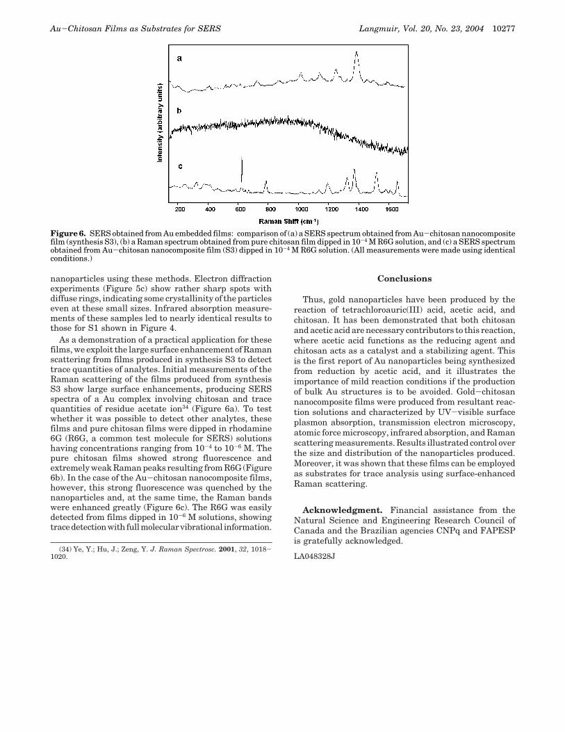
nanoparticles using these methods. Electron diffractionexperiments (Figure 5c) show rather sharp spots withdiffuse rings, indicating some crystallinity of the particleseven at these small sizes. Infrared absorption measure-ments of these samples led to nearly identical results tothose for S1 shown in Figure 4.
As a demonstration of a practical application for thesefilms, we exploit the large surface enhancement of Ramanscattering from films produced in synthesis S3 to detecttrace quantities of analytes. Initial measurements of theRaman scattering of the films produced from synthesisS3 show large surface enhancements, producing SERSspectra of a Au complex involving chitosan and tracequantities of residue acetate ion34 (Figure 6a). To testwhether it was possible to detect other analytes, thesefilms and pure chitosan films were dipped in rhodamine6G (R6G, a common test molecule for SERS) solutionshaving concentrations ranging from 10-4 to 10-6 M. Thepure chitosan films showed strong fluorescence andextremely weak Raman peaks resulting from R6G (Figure6b). In the case of the Au-chitosan nanocomposite films,however, this strong fluorescence was quenched by thenanoparticles and, at the same time, the Raman bandswere enhanced greatly (Figure 6c). The R6G was easilydetected from films dipped in 10-6 M solutions, showingtracedetectionwith full molecular vibrational information.
Conclusions
Thus, gold nanoparticles have been produced by thereaction of tetrachloroauric(III) acid, acetic acid, andchitosan. It has been demonstrated that both chitosanand acetic acid are necessary contributors to this reaction,where acetic acid functions as the reducing agent andchitosan acts as a catalyst and a stabilizing agent. Thisis the first report of Au nanoparticles being synthesizedfrom reduction by acetic acid, and it illustrates theimportance of mild reaction conditions if the productionof bulk Au structures is to be avoided. Gold-chitosannanocomposite films were produced from resultant reac-tion solutions and characterized by UV-visible surfaceplasmon absorption, transmission electron microscopy,atomic force microscopy, infrared absorption, and Ramanscattering measurements. Results illustrated control overthe size and distribution of the nanoparticles produced.Moreover, it was shown that these films can be employedas substrates for trace analysis using surface-enhancedRaman scattering.
Acknowledgment. Financial assistance from theNatural Science and Engineering Research Council ofCanada and the Brazilian agencies CNPq and FAPESPis gratefully acknowledged.
LA048328J(34) Ye, Y.; Hu, J.; Zeng, Y. J. Raman Spectrosc. 2001, 32, 1018-
1020.
Figure 6. SERS obtained from Au embedded films: comparison of (a) a SERS spectrum obtained from Au-chitosan nanocompositefilm (synthesis S3), (b) a Raman spectrum obtained from pure chitosan film dipped in 10-4 M R6G solution, and (c) a SERS spectrumobtained from Au-chitosan nanocomposite film (S3) dipped in 10-4 M R6G solution. (All measurements were made using identicalconditions.)
Au-Chitosan Films as Substrates for SERS Langmuir, Vol. 20, No. 23, 2004 10277