utilization of crab shell-derived chitosan in nanoparticle
TRANSCRIPT

Indian Journal of Geo Marine Sciences Vol. 48 (08), August 2019, pp. 1183-1188
Utilization of crab shell-derived chitosan in nanoparticle synthesis for curcumin delivery
N. Shobana1, P. Senthil Kumar2, P. Raji1, & Antony V. Samrot3* 1Department of Biotechnology, School of Bio and Chemical Engineering, Sathyabama Institute of Science and Technology,
Chennai, Tamil Nadu, India 2Department of Chemical Engineering, School of Bio and Chemical Engineering, Sathyabama Institute of Science and Technology,
Chennai, Tamil Nadu, India 3Department of Biomedical Sciences, Faculty of Medicine and Biomedical Sciences, MAHSA University,
Jalan SP2, Bandar Saujana Putra, 42810 Jenjarom, Selangor, Malaysia *[E-mail: [email protected]]
Chitosan derived from crustaceans is biodegradable as well as biocompatible and can be made into nanoparticles when chelated with chelators, such as sodium tripolyphosphate and barium chloride. In this study, crab shells-derived chitosan was chelated using sodium trimetaphosphate to form nanoparticles. Curcumin was encapsulated into nanoparticles and characterized using Fourier transform infra-red spectroscopy, scanning electron microscopy, atomic force microscopy, and X-ray diffraction analysis. The particles were found to be 18 nm in size, while the curcumin-loaded particles were 25 nm in size. The particles were observed to encapsulate 90% of the drug used. The nanoparticles produced were analyzed for in vitro controlled drug release against Pseudomonas aeruginosa, Bacillus subtilis, and Candida albicans.
[Keywords: Sodium trimetaphosphate (STMP); Chitosan; Curcumin; Drug delivery]
Introduction In the field of nanobiotechnology, there are enormous
expectations from biomaterials-based nanoparticles as they are biodegradable, biocompatible, non/low immunogenic, and nontoxic. Thus, they are well exploited in drug and gene delivery1,3 tissue engineering4 food industries5 etc. Chitosan, an abundant natural polysaccharide found mainly in the exoskeletons of marine organisms and certain fungi and algae6,9 is biocompatible and biodegradable10. Chitosan and its derivatives have been reported as potential carriers for drug delivery systems11,12.
Chitosan nanoparticles are produced by ionic gelation method13 using sodium tripolyphosphate (TPP)14 and barium chloride15. These cross-linking agents combine two components with opposite charges to form nanoparticles16. Size formation can be controlled by ionic gelation method as well as by encapsulation of protein, ions, and drugs17,18. Using an alternate and new cross-linking agent may lead to formation of smaller-sized nanoparticles which may be better than the commonly used cross-linkers such as sodium TPP and barium chloride. Curcumin is a hydrophobic drug with multiple bioactivities, thus it was chosen as a drug of choice to load into the nanocarriers in most studies12,15.
In this study, chitosan was derived from crab shell and an attempt was made to use sodium trimetaphosphate (STMP) as chelator to produce curcumin-loaded chitosan nanoparticle for drug delivery.
Materials and Methods
Materials Curcumin was purchased from Sisco Research
Laboratories Pvt. Ltd; Acetic acid from Qualigens Fine Chemicals, India; STMP, Nutrient agar, and carboxy methyl cellulose (CMC) from LOBA Chemie, HiMedia and Micro Fine Chemicals, India, respectively. Millipore water was used in this study.
Preparation and characterization of crab shell-derived chitosan
Chitosan from crab shells was prepared following Samrot et al.12 and Yen et al.19, mixed with KBr pellets, and subjected to Fourier transform infra-red spectroscopy (FTIR) analysis (Shimadzu, Japan). Synthesis of STMP-chelated chitosan nanoparticles
Chitosan 0.8% was prepared in 50 ml 0.1N acetic acid. The solution was filtered to remove the unsuspended particles. 0.2% STMP in 25 ml of distilled water was added dropwise to the chitosan

1184
solution. 25 was added constant manand then cepellets werecurcumin-encurcumin in solution and
CharacterizaChitosan
curcumin) wJapan). Thechitosan nanelectron mGermany) a(Bruker, Gerof the chitosmart lab X-r
Percentage eAfter the
solution was of the supernAbsorbance wof curcumin drug encapchitosan nanusing the foll
Percentage d
Fig. 1 — FTIRUnloaded chito
ml of 0.4% Cdropwise to
nual stirring, entrifuged at e collected ancapsulated
25 ml ethanochelation was
ation of chitosnanoparticles
were subjectede morphologyoparticles wer
microscope (Sand an atomirmany). X-rasan nanopartray diffractom
encapsulationsynthesis procentrifuged a
natant was withwas taken at 4
n was determisulation effi
noparticles walowing formul
drug loading e
total drug ad
T
R spectroscopy osan nanoparticle
INDIAN
CMC in 25 mo the abovekept undistur5000 rpm fo
and lyophiliznanoparticle
ol was added ts done as desc
san nanoparts (before an
d to FTIR analy, topographre examined uSEM) (Zeisic force mic
ay diffraction ticles were remeter (Rigaku
n efficiency ocess was comat 3500 g for hdrawn every 425 nm15 and tined in the siciency of as calculated la20:
efficiency = dded - free no
Total drug ad
analysis of ches chelated with
N J. MAR. SCI.
ml distilled wae solution wrbed for an hoor 15 min. Tzed to produes; 0.1% to 0.8% chitocribed earlier.
ticles nd after loadlysis (Shimady, and size under a scannss Ultra Plcroscope (AF
(XRD) patteecorded using
u, Japan).
mpleted, a 5 10 min and 1 10 min over 2
the concentratsupernatant. Tcurcumin-loadas a percenta
on-trapped dru
dded
itosan nanopartSTMP, and (c) L
, VOL. 48, NO.
ater with our, The uce
of san
ding dzu,
of ning lus,
FM) erns g a
ml ml
2 h. tion The ded age
ug
In vitrThe
nanopsuch a(0.1N)using Bacilluaerugi Swarm
1.3autoclto soliloadedmixedglucosnutrienand Gat the the bcompa
Resul
Prepachitos
Thein the NHCOconfirpresencm−1 p(Fig. 1
ticles loaded wiLoaded chitosan
08, AUGUST 2
ro controlled e releasing a
particles in the as water, etha), and crude agar well diffus subtilis, inosa, and the
ming motility g of nutrient laved, poured idify. 1 mg/mld or unloadedd with 1 ml se. The abovnt agar with 0
Gram negative centre. The a
bacteria fromaring with con
lts and Discu
aration and chsan e broad O-H crab shell-de
OCH3 and thermed by the nt in the rangproved the pr1a). Similar re
ith curcumin ann nanoparticles c
2019
drug release ability of cupresence of d
anol (25%), Pchitosanase
fusion methodgram ne
fungus, Cand
agar with 1.5%into a sterile l of chitosan nd) in distilled
of steam-steve solution w0.5% agar. GP. aeruginos
ability of the m swarming ntrol22,23.
ssion
haracterizatio
stretch at 355erived chitosane methylene gcorrespondin
ge 2921-2879 resence of δ (esults were rep
nd chelated withchelated with ST
urcumin fromdifferent solvenPBS (pH 6), aenzyme was
d21 against gramegative Pse
dida albicans.
% of agar waspetri plate an
nanoparticles d water (1 mgerilized 10% was added to
Gram positive a were point inanoparticles
was determ
on of crab she
50–3200 cm−1
n. The methygroup in CH2
ng stretching cm−1. A pea
(CH2) of CH2Oported by Sam
h STMP: (a) CTMP
m chitosan nt systems acetic acid
evaluated m positive
eudomonas
s prepared, nd allowed (curcumin g/ml) was (w/v) D-
5 ml of B. subtilis inoculated to inhibit
mined by
ell-derived
was seen yl group in
2OH were vibrations
ak at 1451 OH group
mrot et al12.
Chitosan, (b)
CharacterizaThe chara
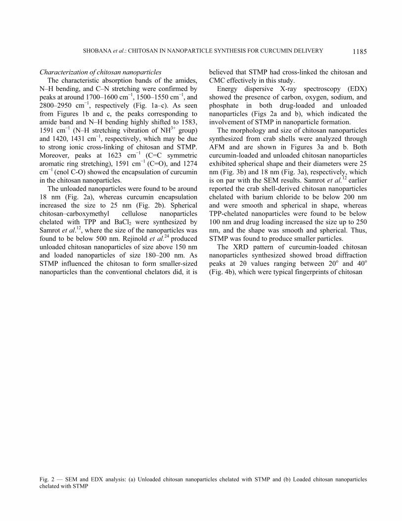
N–H bendinpeaks at arou2800–2950 from Figureamide band 1591 cm−1 (and 1420, 14to strong ionMoreover, paromatic ringcm−1 (enol Cin the chitosa
The unloa18 nm (Figincreased thchitosan–carbchelated witSamrot et al.found to be unloaded chiand loaded STMP influenanoparticles
Fig. 2 — SEMchelated with S
SHOBANA
ation of chitosacteristic absog, and C–N s
und 1700–160cm−1, respec
es 1b and c, and N–H ben
(N–H stretchi431 cm−1, resnic cross-linkpeaks at 16g stretching),
C-O) showed tan nanoparticladed nanopartg. 2a), wherhe size to 2rboxymethyl th TPP and .12, where the below 500 nitosan nanopananoparticles
enced the chs than the con
M and EDX anaSTMP
et al.: CHITOSA
san nanopartiorption bandsstretching we00 cm−1, 1500ctively (Fig.
the peaks cnding highly ing vibration spectively, whking of chito623 cm−1 (C
1591 cm−1 (Cthe encapsulatles. ticles were foureas curcumin25 nm (Fig.
cellulose BaCl2 were size of the na
m. Rejinold earticles of sizes of size 18
hitosan to fornventional ch
alysis: (a) Unloa
AN IN NANOPA
icles s of the amidre confirmed –1550 cm−1, a1a–c). As se
correspondingshifted to 15of NH3+ grou
hich may be dsan and STM
C=C symmetC=O), and 12tion of curcum
und to be aroun encapsulat
2b). Spherinanopartic
synthesized anoparticles wet al.24 produce above 150 80–200 nm. rm smaller-sizhelators did, i
aded chitosan n
ARTICLE SYN
des, by
and een to 83, up) due
MP. tric 274 min
und tion ical cles
by was ced nm As
zed t is
believCMC
Eneshowephospnanopinvolv
ThesyntheAFM curcumexhibinm (Fis on reportchelatand wTPP-c100 nnm, aSTMP
Thenanoppeaks (Fig. 4
nanoparticles che
NTHESIS FOR C
ved that STMeffectively in
ergy dispersed the presenphate in boparticles (Figvement of STe morphologyesized from
and are shmin-loaded aited spherical
Fig. 3b) and 1par with the ted the crab sted with bariwere smooth chelated nano
nm and drug land the shapeP was found te XRD patt
particles syntat 2θ valu
4b), which we
elated with STM
CURCUMIN DE
P had cross-ln this study. sive X-ray nce of carbonoth drug-lo
gs 2a and b)MP in nanopy and size of crab shells w
hown in Figuand unloaded l shape and th18 nm (Fig. 3SEM results.
shell-derived ium chloride
and sphericoparticles weloading increae was smoothto produce smtern of curcthesized show
ues ranging bere typical fin
MP and (b) Loa
ELIVERY
linked the chi
spectroscopyn, oxygen, sodoaded and , which indiarticle formatchitosan nan
were analyzedures 3a andchitosan nan
heir diametera), respective. Samrot et achitosan nanto be below
cal in shape,ere found to ased the size h and spheric
maller particlecumin-loaded wed broad dbetween 20o ngerprints of
aded chitosan n
1185
itosan and
y (EDX) dium, and unloaded
icated the tion.
noparticles d through
d b. Both noparticles rs were 25 ely, which l.12 earlier
noparticles w 200 nm , whereas be below up to 250 cal. Thus,
es. chitosan
diffraction and 40o
chitosan
anoparticles

1186
and curcumidiffraction prevealed that Percentage e
The enccurcumin inwas found todrug encapincreasing wchelated nanscorpion ven
In vitro contThe releasichitosan nadiffusion assthat is, wacid (0.1NNo antibacP. aeruginoswater and P
Fig. 3 — AFM(b) Loaded chit
Fig. 4 — XRDchitosan nanop
in25. The lowpeaks of unlot they were am
encapsulationcapsulation eto STMP-cheo be around 9
psulation effiwith time. Donoparticles tonom into the n
trolled drug ring ability
anoparticles wsay using fivater, ethano
N), and crcterial activsa, and C. a
PBS were use
M analysis of chtosan nanopartic
D of chitosan nanarticles chelated
INDIAN
wer intensity eoaded chitosamorphic in na
n efficiency efficiency oelated chitosa90% (Fig. 5). ficiency was ounighi et ao encapsulatenanoparticles
elease of curcum
was studied ve different sol, PBS (pHrude chitosavity againstalbicans was d as solvents
hitosan nanoparticles chelated with
noparticles chelad with STMP
N J. MAR. SCI.
exhibited by an nanoparticature (Fig. 4a)
of hydrophoan nanoparticThe percenta
found to l.26 found TP
e up to 90% .
min-encapsulaby agar w
solvent systemH 6.8), aceanase enzymt B. subtiobserved wh
s. This might
icles chelated wh STMP
ated with STMP
, VOL. 48, NO.
the cles ).
obic cles age be
PP-of
ated well ms, etic me. ilis, hen
be
due toouter lof inhloadedethanoused suppothe dru
Fig. 5 nanopa
with STMP: (a) U
: (a) Unloaded c
08, AUGUST 2
o the inabilitylayer of chitos
hibition at thed chitosan nol, acetic acid,as solvents rt the findingug release out
— Percentage articles chelated
Unloaded chitos
chitosan nanopar
2019
y of water ansan to release e highest concnanoparticles , and crude ch(Tables 1–3)
g that the acidt of chitosan n
drug encapsulawith STMP
san nanoparticle
rticles chelated w
nd PBS to dicurcumin12,27.
centration of was observ
hitosanase enz). Earlier repdic environmanoparticles12
ation efficiency
es chelated with
with STMP and
ssolve the . The zone curcumin-
ved when zyme were ports also ent favors ,15.
of chitosan
h STMP and
d (b) Loaded
SHOBANA et al.: CHITOSAN IN NANOPARTICLE SYNTHESIS FOR CURCUMIN DELIVERY
1187
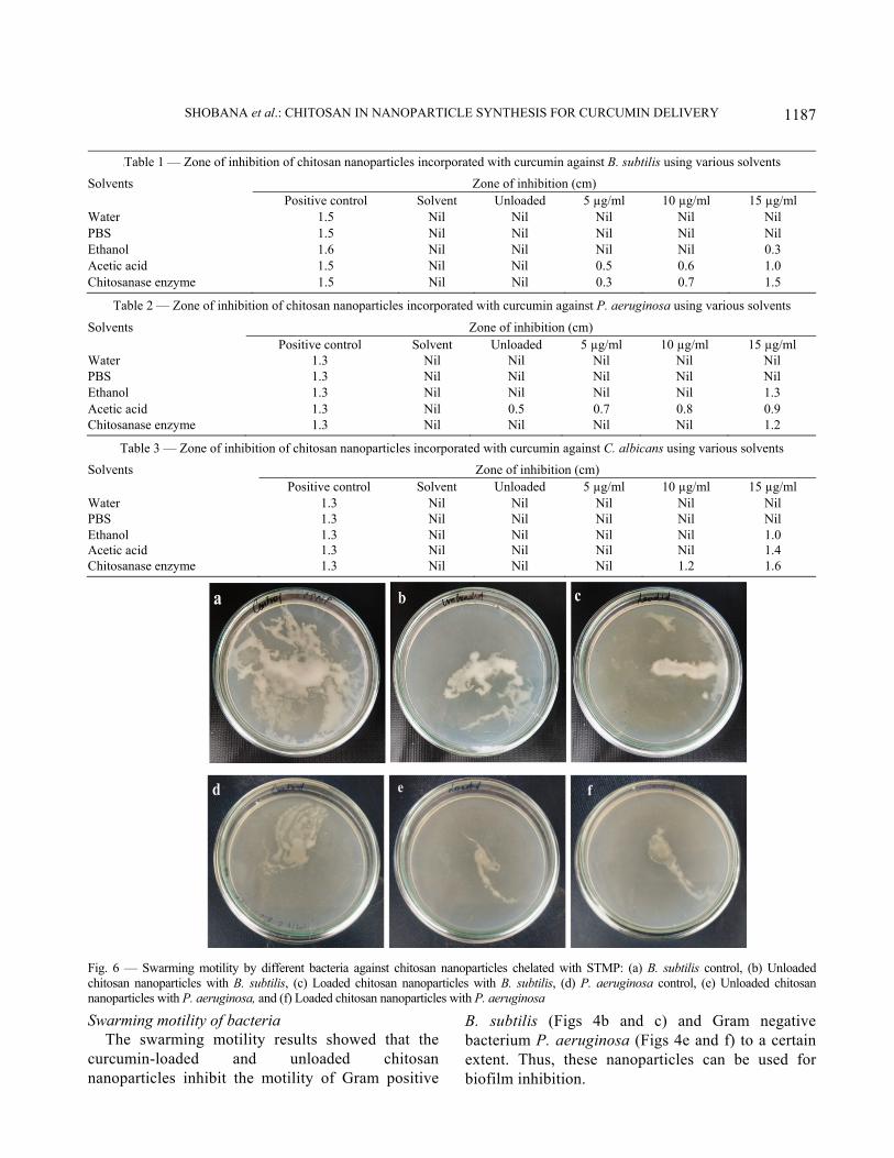
Swarming motility of bacteria The swarming motility results showed that the
curcumin-loaded and unloaded chitosan nanoparticles inhibit the motility of Gram positive
B. subtilis (Figs 4b and c) and Gram negative bacterium P. aeruginosa (Figs 4e and f) to a certain extent. Thus, these nanoparticles can be used for biofilm inhibition.
{Table 1 — Zone of inhibition of chitosan nanoparticles incorporated with curcumin against B. subtilis using various solvents
Solvents Zone of inhibition (cm) Positive control Solvent Unloaded 5 µg/ml 10 µg/ml 15 µg/ml
Water 1.5 Nil Nil Nil Nil Nil PBS 1.5 Nil Nil Nil Nil Nil Ethanol 1.6 Nil Nil Nil Nil 0.3 Acetic acid 1.5 Nil Nil 0.5 0.6 1.0 Chitosanase enzyme 1.5 Nil Nil 0.3 0.7 1.5
Table 2 — Zone of inhibition of chitosan nanoparticles incorporated with curcumin against P. aeruginosa using various solvents
Solvents Zone of inhibition (cm) Positive control Solvent Unloaded 5 µg/ml 10 µg/ml 15 µg/ml
Water 1.3 Nil Nil Nil Nil Nil PBS 1.3 Nil Nil Nil Nil Nil Ethanol 1.3 Nil Nil Nil Nil 1.3 Acetic acid 1.3 Nil 0.5 0.7 0.8 0.9 Chitosanase enzyme 1.3 Nil Nil Nil Nil 1.2
Table 3 — Zone of inhibition of chitosan nanoparticles incorporated with curcumin against C. albicans using various solvents
Solvents Zone of inhibition (cm) Positive control Solvent Unloaded 5 µg/ml 10 µg/ml 15 µg/ml
Water 1.3 Nil Nil Nil Nil Nil PBS 1.3 Nil Nil Nil Nil Nil Ethanol 1.3 Nil Nil Nil Nil 1.0 Acetic acid 1.3 Nil Nil Nil Nil 1.4 Chitosanase enzyme 1.3 Nil Nil Nil 1.2 1.6
Fig. 6 — Swarming motility by different bacteria against chitosan nanoparticles chelated with STMP: (a) B. subtilis control, (b) Unloaded chitosan nanoparticles with B. subtilis, (c) Loaded chitosan nanoparticles with B. subtilis, (d) P. aeruginosa control, (e) Unloaded chitosan nanoparticles with P. aeruginosa, and (f) Loaded chitosan nanoparticles with P. aeruginosa

INDIAN J. MAR. SCI., VOL. 48, NO. 08, AUGUST 2019
1188
Conclusion In this study, chitosan was extracted from crab
shell. The extracted chitosan was chelated with STMP and characterized as 18-25 nm sized spherical nanoparticles. The particles were found to encapsulate curcumin better, that is, with 90% encapsulation. Nanoparticles were found to inhibit the swarming motility to a certain extent. The curcumin-loaded nanoparticles were found to release curcumin in an acidic environment.
References 1 Nitta, S.K., Numata, K., Biopolymer-based nanoparticles for
drug/gene delivery and tissue engineering, Int. J. Mol. Sci., 14(2013) 1629-1654.
2 Kataoka, K., Harada, A., Nagasaki, Y., Block copolymer micelles for drug delivery: Design, characterization and biological significance, Adv. Drug Deliv. Rev., 64(2012) 37-48.
3 Panyam, J., Labhasetwar, V., Biodegradable nanoparticles for drug and gene delivery to cells and tissue, Adv. Drug Deliv. Rev., 55(2003) 329-347.
4 Shi, J., Votruba, A.R., Farokhzad, O.C., Langer, R., Nanotechnology in drug delivery and tissue engineering: From discovery to applications, Nano letters, 10(2010) 3223-3230.
5 Joye, I.J., McClements, D.J., Emulsifying and emulsion-stabilizing properties of gluten hydrolysates, J. Agric. Food Chem., 62(2014) 2623-2630.
6 Knorr, D. Recovery and utilization of chitin and chitosan in food processing waste management, Food Technol.,45 (1991), 114-122.
7 Fenton, D.M., Eveleigh, D.E., Purification and mode of action of a chitosanase from Penicillium islandicum, Microbiology, 126(1981), 151-165.
8 Davis, B., Eveleigh, D.E., Chitosanases: Occurrence, production and immobilization, in: Chitin, chitosan, and related enzymes, edited by John P. Zikakis, (Elsevier, Orland) 1984, pp. 161-179.
9 Pochanavanich, P., Suntornsuk, W., Fungal chitosan production and its characterization. Lett. Appl. Microbiol., 35(2002) 17-21.
10 Struszczyk, M.H., Chitin and chitosan. Part II. Applications of chitosan. Polimery, 47(2002) 396-403.
11 Sivakumar, S.M., Kannadasan, M., Roy, R.K., Review of chitosan and its relevance in pharmaceutical sciences, Res. J. Pharm. Biol. Chem., 5(2014) 425-430.
12 Samrot, A.V., Burman, U., Philip, S.A., Shobana, N., Chandrasekaran, K., Synthesis of curcumin loaded polymeric nanoparticles from crab shell derived chitosan for drug delivery. Informatics in Medicine Unlocked, 10(2018) 159-182.
13 Calvo, P., Remunan-Lopez, C., Vila-Jato, J.L., Alonso, M.J., Novel hydrophilic chitosan–polyethylene oxide nanoparticles as protein carriers, J. Appl. Polym. Sci., 63(1997) 125-132.
14 Kumar, V., Dandapat, S., Kumar, A., Kumar, N., Preparation and characterization of chitosan nanoparticles “alternatively,
carrying potential” for cellular and humoral immune responses, Adv. Anim. Vet. Sci., 2 (2014) 414-417.
15 Samrot, A.V., Jahnavi, T., Padmanaban, S., Philip, S.A., Burman, U., Rabel, A.M., Chelators influenced synthesis of chitosan–carboxymethyl cellulose microparticles for controlled drug delivery, Appl. Nanosci., 6(2016) 1219-1231.
16 Amidi, M., Mastrobattista, E., Jiskoot, W., Hennink, W.E., Chitosan-based delivery systems for protein therapeutics and antigens, Adv. Drug Deliv. Rev., 62(2010) 59-82.
17 Avadi, M.R., Sadeghi, A.M.M., Mohammadpour, N., Abedin, S., Atyabi, F., Dinarvand, R., Rafiee-Tehrani, M., Preparation and characterization of insulin nanoparticles using chitosan and arabic gum with ionic gelation method, Nanomed. Nanotechnol. Biol. Med., 6(2010) 58-63.
18 Du, W.L., Niu, S.S., Xu, Y.L., Xu, Z.R., Fan, C.L., Antibacterial activity of chitosan tripolyphosphate nanoparticles loaded with various metal ions, Carbohydr. Polym., 75(2009) 385-389.
19 Yen, M.T., Yang, J.H., Mau, J.L., Physicochemical characterization of chitin and chitosan from crab shells, Carbohydr. Polym., 75(2009) 15-21.
20 Awotwe-Otoo, D., Zidan, A.S., Rahman, Z., Habib, M.J., Evaluation of anticancer drug-loaded nanoparticle characteristics by non-destructive methodologies, AAPS PharmSciTech., 13(2012) 611-622.
21 Samrot, A.V., Bhavya, K.S., Sahithya, C.S., Sowmya, N., Evaluation of toxicity of chemically synthesised gold nanoparticles against Eudrilus eugeniae, J. Clust. Sci., 10(2018) 1–9.
22 O'May, C., Tufenkji, N., The swarming motility of Pseudomonas aeruginosa is blocked by cranberry proanthocyanidins and other tannin-containing materials, Appl. Environ. Microbiol., 77(2011) 3061-3067.
23 Tremblay, J., Richardson, A.P., Lépine, F., Déziel, E., Self-produced extracellular stimuli modulate the Pseudomonas aeruginosa swarming motility behaviour, Environ. Microbiol., 9(2007) 2622-2630.
24 Rejinold, N.S., Muthunarayanan, M., Divyarani, V.V., Sreerekha, P.R., Chennazhi, K.P., Nair, S.V., Tamura, H., Jayakumar, R., Curcumin-loaded biocompatible thermoresponsive polymeric nanoparticles for cancer drug delivery, J. Colloid Interface Sci., 360(2011) 39-51.
25 Anand, M., Maruthupandy, M., Kalaivani, R., Suresh, S., Kumaraguru, A.K., Larvicidal activity of chitosan nanoparticles synthesized from crab and squilla species against Aedes aegypti, J. Colloid Sci. Biotechnol., 3(2014) 188-193.
26 Dounighi, M.N., Eskandari, R., Avadi, M.R., Zolfagharian, H., Sadeghi, M.M.A., Rezayat, M., Preparation and in vitro characterization of chitosan nanoparticles containing Mesobuthuseupeus scorpion venom as an antigen delivery system, J. Venom. Anim. Toxins Incl. Trop. Dis., 18(2012) 44-52.
27 Samrot, A.V., Senthil Kumar, P., Bhushan, S., Kurup, R., Burman, U., Philip, S.A., Padmanaban, S., Sodium tri poly phosphate mediated synthesis of curcumin loaded chitosan–carboxymethyl cellulose microparticles for drug delivery, Int. Pharmacogn. Phytochem. Res., 9(2017) 694-702.