chitosan treated textile substrates for wound care ...jairjp.com/july 2013/06 chellamani.pdf ·...
TRANSCRIPT

Journal of Academia and Industrial Research (JAIR) Volume 2, Issue 2 July 2013 97
©Youth Education and Research Trust (YERT) jairjp.com Chellamani et al., 2013
ISSN: 2278-5213
Chitosan treated Textile substrates for Wound care applications
K.P. Chellamani*, R.S. Vignesh Balaji and J. Sudharsan The South India Textile Research Association (SITRA), Coimbatore-641 014, TN, India
[email protected]*; 0422-4215347 ______________________________________________________________________________________________
Abstract Wound dressing selection is somewhat challenging because of the availability of large varieties of dressings in the market. Each product has its own specific actions, benefits and drawbacks. Hence, determining which dressing best suits a patient’s needs is a multi-faceted process. Dressing choice depends on factors such as wound type and appearance, exudate, presence or absence of pain and required dressing changing frequency. Nowadays, chitosan incorporated/applied wound dressings are predominant in the wound management system due to their unique properties like biocompatibility, biodegradability, non-toxic nature etc. A review on the literatures published in the area of chitosan coated textile substrates for wound care applications is presented here.
Keywords: Wound dressing, chitosan, biocompatibility, wound management system, textile substrates, wound care.
Introduction Textile substrates are widely used as wound dressing materials. A wound dressing material treated with chitosan, which is effective against bacteria, will be an ideal material for wound care applications without losing their inherent textile characteristics. Textile substrates used as wound closing materials should act as reservoir of antimicrobial agents and should release them gradually at the affected site for a prolonged period of time (Shanmugasundaram et al., 2006; Vangenhove, 2007; Schwartz, 2008; Rajendran, 2009; Shanmugasundaram and Gowda, 2011). Wounds have been categorized in many different ways, but they all reflect commonly the differences in the required treatment, the expected time and prospects of healing. The classification of wounds recognizes the type of injury (blunt contusion, sharp laceration, thermal, chemical, etc.), the extent of tissue loss, the presence of infection, foreign bodies and underlying structural injuries (fracture of bone, exposure of vital parts such as tendons and blood vessels). A general classification of wounds is as follows: 1. Wounds with no tissue loss.
2. Wounds with tissue loss: This generally includes
three types of wounds; those that are (a) Caused by burning, trauma or abrasion (b) The result of secondary events involving chronic ailments, as, for example, venous statis, diabetic ulcers, and pressure sores; or (c) Induced as a part of the treatment of the wound itself, as, for example, the wound arising at the donor site for skin grafting or the wound due to derma-abrasion.
From the standpoint of the extent of injury, a wound may also be classified in terms of the layers involved: 1. The superficial wounds, involving only the epidermis. 2. The partial thickness wounds, involving also the
dermis. 3. The full thickness wounds, involving, additionally, the
subcutaneous fat or deeper tissue. Wounds can also be broadly characterized as acute and chronic. In the former type, that may or may not have tissue loss, healing tends to proceed through a timely and orderly process. In the chronic wounds, on the other hand, healing has failed to proceed through this process or it has proceeded without establishing a sustainable anatomical and functional result. The chronic wounds are classically subdivided into venous statis ulcers, pressure ulcers, and diabetic ulcers. To a lesser extent, the traumatic wound with extensive cutaneous loss that has not been replaced for some reasons also may fall in the category of chronic wounds (Rajendran, 2009). Chitosan is a natural biopolymer that is derived from chitin, a major component of crustacean outer skeletons. Chitosan is the second most common polysaccharide on earth and this material is known in the wound management field for its haemostatic properties. Further, it also possesses other biological activities and affects macrophage function that helps in faster wound healing. It also has an aptitude to stimulate cell proliferation and histoarchitectural tissue organization. The biological properties including bacteriostatic and fungistatic properties are particularly useful for wound treatment (Paul and Sharma, 2004; Shanmugasundaram, 2006; Shanmugasundaram et al., 2006; Sajeev et al., 2008; Lee et al., 2009; Hima Bindu et al., 2010; Shanmugasundaram and Gowda, 2011; Sun and Li, 2011).
REVIEW ARTICLE

Journal of Academia and Industrial Research (JAIR) Volume 2, Issue 2 July 2013 98
©Youth Education and Research Trust (YERT) jairjp.com Chellamani et al., 2013
Fig. 1. Molecular structure of chitosan (Sun and Li, 2011).
Chitosan (1–4)-linked 2-amino-2-deoxy-D-glucopyranose (Fig. 1), is derived from chitin, one of the most abundant natural polysaccharides. Chitosan is well-known for its non-toxic, biocompatible and biodegradable properties. In addition, it has several unique properties: it is antimicrobial and inhibits the growth of a wide variety of fungi, yeasts, and bacteria, which can be beneficial for use in the field of biomedicine. It can also bind toxic metal ions, which can be beneficial for use in air cleaning and water purification applications. These properties arise as a result of protonation of NH2–groups on the chitosan backbone (Hima Bindu et al., 2010). Chitosan provides a non-protein matrix for 3D tissue growth and activates macrophages for tumoricidal activity. It stimulates cell proliferation and histoarchitectural tissue organization. Chitosan is a hemostat, which helps in natural blood clotting and blocks nerve endings reducing pain (Fig. 2).
Fig. 2. Schematic representations of the benefits of chitosan wound
dressing (Paul and Sharma, 2004; Hima Bindu et al., 2010).
Chitosan will gradually depolymerize to release N-acetyl-b-D-glucosamine, which initiates fibroblast proliferation and helps in ordered collagen deposition and stimulates increased level of natural hyaluronic acid synthesis at the wound site. It helps in faster wound healing and scar prevention (Paul and Sharma, 2004; Hima Bindu et al., 2010).
In this review, an attempt has been made to review the different kinds of chitosan treated wound dressings that are used in the wound care applications. They are: 1. Chitosan wound dressings prepared by
Electrospinning method. 2. Cotton yarn/gauze fabrics treated with chitosan
polymers. 3. Chitosan treated wound dressings for drug delivery. 4. Chitosan sponges, composite wound dressings/
non-woven wound dressings for wound care systems. 5. Chitosan films for wound care applications. Chitosan wound dressings prepared by electrospinning method Electrospinning is a straightforward, cost-effective, versatile technique employing electrostatic forces to produce polymer fibers, ranging in diameter from a few microns down to tens of nanometers. Researchers in the past have made attempts to electrospin chitosan in order to further utilize this material. A basic apparatus for electrospinning consists of three major components: a spinneret, a fiber collector and a high-voltage power supply as shown in Fig. 3.
Fig. 3. Electrospinning system (Lee et al., 2009).
Electrospinning of chitosan posses may challenges due to its high solution viscosity. Chitosan’s rigid D-glucosamine structures, high crystallinity and ability to hydrogen bond lead to poor solubility in common organic solvents. Chitosan is a cationic polymer, polyelectrolyte, and subject to the polyelectrolyte effect where in aqueous solution, chitosan’s polymer coils are greatly expanded by the presence of charged groups and if the solution is free of added electrolytes, the polymer coil contracts as the polymer concentration increases. The combination of these properties makes it difficult to create a chitosan-based solution with a high concentration of polymer that has a low enough viscosity to be able to be electrospun (Lee et al., 2009). Poly(vinyl alcohol) (PVA) is a water-soluble, biocompatible and biodegradable synthetic polymer that has been studied intensely because of its good physical properties, high hydrophilicity, processability etc. Electrospinning of PVA solution and its potential applications in the preparation of ultrafine separation filters, biodegradable mats, etc. have been reported by many researchers.

Journal of Academia and Industrial Research (JAIR) Volume 2, Issue 2 July 2013 99
©Youth Education and Research Trust (YERT) jairjp.com Chellamani et al., 2013
In recent years, much attention has been focused on the biomedical applications of PVA hydrogels including contact lenses, artificial organs and drug delivery systems. By incorporating a second polymer component, viz. chitosan, to PVA solution, an improvement in biocompatibility of the blend system can be achieved. This can be attributed to the good biocompatibility, antibacterial properties, appropriate biodegradability, excellent physicochemical properties and its commercial availability at relatively low cost. As candidate materials, chitosan and PVA have been separately investigated for sutures and wound dressings (Sajeev et al., 2008). Cotton yarn/gauze treated with chitosan Chitosan treated cotton yarn: A new cotton yarn with a chitosan coating was prepared by the oxidation of a cotton thread with sodium per-iodate at 60C in water and subsequent treatment with a solution of chitosan in aqueous acetic acid. Infrared spectra of the chitosan coated cotton yarn suggested the formation of Schiff’s base between the chitosan and the oxidized cellulose. Scanning electron microscope (SEM) photographs showed that the surface of the chitosan coated cotton yarn was slightly changed after the series reaction ( Fig. 4 ) . The antimicrobial activity of the chitosan coated cotton yarn exhibits excellent (100%) activity against bacteria E. coli and S. aureus. Hence, this novel thread is suitable for wound healing and medical applications (Shanmugasundaram, 2006).
Fig. 4. SEM analysis of chitosan coated cotton thread.
a. Chitosan coated yarn b. Oxidized yarn
Cotton gauze treated with chitosan: The chitosan coated cotton gauze was developed and evaluated for its chemical, thermal and antimicrobial properties. The chitosan coated cotton gauze was found effective against micro-organisms and thus it is quite suitable as a potential wound dressing (Gupta et al., 2010). Chitosan treated Wound dressings for drug delivery Drug loading on chitosan coated bamboo gauze: Tetracycline hydrochloride, chloramphenicol and rifampicin drugs were applied on the chitosan coated bamboo samples at room temperature. The drugs immobilized substrates were then subjected to antimicrobial sensitivity test against S. aureus and Proteus bacteria and drug release study for 4 d.
Fig. 5. Escherichia coli bacterial growth against
chitosan coated samples.
a. Uncoated sample b. Chitosan coated sample
Fig. 6. Staphylococcus aureus bacterial growth against chitosan coated samples.
a. Uncoated sample b. Chitosan coated sample
The drug loaded chitosan coated bamboo samples exhibited an excellent antibacterial activity and drug loaded samples show the good drug release characteristics in all the 4 d (Gowda, 2011) (Fig. 5 and 6). Fig. 7. Photograph and SEM micrographs of sponges prepared
with chitosan solution at different concentrations (Phaechamud and Charoenteeraboon, 2008).
a b
a b

Journal of Academia and Industrial Research (JAIR) Volume 2, Issue 2 July 2013 100
©Youth Education and Research Trust (YERT) jairjp.com Chellamani et al., 2013
Chitosan sponge loaded with drugs: Chitosan solution was prepared with different concentrations (4, 7 and 10%) and chitosan sponges were prepared using freeze drying technique. Doxycycline hyclate was applied on chitosan sponge as carrier device for sustainable antimicrobial system (Fig. 7). The release of drug from non-cross linked sponge was slower than that from cross-linked sponge owing to the hydration and gel formation retarding the drug diffusion of the first system. It could be inferred drug loaded chitosan sponges exhibit sustained drug release and antibacterial activity (Phaechamud and Charoenteeraboon, 2008). Novel chitosan wound dressing loaded with drugs: Novel wound dressings composed of chitosan (CH) film and minocycline hydrochloride (MH) were prepared using commercial polyurethane film (Tegaderm) as a backing. CHs with deacetylation degrees of 67, 83 and 96% (mol/mol), named CH67, CH83 and CH96, respectively were used. Wound dressing with a large piece of Tegaderm film (4 cm × 4 cm), named CH-MH-N and wound dressing prepared by cutting CH-MH-N to the wound size, named CH-MH-A, were developed. As CH67-MH-N and CH83-MH-N showed the sustained release of minocycline in vitro, CH67 and CH83 were used as chitosan in the in vivo studies. Various formulations were applied to severe burn wounds in rats in the early stage and the wound status and change in the wound surface area were examined. The use of 10 mg of MH and complete sealing with Tegaderm had a negative effect. MH ointment was not effective, but Geben cream was fairly effective. However, CH83-MH-A containing 2 mg of MH (CH83-MH2-A) and CH83 film showed an excellent effect. Considering the elimination of pus, CH83-MH2-A tended to be better than CH83 film. CH83-MH2-A is suggested as a useful formulation for the treatment of severe burn wounds (Aoyagi et al., 2007) Chitosan sponges, composite dressing/non-woven wound dressings for wound care systems Chitosan sponges: Dressing materials were based on prepared polysaccharides, in the form of sponges for treatment of wounds in all phases of healing. Chitosan microfibrids and chitosan-alginate microfibrids with the addition of calcium were used to prepare of dressings. Sponges were prepared (Fig. 8) using freeze drying technique. Developed materials were evaluated for mechanical properties, sorption capacity and biological cytotoxicity and haemostatic. The study showed that the chitosan sponge, meet the basic criteria of the physico-mechanical and biological dressings allows the chitosan sponge. These Sponges were found to have sufficient strength and very good sorption properties (Kucharska et al., 2010). Composite dressings: In cooperation with medical company TRICOMED S.A. in Lodz, a dressing to provide first aid treatment of wounds and trauma TROMBOGUARD+ was developed.
Fig. 8. Surface of sponge with: a) chitosan fibrids,
b) chitosan-alginate fibrids (Kucharska et al., 2010). This is a two-layer dressing consists of hydrophilic polyurethane sponge which is an absorber layer. This is affixed to the biologically active layer containing chitosan, sodium alginate/calcium and silver salts. Studies of in vitro and in vivo on animals have shown that the dressing TROMBOGUARD+ is characterized by its ability to arrest the bleeding. They also have good antimicrobial activity against Escherichia coli and Staphylococcus aureus which allows for protection against infection without the use or the limited participation of antibiotics (Kucharska et al., 2010) (Fig. 9).
Fig. 9. Action schematic of dressing TROMOGUARD+ (Kucharska et al, 2010).
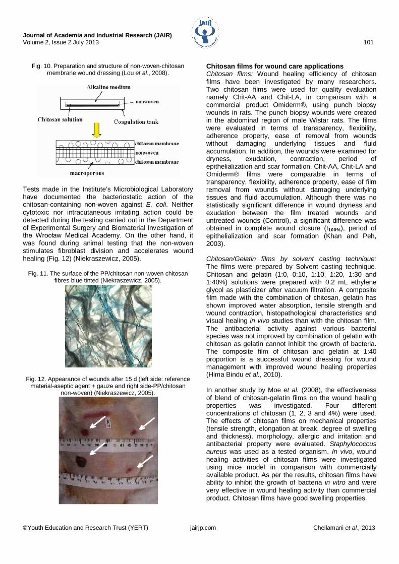
Chitosan non-woven dressing: Different tencel/cotton ratios were used to make non-woven dressings. The Tencel/cotton non-woven with chitosan was prepared by immersion-precipitation phase-inversion using a casting process (Fig. 10). First, the tencel/cotton non-woven was immersed into the chitosan solution with a concentration of 3.0 wt% (Acetic acid aqueous solution 1.0 wt% was used as a solvent) and frozen for 3 h. After defrosting, the non-woven chitosan dressing was immersed into NaOH aqueous solution for 24 h, then washed with deionized water repeatedly. Then, the tencel/cotton non-wovens treated with chitosan samples were subjected to quality evaluation. The results showed that, the non-woven-chitosan membrane exhibited controlled evaporative water loss and promoted fluid drainage ability. They also did not possess antigenicity and toxicity (Lou et al., 2008). In cooperation with the Institute of pulp and papermaking in Lodz, a polypropyl-ene-chitosan non-woven was prepared according to a wet paper method (Fig. 11).
a b
Journal of Academia and Industrial Research (JAIR) Volume 2, Issue 2 July 2013 101
©Youth Education and Research Trust (YERT) jairjp.com Chellamani et al., 2013
Fig. 10. Preparation and structure of non-woven-chitosan
membrane wound dressing (Lou et al., 2008).
Tests made in the Institute’s Microbiological Laboratory have documented the bacteriostatic action of the chitosan-containing non-woven against E. coli. Neither cytotoxic nor intracutaneous irritating action could be detected during the testing carried out in the Department of Experimental Surgery and Biomaterial Investigation of the Wrocław Medical Academy. On the other hand, it was found during animal testing that the non-woven stimulates fibroblast division and accelerates wound healing (Fig. 12) (Niekraszewicz, 2005).
Fig. 11. The surface of the PP/chitosan non-woven chitosan fibres blue tinted (Niekraszewicz, 2005).
Fig. 12. Appearance of wounds after 15 d (left side: reference
material-aseptic agent + gauze and right side-PP/chitosan non-woven) (Niekraszewicz, 2005).
Chitosan films for wound care applications Chitosan films: Wound healing efficiency of chitosan films have been investigated by many researchers. Two chitosan films were used for quality evaluation namely Chit-AA and Chit-LA, in comparison with a commercial product Omiderm®, using punch biopsy wounds in rats. The punch biopsy wounds were created in the abdominal region of male Wistar rats. The films were evaluated in terms of transparency, flexibility, adherence property, ease of removal from wounds without damaging underlying tissues and fluid accumulation. In addition, the wounds were examined for dryness, exudation, contraction, period of epithelialization and scar formation. Chit-AA, Chit-LA and Omiderm® films were comparable in terms of transparency, flexibility, adherence property, ease of film removal from wounds without damaging underlying tissues and fluid accumulation. Although there was no statistically significant difference in wound dryness and exudation between the film treated wounds and untreated wounds (Control), a significant difference was obtained in complete wound closure (t100%), period of epithelialization and scar formation (Khan and Peh, 2003). Chitosan/Gelatin films by solvent casting technique: The films were prepared by Solvent casting technique. Chitosan and gelatin (1:0, 0:10, 1:10, 1:20, 1:30 and 1:40%) solutions were prepared with 0.2 mL ethylene glycol as plasticizer after vacuum filtration. A composite film made with the combination of chitosan, gelatin has shown improved water absorption, tensile strength and wound contraction, histopathological characteristics and visual healing in vivo studies than with the chitosan film. The antibacterial activity against various bacterial species was not improved by combination of gelatin with chitosan as gelatin cannot inhibit the growth of bacteria. The composite film of chitosan and gelatin at 1:40 proportion is a successful wound dressing for wound management with improved wound healing properties (Hima Bindu et al., 2010). In another study by Moe et al. (2008), the effectiveness of blend of chitosan-gelatin films on the wound healing properties was investigated. Four different concentrations of chitosan (1, 2, 3 and 4%) were used. The effects of chitosan films on mechanical properties (tensile strength, elongation at break, degree of swelling and thickness), morphology, allergic and irritation and antibacterial property were evaluated. Staphylococcus aureus was used as a tested organism. In vivo, wound healing activities of chitosan films were investigated using mice model in comparison with commercially available product. As per the results, chitosan films have ability to inhibit the growth of bacteria in vitro and were very effective in wound healing activity than commercial product. Chitosan films have good swelling properties.

Journal of Academia and Industrial Research (JAIR) Volume 2, Issue 2 July 2013 102
©Youth Education and Research Trust (YERT) jairjp.com Chellamani et al., 2013
Moreover, the results have also shown that the chitosan films do not cause any unwilling symptoms (allergic or irritation in skin test). Conclusion Chitosan treated wound dressings have generated considerable interest in medical textiles and wound care management. Chitosan is a safety, eco-friendly substance for wound care system which makes an excellent candidate for the medical applications like promoting tissue growth, accelerating wound-healing, bone regeneration, etc. Wound healing needs an integrated approach and regular monitoring. Chitosan treated dressings play a major role in the wound-healing process and helps for the successful management of a wound. Acknowledgements Authors are thankful to Dr. Prakash Vasudevan, Director, SITRA for his keen interest in this study. Authors acknowledge with thanks the contributions made by research workers in the field of ‘Wound care dressings using chitosan coating’ not only in India but throughout the world, without which this review would not have been completed. References 1. Aoyagi, S., Onishi, H. and Machida, Y. 2007. Novel
chitosan wound dressing loaded with minocycline for the treatment of severe burn wounds. Int. J. Pharm. 330 (1-2): 138-145.
2. Gupta, B., Agarwal, R. and Alam, M.S. 2010. Textile based smart wound dressing. Ind. J. Fibre Text. 35: 174-187.
3. Hima Bindu, T.V.L., Vidyavathi, M., Kavitha, K., Sastry, T.P. and Suresh Kumar, R.V. 2010. Preparation and evaluation of chitosan-gelatin composite films for wound healing activity. Trends Biomater. Artif. Organs. 24(3): 123-130.
4. Khan, T.A. and Peh, K.K. 2003. A preliminary investigation of chitosan film as dressing for punch biopsy wounds in rats. Int. J. Pharm. Pharm. Sci. 6(1): 20-26.
5. Kucharska, M., Ciechańska, D., Niekraszewicz, A., Wiśniewska Wrona, M. and Kardas, I. 2010. Potential use of chitosan–based material in medicine. Prog. Chem. Appl. Chitin Derivatives. 15: 169-176.
6. Lee, D.W., Lim, H., Chong, H.N. and Shim, W.S. 2009.
Advances in chitosan material and its hybrid derivatives: A review. Open Biomater. J. 1: 10-20.
7. Lou, C.W., Lin, C.W., Chen, Y.S., Yao, C.H., Lin, Z.S., Chao, C.Y. and Lin, J.H. 2008. Dressing properties evaluation of tencel/cotton nonwoven fabric coated with chitosan for wound. Text. Res. J. 78(3): 248-253.
8. Moe, T.S., Khaing, T.A., Han, T.Z. and Mon, H.M. 2008. Effects of chitosan films on wound healing and evaluation of their properties, The 3rd GMSARN Int. Conf. 2008, November 12-14, 2008, Kunming, China.
9. Niekraszewicz, A. 2005. Chitosan medical dressings. Fibres Text. East Eur. 13(6): 16-18.
10. Paul, W. and Sharma, C.P. 2004. Chitosan and alginate wound dressings: A short review. Trends Biomater. Artif. Organs. 18(1): 18-23.
11. Phaechamud, T. and Charoenteeraboon, J. 2008. Antibacterial activity and drug release of chitosan sponge containing Doxycycline Hyclate. AAPS. J. 9(3): 829-835.
12. Rajendran, S. 2009. Advanced textiles for wound care, Wood Head Publishing Ltd, CRC press. pp.1-175.
13. Sajeev, U.S., Anoop Anand, K., Deepthy Menon and Shanti Nair. 2008. Control of nanostructures in PVA, PVA/chitosan blends and PCL through electrospinning. B. Mater. Sci. 31(3): 343-351.
14. Schwartz, M. 2008. Smart materials, CRC Press. pp.10.1-10.17.
15. Shanmugasundaram, O. L., Giridev, V.R., Neelakandan, R., Madhusoothanan, M. and Suseela Rajkumar, G. 2006. Drug release and antimicrobial studies on chitosan-coated cotton yarns. Ind. J. Fibre. Text. 31(4): 543-547.
16. Shanmugasundaram, O.L. 2006. Chitosan coated cotton yarn and its effect on antimicrobial activity. J. Text. Apparel Technol. Management. 5(3): 1-6.
17. Shanmugasundaram, O.L. and Gowda, R.V.M. 2011. Development and characterization of bamboo gauze fabric coated with polymer and drug for wound healing. Fiber Polym. 12(1): 15-20.
18. Sun, Z.H. and Li, K. 2011. Preparations, properties and applications of chitosan based nanofibers fabricated by electrospinning. Express Polym. Lett. 5(4): 342-361.
19. Vangenhove, L. 2007. Smart textiles for medicine and healthcare materials, system and applications, Wood Head Publishing Ltd., CRC press. pp.27-104.