gluten sensitivity gluten sensitivity and...
TRANSCRIPT

Year 2012Year 2012Year 2012Year 2012 Thesis N°Thesis N°Thesis N°Thesis N° 84
Gluten sensitivity Gluten sensitivity Gluten sensitivity Gluten sensitivity and neuropathiesand neuropathiesand neuropathiesand neuropathies
A Moroccan cA Moroccan cA Moroccan cA Moroccan caseaseasease----ccccontrol ontrol ontrol ontrol ststststudyudyudyudy
THESIS
Presented to The Medicine Faculty on ……,2012
BYBYBYBY Mr. Bouroumane Mohamed RédaMr. Bouroumane Mohamed RédaMr. Bouroumane Mohamed RédaMr. Bouroumane Mohamed Réda
Born on March 22nd, 1985 in Marrakesh
IN PARTIAL FULFILLEMENT OF THE REQUIREMENTS FOR THE DEGREE OF
DOCTOR OF MEDECINE
Key wordsKey wordsKey wordsKey words::::
Gluten sensitivity, neuropathies, antigliadin antibodies, prevalence.
JURYJURYJURYJURY
M. A. Alaoui Yazidi
Professor of Pneumology and Phthisiology
Director of PCIM research laboratory
M. B. Admou
Aggregate Professor of immunology
Member of PCIM research laboratory
M. N. Kissani
Professor of Neurology
M. M. Bourrous
Aggregate Professor of Neuro-Paediatrics
M. L. Chabaa
Professor of Biochemistry
M. M. Amine
Aggregate Professor of Epidemiology
Member of PCIM research laboratory
CHAIRMAN
ADVISOR
JUDGES
PCIMPCIMPCIMPCIM RRRResearch esearch esearch esearch
LaboratorLaboratorLaboratorLaboratory
UNIVERSITE CADI AYYAD
FACULTE DE MEDECINE ET DE PHARMACIE
MARRAKECH

The Prophet Mohamed (PBUH) said,
"There is no disease that Allah has created except that He
also has created its treatment, apart from aging»

At the time of being admitted as a member of the medical profession: I solemnly pledge myself to consecrate my life to the service of humanity; I will give to my teachers the respect and gratitude which is their due; I will practice my profession with conscience and dignity; the health of my patient will be my first consideration; I will maintain by all the means in my power, the honor and the noble traditions of the medical profession; my colleagues will be my brothers; I will not permit considerations of religion, nationality, race, party politics or social standing to intervene between my duty and my patient; I will maintain the utmost respect for human life from the time of conception, even under threat;

I will not use my medical knowledge contrary to the laws of humanity; I make these promises solemnly, freely and upon my honour.
Geneva Declaration 1948
LIST OF PROFESSORS LIST OF PROFESSORS LIST OF PROFESSORS LIST OF PROFESSORS
AT THE MEDICINE AT THE MEDICINE AT THE MEDICINE AT THE MEDICINE
FACULTY FACULTY FACULTY FACULTY OF OF OF OF
MARRAKESHMARRAKESHMARRAKESHMARRAKESH
---- CADI AYYADCADI AYYADCADI AYYADCADI AYYAD

UNIVERSITE CADI AYYAD FACULTE DE MEDECINE ET DE PHARMACIE
MARRAKECH
Doyen Honoraire : Pr. Badie-Azzamann MEHADJI
ADMINISTRATION
Doyen : Pr. Abdelhaq ALAOUI YAZIDI
Vice doyen à la recherche
Vice doyen aux affaires pédagogiques
: Pr. Badia BELAABIDIA
: Pr. Ag Zakaria DAHAMI
Secrétaire Général : Mr. Azzeddine EL HOUDAIGUI
PROFESSEURS D’ENSEIGNEMENT SUPERIEUR ABOUSSAD Abdelmounaim Néonatologie
AMAL Said Dermatologie
ASMOUKI Hamid Gynécologie – Obstétrique A
ASRI Fatima Psychiatrie
AIT BENALI Said Neurochirurgie
ALAOUI YAZIDI Abdelhaq Pneumo-phtisiologie
BENELKHAIAT
BENOMAR Ridouan Chirurgie – Générale
BELAABIDIA Badia Anatomie-Pathologique
BOUMZEBRA Drissi Chirurgie Cardiovasculaire
BOUSKRAOUI Mohammed Pédiatrie A

CHABAA Laila Biochimie
CHOULLI Mohamed Khaled Neuropharmacologie
ESSAADOUNI Lamiaa Médecine Interne
FIKRY Tarik Traumatologie- Orthopédie A
FINECH Benasser Chirurgie – Générale
KISSANI Najib Neurologie
KRATI Khadija Gastro-Entérologie
LATIFI Mohamed Traumato – Orthopédie B
MOUDOUNI Said mohammed Urologie
MOUTAOUAKIL Abdeljalil Ophtalmologie
RAJI Abdelaziz Oto-Rhino-Laryngologie
SARF Ismail Urologie
SBIHI Mohamed Pédiatrie B
SOUMMANI Abderraouf Gynécologie-Obstétrique A
PROFESSEURS AGREGES
ABOULFALAH Abderrahim Gynécologie – Obstétrique B
ADERDOUR Lahcen Oto-Rhino-Laryngologie
AMINE Mohamed Epidémiologie - Clinique
AIT SAB Imane Pédiatrie B
AKHDARI Nadia Dermatologie
BOURROUS Monir Pédiatrie A
CHELLAK Saliha Biochimie-chimie (Militaire)
DAHAMI Zakaria Urologie
EL ADIB Ahmed rhassane Anesthésie-Réanimation
EL FEZZAZI Redouane Chirurgie Pédiatrique
EL HATTAOUI Mustapha Cardiologie
ELFIKRI Abdelghani Radiologie (Militaire)
ETTALBI Saloua Chirurgie – Réparatrice et plastique
GHANNANE Houssine Neurochirurgie
LMEJJATI Mohamed Neurochirurgie
LOUZI Abdelouahed Chirurgie générale
LRHEZZIOUI Jawad Neurochirurgie(Militaire)
MAHMAL Lahoucine Hématologie clinique
MANOUDI Fatiha Psychiatrie
MANSOURI Nadia Chirurgie maxillo-faciale Et stomatologie
NAJEB Youssef Traumato - Orthopédie B
NEJMI Hicham Anesthésie - Réanimation
OULAD SAIAD Mohamed Chirurgie pédiatrique
SAIDI Halim Traumato - Orthopédie A
SAMKAOUI Mohamed Abdenasser
Anesthésie- Réanimation
TAHRI JOUTEI HASSANI Ali Radiothérapie
TASSI Noura Maladies Infectieuses
YOUNOUS Saïd Anesthésie-Réanimation
PROFESSEURS ASSISTANTS
ABKARI Imad Traumatologie-orthopédie B
ABOU EL HASSAN Taoufik Anesthésie - réanimation
ABOUSSAIR Nisrine Génétique
ADALI Imane Psychiatrie
ADALI Nawal Neurologie
ADMOU Brahim Immunologie
AGHOUTANE El Mouhtadi Chirurgie – pédiatrique
AISSAOUI Younes Anésthésie Reanimation (Militaire)
AIT BENKADDOUR Yassir Gynécologie – Obstétrique A
AIT ESSI Fouad Traumatologie-orthopédie B
ALAOUI Mustapha Chirurgie Vasculaire périphérique
(Militaire)
ALJ Soumaya Radiologie
AMRO Lamyae Pneumo - phtisiologie
ANIBA Khalid Neurochirurgie
ARSALANE Lamiae Microbiologie- Virologie (Militaire)
BAHA ALI Tarik Ophtalmologie
BAIZRI Hicham Endocrinologie et maladies métaboliques
(Militaire)
BASRAOUI Dounia Radiologie
BASSIR Ahlam Gynécologie – Obstétrique B
BELBARAKA Rhizlane Oncologie Médicale
BELKHOU Ahlam Rhumatologie
BEN DRISS Laila Cardiologie (Militaire)
BENCHAMKHA Yassine Chirurgie réparatrice et plastique
BENHADDOU Rajaa Ophtalmologie
BENHIMA Mohamed Amine Traumatologie-orthopédie B
BENJILALI Laila Médecine interne
BENZAROUEL Dounia Cardiologie
BOUCHENTOUF Rachid Pneumo-phtisiologie (Militaire)
BOUKHANNI Lahcen Gynécologie – Obstétrique B
BOURRAHOUAT Aicha Pédiatrie
BSSIS Mohammed Aziz Biophysique
CHAFIK Aziz Chirurgie Thoracique (Militaire)
CHAFIK Rachid Traumatologie-orthopédie A
CHERIF IDRISSI EL
GANOUNI Najat Radiologie
DAROUASSI Youssef Oto-Rhino – Laryngologie (Militaire)
DIFFAA Azeddine Gastro - entérologie
DRAISS Ghizlane Pédiatrie A
EL AMRANI Moulay Driss Anatomie
EL ANSARI Nawal Endocrinologie et maladies métaboliques
EL BARNI Rachid Chirurgie Générale (Militaire)
EL BOUCHTI Imane Rhumatologie
EL BOUIHI Mohamed Stomatologie et chirurgie maxillo faciale
EL HAOUATI Rachid Chirurgie Cardio Vasculaire
EL HAOURY Hanane Traumatologie-orthopédie A
EL HOUDZI Jamila Pédiatrie B
EL IDRISSI SLITINE Nadia Pédiatrie (Néonatologie)
EL KARIMI Saloua Cardiologie
EL KHADER Ahmed Chirurgie Générale (Militaire)
EL KHAYARI Mina Réanimation médicale
EL MANSOURI Fadoua Anatomie – pathologique (Militaire)
EL MEHDI Atmane Radiologie

EL MGHARI TABIB Ghizlane Endocrinologie et maladies métaboliques
EL OMRANI Abdelhamid Radiothérapie
FADILI Wafaa Néphrologie
FAKHIR Bouchra Gynécologie – Obstétrique B
FAKHIR Anass Histologie -embyologie cytogénétique
FICHTALI Karima Gynécologie – Obstétrique B
HACHIMI Abdelhamid Réanimation médicale
HAJJI Ibtissam Ophtalmologie
HAOUACH Khalil Hématologie biologique
HAROU Karam Gynécologie – Obstétrique A
HOCAR Ouafa Dermatologie
JALAL Hicham Radiologie
KADDOURI Said Médecine interne (Militaire)
KAMILI El ouafi el aouni Chirurgie – pédiatrique générale
KHALLOUKI Mohammed Anesthésie-Réanimation
KHOUCHANI Mouna Radiothérapie
KHOULALI IDRISSI Khalid Traumatologie-orthopédie (Militaire)
LAGHMARI Mehdi Neurochirurgie
LAKMICHI Mohamed Amine Urologie
LAKOUICHMI Mohammed Chirurgie maxillo faciale et Stomatologie
(Militaire)
LAOUAD Inas Néphrologie
LOUHAB Nissrine Neurologie
MADHAR Si Mohamed Traumatologie-orthopédie A
MAOULAININE Fadlmrabihrabou Pédiatrie (Néonatologie)
MARGAD Omar Traumatologie – Orthopédie B
MATRANE Aboubakr Médecine Nucléaire
MOUAFFAK Youssef Anesthésie - Réanimation
MOUFID Kamal Urologie (Militaire)
MSOUGGAR Yassine Chirurgie Thoracique
NARJIS Youssef Chirurgie générale
NOURI Hassan Oto-Rhino-Laryngologie
OUALI IDRISSI Mariem Radiologie
OUBAHA Sofia Physiologie
OUERIAGLI NABIH Fadoua Psychiatrie (Militaire)

QACIF Hassan Médecine Interne (Militaire)
QAMOUSS Youssef Anesthésie - Réanimation (Militaire)
RABBANI Khalid Chirurgie générale
RADA Noureddine Pédiatrie
RAIS Hanane Anatomie-Pathologique
ROCHDI Youssef Oto-Rhino-Laryngologie
SAMLANI Zouhour Gastro - entérologie
SORAA Nabila Microbiologie virologie
TAZI Mohamed Illias Hématologie clinique
ZAHLANE Mouna Médecine interne
ZAHLANE Kawtar Microbiologie virologie
ZAOUI Sanaa Pharmacologie
ZIADI Amra Anesthésie - Réanimation
ZOUGAGHI Laila Parasitologie –Mycologie

DEDICATIONSDEDICATIONSDEDICATIONSDEDICATIONS

To the joy of my life,
My mother Mrs. Chaaibi Saida,
My father
Mr. Bouroumane Abdesslam
My sister
Mrs. Bouroumane Meryem
And all my family
The Bouroumanes, the Chaaibis
and the Meskanis

ACKNOWLEDGEMENTSACKNOWLEDGEMENTSACKNOWLEDGEMENTSACKNOWLEDGEMENTS

I am grateful to several people for the role they have played to enable me
undertake and accomplish my studies and dissertation.
In particular, I extend my deepest appreciation to my Thesis Advisor Prof.
Admou Ibrahim, for his unwavering support and enthusiasm during the
entire research period, which enabled me to carry out and complete this
project. I enjoyed his guidance, his modesty and positive criticisms at every
stage of the project. His availability for consultation at any time, weekdays
and weekends, day and night ensured the smooth completion of my project,
and confirmed that I was not working alone.
I am grateful to my University supervisors,
Prof. Najib Kissani, Head of Neurology Department at the University Hospital
of Marrakesh who granted me the permission to work on the patients
hospitalized in the Department and was always ready for help and support.
Prof. Mohamed Amine, Head of Epidemiology Laboratory at the Medicine
Faculty of Marrakesh and Prof. Laila Chabaa, Head of Bio-Chemistry
Laboratory at the University Hospital of Marrakesh also deserve my sincerest
thanks. Their friendship and professional assistance has meant more to me
than I could ever express which helped me overcome the various challenges
of the research project.
I am also very grateful to Prof. Mounir Bourouss, Professor of Neuro-
Paediatrics at the University Hospital of Marrakesh for his invaluable friendly
assistance and support throughout my studies and doctorial process.
This work would not have been possible without the dedicated efforts by
Pr. Janati Idrissi, Director of Ibn-Sina Military Hospital of Marrakesh,
Morocco, and Dr. Othmani, the head of Transfusion Center in the same
hospital, for allowing me to include blood donors from the Transfusion
Center of Ibn-Sina Military Hospital. Besides, I am grateful to its entire staff
for their technical help and assistance.
The staff members of the Neurology Department and Bio-Chemistry
Laboratory were also very helpful, because I often consulted for their
technical advice for the patients’ selection and the immunological screening
process. Hence, special thanks goes to Prof. Nissrine louhab, Dr. Chraa
Mohamed, Dr. Yassine Mebrouk and Dr. Safaa Zahlane, Mrs. Badia Bouzari,
Mrs. Saida Hilm, Mrs. Siham sati and Mrs. Nadia Khalil. Also, I am grateful for
the contribution and support of Dr. Benhiba Imane, Mr. Moumen and Mr.
Sakoura.

Many thanks also go to Mr. Mohamed Lahjouji, Director of Morocco
Diagnostic System Company and member of Orgentec Diagnostika GmbH
(Germany), for sponsoring this project.
Finally, I wish to also acknowledge Prof. Sidihida Abderrahman ( English
Teacher at the American Language Center of Marrakesh) for the literary
revision and assistance, and Ms. Rabiaa Goughri for checking up on the
design of this document with creativity and enthusiasm.

ABBREVIATIONSABBREVIATIONSABBREVIATIONSABBREVIATIONS

AGA : Antigliadin antibodies
AHD : Anterior horn disease
ALS : Amyotrophic lateral sclerosis
CD : Celiac disease
CSF : Cerebro-spinal fluid
CPK : Creatine Phospho-kinase
DGP : deamidated gliadin peptides
DM : Dermato-myositis
EMA : Anti-endomysium antibodies
EMG : Electro-myography
Gamma GT : Gamma glutamyl transferase
GFD : Gluten free diet
GS : Gluten sensitivity
HLA : Human leukocyte Antigen
IELs : Intra-epithelial lymphocytes
IFN : Interferon
IgA : Immunoglobulin A
IgG : Immunoglobulin G
IIM : Inflammatory idiopathic myopathy
IL-15 : interleukine-15
M/F : Male to female
MICA : non-classical MHC molecules
MRI : Magnetic resonance imaging
MS : Multiple sclerosis
MSA : Multiple system atrophy
NKG2D : Natural killer receptor
NP : Not provided
PM : polymyositis
PN : Peripheral neuropathy
PNKD paroxysmal non-kinesigenic dystonia

PT : Prothrombin time
Sd : Syndrome
s-IBM : Sporadic inclusion body myositis
SIgAD : Selective IgA deficiency
SR : Sedimentation rate
TCR : T cell receptor
tTGA : tissue transglutaminase antibodies
WBC : White blood cells

Gluten sensitivity and neuropathiesGluten sensitivity and neuropathiesGluten sensitivity and neuropathiesGluten sensitivity and neuropathies---- A Moroccan case control studyA Moroccan case control studyA Moroccan case control studyA Moroccan case control study
1
SUMMARYSUMMARYSUMMARYSUMMARY

Gluten sensitivity and neuropathiesGluten sensitivity and neuropathiesGluten sensitivity and neuropathiesGluten sensitivity and neuropathies---- A Moroccan case control studyA Moroccan case control studyA Moroccan case control studyA Moroccan case control study
2
Summary 1111
Introduction 3
I. Definitions 4
II. Gluten sensitive enteropathy 5
1. Historical review 5
2. Physiopatholog3 6
3. Epidemiology 12
4. Diagnosis 14
III. Gluten sensitive neuropathies 24
1. Epidemiological data 24
2. Physiology of neural damage 25
3. Pathology of neural damage 26
4. Neurological categories 27
5. Serology testing 33
Patients and Methods 35
Results 38
I. Global data 39
II. Ischemic stroke 49
II. Peripheral neuropathy 51
III. Ataxia 53
IV. Epilepsy 55
V. Myopathy 57
VI. Myelopathy 58
VII. Cerebral thrombophlebitis 59
VIII. Multiple sclerosis 59
IX. Other neurological conditions 60
Discussion 61
I. Relevance of immunological testing 62
II. Socio demographic characteristics of the positive cases 62
III. Epidemiological analysis 63
IV. Clinical and investigations data 69
V. Immunological Investigations 78
Conclusion 88
Abstracts 90
Annex 94
References 99

Gluten sensitivity and neuropathiesGluten sensitivity and neuropathiesGluten sensitivity and neuropathiesGluten sensitivity and neuropathies---- A Moroccan case control studyA Moroccan case control studyA Moroccan case control studyA Moroccan case control study
3
INTRODUCTIONINTRODUCTIONINTRODUCTIONINTRODUCTION

Gluten sensitivity and neuropathiesGluten sensitivity and neuropathiesGluten sensitivity and neuropathiesGluten sensitivity and neuropathies---- A Moroccan case control studyA Moroccan case control studyA Moroccan case control studyA Moroccan case control study
4
IntroductionIntroductionIntroductionIntroduction
IIII.... DefinitionsDefinitionsDefinitionsDefinitions
Gluten sensitivity (GS) is a systemic autoimmune disease with diverse manifestations. This
disorder is characterized by a heightened immunological responsiveness to ingested gluten in
genetically susceptible individuals[2].
It represents a spectrum of diverse manifestations, one of which is gluten-sensitive enteropathy
(or celiac disease), dermatopathy (dermatitis herpetiformis) and neurological disorders (such as
gluten ataxia and neuropathy)[2].
The term celiac disease (CD) should now be restricted to describe gluten-sensitive enteropathy
(including the triad of villous atrophy, crypt hyperplasia and increased intraepithelial
lymphocytes on histological examination of the small-bowel mucosa).
Although neurological manifestations in patients with established CD have been reported since
1966, it was not until 30 years later that, in some individuals, GS was shown to manifest
exclusively with neurological dysfunction. Furthermore, the concept of extra-intestinal
presentations without enteropathy has only recently become accepted[2].
Hadjivassiliou reports in a prospective study on 145 cases of idiopathic axonal neuropathies a
prevalence of 34% of gluten sensitivity. Also, in a study realized on 71 celiac patients, 22,5%
developed neurologic disorders[3]. In addition, other studies showed unusual frequency of

Gluten sensitivity and neuropathiesGluten sensitivity and neuropathiesGluten sensitivity and neuropathiesGluten sensitivity and neuropathies---- A Moroccan case control studyA Moroccan case control studyA Moroccan case control studyA Moroccan case control study
5
CD’s immunologic markers in the idiopathic cerebellar ataxia and peripheral neuropathies with
undetermined etiology[4, 5]. Besides, the prevalence of IgG anti-gliadin antibodies is found to be
57% in idiopathic neuropathies versus 5% in the neuropathies with known aetiology and 12% in
the healthy controls[6].
Otherwise, many researchers shed the light on the efficacy of gluten free diet (GFD) as a
potential treatment for gluten sensitivity linked pathologies[7].
The high frequency of the GS within the population, the impact on the treatment and the absence
of any research about the relation between GS and neuropathies in Morocco motivated us to
carry out this study.
We take the celiac disease as a prototype of the GS in order to describe the physiopathology, and We take the celiac disease as a prototype of the GS in order to describe the physiopathology, and We take the celiac disease as a prototype of the GS in order to describe the physiopathology, and We take the celiac disease as a prototype of the GS in order to describe the physiopathology, and
immunology and pathology and diagnostic timmunology and pathology and diagnostic timmunology and pathology and diagnostic timmunology and pathology and diagnostic tools. ools. ools. ools.
IIIIIIII.... Gluten sensitive enteropathyGluten sensitive enteropathyGluten sensitive enteropathyGluten sensitive enteropathy
1111.... Historical review:Historical review:Historical review:Historical review:
The gluten sensitive enteropathy was first described by Samuel Gee in 1888, a similar
description of malabsorption syndrome by Arateus and Cappadocie (turkey) has been performed
since the 2nd century AD.
The etiology was unknown until a Netherland’s pediatrician,Willem K.Dicke, showed a relation
between the consummation of bread and cereals and chronic diarrhea.
This observation was reinforced during the World War II, when doctors witnessed the
improvement of patients when bread was excluded from soldiers’ food[6].

Gluten sensitivity and neuropathiesGluten sensitivity and neuropathiesGluten sensitivity and neuropathiesGluten sensitivity and neuropathies---- A Moroccan case control studyA Moroccan case control studyA Moroccan case control studyA Moroccan case control study
6
Dick and Van de Kamer made series of experiments, exposing celiac children to diets with a
follow-up of the faeces’ weight and faecal fats in order to measure the malabsorption.
Wheat, rye, barley and oats caused a malabsorption syndrome that could be inversed after
exclusion of these “toxic” cereals. Short after this observation, the role of gluten as a trigger of
this toxicity has been confirmed[8].
The lesion on the proximal duodenum was first described in 1954; the first descriptions were a
mucosa inflammation, cryptic hyperplasia and villous flattening[9]. The first duodenal biopsy was
performed on a child in 1957[10].
The antibodies against transglutaminase II were identified 10 years ago.
The toxic sequences of gluten were also deciphered 10 years ago, (more than 100 different
peptides)
2222.... Physiopathology:Physiopathology:Physiopathology:Physiopathology:
Celiac disease is an inflammatory multifactor disorder of the small intestine caused by an
immune response to ingested wheat gluten and similar proteins of rye and barley.
Data accumulated since the discovery of gluten specific T cells in the intestine of celiac disease
patients the early 1990s have allowed the deciphering of the interplay between the triggering
environmental factor, gluten, the main genetic risk factor, the HLA-DQ2/8 haplotypes and the
auto-antigen: the enzyme tissue transglutaminase (tTG).
More recent work points to an important contribution of innate immunity triggered by a distinct
gluten peptide and driven by the pro-inflammatory cytokine Interleukine-5 (IL-15)[11].

Gluten sensitivity and neuropathiesGluten sensitivity and neuropathiesGluten sensitivity and neuropathiesGluten sensitivity and neuropathies---- A Moroccan case control studyA Moroccan case control studyA Moroccan case control studyA Moroccan case control study
7
2222....1111 Genetic aspects:
A high prevalence (10%) among first-degree relatives of CD patients indicates that susceptibility
to develop CD is strongly influenced by inherited factors[12]. Familial clustering is stronger in CD
than in most other chronic inflammatory diseases with a multi-factorial etiology. The strong
genetic influence in CD is further supported by a high concordance rate (75%) in monozygotic
twins[13].
Both HLA and non-HLA genes contribute to the genetic predisposition. The presence of certain
HLA genes appears to be necessary but sufficient for CD development. The characteristic of the
HLA association suggest that the HLA genes are involved in a process that controls CD
development[14].
For HLA genes, most CD patients carry the DR3-DQ2 haplotype (the DRB1*0301-DQA1*0501-
DQB1*0201 haplotype), or are DR5-DQ7/DR7-DQ2 heterozygotes. Available genetic and
functional data favor DQ8 as the major susceptibility determinant in these patients[15].
Otherwise, much less is known about non-HLA genes in this disorder. There are several reports
that imply involvement of the gene for the negative co-stimulatory molecule CTLA4 (cytotoxic T
lymphocyte associated-4), or a neighbouring gene (such as those encoding CD28 or ICOS);
however, the overall effect of this gene is small[16].
The region that has most consistently been linked to CD is on the long arm of chromosome 5
(5q31-33)[17, 18]; also, there is accumulating evidence for a susceptibility factor on
chromosome 11q32 and on chromosome 19p13 [18].

Gluten sensitivity and neuropathiesGluten sensitivity and neuropathiesGluten sensitivity and neuropathiesGluten sensitivity and neuropathies---- A Moroccan case control studyA Moroccan case control studyA Moroccan case control studyA Moroccan case control study
8
2222....2222 Toxic fractions of gluten:
Wheat gluten is a complex mixture of at least 100 related proteins. The major components of
gluten are the gliadins and glutenins which can both be subdivided into distinct protein families
[14]. The gliadin is composed of monomeric proteines, subdivided into 4 groups (α, β, γ and ω).
The Gliadin A (N-terminal region of fraction α) is suspected to be the toxic molecule resulting in
the majority of the troubles linked to the CD.
As wheat is used in many food products, exposure to relatively large amounts of gluten starts
very early. Usually gluten is introduced into the diet at the age of 6 months and a child of 12
months age eats between 6 and 9 g gluten daily[19].
2222....3333 The immunologic response in CD:
aaaa.... The role of the enzyme tissue transglutaminaseThe role of the enzyme tissue transglutaminaseThe role of the enzyme tissue transglutaminaseThe role of the enzyme tissue transglutaminase
The enzyme tissue transglutaminase (tTG), or transglutaminase 2, is expressed by almost all cell
types and is usually retained intra-cellularly in an enzymatically inactive form. It can be released
to the extracellular space to become associated with the extracellular matrix[20, 21] and this
release is increased when cells are under mechanical or inflammatory stress.
The tTG belongs to a family of at least eight calcium-dependent transamidating enzymes that
catalyze the covalent and irreversible cross-linking of a protein with a glutamine residue
(glutamine donor) to a second protein with a lysine residue (glutamine acceptor), resulting in the
formation of a 3-(gamma-glutamyl)-lysine isopeptide bond[22].
The tTG is only active in the presence of high calcium concentrations, as are found in the

Gluten sensitivity and neuropathiesGluten sensitivity and neuropathiesGluten sensitivity and neuropathiesGluten sensitivity and neuropathies---- A Moroccan case control studyA Moroccan case control studyA Moroccan case control studyA Moroccan case control study
9
extracellular space, where it contributes to the stabilization of the extracellular matrix[23].
However, intracellular activation and subsequent cross- linking can also occur when cellular
integrity is lost and extracellular calcium floods the cell, as found in apoptosis.
Under certain conditions, when no primary lysines are available as glutamine acceptors or at low
pH, as can prevail in intestinal inflammation, tTG merely deamidates a target glutamine in the
substrate protein, transforming the neutral glutamine to a negatively charged glutamic acid
residue[19].
bbbb.... The Gluten specificThe Gluten specificThe Gluten specificThe Gluten specific TTTT----Cell responseCell responseCell responseCell response
The DQ2 and DQ8 molecules predispose to CD by preferential presentation of gluten peptides to
CD-4 T-cells in the lamina propria. HLA-DQ2 and HLA-DQ8 preferentially bind peptides that
contain amino acids with a negative charge, whereas such peptides are not found in the gluten
molecules. It was found that the enzyme tissue transglutaminase (tTG), the target of the
autoantibodies in CD, can modify gluten peptides, which introduces the negative charges
required for binding to HLA-DQ-molecules[24, 25].
Thus, ingested gluten molecules are degraded to peptides by gastrointestinal enzymes, modified
by tTG, bind to HLA-DQ2 or HLA-DQ8, and trigger an inflammatory T cell response.
This inflammatory reaction stimulates the production of antigliadin antibodies (AGA) and anti TG
antibodies IgA and IgG.
In 1998, the identity of the first gluten peptides that were recognised by such T cells was
reported[26](Table I). The now known source proteins for these T cell stimulatory peptides are
the a-gliadins, g-gliadins and the low and high molecular weight glutenins[27, 28].
Gluten sensitivity and neuropathiesGluten sensitivity and neuropathiesGluten sensitivity and neuropathiesGluten sensitivity and neuropathies---- A Moroccan case control studyA Moroccan case control studyA Moroccan case control studyA Moroccan case control study
10
Figure I:Figure I:Figure I:Figure I: Depiction of the intestinal mucosa with emphasis on the factors taking part in the Depiction of the intestinal mucosa with emphasis on the factors taking part in the Depiction of the intestinal mucosa with emphasis on the factors taking part in the Depiction of the intestinal mucosa with emphasis on the factors taking part in the
development and control of celiac disease.development and control of celiac disease.development and control of celiac disease.development and control of celiac disease. ((((aaaa)))) The parts of gluten which are resistant to luminal and brush border enzymes will survive digestion, and can be
transported across the epithelial barrier as polypeptides.
- Gluten peptides are deamidated by tissue transglutaminase (tTG or TG2), which, in the intestinal mucosa, is mainly
located extra-cellularly in the sub-epithelial region, but is also found in the brush border.
- CD4C T-cells in the lamina propria recognize predominantly deamidated gluten peptides, presented by HLA-DQ2 or -
DQ8 molecules on the cell surface of antigen presenting cells (APC).
- The activation of the CD4 T-cells triggers an inflammatory T cell response.
((((bbbb)))) Immunofluorescence staining of TG2 (red), HLA-DQ (green) and T cells (CD3; blue) in the small
Intestinal mucosa of an untreated celiac disease patient.
(Immunofluorescent image courtesy of H. Scott, Riksh
Table I: Amino acid sequence of Table I: Amino acid sequence of Table I: Amino acid sequence of Table I: Amino acid sequence of typical immune stimulating gluten peptides.typical immune stimulating gluten peptides.typical immune stimulating gluten peptides.typical immune stimulating gluten peptides. a-gliadin PQPQLPYPQ and PFPQPQLPY
g-gliadin FPQQPQQPF and PQQSFPQQQ Low molecular weight glutenin FSQQQQSPF
High molecular weight glutenin QGYYPTSPQ

Gluten sensitivity and neuropathiesGluten sensitivity and neuropathiesGluten sensitivity and neuropathiesGluten sensitivity and neuropathies---- A Moroccan case control studyA Moroccan case control studyA Moroccan case control studyA Moroccan case control study
11
CCCC.... The role of ILThe role of ILThe role of ILThe role of IL----15 in gluten sensitivity pathogenesis 15 in gluten sensitivity pathogenesis 15 in gluten sensitivity pathogenesis 15 in gluten sensitivity pathogenesis
The massive increase in IEL is considered a diagnostic criterion of GSE, and is not a usual feature
of inflammatory conditions in the small intestine.
The Gliadin-Peptides 31-49, common to the N-termini of a-gliadins, induce the production of
the cytokine IL-15 in epithelial cells and macrophages via -as yet unknown- relays[29].
In turn, IL-15 arms IEL by stimulating their cytotoxic properties and their expression of the
innate immune receptor NKG2D (the activating natural killer NK receptor also expressed by
intraepithelial T- cells)[30]. Furthermore, IL-15 induces the expression of MICA (non-classical
MHC molecules), the epithelial ligand of NKG2D. Binding of NKG2D to MIC can then trigger the
cytotoxicity of IEL against epithelial cells.[29, 30] .
Figure II : Mechanisms leading to the activation of IEL by IL-15 in celiac disease. NKG2D: natural killer receptor MICA : non-classical MHC molecules IL-15 : interleukine-15 TCR : T-Cell receptor IFN : Interferon IEL : Intra-epithelial lymphocytes

Gluten sensitivity and neuropathiesGluten sensitivity and neuropathiesGluten sensitivity and neuropathiesGluten sensitivity and neuropathies---- A Moroccan case control studyA Moroccan case control studyA Moroccan case control studyA Moroccan case control study
12
3. EpidemiologyEpidemiologyEpidemiologyEpidemiology
3333....1111.... The prevalence of GSE
The prevalence rate of GSE in the population generally depends on the forms of the disease.
Actually, the estimate frequency of combined undiagnosed (or silent form) and diagnosed (active
form) GSE was remarkably similar, between 0.7%–2% in most of the populations, including the
United States [31]. The prevalence of childhood GSE has been reported to be between 1:285 and
1:77 in Sweden, 1:99 and 1:67 in Finland, 1:230 and 1:106 in Italian schoolchildren. Generally,
similar rates have been reported for non-European white populations, such as New Zealand and
Australia[31].
In US adults, the prevalence varied from 1:1750 (clinically diagnosed GSE, including dermatitis
herpetiformis) to 1:105 (presence of IgA endomysial antibodies) [31].
GSE is virtually unknown in East Asian populations who also lack this HLA haplotype; however,
rates close to those in Europe have been reported from the Middle East and India. Although the
disease is believed to be rare in Africa (and in Afro-Americans), a highest prevalence (5,6%) has
been reported for Saharaouii in North Africa [31-34]; thus, the estimates were 1 :187 in Egypt
[35] and 1 :157 in Tunisia [36].
The estimates based on sero-epidemiologic studies suggest that for each diagnosed case of GSE,
there may be many undiagnosed cases [37] and that 1%–3% of the general population in Europe
and the United States becomes affected at some point in life. [31]
The prevalence was found to be higher among type 1 diabetics and patients with immunologic

Gluten sensitivity and neuropathiesGluten sensitivity and neuropathiesGluten sensitivity and neuropathiesGluten sensitivity and neuropathies---- A Moroccan case control studyA Moroccan case control studyA Moroccan case control studyA Moroccan case control study
13
pathologies, Down syndrome, Turner syndrome…etc [33, 38, 39].
The contrast between the high rate of the GSE and increased number of undiagnosed cases could
be explained by the frequency of asymptomatic forms and also by the fact that physicians don’t
usually think about this condition.
To sum up, GSE is a systemic disease affecting almost 1% of the general population.
3333....2222.... The incidence of CD
Population-based estimates of the incidence of small bowel biopsy (SBB)- confirmed GSE in
adults vary from 2–13/100,000 per year [40, 41]. Those rates have to be interpreted with caution
because many patients diagnosed as adults likely have had 20–60 years of untreated GSE, thus
hardly represent truly incident new cases of disease.
The recent raise in the incidence rates is likely due to increasing use of serologic screening
leading to diagnosis in milder cases. However, there is a paucity of incidence data that would
represent the full spectrum of disease, including silent and latent cases.[31]
The variation of nutrition regimens in early childhood and infants among populations and
differences in the prevalence of susceptible HLA alleles may explain inter-population variation in
the incidence of GSE[31].
The effects of nutritional practices on the risk and severity of GSE may also account for
geographic and temporal variation in the incidence and be of great public health importance. [31]
3333....3333.... The Progression
Over time, individuals progress from latent to silent or active disease and can reverse to the

Gluten sensitivity and neuropathiesGluten sensitivity and neuropathiesGluten sensitivity and neuropathiesGluten sensitivity and neuropathies---- A Moroccan case control studyA Moroccan case control studyA Moroccan case control studyA Moroccan case control study
14
latent subclinical state on a strict GFD [31].
The higher frequencies are registered in two periods of age :
-The first one is ranging between 1 and 5 years old, maximum at 2 years old.[42]
-The second one in adulthood, with a slight sex difference: between the 3rd and 5th decade for
females, and later for males [42].
3333....4444.... Gender
Most studies report the female predominance in childhood. The male to female sex ratio ranges
between 1/1 and ½; however, the complications are more likely to happen with males[42]. In the
region of Marrakesh, series of studied patients showed that 58,62% of GSE patients were females
[33].
4. DiagnosisDiagnosisDiagnosisDiagnosis
The presumption of GSE is mainly clinical; however, the high rate of atypical or asymptomatic
forms pushes to consider serologic and histologic parameters to establish the diagnosis.
4.1 Clinical description[43]
GSE is diagnosed typically in early childhood around the age of 2 years and a second peak is
found around age of 40 years [43]
The clinical manifestations differ greatly, depending on each case and ranging from
asymptomatic to full blown CD. The severity of symptoms is not necessarily proportional to the
severity of the mucosal lesions and patients with total atrophy can be asymptomatic or present
with subclinical symptoms such as iron deficiency or muscle cramps. Recently, more subjects
present with asymptomatic or mild GSE than with the classical symptoms of severe
malabsorption[43] .

Gluten sensitivity and neuropathiesGluten sensitivity and neuropathiesGluten sensitivity and neuropathiesGluten sensitivity and neuropathies---- A Moroccan case control studyA Moroccan case control studyA Moroccan case control studyA Moroccan case control study
15
Different forms of GSE are listed below (table II).
Table II : Definition states of GSE.
States of GSEStates of GSEStates of GSEStates of GSE DefinitionDefinitionDefinitionDefinition
Clinically
overt GSE
Typical gastrointestinal symptoms and signs of malabsorption. Histological
changes with villous atrophy and hypertrophic crypts.
Silent GSE Asymptomatic patients with typical histological changes
Asymptomatic
GSE
Same findings as in silent GSE.
Atypical GSE Extra-intestinal findings such as IgA-nephropathy symptoms. Typical
histological symptoms.
Latent GSE/
Potential GSE
Subjects with genetic predisposition who have initially a normal histology with
no atrophy or crypt hyperplasia. Immunological abnormalities such as
increased count of IELs and positive EMA or tTG-antibody tests are sometimes
present. These subjects may develop clinically overt GSE later in life.
Refractory
GSE
Patients who do not respond to gluten-free diet or who previously responded
but later become non-responsive to a gluten-free diet. Intestinal lymphoma
may have developed.
Symptoms begin at various times after the ingestion of gluten. Patients generally present with various
presentations (see below). Beside the typical symptoms mainly occurring in infants and young children
GSE may manifest at any age :

Gluten sensitivity and neuropathiesGluten sensitivity and neuropathiesGluten sensitivity and neuropathiesGluten sensitivity and neuropathies---- A Moroccan case control studyA Moroccan case control studyA Moroccan case control studyA Moroccan case control study
16
a. Typical presentation: [44]
� Chronic diarrhea
� Anorexia
� Abdominal distension
� Abdominal pain
� Poor weight gain
� Weight loss
� Vomiting
� Severe malnutrition can occur if the diagnosis is delayed
� Behavioral changes are common and include irritability and an introverted attitude
Rarely, severely affected infants present with a celiac crisis, which is characterized by explosive watery
diarrhea, marked abdominal distension, dehydration, hypotension, and lethargy, often with profound
electrolyte abnormalities, including severe hypokalemia.
The variability in the age of symptom onset possibly depends on the amount of gluten in the diet and
other environmental factors, such as duration of breast feeding.
o Extra-intestinal symptoms secondary to malabsorption: [43]
� Peripheral neuropathy (vitamin B12 and B1 deficiency)
� Anemia (iron, vitamin B12 and folate deficiency)
� Growth failure in children
� Bone pain (osteoporosis and osteopenia, vitamin D and calcium deficiency)
� Muscle cramps (magnesium and calcium deficiency)
� Night blindness (vitamin A deficiency)

Gluten sensitivity and neuropathiesGluten sensitivity and neuropathiesGluten sensitivity and neuropathiesGluten sensitivity and neuropathies---- A Moroccan case control studyA Moroccan case control studyA Moroccan case control studyA Moroccan case control study
17
� Weight loss (impaired absorption of most nutrients)
� Edema (Protein and albumin loss)
� Weakness (hypokalemia and electrolyte depletion)
� Bleeding and hematoma (vitamin K deficiency)
b. Atypical manifestations
� Neurological disorders such as peripheral neuropathies, ataxia, epilepsy.
� Dermatitis herpetoformis.
� Elevetad liver enzymes, liver failure.
� Infertility
� Stomatitis
� Myocarditis
� IgA nephritis
� Idiopathic pulmonary hemosiderosis
� Arthritis
c. Conditions associated with GSE
� Autoimmune diseases such such as type 1 diabetes, sjorgen syndrome, thyroid dieases
(Hashimoto’s thyroiditis and Graves’s disease), autoimmune and primary biliary cirrhosis.
� Selective IgA deficiency
� Turner’s syndrome
� Down’s syndrome

Gluten sensitivity and neuropathiesGluten sensitivity and neuropathiesGluten sensitivity and neuropathiesGluten sensitivity and neuropathies---- A Moroccan case control studyA Moroccan case control studyA Moroccan case control studyA Moroccan case control study
18
4.2 Serology Testing
The widespread availability of serologic tests has permitted disease to be considered and tested
for by any physician.
Serologic testing is recommended in different clinical situations showed in table (III)
Table III: Clinical indications for serologic testing
Unexplained, chronic diarrhea with and without malabsorption.
Unexplained weight loss.
Iron deficiency anemia
Folate deficiency
Vitamin E or K deficiency
Osteoporosis
Hypocalcaemia or vitamin D deficiency, secondary hyperparathyroidism
Unexplained elevation of transminases
First degree relatives of celiac patients
Associated autoimmune diseases: Type 1 diabetes, Sjorgen’s syndrome primary billiary
cirrhosis
Down and Turner syndromes
Neurologic disorders: Unexplained peripheral neuropathy, epilepsy and ataxia.
Serologic tests allowed to diagnose both classical and atypical forms of GSE and to specify
patients whom jejunal biopsy is required, to screen patients likely to develop GSE and evaluate
observance to GFD. The most sensitive tests are based on the use of IgA isotypes.
The available tests include antigliadin antibodies and anti-endomysial and/or tissue
transglutaminase antibodies).
The gold standard in celiac serologies remains the IgA endomysial antibody (EMA) with high
specificity for celiac disease that approaches 100%.
The endomysium is a protein located within the collagene of the human connective tissue and
monkey esophagus. The titer of EMA correlates with the degree of mucosal damage. Accordingly,

Gluten sensitivity and neuropathiesGluten sensitivity and neuropathiesGluten sensitivity and neuropathiesGluten sensitivity and neuropathies---- A Moroccan case control studyA Moroccan case control studyA Moroccan case control studyA Moroccan case control study
19
the sensitivity declines when greater number of patients with less degrees of villous flattening
are included in the studies[5]
The EMA is an observer-dependent immunoflourescence test that requires expertise in reading it
and the use of either primate esophagus or human umbilical cord as tissue substrate.
The IgA and IgG antigliadin and tTG antibodies are produced against deamidated gliadin
peptids.[45]
The use of an antigliadin IgA with a biopsy increases the rate of diagnosis of coeliac disease by
up to 20% [46]
The recognition of the tissue transglutaminase (tTG), an ubiquitary intracellular enzyme, as the
autoantigen for the EMA allowed development of an enzyme-linked immunoassay (ELISA) [47].
Initially the antigen in the assay was tTG derived from guinea pig liver (GP-tTG): subsequently
human tTG (H-tTG), either recombinant or derived from human red cells has replaced the assays
using GP-tTG.
Overall, the sensitivity of both the EMA and tTG is greater than 90%[48]. While the specificity of
the EMA is considered to be virtually 100%, the tTG test does not achieve that degree of
specificity.
There are numerous reports of positive tTG results in the absense of GSE. They may be seen in
Type 1 diabetes, chronic liver disease, psoriatic or rheumatoid arthritis and heart failure, though
biopsy has not been performed in most of these studies.[49-52]
a. Selective Iga deficiency
Selective IgA deficiency (SIgAD) occurs more commonly in patients with GSE than the general
population. As a result, patients with CD lack IgA-EMA, IgA-tTG and IgA-antigliadin
Gluten sensitivity and neuropathiesGluten sensitivity and neuropathiesGluten sensitivity and neuropathiesGluten sensitivity and neuropathies---- A Moroccan case control studyA Moroccan case control studyA Moroccan case control studyA Moroccan case control study
20
antibodies. In order to detect GSE in those with SIgAD, a total IgA level should be incorporated
into the testing for CD, as well as an IgG antibody-based test, either IgG-antigliadin or IgG-
tTG.[53, 54]
b. Seronegative celiac disease
Several studies have demonstrated that serologic studies may lack sensitivity when used in the
practice setting. Reliance on EMA as a single test has, in fact, underestimated the prevalence of
GSE by at least 20–25%. This is mainly due to the inclusion of patients with mild mucosal
changes, a situation when patients may not express an EMA. A similar situation occurs with
tissue transglutaminase, with titers decreasing as the mucosal lesion becomes less marked. [55,
56]
c. Role of HLA DQ2/DQ8 assessmentRole of HLA DQ2/DQ8 assessmentRole of HLA DQ2/DQ8 assessmentRole of HLA DQ2/DQ8 assessment
The HLA DQ2 is found in up to 90–95% of patients with GSE, while most of the remaining
patients are HLA DQ8. However, these HLA alleles are found in up to 40% of the general
population. They appear to be a necessary, but not sufficient, factor in the pathogenesis of
coeliac disease. The role of determining whether an individual carries HLA DQ2 or DQ8 in the
assessment of coeliac disease lies in their high negative predictive value.
The main roles are in determining whether family members require screening for coeliac disease,
in excluding coeliac disease when patients are already on a gluten-free diet and in the situation,
where the diagnosis of coeliac disease is unclear.[57-59].
Gluten sensitivity and neuropathiesGluten sensitivity and neuropathiesGluten sensitivity and neuropathiesGluten sensitivity and neuropathies---- A Moroccan case control studyA Moroccan case control studyA Moroccan case control studyA Moroccan case control study
21
Table IV : sensitivity and specificity of serology tests in children
Test performance (%) IgA-EMA IgA-tTGA IgA-AGA
Sensitivity
Specificity
Reproducibility
90
99
93
93
95
83
83
82
62
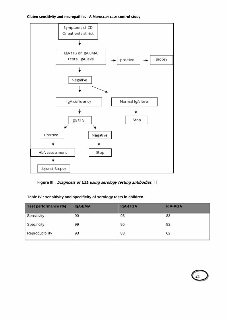
Figure IIIFigure IIIFigure IIIFigure III : Diagnosis of Diagnosis of Diagnosis of Diagnosis of GSEGSEGSEGSE using serology testing antibodiesusing serology testing antibodiesusing serology testing antibodiesusing serology testing antibodies.[1]
Gluten sensitivity and neuropathiesGluten sensitivity and neuropathiesGluten sensitivity and neuropathiesGluten sensitivity and neuropathies---- A Moroccan case control studyA Moroccan case control studyA Moroccan case control studyA Moroccan case control study
22
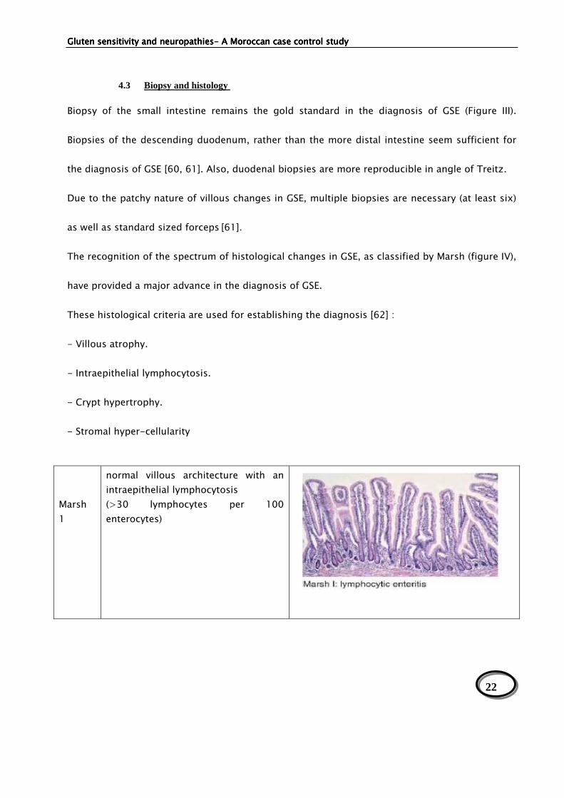
4.3 Biopsy and histology
Biopsy of the small intestine remains the gold standard in the diagnosis of GSE (Figure III).
Biopsies of the descending duodenum, rather than the more distal intestine seem sufficient for
the diagnosis of GSE [60, 61]. Also, duodenal biopsies are more reproducible in angle of Treitz.
Due to the patchy nature of villous changes in GSE, multiple biopsies are necessary (at least six)
as well as standard sized forceps [61].
The recognition of the spectrum of histological changes in GSE, as classified by Marsh (figure IV),
have provided a major advance in the diagnosis of GSE.
These histological criteria are used for establishing the diagnosis [62] :
- Villous atrophy.
- Intraepithelial lymphocytosis.
- Crypt hypertrophy.
- Stromal hyper-cellularity
Marsh
1
normal villous architecture with an
intraepithelial lymphocytosis
(>30 lymphocytes per 100
enterocytes)

Gluten sensitivity and neuropathiesGluten sensitivity and neuropathiesGluten sensitivity and neuropathiesGluten sensitivity and neuropathies---- A Moroccan case control studyA Moroccan case control studyA Moroccan case control studyA Moroccan case control study
23
Marsh
2
intraepithelial lymphocytosis is
accompanied by crypt hypertrophy
Marsh
3
Moderate to very severe reduction in villous height.
The majority of patients diagnosed with GSE (50–60%) fall into this category
Figure IV: Figure IV: Figure IV: Figure IV: HistoHistoHistoHisto----papapapathological classification of GSE according to Marsh criteria.thological classification of GSE according to Marsh criteria.thological classification of GSE according to Marsh criteria.thological classification of GSE according to Marsh criteria.

Gluten sensitivity and neuropathiesGluten sensitivity and neuropathiesGluten sensitivity and neuropathiesGluten sensitivity and neuropathies---- A Moroccan case control studyA Moroccan case control studyA Moroccan case control studyA Moroccan case control study
24
IIIIIIII.... Gluten sensitive neuropathiesGluten sensitive neuropathiesGluten sensitive neuropathiesGluten sensitive neuropathies
Gluten sensitivity is associated with multiple neurological abnormalities including gluten ataxia,
peripheral neuropathy, and ischemic stroke [63].
1. 1. 1. 1. Epidemiologic dataEpidemiologic dataEpidemiologic dataEpidemiologic data
There are no accurate estimates of the prevalence of the neurological expressions of GS in the
general population.[64]. Otherwise, the reported frequency of neurological abnormalities among
patients with established GSE varies between 10% to 22,5% [65, 66].
A japanese research showed that patients with cerebellar ataxia have significantly higher rates of
AGA positivity than patients without cerebellar ataxia or normal controls, which suggests that
ataxic patients are more likely to have gluten sensitivity [63].
In another study in Sheffield Hospital, UK, series of 500 patients with progressive ataxia were
evaluated over a period of 13 years and showed that 46,97 % (101/215) of patients with
idiopathic sporadic ataxia had serological evidence of gluten sensitivity [67]. The prevalence of
gluten ataxia was 20% among all patients with ataxias, 25% among patients with sporadic
ataxias, and 45% among those with idiopathic sporadic ataxias [67, 68].
Otherwise, peripheral neuropathy was found in up to 23% of patients with established GSE on a
gluten-free diet [69]. Also, in a large population-based study (84.000 participants) in Sweden
that examined the risk of neurological disease in patients with GSE, polyneuropathy was
significantly associated with GSE (odds ratio 5,4; 95% CI 3,6–8,2) [70]. Besides, a UK-based study
showed that 47 of 140 (34%) patients with idiopathic sporadic axonal neuropathy had circulating
AGA [71]. In an Italian study, a greater proportion of patients with various types of

Gluten sensitivity and neuropathiesGluten sensitivity and neuropathiesGluten sensitivity and neuropathiesGluten sensitivity and neuropathies---- A Moroccan case control studyA Moroccan case control studyA Moroccan case control studyA Moroccan case control study
25
neuropathies were positive for IgA anti-TG2 (68 of 330 ;21%) compared with controls (1of 68;
1,5 %; p<0�0001)[72]. Further more, a retrospective evaluation of 400 patients with neuropathy
showed the prevalence of GSE to be between 2,5% and 8% (compared with 1% in the healthy
population) [73].
Finally, rare cases of ischemic stroke occurring in young adults have lead to the diagnosis of GSE
[74, 75].
2. 2. 2. 2. PhysiologyPhysiologyPhysiologyPhysiology of neural damageof neural damageof neural damageof neural damage
The search for causes of neurological dysfunction in GS has largely ignored the immunological
aspect, and has concentrated on vitamin deficiencies (B12, E, D, folic acid, pyridoxine) as a result
of malabsorption [76]. Vitamin replacement rarely improves the neurological deficit [76, 77].
Alternative hypotheses are that antigliadin antibodies are more directly involved in the
neuropathological process, or are markers of autoimmune activity with an unidentified
neurotoxic antibody[76].
If these antibodies are directly or indirectly neurotoxic, why do patients with neurological
dysfunction and on gluten-free diet not always improve? One possibility is that damaged neural
tissue (e.g. cerebellar Purkinje cells) does not regenerate[76]; the second is that patients may not
strictly adhere to their gluten-free diet or that the diet may be insufficient to suppress the
immunological process completely, especially since patients without gastrointestinal symptoms
are unlikely to adhere to a gluten-free diet [76, 78].
Evidence suggests there might be antibody cross-reactivity between antigenic epitopes on
Purkinje cells and gluten proteins. Serum from patients with gluten ataxia and from patients with

Gluten sensitivity and neuropathiesGluten sensitivity and neuropathiesGluten sensitivity and neuropathiesGluten sensitivity and neuropathies---- A Moroccan case control studyA Moroccan case control studyA Moroccan case control studyA Moroccan case control study
26
CD without neurological symptoms showed cross-reactivity with epitopes on Purkinje cells of
both human and rat cerebellum[2, 79].
When using sera from patients with gluten ataxia, there is evidence of additional antibodies
targeting Purkinje cell epitopes, because elimination of AGA alone is not sufficient to remove
such reactivity. Additional antibodies might be causing this reactivity, such as antibodies against
one or more transglutaminase isozymes. Furthermore, shared epitopes between TG2 and
deamidated gliadin peptides (DGPs) could provide a link between these seemingly unrelated
immunological targets. In the case of gluten neuropathy there is evidence of antibody cross-
reactivity with the neuronal protein synapsin I [2, 80]. Additionally, gliadin can bind to GM1
gangliosides that are known to be associated with autoimmune peripheral neuropathies[2, 81].
Finally, sera from patients with celiac disease and neurological manifestations also evoke a
mitochondrial-dependent apoptosis in vitro, suggesting that neurotoxic antibodies might be
present[64, 82]. However, the nature of these antibodies and their role in vivo neurotoxicity
remains to be shown[83].
3. 3. 3. 3. Pathology of neural damagePathology of neural damagePathology of neural damagePathology of neural damage
Post-mortem examination from patients with gluten ataxia showed irregular loss of Purkinje
cells throughout the cerebellar cortex, which is common in many end-stage diseases of the
cerebellum (figure 5D)[84, 85] . However, additional findings supporting an immune-mediated
pathogenesis include diffuse infiltration mainly of T-lymphocytes within the cerebellar white
matter as well as marked peri-vascular cuffing with inflammatory cells (figure 5A) [2].
The peripheral nervous system also showed sparse lymphocytic infiltrates with perivascular

Gluten sensitivity and neuropathiesGluten sensitivity and neuropathiesGluten sensitivity and neuropathiesGluten sensitivity and neuropathies---- A Moroccan case control studyA Moroccan case control studyA Moroccan case control studyA Moroccan case control study
27
cuffing in sural nerve biopsy samples of patients with gluten neuropathy[71] and in dorsal root
ganglia in patients with sensory neuronopathy and myopathy caused by gluten sensitivity [86].
Similar findings have been described in patients with established celiac disease who then
developed neurological dysfunction[83, 84].
4. 4. 4. 4. Neurologic categoriesNeurologic categoriesNeurologic categoriesNeurologic categories
Although neurological manifestations in patients with established GSE have been reported since
1966, it was not until 30 years later that, in some individuals, gluten sensitivity was shown to
manifest only with neurological dysfunction[2] .
Gluten Sensitive Neuropathies (GSN) can present with different symptoms, thus many categories
of GSN are known nowadays:
4.1. Gluten ataxia
Cerebellar ataxia is one of the two most common neurological manifestations of gluten
sensitivity. Gluten ataxia was defined in 1996 as apparently sporadic ataxia with positive
serological markers for gluten sensitivity; this definition was based on the serological tests
available at the time (AGA)[2]

Gluten sensitivity and neuropathiesGluten sensitivity and neuropathiesGluten sensitivity and neuropathiesGluten sensitivity and neuropathies---- A Moroccan case control studyA Moroccan case control studyA Moroccan case control studyA Moroccan case control study
28
Figure 5:Figure 5:Figure 5:Figure 5: The immuno The immuno The immuno The immuno----pathology of gluten ataxiapathology of gluten ataxiapathology of gluten ataxiapathology of gluten ataxia
A: Cerebellar tissue from a patient with gluten ataxia.
B: This perivascular inflammatory infiltrate is a characteristic finding in patients with neurological manifestations
of gluten sensitivity and might contribute to the loss of the integrity of the blood–brain barrier, enabling
circulating antibodies to enter the CNS. Serum from patients with gluten ataxia reacts with Purkinje cell epitopes.
C: Perivascular TG6 deposits are present in the cerebellum of a patient with gluten ataxia.
D: Cerebellar section from a patient with gluten ataxia showing profound loss of Purkinje cells.
a. History snapshot:
Elders first reported the association between sprue and ataxia in 1925 [85]. However, before the
introduction of biopsies on the small bowel in 1953, the causes of steatorrhoea could not be
verified, and neurological details were sparse. Cooke and colleagues reported in 1966 the first
detailed study of patients with severe GSE and neurological complications [84]; all 16 patients in
the study showed gait ataxia.
Since then many case reports of ataxia with or without myoclonus and GSE have been published
[87]; most describe patients with established GSE and prominent gastrointestinal

Gluten sensitivity and neuropathiesGluten sensitivity and neuropathiesGluten sensitivity and neuropathiesGluten sensitivity and neuropathies---- A Moroccan case control studyA Moroccan case control studyA Moroccan case control studyA Moroccan case control study
29
symptoms who then develop ataxia. Some have shown improvement of ataxia [87] and
peripheral neuropathy [88] on a gluten-free diet.
b. Clinical manifestations:
Table V : Clinical, radiological and neurophysiological characteristics of 68 patients with gluten Table V : Clinical, radiological and neurophysiological characteristics of 68 patients with gluten Table V : Clinical, radiological and neurophysiological characteristics of 68 patients with gluten Table V : Clinical, radiological and neurophysiological characteristics of 68 patients with gluten
ataxiaataxiaataxiaataxia (54 patients from North Trent plus 14 from The Institute of Neurology, London) [89]
Characteristics of the patientsCharacteristics of the patientsCharacteristics of the patientsCharacteristics of the patients ResultsResultsResultsResults
Male to female ratio 35 : 33
Mean age at onset of ataxia (range) 48 years (14±78 years)
Mean duration of ataxia (range) 9.7 years (1±40 years)
Occular signs 84%
Dysarthria 66%
Upper limb ataxia 75%
Lower limb ataxia 90%
Gait ataxia 100%
Gastrointestinal symptoms 13%
Cerebellar atrophy on MRI 79%
White matter hyperintensities on MRI 19%
Sensorimotor axonal neuropathy on neurophysiology 45%
Gluten-sensitive enteropathy on biopsy 24%
HLA DQ2 72%
Gluten ataxia usually presents with pure cerebellar ataxia or, rarely, ataxia in combination with
myoclonus (see above), palatal tremor,[90] opsoclonus, or chorea [91].
Gluten ataxia usually has an insidious onset with a mean age at onset of 53 years. Rarely, the
ataxia can be rapidly progressive, mimicking paraneoplastic cerebellar degeneration[92]. Gaze-
evoked nystagmus and other ocular signs of cerebellar dysfunction are seen in up to 80% of
cases [93]. All patients have gait ataxia and most have limb ataxia. Less than 10% of patients
Gluten sensitivity and neuropathiesGluten sensitivity and neuropathiesGluten sensitivity and neuropathiesGluten sensitivity and neuropathies---- A Moroccan case control studyA Moroccan case control studyA Moroccan case control studyA Moroccan case control study
30
with gluten ataxia will have any gastrointestinal symptoms but a third will have evidence of
enteropathy on biopsy [90].
Up to 60% of patients have neurophysiological evidence of sensorimotor, length-dependent
axonal neuropathy [90]; this neuropathy is usually mild and does not contribute to the ataxia.
c. Genetic findings:
The HLA type DQ2 is found in 70% of patients with ataxia who are positive for AGA (present in
90% of patients with GSE and in 36% of healthy controls); the remaining 30% carry the HLA DQ8
(10%) and HLA DQ1 (20%) variants [2]. These reported occurrences are consistent with strict
association with the HLA risk genotype of GSE [2].
d. Radiological findings:
Up to 60% of patients with gluten ataxia have evidence of cerebellar atrophy on MRI[94]. The
investigation of the metabolic status of the cerebellum in 15 patients with gluten ataxia and 10
controls by use of proton magnetic resonance spectroscopy showed significant differences in
mean N-acetyl concentrations at short echo-time and in N-acetyl aspartate to choline ratios at
long echo-time between patients with gluten ataxia and healthy controls, suggesting that
cerebellar neuronal physiology is abnormal [95]. Even in patients without cerebellar atrophy,
proton magnetic resonance spectroscopy of the cerebellum was abnormal[95].
4.2. Gluten neuropathy
Peripheral neuropathy is the other most common manifestation of gluten sensitivity. It is defined
as apparently sporadic idiopathic neuropathy in the absence of an alternative etiology and in the
presence of serological evidence of gluten sensitivity[2, 71].

Gluten sensitivity and neuropathiesGluten sensitivity and neuropathiesGluten sensitivity and neuropathiesGluten sensitivity and neuropathies---- A Moroccan case control studyA Moroccan case control studyA Moroccan case control studyA Moroccan case control study
31
a. Clinical characteristics
Gluten neuropathy is a slowly progressive disease with a mean age at onset of 55 years (range
24–77) and a mean duration of neuropathy to diagnosis of gluten sensitivity of 9 years (range 1–
33) [71].
Sensory ganglionopathies can also be a manifestation of gluten sensitivity and might require
immunosuppressive medication in addition to a strict gluten-free diet to achieve stabilization
[86].
Chronic distal, symmetric, predominantly sensory neuropathy is described most commonly in
patients with GSE; however, pure motor neuropathy, mononeuritis multiplex, Guillain–Barré–like
syndrome and autonomic neuropathy also have been reported [84, 96].
b. Electrophysiological findings
The most common type is symmetrical sensorimotor axonal peripheral neuropathy [2], but other
types of neuropathies have also been reported (asymmetrical neuropathy [97], sensory
ganglionopathy [86], small fiber neuropathy [98], and, rarely, pure motor neuropathy or
autonomic neuropathy [99] ).
However, electrophysiologic studies can be normal or mildly abnormal in many GS patients with
neuropathy [96].
c. Pathology:
A third of patients have evidence of enteropathy on biopsy[2]. The few data on pathology
available from post mortems and nerve biopsy samples are consistent with an inflammatory
etiology (perivascular lymphocytic infiltration) [71].

Gluten sensitivity and neuropathiesGluten sensitivity and neuropathiesGluten sensitivity and neuropathiesGluten sensitivity and neuropathies---- A Moroccan case control studyA Moroccan case control studyA Moroccan case control studyA Moroccan case control study
32
d. Follow-up
The capacity for recovery of the peripheral nerves might be reduced when the neuropathy is
severe or that more time might be needed for such recovery to manifest [2, 71].
As there was a correlation between disease severity and longer disease duration, gluten
neuropathy could be considered as a progressive disease if untreated[2].
4.3. Ischemic stroke
a. Pathogenesis:
Hyperhomocysteinemia, cerebral arterial vasculopathy and antiphospholipid syndrome are
thought to be involved in the pathogenesis of stroke during GSE [74, 75]. Also, studies both in
vitro and in vivo point to several possible mechanisms of vascular damage mediated by high
homocysteine levels[100]. These include endothelial dysfunction, activation of factor V and
tissue-type plasminogen activator, enhanced platelet aggregation, and inhibition of protein
C[100].
In fact, recent data indicate that an elevated plasma level of the thiol-containing amino acid
homocysteine is a common, independent, easily modifiable and possibly causal risk factor for
atherosclerosis, which may be no less important than hypertension, hypercholesterolemia or
smoking[101].
The tTG in the cerebral tissue has a key role in the integrity of the endothelium and the
metabolism of neuronal cells [79]. The auto-antibodies anti-tTG IgA against the endothelial tTG
may cause a cerebral vasculopathy that weakens the hematomeningial barrier and therefore
expose the central nervous system to the circulating AGA.[102]
Also, the enterocyts apoptosis expose the cardiolipins on the cell’s membrane in the digestive

Gluten sensitivity and neuropathiesGluten sensitivity and neuropathiesGluten sensitivity and neuropathiesGluten sensitivity and neuropathies---- A Moroccan case control studyA Moroccan case control studyA Moroccan case control studyA Moroccan case control study
33
mucosa, these are thought to be involved in the production of IgA anticardiolipins antibodies
[75]. Otherwise, the children who have GSE are thought to develop an auto-immune angiopathy
that may cause stroke [103].
In addition to the neurotoxic effects of gluten [89], authors suggested that the mechanism might
be arythmogenic or thromboembolic regarding the high prevalence of GSE in idiopathic
cardiomyopathy [104].
b. Clinical characteristics:
The GSE is suspected when the patient is young, with no vascular risk factors or cardiac cause
identified [74]. However in some observations, the thrombotic manifestations during GSE are
referred to an antiphospholipid syndrom [74, 105].
5. 5. 5. 5. Serology testingSerology testingSerology testingSerology testing
In a genetically predisposed individual, the consumption of gluten exposes the bowel to
immuno-reactive epitopes that initiate a mal-adaptive immune response [106]. In patients with
biopsy confirmed GSE, IgA antigliadin antibodies (AGA) have a sensitivity of (81%-83%)7 and a
specificity of (82%-89%)7 where as the sensitivity of IgG AGA is (82-99%)[107] and the specificity
is (76%-92%)[107]. It has been proposed that AGA testing, a marker of gluten sensitivity, is an
essential investigation for patients with sporadic ataxia [89, 108] and that AGA of the IgG type is
the best marker for neurological manifestations of gluten sensitivity[89].
Antiendomysium antibodies are detectable in only 22% of patients [90]. By use of ELISA, anti-TG2
IgA antibodies are present in up to 38% of patients with gluten ataxia, but often at lower titres
than patients with GSE; however, unlike in GSE, IgG class antibodies to TG2 in patients with

Gluten sensitivity and neuropathiesGluten sensitivity and neuropathiesGluten sensitivity and neuropathiesGluten sensitivity and neuropathies---- A Moroccan case control studyA Moroccan case control studyA Moroccan case control studyA Moroccan case control study
34
gluten ataxia are more common than IgA.
This finding is in line with data that have provided evidence for intra-thecal antibody production
against tTG in patients with neurological diseases; that is because the high prevalence of IgG
class antibodies to tTG2 and TG6 is consistent with an immune response in the CNS[109].
Antibodies against either TG2 or TG6, or both, can be found in 85% of patients with ataxia and
AGA antibodies [110].
In reverse, antibodies to TG2 and TG6 can also be detected in patients with idiopathic sporadic
ataxia who are negative for AGA, although at much lower frequency compared with patients with
circulating antigliadin antibodies [92].
Some patients also are positive for anti-TG3 antibodies, although the frequency of such
antibodies is low when compared with patients with dermatitis herpetiformis, and no patients
tested positive for such antibodies in isolation [2].
Whether combined detection of TG2 and TG6 IgA/IgG can identify all patients with gluten
sensitivity remains unclear; however, detection of anti-DGP (deamidated gliadin peptides)
antibodies did not identify any additional patients[92].
The discrepancy between antitransglutaminase antibody and AGA detection is in agreement with
the expected rate of false-positive results (about 12%) and with the sensitivity reported for GSE
[111].
For ischemic stroke, the screening of classic immunological disorders (anti-nuclear antibodies,
anti-DNA antibodies) is negative[112] . In a recent study, there was no important difference in
the prevalence of anti-cardiolipin and and B-2GPI antibodies; however, the anticardiolipin IgA
antibodies were more frequent during GSE [112].

Gluten sensitivity and neuropathiesGluten sensitivity and neuropathiesGluten sensitivity and neuropathiesGluten sensitivity and neuropathies---- A Moroccan case control studyA Moroccan case control studyA Moroccan case control studyA Moroccan case control study
35
PATIENTS AND PATIENTS AND PATIENTS AND PATIENTS AND
METHODSMETHODSMETHODSMETHODS

Gluten sensitivity and neuropathiesGluten sensitivity and neuropathiesGluten sensitivity and neuropathiesGluten sensitivity and neuropathies---- A Moroccan case control studyA Moroccan case control studyA Moroccan case control studyA Moroccan case control study
36
Patients and MethodsPatients and MethodsPatients and MethodsPatients and Methods
1. Patients’ selection1. Patients’ selection1. Patients’ selection1. Patients’ selection
We performed a prospective study about 60 patients with different categories of idiopathic
neuropathies and 57 controls. The patients were recruited at the department of neurology in the
University Hospital of Marrakesh over a period of one year (from June 2010 to June 2011).
Patients with known CD undergoing gluten free diet and those with etiological established
diagnosis of their neuropathy were excluded from the study.
The control individuals were selected from blood transfusion center affiliated to Ibn-Sina Military
Hospital of Marrakesh.
2. Clinical examination and investigations2. Clinical examination and investigations2. Clinical examination and investigations2. Clinical examination and investigations
Using a preset questionnaire, the clinical data of the population were picked up, including:
- Socio-demographic characteristics : sex, age, origin, education level and occupation;
- Medical history: diabetes, HBP, smoking, alcohol intake, nutrition deficiency, known GSE,
gluten introduction age, digestive symptoms, tuberculosis, and the type of onset and
progression;
- Clinical examination: all the patients had neurological and general physical examination,
letting to characterize the type of the neurological disorder.
- Investigations: according to the neuropathy’s aetiology categories, patients underwent
different investigations, which are cerebral and/or medullar neuro-imaging (scanner,

Gluten sensitivity and neuropathiesGluten sensitivity and neuropathiesGluten sensitivity and neuropathiesGluten sensitivity and neuropathies---- A Moroccan case control studyA Moroccan case control studyA Moroccan case control studyA Moroccan case control study
37
magnetic resonance imaging), chest radiograph and echocardiography and vascular echo-
Doppler. The electromyography testing was performed in cases of peripheral neuropathy; and
the electro-encephalogram in patients presenting with epilepsy. In myopathy suspicious
cases, muscular biopsy was performed. In addition, depending on the clinical context, a
complementary laboratory investigations have been indicated, including blood cell count,
inflammatory markers, syphilis and HIV serology, urea and electrolytes, liver and thyroid
function tests, glycaemia, lipid profile, and proteins electrophoresis. Additional investigations
were carried out if clinically indicated, and included vitamin B12, serum folate, antinuclear
antibodies, hepatitis B and C serology, complement levels, serum homocystein, vitamin E.
3. Immunologic testing3. Immunologic testing3. Immunologic testing3. Immunologic testing
All the patients and controls were screened for both IgG and IgA antigliadin antibodies, using an
immuno-enzymological method (ELISA IgG-Gliadin, IgA-Gliadin, Diagnostic System, Germany,
threshold: 12 IU/ml), followed by the anti-IgA tissue transglutaminase antibodies using the ELISA
system (tGT IgA, DRG instruments, GmbH, Germany, threshold 10 IU/mL).
4. Statistical analysis4. Statistical analysis4. Statistical analysis4. Statistical analysis
All statistical analysis was performed in the laboratory of Epidemiology, Faculty of Medicine of
Marrakesh, using Epi Info™ version 6.0 and SPSS program.

Gluten sensitivity and neuropathiesGluten sensitivity and neuropathiesGluten sensitivity and neuropathiesGluten sensitivity and neuropathies---- A Moroccan case control studyA Moroccan case control studyA Moroccan case control studyA Moroccan case control study
38
RESULTSRESULTSRESULTSRESULTS

Gluten sensitivity and neuropathiesGluten sensitivity and neuropathiesGluten sensitivity and neuropathiesGluten sensitivity and neuropathies---- A Moroccan case control studyA Moroccan case control studyA Moroccan case control studyA Moroccan case control study
39
ResultsResultsResultsResults
I. Global dataGlobal dataGlobal dataGlobal data
1.1 SocioSocioSocioSocio----demographic characteristicsdemographic characteristicsdemographic characteristicsdemographic characteristics
The study included 60 patients. The median of age was 43 years (+/- 13,91years) ranging
between 13 and 76 years, with a female predominance (36 females versus 24 males, sex-ratio
M/F = 0.66).
The standard of living was medium for 55 % (n=33) of patients, low for 36,66% (n=22) , and high
for 3,33% (n=2), and not defined for 3 other patients.
About half of patients (n=29, 48,33%) had primary school level, 26,66% (n=16) of them were
illiterate, 15% (n=9) had high school level, while only one patient (1,6%) had university status of
education.
Regarding the social status of patients, 77,77% (n=28) of females were housewives, 11,6% (n=7)
were labourers, 8,33% (n= 5) were employees, 8,33% (n= 5) were traders, 6,6% (n= 4) were
students; the five other occupations corresponded to 1 imam, 2 hairdressers, 1 sailor and 1
cook, however 5 patients didn’t precise their job. The level of education was primary school for
29 patients (48,33%), high school for 9 patients (15%), and only one patient (1,6%) had an
university status, while 16 patients (26,66%) were illiterate. (Table VI)

Gluten sensitivity and neuropathiesGluten sensitivity and neuropathiesGluten sensitivity and neuropathiesGluten sensitivity and neuropathies---- A Moroccan case control studyA Moroccan case control studyA Moroccan case control studyA Moroccan case control study
40
Table VI: socio-demographic characteristics of the patients
CharacteristicsCharacteristicsCharacteristicsCharacteristics NumberNumberNumberNumber %%%%
StStStStandard of livingandard of livingandard of livingandard of living
- Medium 32 53,3
- Low 22 36,6
- High 2 3,3
- Not defined 4 6,6
Level of educationLevel of educationLevel of educationLevel of education
- Primary school 29 48,3
- Illiteracy 16 26,6
- High school 9 15
- University 1 1,6
Jobs % (total)Jobs % (total)Jobs % (total)Jobs % (total)
- Housewives 28 46,6
- Labourers 7 11,6
- Employees 5 8,3
- Trader 5 8,3
- Students 4 6,6
- Hairdresser 2 3,3
- Imam 1 1,6
- Sailor 1 1,6
- Cook 1 1,6
- Not defined 5 8,3
1.2 Clinical dataClinical dataClinical dataClinical data
Among our patients, 8 had gluten sensitivity risk factors, corresponding to 1 case of confirmed
celiac disease, 2 cases of consanguinity status, 4 cases of type 2 diabetes and 3 had
autoimmune conditions: rheumatoid polyarthritis, livedo and Raynaud disease.
Digestive symptoms were reported by 10% of patients (n=6) that were chronic diarrhea and
dyspepsia (Table X) and one patient had nutrition deficiency.
Cardiovascular risk factors were found in 8 patients: high blood pressure, type 2 diabetes, heart
disabilities (one case of valvulopathy and another with cardiac insufficiency) while tobacco

Gluten sensitivity and neuropathiesGluten sensitivity and neuropathiesGluten sensitivity and neuropathiesGluten sensitivity and neuropathies---- A Moroccan case control studyA Moroccan case control studyA Moroccan case control studyA Moroccan case control study
41
addiction was found in11 patients (18,3%); besides, 3 patients had miscarriage.
The gluten introduction age ranged between 5 to 9 months with a median of 6,25 months. For
28,3% of the patients (n=17), which represent the majority, gluten has been introduced in the
6th month.
The onset mode of the clinical symptoms was acute in 51,66% ( n=31 patients), and progressive
in 48,33 % (29 patients); furthermore, the progression was chronic in 48,33% (29 patients),
remittent in 31,66% (16 patients) and one episode in 25% of cases (n=15 patients).
The global clinical data of the patients corresponded to 18 stroke cases (28,33%), 16 cases of
peripheral neuropathy (26,66%), 7 cases of epilepsy (11,66%), 7 cases of cerebellar ataxia
(11,66%), 3 cases of myopathy ( 6,66%) and 9 other cases (15%) corresponding to 2 patients with
myelopathy, 2 patients with anterior horn disease, 1 with Parkinson disease,1 with lymphocytic
meningitis,1 with multiple sclerosis, 1 with thrombophlebitis, and 1 with dystonia and (figure 6).

Gluten sensitivity and neuropathiesGluten sensitivity and neuropathiesGluten sensitivity and neuropathiesGluten sensitivity and neuropathies---- A Moroccan case control studyA Moroccan case control studyA Moroccan case control studyA Moroccan case control study
42
Peripheral
neuropathy
28%
Ischemic stroke
30%
Myopathy
5%
cerebellar ataxia
7%
Epilepsy
12%
Other
15%
Figure 6Figure 6Figure 6Figure 6: Distribu: Distribu: Distribu: Distribution of the studied neurologic diseases tion of the studied neurologic diseases tion of the studied neurologic diseases tion of the studied neurologic diseases
Table VIITable VIITable VIITable VII: General Clinical data of the patients: General Clinical data of the patients: General Clinical data of the patients: General Clinical data of the patients
CharacteristicsCharacteristicsCharacteristicsCharacteristics NumberNumberNumberNumber %%%%
Known GSE 1 1,6
GI symptoms 6 10
Onset:
Acute 31 51,66
Progressive 29 48,3
Progression:
- Remittent
16
26,7
- One episode 15 25
- Chronic 29 48,3

Gluten sensitivity and neuropathiesGluten sensitivity and neuropathiesGluten sensitivity and neuropathiesGluten sensitivity and neuropathies---- A Moroccan case control studyA Moroccan case control studyA Moroccan case control studyA Moroccan case control study
43
1111....3333 Biological investigationsBiological investigationsBiological investigationsBiological investigations
The biologic investigations showed an hypochromic microcytic anaemia in 32 patients (53,33%)
corresponding to 12 (37,5%) and 7 and 4 and 2 and 1 that are respectively stroke, peripheral
neuropathy, myopathy, ataxia and epileptic cases.
An inflammatory syndrome, (increased CRP with or without sedimentation rate levels) was
detected in 23 patients (28,3%) including 12 cases (52,17%) of peripheral neuropathies, 4 cases
of strokes and 2 cases for each of the following, epilepsy, ataxia and myopathy.
Total Hypo-cholesterolemia was found in 10 patients (16,66%) which were 4 cases of stroke , 3
peripheral neuropathies and 2 epileptic patients.
Figure 7: Major biological features of the patientsFigure 7: Major biological features of the patientsFigure 7: Major biological features of the patientsFigure 7: Major biological features of the patients
Diabetes was detected in 10 patients (16,7%); 6 of them had stroke, 1 had cerebellar ataxia and
2 had epilepsy.
0,00%
10,00%
20,00%
30,00%
40,00%
50,00%
60,00%
anemiaInflammation
Total hypocholesterolemiaHyperglycemia
53,33%
26,66%
16,16%
15%

Gluten sensitivity and neuropathiesGluten sensitivity and neuropathiesGluten sensitivity and neuropathiesGluten sensitivity and neuropathies---- A Moroccan case control studyA Moroccan case control studyA Moroccan case control studyA Moroccan case control study
44
The other biologic parameters including renal function, lipid profile and haemostasis testing
were normal. The screening for HIV and syphilis infections was negative for all patients.
1111....4444 Immunologic findingsImmunologic findingsImmunologic findingsImmunologic findings
The testing of AGA was positive for 26,7% (n=16) of all the patients, including 16,6% (n=10) who
had only IgA antigliadin Abs, and 10% (n=6) who were positive for IgG isotype (table XVIII). Only
one patient was positive for both IgA and IgG AGAs (table XVIII). The antibodies titers varied
between 14,18 u/l to more than 200 u/l for IgA AGA , between 14,4 u/l to more than 100 u/l for
IgG AGA (table IX).
7
1
1
1
43
2
31
11
1
Positive AGANeuropathy
Stroke
Myopathy
Ataxia
Epilepsy
Cerebral thrombophlebitis
Myelopathy
Multiple sclerosis
Figure 8:Figure 8:Figure 8:Figure 8: Distribution of positive Distribution of positive Distribution of positive Distribution of positive AGA cases according to neuropathy categoriesAGA cases according to neuropathy categoriesAGA cases according to neuropathy categoriesAGA cases according to neuropathy categories
Among the positive IgA-AGA, 4 patients had peripheral neuropathy, one had stroke, 3 had
cerebellar ataxias, one had epilepsy, and 1 patient had thrombophlebitis (figure 9).

Gluten sensitivity and neuropathiesGluten sensitivity and neuropathiesGluten sensitivity and neuropathiesGluten sensitivity and neuropathies---- A Moroccan case control studyA Moroccan case control studyA Moroccan case control studyA Moroccan case control study
45
0
2
4
6
8
10
12
14
16
18
4
13
11
12
17
46
0
Pos IgA
Neg IgA
Figure 9: ResulFigure 9: ResulFigure 9: ResulFigure 9: Results of IgA antigliadin antibodies according to different forms of neuropathiests of IgA antigliadin antibodies according to different forms of neuropathiests of IgA antigliadin antibodies according to different forms of neuropathiests of IgA antigliadin antibodies according to different forms of neuropathies
Pos iga: positive IgA antigliadins; neg iga: negative IgA antigliadins; PN: peripheral neuropathy
Among the positive IgG-AGA patients, we found 2 cases of stroke, 2 cases of myopathy, one
case of myelopathy and one case of multiple sclerosis (figure 10).
0
2
4
6
8
1 0
1 2
1 4
1 6
S tro ke
M y o p a th yE p ile sp y
M ye lo p ath y
2
2
1
1
1 6
1
6
0
P o s Ig G
N e g Ig G
Figure 10: Results of IgG antigliadin antibodies according to different forms of neuropathiesFigure 10: Results of IgG antigliadin antibodies according to different forms of neuropathiesFigure 10: Results of IgG antigliadin antibodies according to different forms of neuropathiesFigure 10: Results of IgG antigliadin antibodies according to different forms of neuropathies Pos IgG: positive IgG antigliadins; neg IgG: negative IgG antigliadins

Gluten sensitivity and neuropathiesGluten sensitivity and neuropathiesGluten sensitivity and neuropathiesGluten sensitivity and neuropathies---- A Moroccan case control studyA Moroccan case control studyA Moroccan case control studyA Moroccan case control study
46
Only one patient who had cerebellar ataxia showed positivity for both IgA and IgG AGA.
Anti-IgA-tTG was negative for all the patients(table XVIII).
IgG AGA was negative for 54 patients (90%), and IgA AGA testing was negative for 50 patients
(83,33%), and 44 patients (73,33%) were negative for both IgA and IgG AGA; None of
neuropathic, nor ataxic nor epileptic patients had IgG AGA.
Table VIII: AGA and tTG testing results for the patients and healthy donorsTable VIII: AGA and tTG testing results for the patients and healthy donorsTable VIII: AGA and tTG testing results for the patients and healthy donorsTable VIII: AGA and tTG testing results for the patients and healthy donors
Neuropathy Neuropathy Neuropathy Neuropathy
categoriescategoriescategoriescategories
AGA IgGAGA IgGAGA IgGAGA IgG AGA IgAAGA IgAAGA IgAAGA IgA CoCoCoCombined mbined mbined mbined
IgA & IgG IgA & IgG IgA & IgG IgA & IgG
AGAAGAAGAAGA
tTGtTGtTGtTG----IgAIgAIgAIgA Total Total Total Total
Pos Pos Pos Pos
n (%)n (%)n (%)n (%)
Neg Neg Neg Neg
n (%)n (%)n (%)n (%)
Pos Pos Pos Pos
n (%)n (%)n (%)n (%)
Neg Neg Neg Neg
n (%)n (%)n (%)n (%)
Pos Pos Pos Pos
n (%)n (%)n (%)n (%)
Neg Neg Neg Neg
n (%)n (%)n (%)n (%)
Peripheral
neuropathy
0 16 (100) 4 (25) 12 (75) 0 0 16(100) 16
Stroke 2(11) 16(88,8) 1(5,5) 17(94,4) 0 0 18(100) 18
Myopathy 2 1 0 3 0 0 3 3
Ataxia 1 6 3 6 1 0 7 7
Epilepsy 0 7 1 6 0 0 7 7
Myelopathy 1 1 0 2 0 0 2 2
Multiple sclerosis 1 1 1 0 0 0 1 1
Anterior horn sd 0 2 0 2 0 0 2 2
Thrombophlebitis 0 1 1 0 0 0 1 1
dystonia 0 1 0 1 0 0 1 1
Parkinson sd 0 1 0 1 0 0 1 1
Lymphocytic
Meningitis
0 1 0 1 0 0 1 1
Total of Patients 6(10) 54(90) 9(15) 51(85) 1(1,7) 0 60
(100)
60
Healthy donors 4(7) 53 (93) 7(12,3) 50(87,7) 1(1,7) 1(1,7) 56(98,2) 57

Gluten sensitivity and neuropathiesGluten sensitivity and neuropathiesGluten sensitivity and neuropathiesGluten sensitivity and neuropathies---- A Moroccan case control studyA Moroccan case control studyA Moroccan case control studyA Moroccan case control study
47
Table IX: The immunologic profile of patients with positive serology testing Table IX: The immunologic profile of patients with positive serology testing Table IX: The immunologic profile of patients with positive serology testing Table IX: The immunologic profile of patients with positive serology testing
Neuropathy cateNeuropathy cateNeuropathy cateNeuropathy categoriesgoriesgoriesgories Patients Patients Patients Patients IgG AGA* resultIgG AGA* resultIgG AGA* resultIgG AGA* result IgA AGA**IgA AGA**IgA AGA**IgA AGA**
resultresultresultresult
IgA tTG***IgA tTG***IgA tTG***IgA tTG***
result result result result
Peripheral neuropathy n°1 neg 18,25 UI/mL neg
n°2 neg 24,24 UI/mL neg
n°3 neg 16,56 UI/mL Neg
n°4 neg 20,39 UI/mL Neg
Stroke n°5 22,49 UI/mL neg Neg
n°6 Neg 23,33 UI/mL neg
n°7 > 100 UI/mL neg Neg
Ataxia n°8 > 200 UI/mL > 200 UI/mL Neg
n°9 neg 14,18 UI/mL Neg
n°10 Neg 22,24 UI/mL neg
Epilepsy n°11 neg 16,25 UI/mL Neg
Myopathy n°12 27,41 UI/mL Neg Neg
n°13 14,4 UI/mL neg Neg
Myeolpathy n°14 34,25 UI/mL neg Neg
Cerebral Thrombophlebitis n°15 neg 14,32 UI/mL neg
multiple sclerosis n°16 97 UI/mL neg neg
*threshold>12 IU/mL ** threshold >12 IU/mL *** threshold >12 IU/mL
Among the 57 healthy donors tested for IgG and IgA AGA and tTG, 9 (15,7%) were positive for
AGA; 6 of them were positive for IgA-AGA and 3 were positive for IgG-AGA while one patient
was positive for both AGA and tTG (Table X).
Besides, other immunologic features were found. In fact, 2 patients had positive ANAs and the
anti DNA antibodies testing were negative for all.

Gluten sensitivity and neuropathiesGluten sensitivity and neuropathiesGluten sensitivity and neuropathiesGluten sensitivity and neuropathies---- A Moroccan case control studyA Moroccan case control studyA Moroccan case control studyA Moroccan case control study
48
Table X: The immunologic profile of healthy donors with positive serology testingTable X: The immunologic profile of healthy donors with positive serology testingTable X: The immunologic profile of healthy donors with positive serology testingTable X: The immunologic profile of healthy donors with positive serology testing
List of Control List of Control List of Control List of Control
groupgroupgroupgroup
IgGIgGIgGIgG---- AGA resultAGA resultAGA resultAGA result
IgAIgAIgAIgA----AGA resultAGA resultAGA resultAGA result
IgAIgAIgAIgA---- tTG resulttTG resulttTG resulttTG result
C1 39 IU/mL > 200 IU/mL Neg
C2 31,25 IU/mL neg neg
C3 More than 200 neg neg
C4 neg 36,10 IU/mL neg
C5 neg 30,38 IU/mL neg
C6 26,14 IU/mL 69,78 IU/mL > 200 IU/mL
C7 neg > 200 IU/mL neg
C8 neg > 200 IU/mL neg
C9 neg 42,12 IU/mL neg
Table XI: Comparison of the serology testing results in patients and controlsTable XI: Comparison of the serology testing results in patients and controlsTable XI: Comparison of the serology testing results in patients and controlsTable XI: Comparison of the serology testing results in patients and controls
PositivePositivePositivePositive subjectssubjectssubjectssubjects Total Total Total Total pppp---- valuevaluevaluevalue
Patients with neuropathies 16 60 0,151
Control group 9 57
1111....4444 Imaging data:Imaging data:Imaging data:Imaging data:
36 patients (60% of the patients) included in the study had MRI and/or TDM imaging.
It showed Cerebral and/or cerebellar atrophy in 6 patients (Total: 6 patients).
Table XII: Biology and imaging findings.Table XII: Biology and imaging findings.Table XII: Biology and imaging findings.Table XII: Biology and imaging findings.
Biological dataBiological dataBiological dataBiological data n (%)n (%)n (%)n (%)
Hypochrome microcytic anemia 32 (53,33)
Biological Inflammation 28 (26,66%)
Hyperglycemia 9 (15%)
Medical ImagingMedical ImagingMedical ImagingMedical Imaging
Cerebral and/or cerebellar atrophy 6 (10%)

Gluten sensitivity and neuropathiesGluten sensitivity and neuropathiesGluten sensitivity and neuropathiesGluten sensitivity and neuropathies---- A Moroccan case control studyA Moroccan case control studyA Moroccan case control studyA Moroccan case control study
49
2222.... Ischemic strokeIschemic strokeIschemic strokeIschemic stroke The clinical and investigations data of 18 patients having stroke are described below:
2222....1111.... Clinical featuresClinical featuresClinical featuresClinical features
The median of age at onset was 40 years ranging between 25 and 76 years. The sex ratio was
0,5 (6 males vs12 females).
The medical history showed 3 cases of miscarriage, 2 cases of smoking, 3 patients had high
blood pressure (HBP), and 4 patients were diabetics. Chronic diarrhoea was reported by 2
patients. No patient had a family history of consanguinity (table XIII).
A total of 17 patients had hemipyramidal syndrome (94,11%), affecting the right side in 9
patients and the left side in 7 patients; for the majority, it was pure in 12 cases (66,66%) and
proportional hemi pyramidal syndrome in 13 cases (72,22%). Eight patients had aphasia and 17
had normal sensitivity (table XIII).
Table XIII: Clinical characteristics of patients with Brain ischemic stroke Table XIII: Clinical characteristics of patients with Brain ischemic stroke Table XIII: Clinical characteristics of patients with Brain ischemic stroke Table XIII: Clinical characteristics of patients with Brain ischemic stroke
Clinical dataClinical dataClinical dataClinical data NumberNumberNumberNumber %%%%
Hemipyramidal syndrome 17 94,44
pure 12 66,6
Proportional 13 72,2
aphasia 8 44,4 Tactile hypoesthesia 1 5,5
Total of patieTotal of patieTotal of patieTotal of patientsntsntsnts 18 18
2222....2222.... Imaging resultsImaging resultsImaging resultsImaging results
The medical imaging showed hypo-densities on the Scanner and hypo-intensities on MRI, mostly
in the MCA areas (82%). Also, cortical and sub-cortical atrophy was observed in only two patients
(table XIV).

Gluten sensitivity and neuropathiesGluten sensitivity and neuropathiesGluten sensitivity and neuropathiesGluten sensitivity and neuropathies---- A Moroccan case control studyA Moroccan case control studyA Moroccan case control studyA Moroccan case control study
50
Table XIV: The results oTable XIV: The results oTable XIV: The results oTable XIV: The results of medical imaging of ischemic stroke patientsf medical imaging of ischemic stroke patientsf medical imaging of ischemic stroke patientsf medical imaging of ischemic stroke patients
Medical Imaging (TDM & MRI)Medical Imaging (TDM & MRI)Medical Imaging (TDM & MRI)Medical Imaging (TDM & MRI) Number (%)Number (%)Number (%)Number (%)
Type of lesionsType of lesionsType of lesionsType of lesions
Heterogeneous Lesions 1
Hypodensities 18 (100)
TopographyTopographyTopographyTopography ::::
Left MCA area 5
Superficial area of the right ACA and MCA 2
Profound area of the right MCA 2
Profound and superficial area of right MCA 3
left ICA 1
Associated Lesions:Associated Lesions:Associated Lesions:Associated Lesions:
corticale and sub-corticale atrophy 2
sequellae lesions 2
demyelinating white matter 1
2222....3333 BiologicBiologicBiologicBiologicalalalal datadatadatadata
The immunologic testing detected 3 positive cases of AGA, in which 1 had elevated IgA AGA
(23,33 UI/mL) and 2 had elevated IgG AGA titers, superior to 100 UI/ml in one case, and equal to
22,49 UI/mL.
The biological testing showed 6 cases of hypochromic microcytic anaemia and 6 cases of
inflammatory syndrome, also, 5 patients had diabetes and 4 had hypocholesterolemia.
Hemostasis testing (prothrombin time, S and C proteins, factor V mutation, antithrombin III) was
normal. One patient had an intermediate rate of Homocysteine (1,37 mg/l) (tables XV and XVI).

Gluten sensitivity and neuropathiesGluten sensitivity and neuropathiesGluten sensitivity and neuropathiesGluten sensitivity and neuropathies---- A Moroccan case control studyA Moroccan case control studyA Moroccan case control studyA Moroccan case control study
51
Table XV: Immunological and biological results in patients with ischemic stroke Table XV: Immunological and biological results in patients with ischemic stroke Table XV: Immunological and biological results in patients with ischemic stroke Table XV: Immunological and biological results in patients with ischemic stroke
Immunologic markersImmunologic markersImmunologic markersImmunologic markers numbernumbernumbernumber
AGA IgA 1
AGA IgG 2
Total 3
Biologic testsBiologic testsBiologic testsBiologic tests
Intermediate rate of Homocystein (1.37 mg/l) 1
Hypochromic microcytic anemia 6
Inflammatory syndrome 6
Hyperglycemia 5
Hypertriglyceridemia 2
Decreased PT 0
Decreased Prot S 0
Elevated AT III 0
Factor V mutation 0
3333.... Peripheral neuropathyPeripheral neuropathyPeripheral neuropathyPeripheral neuropathy
The analysis of all clinical and imaging and biological data of the 16 patients presenting
peripheral neuropathies is detailed as follows:
3333....1111.... Clinical findingsClinical findingsClinical findingsClinical findings
These cases include 8 males and 8 females (sex-ratio =1), and the median of age was 44 years
ranging between 18 and 63 years. There was neither history of consanguinity nor diabetes nor
alcoholism nor thyroid disorders nor chronic nephropathies.
However, two patients had HBP, 3 others had recurrent miscarriage and three were chronic
smokers. One patient had nutrition deficiency (diagnosis made according to diminished values of
mid-arm muscle circumference and hypo-protidemia).
Among these patients, only 1 had evidence of GSE including chronic diarrhoea, loose stools,
Gluten sensitivity and neuropathiesGluten sensitivity and neuropathiesGluten sensitivity and neuropathiesGluten sensitivity and neuropathies---- A Moroccan case control studyA Moroccan case control studyA Moroccan case control studyA Moroccan case control study
52
weight loss and anaemia, associated to neurologic symptoms.
The onset was acute for 3 patients and progressive for the others and progression was chronic in
15 cases and remittent in one.
Weakness and sensitivity impairment such as paresthesias and dysesthesias were reported by 15
patients (93,75%). They were distal for 12 patients, proximal and distal in 2 patients and
proximal in 1 patient. Those symptoms corresponded to burning, tingling, numb and “pins and
needles” sensations.
On neurological examination, 10 patients had sensory abnormalities with predominant tactile
hypoesthesia, associated to thermo-algic hypoesthesia in 2 cases. Meanwhile, no patient had
muscular atrophy (Table XVII).
3333....2222.... Electromyography results Electromyography results Electromyography results Electromyography results
The electro-diagnostic testing showed combined motor and sensitive, motor, and sensitive
neuropathies in respectively 7, 2, and 2 cases.
Ten (58,8%) patients had polyneuropathy, 3 patients had mononeuropathy multiplex and 2
others had polyradiculoneuropathy (Table XVII).
Besides, 4 patients had the demyelinating form and 5 patients had the axonal form.
3333....3333.... NeuroNeuroNeuroNeuro----imagingimagingimagingimaging
The MRI was performed in only 3 patients and showed a discal protrusion L3-L4 in 1 of them.
3333....5555.... Immunology testingImmunology testingImmunology testingImmunology testing
The AGA Abs were detected in 4 patients, all of them had an IgA isotype. Their titers varied
between 16, 25, 18,25, 20,35 and 24,24 UI/l. None of them had IgG isotype or the tTG (Table
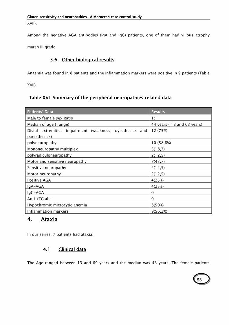
Gluten sensitivity and neuropathiesGluten sensitivity and neuropathiesGluten sensitivity and neuropathiesGluten sensitivity and neuropathies---- A Moroccan case control studyA Moroccan case control studyA Moroccan case control studyA Moroccan case control study
53
XVII).
Among the negative AGA antibodies (IgA and IgG) patients, one of them had villous atrophy
marsh III grade.
3333....6666.... Other biological resultsOther biological resultsOther biological resultsOther biological results
Anaemia was found in 8 patients and the inflammation markers were positive in 9 patients (Table
XVII).
Table XVI: Summary of the perTable XVI: Summary of the perTable XVI: Summary of the perTable XVI: Summary of the peripheral neuropathies related data ipheral neuropathies related data ipheral neuropathies related data ipheral neuropathies related data
Patients’ Data Patients’ Data Patients’ Data Patients’ Data ResultsResultsResultsResults
Male to female sex Ratio 1:1
Median of age ( range) 44 years ( 18 and 63 years)
Distal extremities impairment (weakness, dysethesias and
paresthesias)
12 (75%)
polyneuropathy 10 (58,8%)
Mononeuropathy multiplex 3(18,7)
polyradiculoneuropathy 2(12,5)
Motor and sensitive neuropathy 7(43,7)
Sensitive neuropathy 2(12,5)
Motor neuropathy 2(12,5)
Positive AGA 4(25%)
IgA-AGA 4(25%)
IgG-AGA 0
Anti-tTG abs 0
Hypochromic microcytic anemia 8(50%)
Inflammation markers 9(56,2%)
4444.... AtaxiaAtaxiaAtaxiaAtaxia
In our series, 7 patients had ataxia.
4444....1111 Clinical dataClinical dataClinical dataClinical data
The Age ranged between 13 and 69 years and the median was 43 years. The female patients

Gluten sensitivity and neuropathiesGluten sensitivity and neuropathiesGluten sensitivity and neuropathiesGluten sensitivity and neuropathies---- A Moroccan case control studyA Moroccan case control studyA Moroccan case control studyA Moroccan case control study
54
were predominant (sex-ratio=0,16)
The onset of ataxia symptoms was progressive for all the patients, and the evolution was chronic
for 6 of them and remittent for 1 patient.
Five cases of gait and limb cerebellar ataxia were found on the clinical examination as well as 2
cases of sensory ataxia.
One patient had a history of tuberculosis and another one had HBP and type 2 diabetes.
Digestive symptoms were reported by one subject who had chronic diarrhoea and moderate
dyspepsia.
4444....2222.... Immunological testingImmunological testingImmunological testingImmunological testing
Three patients had high titers of AGA antibodies which represent 42,85% of included ataxic
patients.
Positive IgA AGA were found in two patients with the following titers: 14,18 UI/L and 22,24 UI/L.
Additionally, one patient had both IgA (more than 200 UI/L) and IgG (more than 200 UI/L) AGA
antibodies.
4444....3333.... Other biological featuresOther biological featuresOther biological featuresOther biological features
Anaemia was present in 5 patients and inflammation in 4 patients.
In addition, we found hypocholesterolemia and hyperglycemia in one case.
4444....4444.... NeuroNeuroNeuroNeuro----imagingimagingimagingimaging
MRI revealed global cerebellar atrophy in 4 patients.

Gluten sensitivity and neuropathiesGluten sensitivity and neuropathiesGluten sensitivity and neuropathiesGluten sensitivity and neuropathies---- A Moroccan case control studyA Moroccan case control studyA Moroccan case control studyA Moroccan case control study
55
Table XVII: Table XVII: Table XVII: Table XVII: Summary of clinical, biological, immunoSummary of clinical, biological, immunoSummary of clinical, biological, immunoSummary of clinical, biological, immunological and imaging investigations on logical and imaging investigations on logical and imaging investigations on logical and imaging investigations on
ataxic patients.ataxic patients.ataxic patients.ataxic patients.
Patient’s dataPatient’s dataPatient’s dataPatient’s data ResultsResultsResultsResults
Median of age ( range) 43 years (13 and 69 years)
Male to female sex ratio 2:5
Cerebellar ataxia 5 patients
Sensory ataxia 2 patients
Associated digestive symptoms 1 patients
Positive AGA Abs 3 patients
IgA AGA Abs 2 patients
Both IgG and IgA AGA Abs 1 patients
Hypochromic microcytic anaemia 5 patients
Inflammation markers 4 patients
Global atrophy (cerebral and cerebellar atrophy) 4 patients
5555.... EpilepsyEpilepsyEpilepsyEpilepsy
Our study included 7 patients with epilepsy, with the following characteristics:
5555....1111.... Clinical dataClinical dataClinical dataClinical data
All the patients reported an acute onset with a remittent evolution of their condition.
Two patients were type 2 diabetics.
The patients had neither history of GSE nor digestive symptoms suggesting this disease.
On the clinical description, 5 patients had the generalized tonico-clonic form followed by
confusion, 2 had partial seizures and one of them had partial status epilepticus.
Six patients were under a treatment based on sodium valproates, with a bad outcome while one
patient was under Phenobarbital.

Gluten sensitivity and neuropathiesGluten sensitivity and neuropathiesGluten sensitivity and neuropathiesGluten sensitivity and neuropathies---- A Moroccan case control studyA Moroccan case control studyA Moroccan case control studyA Moroccan case control study
56
5555....2222.... ElectroElectroElectroElectro----encephalogram resultsencephalogram resultsencephalogram resultsencephalogram results
Six patients had spikes and waves at the EEG investigation; they were generalized in 4 cases and
localized to the left parietal lobe in 2 other cases, while the EEG was normal in one case.
5555....3333.... Immunologic testingImmunologic testingImmunologic testingImmunologic testing
One patient had positive IgA AGA with 16,25 IU/ml as a titre.
All the patients were negative for both IgG-AGA and IgA-tTG.
5555....4444.... Biologic resultsBiologic resultsBiologic resultsBiologic results
Three patients had microcytic hypochromic anaemia, while 4 patients had increased
inflammation markers.
5555....5555.... NeuroNeuroNeuroNeuro----imaging dataimaging dataimaging dataimaging data
MRI and scanner imaging were normal for all epileptic patients. They showed neither atrophy nor
focal lesions that may explain the seizures.
Table XVIII: Table XVIII: Table XVIII: Table XVIII: Characteristics of the epileptiCharacteristics of the epileptiCharacteristics of the epileptiCharacteristics of the epileptic patientsc patientsc patientsc patients
Patient’s dataPatient’s dataPatient’s dataPatient’s data ResultResultResultResult
Median of age (range) 45 years +/+/+/+/---- 16,31
Sex ratio (M/F) 0,75
generalized tonico-clonic form 5
Partial epilepsy 2
Status epilepticus 1
Generalized spikes and waves 4
partial spikes and waves 2
Normal EEG 1
Neuro-imaging data (MRI and TDM) Normal
positive IgA AGA Abs 1

Gluten sensitivity and neuropathiesGluten sensitivity and neuropathiesGluten sensitivity and neuropathiesGluten sensitivity and neuropathies---- A Moroccan case control studyA Moroccan case control studyA Moroccan case control studyA Moroccan case control study
57
6666.... MyopathyMyopathyMyopathyMyopathy
Our series included 3 patients with myopathy; their clinical and immunological and biological
data are described below:
6666....1111.... Clinical findingsClinical findingsClinical findingsClinical findings
The median of age was 45 years (range: 25-55 years), with 2 females and one male (sex ratio
M/F =0,5). These patients had neither digestive symptoms, nor HBP, nor diabetes, nor nutrition
deficiency nor cardiopathies.
For all the patients, the disease’s symptoms were progressive and chronic.
On neurologic examination, all the patients had bilateral and symmetric myogenic syndrome,
associated to muscular atrophy in one case. Besides, one of them had total muscular deficit and
therefore confined to wheelchair. Cutaneous signs were found in one patient who had vasculitis
of the hands with manicure sign and Gottron papules suggesting dermato-myositis (Table XX).
6666....2222.... Immunological resultsImmunological resultsImmunological resultsImmunological results
Two patients were positive for AGA and both of them had the IgG isotype. The titres were
respectively 27,41 IU/ml and 14,4 IU/ml (Table XX). All of them were negative for IgA-tTG.
6666....3333.... Biology resultsBiology resultsBiology resultsBiology results
Two patients had hypochromic microcytic anaemia and increased inflammation markers.
The patients had neither hyperglycemia nor hypocholesterolemia.
Two patients had increased CPK titers with respectively 1067 and 5213 U/L (Table XX).

Gluten sensitivity and neuropathiesGluten sensitivity and neuropathiesGluten sensitivity and neuropathiesGluten sensitivity and neuropathies---- A Moroccan case control studyA Moroccan case control studyA Moroccan case control studyA Moroccan case control study
58
6666....4444.... PathologyPathologyPathologyPathology
Two patients underwent muscular biopsy. One patient had mitochondriopathy with no
expression of dystrophine or alpha sarcoglycan while the other one had chronic myositis with
vasculitis lesions (Table XIX).
Table XIXTable XIXTable XIXTable XIX: Characteristics of the patients with myopathy.Characteristics of the patients with myopathy.Characteristics of the patients with myopathy.Characteristics of the patients with myopathy.
Patient’s dataPatient’s dataPatient’s dataPatient’s data ResultsResultsResultsResults
Median of age ( range) 45 (25 to 55 years)
Sex-ratio 1:2
Onset Progressive
Progression Chronic
Bilateral and symmetric myogenic Sd 3
Muscular atrophy 1
Dermato-myositis 1
Increased CPK titers 2
IgG AGA Abs 2
7777.... MyelopathyMyelopathyMyelopathyMyelopathy
Two cases of myelopathy are included in the study.
The first one is about a masculine patient aged 48 years old and having a HBP; otherwise, his
medical record showed neither diabetes nor auto-immune conditions nor cancer nor GSE’s
evoking symptoms.
The symptoms (weakness and paresthesias of the lower limbs) were progressive and neurological
examination found a medullar syndrome; also, the MRI and CSF analysis were normal.
The immunological testing showed a positive IgG-AGA with 34,25 IU/ml, but was negative for
both IgA AGA and IgA-tTG.

Gluten sensitivity and neuropathiesGluten sensitivity and neuropathiesGluten sensitivity and neuropathiesGluten sensitivity and neuropathies---- A Moroccan case control studyA Moroccan case control studyA Moroccan case control studyA Moroccan case control study
59
The second case corresponds to a 67 years old man, with normal medical history, who presented
progressively a weakness with proximal and distal paresthesias and pain affecting both of the
lower limbs. Neurological examination found a tetra-pyramidal syndrome. The scanner showed a
tight lumbar canal and normal cervical medulla, however, MRI results were not provided. The
electro-diagnosis exploration revealed a myelopathy evoking aspect with an impairment of the
S1 sensitive root.
The patient was offered an immunological analysis (for both IgA and IgG AGAs and ant-tTG) that
was negative.
8888.... Cerebral thrombophlebitisCerebral thrombophlebitisCerebral thrombophlebitisCerebral thrombophlebitis
One patient included in the study was diagnosed as cerebral thrombophlebitis. This female
patient was 38 years old; she had neither history of GSE nor digestive symptoms.
Also, the patient didn’t have a history of surgery, or immobilization; there were neither
obstetrical conditions (pregnancy, delivery and abortion) nor medications (such as
anticoagulants, contraception). She had intracranial hypertension syndrome on physical
examination.
The MRI was normal without any atrophy of the cerebral parenchyma, however, scanner imaging
showed a thrombophlebitis evoking aspect without parenchyma lesions.
The IgA AGA Abs were positive (14,32 IU/mL), and both IgG AGA and tTG were negative.
9999.... Multiple sclerosisMultiple sclerosisMultiple sclerosisMultiple sclerosis
A 44 years old man, with no previous medical conditions presented an acute episode of

Gluten sensitivity and neuropathiesGluten sensitivity and neuropathiesGluten sensitivity and neuropathiesGluten sensitivity and neuropathies---- A Moroccan case control studyA Moroccan case control studyA Moroccan case control studyA Moroccan case control study
60
headache, dizziness, nausea and gait impairment that progressed in a remittent way.
Neurological examination showed tetra-pyramidal syndrome.
There was hyper-gammaglobulinemia on serum proteins’ electrophoresis without oligoclonal
aspect in the CSF. Brain and cervical spine MRI showed peri-ventricular, bilateral and asymmetric
hyposignal in T1 sequence and hypersignal in T2 and Flair mode. According to McDonald’s
criteria, the diagnosis of MS was made.
The patient was positive for IgG AGA Abs with 97 IU/mL, however, he was negative for IgA-AGA
and IgA-tTG.
10101010.... Others neurologicOthers neurologicOthers neurologicOthers neurologicalalalal conditions conditions conditions conditions
The 3 others cases of this study corresponded to anterior horn disease, dystonia, Parkinson
disease associated to global atrophy and demyelinating lesions of white matter on MRI, and
lymphocytic meningitis (CSF analysis showing increased levels of lymphocytes, normal
glycorachy and proteinorachy and negative bacteriology testing) with respectively 2 cases, 1
case, 1 case, 1 case. The immunologic testing of all these patients was negative.

Gluten sensitivity and neuropathiesGluten sensitivity and neuropathiesGluten sensitivity and neuropathiesGluten sensitivity and neuropathies---- A Moroccan case control studyA Moroccan case control studyA Moroccan case control studyA Moroccan case control study
61
DISCUSSIONDISCUSSIONDISCUSSIONDISCUSSION
Gluten sensitivity and neuropathiesGluten sensitivity and neuropathiesGluten sensitivity and neuropathiesGluten sensitivity and neuropathies---- A Moroccan case control studyA Moroccan case control studyA Moroccan case control studyA Moroccan case control study
62
DiscussionDiscussionDiscussionDiscussion
In our study, we screened for GS among patients with idiopathic neurologic disorders. IgG and
IgA AGA were first tested in patients and controls. To rule out an authentic celiac disease (GSE) in
our patients, we performed an anti-tTG testing for both positive and negative AGA patients, and
also in the controls.
IIII.... Relevance of the immunological testing Relevance of the immunological testing Relevance of the immunological testing Relevance of the immunological testing
The AGA testing is used by a large number of researchers in patients presenting neurologic
diseases to check out GS[2, 113, 114] . Even if they lack sensitivity and specificity for the
diagnosis [106, 107], they might be useful for detection of GS in unexplained neurological
diseases[2].
It has been suggested that AGA testing remains an essential investigation in patients with
sporadic idiopathic ataxia [89, 108] and IgG-AGA isotype is the best marker of GS in
neurological disorders[2, 89]
Meanwhile, tTG and/or EMA testing are principally used to diagnose CD, regarding their high
sensitivity and specificity [2, 48, 113, 115, 116].
IIIIIIII.... SocioSocioSocioSocio----demographic characteristics of the positive casesdemographic characteristics of the positive casesdemographic characteristics of the positive casesdemographic characteristics of the positive cases
The male to female ratio among our AGA positive individuals is 1,6 which differs from the sex
ratio of the whole group of patients, and the median of age was 45,5 +/- 11,9 years old.
As shown in table XXI, demographic data of other series, patients with gluten sensitive

Gluten sensitivity and neuropathiesGluten sensitivity and neuropathiesGluten sensitivity and neuropathiesGluten sensitivity and neuropathies---- A Moroccan case control studyA Moroccan case control studyA Moroccan case control studyA Moroccan case control study
63
neuropathies are mostly males and youth.
Table XX: Summary of demographic characteristics of patients with GS neuropathiesTable XX: Summary of demographic characteristics of patients with GS neuropathiesTable XX: Summary of demographic characteristics of patients with GS neuropathiesTable XX: Summary of demographic characteristics of patients with GS neuropathies
M/F sex rM/F sex rM/F sex rM/F sex ratio atio atio atio Mean age (range)Mean age (range)Mean age (range)Mean age (range)
Hadjivassiliou and al, 1996 [117] 2 NP*
Hadjivassiliou and al, 1998[85] 3 61 years (21-79)
Ihara and al, 2006, [118] 1,5 69 years (53-88 years)
Hadjivassiliou and al, 2010[119] 0,4 67 years (47-58 years)
Chin and al, 2003 [120] 1,5 53 years (31-79 years)
Our study, 2011 1,6 45,8 years (13-69 years)
*Not provided
By contrast, In one of the studies led by Hadjivassiliou et al, the male to female ratio among AGA
positive patients was 0,4 which means that females were prominent (Table XX).
IIIIIIIIIIII.... EpidemiologicEpidemiologicEpidemiologicEpidemiologicalalalal analysisanalysisanalysisanalysis
Our study found a high prevalence (26.7%) of antigliadin antibodies in patients with neuropathies
of unknown cause versus 15.7% In the control group. Even if the association of neuropathies and
GS in our series is not significant (p=0,151), these findings support the fact that GS may
represent a potential aetiology of idiopathic neuropathies in our patients.
In fact, the studies that assessed the prevalence of AGA abs in idiopathic neuropathies showed
different results (table XXI). Our prevalence is among the highest ones published by authors,
such us Hadjivassiliou et al who found respectively 34%, 41% and 57% in his series [71, 89, 117].
Meanwhile, our results surpassed other published ones, such us Pellecchia et al, Burk et al and
Abele et al who found respectively 13%, 12% and 13% of AGA positive patients[108, 121, 122].

Gluten sensitivity and neuropathiesGluten sensitivity and neuropathiesGluten sensitivity and neuropathiesGluten sensitivity and neuropathies---- A Moroccan case control studyA Moroccan case control studyA Moroccan case control studyA Moroccan case control study
64
Table XXI: PrevaleTable XXI: PrevaleTable XXI: PrevaleTable XXI: Prevalence of AGA abs in idiopathic neuropathiesnce of AGA abs in idiopathic neuropathiesnce of AGA abs in idiopathic neuropathiesnce of AGA abs in idiopathic neuropathies
Idiopathic neuropathies Idiopathic neuropathies Idiopathic neuropathies Idiopathic neuropathies
with positive AGA abs with positive AGA abs with positive AGA abs with positive AGA abs
Healthy controls Healthy controls Healthy controls Healthy controls
%%%%
P ValueP ValueP ValueP Value ReferencesReferencesReferencesReferences
Hadjivassiliou, 2006
(n=140)
34% 12% <0,001 [71]
Ihara, 2006, (n=14) 36% 2% <0,001 [118]
Pellecchia, 1999, (n=24) 13% NP <0,05 [108]
Hadjivassiliou, 2003,
(n =53)
57 % 12% <0,0001 [89]
Wong (n=56) 11% 8% 0,68 [123]
Our study (n=60) 16 (26,7%) 9/57 (15,75%) 0.151 -
NP=Not provided
The positive GS cases of our series (n=16) correspond to peripheral neuropathies (n=4), followed
by ataxia (n=3), ischemic stroke (n=3), myopathy (n=2) and 1 case for each of the following
diseases: epilepsy, cerebral thrombo-phlebitis, myelopathy and multiple sclerosis.
Our results are supporting those of previous studies (table XXII) where peripheral neuropathy
and ataxia are found to be the most common neurological manifestations of GS. We found that
ischemic stroke was highly prevalent (18,75%) among positive AGA patients; it wasn’t mentioned
in the large based studies listed in the comparative table (table XXII),however, there are case-
reports describing the association between stroke and GS and will be detailed in the next chapter
(Clinical and investigation data) [74, 75].

Gluten sensitivity and neuropathiesGluten sensitivity and neuropathiesGluten sensitivity and neuropathiesGluten sensitivity and neuropathies---- A Moroccan case control studyA Moroccan case control studyA Moroccan case control studyA Moroccan case control study
65
Table XXII: Categories of gluten sensitivity neuropathies in differTable XXII: Categories of gluten sensitivity neuropathies in differTable XXII: Categories of gluten sensitivity neuropathies in differTable XXII: Categories of gluten sensitivity neuropathies in different studiesent studiesent studiesent studies
Neurological Neurological Neurological Neurological
manifestationsmanifestationsmanifestationsmanifestations
Hadjivassiliou Hadjivassiliou Hadjivassiliou Hadjivassiliou [2][2][2][2]
n (%)n (%)n (%)n (%)
Hadjivassiliou Hadjivassiliou Hadjivassiliou Hadjivassiliou
[117][117][117][117] n (%)n (%)n (%)n (%)
BBBBƼrkƼrkƼrkƼrk [124][124][124][124]
n (%)n (%)n (%)n (%)
Our studyOur studyOur studyOur study
n (%)n (%)n (%)n (%)
Ataxia 184 (43,4) 17/53 (32,07) 27 (37,5) 3/16 (18,75)
Peripheral neuropathy 174 (41,03) 28/53 (52,8) 10 (14) 4/16 (25)
Ischemic stroke NP* NP NP 3/16 (18,75)
Encephalopathy 62 (14,62) NP NP NP
Myopathy 18 (4,2) 4/53 (7,5) NP 2/16 (12,5)
Venous thrombosis NP NP NP 1/16 (6,25)
Multiple sclerosis NP NP NP 1/16 (6,25)
Myelopathy 6 (1,4) 2/53 ( 3,7) NP 1/16 (6,25)
Stiff-man syndrome 6 (1,4) NP NP NP
Chorea 3 (0,7) NP NP NP
Neuromyotonia 1 (0,2) NP NP NP
Dystonia NP NP 3 (4) NP
Parkinson disease NP NP 2 (2,77) NP
Epilepsy 1 (0,2) NP NP 1/16 (6,25)
* NP= Not provided or not studied
The data from several studies investigating the occurrence of AGA in ataxias have been
published and are summarised in table XXIV.
Table XXIII: prevalence of antigliadin antibodies in idiopathic sporadTable XXIII: prevalence of antigliadin antibodies in idiopathic sporadTable XXIII: prevalence of antigliadin antibodies in idiopathic sporadTable XXIII: prevalence of antigliadin antibodies in idiopathic sporadic ataxia ic ataxia ic ataxia ic ataxia
Studies and authorsStudies and authorsStudies and authorsStudies and authors Sporadic ataxia (%)Sporadic ataxia (%)Sporadic ataxia (%)Sporadic ataxia (%) Healthy controls (%)Healthy controls (%)Healthy controls (%)Healthy controls (%) ReferencesReferencesReferencesReferences
Hadjivassiliou and al, UK 47% 149 (12%) [85]
Hadjivassiliou and al, UK 41% 12% [89]
Pellecchia and al, Italy 13% NP [108]
Bürk and al, Germany 12% 5% [121]
Bushara and al, USA 27% NP [125]
Abele and al, Germany 15% 5% [122]
Luostarinen and al, Finland 17% 2% [126]
Ihara and al, Japan 36% 2% [118]
Anheim and al, France 36% NP [127]
Our study 42,85% (n=3/7) 15,78% -

Gluten sensitivity and neuropathiesGluten sensitivity and neuropathiesGluten sensitivity and neuropathiesGluten sensitivity and neuropathies---- A Moroccan case control studyA Moroccan case control studyA Moroccan case control studyA Moroccan case control study
66
The prevalence of GS in our ataxic patients is among the highest ones; close to the English [85],
Japanese [118], and French [127] series (table XIII). Nonetheless, the lowest prevalence were
reported in the Finnish [126] and German [121] series. Actually, the discrepancy in frequency
might be explained by several factors including geographical differences, culinary habits,
variability in the AGA assays used, referral bias in the selection of patients (for example, some
studies categorised patients with cerebellar variant of multisystem atrophy as idiopathic sporadic
ataxia), small study size, and absence of controls[2].
Sporadic idiopathic ataxia seems to be more linked to GS than the hereditary ataxia or the
sporadic form with multiple system atrophy (MSA). This fact is supported by Hadjivassiliou [67,
68, 89, 128] and Pellecchia [108] and Luostarinen[126], and Abele[122, 129].
In the large study lead in England on 268 patients with ataxia, GS accounted for up to 41% of
cases of sporadic idiopathic ataxia which makes it the single most common cause of ataxia in
this cohort of patients. Besides, 14% of familial ataxia’s cases and 15% of the MSA-C (cerebellar
variant of MSA) group had evidence of GS but were not significantly different from the healthy
controls (12%) [89]. However, a study from the USA found a high prevalence of AGA positivity in
both sporadic (27%) and familial ataxias (37%); however, the numbers screened were small (26
sporadic form and 24 familial form) [130]. On the other hand, all our 3 positive AGA ataxic
patients correspond to cerebellar sporadic idiopathic form which is well supported by the
literature data above.
Peripheral neuropathy (PN) is the other most common manifestation of GS[2]. Actually, many
authors relate that PN is frequently associated with GSE, and it might be detected even in celiac
patients under GFD [2, 131, 132]. Otherwise, PN was recognised as an exclusive

Gluten sensitivity and neuropathiesGluten sensitivity and neuropathiesGluten sensitivity and neuropathiesGluten sensitivity and neuropathies---- A Moroccan case control studyA Moroccan case control studyA Moroccan case control studyA Moroccan case control study
67
manifestation of GS in numerous studies[2, 71, 72, 120]. Concerning our series, 25% of cases
(n=4) had evidence of gluten sensitive neuropathies, which is close to the English [71], and
Italian [72] and Finnish [131] studies; but remains higher than the American one [120] (table
XXIV).
Table XXIVTable XXIVTable XXIVTable XXIV: Prevalence of GS in peripheral neuropathy patients : Prevalence of GS in peripheral neuropathy patients : Prevalence of GS in peripheral neuropathy patients : Prevalence of GS in peripheral neuropathy patients
Studies and authorsStudies and authorsStudies and authorsStudies and authors Gluten neuropathies Gluten neuropathies Gluten neuropathies Gluten neuropathies
n (%)n (%)n (%)n (%)
Control group Control group Control group Control group
n (%)n (%)n (%)n (%)
p Valuep Valuep Valuep Value ReferencesReferencesReferencesReferences
Luostarinen et al, Finland
(n=26)
23% 4,3% <0,001 [131]
Hadjivassiliou et al, UK
(n=140)
34% 149 (12%) <0.001 [71]
Mata et al, Italy (n=330) 21% 1 (1,5%) <0�0001 [72]
Chin et al, USA (n=400) Between 2,5% and 8% 1% <0,001 [120]
Our study 4 (25%) 15,78% - -
According to literature, the association of ischemic stroke with GS is less frequent and has not
been well documented [74, 75]. A recent study suggested that childhood GSE is associated with a
10-fold increased risk of death from stroke, although it was based on small numbers[133].
However, a nationwide Swedish study that evaluated the risk of stroke in 28,000 patients
with GSE concluded that celiac patients are at only a small increased risk of stroke and that CD
does not seem to be a major risk factor for stroke[133].
The risk of cardiovascular disease in individuals with GSE has been evaluated by Swedish authors
on a large cohort of patients (13 358 individuals diagnosed with GSE and 64 118 age-matched
and sex-matched controls without GSE). They found a positive association between GSE and
myocardial infarction, angina pectoris, heart failure, brain haemorrhage and ischemic stroke
[134].

Gluten sensitivity and neuropathiesGluten sensitivity and neuropathiesGluten sensitivity and neuropathiesGluten sensitivity and neuropathies---- A Moroccan case control studyA Moroccan case control studyA Moroccan case control studyA Moroccan case control study
68
West and al studied the risk of vascular disease in adults with diagnosed GSE compared with the
general population. Consequently, the author found a slight increase in the risk of atrial
fibrillation and ischemic stroke. In our series, 3 of 18 ischemic stroke cases were positive for
AGA which is important.
Myopathy is considered by Hadjivassiliou [2] and Henriksson [135] as rare neurological
manifestation of GS[2]. However, Selva-O’Callaghan and colleagues found that GS might be a
probable aetiology for idiopathic and inflammatory myopathies[136]. They supported their
statement by the high prevalence of AGA positivity (31%) within inflammatory myopathies
compared to the general population, certainly because of the shared HLA class II haplotypes
(DQ2) between the two diseases [136, 137]. Moreover, our series showed also a high prevalence
of GS since 2 have tested positive for AGA among the 3 myopathy patients.
Several reports have suggested a link between epilepsy and GSE[2, 5, 138, 139] which was first
described by Visakorpi and al in 1970 [140]. Nevertheless, some authors didn’t confirm these
findings, such us Ranua and al who found no difference in the prevalence of AGA and anti-EMA
and anti-tTG abs between epileptic patients and controls [141]. Moreover, authors disagreed on
the frequency of this association; while Hadjivassiliou considered epilepsy as a rare
manifestation of GS; It was reported to be common in Italy, Spain, and Argentina suggesting a
geographical factor [142]. In our series, 1 of the 7 epileptic patients had evidence of GS.
Further more, the association between GS and relapsing-remitting or secondary-progressive
multiple sclerosis was not significant in all studies performed till now[143, 144].
Additionally, clinical evidence of myelopathy without vitamin or other deficiencies (particularly
copper) can be a rare manifestation of GS[2, 117]. There have been reports of patients

Gluten sensitivity and neuropathiesGluten sensitivity and neuropathiesGluten sensitivity and neuropathiesGluten sensitivity and neuropathies---- A Moroccan case control studyA Moroccan case control studyA Moroccan case control studyA Moroccan case control study
69
with neuromyelitis optica (Devic’s disease) and GS associated with antibodies to aquaporin-
4[145, 146]. In our series, only 1 patient who had myelopathy with negative etiological
investigations was positive for AGA abs.
The cases of thrombophlebitis associated with GSE are very scarce[147]. Bahloul and al reported
a case of GSE who developed cerebral venous thrombosis associated with protein S
deficiency[148] . Moreover, a Turkish case-report about GSE associated with deep venous
thrombosis of the leg was published[147]. Our series comprises a 38-years old female patient
diagnosed with cerebral thrombophlebitis who tested positive for AGA.
Finally, all patients in our series who had dystonia, and anterior horn disease, and Parkinson
disease and lymphocytic meningitis were tested negative for AGA. In fact, case reports about a
possible association between GS and those conditions are scarce; however, there is no evidence
in literature of such association with Parkinson disease. Thus, 1 case of chronic meningitis [149]
and 2 cases of dystonia [150, 151] and 3 cases of anterior horn disease [118, 152, 153]
revealing GS are reported by the literature.
IVIVIVIV.... Clinical and investigations dataClinical and investigations dataClinical and investigations dataClinical and investigations data
1111.... Gluten AtaxiaGluten AtaxiaGluten AtaxiaGluten Ataxia
The patients with gluten ataxia are usually males, however, female predominance was reported
by some authors [121]. Moreover, gluten ataxia has generally an insidious onset with a mean age
at diagnosis of 41 years which is in line with Hadjivassiliou and Burk’s findings[89, 121];
however, old patients prevailed in a Japanese study[118] (Table XXV).

Gluten sensitivity and neuropathiesGluten sensitivity and neuropathiesGluten sensitivity and neuropathiesGluten sensitivity and neuropathies---- A Moroccan case control studyA Moroccan case control studyA Moroccan case control studyA Moroccan case control study
70
Table XXV: Characteristics of the patients with gluten ataxiaTable XXV: Characteristics of the patients with gluten ataxiaTable XXV: Characteristics of the patients with gluten ataxiaTable XXV: Characteristics of the patients with gluten ataxia
Hadjivassiliou et Hadjivassiliou et Hadjivassiliou et Hadjivassiliou et
al, 2003, al, 2003, al, 2003, al, 2003, [89][89][89][89]
n= 6n= 6n= 6n= 68888
Hadjivassiliou Hadjivassiliou Hadjivassiliou Hadjivassiliou
et al,1998,et al,1998,et al,1998,et al,1998,[85][85][85][85]
n=28n=28n=28n=28
Burk et al, Burk et al, Burk et al, Burk et al,
2001, 2001, 2001, 2001, [121][121][121][121]
n=12n=12n=12n=12
IharIharIharIhara et al, a et al, a et al, a et al,
2006,2006,2006,2006,[118][118][118][118]
n=5n=5n=5n=5
Our study, Our study, Our study, Our study,
2012201220122012
n=3n=3n=3n=3
M/F sex ratio 1,06 3 0,71 1,5 2
Mean age at
study (range)
NP 61 years (21–79
years)
55 years+/-
17,4 (30-76)
69 years
(53-88)
41 years
(13-69)
Mean age at
onset (range)
48 years
(14± 78 years)
53,57 years
(18-76 years)
44,8 years+/-
23,2 (2-68
years)
NP NP
Dysarthria 45 (66%) NP 12 (100%) NP 3 (100%)
Limb ataxia (LA) Upper LA
n=51(75%)
Lower LA
n=61(90%)
26 (92,8%) 11 (91,7%) NP 3 (100%)
Gait ataxia 68 (100%) 28 (100%) 12 (100%) NP 2 (67%)
Gastrointestinal symptoms
9 (13%) 12 (43%) 2 (16,7%) NP -
Loss of proprioception
NP NP 7 (58,3%) NP 1 (33,33%)
PN clinical signs NP 10 (35,7%) 4 (33,3%) 3 (60%) -
Amyotrophy - - 1 (8,3%) NP -
Cerebellar atrophy on MRI
n=52 (79%) 6 (21,4%) 12 (100%) 4 (80%) 2 (67%)
White matter hyper-signal on MRI
n=13 (19%) - - NP -
axonal neuropathy on neurophysiology (EMG)
31 (45%) 10 (35,7%) 2 (16,7%) 3 (60%) -
In addition, gluten ataxia presents usually with the cerebellar form [2, 89, 121] which matches
our results (Table XXV). However, the association between GS and sensory ataxia has been
described [121]. Otherwise, the association of ataxia and peripheral neuropathy was reported by
authors; usually it’s an axonal neuropathy on EMG exploration[85, 118, 121, 154]. This
neuropathy is frequently mild and does not contribute to the ataxia[2]; however, none of our
Gluten sensitivity and neuropathiesGluten sensitivity and neuropathiesGluten sensitivity and neuropathiesGluten sensitivity and neuropathies---- A Moroccan case control studyA Moroccan case control studyA Moroccan case control studyA Moroccan case control study
71
patients diagnosed with gluten ataxia had such signs. Gaze-evoked nystagmus and other ocular
signs of cerebellar dysfunction are seen in up to 80% of cases[154].
Besides, cerebellar atrophy on MRI is commonly found in gluten ataxia reaching 100% in some
studies [85, 89, 121] (table XXV).
Finally, there is no specific clinical features to enable a confident diagnosis of gluten ataxia,
except perhaps in patients with established GSE[108]. The clinical features described in table
XXV are also seen in patients with other forms of sporadic and inherited ataxias. This
emphasizes the importance to keep in mind the possibility of GS in patients presenting with
ataxia[89].
2222.... Gluten neuropathyGluten neuropathyGluten neuropathyGluten neuropathy
Gluten neuropathy is defined as apparently sporadic idiopathic neuropathy in the absence of an
alternative etiology and in the presence of serological evidence of GS. It is a progressive disease
affecting mostly young subjects with a mean age at onset of 55 years (range 24–77 years) [71]
which supports our findings (mean age at diagnosis: 45 years).
The mean diagnostic delay before introducing GFD is 9 years (range 1-33 years)[2]; this long
delay explains the occurrence of neurologic disorders in GS patients[131].
The majority of patients are masculine (male to female sex ratio 3:1) and matches the sex ratio
of the whole group of positive AGA patients, which is supported by other studies [117, 120].
Also, Chin and al [120] found an important frequency of positive AGA cases in patients with
associated digestive symptoms whereas one positive AGA case in our series reported chronic
diarrhea; thus, it could be considered as an orientating element.

Gluten sensitivity and neuropathiesGluten sensitivity and neuropathiesGluten sensitivity and neuropathiesGluten sensitivity and neuropathies---- A Moroccan case control studyA Moroccan case control studyA Moroccan case control studyA Moroccan case control study
72
Positive patients often express pain and paresthesias, and on clinical examination, PN syndrome
may be associated with sensory abnormalities (tactile, heat pain and vibratory hypoesthesia),
which is consistent with our findings. This provides evidence that peripheral nerve fibres, even
small non-myelinated ones, are affected in GS[131]. Otherwise, amyotrophy or handicap were
neither found in our study nor reported by authors (Table XXVI).
Symmetrical sensori-motor axonal neuropathy is the prominent electrophysiological finding[2,
71, 155] which is consistent with EMG results of our positive AGA cases. Other types of
neuropathies have also been reported such as asymmetrical neuropathy[155], sensory
ganglionopathy[156], small fiber neuropathy[157], pure motor neuropathy[71], and autonomic
neuropathy[99]. Meanwhile, 2 positive AGA patients, in our series, have polyradiculo-neuropathy
for one and elongation of distal latencies with abnormalities of “F” waves for the other.
Brannagan and al reported up to 8% of gluten neuropathy with normal electrophysiologic testing
[120]. Similar to our results, Hadjivassiliou and Chin didn’t find cerebral and/or cerebellar
atrophy on neuro-imaging in patients with gluten neuropathy[71, 120].

Gluten sensitivity and neuropathiesGluten sensitivity and neuropathiesGluten sensitivity and neuropathiesGluten sensitivity and neuropathies---- A Moroccan case control studyA Moroccan case control studyA Moroccan case control studyA Moroccan case control study
73
Table XXVI: Clinical and investigations’ characteristics of patientTable XXVI: Clinical and investigations’ characteristics of patientTable XXVI: Clinical and investigations’ characteristics of patientTable XXVI: Clinical and investigations’ characteristics of patients with idiopathic neuropathy s with idiopathic neuropathy s with idiopathic neuropathy s with idiopathic neuropathy
associated to positive AGA antibodiesassociated to positive AGA antibodiesassociated to positive AGA antibodiesassociated to positive AGA antibodies
CharacteristicsCharacteristicsCharacteristicsCharacteristics Hadjivassiliou et al, Hadjivassiliou et al, Hadjivassiliou et al, Hadjivassiliou et al,
2006,2006,2006,2006,[71][71][71][71], n=47, n=47, n=47, n=47
Chin et al, Chin et al, Chin et al, Chin et al,
2003,2003,2003,2003,[120][120][120][120],n=28,n=28,n=28,n=28
Our studyOur studyOur studyOur study
n=4n=4n=4n=4 Male to female ratio NP 1,75 3
Mean age at onset of neuropathy 55 years (24–77) 56 years (31-79) 45 years* (40-52)
Mean duration of disease 9 years (range 1–33) NP NP
Known GSE NP 45% -
Digestive symptoms NP NP 25 %
amyotrophy NP NP -
Need walking aid 22% NP -
Pain a prominent feature 14% 27% -
Paresthesias NP 100% 3/4
Objective sensory abnormalities NP 85% 2/4 (50%)
Cerebral an/or cerebellar atrophy on MRI NP - -
White matter abnormalities on MRI NP - -
Sensorimotor axonal neuropathy 67% 35% -
Sensitive axonal polyneuropathy - - 1/4 (25%)
Motor neuropathy 9% - -
Mononeuropathy multiplex 17% - 1/4(25%)
Small-fibre neuropathy 7% - -
Motor polyradiculoneuropathy - - 1/4(25%)
Other EMG findings - - 1**
Only antigliadin IgG positive 57% 37% -
Only antigliadin IgA positive 16% 18% 4/4(100%)
Both antigliadin IgG and IgA positive 27% 45% -
* Age at diagnosis; ** Elongation of distal latencies with abnormalities of “F” waves; NP=not provided

Gluten sensitivity and neuropathiesGluten sensitivity and neuropathiesGluten sensitivity and neuropathiesGluten sensitivity and neuropathies---- A Moroccan case control studyA Moroccan case control studyA Moroccan case control studyA Moroccan case control study
74
3333.... Ischemic strokeIschemic strokeIschemic strokeIschemic stroke
The majority of patients with GS are young [74, 75, 158] and the median of age at onset, in our
series, is 40 years ranging between 25 and 74 years. Besides, case-reports about childhood
stroke revealing GS have already been published [103, 159]. The medical record of the gluten
stroke cases didn’t show any cardiovascular risk factors except 1 patient who is masculine. Also,
digestive symptoms were absent for all of them. Moreover, recurrent episodes of transient
ischemic strokes are reported by authors [100, 160]. The main clinical presentation corresponds
to pure and proportional pyramidal syndrome, and rarely pseudo-bulbar syndrome [75], and the
right side was mostly concerned in our study.
On neuro-imaging, the middle cerebral artery’s area is the most affected with seldom cerebral or
cerebellar atrophy[75], however, 2 of the 3 positive cases in our study presented global atrophy.
Consistent with literature, echography showed elevated resistance in the right primitive carotids
and vertebral arteries in 1 patient and hypertensive cardiomyopathy in another one. In fact, cases
of thrombus of the ventricles[100], arteriosclerosis of the coronary artery[100] and dilated
cardiomyopathy [159] have been described.
Laboratory evaluations for prothrombotic conditions (including coagulation examinations, full
blood count, factor V Leyden and prothrombin mutations, serum homocystein, glycaemia) can be
either normal [74, 159] or abnormal [100, 160]. In this disease, AGA frequently coexists with
hyper-homocysteinemia and decreased folates’ levels [75]. This hyper-homocysteinemia is a
known risk factor for atherosclerosis and ischemic strokes [75, 101].
In our series, 3 of 18 ischemic stroke cases had positive AGA which is important regarding the

Gluten sensitivity and neuropathiesGluten sensitivity and neuropathiesGluten sensitivity and neuropathiesGluten sensitivity and neuropathies---- A Moroccan case control studyA Moroccan case control studyA Moroccan case control studyA Moroccan case control study
75
rare reported cases over the world. One of them has an intermediate homocycteinemia level, 2
patients had hyperglycaemia and 1 had intermediate rate of serum homocysteine; meanwhile,
the lipid profile and coagulation examinations were normal for all. Hence, our results are
approximate to literature.
4444.... MyopathyMyopathyMyopathyMyopathy
AGA antibodies have been detected in various clinical forms of myopathies including proximal
myopathy due to vitamin E deficiency[161], osteomalacia linked to vitamin D deficiency[162],
polymyositis [135], juvenile form of dermatomyositis (DM) [163] and sporadic inclusion body
myositis (s-IBM) [135]. Besides, no intestinal symptoms were reported in most cases.
In addition, Spanish authors found that 31% of patients with inflammatory myopathy were AGA
positive and the prevalence was significantly more frequent in patients with s-IBM than DM
(P<0.001)[136].
On neurologic examination, patients usually have bilateral and symmetric myogenic syndrome
with progressive muscular atrophy [2]. Moreover, the association with ataxia and neuropathy was
described by some authors[164].
Gluten myopathy can be combined to normal or increased CPK rate[136, 164]. On neuro-
pathology, inflammatory myopathy is the most common finding[136, 164]. Finally, some authors
reported an improvement of the myopathy after GFD introduction [2].

Gluten sensitivity and neuropathiesGluten sensitivity and neuropathiesGluten sensitivity and neuropathiesGluten sensitivity and neuropathies---- A Moroccan case control studyA Moroccan case control studyA Moroccan case control studyA Moroccan case control study
76
5555....1111.... EpilepsyEpilepsyEpilepsyEpilepsy
Epilepsy associated with GS tends to affect young patients; however, paediatric cases have also
been reported [165]. The seizures mostly resist to antiepileptic drugs[142, 166], and may be
benign or sometimes evolves into severe epileptic encephalopathy[142].
The epilepsy related to GS is more often focal [139, 165, 167], besides, Magaudda and al found
that the majority of patients had complex partial seizures linked to the occipital or temporal
lobes. In addition, secondarily generalized seizures, or other seizure types, and episodic
headaches were also described [138, 168]. This is in line with clinical presentation of our only
positive patient who is young (44 years) and had a partial and remittent tonic-clonic status
epilepticus.
On neuro-imaging, authors found that cerebral calcifications of the occipital lobe without
contrast enhancement are tightly associated with GSE [5, 165, 167, 169]. However, the CT-scan
performed for our patient showed a frontal sequellae hypo-density without cerebral atrophy or
calcifications.
6666.... Other less common manifestationsOther less common manifestationsOther less common manifestationsOther less common manifestations
6.1. Thrombophlebitis6.1. Thrombophlebitis6.1. Thrombophlebitis6.1. Thrombophlebitis
The cases of thrombophlebitis associated with GSE have been rarely published[147]. The disease
usually affects young patients with no pro-thrombotic risk factors[147], involving different
vascular territories: deep venous proximal thrombosis of the leg[147] , portal vein
thrombosis[105], and non-ischemic central retinal vein occlusion[170]. Also, Bahloul and al

Gluten sensitivity and neuropathiesGluten sensitivity and neuropathiesGluten sensitivity and neuropathiesGluten sensitivity and neuropathies---- A Moroccan case control studyA Moroccan case control studyA Moroccan case control studyA Moroccan case control study
77
reported a case of GSE who developed cerebral venous thrombosis associated with protein S
deficiency[148] . In addition, some patients improved after introduction of GFD[147].
6.2. Dystonia6.2. Dystonia6.2. Dystonia6.2. Dystonia
The case-reports about a possible association between dystonia and GS are rare. Hall and al
reported a child with biopsy-proven GSE associated to movement disorder since the age of 6
months that evoked paroxysmal non-kinesigenic dystonia (PNKD). This patient had complete
resolution of her neurological symptoms after introduction of GFD[151]. Another case-report
about 14 years-old girl who developed dystonic movements in her left lower limb and became
handicapped; she was diagnosed later with pathology-proven GSE[150]. In our study, one patient
with dystonia was admitted and has no evidence of GS after immunological screening.
6.3. Anterior horn disease (AHD)6.3. Anterior horn disease (AHD)6.3. Anterior horn disease (AHD)6.3. Anterior horn disease (AHD)
The amyotrophic lateral sclerosis (ALS) is one of the most common diseases that affect the
anterior medullar horn. Extremely few Case-reports about the association of ALS with GS are
published [118, 152, 153]. Actually, in a Japanese study, among the 18 recruited patients with
ALS, 1 had evidence of GS; he was a 56 years-old man who had an active denervation of the
tongue and the 4 extremities on electrophysiological testing, with abnormal T2 signal
intensities in the bilateral pyramidal tract on cranial MRI[118]. In addition, Turner and Brown
published 2 cases of confirmed GSE that manifested by ALS associated with MRI abnormalities.
One of them had T2/FLAIR signal-intensity abnormalities in the left cortico-spinal tract and the
other had abnormal T2 signal-intensity abnormalities in the bilateral pyramidal tract on cranial
MRI. After GFD introduction, the MRI ALS suggesting features showed improvement[152, 153].

Gluten sensitivity and neuropathiesGluten sensitivity and neuropathiesGluten sensitivity and neuropathiesGluten sensitivity and neuropathies---- A Moroccan case control studyA Moroccan case control studyA Moroccan case control studyA Moroccan case control study
78
Otherwise, our study included 2 female patients with AHD; both of them were negative for AGA
and anti-tTG testing.
6.4. Lymphocytic meningitis6.4. Lymphocytic meningitis6.4. Lymphocytic meningitis6.4. Lymphocytic meningitis
The first observation of 56 years-old woman with chronic meningitis revealing GSE is reported
by Cartalat-catel and al. Seven years after the onset of meningitis, she presented neuralgia of
the trigeminal nerve and cerebellar syndrome. The MRI examination showed cerebellar atrophy
with leuco encephalopathy[149].
6.5. Myelopathy6.5. Myelopathy6.5. Myelopathy6.5. Myelopathy
Clinical evidence of myelopathy in the absence of vitamin and other deficiencies (particularly
copper) can be a rare manifestation of GS[2, 117]. This myelopathy can present with progressive
medullar syndrome and is usually associated with normal imaging of the spinal cord[2]. In
addition, there have been reports of patients with neuromyelitis optica (Devic’s disease) and GS
who have antibodies to aquaporin-4[145, 146]; these patients had abnormal MRI of the spinal
cord. This form has to be determined whether this is simply an association based on the same
genetic susceptibility or due to immunological mechanisms[2].
VVVV.... Immunological investigationsImmunological investigationsImmunological investigationsImmunological investigations
1111.... Global findingsGlobal findingsGlobal findingsGlobal findings
We found an important prevalence of AGA abs among patients with idiopathic neuropathies
reaching 26.7% (n=16). This finding is in line with previous studies, some of them are close to
our results such as Hadjivassiliou[89]. Other ones show higher frequency [117], and the
published prevalence by some German studies are the lowest[121] (Table XXVII).

Gluten sensitivity and neuropathiesGluten sensitivity and neuropathiesGluten sensitivity and neuropathiesGluten sensitivity and neuropathies---- A Moroccan case control studyA Moroccan case control studyA Moroccan case control studyA Moroccan case control study
79
Even if the association of GS and neuropathies in our series is not significant (p=0,151),
compared to control individuals, GS can be considered as a substantial aetiology of unknown
neuropathies of our patients.
Table XXVII: Immunological findings in Table XXVII: Immunological findings in Table XXVII: Immunological findings in Table XXVII: Immunological findings in series about the association of GS and idiopathic series about the association of GS and idiopathic series about the association of GS and idiopathic series about the association of GS and idiopathic
neuropathiesneuropathiesneuropathiesneuropathies
Hadjivassiliou Hadjivassiliou Hadjivassiliou Hadjivassiliou
[117][117][117][117]
n(%)n(%)n(%)n(%)
Hadjivassiliou Hadjivassiliou Hadjivassiliou Hadjivassiliou
[85][85][85][85] n(%)n(%)n(%)n(%)
Hadjivassiliou Hadjivassiliou Hadjivassiliou Hadjivassiliou
[89][89][89][89] n(%)n(%)n(%)n(%)
Chin; Chin; Chin; Chin;
[120][120][120][120]
n(%)n(%)n(%)n(%)
Burk Burk Burk Burk
[121][121][121][121]
n(%)n(%)n(%)n(%)
Our studyOur studyOur studyOur study
n(%)n(%)n(%)n(%)
Number 53 NP* 176 20 104 60
Only IgG-AGA
6 16 62 4 2 6
Only IgA-AGA
12 6 6 2 6 9
Both IgG and IgA AGA positive
12 6 NP 5 3 1
Total of AGA-positive patients
30 (57%) 28 68 (38,6%) 11 (55%) 11(10,5%) 16(26,7%)
Anti IgA-tTG
NP NP NP 8 NP 0
The author reports that he included patients seen in the neurology outpatient department at the Royal Hallamshire Hospital from January
1994, to April 1998 and 28 were diagnosed with GS after immunological screening.
IgA-AGA is the most common isotype that we identified in our gluten neuropathies (16,7%;n=9),
versus 10% (n=6) for IgG-AGA. This finding is coherent with Burk’s series [121]. However, many
authors found that IgG-AGAs were more common in neurologic diseases [85, 89, 120].
Therefore, IgG-AGA isotype is considered to be more important in the context of extra-
intestinal manifestations of GS, by comparison to IgA class antibodies, which seems to be more
linked to gut clinical forms [2, 114, 171]. This finding can be supported by the fact that IgG-AGA
titres are higher than IgA-AGA’s ones in our series (figure 11). However, this fact needs further

Gluten sensitivity and neuropathiesGluten sensitivity and neuropathiesGluten sensitivity and neuropathiesGluten sensitivity and neuropathies---- A Moroccan case control studyA Moroccan case control studyA Moroccan case control studyA Moroccan case control study
80
Figure 11: Figure 11: Figure 11: Figure 11: Comparison between IgGComparison between IgGComparison between IgGComparison between IgG----AGA and IgAAGA and IgAAGA and IgAAGA and IgA----AGA titresAGA titresAGA titresAGA titres
consideration because of the absence of analogous data.
Nevertheless, some authors report a non-significant increase of AGA either in ataxia or PN, and
no over representation of HLA DQ2 or HLA DQ8 is detected in their series [113, 129]. Lock and al
argue that it might simply reflect an age related phenomenon or may imply a general heightened
responsiveness of the gut immune system in these individuals. Moreover, it could be explained
by a gut permeability’s impairment or inflammation, and adds that AGA in hereditary ataxias
must be an epiphenomenon[113].
The introduction of more specific CD serological markers such as anti-EMA and more recently,
tissue transglutaminase abs may help in diagnosing GSE, but their sensitivity as markers of other
manifestations of GS (where the bowel is not affected) is low[85]. Hadjivassiliou explained that
without overt gastrointestinal involvement, serum antibodies to transglutaminase-2 (TG2) can be
absent[2].
Our results are supporting previous findings and all of our patients were negative for anti-tTG
IgA, which makes occult CD unlikely.
0
10
20
30
40
50
60
70
80
90
100
110
120
0 1 2 3 4 5 6 7 8 9 10 11 12 13 14 15 16
IgA-AGA (IU/ml) IgG-AGA (IU/ml)

Gluten sensitivity and neuropathiesGluten sensitivity and neuropathiesGluten sensitivity and neuropathiesGluten sensitivity and neuropathies---- A Moroccan case control studyA Moroccan case control studyA Moroccan case control studyA Moroccan case control study
81
2222.... Gluten AtaxiaGluten AtaxiaGluten AtaxiaGluten Ataxia
Two of our ataxic patients have positive IgA-AGA. (14,18 IUI/mL and 22,24 IU/mL), and 1 has
both IgA (more than 200 UI/L) and IgG (more than 200 UI/L) AGA. None of them is positive for
IgA anti-tTG.
In many studies, patients with idiopathic sporadic ataxias were found to have frequent
occurrence of AGA compared with healthy controls (table XXVIII), which makes GS a potential
cause for this condition [2, 89, 108, 118, 121]
Table XXVIII: Immunological profile of gluten ataxia. Table XXVIII: Immunological profile of gluten ataxia. Table XXVIII: Immunological profile of gluten ataxia. Table XXVIII: Immunological profile of gluten ataxia.
Ihara, Ihara, Ihara, Ihara,
2006, 2006, 2006, 2006,
[118][118][118][118]
Hadjivassiliou Hadjivassiliou Hadjivassiliou Hadjivassiliou
et al, et al, et al, et al,
1998, 1998, 1998, 1998, [85][85][85][85], , , ,
(n=NP)(n=NP)(n=NP)(n=NP)
Hadjivassiliou Hadjivassiliou Hadjivassiliou Hadjivassiliou
et al,et al,et al,et al,
2003,2003,2003,2003,[89][89][89][89]
(n=176)(n=176)(n=176)(n=176)
PellecchPellecchPellecchPellecchia et ia et ia et ia et
al, al, al, al,
1999,1999,1999,1999,[108][108][108][108]
(n=24)(n=24)(n=24)(n=24)
Burk et al, Burk et al, Burk et al, Burk et al,
2003, 2003, 2003, 2003,
[121][121][121][121]
(n=104)(n=104)(n=104)(n=104)
Our Our Our Our
studystudystudystudy
n=7n=7n=7n=7
AGA
positive
(IgG or IgA)
36% n=28 68 (38,63%) 12,5% 10,5% N=3
IgG-AGA
positive
3 (21,4) n=16 (57,1%) 62 (35,2%) 3(8,3%) 2 (2%) N=0
IgA-AGA
positive
2
(14,3)
n=6 (21,4%) 6 (3,4%) 0 6 (5,7%) N=2
Both IgG
and IgA
positive
- n=6 (21,4%) NP 1 (4,1%) 3 (2,9%) n=1
n: number
IgG-AGAs are better markers of the whole spectrum of GS irrespective of the organ involved and
remain the best diagnostic markers for gluten ataxia[89, 130]. Moreover, IgA-AGA and anti-EMA
may lack sensitivity and specificity when used in a neurological population[172]. In fact, HLA
studies supports theses findings; thus, within a group of patients with gluten ataxia defined by
the presence of IgG-AGA antibodies (n=68), authors have found an HLA association similar to

Gluten sensitivity and neuropathiesGluten sensitivity and neuropathiesGluten sensitivity and neuropathiesGluten sensitivity and neuropathies---- A Moroccan case control studyA Moroccan case control studyA Moroccan case control studyA Moroccan case control study
82
that seen in patients with CD: 72% of patients have the HLA DQ2 (35% in the general population),
6% have the HLA DQ8 and the remainder have HLA DQ1 [89].
However, Burk and al found that IgA-AGA was the most frequent isotype among gluten ataxia
patients[121] which is similar to our findings ( all gluten ataxia patients have the IgA-AGA
isotype). The genetic and geographical factors might cause this difference in immunologic
profiles but it remains to be demonstrated.
Finally, combined IgA and IgG-AGA abs can be present in those patients, but at a lower
proportion compared with the other immunological features (table XXVIII).
IgG-anti-tTG2 in patients with gluten ataxia are found to be more common than IgA, also, IgA-
anti-tTG2’ titres in patients with gluten ataxia are lower than those seen in celiac patients [2]. In
our study, all patients were negative for IgA-anti-tTG but none of them was screened for IgG-
anti TG2.
3333.... Gluten neuropathyGluten neuropathyGluten neuropathyGluten neuropathy
The literature highlights an important prevalence rate of GS in a considerable number of
idiopathic axonal PN (table XXIX); suggesting the aetiological link between the two conditions.

Gluten sensitivity and neuropathiesGluten sensitivity and neuropathiesGluten sensitivity and neuropathiesGluten sensitivity and neuropathies---- A Moroccan case control studyA Moroccan case control studyA Moroccan case control studyA Moroccan case control study
83
Table XXIX: Immunological characteristics of patients with idiopathic PNTable XXIX: Immunological characteristics of patients with idiopathic PNTable XXIX: Immunological characteristics of patients with idiopathic PNTable XXIX: Immunological characteristics of patients with idiopathic PN
Immunological characteristicsImmunological characteristicsImmunological characteristicsImmunological characteristics Hadjivassiliou et al, Hadjivassiliou et al, Hadjivassiliou et al, Hadjivassiliou et al,
2002002002006,6,6,6,[71][71][71][71] n=140n=140n=140n=140
Chin et al,2003,Chin et al,2003,Chin et al,2003,Chin et al,2003,[120][120][120][120]
n=20n=20n=20n=20
Our studyOur studyOur studyOur study
n=16n=16n=16n=16 Positive AGA abs (of any isotype) 34% 55% 25%
Positive IgG AGA Only 57% 37% 0
Positive IgA AGA Only 16% 18% 25%
Positive of Both IgG and IgA AGA 27% 45% 0
Positive IgA anti-tTG 36% 40% 0
Moreover, IgG-AGA remains the most frequent isotype among neuropathic patients[2, 71, 120].
However, the IgA-AGA can be the only positive isotype but at lower proportions [71, 120], which
is coherent with our results. On the other hand, IgA anti-tTG2 detection is specific for the
presence of enteropathy but often not detectable in patients with neurological manifestations[2,
155]. Nevertheless, an Italian study found an important number of patients (68 of 330; 21%) with
various types of peripheral neuropathies who were positive for IgA anti-tTG2 compared to
controls (1 of 68; 1,5%) (p<0�0001) [72]. In addition, the HLA types associated with GSE were
seen in 80% of the patients with PN (62% HLA-DQ2, 10% HLA-DQ8, 8% HLA-DR3, HLA-DR5 or
HLA-DR7, without HLA-DQ2 or HLA-DQ8) [71], thus, it may offer additional diagnostic value.
4444.... Ischemic strokeIschemic strokeIschemic strokeIschemic stroke
Even if the published case-reports on the stroke-GS association are scarce, the following
remarks are made on the basis of the available literature [74, 75, 103, 160] and our data,
- IgG-AGA isotype is more frequent than IgA-AGA, and it has high titres, which supports the fact
that AGA is more specific to GS without gastrointestinal involvement;
- IgA anti-tTG abs and anti-EMA abs are often detected, while, none of our patients was positive

Gluten sensitivity and neuropathiesGluten sensitivity and neuropathiesGluten sensitivity and neuropathiesGluten sensitivity and neuropathies---- A Moroccan case control studyA Moroccan case control studyA Moroccan case control studyA Moroccan case control study
84
for IgA anti-tTG abs.
Being a potentially treatable cause of ischemic stroke, GS can be considered as a potential
aetiology of stroke of unknown cause, particularly in young patients, and even without
gastrointestinal manifestations[173].
5555.... MyopathyMyopathyMyopathyMyopathy
GS is more prevalent in patients with inflammatory myopathies than in the general population
[136, 174]. The positive status of HLA-DQ2 allele, which is known to be more frequent in
patients with inflammatory myopathies (IM), could explain the high prevalence of AGA in this
population[136].
The data available from literature show that IgA-AGA is the prominent isotype[136, 174]. In a
study evaluating the titres of auto-antibodies specific to various autoimmune diseases, including
GS, in 99 patients with inflammatory myopathies compared with 100 healthy controls, found that
IgA-AGA levels were significantly elevated in IM patients compared with controls (p= 0.017)
[174]. Also, Selva O’Callaghan and al found that 31% of patients (n=17) were positive for IgA-
AGA which were significantly more frequent in patients with inclusion body myositis than
dermatomyositis (p <0.001)[136]. However, 2 of 3 patients with myopathy were positive for IgG-
AGA isotype.
Furthermore, anti-tTG abs are not necessarily positive in gluten inflammatory myopathies[174],
which is coherent with our results. The following factors could contribute to explain these
findings[136]. First, low positive values of IgA-class AGA antibodies can be present in patients
with autoimmune disease, as has been demonstrated in Sjogren’s syndrome and lupus [175].

Gluten sensitivity and neuropathiesGluten sensitivity and neuropathiesGluten sensitivity and neuropathiesGluten sensitivity and neuropathies---- A Moroccan case control studyA Moroccan case control studyA Moroccan case control studyA Moroccan case control study
85
Therefore, we believe that autoimmune myositis patients might show similar findings, and the
negative anti-tTG abs might be due to the antigen binding to the bowel mucosa, making it
undetectable in serum. Actually, this hypothesis was suggested in some patients with gluten
ataxia [102]. However, compared to patients with normal muscle, the elevated rate of tTG in
subjects with sporadic inclusion-body myositis, suggests the possible contribution of these abs
in the pathogenesis of myopathy [176]. Nonetheless, more large-based population studies are
needed to confirm or reject this hypothesis.
On the basis of these findings, some practical observations were offered by authors [136]. First,
a high degree of awareness is required to detect GS in patients with idiopathic inflammatory
myopathies (IIM). In fact, intestinal symptoms are usually absent, and AGA abs are mildly
positive, and anti-tTG abs, the most specific and sensitive serological screening test for GSE, are
usually negative. The HLA-DQ2 haplotype does not seem to help in the diagnosis of GS in these
patients because of the shared HLA alleles in both diseases (IIM and GS)[136]. Thus, moderate
AGA values (around 7 IU/mL) in these patients may warrant a bowel biopsy to exclude GSE[136].
Secondarily, the risk of cancer, which is increased in patients with PM/DM, may be even higher in
patients with associated GSE, particularly gastrointestinal cancer. Consequently, a correct
diagnosis of GSE may help to reduce this risk[136]
6666.... Other less common manifestationsOther less common manifestationsOther less common manifestationsOther less common manifestations
6.1. Multiple sclerosis
An Iranian study, lead on 161 clinically defined MS patients and 166 control group who were
screened for IgG and IgA antigliadin abs, showed no significant differences between MS patients

Gluten sensitivity and neuropathiesGluten sensitivity and neuropathiesGluten sensitivity and neuropathiesGluten sensitivity and neuropathies---- A Moroccan case control studyA Moroccan case control studyA Moroccan case control studyA Moroccan case control study
86
and controls regarding the IgG and IgA-AGA status and anti-tTG abs as well as intestinal biopsy
were negative in all positive IgG or IgA AGA [143]. Nicoletti and al described similar observations
in a case-control (217 patients vs 200 controls) Italian study [177].
In our study, the only 1 patient with MS was positive for IgG-AGA abs and the titre was 97
IU/mL, the IgA-AGA abs and anti-tTG abs were negative.
6.2. Thrombophlebitis
A Turkish study reported 1 patient with deep venous thrombosis of the leg associated to GSE;
the diagnosis was made according to the elevated rate of IgA-AGA and IgA-EMA abs along with
the jejunal biopsy results[147]. Also, Lee and Pulido described a woman with non-ischemic
central retinal vein occlusion associated with GSE and suggested that hyper-viscosity due to
circulating AGA abs or dehydration due to diarrhoea might cause the thrombotic event[170].
Saibeni and al found that hyper-homocysteinemia is more frequent in patients with GSE
compared with the control group, which results from vitamin deficiency caused by malabsorption
and represent a risk factor for thrombosis [178]. Similar to the Turkish study, our corresponding
patient had cerebral thrombo-phlebitis and positive IgA-AGA Abs (14,32 UI/mL) but both IgG
AGA and tTG were negative; unfortunately, he did not have the serum homocystein testing.
6.3. Myelopathy
Similarly to our study, among 53 unknown etiologic neuropathy patients, Hadjivassiliou included
2 cases of myelopathy. One of them had IgG-AGA abs and the other one had both IgG and IgA-
AGA [117]. Two patients with myelopathy were enrolled in our series and the immunological
testing showed 1 positive IgG-AGA (34,25 IU/ml), but negative for both IgA-AGA and IgA-tTG.

Gluten sensitivity and neuropathiesGluten sensitivity and neuropathiesGluten sensitivity and neuropathiesGluten sensitivity and neuropathies---- A Moroccan case control studyA Moroccan case control studyA Moroccan case control studyA Moroccan case control study
87
Finally, IgG-AGA isotype seems more frequent in this condition, nevertheless, the small size of
patients with myelopathy in both Hadjivassiliou and our series makes difficult to elaborate
concrete conclusions.

Gluten sensitivity and neuropathiesGluten sensitivity and neuropathiesGluten sensitivity and neuropathiesGluten sensitivity and neuropathies---- A Moroccan case control studyA Moroccan case control studyA Moroccan case control studyA Moroccan case control study
88
CONCLUSIONCONCLUSIONCONCLUSIONCONCLUSION

Gluten sensitivity and neuropathiesGluten sensitivity and neuropathiesGluten sensitivity and neuropathiesGluten sensitivity and neuropathies---- A Moroccan case control studyA Moroccan case control studyA Moroccan case control studyA Moroccan case control study
89
We conclude that serological evidence of gluten sensitivity is common in neurological diseases of
unknown cause and may be etiologically linked.
The diagnosis is based on AGA testing which might be the best marker for gluten neuropathies.
The clinical assessment showed that peripheral neuropathy and ataxia and even ischemic stroke
of young adults are commonly associated with GS. Otherwise, the effect of gluten free diet on
the neuropathy will be an additional argument for this association and, therefore, offers the
prospect of a realistic therapeutic possibility for some untreatable neuropathies.
Indeed, further studies with larger sampling of patients as well as experiments on the
effectiveness of the gluten free diet are strongly needed.

Gluten sensitivity and neuropathiesGluten sensitivity and neuropathiesGluten sensitivity and neuropathiesGluten sensitivity and neuropathies---- A Moroccan case control studyA Moroccan case control studyA Moroccan case control studyA Moroccan case control study
90
ABSTRACTSABSTRACTSABSTRACTSABSTRACTS
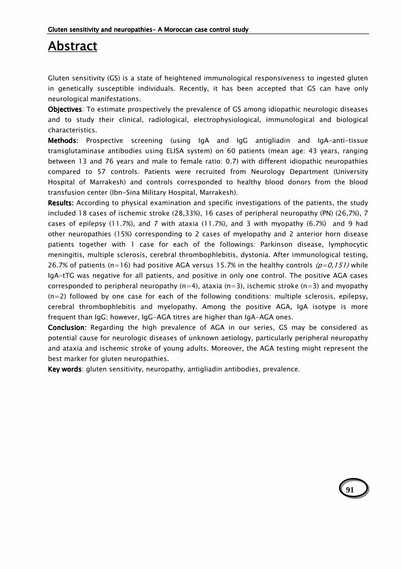
Gluten sensitivity and neuropathiesGluten sensitivity and neuropathiesGluten sensitivity and neuropathiesGluten sensitivity and neuropathies---- A Moroccan case control studyA Moroccan case control studyA Moroccan case control studyA Moroccan case control study
91
AbstractAbstractAbstractAbstract
Gluten sensitivity (GS) is a state of heightened immunological responsiveness to ingested gluten
in genetically susceptible individuals. Recently, it has been accepted that GS can have only
neurological manifestations.
ObjectivesObjectivesObjectivesObjectives: To estimate prospectively the prevalence of GS among idiopathic neurologic diseases
and to study their clinical, radiological, electrophysiological, immunological and biological
characteristics.
Methods: Methods: Methods: Methods: Prospective screening (using IgA and IgG antigliadin and IgA-anti-tissue
transglutaminase antibodies using ELISA system) on 60 patients (mean age: 43 years, ranging
between 13 and 76 years and male to female ratio: 0.7) with different idiopathic neuropathies
compared to 57 controls. Patients were recruited from Neurology Department (University
Hospital of Marrakesh) and controls corresponded to healthy blood donors from the blood
transfusion center (Ibn-Sina Military Hospital, Marrakesh).
Results: Results: Results: Results: According to physical examination and specific investigations of the patients, the study
included 18 cases of ischemic stroke (28,33%), 16 cases of peripheral neuropathy (PN) (26,7%), 7
cases of epilepsy (11.7%), and 7 with ataxia (11.7%), and 3 with myopathy (6.7%) and 9 had
other neuropathies (15%) corresponding to 2 cases of myelopathy and 2 anterior horn disease
patients together with 1 case for each of the followings: Parkinson disease, lymphocytic
meningitis, multiple sclerosis, cerebral thrombophlebitis, dystonia. After immunological testing,
26.7% of patients (n=16) had positive AGA versus 15.7% in the healthy controls (p=0,151) while
IgA-tTG was negative for all patients, and positive in only one control. The positive AGA cases
corresponded to peripheral neuropathy (n=4), ataxia (n=3), ischemic stroke (n=3) and myopathy
(n=2) followed by one case for each of the following conditions: multiple sclerosis, epilepsy,
cerebral thrombophlebitis and myelopathy. Among the positive AGA, IgA isotype is more
frequent than IgG; however, IgG-AGA titres are higher than IgA-AGA ones.
Conclusion: Conclusion: Conclusion: Conclusion: Regarding the high prevalence of AGA in our series, GS may be considered as
potential cause for neurologic diseases of unknown aetiology, particularly peripheral neuropathy
and ataxia and ischemic stroke of young adults. Moreover, the AGA testing might represent the
best marker for gluten neuropathies.
Key wordsKey wordsKey wordsKey words: gluten sensitivity, neuropathy, antigliadin antibodies, prevalence.

Gluten sensitivity and neuropathiesGluten sensitivity and neuropathiesGluten sensitivity and neuropathiesGluten sensitivity and neuropathies---- A Moroccan case control studyA Moroccan case control studyA Moroccan case control studyA Moroccan case control study
92
ملخصملخصملخصملخصو . الحساسية المفرطة للغلوتين هي استجابة مناعية مبالغة ضد الغلوتين و مشتقـاته عند أشخاص ذو استعداد وراثي
و لهذا . أن يظهر بأعراض عصبية فقط دونما عوارض هضمية قد بينت األبحاث الطبية الحديثة أن هذا المرض يمكنهذه الحساسية المفرطة في األمراض العصبية المجهولة تأتي هذه الدراسة االستباقية من أجل تحديد معدل انتشار
متوسط (مريضا 60تم انجاز هذا البحث على . السبب و تبيان خواصها السريرية، اإلشعاعية، الفيزيولوجية و اإلحيائيةشاهدا سليما تم انتقـاؤهم 57في مقـابل ) 0,7: سنة و نسبة الذكور لإلناث 76و 13سنة محصورة بين 43العمر
استفـاد المشاركون من فحص بدني وفحص باألشعة و تحاليل مخبرية ضمت قياس تركيز . من بين المتبرعين بالدمى مضادات األجسام ضد ناقـالت الغليادين من نوع إضافة إل" ج"و " أ"مضادات األجسام ضد الغليادين من نوع
، )% 26,7(حالة من مرض اعتالل األعصاب 16، )28,3%(حالة جلطة دماغية 18أسفرت هذه الفحوصات عن ".أ"،حالتان من )6,7%(حاالت من االعتالل العضلي 3، )11,7%(حاالت من الرنح 7،)11,7%(حاالت من الصرع 7
مامي و حالة واحدة لكل من متالزمة باركنسون، التهاب السحايا، متالزمة االنضغاط النخاعي، مرض القرن النخاعي األأما التحليل . التصلب اللويحي، مرض اعتالل النخاع الشوكي و مرض خلل التوتر العضلي و التهاب الوريد الخثاري
وجب لمضادات األجسام ضد ذو تركيز م%) 15,7(شهود 9و ) %26,7(مريضا 16المناعي فـأسفر عن تسجيل في حين أن مضادات ناقـالت الغلوتامين كانت ذات تركيز موجب عند ) 0,151=الرقم االستداللي (الغليادين 3حاالت من مرض اعتالل األعصاب، 4: يتوزع المصابون بالحساسية الزائدة للغلوتين على الشكل اآلتي. شاهد واحد
ة الدماغية، حالتين لالعتالل العضلي إضافة الى حالة واحدة لكل من التصلب حاالت لكل من الرنح المخيخي و الجلطو قد أثبثت التحاليل المخبرية أن . اللويحي و الصرع و التهاب األوردة الدماغية الخثاري و متالزمة االنضغاط النخاعي
". ج"الغليادين من نوع هي األكثر ترددا لكن تركيزها يبقى أقـل من مضادات " أ"مضادات الغليادين من نوع نستنتج من كل ما سبق ان معدل االنتشار المهم للحساسية المفرطة للغلوتين تجعلها سببا محتمال لألمراض العصبية
. المجهولة المصدر و خصوصا مرض اعتالل األعصاب و الرنح المخيخي إضافة إلى الجلطة الدماغية .تين، المرض العصبي، مضادات األجسام ضد الغليادين، معدل االنتشارالحساسية المفرطة للغلو : الكلمات األساسيةالكلمات األساسيةالكلمات األساسيةالكلمات األساسية
RésuméRésuméRésuméRésumé
La sensibilité au gluten (SaG) est un état de réponse immunologique excessive aux protéines
ingérées du gluten, chez un individu génétiquement prédisposé. Récemment, il est apparu
Gluten sensitivity and neuropathiesGluten sensitivity and neuropathiesGluten sensitivity and neuropathiesGluten sensitivity and neuropathies---- A Moroccan case control studyA Moroccan case control studyA Moroccan case control studyA Moroccan case control study
93
que la SaG peut se manifester uniquement par des troubles neurologiques en dehors de tout
symptôme digestif.
ObjectifObjectifObjectifObjectif : Notre étude a pour objectif de mesurer la prévalence de la SaG au cours des
neuropathies idiopathiques et de déterminer leurs caractéristiques cliniques, radiologiques,
électro-physiologiques et biologiques.
Patients et méthodesPatients et méthodesPatients et méthodesPatients et méthodes : Nous avons recherché des marqueurs de la SaG, représentés par les
anticorps anti-gliadine (AGA, IgA et IgG) couplés aux anticorps anti-transglutaminase tissulaire
(Ac anti-tTG), chez 60 patients (âge moyen: 43 ans, allant de 13 à 76 ans, sex-ratio M/F: 0,7)
recrutés à partir du service de neurologie du Centre Hospitalier Universitaire de Marrakech et 57
témoins sélectionnés parmi des donneurs de sang sains.
RésultRésultRésultRésultatsatsatsats : Selon les données cliniques et para-cliniques, les patients de l’étude correspondaient
à 18 cas d’accident vasculaire cérébral ischémique (AVCi) (28,33%), 16 cas de neuropathies
périphériques (NP) (26,7%), 7 cas d’épilepsie (11,7%), 7 cas d’ataxie (11,7%), 3 cas de myopathies
(6,7%), 2 cas de maladie de la corne antérieure, et 1 cas pour chacune des pathologies
suivantes : syndrome de compression médullaire, syndrome parkinsonien, méningite
lymphocytaire, sclérose en plaques (SEP), myélopathie, la thrombophlébite cérébrale et la
dystonie.
Les AGA étaient positifs chez 26,7% de nos patients contre 15,7% chez les témoins (p=0,151),
alors que le dosage des anti-tTG était négatif pour tous les patients et positif chez un seul
témoin. Les patients AGA positifs correspondaient à 4 cas de NP, 3 cas d’ataxie (n=3), 3 cas
d’AVCi, 2 cas de myopathie suivi d’un cas pour chacune des neuropathies suivantes : la Sclérose
en plaques , l’épilepsie, la thrombophlébite cérébrale et le syndrome de compression médullaire.
ConclusionConclusionConclusionConclusion :::: Confrontées aux données de la littérature, les résultats de notre série permettraient
d’attribuer l’étiologie des neuropathies idiopathiques à une SaG, essentiellement la NP, l’ataxie
et l’AVCi du sujet jeune. En plus, le dosage des AGA constituerait un meilleur marqueur pour les
neuropathies au gluten.
Mots clésMots clésMots clésMots clés : Sensibilité au gluten, neuropathie, anticorps antigliadin, prévalence.

Gluten sensitivity and neuropathiesGluten sensitivity and neuropathiesGluten sensitivity and neuropathiesGluten sensitivity and neuropathies---- A Moroccan case control studyA Moroccan case control studyA Moroccan case control studyA Moroccan case control study
94
The QuestionnaireThe QuestionnaireThe QuestionnaireThe Questionnaire 1111----IdentityIdentityIdentityIdentity Full name : ……………………………………
…………….. Age :
……….
Sex : M : F :
ANNEXANNEXANNEXANNEX
Gluten sensitivity and neuropathiesGluten sensitivity and neuropathiesGluten sensitivity and neuropathiesGluten sensitivity and neuropathies---- A Moroccan case control studyA Moroccan case control studyA Moroccan case control studyA Moroccan case control study
95
2222----Medical Record Medical Record Medical Record Medical Record Diabetes
Type 1
Type 2
Treatment : Insulin Pills Diet
HBP
Treatment Yes No
Cardiopathy Type:
Tobaccoc …………….P.Y
3333----Disease’s evolution:Disease’s evolution:Disease’s evolution:Disease’s evolution: Mode of onset
Acute
Origin
:
Rural
Urban
Occupation
:
…………………………….
Department : …………………………………………………..
Intellectual level : Illiterate Primary Secondary university
Inclusion date : …………………………………
………………..
Coagulation dysfunction type :
Systemic diseases
Type:
Alcoholism
Amount :
Nephropathy
Type :
Drug’s Intoxication Type : nutrition deficiency Type : Tuberculosis yes
No: Treatment for tuberculosis Yes:
No: Recent viral infection Type : Celiac disease of the parents Yes :
No : Celiac disease in the family Yes :
No : Similar cases in the family Yes :
No :

Gluten sensitivity and neuropathiesGluten sensitivity and neuropathiesGluten sensitivity and neuropathiesGluten sensitivity and neuropathies---- A Moroccan case control studyA Moroccan case control studyA Moroccan case control studyA Moroccan case control study
96
Chronic
Evolution remittent progressive
One Episode
-Introduction age of gluten …………………………………………….. - Gluten free diet Yes No - Reasons for GFD …………………………………………………………………
4444----Clinical analysisClinical analysisClinical analysisClinical analysis :::: AAAA.... AtaxiasAtaxiasAtaxiasAtaxias Proprioceptive Cerebellar Peripheral vestibular Central vestibular
B. Ischemic strokeIschemic strokeIschemic strokeIschemic stroke • Motor signs : • Sensitive signs : • Consciousness and Glasgow score : • Seizures : yes No Type :………………………………. • Topography : • Dysarthria :
CCCC.... Peripheral neuropathiesPeripheral neuropathiesPeripheral neuropathiesPeripheral neuropathies Weakness Proximal Distal
Paresthesia Distal Radicular
Hypoesthesia Superficial Tactile
Profound Vibratory
Heat and pain
arthrocinetic
Cranial nerves dysfunction
Type : …………………………………………
Complications :
Respiratory failure
Swallowing dysfunction

Gluten sensitivity and neuropathiesGluten sensitivity and neuropathiesGluten sensitivity and neuropathiesGluten sensitivity and neuropathies---- A Moroccan case control studyA Moroccan case control studyA Moroccan case control studyA Moroccan case control study
97
D. MyopathiesD. MyopathiesD. MyopathiesD. Myopathies • Weakness :
Bilateral : Yes No Symmetric : Yes No
• Dermatologic signs : yes No description……………………..........................
5555----InvestigationsInvestigationsInvestigationsInvestigations A/ NeuroA/ NeuroA/ NeuroA/ Neuro----ImagingImagingImagingImaging Brain scanner :
MRI :
Echography-Echo Doppler :
B/ElectroB/ElectroB/ElectroB/Electro----physiological explorationsphysiological explorationsphysiological explorationsphysiological explorations :::: • EMG :
Axonal :
Demyelinating : Mononeuropathy : Polyneuropathy Mononeuropathy multiplex : Polyradiculoneuropathy : Motor : Sensitive : Other features :
• EEG : • PEV : • PEA :
C/ BiologyC/ BiologyC/ BiologyC/ Biology
HgB : Urea : WBC : Serum creatinine : CRP : SR : Serum glycaemia : HIV serology Lipid Profile Triglycerids : Total Cholesterol HDL : LDL : VLDL :
Liver function ALAT : ASAT : Total Bilirubin: Unconjugated Bilirubin: Conjugated Bilirubin: alkaline phosphatase : Gamma GT :
Apo A : ………………………. ApoB :………………………..
Muscle enzymes CPK: CSF exploration Cells : Proteins : Glucose : Bactériology : Abnormal cells research
Serum proteins electrophoresis
AAN :………………… Anti SSb :………………………… Anti SSa :……………………….. AntiDNA :………………..

Gluten sensitivity and neuropathiesGluten sensitivity and neuropathiesGluten sensitivity and neuropathiesGluten sensitivity and neuropathies---- A Moroccan case control studyA Moroccan case control studyA Moroccan case control studyA Moroccan case control study
98
Measurement of IgGMeasurement of IgGMeasurement of IgGMeasurement of IgG----AGA: AGA: AGA: AGA: Positive Negative The rate : …………………..
Measurement of IgA antiMeasurement of IgA antiMeasurement of IgA antiMeasurement of IgA anti----tTG:tTG:tTG:tTG: Positive Negative IgA deficiency: yes No The rate of IgA-tTG: ………………… The rate of IgG-tTG: ………………...
CONCLUSION:CONCLUSION:CONCLUSION:CONCLUSION: ………………………………………………………….…….

Gluten Gluten Gluten Gluten sensitivity and neuropathiessensitivity and neuropathiessensitivity and neuropathiessensitivity and neuropathies---- A Moroccan case control studyA Moroccan case control studyA Moroccan case control studyA Moroccan case control study
99
REFERENCESREFERENCESREFERENCESREFERENCES

Gluten Gluten Gluten Gluten sensitivity and neuropathiessensitivity and neuropathiessensitivity and neuropathiessensitivity and neuropathies---- A Moroccan case control studyA Moroccan case control studyA Moroccan case control studyA Moroccan case control study
100
[1] B.Admou MS, F.Bienvenu, L.Chabaa. Immunologic diagnosis of celiac disease in children. Report progress. 2009.
[2] Hadjivassiliou M SD, et al. Gluten sensitivity: from gut to brain. Lancet Neurol. 2010;9:318–30.
[3] Dowd B. WSJ, Gee S. Aretaeus, and the Coeliac Affection. Br Med J. 1974;2(5909):45-7 [4] FW. P. From Aretaeus to Crosby: a history of coeliac disease. BMJ. 1988;
( 297): 1646-9 [5] Gobbi G BF, Greco L et al. Coeliac disease, epilepsy, and cerebral calcifications. Lancet.
1992;340(8817): 439–43. [6] SV. H. Celiac disease, its specific treatment andcure without nutritional relapse.JAMA.
1932: 99: 448. [7] Dicke WK. Simple dietary treatment for the syndrome of GheeHerter. Ned Tijdschr
Geneeskd. 1941; 85:1715. [8] Van De Kamer JH WH, Dicke WK. . An investigation into the injurious constituents of
wheat in connection with their action on patients with coeliac disease. Acta Paediatr 1953;42: 232.
[9] JW. P. Observation on the aetiology of idiopathic steatorrhoea; jejunal and lymph-node biopsies. . Br Med J 1954( 4900): 1318-21.
[10] Sakula j SM. celiac disease with atrophy of the small-intestine mucosa. Lancet 1957(273):876-7.
[11] Koning F SD, Cerf-Bensussan N, Sollid M.L. Pathomechanisms in celiac disease. Best Practice and Research Clinical Gastroenterology 2005;19,(3): 373-87.
[12] A. E. Coeliac disease: previous family studies. Genetics of coeliac disease. Lancaster: MTP:197–200.
[13] Greco L RR, Coto I et al. The first large population based twin study of coeliac disease. . Gut. 2002( 50):624–8.
[14] Shewry P.R. TA. The prolamin storage proteins of cereal seeds: structure and evolution. . Biochem J 1990(276): 1–12.
[15] Holopainen P AM, Sistonen P et al. CD28/CTLA4 gene region on chromosome 2q33 confers genetic susceptibility to celiac disease. A linkage and family-based association study. Tissue Antigens 1999(53):470–5.
[16] Rioux JD KH, Kocher K et al. . Genomewide search and association studies in a Finnish celiac disease population: Identification of a novel locus and replication of the HLA and CTLA4 loci. Am J Med Genet 2004( 130):345–50.
[17] Neuhausen SL FM, Camp NJ et al. Genome-wide linkage analysis for celiac disease in North American families. Am J Med Genet 2002(111): 1.
[18] Greco L BM, Corazza GR et al. . Existence of a genetic risk factor on chromosome 5q in Italian coeliac disease families. Ann Hum Genet 2001(65):35.
[19] Folk JE CS. Transglutaminases. . Meth Enzymol 1985( 113): 358–75. [20] Piacentini M RC, Farrace MG & Autuori F. Tissue transglutaminase in animal development.
. Int J Dev Biol 2002(44.): 655–62. [21] Aeschlimann D TV. Protein crosslinking in assembly and remodelling of extracellular
matrices: the role of transglutaminases. . Connect Tissue Res 2000( 41): 1–27. [22] Lorand L. GR. Transglutaminases: crosslinking enzymes with pleiotropic functions. . Nat
Rev Mol Cell Biol 2003( 4):140–56. [23] Haroon ZA HJ, Lai TS et al. . Tissue transglutaminase is expressed, active, and directly
involved in rat dermal wound healing and angiogenesis. . FASEB J 1999( 13): 1787–815. [24] Molberg Ø MS, Ko¨rner R et al. . Tissue transglutaminase selectively modifies gliadin
peptides that are recognized by gut derived T cells in celiac disease. . Nat Med 1998( 4): 713–7.
[25] Van de Wal Y KY, van Veelen P et al. . Selective deamidation by tissue transglutaminase strongly enhances gliadin-specific T cell reactivity. J Immunol 1998( 161):1585–8.

Gluten Gluten Gluten Gluten sensitivity and neuropathiessensitivity and neuropathiessensitivity and neuropathiessensitivity and neuropathies---- A Moroccan case control studyA Moroccan case control studyA Moroccan case control studyA Moroccan case control study
101
[26] Sjostrom H LK, Molberg Ø et al. Identification of a gliadin T cell epitope in coeliac disease: general importance of gliadin deamidation for intestinal T cell recognition. Scandanavian J Immunol. 1998(48):111–5.
[27] Vader W dRA, van de Wal Y et al. . Specificity of tissue transglutaminase explains cereal toxicity in celiac disease. J Exp Med 2002( 195):2002.
[28] VaderW KY, van Veelen P et al. . The gluten response in children with recent onset celiac disease. A highly diverse response towards multiple gliadin and glutenin derived peptides. . Gastroenterology. 2002(122):1729–37.
[29] Meresse B CZ, Ciszewski C et al. . Coordinated Induction by IL15 of a TCR-Independent NKG2D signaling pathway converts CTL into Lymphokine-activated killer cells in celiac disease. . Immunity 2004;21:357–66.
[30] Hu¨e S MJ-J, Monteiro RC et al. . A direct role for NKG2D/MICA interaction in villous atrophy during celiac disease. . Immunity. 2004(21):367-77.
[31] M.Rewers. Epidemiology of Celiac Disease: What Are the Prevalence, Incidence, and Progression of Celiac Disease? . Gastroenterology. 2005(128):S47–S51.
[32] Catassi C RI, Gandolfi L, Pratesi R, Fabiani E, El Asmar R, Frijia M, Bearzi I, Vizzoni L. Why is coeliac disease endemic in the people of the Sahara? Lancet 1999;354:647–8.
[33] S.Ridouane MB. Profil épidemiologique, clinique et évolutif de la maladie coeliaque dans la région de Marrakech Marrakech: Université Cadi Ayyad 2008.
[34] J W. Coeliac disease. Gastroenterology. 2007 April;35(4):226-30. [35] Abu-Zekry M DM, Catassi C and Fasano A. Prevalence of Celiac Disease in Egyptian
Children Disputes the East-West Agriculture-dependent Spread of the Disease. J Pediatr Gastroenterol Nutr. 2008 August;47(2):136-40.
[36] Ben Hariz M K-SM, Kallel L, Lahmer A, Halioui S, Bouraoui S, Laater A, Sliti A, Mahjoub A, Zouari B, Makni S, Maherzi A. Prevalence of celiac disease in Tunisia: mass-screening study in schoolchildren. Eur J Gastroenterol Hepatol. 2007 August;19(8):687-94.
[37] Fasano A CC. Current approaches to diagnosis and treatment of celiac disease: an evolving spectrum. Gastroenterology 2001;120:636–51.
[38] Book L HA, Black J, Feolo M, Zone JJ, Neuhausen SL. Prevalence and clinical characteristics of celiac disease in Down’s syndrome in a US study. Am J Med Genet. 2001;98:70–4.
[39] Giannotti A TG, Castro M, Virgilii F, Colistro F, Ferretti F, Digilio MC et al. Coeliac disease in Williams syndrome. J Med Genet 2001;38:767–8.
[40] Murray JA VDC, Plevak MF, Dierkhising RA, Zinsmeister AR, Melton LJ. Trends in the identification and clinical features of celiac disease in a North American community, 1950–2001. Clin Gastroenterol Hepatol. 2003;1:19–27.
[41] Cook B OR, Chapman B, Whitehead M, Burt M. A thirty-year (1970–1999) study of coeliac disease in the Canterbury region of New Zealand. N Z Med J. 2004;117:U772.
[42] Roux K. Formes cliniques de la maladie coeliaque en Pédiatrie. [Thèse de mèd.]. DIJON: Faculté de Médecine de DIJON; 2002.
[43] Holtmeier W cWF. Celiac disease. 2005 [cited; Available from: http://www.orphanet.net/data/patho/GB/uk-CD.pdf
[44] S.Guandalini PA, Vallee. Celiac Disease. [cited; Available from: http://emedicine.medscape.com/article/932104-overview
[45] Farell RJ KC. Celiac sprue. N Engl J Med. 2002;346(3):180-2. [46] Sanders DS CM, Hurlstone DP et al. . Association of adult coeliac disease with irritable
bowel syndrome: a case-control study in patients fulfilling ROME II criteria referred to secondary care. Lancet. 2001;358(9292):1504–8.
[47] DieterichW LE, Schopper H et al. Autoantibodies to tissue transglutaminase as predictors of celiac disease. Gastroenterology. 1998;115(6):1317–21.
[48] Rostom A DC, Cranney A et al. Celiac disease. . Celiac disease. In: Quality MAfHRa, ed.

Gluten Gluten Gluten Gluten sensitivity and neuropathiessensitivity and neuropathiessensitivity and neuropathiessensitivity and neuropathies---- A Moroccan case control studyA Moroccan case control studyA Moroccan case control studyA Moroccan case control study
102
Evidence Report/Technology Assessment: AHRQ Publication No. 04-E029-2. Rockville 2004.
[49] Clemente MG MM, Frau F et al. . Antitissue transglutaminase antibodies outside celiac disease. J Pediatr Gastroenterol Nutr 2002; 34(1): 31–4.
[50] Freeman HJ. Strongly positive tissue transglutaminase antibody assays without celiac disease. Can J Gastroenterol 2004;18(1):25–8.
[51] Dahele AV AM, Humphreys K & Ghosh S. Serum IgA tissue transglutaminase antibodies in coeliac disease and other gastrointestinal diseases. QJM 2001. 2001;94(4):195–205.
[52] Weiss B BY, Avidan B et al. Positive tissue transglutaminase antibodies with negative endomysial antibodies. Isr Med Assoc J 2004;6(1):9–12.
[53] Rittmeyer C. RJ. IgA deficiency causes false-negative endomysial antibody results in celiac disease. J Pediatr Gastroenterol Nutr. 1996;23(4):504–6.
[54] Cataldo F MV, Bottaro G et al. Celiac disease and selective immunoglobulin A deficiency. J Pediatr. 1997;131(2):306–8.
[55] Dickey W HDMS. Reliance on serum endomysial antibody testing underestimates the true prevalence of coeliac disease by one fifth. Scand J Gastroenterol. 2000;35(2):181–3.
[56] Dahele A KK, Bode J et al. Anti-endomysial antibody negative celiac disease: does additional serological testing help? . Dig Dis Sci 2001;46(1):214–21.
[57] Johnson TC DB, Memeo L et al. Relationship of HLA-DQ8 and severity of celiac disease: comparison of New York and Parisian cohorts. Clin Gastroenterol Hepatol. 2004;2(10):888–94.
[58] Ploski R EJ, Thorsby E & Sollid LM. On the HLA-DQ(alpha 1*0501, beta 1*0201)-associated susceptibility in celiac disease: a possible gene dosage effect of DQB1*0201. Tissue Antigens. 1993;41(4):173–7.
[59] Kaukinen K PJ, Maki M & Collin P. HLA-DQ typing in the diagnosis of celiac disease. Am J Gastroenterol. 2002;97(3):695–9.
[60] Thijs WJ vBJ, Kleibeuker JH & Kolkman JJ. Duodenal versus jejunal biopsies in suspected celiac disease. Endoscopy 2004;36(11):993–6.
[61] Dandalides SM CW, Petras R & Achkar E. Endoscopic small bowel mucosal biopsy: a controlled trial evaluating forceps size and biopsy location in the diagnosis of normal and abnormal mucosal architecture Gastrointest Endosc. 1989;35(3):197–200.
[62] Mariaud de Serre NP VV, Cellier C et al. Diagnostic étiologique d’une atrophie villositaire. Ann Pathol. 2001;21:319–33.
[63] Masafumi Ihara FM, Hideyuki Sawada, Takahiro Mezaki, Kotaro Mizutani,, Hiroshi Nakase MM, Hidekazu Tomimoto and Shun Shimohama. Gluten Sensitivity in Japanese Patients with adult-onset cerebellar ataxia. Internal Medicine. 2005;1351(.45).
[64] M.Hadjivassiliou DSS, et al. Gluten sensitivity: from gut to brain. Lancet Neurol. 2010;9:318–30.
[65] Holmes G. Neurological and psychiatric complications in celiac disease. In: Gobbi G, Anderman F, Naccarato S, Banchini G, eds. Epilepsy and other neurological disorders in coeliac disease. London: John Libbey 1997.
[66] Briani C ZG, Alaedini A, et al. Neurological complications of coeliac disease and autoimmune mechanisms: a prospective study. J Neuroimmunol. 2008(195):171–75.
[67] Hadjivassiliou M BS, Tongiorgi E, et al. Immune mediated acquired ataxias: Elsevier (in press).
[68] Hadjivassiliou M BS, Tongiorgi E, et al. Cerebellar ataxia as a possible organ specific autoimmune disease. Mov Disord. 2008;23:1370–77.
[69] Luostarinen L HS, Luostarinen M, Collin P, Pirttila T. Neuromuscular and sensory disturbances in patients with well treated celiac disease J Neurol Neurosurg Psychiatry 2003;74:490–94.
[70] Ludvigsson JF OT, Ekbom A, Montgomery SM. A population based study of celiac disease,

Gluten Gluten Gluten Gluten sensitivity and neuropathiessensitivity and neuropathiessensitivity and neuropathiessensitivity and neuropathies---- A Moroccan case control studyA Moroccan case control studyA Moroccan case control studyA Moroccan case control study
103
neurodegenerative and neuroinflammatory diseases. Aliment Pharmacol Ther 2007;25:1317–27.
[71] Hadjivassiliou M GR, Kandler RH, et al. Neuropathy associated with gluten sensitivity. J Neurol Neurosurg Psychiatry. 2006;77:1262–66.
[72] Mata S RD, Pinto F, Calabro A. Anti-tissue transglutaminase IgA antibodies in peripheral neuropathy and motor neuronopathy. Acta Neurol Scand. 2006;114:54–8.
[73] Chin RL SH, Brannagan TH, et al. Celiac neuropathy. Neurology 2003;60:1581–85. [74] rachid belfkih ea. Accident vasculaire cérébral ischémique révélant une maladie coeliaque.
Pan African Medical Journal. 2009;5(2). [75] S. Audia a, C. Duchêne a, M. Samsona, G. Muller a, P. Bielefeld a, F. Ricolfi b, M. Giroud
c, J.-F. Besancenot. Accident vasculaire cérébral ischémique de l’adulte jeune au cours de la maladie coeliaque. À propos de deux observations. La Revue de médecine interne. 2008;29:228-31.
[76] M Hadjivassiliou AG, G A B Davies-Jones, A J Lobo, T J Stephenson, A Milford-Ward. Does cryptic gluten sensitivity play a part in neurological illness? The Lancet. 1996 February 10;347.
[77] Cooke WT HG. Neurological and psychiatric complications in coeliac disease. Churchill Livingstone. 1984:197-213.
[78] Mayer M GL, Troncone R, Auricchio S, Marsh MN. Compliance of adolescents with coeliac disease with a gluten free diet. Gut. 1991;32:881-85.
[79] Hadjivassiliou M BS, Davies-Jones GA, Grunewald RA, Not T, Sanders DS, et al. . The humoral response in the pathogenesis of gluten ataxia. . Neurology. 2002;58:1221–6.
[80] Alaedini A OH, Briani C, et al. Immune cross-reactivity in coeliac disease: antigliadin antibodies bind to neuronal symapsin I. J Immunol 2007;178:6590–95.
[81] Alaedini A LN. Transglutaminase-independent binding of gliadin to intestinal brush border membrane and GM1 ganglioside. J Neuroimmunol 2006;177:167–72.
[82] Cervio E VU, Verri M, et al. Sera from patients with celiac disease and neurologic disorders evoke a mitochondrial-dependent apoptosis in vitro. Gastroenterol 2007;133:195–206.
[83] Hadjivassiliou M DSS, Richard A Grünewald, Nicola Woodroofe, Sabrina Boscolo, Daniel Aeschlimann. Gluten sensitivity: from gut to brain. Lancet Neurol. 2010;9:318-30.
[84] Cooke WT T-SW. Neurological disorders associated with adult coeliac disease. Brain 1966;89:683–722.
[85] Hadjivassiliou M GRA, Chattopadhyay A K. Clinical, radiological, neurophysiological, and neuropathological characteristics of gluten ataxia. Lancet. 1998;352:1582–85.
[86] Rao DG HM. Sensory neuronopathy due to gluten sensitivity. Ann Indian Acad Neurol 2007;10 (suppl 2):16.
[87] Finelli PF MW, Ambler M, Kestenbaum D. Adult celiac disease presenting as cerebellar syndrome. Neurology 1980;30:245–49.
[88] Kaplan JG PD, Horoupian D, DeSouza T, Brin M, Schaumburg H. . Distal axonopathy associated with chronic gluten enteropathy: a treatable disorder. Neurology. 1988;38:642–45.
[89] Hadjivassiliou M GR, Sharrack B,Sanders D et al. Gluten ataxia in perspective: epidemiology, genetic susceptibility and clinical characteristics. Brain. 2003;126:685-91.
[90] Hadjivassiliou M SD, Woodroofe N, Williamson C, Grunewald RA. . Gluten ataxia. Cerebellum 2008;7:494–98.
[91] Pereira AC EM, Buttery PC, et al. Choreic syndrome and coeliac disease: a hitherto unrecognised association. Mov Disord. 2004;19:478–82.
[92] Hadjivassiliou M AP, Sanders DS, et al. Antibodies against TG6 as the only serological marker of gluten ataxia. Proceedings of the 13th International Coeliac Disease Symposium; 2009 April 6–8, 2009; Amsterdam; 2009.
[93] Luostarinen LK CP, Paraaho MJ, Maki MJ, Pirttila TA. Coeliac disease in patients with cerebellar ataxia of unknown origin. Ann Med. 2001;33:445–49.

Gluten Gluten Gluten Gluten sensitivity and neuropathiessensitivity and neuropathiessensitivity and neuropathiessensitivity and neuropathies---- A Moroccan case control studyA Moroccan case control studyA Moroccan case control studyA Moroccan case control study
104
[94] Wilkinson ID HM, Dickson JM et al. Cerebellar abnormalities on proton MR spectroscopy in gluten ataxia. J Neurol Neurosurg Psychiatry. 2005;76:1011-13.
[95] Wilkinson ID HM, Dickson JM, et al. Cerebellar abnormalities on proton MR spectroscopy in gluten ataxia. J Neurol Neurosurg Psychiatry 2005;76:1011–13.
[96] Chin RL SH, Brannagan TH, Green PH, Hays AP, Alaedini A, Latov N. Celiac neuropathy. Neurology. 2003;60:1581–5.
[97] Kelkar P RM, Murray J. Mononeuropathy multiplex associated with celiac disease. Muscle Nerve 1996;19:234–36.
[98] Brannagan TH HA, Chin SS, et al. Small-fiber neuropathy/ neuronopathy associated with celiac disease: skin biopsy findings. Arch Neurol. 2005;62:1574–78.
[99] Gibbons CH FR. Autonomic neuropathy and celiac disease. J Neurol Neurosurg Psychiatry 2005;76:579–81.
[100] Gefel D. DM, Ben-Valid E., Daraushe A.E., Lugassy G, Ben-Ami S. Recurrent stroke in a young patient with celiac disease and hyperhomocysteinemia. IMAJ 2002;4:222-3.
[101] El-Khairy L UPM, Nygard O, Refsum H., Vollset SE. Lifestyle and cardiovascular disease risk factors as determinants of total homocysteine in plasma: the Hordaland Homocysteine Study. Am J Clin Nutr 1999;70:1016±24.
[102] Hadjivassiliou M MM, Sanders DS, Williamson CA, Grunewald RA, Woodroofe NM, et al. Autoantibody targeting of brain and intestinal transglutaminase in gluten ataxia. . Neurology 2006;66:373–7.
[103] Goodwin FC BR, Millar J, Kirkham FJ. Celiac disease and childhood stroke. Pediatr Neurol 2004;31:139–42.
[104] Curione M BM, De Biase L, Viola F, Lo Russo L, Cardi E. Prevalence of coeliac disease in idiopathic dilated cardiomyopathy. Lancet. 1999;354:222-3.
[105] Karoui S SS, Kallel M, Boubaker J, Makni S, Filali A. Syndrome des antiphospholipides révélé par une thrombose porte au cours d’une maladie coeliaque. Rev Med Interne 2004;25:471–3.
[106] Dickson BC SC, Chetty R. Coeliac disease: an update for pathologists. J Clin Pathol. 2006;59(10):1008-16.
[107] Baudon JJ JC, Absalon YB, Morgant G, Cabrol S, Mougenot JF. Diagnosing celiac disease: a comparison of human tissue transglutaminase antibodies with antigliadin and antiendomysium antibodies. Arch Pediatr Adolesc Med. 2004 Jun;158(6):584-8.
[108] Pellecchia MT SR, Filla A, De Michele G, Ciacci C, Barone P. Idiopathic cerebellar ataxia associated with celiac disease: lack of distinctive neurological features. J Neurol Neurosurg Psychiatry 1999;66:32–5.
[109] Schrodl D KF, Peter-Zimmer K, et al. Intrathecal synthesis of autoantibodies against tissue transglutaminase. J Autoimmun. 2004;22:335–40.
[110] Hadjivassiliou M AP, Sanders DS, et al. Antibodies against TG6 as the only serological marker of gluten ataxia. Proceedings of the 13th International Coeliac Disease Symposium April 6–8, 2009. 09.1; Amsterdam
[111] Kaukinen K C, P, Laurila, K et al. Resurrection of gliadin antibodies in celiac disease. Deamidated gliadin peptide antibody test provides additional diagnostic benefit. Scand J Gastroenterol. 2007;42:1428–33.
[112] Karoui S SM, Laatar AB, Zitouni M, Matri S, Laadhar L, et al. Prevalence of anticardiolipin and antibeta2-glycoprotein I antibodies in celiac disease. Dig Dis Sci. 2007;52:1096–100.
[113] R J Lock DSNAPT, D J Unsworth, J J Ward, A J Wills. Ataxia, peripheral neuropathy, and anti-gliadin antibody. Guilt by association? J Neurol Neurosurg Psychiatry. 2005(76):1601-3.
[114] Hadjivassiliou M, Williamson C.A, Woodroofe N. The immunology of gluten sensitivity: beyond the gut. Trends in immunology. 2004 November;25(11).
[115] Neto J I S VCACL, Magalhães F G, Silva G S. NEUROLOGICAL MANIFESTATIONS OF CELIAC DISEASE. Arq Neuropsiquiatr. 2004;62(4):969-72.

Gluten Gluten Gluten Gluten sensitivity and neuropathiessensitivity and neuropathiessensitivity and neuropathiessensitivity and neuropathies---- A Moroccan case control studyA Moroccan case control studyA Moroccan case control studyA Moroccan case control study
105
[116] Lahat E BE, Leshem M, Evans S, Scapa E. Prevalence of Celiac Antibodies in Children With Neurologic Disorders. Pediatr Neurol. 2000;22:393-6.
[117] Hadjivassiliou M GA, Davies-Jones GAB, et al. Does cryptic gluten sensitivity play a part in neurological illness? Lancet. 1996(347):369–71.
[118] Ihara M MF, Sawada H, et al. Gluten sensitivity in Japanese patients with adult-onset cerebellar ataxia. Intern Med 2006;45:135–40.
[119] Hadjivassiliou M DGR, S.B. Wharton, D.S. Sanders, R.A. Grunewald, Davies-Jones,MD. Sensory ganglionopathy due to gluten sensitivity. Neurology. 2010 September 14(75):1003-08.
[120] Chin RL SH, Brannagan TH, et al. Celiac neuropathy. Neurology. 2003(60):1566-68. [121] Bürk K BS, Müller CA, et al. Sporadic cerebellar ataxia associated with gluten sensitivity.
Brain. 2001;124:1013–19. [122] Abele M SL, Schwartz S, Klockgether T. Prevalence of antigliadin antibodies in ataxia
patients. Neurology 2003;60:1674–75. [123] D. Wong MD, M. Schulzer, M. Nimmo, B.R. Leavitt, S.D. Spacey. Ataxia and the role of
antigliadin antibodies. Can J Neurol Sci. 2007;34:193-6. [124] Burk K M-LF, Georg Lamprecht,Guenter Roth, Patrice Decker,Michael Weller, Hans-Georg
Rammensee, Wolfang Oertel. Neurological Symptoms in Patients with Biopsy Proven Celiac Disease. Mov Disord. 2009;24(16):2358-62.
[125] BUSHARA KO. Neurologic Presentation of Celiac Disease. GASTROENTEROLOGY 2005;128:S92–S97. 2005;128:S92–S7.
[126] Luostarinen LK CP, Paraaho MJ, Maki MJ, Pirttila TA. Coeliac disease in patients with cerebellar ataxia of unknown origin. Ann Med 2001;33:445–49.
[127] Anheim M DB, Echaniz-Laguna A, Fleury M, Grucker M, Tranchant C. Ataxia associated with gluten sensitivity, myth or reality? . Rev Neurol 2006;162: 214–21.
[128] Hadjivassiliou M GA, Davies-Jones GAB, Lobo A, Stephenson TJ, Milford-Ward A. Is cryptic gluten sensitivity an important cause of neurological illness? . Lancet 1996;347:369–71.
[129] Abele M BK, Schöls L, et al. The aetiology of sporadic adult onset ataxia. Brain. 2002;125:961–68.
[130] Bushara KO GS, Shill H, Goldfarb LG, Hallett M. Gluten sensitivity in sporadic and hereditary ataxia. Ann Neurol. 2001;49(540–43):540–43.
[131] Luostarinen L HS, Luostarinen M, Collin P, Pirttila T. Neuromuscular and sensory disturbances in patients with well treated celiac disease. J Neurol Neurosurg Psychiatry 2003;74:490–94.
[132] Ludvigsson JF OT, Ekbom A, Montgomery SM. A population based study of celiac disease, neurodegenerative and neuroinfl ammatory diseases. Aliment Pharmacol Ther. 2007;25:1317–27.
[133] Ludvigsson JF WJ, Card T, Appelros P. Risk of Stroke in 28,000 Patients with Celiac Disease: A Nationwide Cohort Study in Sweden. J Stroke Cerebrovasc Dis. 2011 Jul 4.
[134] Ludvigsson JF. DFU, Ekbom A., Montgomery SM. Vascular disease in a population-based cohort of individuals hospitalised with coeliac disease. Heart. 2007;93:1111-5.
[135] Henriksson KG HC, Norrby K, Walan A. Polymyositis and adult celiac disease. Acta Neurol Scand 1982(65):301–19.
[136] Selva-O’Callaghan A CF, De Torres I, Palou E, Grau-Junyent JM, Villardell-Tarres M. Celiac disease and antibodies associated with celiac disease in patients with inflammatory myopathy. Muscle Nerve 2007(35):49–54.
[137] Farre´ C HP, Vilar P, Varea V, Aldeguer X, Carnicer J, et al. Serological markers and HLA-DQ2 haplotype among first-degree relatives of celiac patients. Dig Dis Sci 1999;44:2344-9.
[138] Cronin CC JL, Feighery C, Shanahan F, Abuzakouk M, Ryder DQ, Whelton M, Callaghan N. . Coeliac disease and epilepsy. QJM 1998;91:303–8.

Gluten Gluten Gluten Gluten sensitivity and neuropathiessensitivity and neuropathiessensitivity and neuropathiessensitivity and neuropathies---- A Moroccan case control studyA Moroccan case control studyA Moroccan case control studyA Moroccan case control study
106
[139] Gobbi G AP, Zaniboni MG, Lambertini A, Ambrosioni G, Tassinari CA. . Celiac disease, posterior cerebral calcifications and epilepsy. Brain Dev 1992(14):23-9.
[140] Visakorpi J KP, Pelkonen P. Intestinal malabsorption: a clinical study of 22 children over 2 years of age. Acta Paediatr Scand 1970(59):273–80.
[141] Ranua J LK, Auvinen A, et al. Celiac disease-related antibodies in an epilepsy cohort and matched reference population. Epilepsy Behav 2005(6):388–92.
[142] Gobbi G. Coeliac disease, epilepsy and cerebral calcifications. Brain Dev 2005 Apr;27(3):189-200.
[143] Haghighi AB AN, Mokhtari M, Geramizadeh B, Lankarani KB. Multiple sclerosis and gluten sensitivity. Clin Neurol Neurosurg 2007(109):651-53.
[144] Hadjivassiliou M SD, Grunewald RA. Multiple sclerosis and occult gluten sensitivity. Neurology 2004;62:2326–27.
[145] Jacob S ZM, Kenton A, Allroggen H. Gluten sensitivity and neuromyelitis optica: two case reports. J Neurol Neurosurg Psychiatry. 2005;76:1028–30.
[146] Jarius S JS, Waters P, Jacob A, Littleton E, Vincent A. Neuromyelitis optica in patients with gluten sensitivity associated with antibodies to aquaporin-4. J Neurol Neurosurg Psychiatry. 2008;79:1084.
[147] Esin Beyan MP, Cengiz Beyan. Deep vein thrombosis associated with celiac disease. Brastil Lek Listy. 2009;110(4):263-4.
[148] Bahloul M CA, Khlaf-Bouaziz N et al. Celiac disease, cerebral venous thrombosis and protein S deficiency, a fortuitous association? J Mal Vasc. 2005;30:228-30.
[149] Cartalat-Carel S PP, Carpentier A, Delattre JY. Chronic meningitis before diagnosis of celiac disease. Rev Neurol (Paris). 2002;158(4):467-9.
[150] Sobstyl M ZM, Dzierzęcki S, Mossakowski Z, Szczałuba K. Successful bilateral pallidal stimulation in a patient with isolated lower limb dystonia coexistent with Langerhans cell histiocytosis and coeliac disease. Neurol Neurochir Pol. 2011 Sep-Oct;45(5):514-9.
[151] Hall DA PJ, Benke T. Paroxysmal nonkinesigenic dystonia and celiac disease. Mov Disord. 2007 Apr 15;22(5):708-10.
[152] K.J. Brown VJ, H. Herfarth, M. Castillo. White Matter Lesions Suggestive of Amyotrophic Lateral Sclerosis Attributed to Celiac Disease. Am J Neu Rad 2010 May;31:880-1.
[153] Turner M CG, Quaghebeur G, et al. A case of celiac disease mimicking amyotrophic lateral sclerosis. Nat Clin Pract Neurol. 2007;3:581–84.
[154] Hadjivassiliou M SD, Woodroofe N, Williamson C, Grunewald RA. Gluten ataxia. Cerebellum. 2008(7):494–98.
[155] Chin RL TV, Green PHR, Sander HW, Brannagan TH, Latov N. Multifocal axonal polyneuropathy in celiac disease. Neurology 2006(66):1923–25.
[156] Rao DG HM. Sensory neuronopathy due to gluten sensitivity. Ann Indian Acad Neurol. 2007;10(suppl 2):16.
[157] Brannagan TH HA, Chin SS, et al. Small-fi ber neuropathy/ neuronopathy associated with celiac disease: skin biopsy findings. Arch Neurol 2005;62:1574–78.
[158] Goodwin FC BR, Millar J, Kirkham FJ. Celiac Disease and Childhood Stroke. Pediatr Neurol. 2004;31:139-43.
[159] Doğan M. PE, Cagan E., Akbayram S, Acikgoz M, Caksen H, Uner A, Cesur Y. Stroke and dilated cardiomyopathy associated with celiac disease. World J Gastroenterol 2010;16(18):2302-4.
[160] El Moutawakil B CN, Sibai M, Moutawakil F, Rafai M, Bourezgui, Sissi I. Celiac disease and ischemic stroke. Rev Neurol. 2009 Nov;165(11):962-6.
[161] Kleopa KA KK, Zamba-Papanicolau E, Kyriakides T. Reversible inflammation and vacuolar myopathy with vitamin. Muscle Nerve. 2005;31:260 –5.
[162] Kozanoglu E BS, Goncu MK. Proximal myopathy as an unusual presenting feature of celiac disease. Clin Rheumatol. 2005;24:76 –8.

Gluten Gluten Gluten Gluten sensitivity and neuropathiessensitivity and neuropathiessensitivity and neuropathiessensitivity and neuropathies---- A Moroccan case control studyA Moroccan case control studyA Moroccan case control studyA Moroccan case control study
107
[163] Marie I LF, Hachulla E, Antonietti M, François A, Levesque H, et al. An uncommon association: celiac disease and dermatomyositis in adults. Clin Exp Rheumatol. 2001;19:201-3.
[164] Hadjivassiliou M CA, Grünewald RA, et al. Myopathy associated with gluten sensitivity Muscle Nerve 2007;35:443–50.
[165] Fois A VM, Di Bartolo RM, Di Marco V. Celiac disease and epilepsy in pediatric patients. Childs Nerv Syst 1994;10:450–54.
[166] Toti P BP, Cano M, et al. Celiac disease with cerebral calcium and silica deposits: X-ray spectroscopic findings, an autopsy study. Neurology 1996;46:1088–92.
[167] Magaudda A DBB, De Marco P, Sfaello Z, Longo M, Colamaria V, Daniele O, Tortorella G, Tata MA, Di Perri R, et al. Bilateral occipital calcification, epilepsy and coeliac disease: clinical and neuroimaging features of a new syndrome. J Neurol Neurosurg Psychiatry. 1993(56):885-9.
[168] Luostarinen L DP, Collin P, Peraaho M, Maki M, Erila T, Pirttila T. Association between coeliac disease, epilepsy and brain atrophy. 2001(46):187–91.
[169] Paltola M KK, Dastidar P, et al. Hippocampal sclerosis in refractory temporal lobe epilepsy is associated with gluten sensitivity. J Neurol Neurosurg Psychiatry 2009(80):626–30.
[170] Lee ES PJ. Non ischemic central retinal vein occlusion associated with celiac disease. Rev Med Interne. 2005;80(2):157.
[171] Marzari Rea. Molecular dissection of the tissue transglutaminase autoantibody response in coeliac disease. J Immunol. 2001;166:4170-6.
[172] Fasano A CC. Current approaches to diagnosis and treatment of celiac disease: an evolving spectrum. [Review]. Gastroenterology. 2001;120:636±51.
[173] El Moutawakil B CN, Sibai M, Moutaouakil F, Rafai M, Bourezgui M, Slassi I. Ischemic stroke and celiac disease. Rev Neurol (Paris). 2009 Nov;165(11):962-6.
[174] Orbach H AN, Barzilai O, Boaz M, Ram M, Zandman-Goddard G, Shoenfeld Y. Autoantibody screen in inflammatory myopathies high prevalence of antibodies to gliadin. Ann N Y Acad Sci. 2009;1173:174-9.
[175] Iltanen S CP, Korpela M, Holm K, Partanen J, Polvi, A, et al. Celiac disease and markers of celiac disease latency in patients with primary Sjogren’s syndrome. Am J Gastroenterol. 1999;94:1042-6.
[176] Choi Y-C PG, Kim T-S, Sunwoo I-N, Steinert PM, Kim S-Y. Sporadic inclusion body myositis correlates with increased expression and cross-linking by transglutaminases 1 and 2. J Biol Chem. 1999;275:8703–10.
[177] Nicoletti A PF, Lo Fermo S, Sciacca A, Laisa P, Liberto A, Lanzafame S, Contraffatto D, D'Agate C, Russo A, Zappia M. Frequency of celiac disease is not increased among multiple sclerosis patients. Mult Scler. 2008;14(5):698-700.
[178] Saibeni S LA, Meucci G et al. Prevalence of hyperhomocysteinemia in adult gluten-sensitive enteropathy at diagnosis: role of B12, folate and genetics. Clin Gastroenterol Hepatol. 2005;3(6):574-80.

Gluten Gluten Gluten Gluten sensitivity and neuropathiessensitivity and neuropathiessensitivity and neuropathiessensitivity and neuropathies---- A Moroccan case control studyA Moroccan case control studyA Moroccan case control studyA Moroccan case control study
108
قسم الطبيبقسم الطبيبقسم الطبيبقسم الطبيب اقسم باهللا العظيم اقسم باهللا العظيم اقسم باهللا العظيم اقسم باهللا العظيم
. . . .أن أراقب الله في مهنتيأن أراقب الله في مهنتيأن أراقب الله في مهنتيأن أراقب الله في مهنتي
الظروف واألحوال بـاذال الظروف واألحوال بـاذال الظروف واألحوال بـاذال الظروف واألحوال بـاذال في كلفي كلفي كلفي كل. . . . وأن أصون حياة اإلنسان في كافة أدوارهاوأن أصون حياة اإلنسان في كافة أدوارهاوأن أصون حياة اإلنسان في كافة أدوارهاوأن أصون حياة اإلنسان في كافة أدوارها
. . . .والقـلقوالقـلقوالقـلقوالقـلق نقـاذها من الهالك والمرض واأللمنقـاذها من الهالك والمرض واأللمنقـاذها من الهالك والمرض واأللمنقـاذها من الهالك والمرض واأللموسعي في استوسعي في استوسعي في استوسعي في است
. . . . وأكتم سرهم وأكتم سرهم وأكتم سرهم وأكتم سرهم وأن أحفظ للنـاس كرامتهم ، وأستر عورتهم ،وأن أحفظ للنـاس كرامتهم ، وأستر عورتهم ،وأن أحفظ للنـاس كرامتهم ، وأستر عورتهم ،وأن أحفظ للنـاس كرامتهم ، وأستر عورتهم ،
تي الطبية للقريب تي الطبية للقريب تي الطبية للقريب تي الطبية للقريب رعاي رعاي رعاي رعاي وأن أكون على الدوام من وسائل رحمة اهللا ، باذالوأن أكون على الدوام من وسائل رحمة اهللا ، باذالوأن أكون على الدوام من وسائل رحمة اهللا ، باذالوأن أكون على الدوام من وسائل رحمة اهللا ، باذال
. . . . والبعيد ، للصالح والخاطئ ، والصديق والعدووالبعيد ، للصالح والخاطئ ، والصديق والعدووالبعيد ، للصالح والخاطئ ، والصديق والعدووالبعيد ، للصالح والخاطئ ، والصديق والعدو
. . . .ال ألذاهال ألذاهال ألذاهال ألذاه أسخره لنفع اإلنسانأسخره لنفع اإلنسانأسخره لنفع اإلنسانأسخره لنفع اإلنسان العلم،العلم،العلم،العلم،لب لب لب لب وأن أثابر على طوأن أثابر على طوأن أثابر على طوأن أثابر على ط
وأكون أخا لكل زميـل في المهنة وأكون أخا لكل زميـل في المهنة وأكون أخا لكل زميـل في المهنة وأكون أخا لكل زميـل في المهنة وأعلم من يصغرني،وأعلم من يصغرني،وأعلم من يصغرني،وأعلم من يصغرني، علمني،علمني،علمني،علمني،وأن أوقر من وأن أوقر من وأن أوقر من وأن أوقر من
الطبية متعاونين على البر الطبية متعاونين على البر الطبية متعاونين على البر والتقوىوالتقوىوالتقوىوالتقوى الطبية متعاونين على البر. . . .
نها تجاه اهللا نها تجاه اهللا نها تجاه اهللا نها تجاه اهللا نقية مما يشينقية مما يشينقية مما يشينقية مما يشي وأن تكون حياتي مصداق إيماني في سري وعالنيتي ،وأن تكون حياتي مصداق إيماني في سري وعالنيتي ،وأن تكون حياتي مصداق إيماني في سري وعالنيتي ،وأن تكون حياتي مصداق إيماني في سري وعالنيتي ،
.... ورسوله والمؤمنينورسوله والمؤمنينورسوله والمؤمنينورسوله والمؤمنين
شهيدشهيدشهيدشهيد واهللا على ما أقولواهللا على ما أقولواهللا على ما أقولواهللا على ما أقول

جامعة القاضي عياض كلية الطب و الصيدلة
مراكش84أطروحة رقم 2012 سنة
الحساسية المفرطة للغلوتين وأمراض الجھاز العصبي لحا+ت و الش'واھدل دراسة مغربية
,طروحةا
قدمت ونوقشت ع�نية يوم
من طرف رمانمحمد رضى بو السيد
مراكشب 1985مارس 22المزداد في
ة في الطبالنيل شھادة الدكتور
: الكلمات ا,ساسية معدل ا)نتشار، مضادات ا'جسام ضد الغليادين، صبيالحساسية المفرطة للغلوتين، المرض الع
اللجنة
ا,بحاث ..... مركز
PCIM
الرئيس
المشرف
القضاة
العلوي اليزيدي .ع السيد الجھاز التنفسي علم أمراض أستاذ في PCIMمدير مختبر ا'بحاث
أدمو .ا السيد علم المناعة فيمبرز تاذأس كساني .ن السيد
أمراض الجھاز العصبيعلم في أستاذ روسالبو .م السيد
تخصص ا'عصاب–ا'طفال طب فيمبرز أستاذ شبعى .ل السيدة
علم ا'حياء أستاذة في أمين.م السيد علم ا)حصاءأستاذ مبرز في

Gluten Gluten Gluten Gluten sensitivity and neuropathiessensitivity and neuropathiessensitivity and neuropathiessensitivity and neuropathies---- A Moroccan case control studyA Moroccan case control studyA Moroccan case control studyA Moroccan case control study
110