giovanoli et al bbi 2015 zora
TRANSCRIPT
Zurich Open Repository andArchiveUniversity of ZurichMain LibraryStrickhofstrasse 39CH-8057 Zurichwww.zora.uzh.ch
Year: 2016
Prenatal immune activation causes hippocampal synaptic deficits in theabsence of overt microglia anomalies
Giovanoli, Sandra ; Weber-Stadlbauer, Ulrike ; Schedlowski, Manfred ; Meyer, Urs ; Engler, Harald
Abstract: Prenatal exposure to infectious or inflammatory insults can increase the risk of developingneuropsychiatric disorder in later life, including schizophrenia, bipolar disorder, and autism. Thesebrain disorders are also characterized by pre- and postsynaptic deficits. Using a well-established mousemodel of maternal exposure to the viral mimetic polyriboinosinic-polyribocytidilic acid [poly(I:C)], weexamined whether prenatal immune activation might cause synaptic deficits in the hippocampal formationof pubescent and adult offspring. Based on the widely appreciated role of microglia in synaptic pruning,we further explored possible associations between synaptic deficits and microglia anomalies in offspringof poly(I:C)-exposed and control mothers. We found that prenatal immune activation induced an adultonset of presynaptic hippocampal deficits (as evaluated by synaptophysin and bassoon density). Theearly-life insult further caused postsynaptic hippocampal deficits in pubescence (as evaluated by PSD95and SynGAP density), some of which persisted into adulthood. In contrast, prenatal immune activationdid not change microglia (or astrocyte) density, nor did it alter their activation phenotypes. The prenatalmanipulation did also not cause signs of persistent systemic inflammation. Despite the absence of overtglial anomalies or systemic inflammation, adult offspring exposed to prenatal immune activation displayedincreased hippocampal IL-1 levels. Taken together, our findings demonstrate that age-dependent synapticdeficits and abnormal pro-inflammatory cytokine expression can occur during postnatal brain maturationin the absence of microglial anomalies or systemic inflammation.
DOI: https://doi.org/10.1016/j.bbi.2015.09.015
Posted at the Zurich Open Repository and Archive, University of ZurichZORA URL: https://doi.org/10.5167/uzh-120354Journal ArticleAccepted Version
The following work is licensed under a Creative Commons: Attribution-NonCommercial-NoDerivatives4.0 International (CC BY-NC-ND 4.0) License.
Originally published at:Giovanoli, Sandra; Weber-Stadlbauer, Ulrike; Schedlowski, Manfred; Meyer, Urs; Engler, Harald (2016).Prenatal immune activation causes hippocampal synaptic deficits in the absence of overt microglia anoma-lies. Brain, Behavior, and Immunity, 55:25-38.DOI: https://doi.org/10.1016/j.bbi.2015.09.015
1
Giovanoli et al (in press) Brain Behavior Immunity
Prenatal immune activation causes hippocampal synaptic deficits in
the absence of overt microglia anomalies
Sandra Giovanoli1, Ulrike Weber-‐Stadlbauer1,2, Manfred Schedlowski3, Urs Meyer1,2,*,
Harald Engler3
1Physiology and Behavior Laboratory, ETH Zurich, Schwerzenbach, Switzerland.
2Institute of Pharmacology and Toxicology, University of Zurich-‐Vetsuisse, Zurich, Switzerland.
3Institute of Medical Psychology and Behavioral Immunobiology, University Hospital Essen,
University of Duisburg-‐Essen, Essen, Germany.
*Correspondence:
Urs Meyer, Ph.D.
Institute of Pharmacology and Toxicology
University of Zurich-‐Vetsuisse
Winterthurerstrasse 260,
8057 Zurich,
Switzerland
E-‐mail:[email protected]
Tel.: +41 44 635 88 44; Fax.:+41 44 635 89 10
Running title: Prenatal immune activation and synaptic development.
Key words: Autism; cytokines; inflammation; maternal immune activation; microglia;
poly(I:C); schizophrenia; synapse.

2
Abstract
Prenatal exposure to infectious or inflammatory insults can increase the risk of developing
neuropsychiatric disorder in later life, including schizophrenia, bipolar disorder, and
autism. These brain disorders are also characterized by pre-‐ and postsynaptic deficits.
Using a well-‐established mouse model of maternal exposure to the viral mimetic
polyriboinosinic-‐polyribocytidilic acid [poly(I:C)], we examined whether prenatal immune
activation might cause synaptic deficits in the hippocampal formation of pubescent and
adult offspring. Based on the widely appreciated role of microglia in synaptic pruning, we
further explored possible associations between synaptic deficits and microglia anomalies in
offspring of poly(I:C)-‐exposed and control mothers. We found that prenatal immune
activation induced adult onset of presynaptic hippocampal deficits (as evaluated by
synaptophysin and bassoon density). The early-‐life insult further caused postsynaptic
hippocampal deficits in pubescence (as evaluated by PSD95 and SynGAP density), some of
which persisted into adulthood. In contrast, prenatal immune activation did not change
microglia (or astrocyte) density, nor did it alter their activation phenotypes. The prenatal
manipulation did also not cause signs of persistent systemic inflammation. Despite the
absence of overt glial anomalies or systemic inflammation, adult offspring exposed to
prenatal immune activation displayed increased hippocampal IL-‐1β levels. Taken together,
our findings demonstrate that age-‐dependent synaptic deficits and abnormal pro-‐
inflammatory cytokine expression can occur during postnatal brain maturation in the
absence of microglial anomalies or systemic inflammation.

3
1. Introduction
Maternal infectious or inflammatory insults during pregnancy have been repeatedly
implicated in the etiology of developmental neuropsychiatric disorders, including
schizophrenia (Brown and Derkits, 2010; Canetta et al., 2014b), autism (Atladóttir et al.,
2010; Brown et al., 2014), and bipolar disorder (Canetta et al., 2014a; Parboosing et al.,
2013). Preclinical support for these epidemiological associations has been obtained by
various translational rodent models demonstrating multiple brain and behavioral
abnormalities following prenatal exposure to infection and/or immune activation
(reviewed in Boksa, 2010; Harvey and Boksa, 2012; Meyer, 2014; Meyer and Feldon, 2010).
Increasing evidence suggests that cytokine-‐associated inflammatory events, together with
downstream pathophysiological effects such as oxidative stress and (temporary)
macronutrient and micronutrient deficiency, are critical in mediating the adverse effects of
maternal infection on the fetal system (reviewed in Meyer, 2014; Miller et al., 2013).
Disruption of normal fetal development (Meyer et al., 2008; Vuillermot et al., 2010),
together with subsequent changes in brain maturation (Hadar et al., 2015; Piontkewitz et
al., 2011; Richetto et al., 2014), may then confer increased risk of behavioral and cognitive
dysfunctions in later life.
In recent years, there has been growing interest in the potential role of microglia in
normal brain development and maturation, especially in those events that relate to
synaptic pruning (Bilimoria and Stevens, 2015; Kreisel et al., 2014; Paolicelli et al., 2011;
Schafer et al., 2012; Schafer and Stevens, 2013). Microglia are the major immunocompetent
cells residing in the brain parenchyma and can adopt different morphological
characteristics and functions: In their ramified state, they constantly survey the brain

4
microenvironment to detect and to respond to alterations in brain homeostasis, which in
turn can induce the transition of ramified into phagocytic microglia (Gomez-‐Nicola and
Perry, 2015). In contrast to ramified microglia with small cell bodies and extensive
arborization, phagocytic microglia are characterized by enlarged cell soma and less
arborization. As phagocytes, they can quickly remove cellular debris and dying neurons,
thereby preventing possible toxic damage to neighboring cells.
The phagocytic capacity of microglia is particularly important during early brain
development and subsequent maturation, where they can contribute to the removal of
excessive synapses in certain neuronal pathways (Bilimoria and Stevens, 2015; Kreisel et
al., 2014; Paolicelli et al., 2011; Schafer et al., 2012). For these reasons, it has been
suggested that abnormal microglia functions during critical periods of brain development
and maturation may be an important etiopathological mechanism linking
neuroinflammation to synaptic deficits (Rao et al., 2012). Synaptic deficits are a hallmark of
various neurodevelopmental brain disorders, including schizophrenia and autism
(reviewed in Eastwood, 2004; Ebrahimi-‐Fakhari and Sahin, 2015; Harrison, 2004). These
disorders have also been associated with altered microglia functions and associated
neuroinflammatory changes (Arion et al., 2007; Doorduin et al., 2009; Fillman et al., 2013;
Fung et al., 2014; Morgan et al., 2010; Tetreault et al., 2012; van Berckel et al., 2008; Volk et
al., 2015). It remains elusive, however, whether and to what extent such microglial
abnormalities may be responsible for the development of synaptic deficits as, for example,
seen in schizophrenia or autism (Catts et al., 2013; Onore et al., 2012).
Against these backgrounds, the present study aimed at exploring potential
associations between impaired synaptic development and microglial abnormalities in a
model of prenatal immune activation with relevance to neurodevelopmental disorders. We
5
used a well-‐established mouse model of maternal treatment with the viral mimetic
polyriboinosinic-‐polyribocytidilic acid [poly(I:C)], which is known to capture a wide
spectrum of neuronal and behavioural abnormalities relevant to psychiatric disorders,
especially schizophrenia and autism (reviewed in Meyer, 2014; Reisinger et al., 2015).
Prenatal poly(I:C) exposure in rats has also been shown to cause synaptic deficits (Forrest
et al., 2012; Oh-‐Nishi et al., 2010), but their possible association with microglial
abnormalities and related neuroimmune changes remain unexplored thus far.
Using the maternal poly(I:C) administration model in mice, we assessed the
expression of synaptic proteins, microglia density and activation, systemic and peripheral
inflammation, and selected behavioral functions. We focused our neuroanatomical
investigations on the hippocampus for two main reasons. First, it is one of the brain areas
strongly implicated in the neuropathology of schizophrenia and other brain disorders with
neurodevelopmental components (Harrison, 2004; Tamminga et al., 2010). Second, the
hippocampal formation is markedly affected by prenatal immune challenge and is of key
interest to current research in this field (reviewed in: Meyer and Feldon, 2009; Piontkevitz
et al., 2012a). The assessment of synaptic proteins included synaptophysin, bassoon,
synaptic Ras GTPase-‐activating protein 1 (SynGAP), and postsynaptic density protein 95
(PSD95). Synaptophysin and bassoon are two major presynaptic proteins that are localized
in or associated with presynaptic vesicles (Elferink and Scheller, 1995; Schoch and
Gundelfinger, 2006; Shin, 2014; Wiedenmann and Franke, 1985). On the other hand,
SynGAP and PSD95 are two major postsynaptic proteins that are enriched at postsynaptic
sites of excitatory synapses, where they critically help organizing and strengthening
excitatory receptor complexes and their associated signaling proteins (Kim et al., 1998;
Kim and Sheng, 2004; Sheng and Hoogenraad, 2007). Microglia density was examined by
6
quantification of cells immunoreactive for the ionized calcium-‐binding adaptor molecule 1
(Iba1), whereas their activation status was assessed through the analysis of microglia
morphology and cluster of differentiation 68 (CD68) immunoreactivity (Colton and
Wilcock, 2010; Franco and Fernández-‐Suárez, 2015; Ransohoff and Perry, 2015). In
addition, we measured the density of glial fibrillary acidic protein (GFAP)-‐positive
astrocytes and the levels of cytokines in plasma and hippocampus to assess the effects of
prenatal immune activation on astrogliosis and inflammatory signaling, respectively. To
confirm the negative influence of prenatal immune challenge on behavior, we measured
sensorimotor gating using the paradigm of prepulse inhibition (PPI) of the acoustic startle
reflex. PPI deficiency is a well-‐replicated behavioral manifestation emerging in rats and
mice exposed to prenatal immune activation (for a review, see Boksa, 2010; Meyer, 2014;
Meyer and Feldon, 2010; Reisinger et al., 2015) and is frequently observed in
neuropsychiatric disorders with neurodevelopmental components (Braff et al., 2001;
Swerdlow et al., 2008). All analyses were conducted during puberty and adulthood in order
to identify possible maturation-‐dependent effects of prenatal immune activation.
2. Methods
2.1. Animals
C57BL6/N mice were used throughout the study. Female and male breeders were obtained
from Charles River Laboratories (Sulzfeld, Germany) at the age of 10–14 weeks. Breeding
began after 2 weeks of acclimatization to the animal holding rooms, which were
temperature-‐ and humidity-‐controlled (21 ± 1°C, 55 ± 5%) facilities under a reversed light–
dark cycle. All animals had ad libitum access to food (Kliba 3430, Kaiseraugst, Switzerland)
7
and water. All procedures described in the present study had been previously approved by
the Cantonal Veterinarian's Office of Zurich. All efforts were made to minimize the number
of animals used and their suffering.
2.2. Maternal immune activation during pregnancy
For the purpose of the maternal immune challenge, C57BL6/N female mice were subjected
to a timed mating procedure as described previously (Meyer et al., 2005). Pregnant dams on
gestation day (GD) 9 were randomly assigned to receiving either a single injection of
poly(I:C) (potassium salt; Sigma-‐Aldrich, Buchs, St. Gallen, Switzerland) or vehicle. Poly(I:C)
(5 mg/kg; calculated based on the pure form poly(I:C)) was dissolved in sterile pyrogen-‐
free 0.9% NaCl (vehicle) solution to yield a final concentration of 1 mg/ml and was
administered intravenously (i.v.) into the tail vein under mild physical constraint. The dose
of poly(I:C) was selected based on previous dose-‐response studies (Meyer et al., 2005). A
total of 16 pregnant dams were used, half of which were allocated to the poly(I:C)
treatment, and the other half to the vehicle treatment.
The selected gestational window (i.e., GD 9) in mice corresponds roughly to the middle
of the first trimester of human pregnancy with respect to developmental biology and
percentage of gestation from mice to humans (Clancy et al., 2007). It was selected based on
our previous findings showing that poly(I:C) exposure on GD 9 leads to multiple behavioral
abnormalities in the adult offspring, including deficits in PPI (Meyer et al., 2005, 2006). We
have also verified the effectiveness of this poly(I:C) administration protocol in mice in
terms of the elicited cytokine-‐associated inflammatory response in maternal and fetal
tissues (e.g., Meyer et al., 2006).

8
2.3. Allocation and testing of the offspring
All offspring were weaned and sexed on postnatal day (PND) 21. Littermates of the same
sex were caged separately and maintained in groups of 3-‐5 animals per cage. Only male
animals were included in all experiments. Poly(I:C) and control offspring stemmed from
multiple independent litters (N = 8 for each prenatal treatment group) to avoid possible
confounds arising from litter effects. For each maturational stage (i.e., pubescence and
adulthood), 1 male offspring per litter was randomly selected for the initial PPI test and
subsequent immunohistochemical evaluations of glial and synaptic markers, leading to a
group size of N=8 offspring per treatment group and age. Pubescent and adult testing began
with the assessment of PPI on PND 35 and PND 84, respectively. Following a resting period
of 5-‐6 days, the animals were then sacrificed on PND 40 and PND 90, respectively, for the
purpose of the immunohistochemical evaluations in pubescence and adulthood.
The remaining offspring from poly(I:C) and control litters were used for the analysis of
plasma and hippocampal cytokines in pubescence (N = 10 per treatment group) and in
adulthood (N = 8 per treatment group). The animals were killed on PND 40 and PND 90 to
match the age of those offspring, which were assigned to the immunohistochemical
evaluations of glial and synaptic markers.
The pubescent and adult stages were defined based on the gradual attainment of sexual
maturity and age-‐specific behavioral discontinuities from younger to older animals (Spear,
2000) and based on our previous findings revealing a post-‐pubertal onset of PPI deficits
following poly(I:C)-‐induced prenatal immune activation in mice (Vuillermot et al., 2010).
9
2.4. Prepulse inhibition of the acoustic startle reflex
Sensorimotor gating was assessed using the paradigm of prepulse inhibition (PPI) of the
acoustic startle reflex. PPI of the acoustic startle reflex refers to the reduction in startle
reaction in response to a startle-‐eliciting pulse stimulus when it is shortly preceded by a
weak prepulse stimulus (Braff et al., 2001; Swerdlow et al., 2008). The apparatus consisted
of four startle chambers for mice (San Diego Instruments, San Diego, CA, USA) and has been
fully described elsewhere (Meyer et al., 2005). In the demonstration of PPI, the animals
were presented with a series of discrete trials comprising a mixture of 4 trial types. These
included pulse-‐alone trials, prepulse-‐plus-‐pulse trials, prepulse-‐alone trials, and no-‐
stimulus trials in which no discrete stimulus other than the constant background noise was
presented. The pulse and prepulse stimuli used were in the form of a sudden elevation in
broadband white noise level (sustaining for 40 and 20 ms, respectively) from the
background (65 dBA), with a rise time of 0.2–1.0 ms. In all trials, three different intensities
of pulse (100, 110, and 120 dBA) and three intensities of prepulse (71, 77, and 83 dBA,
which corresponded to 6, 12, and 18 dBA above background, respectively) were used. The
stimulus-‐onset asynchrony of the prepulse and pulse stimuli on all prepulse-‐plus-‐pulse
trials was 100 ms (onset-‐to-‐onset).
The protocol used for the PPI test was extensively validated before (e.g., Vuillermot et
al., 2010). A session began with the animals being placed into the Plexiglas enclosure. They
were acclimatized to the apparatus for 2 min before the first trial began. The first 6 trials
consisted of 6 startle-‐alone trials; such trials served to habituate and stabilize the animals’
startle response and were not included in the analysis. Subsequently, the animals were
presented with 10 blocks of discrete test trials. Each block consisted of the following: three
pulse-‐alone trials (100, 110, or 120 dBA), 3 prepulse-‐alone trials (+6, +12, or +18 dBA above
10
background), 9 possible combinations of prepulse-‐plus-‐pulse trials (3 levels of pulse × 3
levels of prepulse), and one no stimulus trial. The 16 discrete trials within each block were
presented in a pseudorandom order, with a variable interval of 15 s on average (ranging
from 10 to 20 s). For each of the 3 pulse intensities (100, 110, or 120 dBA), PPI was indexed
by percent inhibition of the startle response obtained in the pulse-‐alone trials by the
following expression: 100% × [1 − (mean reactivity on prepulse-‐plus-‐pulse trials/mean
reactivity on pulse-‐alone trials)], for each animal, and at each of the three possible prepulse
intensities (+6, +12, or +18 dBA above background). In addition to PPI, reactivity to pulse-‐
alone trials and prepulse-‐alone trials were also analyzed.
2.5. Immunohistochemistry
The animals were deeply anesthetized with an overdose of Nembutal (Abbott Laboratories,
North Chicago, IL, USA) and perfused transcardially with 0.9% NaCl, followed by 4%
phosphate-‐buffered paraformaldehyde solution containing 15% picric acid. The dissected
brains were postfixed in the same fixative for 6 h and processed for antigen retrieval
involving overnight incubation in citric acid buffer (pH 4.5) followed by a 90 s microwave
treatment at 480 W according to protocols established before (Giovanoli et al, 2013;
Vuillermot et al., 2010). The brains were then cryoprotected using 30% sucrose in PBS,
frozen with powdered dry ice, and stored at −80°C until further processing.
Perfused brain samples were cut coronally at 30 μm thickness from frozen blocks with
a sliding microtome. Eight serial sections were prepared for each animal and, after rinsing
in PBS, stored at −20°C in antifreeze solution (30% glycerol and 30% ethylene glycol in PBS
at 25 mM and pH 7.4) until further processing. For immunohistochemical staining, the slices
were rinsed three times for 10 min in PBS, and blocking was done in PBS, 0.3% Triton X-‐

11
100, 10% normal serum for 1 h at room temperature. The following primary antibodies
were used: Rabbit anti-‐Iba1 (Wako, Neuss, Germany; diluted 1:2,000), rat anti-‐CD68 (AbD
Serotec, Oxford, UK; diluted 1:5,000), rabbit anti-‐GFAP (Dako, Baar, Switzerland; diluted
1:5,000), rabbit anti-‐synaptophysin (Sigma, diluted 1:3,000), mouse anti-‐bassoon
(Stressgen Biotechnologies, Victoria, Canada; diluted 1:5,000), mouse anti-‐PSD95 (Pierce
Antibody Products, diluted 1:800), and rabbit anti-‐SynGap (Affinity BioReagents, Colorado,
USA; diluted 1:400). All primary antibodies were validated before (Giovanoli et al, 2013;
Nyffeler et al., 2006, 2007). They were diluted in PBS containing 0.3% Triton X-‐100 and 2%
normal serum, and the sections were incubated free-‐floating overnight at room
temperature. After three washes with PBS (10 min each), the sections were incubated for 1
h with the biotinylated secondary antibodies diluted 1:500 in PBS containing 2% NGS and
0.3% Triton X-‐100. Sections were washed again three times for 10 min in PBS and
incubated with Vectastain kit (Vector Laboratories, Burlingame, CA, USA) diluted in PBS for
1 h. After three rinses in 0.1 M Tris-‐HCl, pH 7.4, the sections were stained with 1.25% 3,3-‐
diaminobenzidine and 0.08% H2O2 for 10–15 min, rinsed again four times in PBS,
dehydrated, and coverslipped with Eukitt (Kindler, Freiburg, Germany).
2.6. Unbiased stereological estimations
The numbers of Iba1-‐, CD68-‐, or GFAP-‐immunoreactive cells were determined by unbiased
stereological estimations using the optical fractionator method (Gundersen et al., 1988).
With the aid of the image analysis computer software Stereo Investigator (version 6.50.1;
MicroBrightField, Williston, VT, USA), every section of a one-‐in-‐eight series was measured,
resulting in an average of 4-‐5 sections per brain sample. The following sampling
parameters were used: (1) a fixed counting frame with a width of 60 μm and a length of 60

12
μm; and (2) a sampling grid size of 200 × 150 μm. The counting frames were placed
randomly at the intersections of the grid within the outlined structure of interest by the
software. The cells were counted following the unbiased sampling rule using the 40× oil
lens [numerical aperture (NA), 1.3] and included in the measurement when they came into
focus within the optical dissector (Howard and Reed, 2005). All immunohistochemical
preparations were quantified in the dorsal cornu amonis 1 to 3 (CA1 –CA3) regions of the
hippcoampus (Bregma -‐1.3 to -‐2.7 mm), thereby including stratum oriens (so), stratum
radiatum (sr), and stratum lacunosum moleculare (slm). Preliminary analyses have
revealed no layer-‐specific effects of prenatal immune activation (data not shown), so that
the data representing the entire dorsal CA region are presented. In addition, the
immunohistochemical preparations were quantified in the dorsal stratum moleculare (sm)
of the dentate gyrus (DG; Bregma -‐1.3 to -‐2.7 mm). In view of the dorsal-‐ventral dichotomy
of the hippocampus, we extended parts of the stereological estimations to the ventral part
of the hippocampus (Bregma -‐2.9 to -‐3.5 mm), thereby including the same ventral layers
(CA: so, sr, slm; DG: sm) as for the dorsal subregion. All brain regions and layers were
delineated according to the “Mouse Brain in Stereotaxic Coordinates” (Franklin and Paxinos,
2008). These stereological methods were previously validated to capture overt microglia
anomalies under conditions of hippocampal inflammation (Giovanoli et al., 2013).
2.7. Assessment of microglia morphology
Iba1-‐immunoreactive microglia were visualized under the 63× oil lens [numerical aperture
(NA), 1.4] using a Zeiss Axiophot microscope (Carl Zeiss, Jena, Germany). Various
parameters of microglia cell morphology were assessed in the CA1 slm and DG sm
according to methods described before (Giovanoli et al., 2013). In brief, a counting frame of

13
100 μm × 100 μm was randomly placed into three sections of a one-‐in-‐eight series. All
microglia cells captured by the counting frame were included in the morphological
analyses, except when microglial processes were obscured by either background labeling or
other cells. 3 to 4 microglia cells in every section of a one-‐in-‐eight series (see above) were
traced using the software Stereo Investigator (version 6.50.1; MicroBrightField), for which
cell soma area and the number of primary and secondary processes were estimated, giving
a total of 12 to 16 cells per animal, as described before (Giovanoli et al., 2013).
2.8. Optical densitometry
Quantification of synaptophysin, bassoon, PSD95, and SynGap immunoreactivity was
achieved by means of optical densitometry using NIH ImageJ software as described before
(Giovanoli et al, 2013; Nyffeler et al., 2007). In brief, digital images were acquired at a
magnification of 5.0× using a digital camera (Axiocam MRc5; Carl Zeiss) mounted on a Zeiss
Axioplan microscope. Exposure times were set so that pixel brightness was never saturated.
Pixel brightness was measured in the respective areas of one randomly selected brain
hemisphere. In addition, pixel brightness was measured in non-‐immunoreactive areas of
the corpus callosum as background measurements. The background-‐corrected relative
optical densities were averaged per brain region and animal. Four to 5 sections per animal
were analyzed for each brain region of interest. All immunohistochemical preparations
were quantified in the dorsal CA and DG regions (Bregma -‐1.3 to -‐2.7 mm) as described
above. Synaptophysin and PSD95 immunoreactivities were also quantified in the ventral
subregions of the hippocampus (Bregma -‐2.9 to -‐3.5 mm), thereby including the same
layers (CA: so, sr, slm; DG: sm) as for the dorsal subregions.

14
2.9. Preparation of plasma samples and hipocampal homogenates
Mice were killed by decapitation and trunk blood was collected in heparinized tubes
(Microvette CB 300 LH, Sarstedt, Nümbrecht, Germany). Plasma was separated by
centrifugation (2000 × g, 5 min) and stored at −20°C until later analyses. The brains were
extracted from the skull and placed ventral side up on an ice-‐chilled plate for extraction of
the left and right hippocampi. The entire hippocampi were weighed and stored at −80°C
until further processing. Frozen hippocampal samples were placed in 300 μl lysis buffer
containing 50 mM Tris-‐HCl (pH 7.4), 0.6 M NaCl, 0.2% Triton X-‐100, 0.5% bovine serum
albumin, and protease inhibitors (1 mM benzamidine, 0.1 mM benzethonium chloride and
0.1 mM phenylmethylsulfonyl fluoride). Once placed in the lysis buffer, samples were
allowed to thaw and were then homogenized (TissueTearor; BioSpec Products, Bartlesville,
OK, USA) for 10 s, sonicated (Vibra Cell; Sonics & Materials, Newtown, CT, USA) for 20 s at
10 mV, and centrifuged as described before (Giovanoli et al., 2013). The hippocampal
supernatants were aliquoted and frozen at –80°C until the cytokine assays were performed
(see below).
2.10. Cytokine assay
Cytokine levels in plasma and hippocampal supernatants were quantified using a
customized Meso-‐Scale Discovery (MSD) V-‐Plex electrochemiluminescence assay for mice,
which allows ultralow detection of multiple cytokines in mouse plasma and supernatants
(Burguillos, 2013). V-‐plex plus 96-‐well plates coated with primary antibodies directed
against interleukin (IL)-‐1β, IL-‐4, IL-‐6, and tumor necrosis factor (TNF)-‐α were used and
were treated with the corresponding detecting antibodies, which were prelabeled with
SULFO-‐TAGTM (MSD, Rockville, Maryland, USA). The plates were read using the SECTOR PR
15
400 (MSD) imager and analyzed using MSD’s Discovery Workbench analyzer and software
package. All assays were run according to the manufacturer’s instructions. IL-‐1β, IL-‐6 and
TNF-‐α were selected to cover prototypical pro-‐inflammatory cytokines secreted by
classically activated (M1) microglia, and IL-‐4 was selected to probe a prototypical anti-‐
inflammatory cytokine inducing alternative (M2) microglia activation (Cherry et al., 2014).
The detection limits were 0.04 pg/ml for IL-‐1β, 0.06 pg/ml for IL-‐4, 0.5 pg/ml for IL-‐6, and
0.03 pg/ml for TNF-‐α. To express hippocampal cytokine levels, the cytokine concentrations
quantified in the corresponding hippocampal lysates were normalized to the animals’
hippocampal weights measured immediately after hippocampal dissection.
2.11. Statistical analyses
All data were analyzed using full-‐factorial parametric analysis of variance (ANOVA),
followed by Fisher's least significant difference (LSD) post-‐hoc comparisons or restricted
ANOVAs whenever appropriate. Percent PPI was analyzed using a 2 × 2 × 3 × 3 (prenatal
treatment × age × prepulse level × pulse level) ANOVA, and reactivities to pulse-‐alone trials
and prepulse-‐alone trials were analyzed using 2 × 2 × 3 (prenatal treatment × age × pulse
level) and 2 × 2 × 3 (prenatal treatment × age × prepulse level) ANOVAs, respectively. All
immunohistochemical and cytokine data were analyzed using a 2 × 2 (prenatal treatment ×
age) ANOVA. Statistical significance was set at p < 0.05. All statistical analyses were
performed using the statistical software StatView software (version 5.0) implemented on a
PC running the Windows XP operating system.

16
3. Results 3.1. Prenatal immune activation induces an adult onset of prepulse inhibition deficits
Consistent with previous reports (Lipina et al., 2013; Vuillermot et al., 2010), we found that
poly(I:C)-‐induced prenatal immune activation induced an adult onset of PPI deficits
(Fig.1A). ANOVA yielded a significant main effect of age [F(1,28) = 136.75, p < 0.001],
reflecting the general increase in % PPI displayed by adult relative to pubescent animals, as
well as a significant interaction between age and prenatal treatment [F(1,28) = 4.80, p <
0.05]. Subsequent post-‐hoc analyses verified that adult but not pubescent offspring of
poly(I:C)-‐exposed mothers displayed a significant (p < 0.05) reduction in %PPI compared to
adult control offspring (Fig. 1A).
Prenatal immune activation did not significantly affect the reactivity to pulse-‐alone
trials (Fig. 1B) or prepulse-‐alone trials (Fig. 1C). For both measures, ANOVA only revealed
a significant main effect of age [pulse: F(1,28) = 5.25, p < 0.05; prepulse: F(1,28) = 59.80, p <
0.001], reflecting the general increases in pulse-‐ or prepulse-‐induced reactivity from
pubescence to adulthood.
3.2. Prenatal immune activation induces an adult onset of presynaptic hippocampal
deficits
As depicted in Fig. 2A, the relative density of the presynaptic marker synaptophysin was
generally increased in the dorsal CA and DG of adult relative to pubescent mice, leading to a
significant main effect of age [CA: F(1,28) = 44.87, p < 0.001; DG: F(1,28) = 47.94, p < 0.001].
Adult (but not pubescent) offspring of poly(I:C)-‐exposed mothers displayed a significant
decrease in the relative density of synaptophysin compared to adult control offspring. This
effect emerged similarly in the dorsal CA and DG (Fig. 2A,B) and was supported by the

17
significant interaction between prenatal treatment and age [CA: F(1,28) = 8.64, p < 0.01;
DG: F(1,28) = 5.95, p < 0.05], and by the subsequent post-‐hoc comparisons confirming the
significant group differences in adulthood (for both CA and DG, p < 0.05). Similar findings
were obtained with regards to synaptophysin expression in the ventral CA and DG
subregions (Suppl. Fig. S1).
In contrast to synaptophysin, the density of the presynaptic marker bassoon generally
decreased from pubescence to adulthood (Fig. 2C), leading to a significant main effect of
age [CA: F(1,28) = 76.18, p < 0.001; DG: F(1,28) = 86.80, p < 0.001]. Prenatal immune
activation significantly reduced the relative density of bassoon in the dorsal CA and DG of
adult but not pubescent offspring (Fig. 2C,D), as supported by the significant interaction
between prenatal treatment and age [CA: F(1,28) = 5.84, p < 0.05; DG: F(1,28) = 7.29, p <
0.01]. Subsequent post-‐hoc comparisons verified the significant group differences in adult
offspring (for both CA and DG, p < 0.01).
3.3. Prenatal immune activation induces a pubescent onset of postsynaptic
hippocampal deficits
The relative density of the postsynaptic protein PSD95 in both the dorsal CA and DG
generally decreased from pubescence to adulthood, leading to a significant main effect of
age [CA: F(1,28) = 27.87, p < 0.001; DG: F(1,28) = 36.98, p < 0.001]. Offspring of poly(I:C)-‐
exposed mothers displayed a significant reduction of PSD95 in the dorsal CA and DG. This
effect was already present in pubescence and persisted into adulthood (Fig. 3A,B), as
supported by the significant main effect of prenatal treatment [CA: F(1,28) = 23.99, p <
0.001; DG: F(1,28) = 32.19, p < 0.001]. Prenatal immune activation led to a similar reduction
in PSD95 expression in the ventral CA and DG subregions, which was evident in pubescent

18
and adult offspring of poly(I:C)-‐exposed offspring relative to age-‐matched controls (Suppl.
Fig. S2).
In addition to its age-‐independent effect on PSD95 (Fig. 3A,B), prenatal immune
activation also reduced the relative density of the postsynaptic protein SynGap. In contrast
to PSD95, however, prenatal poly(I:C) treatment selectively decreased SynGap density in
the dorsal CA region at pubescent but not adult age (Fig. 3C,D). This age-‐dependent effect in
the dorsal CA region was supported by the significant interaction between prenatal
treatment and age [F(1,28) = 6.93, p < 0.05], and by the subsequent post-‐hoc analysis
confirming the significant group difference in pubescent animals (p < 0.01). No significant
effects were obtained with regards to SynGAP density in the dorsal DG.
3.4. Prenatal immune activation does not alter hippocampal microglia density or
activation statuses
Unbiased stereological estimations of Iba1-‐positive microglia showed that prenatal immune
activation did not alter the total number of microglia cells in the dorsal CA and DG, neither
in pubescence nor in adulthood (Fig. 4A,B). There were also no group differences with
regards to Iba1-‐positive microglia in the ventral CA and DG subregions (Suppl. Fig. S3A).
The number of Iba1-‐positive microglia was generally lower in the dorsal CA of adult as
compared to pubescent offspring, leading to a significant main effect of age [F(1,28) = 6.06,
p < 0.05]. This age-‐dependent decrease, however, similarly emerged in poly(I:C)-‐exposed
and control offspring (Fig. 4A,B; Suppl. Fig. S3A).
There were also no signs of altered microglia activation in poly(I:C)-‐exposed relative to
control offspring. We found no group differences with respect to cell soma area and
primary or secondary branches of Iba1-‐positive cells in the CA (Fig. 4C) or DG subregions
19
(Suppl. Fig. S4), suggesting that microglia morphology was not changed between poly(I:C)-‐
exposed relative to control offspring. Furthermore, the number of microglia expressing
CD68, which is typically increased in the lysosome of activated (phagocytic) cells (Colton
and Wilcock, 2010), was not changed in the dorsal CA and DG subregions of poly(I:C)-‐
treated offspring relative to controls (Fig. 4D,E). There were also no group differences with
regards to CD68-‐positive cells in the ventral CA and DG subregions (Suppl. Fig. S3B).
Consistent with the age-‐dependent decrease of Iba1-‐positive cells (Fig. 4A), the number of
CD68-‐positive microglia was generally lower in the dorsal CA and DG of adult as compared
to pubescent offspring (Fig. 4D), leading to a significant main effect of age [CA: F(1,28) =
4.78, p < 0.05; DG: F(1,28) = 6.35, p < 0.05]. Similar findings were obtained for the ventral
CA and DG subregions (Suppl. Fig. S3B).
We further examined whether possible microglia alterations might exist in offspring of
poly(I:C)-‐mothers at an earlier maturational stage, namely at juvenile age (PND 21). These
additional analyses, however, similarly revealed no significant group differences with
regards to Iba1-‐or CD68-‐positive microglia cells in the juvenile hippocampus (Suppl. Fig.
S5). Hence, we found no evidence for microgliosis or altered microglia activation in juvenile
offspring exposed to prenatal viral-‐like immune activation.
Consistent with previous studies (Nyffeler et al., 2006), poly(I:C)-‐induced prenatal
immune activation did also not alter the numbers of GFAP-‐positive in the hippocampus at
pubescent or adult ages (Fig. 4F,G). In the dorsal CA region, there was a general decrease in
the number of astrocytes in adult relative to pubescent animals [main effect of age: F(1,28)
= 7.50, p < 0.05], whereas an opposite age-‐dependent effect emerged in the dorsal DG
region [main effect of age: F(1,28) = 9.16, p < 0.01].
20
3.5. Prenatal immune activation increases hippocampal IL-‐1β levels in adulthood
As depicted in Fig. 5A, we found that prenatal immune activation selectively increased
hippocampal IL-‐1β levels in adult but not pubescent offspring. ANOVA of hippocampal IL-‐1β
revealed a significant interaction between prenatal treatment and age [F(1,32) = 4.90, p <
0.05], and subsequent post-‐hoc analyses confirmed the significant group difference in
hippocampal IL-‐1β levels at adult age (p< 0.05). Prenatal immune activation did not affect
the hippocampal levels of the other cytokines of interest (IL-‐4, IL-‐6 and TNF-‐α; Fig. 5A).
There was only a general age-‐dependent effect for the hippocampal levels of IL-‐4 [main
effect of age: F(1,32) = 5.63, p < 0.05] and TNF-‐α [main effect of age: F(1,32) = 4.82, p <
0.05], with lower levels measured in adult as compared to pubescent offspring (Fig. 5A).
No group differences in plasma levels of IL-‐1β, IL-‐4, IL-‐6 or TNF-‐α were detected (Fig.
5B), indicating that prenatal immune activation did not induce systemic inflammation at
pubescent or adult ages. Plasma levels of IL-‐1β and TNF-‐α generally decreased as a function
of age (Fig. 5B), leading to significant main effects of age [IL-‐1β: F(1,32) = 17.29, p < 0.001;
TNF-‐α: F(1,32) = 6.72, p < 0.05]. No other main effects or interactions attained significance.
4. Discussion The present study demonstrates that viral-‐like prenatal immune activation in mice impairs
the expression of major pre-‐ and post-‐synaptic synaptic proteins in the hippocampal
formation. Consistent with other findings (Oh-‐Nishi et al., 2010), we have previously shown
that the same prenatal manipulation does not lead to neuronal loss in this brain area
(Nyffeler et al., 2006). It thus follows that hippocampal synaptic deficits emerging following
prenatal viral-‐like immune activation are not secondary to neuronal loss. Interestingly, the
21
nature of synaptic deficits induced by this prenatal insult was markedly influenced by the
maturational stage of the offspring. Clear deficits in the expression of the presynaptic
proteins synaptophysin and bassoon were evident only in adult but not pubescent offspring
of immune-‐challenged mothers. In contrary, prenatal immune activation led to a decrease
in the expression of the major postsynaptic protein PSD95 regardless of the offspring’s age.
In addition, it impaired hippocampal SynGap expression specifically in pubescent but not
adult offspring. It thus seems that prenatal viral-‐like immune activation can led to an early
pubescent onset of postsynaptic hippocampal deficits, whereas presynaptic abnormalities
are only manifested once the offspring reach adulthood. Our study does not provide
insights into the developmental processes underlying this differential temporal onset.
Hence, it remains elusive whether they may be somehow interrelated, or alternatively,
whether distinct developmental processes may be involved. In support of the former
possibility, however, it has been shown that postsynaptic proteins can induce presynaptic
assembly and drive the formation of new presynaptic contacts (Biederer et al., 2002;
Scheiffele et al., 2000). Furthermore, the postsynaptic scaffolding protein PSD95, along with
its interaction partners, can modulate the release probability of presynaptic transmitter
vesicles in a retrograde manner (Futai et al., 2007). The early onset of postsynaptic
deficiencies identified here is consistent with previous findings showing that prenatal
immune activation induces early developmental changes in the expression of N-‐methyl-‐D-‐
aspartate (NMDA) receptor subunits (Khalil et al., 2013), which co-‐localize with several PSD
proteins such as PSD95 (Sheng, 2001). Therefore, it could be speculated that the presence
of early (pubescent) postsynaptic deficits may facilitate or even drive the subsequent
development of presynaptic deficits, which in turn would appear only with a certain delay
in adulthood.

22
Interestingly, the delayed emergence of presynaptic hippocampal deficits coincided
with the adult onset of prenatal infection-‐induced PPI deficits. The adult emergence of
poly(I:C)-‐induced PPI deficit is consistent with numerous previous studies in mice (e.g.,
Lipina et al., 2013; Pacheco-‐López et al., 2013; Vuillermot et al., 2010) and rats (Hadar et al.,
2015; but see also Wolff and Bilkey, 2010). Since the hippocampal formation is one of
several neuronal substrates modulating sensorimotor gating (Bast and Feldon, 2003),
deficient hippocampal expression of presynaptic proteins may readily contribute to the
attenuation of PPI in immune-‐challenged offspring. Even though this hypothesis warrants
further examination, it would be consistent with other environmental stress models
showing an association between reduced hippocampal synaptophysin expression and
emergence of PPI deficits (Varty et al., 1999). Besides post-‐pubertal synaptic deficits, the
delayed onset of prenatal poly(I:C)-‐induced deficits in PPI may also involve altered
functional maturation of the subcortical dopamine system (Hadar et al., 2015; Vuillermot et
al., 2010), brain volumetric changes throughout adolescence (Piontkewitz et al., 2011), and
altered maturation of prefrontal GABAergic systems (Richetto et al., 2014). It should also be
noted that prenatal poly(I:C)-‐induced immune activation can lead to various behavioral and
neuronal abnormalities with early juvenile or adolescent onsets, including hypersensitivity
to dopamine-‐stimulating psychotomimetic drugs (Meyer et al., 2008,; Vuillermot et al.,
2010), cognitive deficits (Richetto et al., 2013), impairments in social interaction (Aavani et
al., 2015), and deficits in hippocampal neurogenesis (Meyer et al., 2006). Hence, whereas
the present findings may be more important for neuropsychiatric disorders with adult
onsets, prenatal immune activation models are generally also relevant for
neurodevelopmental disorders that are characterized by overt symptomatology in
childhood or early adolescence.
23
Besides their potential involvement in PPI, the synaptic deficits identified here are
likely to contribute to other functional abnormalities typically associated with prenatal
viral-‐like immune activation. In particular, the disruption of hippocampal synaptic integrity
may change the electrophysiological properties of hippocampal cells, thereby altering the
firing activity of hippocampal neurons. Such abnormalities have indeed been noted by
numerous previous investigations using prenatal poly(I:C) models in rodents (Dickerson et
al., 2010, 2014; Savanthrapadian et al., 2013; Wolff and Bilkey, 2015; Zhang and van Praag,
2015). Furthermore, the synaptic deficits may also contribute to the cognitive impairments
that have frequently been observed following prenatal immune activation (reviewed in
Boksa, 2010; Harvey and Boksa, 2012; Meyer and Feldon, 2010; Reisinger et al., 2015).
Based on the growing evidence suggesting an important role of microglia in forming
and maintaining synaptic integrity (Bilimoria and Stevens, 2015; Kreisel et al., 2014;
Paolicelli et al., 2011; Schafer et al., 2012; Schafer and Stevens, 2013), we hypothesized that
the prenatal infection-‐induced synaptic abnormalities would be associated with altered
densities and/or activation patterns of microglia. Contrary to this hypothesis, we did not
find any evidence for overt microglia abnormalities in offspring exposed to poly(I:C)-‐
induced prenatal immune activation. In fact, our data highlight that a prenatal poly(I:C)
challenge, which is effective in causing marked deficits in hippocampal synaptic protein
expression and behavioral abnormalities, does not necessarily lead to increased Iba1-‐
postive microglia (or GFAP-‐positive astrocyte) density in the offspring’s hippocampal
formation. We were also unable to detect signs of altered microglia activation in poly(I:C)-‐
exposed offspring, as evaluated by microglia morphology and CD68 immunoreactivity. We
acknowledge that our study was not designed to detect possible changes in microglia
functions that could occur at early fetal or neonatal developmental windows (Arsenault et
24
al., 2014; Pratt et al., 2013). Therefore, our study does not negate a possible role of
microglia in mediating early neurodevelopmental effects of prenatal immune activation. At
the same time, however, the design of our study readily allowed us to identify potential
microglia abnormalities from the pubescent (PND 40) to the adult (PND 90) stage and to
establish a putative association with pre-‐ and postsynaptic hippocampal deficits. Since our
study failed to establish such an association, we feel it is reasonable to conclude that the
adult onset of hippocampal synaptic deficits was not primarily the result of altered synaptic
pruning by microglia. Based on our additional evaluation of possible microglia
abnormalities at an earlier juvenile (PND 21) stage, we believe that altered synaptic
pruning by microglia similarly plays only a minor role in inducing the hippocampal synaptic
deficits in the pubescent offspring of infected mothers.
Hence, even though abnormalities in microglia functions can occur following
prenatal immune challenge (Borrell et al., 2002; Juckel et al., 2011; Van den Eynde et al.,
2014; Zhu et al., 2014), our findings suggest that such changes are not a prerequisite for
synaptic deficits to occur, at least within the dorsal and ventral hippocampus. This
interpretation is consistent with the findings derived from disease models of chronic
neurodegeneration, suggesting that microglia do not play an active role in either synaptic
stripping or synapse degeneration in the hippocampal formation (Perry and O'Connor,
2010; Sisková et al., 2009). Instead, these models highlight that synaptic elimination and
envelopment of degenerating terminals can be a neuron autonomous event. Furthermore,
our interpretation is also in line with other studies using the prenatal poly(I:C)
administration model showing that significant neuronal and behavioral abnormalities can
occur in the absence of overt microglia abnormalities (Garay et al., 2013; Missault et al.,
2914; Pineda et al., 2013; Willi et al., 2013). Additional investigations will therefore be
25
needed to identify the cellular and molecular processes underlying the emergence of
hippocampal synaptic deficits following prenatal immune challenge. On speculative
grounds, one possible mechanism may be related to impairments in postnatal hippocampal
neurogenesis, which have been repeatedly found in various models of prenatal immune
challenge (Cardon et al., 2010; Cui et al., 2009; Meyer et al., 2006, 2010; Piontkewitz et al.,
2012b; Zhang and van Praag, 2015). An alternative (but not mutually exclusive) mechanism
may relate to epigenetic modifications impacting on the transcription of genes that encode
for synaptic proteins. Even though this possibility warrants direct examination, epigenetic
modifications at other gene loci have recently been identified in mouse models of prenatal
immune activation (Basil et al., 2014; Connor et al., 2012; Hollins et al., 2014; Tang et al.,
2013). Yet another possibility would be that the emergence of hippocampal synaptic
deficits following prenatal immune activation might involve (developmental) changes in the
kynurenine pathway. As discussed in detail elsewhere (Schwarcz et al., 2012), this pathway
is strongly influenced by inflammatory processes, so that some effects of prenatal poly(I:C)-‐
induced immune activation might be mediated by transient and/or long-‐term changes in
the synthesis of kynurenine and its metabolites. Circumstantial support for this hypothesis
derives from studies showing that inhibition of the kynurenine pathway during gestation
can lead to changes in synaptic transmission, neuronal morphology and plasticity in the rat
hippocampus (Forrest et al., 2013a,b; Khalil et al., 2014). Whatever precise mechanism
involved, we believe that the deficits in hippocampal synaptic proteins emerging in
pubescent and/or adult offspring of infected mothers may have an early developmental
origin rather than being the result of abnormal microglia functions and associated changes
in adolescent synaptic pruning.
26
In fact, the age-‐dependent increase in hippocampal synaptophysin expression, which
was seen in both poly(I:C)-‐exposed and control offspring, may even more generally
question whether the mouse hippocampal formation undergoes marked synaptic pruning
between pubescence and adulthood. Synaptophysin is one of the most widely used synaptic
markers, which is typically taken to index synaptic integrity and presynaptic density
(Elferink and Scheller, 1995; Wiedenmann and Franke, 1985). Synaptophysin is already
expressed during early neuronal differentiation, where it appears to have important roles
in synapse formation (Eastwood et al., 2006; Friedman et al., 2000; Zai et al., 2000). The
present findings of an age-‐dependent increase in hippocampal synaptophysin expression
are consistent with previous studies that examined its basal expression from the early
neonatal to the adolescent period (Eastwood et al., 2006). Similar to our results, Eastwood
et al. (2006) did not find any evidence for an adolescence-‐related decline in basal
hippocampal synaptophysin expression, as it is seen, for example, in cortical brain areas
(Glantz et al., 2007; but see also Webster et al., 2011). In as much as synaptophysin can be
considered a presynaptic marker (Elferink and Scheller, 1995; Wiedenmann and Franke,
1985), our findings presented here, together with those reported by Eastwood et al. (2006),
suggest that the overall presynaptic density in the hippocampus is not reduced but rather
increased between pubescence and adulthood.
At the same time however, we found that the hippocampal expression of the
presynaptic protein bassoon generally decreased from pubescence to adulthood. This is in
stark contrast to the expression pattern of synaptophysin, which generally increased from
pubescent to adulthood. We admit that we have no parsimonious explanation for this
differential pattern of temporal expression. On speculative grounds, however, this
difference may be explained by the distinct role of these two presynaptic proteins in
27
governing synaptic strength and neurotransmitter release. The vesicle-‐associated protein
synaptophysin is involved in the regulation of synaptic strength (Han and Stevens, 2009;
Schmitt et al., 2009), but it does not seem to be required for the actual release of
neurotransmitters from presynaptic vesicles (McMahon et al., 1996). On the other hand,
bassoon is a scaffold protein of the presynaptic active zone and critically determines
presynaptic neurotransmitter release through various (but not mutually exclusive)
mechanisms (Davydova et al., 2014; Matz et al., 2010; Mendoza Schulz et al., 2014; Schröder
et al., 2013;), including reloading of presynaptic vesicles to release sites at excitatory
synapses (Hallermann et al., 2010). Based on our data, it may be speculated that
hippocampal synaptic strength generally increases from pubescence to adulthood (as
indexed by the overall increase in synaptophysin expression in adult relative to pubescent
animals), which is consistent with the maturation-‐dependent increases in synaptic strength
occurring in other brain areas (Kasanetz and Manzoni, 2009). One limitation of our study is,
however, that we cannot further define whether the age-‐dependent changes in
synaptophysin expression may reflect alterations in the number of presynaptic contacts
and/or variations in the number of presynaptic vesicles per synapse. Moreover, it remains
to be further explored whether the age-‐dependent changes in hippocampal bassoon
expression may reflect changes in neurotransmitter reloading and/or basal
neurotransmitter release, and whether they are specific to a certain neuronal population
(e.g., excitatory pyramidal cells or inhibitory interneurons). Despite these limitations, our
results tentatively suggest that prenatal immune activation impairs the maturation of
presynaptic cellular processes that govern synaptic strength and neurotransmitter release.”
Another intriguing finding of our study is that adult offspring of poly(I:C)-‐exposed
mothers display a significant increase in hippocampal IL-‐1β expression even in the absence
28
of overt changes in microglia density and/or activation status. Even though in line with
other studies (Garay et al., 2013), this effect may seem paradoxical in view of the numerous
observations suggesting that microglia, especially those acquiring a pro-‐inflammatory (M1)
phenotype, are the major source of IL-‐1β (and other pro-‐inflammatory cytokines) in the
brain parenchyma (Hanisch, 2002; Ransohoff and Perry, 2009). Although this may hold true
for various neuroinflammatory conditions, in which overt microgliosis and increased pro-‐
inflammatory microglia activity exist, it is unlikely to be the case in our model. We also did
not find associations between IL-‐1β expression in the hippocampus and plasma IL-‐1β
concentrations, or between hippocampal IL-‐1β expression and astrocyte numbers. These
findings indicate that neither systemic inflammation nor astrogliosis may be responsible for
the increased hippocampal IL-‐1β levels in adult offspring of poly(I:C)-‐exposed mothers. In
addition to microglia and astrocytes, however, neurons are known to express cytokines,
including IL-‐1β, as well (Acarin et al., 2000; Erta et al., 2012; Freidin et al., 1992; Liu et al.,
2005). Neuronal sources of cytokines have received somewhat less attention, but still
appear to be relevant under various pathological conditions such as stress exposure (Kwon
et al., 2008) and gray matter damage (Acarin et al., 2000; Liu et al., 2005). Even though the
underlying mechanisms and functional role of increased hippocampal IL-‐1β expression
remains to be examined in our model, our data clearly demonstrate that abnormal pro-‐
inflammatory cytokine expression in the brain can occur without concomitant microglia
abnormalities. These findings may also have important implications for the current
attempts to define “neuroinflammation” in neurodevelopmental disorders such as
schizophrenia and autism, especially for those that rely on the examination of microglia
only (Doorduin et al., 2009; Kenk et al., 2015; Morgan et al., 2010; Pasternak et al., 2015;
Tetreault et al., 2012; van Berckel et al., 2008).

29
In conclusion, our study shows that maternal poly(I:C)-‐induced immune activation in
mice causes age-‐dependent hippocampal deficits in the offspring. Given the validity of the
prenatal poly(I:C) administration model for neuropsychiatric disorders with
neurodevelopmental components (Meyer and Feldon, 2010; Meyer, 2014; Reisinger et al.,
2015), our findings suggest that prenatal infection and/or inflammation may be an early
environmental risk factor for the development of synaptic abnormalities in these disorders.
Another important implication of our findings is that the hippocampal synaptic deficits in
offspring exposed to prenatal immune activation were not paralleled by overt microglial
abnormalities, suggesting that the latter is not a prerequisite for the former. Interestingly, a
recent study using the same poly(I:C)-‐based immune activation model in mice showed that
there were no significant differences in fetal microglial cell density or activation levels
between offspring of immune-‐stimulated and control mothers (Smolders et al., 2015). Our
interpretations are consistent with these findings and generally question a major
involvement of microglia-‐driven processes in precipitating short-‐ and long-‐term
(hippocampal) abnormalities following prenatal immune activation. Future attempts to
identify the mechanisms underlying these hippocampal deficits should thus go beyond the
possible role of altered microglia functions.

30
Acknowledgements
We thank Liz Weber for her technical assistance in the immunohistochemical analyses, and
Stéphanie Vuillermot for her technical assistance in the cytokine assays. We also remain
indebted to Wolfgang Langhans for the continuous infrastructural support. This work was
supported by a grant from the German Research Foundation (DFG) awarded to HE (EN
814/2-‐1) and by a grant from the Swiss National Science Foundation (Grant Nr.
310030_146217) awarded to UM. The funding organizations were not involved in the study
design, the collection, analysis, and interpretation of the data, the writing of the report, or
the decision to submit the article for publication.
Conflict of interest
All authors declare no conflict of interest.

31
References
Aavani, T., Rana, S.A., Hawkes, R., Pittman, Q.J., 2015. Maternal immune activation produces
cerebellar hyperplasia and alterations in motor and social behaviors in male and female mice.
Cerebellum, in press (Epub ahead of print [PMID: 25863812]).
Acarin, L., González, B., Castellano, B., 2000. Neuronal, astroglial and microglial cytokine expression
after an excitotoxic lesion in the immature rat brain. Eur. J. Neurosci. 12, 3505-‐3520.
Arion, D., Unger, T., Lewis, D.A., Levitt, P., Mirnics, K., 2007. Molecular evidence for increased
expression of genes related to immune and chaperone function in the prefrontal cortex in
schizophrenia. Biol. Psychiatry 62, 711-‐721.
Arsenault, D., St-‐Amour, I., Cisbani, G., Rousseau, L.S., Cicchetti, F., 2014. The different effects of LPS
and poly I:C prenatal immune challenges on the behavior, development and inflammatory
responses in pregnant mice and their offspring. Brain Behav. Immun. 38, 77-‐90.
Atladóttir, H.Ó., Henriksen, T.B., Schendel, D.E., Parner, E.T., 2012. Autism after infection, febrile
episodes, and antibiotic use during pregnancy: an exploratory study. Pediatrics 130, e1447–
1454.
Basil, P., Li ,Q., Dempster, E.L., Mill, J., Sham, P.C., Wong, C.C., McAlonan, G.M., 2014. Prenatal
maternal immune activation causes epigenetic differences in adolescent mouse brain. Transl.
Psychiatry 4, e434.
Bast, T., Feldon, J., 2003. Hippocampal modulation of sensorimotor processes. Prog. Neurobiol. 70,
319-‐345.
Biederer, T., Sara, Y., Mozhayeva, M., Atasoy, D., Liu, X., Kavalali, E.T., Südhof, T.C., 2002. SynCAM, a
synaptic adhesion molecule that drives synapse assembly. Science 297, 1525-‐1531.
Bilimoria, P.M., Stevens, B., 2015. Microglia function during brain development: New insights from
animal models. Brain Res, 1617, 7-‐17.
Boksa, P., 2010. Effects of prenatal infection on brain development and behavior: a review of
findings from animal models. Brain Behav. Immun. 24, 881-‐897.
Borrell, J., Vela, J.M., Arévalo-‐Martin, A., Molina-‐Holgado, E., Guaza, C., 2002. Prenatal immune
challenge disrupts sensorimotor gating in adult rats. Implications for the etiopathogenesis of
schizophrenia. Neuropsychopharmacology 26, 204–215.
Braff, D.L., Geyer, M.A., Swerdlow, N.R., 2001. Human studies of prepulse inhibition of startle:
normal subjects, patient groups, and pharmacological studies. Psychopharmacology 156, 234-‐
258.
Brown, A.S., Derkits, E.J., 2010. Prenatal infection and schizophrenia: a review of epidemiologic and
translational studies. Am. J. Psychiatry 167, 261-‐280.
32
Brown, A.S., Sourander, A., Hinkka-‐Yli-‐Salomäki, S., McKeague, I.W., Sundvall, J., Surcel, H.M., 2014.
Elevated maternal C-‐reactive protein and autism in a national birth cohort. Mol. Psychiatry 19,
259-‐264.
Burguillos, M.A., 2013. Use of meso-‐scale discovery™ to examine cytokine content in microglia cell
supernatant. Methods Mol. Biol. 1041, 93-‐100.
Canetta, S., Sourander, A., Surcel, H.M., Hinkka-‐Yli-‐Salomäki, S., Leiviskä, J., Kellendonk, C., McKeague,
I.W., Brown, A.S., 2014b. Elevated maternal C-‐reactive protein and increased risk of
schizophrenia in a national birth cohort. Am. J. Psychiatry 171, 960-‐968.
Canetta, S.E., Bao, Y., Co, M.D., Ennis, F.A., Cruz, J., Terajima, M., Shen, L., Kellendonk, C., Schaefer, C.A.,
Brown, A.S., 2014a. Serological documentation of maternal influenza exposure and bipolar
disorder in adult offspring. Am. J. Psychiatry 171, 557-‐563.
Cardon, M., Ron-‐Harel, N., Cohen, H., Lewitus, G.M., Schwartz, M., 2010. Dysregulation of kisspeptin
and neurogenesis at adolescence link inborn immune deficits to the late onset of abnormal
sensorimotor gating in congenital psychological disorders. Mol. Psychiatry 15, 415-‐425.
Catts, V.S., Fung, S.J., Long, L.E., Joshi, D., Vercammen, A., Allen, K.M., Fillman, S.G., Rothmond, D.A.,
Sinclair, D., Tiwari, Y., Tsai, S.Y., Weickert, T.W., Shannon Weickert, C., 2013. Rethinking
schizophrenia in the context of normal neurodevelopment. Front. Cell. Neurosci. 7, 60.
Cherry, J.D., Olschowka, J.A., O'Banion, M.K., 2014. Neuroinflammation and M2 microglia: the good,
the bad, and the inflamed. J. Neuroinflammation 11, 98.
Clancy, B., Kersh, B., Hyde, J., Darlington, R.B., Anand, K.J., Finlay, B.L., 2007. Web-‐based method for
translating neurodevelopment from laboratory species to humans. Neuroinformatics. 5, 79-‐
94.
Colton, C., Wilcock, D.M., 2010. Assessing activation states in microglia. CNS Neurol. Disord. Drug
Targets 9, 174-‐91.
Connor, C.M., Dincer, A., Straubhaar, J., Galler, J.R., Houston, I.B., Akbarian, S., 2012. Maternal immune
activation alters behavior in adult offspring, with subtle changes in the cortical transcriptome
and epigenome. Schizophr. Res. 140, 175-‐184.
Cui, K., Ashdown, H., Luheshi, G.N., Boksa, P., 2009. Effects of prenatal immune activation on
hippocampal neurogenesis in the rat. Schizophr. Res. 113, 288-‐297.
Davydova, D., Marini, C., King, C., Klueva, J., Bischof, F., Romorini, S., Montenegro-‐Venegas, C, Heine,
M., Schneider, R., Schröder, M.S., Altrock, W.D., Henneberger, C., Rusakov, D.A., Gundelfinger,
E.D., Fejtova, A., 2014. Bassoon specifically controls presynaptic P/Q-‐type Ca(2+) channels via
RIM-‐binding protein. Neuron 82, 181-‐194.
33
Dickerson, D.D., Overeem, K.A., Wolff, A.R., Williams, J.M., Abraham, W.C., Bilkey, D.K., 2014.
Association of aberrant neural synchrony and altered GAD67 expression following exposure
to maternal immune activation, a risk factor for schizophrenia. Transl. Psychiatry 4, e418.
Dickerson, D.D., Wolff, A.R., Bilkey, D.K., 2010. Abnormal long-‐range neural synchrony in a maternal
immune activation animal model of schizophrenia. J. Neurosci. 30, 12424-‐12431.
Doorduin, J., de Vries, E.F., Willemsen, A.T., de Groot, J.C., Dierckx, R.A., Klein, H.C., 2009.
Neuroinflammation in schizophrenia-‐related psychosis: a PET study. J. Nucl. Med. 50, 1801-‐
1807.
Eastwood, S.L., 2004. The synaptic pathology of schizophrenia: is aberrant neurodevelopment and
plasticity to blame? Int. Rev. Neurobiol. 59, 47-‐72.
Eastwood, S.L., Weickert, C.S., Webster, M.J., Herman, M.M., Kleinman, J.E., Harrison, P.J., 2006.
Synaptophysin protein and mRNA expression in the human hippocampal formation from birth
to old age. Hippocampus 16, 645-‐654.
Ebrahimi-‐Fakhari, D., Sahin, M., 2015. Autism and the synapse: emerging mechanisms and
mechanism-‐based therapies. Curr. Opin. Neurol. 28, 91-‐102.
Elferink, L.A., Scheller, R.H., 1995. Synaptic vesicle proteins and regulated exocytosis. Prog. Brain
Res. 105, 79-‐85.
Erta, M., Quintana, A., Hidalgo, J., 2012. Interleukin-‐6, a major cytokine in the central nervous
system. Int. J. Biol. Sci. 8, 1254-‐1266.
Fillman, S.G., Cloonan, N., Catts, V.S., Miller, L.C., Wong, J., McCrossin, T., Cairns, M., Weickert, C.S.,
2013. Increased inflammatory markers identified in the dorsolateral prefrontal cortex of
individuals with schizophrenia. Mol. Psychiatry 18, 206-‐214.
Forrest, C.M., Khalil, O.S., Pisar, M., Darlington, L.G., Stone, T.W., 2013a. Prenatal inhibition of the
tryptophan-‐kynurenine pathway alters synaptic plasticity and protein expression in the rat
hippocampus. Brain Res. 1504, 1-‐15.
Forrest, C.M., Khalil, O.S., Pisar, M., McNair, K., Kornisiuk, E., Snitcofsky, M., Gonzalez, N., Jerusalinsky,
D., Darlington, L.G., Stone, T.W., 2013b. Changes in synaptic transmission and protein
expression in the brains of adult offspring after prenatal inhibition of the kynurenine
pathway. Neuroscience 254, 241-‐259.
Forrest, C.M., Khalil, O.S., Pisar, M., Smith, R.A., Darlington, L.G., Stone, T.W., 2012. Prenatal activation
of Toll-‐like receptors-‐3 by administration of the viral mimetic poly(I:C) changes synaptic
proteins, N-‐methyl-‐D-‐aspartate receptors and neurogenesis markers in offspring. Mol. Brain.
5, 22.

34
Franco, R., Fernández-‐Suárez, D., 2015. Alternatively activated microglia and macrophages in the
central nervous system. Prog. Neurobiol., in press (Epub ahead of print [PMID: 26067058]).
Franklin, K.B.J., Paxinos, G., 2008. The Mouse Brain in Stereotaxic Coordinates. Elsevier Academic,
Amsterdam.
Freidin, M., Bennett, M.V., Kessler, J.A., 1992. Cultured sympathetic neurons synthesize and release
the cytokine interleukin 1 beta. Proc. Natl. Acad. Sci. U. S. A. 89, 10440-‐10443.
Friedman, H.V., Bresler, T., Garner, C.C., Ziv, N.E., 2000. Assembly of new individual excitatory
synapses: time course and temporal order of synaptic molecule recruitment. Neuron 27, 57–
69.
Fung, S.J., Joshi, D., Fillman, S.G., Weickert, C.S., 2014. High white matter neuron density with
elevated cortical cytokine expression in schizophrenia. Biol. Psychiatry 75, e5-‐7.
Futai, K., Kim, M.J., Hashikawa, T., Scheiffele, P., Sheng, M., Hayashi, Y., 2007. Retrograde modulation
of presynaptic release probability through signaling mediated by PSD-‐95-‐neuroligin. Nat.
Neurosci. 10, 186-‐195.
Garay, P.A., Hsiao, E.Y., Patterson, P.H., McAllister, A.K., 2013. Maternal immune activation causes
age-‐ and region-‐specific changes in brain cytokines in offspring throughout development.
Brain Behav. Immun. 31, 54-‐68.
Giovanoli, S., Engler, H., Engler, A., Richetto, J., Voget, M., Willi, R., Winter, C., Mortensen, P.B., Feldon,
J., Schedlowski, M., Meyer, U., 2013. Stress in puberty unmasks latent neuropathological
consequences of prenatal immune activation in mice. Science 339, 1095-‐1099.
Glantz, L.A., Gilmore, J.H., Hamer, R.M., Lieberman, J.A., Jarskog, L.F., 2007. Synaptophysin and
postsynaptic density protein 95 in the human prefrontal cortex from mid-‐gestation into early
adulthood. Neuroscience 149, 582-‐591.
Gomez-‐Nicola, D., Perry, V.H., 2015. Microglial dynamics and role in the healthy and diseased brain:
a paradigm of functional plasticity. Neuroscientist 21, 169-‐184.
Gundersen, H.J., Bagger, P., Bendtsen, T.F., Evans, S.M., Korbo, L., Marcussen, N., Møller, A., Nielsen,
K., Nyengaard, J.R., Pakkenberg, B., et al., 1988. The new stereological tools: disector,
fractionator, nucleator and point sampled intercepts and their use in pathological research
and diagnosis. APMIS 96, 857-‐881.
Hadar, R., Soto-‐Montenegro, M.L., Götz, T., Wieske, F., Sohr, R., Desco, M., Hamani, C., Weiner, I.,
Pascau, J., Winter, C., 2015. Using a maternal immune stimulation model of schizophrenia to
study behavioral and neurobiological alterations over the developmental course. Schizophr.
Res. 166, 238-‐247.

35
Hadar, R., Soto-‐Montenegro, M.L., Götz, T., Wieske, F., Sohr, R., Desco, M., Hamani, C., Weiner, I.,
Pascau, J., Winter, C., 2015. Using a maternal immune stimulation model of schizophrenia to
study behavioral and neurobiological alterations over the developmental course. Schizophr.
Res., in press (Epub ahead of print [PMID: 26055633]).
Han, E.B., Stevens, C.F., 2009. Development regulates a switch between post-‐ and presynaptic
strengthening in response to activity deprivation. Proc. Natl. Acad. Sci. USA 106, 10817-‐10822.
Hanisch, U.K. 2002. Microglia as a source and target of cytokines. Glia 40, 140-‐155.
Harrison, P.J., 2004. The hippocampus in schizophrenia: a review of the neuropathological evidence
and its pathophysiological implications. Psychopharmacology 174, 151-‐162.
Harvey, L., Boksa, P., 2012. Prenatal and postnatal animal models of immune activation: relevance to
a range of neurodevelopmental disorders. Dev. Neurobiol. 72, 1335-‐1348.
Hollins, S.L., Zavitsanou, K., Walker, F.R., Cairns, M.J., 2014. Alteration of imprinted Dlk1-‐Dio3 miRNA
cluster expression in the entorhinal cortex induced by maternal immune activation and
adolescent cannabinoid exposure. Transl. Psychiatry 4, e452.
Howard, C.V., Reed, M.G., 2005. Garland Science/BIOS Scientific, New York.
Juckel, G., Manitz, M.P., Brüne, M., Friebe, A., Heneka, M.T., Wolf, R.J., 2011. Microglial activation in a
neuroinflammational animal model of schizophrenia-‐-‐a pilot study. Schizophr. Res. 131, 96-‐
100.
Kasanetz, F., Manzoni, O.J., 2009. Maturation of excitatory synaptic transmission of the rat nucleus
accumbens from juvenile to adult. J. Neurophysiol. 101, 2516-‐2527.
Kenk, M., Selvanathan, T., Rao, N., Suridjan, I., Rusjan, P., Remington, G., Meyer, J.H., Wilson, A.A.,
Houle, S., Mizrahi, R., 2015. Imaging neuroinflammation in gray and white matter in
schizophrenia: an in-‐vivo PET study with [18F]-‐FEPPA. Schizophr. Bull. 41, 85-‐93.
Khalil, O.S., Forrest, C.M., Pisar, M., Smith, R.A., Darlington, L.G., Stone, T.W., 2013. GluN2B expression
in embryos and sonic hedgehog in offspring in the absence of kynurenine pathway activation.
Immunopharmacol. Immunotoxicol. 35, 581-‐593.
Khalil, O.S., Pisar, M., Forrest, C.M., Vincenten, M.C., Darlington, L.G., Stone, T.W., 2014. Prenatal
inhibition of the kynurenine pathway leads to structural changes in the hippocampus of adult
rat offspring. Eur. J. Neurosci. 39, 1558-‐1571.
Kim, E., Sheng, M., 2004. PDZ domain proteins of synapses. Nat. Rev. Neurosci. 5, 771-‐781.
Kim, J.H., Liao, D., Lau, L.F., Huganir, R.L., 1998. SynGAP: a synaptic RasGAP that associates with the
PSD-‐95/SAP90 protein family. Neuron 20, 683-‐691.

36
Kreisel, T., Frank, M.G., Licht, T., Reshef, R., Ben-‐Menachem-‐Zidon, O., Baratta, M.V., Maier, S.F.,
Yirmiya, R., 2014. Dynamic microglial alterations underlie stress-‐induced depressive-‐like
behavior and suppressed neurogenesis. Mol. Psychiatry 19, 699-‐709.
Kwon, M.S., Seo, Y.J., Lee, J.K., Lee, H.K., Jung, J.S., Jang, J.E., Park, S.H., Suh, H.W., 2008. The repeated
immobilization stress increases IL-‐1beta immunoreactivities in only neuron, but not astrocyte
or microglia in hippocampal CA1 region, striatum and paraventricular nucleus. Neurosci. Lett.
430, 258-‐263.
Lipina, T.V., Zai, C., Hlousek, D., Roder, J.C., Wong, A.H., 2013. Maternal immune activation during
gestation interacts with Disc1 point mutation to exacerbate schizophrenia-‐related behaviors
in mice. J. Neurosci. 33, 7654-‐7666.
Liu, L., Li, Y., Van Eldik, L.J., Griffin, W.S., Barger, S.W., 2005. S100B-‐induced microglial and neuronal
IL-‐1 expression is mediated by cell type-‐specific transcription factors. J. Neurochem. 92, 546-‐
553.
Matz, J., Gilyan, A., Kolar, A., McCarvill, T., Krueger, S.R., 2010. Rapid structural alterations of the
active zone lead to sustained changes in neurotransmitter release. Proc. Natl. Acad. Sci. USA
107, 8836-‐8841.
Matz, J., Gilyan, A., Kolar, A., McCarvill, T., Krueger, S.R., 2010. Rapid structural alterations of the
active zone lead to sustained changes in neurotransmitter release. Proc Natl Acad Sci U S A
107, 8836-‐8841.
McMahon, H.T., Bolshakov, V.Y., Janz, R., Hammer, R.E., Siegelbaum, S.A., Südhof, T.C., 1996.
Synaptophysin, a major synaptic vesicle protein, is not essential for neurotransmitter release.
Proc. Natl. Acad. Sci. USA 93, 4760-‐4764.
Mendoza Schulz, A., Jing, Z., Sánchez Caro, J.M., Wetzel, F., Dresbach, T., Strenzke, N., Wichmann, C.,
Moser, T., 2014. Bassoon-‐disruption slows vesicle replenishment and induces homeostatic
plasticity at a CNS synapse. EMBO J 33, 512-‐527.
Meyer, U., 2014. Prenatal poly(I:C) exposure and other developmental immune activation models in
rodent systems. Biol. Psychiatry. 75, 307-‐315.
Meyer, U., Engler, A., Weber, L., Schedlowski, M., Feldon, J., 2008. Preliminary evidence for a
modulation of fetal dopaminergic development by maternal immune activation during
pregnancy. Neuroscience 154, 701-‐709.
Meyer, U., Feldon, J., 2009. Neural basis of psychosis-‐related behaviour in the infection model of
schizophrenia. Behav. Brain Res. 204, 322-‐334.
Meyer, U., Feldon, J., 2010. Epidemiology-‐driven neurodevelopmental animal models of
schizophrenia. Prog. Neurobiol. 90, 285-‐326.

37
Meyer, U., Feldon, J., Schedlowski, M., Yee, B.K., 2005. Towards an immuno-‐precipitated
neurodevelopmental animal model of schizophrenia. Neurosci. Biobehav. Rev. 29, 913-‐947.
Meyer, U., Knuesel, I., Nyffeler, M., Feldon, J., 2010. Chronic clozapine treatment improves prenatal
infection-‐induced working memory deficits without influencing adult hippocampal
neurogenesis. Psychopharmacology 208, 531-‐543.
Meyer, U., Nyffeler, M., Engler, A., Urwyler, A., Schedlowski, M., Knuesel, I., Yee, B.K., Feldon, J., 2006.
The time of prenatal immune challenge determines the specificity of inflammation-‐mediated
brain and behavioral pathology. J. Neurosci. 26, 4752-‐4762.
Miller, B.J., Culpepper, N., Rapaport, M.H., Buckley, P., 2013. Prenatal inflammation and
neurodevelopment in schizophrenia: a review of human studies. Prog.
Neuropsychopharmacol. Biol. Psychiatry 42, 92-‐100.
Missault, S., Van den Eynde, K, Vanden Berghe, W., Fransen, E., Weeren, A., Timmermans, J.P.,
Kumar-‐Singh, S., Dedeurwaerdere, S., 2014. The risk for behavioural deficits is determined by
the maternal immune response to prenatal immune challenge in a neurodevelopmental
model. Brain Behav. Immun. 42, 138-‐146.
Morgan, J.T., Chana, G., Pardo, C.A., Achim, C., Semendeferi, K., Buckwalter, J., Courchesne, E., Everall,
I.P., 2010. Microglial activation and increased microglial density observed in the dorsolateral
prefrontal cortex in autism. Biol. Psychiatry 68, 368-‐376.
Nyffeler, M., Meyer, U., Yee, B.K., Feldon, J., Knuesel, I., 2006. Maternal immune activation during
pregnancy increases limbic GABAA receptor immunoreactivity in the adult offspring:
implications for schizophrenia. Neuroscience 143, 51-‐62.
Nyffeler, M., Zhang, W.N., Feldon, J., Knuesel, I., 2007. Differential expression of PSD proteins in age-‐
related spatial learning impairments. Neurobiol. Aging. 28, 143-‐155.
Oh-‐Nishi, A., Obayashi, S., Sugihara, I., Minamimoto, T., Suhara, T., 2010. Maternal immune activation
by polyriboinosinic-‐polyribocytidilic acid injection produces synaptic dysfunction but not
neuronal loss in the hippocampus of juvenile rat offspring. Brain Res. 1363, 170-‐179.
Onore, C., Careaga, M., Ashwood, P., 2012. The role of immune dysfunction in the pathophysiology of
autism. Brain Behav. Immun. 26, 383-‐392.
Pacheco-‐López, G., Giovanoli, S., Langhans, W., Meyer, U., 2013. Priming of metabolic dysfunctions by
prenatal immune activation in mice: relevance to schizophrenia. Schizophr. Bull. 39, 319-‐329.
Paolicelli, R.C., Bolasco, G., Pagani, F., Maggi, L., Scianni, M., Panzanelli, P., Giustetto, M., Ferreira, T.A.,
Guiducci, E., Dumas, L., Ragozzino, D., Gross, C.T., 2011. Synaptic pruning by microglia is
necessary for normal brain development. Science 333, 1456-‐1458.

38
Parboosing, R., Bao, Y., Shen, L., Schaefer, C.A., Brown, A.S., 2013. Gestational influenza and bipolar
disorder in adult offspring. JAMA Psychiatry 70, 677-‐685.
Pasternak, O., Kubicki, M., Shenton, M.E., 2015. In vivo imaging of neuroinflammation in
schizophrenia. Schizophr. Res., in press (Epub ahead of print [PMID: 26048294]).
Perry, V.H., O'Connor, V., 2010. The role of microglia in synaptic stripping and synaptic
degeneration: a revised perspective. ASN Neuro. 2, e00047.
Pineda, E., Shin, D., You, S.J., Auvin, S., Sankar, R., Mazarati, A., 2013. Maternal immune activation
promotes hippocampal kindling epileptogenesis in mice. Ann. Neurol. 74, 11-‐19.
Piontkewitz, Y., Arad, M., Weiner, I., 2011. Abnormal trajectories of neurodevelopment and behavior
following in utero insult in the rat. Biol. Psychiatry. 70, 842-‐851.
Piontkewitz, Y., Arad, M., Weiner, I., 2012a. Tracing the development of psychosis and its
prevention: what can be learned from animal models. Neuropharmacology 62, 1273-‐1289.
Piontkewitz, Y., Bernstein, H.G., Dobrowolny, H., Bogerts, B., Weiner, I., Keilhoff, G., 2012b. Effects of
risperidone treatment in adolescence on hippocampal neurogenesis, parvalbumin expression,
and vascularization following prenatal immune activation in rats. Brain Behav. Immun. 26,
353-‐263.
Pratt, L., Ni, L., Ponzio, N.M., Jonakait, G.M., 2013. Maternal inflammation promotes fetal microglial
activation and increased cholinergic expression in the fetal basal forebrain: role of
interleukin-‐6. Pediatr. Res. 74, 393-‐401.
Ransohoff, R.M., Perry, V.H., 2009. Microglial physiology: unique stimuli, specialized responses.
Annu. Rev. Immunol. 27, 119-‐145.
Rao, J.S., Kellom, M., Kim, H.W., Rapoport, S.I., Reese, E.A., 2012. Neuroinflammation and synaptic
loss. Neurochem. Res. 37, 903-‐910.
Reisinger, S., Khan, D., Kong, E., Berger, A., Pollak, A., Pollak, D.D., 2015. The Poly(I:C)-‐induced
maternal immune activation model in preclinical neuropsychiatric drug discovery. Pharmacol.
Ther. 149, 213-‐226.
Richetto, J., Calabrese, F., Meyer, U., Riva, M.A., 2013. Prenatal versus postnatal maternal factors in
the development of infection-‐induced working memory impairments in mice. Brain Behav.
Immun. 33, 190-‐200.
Richetto, J., Calabrese, F., Riva, M.A., Meyer, U., 2014. Prenatal immune activation induces
maturation-‐dependent alterations in the prefrontal GABAergic transcriptome. Schizophr. Bull.
40, 351–361.
Savanthrapadian, S., Wolff, A.R., Logan, B.J., Eckert, M.J., Bilkey, D.K., Abraham, W.C., 2013. Enhanced
hippocampal neuronal excitability and LTP persistence associated with reduced behavioral

39
flexibility in the maternal immune activation model of schizophrenia. Hippocampus 23, 1395-‐
1409.
Schafer, D.P., Lehrman, E.K., Heller, C.T., Stevens, B., 2014. An engulfment assay: a protocol to assess
interactions between CNS phagocytes and neurons. J. Vis. Exp. 88, 51482.
Schafer, D.P., Lehrman, E.K., Kautzman, A.G., Koyama, R., Mardinly, A.R., Yamasaki, R., Ransohoff,
R.M., Greenberg, M.E., Barres, B.A., Stevens, B., 2012. Microglia sculpt postnatal neural circuits
in an activity and complement-‐dependent manner. Neuron 74, 691-‐705.
Schafer, D.P., Stevens, B., 2013. Phagocytic glial cells: sculpting synaptic circuits in the developing
nervous system. Curr. Opin. Neurobiol. 23, 1034-‐1040.
Scheiffele, P., Fan, J., Choih, J., Fetter, R., Serafini, T., 2000. Neuroligin expressed in nonneuronal cells
triggers presynaptic development in contacting axons. Cell 101, 657-‐669.
Schmitt, U., Tanimoto, N., Seeliger, M., Schaeffel, F., Leube, R.E., 2009. Detection of behavioral
alterations and learning deficits in mice lacking synaptophysin. Neuroscience 162, 234-‐243.
Schoch, S., Gundelfinger, E.D., 2006. Molecular organization of the presynaptic active zone. Cell
Tissue Res. 326, 379–391.
Schröder, M.S., Stellmacher, A., Romorini, S., Marini, C., Montenegro-‐Venegas, C., Altrock, W.D.,
Gundelfinger, E.D., Fejtova, A., 2013. Regulation of presynaptic anchoring of the scaffold
protein Bassoon by phosphorylation-‐dependent interaction with 14-‐3-‐3 adaptor proteins.
PLoS One 8, e58814.
Schwarcz, R., Bruno, J.P., Muchowski, P.J., Wu, H.Q., 2012. Kynurenines in the mammalian brain:
when physiology meets pathology. Nat. Rev. Neurosci. 13, 465-‐477.
Sheng, M., 2001. Molecular organization of the postsynaptic specialization. Proc. Natl. Acad. Sci. USA
98, 7058-‐7061.
Sheng, M., Hoogenraad, C.C., 2007. The postsynaptic architecture of excitatory synapses: a more
quantitative view. Annu. Rev. Biochem. 76, 823-‐847.
Shin, O.H., 2014. Exocytosis and synaptic vesicle function. Compr. Physiol. 4, 149-‐175.
Sisková, Z., Page, A., O'Connor, V., Perry, V.H., 2009. Degenerating synaptic boutons in prion disease:
microglia activation without synaptic stripping. Am. J. Pathol. 175, 1610-‐1621.
Smolders, S., Smolders, S.M., Swinnen, N., Gärtner, A., Rigo, J.M., Legendre, P., Brône, B., 2015.
Maternal immune activation evoked by polyinosinic:polycytidylic acid does not evoke
microglial cell activation in the embryo. Front. Cell. Neurosci. 9, 301.
Spear, L.P., 2000. The adolescent brain and age-‐related behavioral manifestations. Neurosci.
Biobehav. Rev. 24, 417-‐463.
40
Swerdlow, N.R., Weber, M., Qu, Y., Light, G.A., Braff, D.L., 2008. Realistic expectations of prepulse
inhibition in translational models for schizophrenia research. Psychopharmacology 199, 331-‐
388.
Tamminga, C.A., Stan, A.D., Wagner, A.D., 2010. The hippocampal formation in schizophrenia. Am. J.
Psychiatry. 167, 1178-‐1193.
Tang, B., Jia, H., Kast, R.J., Thomas, E.A., 2013. Epigenetic changes at gene promoters in response to
immune activation in utero. Brain Behav. Immun. 30, 168-‐175.
Tetreault, N.A., Hakeem, A.Y., Jiang, S., Williams, B.A., Allman, E., Wold, B.J., Allman, J.M., 2012.
Microglia in the cerebral cortex in autism. J. Autism Dev. Disord. 42, 2569-‐2584.
van Berckel, B.N., Bossong, M.G., Boellaard, R., Kloet, R., Schuitemaker, A., Caspers, E., Luurtsema, G.,
Windhorst, A.D., Cahn, W., Lammertsma, A.A., Kahn, R.S., 2008. Microglia activation in recent-‐
onset schizophrenia: a quantitative (R)-‐[11C]PK11195 positron emission tomography study.
Biol. Psychiatry 64, 820-‐822.
Van den Eynde, K., Missault, S., Fransen, E., Raeymaekers, L., Willems, R., Drinkenburg, W.,
Timmermans, J.P., Kumar-‐Singh, S., Dedeurwaerdere, S., 2014. Hypolocomotive behaviour
associated with increased microglia in a prenatal immune activation model with relevance to
schizophrenia. Behav. Brain Res. 258, 179-‐186.
Varty, G.B., Marsden, C.A., Higgins, G.A., 1999. Reduced synaptophysin immunoreactivity in the
dentate gyrus of prepulse inhibition-‐impaired isolation-‐reared rats. Brain Res. 824, 197-‐203.
Volk, D.W., Chitrapu, A., Edelson, J.R., Roman, K.M., Moroco, A.E., Lewis, D.A., 2015. Molecular
mechanisms and timing of cortical immune activation in schizophrenia. Am. J. Psychiatry, in
press (Epub ahead of print [PMID: 26133963]).
Vuillermot, S., Weber, L., Feldon, J., Meyer, U., 2010. A longitudinal examination of the
neurodevelopmental impact of prenatal immune activation in mice reveals primary defects in
dopaminergic development relevant to schizophrenia. J. Neurosci. 30, 1270-‐1287.
Webster, M.J., Elashoff, M., Weickert, C.S., 2011. Molecular evidence that cortical synaptic growth
predominates during the first decade of life in humans. Int. J. Dev. Neurosci. 29, 225-‐236.
Wiedenmann, B., Franke, W.W., 1085. Identification and localization of synaptophysin, an integral
membrane glycoprotein of Mr 38,000 characteristic of presynaptic vesicles. Cell 1985 41,
1017-‐1028.
Willi, R., Harmeier, A., Giovanoli, S., Meyer, U., 2013. Altered GSK3β signaling in an infection-‐based
mouse model of developmental neuropsychiatric disease. Neuropharmacology 73, 56-‐65.
Wills, T.J., Cacucci, F., Burgess, N., O'Keefe, J., 2010 Development of the hippocampal cognitive map
in preweanling rats. Science. 328, 1573-‐1576.

41
Wolff, A.R., Bilkey, D.K., 2010. The maternal immune activation (MIA) model of schizophrenia
produces pre-‐pulse inhibition (PPI) deficits in both juvenile and adult rats but these effects
are not associated with maternal weight loss. Behav. Brain Res. 213, 323-‐327.
Wolff, A.R., Bilkey, D.K., 2015. Prenatal immune activation alters hippocampal place cell firing
characteristics in adult animals. Brain Behav. Immun., in press (Epub ahead of print [PMID:
25843370]).
Zhai, R., Olias, G., Chung, W.J., Lester, R.A., tom Dieck, S., Langnaese, K., Kreutz, M.R., Kindler, S.,
Gundelfinger, E.D., Garner, C.C., 2000. Temporal appearance of the presynaptic cytomatrix
protein bassoon during synaptogenesis. Mol. Cell. Neurosci. 15, 417–428.
Zhang, Z., van Praag, H., 2015 Maternal immune activation differentially impacts mature and adult-‐
born hippocampal neurons in male mice. Brain Behav. Immun. 45, 60-‐70.
Zhu, F., Zheng, Y., Liu, Y., Zhang, X., Zhao, J., 2014. Minocycline alleviates behavioral deficits and
inhibits microglial activation in the offspring of pregnant mice after administration of
polyriboinosinic-‐polyribocytidilic acid. Psychiatry Res. 219, 680-‐686.
Zuckerman, L., Rehavi, M., Nachman, R., Weiner, I., 2003. Immune activation during pregnancy in
rats leads to a postpubertal emergence of disrupted latent inhibition, dopaminergic
hyperfunction, and altered limbic morphology in the offspring: a novel neurodevelopmental
model of schizophrenia. Neuropsychopharmacology 28, 1778-‐1789.

42
Figures
Figure 1. Effects of prenatal immune activation on prepulse inhibition of the acoustic startle reflex. (A) The line plot shows percent prepulse inhibition (%PPI) as a function of the 3 distinct pulse
levels (P-‐100, P-‐110 and P-‐120, which correspond to 100, 110 and 120 dBA) and prepulse levels (+6,
+12 and +18 dBA above background of 65 dBA) for pubescent and adult offspring of poly(I:C)-‐
exposed (POL) and control (CON) mothers, and the bar plot depicts the mean %PPI across all
prepulse and pulse stimuli used. §p < 0.001, reflecting the significant difference between pubescent and adult offspring based on ANOVA; *p < 0.05, reflecting the significant difference between adult
CON and adult POL offspring based on Fisher’s LSD post-‐hoc test. (B) The line plot illustrates the
startle reactivity (in arbitrary units, AU) to pulse-‐alone trials for the 3 different pulse levels. §p <
0.05, reflecting the significant difference between pubescent and adult offspring based on ANOVA. (C) The line plot shows the prepulse-‐elicited reactivity (AU) for the 3 different prepulse levels
(above background of 65 dBA). §p < 0.001, reflecting the significant difference between pubescent
and adult offspring based on ANOVA. N = 8 in each group; all values are means ± s.e.m.

43
Figure 2. Effects of prenatal immune activation on presynaptic proteins in the hippocampus. (A)
Relative optical density (in arbitrary units, AU) of synaptophysin in the dorsal cornu amonis (CA;
CA1–CA3) and dentate gyrus (DG) of pubescent and adult offspring born to poly(I:C)-‐exposed (POL)
and control (CON) mothers. §p < 0.001, reflecting the significant difference between pubescent and
adult offspring based on ANOVA; *p < 0.05, reflecting the significant difference between adult CON and adult POL offspring based on Fisher’s LSD post-‐hoc test. (B) The photomicrographs show
representative hippocampal sections of adult CON and POL offspring stained with anti-‐
synaptophysin antibody. (C) Relative optical density (AU) of bassoon in the CA and DG of pubescent
and adult offspring born to poly(I:C)-‐exposed (POL) or control (CON) mothers. §p < 0.001, reflecting
the significant difference between pubescent and adult offspring based on ANOVA; **p < 0.01, reflecting the significant difference between adult CON and adult POL offspring based on Fisher’s
LSD post-‐hoc test. (D) The photomicrographs show representative hippocampal sections of adult
CON and POL offspring stained with anti-‐bassoon antibody. N = 8 in each group; all values are
means ± s.e.m.

44
Figure 3. Effects of prenatal immune activation on postsynaptic proteins in the hippocampus. (A)
Relative optical density (in arbitrary units, AU) of PSD95 in the dorsal cornu amonis (CA; CA1–CA3)
and dentate gyrus (DG) of pubescent and adult offspring born to poly(I:C)-‐exposed (POL) and
control (CON) mothers. +p < 0.001, reflecting the significant difference between CON and POL
offspring associated with the main effect of prenatal treatment by ANOVA; §p < 0.001, reflecting the significant difference between pubescent and adult offspring based on ANOVA. (B) The
photomicrographs show representative hippocampal sections of adult CON and POL offspring
stained with anti-‐PSD95 antibody. (C) Relative optical density (AU) of SynGap in the CA and DG of
pubescent and adult offspring born to poly(I:C)-‐exposed (POL) or control (CON) mothers. **p < 0.01,
reflecting the significant difference between pubescent CON and pubescent POL offspring based on Fisher’s LSD post-‐hoc test. (D) The photomicrographs show representative hippocampal sections of
pubescent CON and POL offspring stained with anti-‐SynGap antibody. N = 8 in each group; all values
are means ± s.e.m.
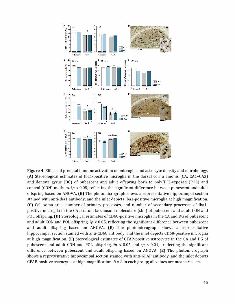
45
Figure 4. Effects of prenatal immune activation on microglia and astrocyte density and morphology.
(A) Stereological estimates of Iba1-‐positive microglia in the dorsal cornu amonis (CA; CA1–CA3)
and dentate gyrus (DG) of pubescent and adult offspring born to poly(I:C)-‐exposed (POL) and
control (CON) mothers. §p < 0.05, reflecting the significant difference between pubescent and adult
offspring based on ANOVA. (B) The photomicrograph shows a representative hippocampal section stained with anti-‐Iba1 antibody, and the inlet depicts Iba1-‐positive microglia at high magnification.
(C) Cell soma area, number of primary processes, and number of secondary processes of Iba1-‐
positive microglia in the CA stratum lacunosum moleculare (slm) of pubescent and adult CON and
POL offspring. (D) Stereological estimates of CD68-‐positive microglia in the CA and DG of pubescent
and adult CON and POL offspring. §p < 0.05, reflecting the significant difference between pubescent and adult offspring based on ANOVA. (E) The photomicrograph shows a representative
hippocampal section stained with anti-‐CD68 antibody, and the inlet depicts CD68-‐positive microglia
at high magnification. (F) Stereological estimates of GFAP-‐positive astrocytes in the CA and DG of
pubescent and adult CON and POL offspring. §p < 0.05 and +p < 0.01, reflecting the significant
difference between pubescent and adult offspring based on ANOVA. (E) The photomicrograph shows a representative hippocampal section stained with anti-‐GFAP antibody, and the inlet depicts
GFAP-‐positive astrocytes at high magnification. N = 8 in each group; all values are means ± s.e.m.
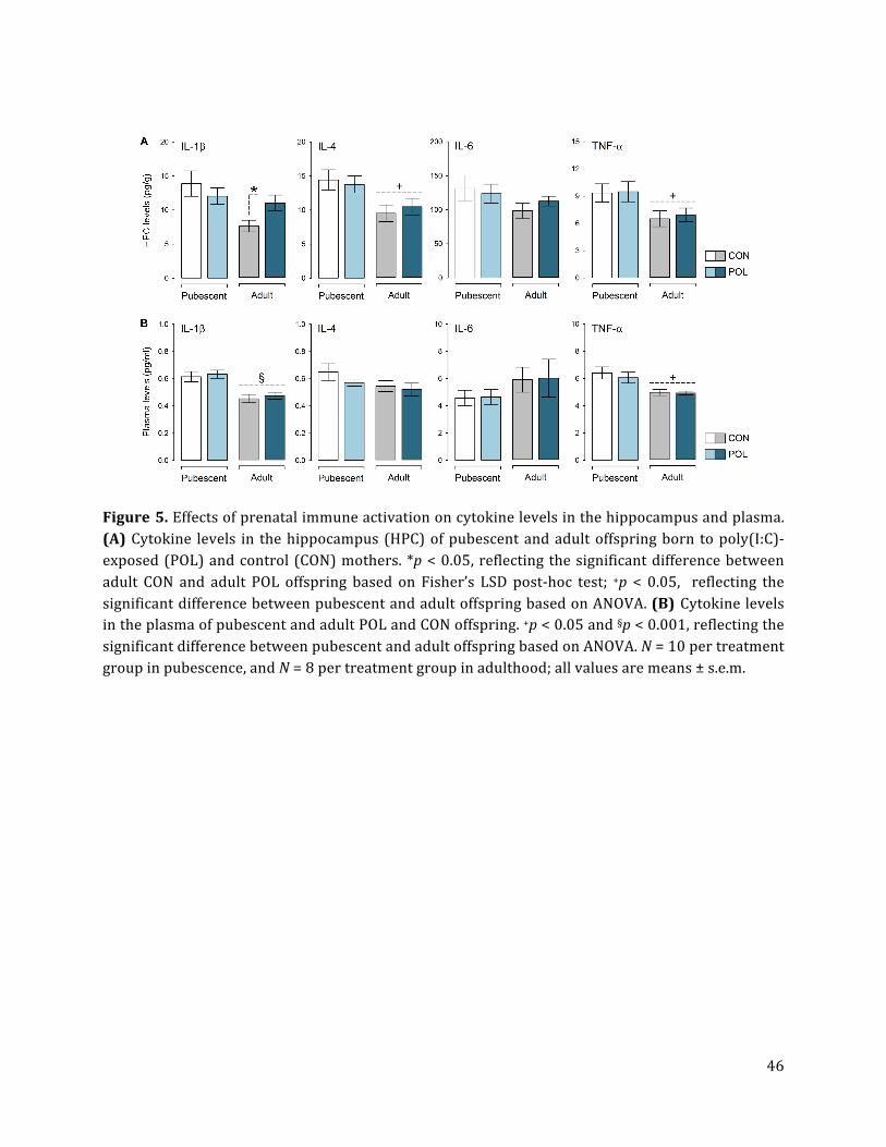
46
Figure 5. Effects of prenatal immune activation on cytokine levels in the hippocampus and plasma.
(A) Cytokine levels in the hippocampus (HPC) of pubescent and adult offspring born to poly(I:C)-‐
exposed (POL) and control (CON) mothers. *p < 0.05, reflecting the significant difference between
adult CON and adult POL offspring based on Fisher’s LSD post-‐hoc test; +p < 0.05, reflecting the
significant difference between pubescent and adult offspring based on ANOVA. (B) Cytokine levels in the plasma of pubescent and adult POL and CON offspring. +p < 0.05 and §p < 0.001, reflecting the
significant difference between pubescent and adult offspring based on ANOVA. N = 10 per treatment
group in pubescence, and N = 8 per treatment group in adulthood; all values are means ± s.e.m.
47
SUPPLEMENTARY DATA
Supplementary Figure S1. Relative optical density (in arbitrary units, AU) of synaptophysin in the ventral cornu amonis (CA; CA1–CA3) and dentate gyrus (DG) of pubescent and adult offspring born
to poly(I:C)-‐exposed (POL) and control (CON) mothers. §p < 0.001, reflecting the significant
difference between pubescent and adult offspring based on ANOVA [CA: F(1,28) = 36.43; DG:
F(1,28) = 39.01]; *p < 0.05, reflecting the significant difference between adult CON and adult POL
offspring based on Fisher’s LSD post-‐hoc test following the presence of a significant interaction between prenatal treatment and age [CA: F(1,28) = 5.26, p < 0.05; DG: F(1,28) = 5.83, p < 0.05]. N = 8
in each group; all values are means ± s.e.m.
Supplementary Figure S2. Relative optical density (in arbitrary units, AU) of PSD95 in the ventral
cornu amonis (CA; CA1–CA3) and dentate gyrus (DG) of pubescent and adult offspring born to
poly(I:C)-‐exposed (POL) and control (CON) mothers. +p < 0.001, reflecting the significant difference between CON and POL offspring associated with the main effect of prenatal treatment [CA: F(1,28) =
28.71; DG: F(1,28) = 20.36] revealed by ANOVA; §p < 0.001, reflecting the significant difference
between pubescent and adult offspring based on the main effect of age [CA: F(1,28) = 26.13; DG:
F(1,28) = 24.22, p < 0.001] revealed by ANOVA. N = 8 in each group; all values are means ± s.e.m.
48
Supplementary Figure S3. (A) Stereological estimates of Iba1-‐positive microglia in the ventral cornu amonis (CA; CA1–CA3) and dentate gyrus (DG) of pubescent and adult offspring born to
poly(I:C)-‐exposed (POL) and control (CON) mothers. #p < 0.05 and §p < 0.01, reflecting the
significant difference between pubescent and adult offspring based on the main effect of age [CA:
F(1,28) = 9.43, p < 0.01; DG: F(1,28) = 5.92, p < 0.05) revealed by ANOVA. (B) Stereological
estimates of CD68-‐positive microglia in the CA and DG of pubescent and adult CON and POL offspring. #p < 0.05 and §p < 0.01, reflecting the significant difference between pubescent and adult
offspring based on the main effect of age [CA: F(1,28) = 5.08, p < 0.05; DG: F(1,28) = 9.38, p < 0.01]
revealed by ANOVA. N = 8 in each group; all values are means ± s.e.m.
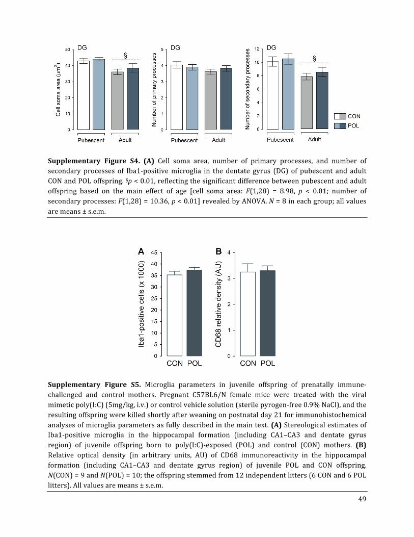
49
Supplementary Figure S4. (A) Cell soma area, number of primary processes, and number of secondary processes of Iba1-‐positive microglia in the dentate gyrus (DG) of pubescent and adult
CON and POL offspring. §p < 0.01, reflecting the significant difference between pubescent and adult
offspring based on the main effect of age [cell soma area: F(1,28) = 8.98, p < 0.01; number of
secondary processes: F(1,28) = 10.36, p < 0.01] revealed by ANOVA. N = 8 in each group; all values
are means ± s.e.m.
Supplementary Figure S5. Microglia parameters in juvenile offspring of prenatally immune-‐
challenged and control mothers. Pregnant C57BL6/N female mice were treated with the viral
mimetic poly(I:C) (5mg/kg, i.v.) or control vehicle solution (sterile pyrogen-‐free 0.9% NaCl), and the
resulting offspring were killed shortly after weaning on postnatal day 21 for immunohistochemical
analyses of microglia parameters as fully described in the main text. (A) Stereological estimates of Iba1-‐positive microglia in the hippocampal formation (including CA1–CA3 and dentate gyrus
region) of juvenile offspring born to poly(I:C)-‐exposed (POL) and control (CON) mothers. (B)
Relative optical density (in arbitrary units, AU) of CD68 immunoreactivity in the hippocampal
formation (including CA1–CA3 and dentate gyrus region) of juvenile POL and CON offspring.
N(CON) = 9 and N(POL) = 10; the offspring stemmed from 12 independent litters (6 CON and 6 POL
litters). All values are means ± s.e.m.