freely available online research biodistribution of ... · project director nagoya university ......
TRANSCRIPT
VOL. 3, No. 3, MARCH 2014 76
RESEARCH
Biodistribution of locally or systemically transplanted osteoblast-like cells
Y. T. Okabe,T. Kondo,K. Mishima,Y. Hayase,K. Kato,M. Mizuno,N. Ishiguro,H. Kitoh
From Nagoya University Hospital, Center for Advanced Medicine and Clinical Research, Showa-ku, Nagoya, Japan
Y. T. Okabe, PhD, Biomedical Scientist T. Kondo, PhD, Biomedical Scientist Y. Hayase, BHS, Biomedical Scientist K. Kato, PhD, Assistant Professor M. Mizuno, MD, PhD, General Project Manager N. Ishiguro, MD, PhD, General Project DirectorNagoya University Hospital, Center for Advanced Medicine and Clinical Research, 65 Tsurumai, Showa-ku, Nagoya, Aichi 466-8550, Japan.
K. Mishima, MD, Orthopaedic Surgeon H. Kitoh, MD, PhD, Associate ProfessorNagoya University Graduate School of Medicine, Department of Orthopedic Surgery, 65 Tsurumai, Showa-ku, Nagoya, Aichi 466-8550, Japan.
Correspondence should be sent to Dr H. Kitoh; e-mail: [email protected]
10.1302/2046-3758.33.2000257 $2.00
Bone Joint Res 2014;3:76–81. Received 13 November 2013; Accepted after revision 17 December 2013
ObjectivesIn order to ensure safety of the cell-based therapy for bone regeneration, we examined in vivo biodistribution of locally or systemically transplanted osteoblast-like cells generated from bone marrow (BM) derived mononuclear cells.
MethodsBM cells obtained from a total of 13 Sprague-Dawley (SD) green fluorescent protein transgenic (GFP-Tg) rats were culture-expanded in an osteogenic differentiation medium for three weeks. Osteoblast-like cells were then locally transplanted with collagen scaffolds to the rat model of segmental bone defect. Donor cells were also intravenously infused to the normal Sprague-Dawley (SD) rats for systemic biodistribution. The flow cytometric and histological analyses were performed for cellular tracking after transplantation.
ResultsLocally transplanted donor cells remained within the vicinity of the transplantation site without migrating to other organs. Systemically administered large amounts of osteoblast-like cells were cleared from various organ tissues within three days of transplantation and did not show any adverse effects in the transplanted rats.
ConclusionsWe demonstrated a precise assessment of donor cell biodistribution that further augments prospective utility of regenerative cell therapy.
Cite this article: Bone Joint Res 2014;3:76–81.
Article focus To ensure safety of the cell-based therapy
for bone regeneration, we examined bio-distribution of locally or systemicallytransplanted osteoblast-like cells.
Key messages Locally transplanted osteoblast-like cells
resided within the transplanted site andwere not detected in other organs at twoweeks after transplantation.
Intravenously infused cells were clearedfrom various organ tissues within threedays of transplantation, without showingany adverse effects in the transplanted rats.
Strengths and limitations Strengh: This is the first validation of bio-
distribution of locally or systemically
transplanted osteoblast-like cells. Weclearly demonstrated the safety of celltherapies using culture-expanded osteo-blast-like cells.
Limitation: Transplanted cells were hetero-geneous and the cellular localisation hasnot been analysed in vascular or otherorgans including heart, pancreas, andintestine.
Introduction Distraction osteogenesis (DO) is a well-estab-lished therapeutic technique used for limblengthening and bone regeneration with anexternal device following osteotomy.1-5 Long-term treatment, however, is essential toextend the bone by DO, which may lead to arisk of adjacent joint contractures and pintrack infection. In order to minimise these
Freely available online
Keywords: Biodistribution, Osteoblast-like cells, Transplantation, Cellular tracking, GFP rats, Bone regenerative medicine
77 Y. T. OKABE, T. KONDO, K. MISHIMA, Y. HAYASE, K. KATO, M. MIZUNO, N. ISHIGURO, H. KITOH
BONE & JOINT RESEARCH
DO-related complications, several trials have been carriedout such as ultrasonic stimulation,6,7 electronic stimula-tion,8 hyperbaric oxygen exposure,9 transplantation offresh bone marrow (BM) cells or administration of growthhormone or cytokines to shorten the treatment periodand enhance osteogenesis in DO.10-13 We have developeda novel DO technique to shorten the consolidation periodby performing transplantation of culture-expandedosteoblast-like cells together with platelet rich plasma(PRP), thrombin and calcium.14-16 Our cell therapy accel-erated bone regeneration and decreased the associatedadverse effects, which indicated the utility of combiningosteoblast-like cells and biomaterials for DO.
Systemic adverse effects regarding BM cell transplanta-tion still remain controversial. Several studies havereported the safety of transplanting both fresh and cul-tured BM cells,17,18 On the other hand, some investigatorshave found that autologous transplantation of BM cellsdeveloped unexpected adverse effects, including unfa-vourable angiogenesis and even death.19,20 It is impor-tant to assess the systemic biodistribution of transplantedBM cells in the recipient to ensure secure use of BM cellsin regeneration medicine.
In this study, local and systemic biodistribution oftransplanted osteoblast-like cells was analysed in a ratmodel. For local biodistribution of transplanted cells, weused a segmental bone defect model instead of a DOmodel to simplify the experimental procedure. Donor BMcells were harvested from green fluorescent protein(GFP)-transgenic (GFP-Tg) rats in order to trace the trans-planted cells in the recipient rat. Flow cytometric and
histological analyses were performed to assess the bio-distribution of donor cells after transplantation.
Materials and MethodsCell cultures for rat bone marrow cells . A total of31 Sprague-Dawley (SD) rats and 17 GFP-Tg rats werepurchased (Japan SLC, Inc. Shizuoka, Japan). The experi-mental procedures were approved by the Animal Experi-mentation and Ethics Committee of the NagoyaUniversity School of Medicine, and all animal experi-ments were conducted following the guidelines of thecommittee for the care and use of experimental animals.
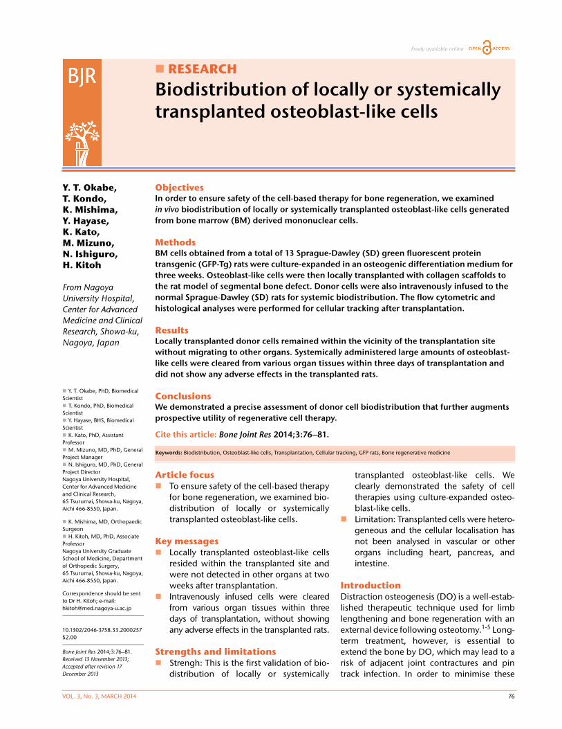
The standard culture medium for osteoblastic differentia-tion (OS medium) used in the current study consisted ofDulbecco’s modified Eagle’s medium (Sigma-Aldrich, StLouis, Missouri), 1.0x10⁻⁷ M Dexamethasone (Sigma-Aldrich), 10 mM Glycerol-2-phosphate disodium salthydrate (Sigma-Aldrich), 2 uM Ascorbic acid (Wako PureIndustries, Ltd, Osaka, Japan), 100 unit/ml penicillin,100 ug/ml streptomycin (Gibco, Grand Island, New York),0.75 ug/ml FUNGIZONE (Gibco) and 10% heat inactivatedfetal calf serum (Thermo Scientific, Waltham, Massachu-setts), under 5% CO2, 37°C. Rat BM cells were obtainedfrom the femur of eight-week-old SD-GFP Tg rats (n = 13).The ends of the femur were cut and the marrow was flushedout using culture medium in a 5 mL syringe (Terumo, Tokyo,Japan) with a 21-gauge needle (Terumo). Nonadherent cellswere removed and adherent cells were expanded as amonolayer culture, with medium changes every two orthree days. When culture dishes became near confluent,cells were dissociated with 0.025% trypsin-EDTA (Gibco)and suspended for continued passage. Cells used for trans-plantation were passaged three times and cultured over athree week period.Transplantation of culture-expanded osteoblastic cells. Asegmental bone defect rat model was prepared as describedpreviously.21 Briefly, rats were anesthetised with an intraperi-toneal infusion of Nembutal (30 mg/kg to 40 mg/kg: dilutedwith saline solution) (Dainippon Pharmaceutical Co, Ltd,Osaka, Japan) and a mini external fixator-distractor (NagoyaScrew Manufacturing Co., Ltd, Nagoya, Japan) was securedto the rat’s right femoral bone using four threaded pins. Themid-third of the bone shaft was osteotomised. After fixationof the distraction device, a bone defect 5 mm in length wasprepared by lengthening the femur and collagen sponge(Koken Co., Ltd., Tokyo Japan) containing 5 x 10⁶ of cultureBM cells, which were put on the site of the bone defect(Fig. 1a). The animals were allowed to move freely in cagesafter recovery from anesthesia. For intravenously infusionexperiments, five million osteoblast-like cells were systemi-cally infused into the normal SD rats via puncture of the fem-oral vein (Fig. 1b). Infusion of PBS without osteoblast-like cellswas used as a negative control. To analyse donor-cell contri-bution, recipient-rat tissues were periodically assessed at oneand two weeks after transplantation in bone defect rats, andat two hours, one and three days after intravenous infusion.
Fig. 1b
Figure 1a – Schematic diagrams of the experimental procedure of a rat seg-mental bone defect model. BM cells harvested from GFP-Tg rat were culturedin a medium for osteoblastic differentiation (OS medium) for three weeks.Three passaged (P3) 5 x 10⁶ cells were embedded in a collagen sponge andtransplanted into bone defect rats. Figure 1b – The experimental procedurefor intravenous infusion of osteoblast-like cells into the healthy SD rats. BMcells harvested from GFP-Tg rat were cultured in an OS medium for threeweeks. Three passaged (P3) 5 x 10⁶ cells were systemically infused into the SDrats via femoral vein.
Fig. 1a
BIODISTRIBUTION OF LOCALLY OR SYSTEMICALLY TRANSPLANTED OSTEOBLAST-LIKE CELLS 78
VOL. 3, No. 3, MARCH 2014
Flow cytometry, histological analysis and real-timepolymerase chain reaction (PCR). A total of thirty-fiverats were euthanised under anesthesia with CO2, and tis-sue was collected at various time points (two hours, one,three, seven and 14 days) following local transplantationor systemic infusion of osteoblast-like cells. Peripheralblood (PB) was collected from the heart. Lungs, livers andbrains were digested with digestion buffer, 1 mg/mlcollagenase (Wako Pure Industries, Ltd, Osaka, Japan),1 mg/ml dispase (Gibco) and DNase (Sigma-Aldrich) inPBS at 37° C for 30 minutes. These tissues and spleenswere pressed between two frosted glass slides (Matsu-nami Glass, Osaka, Japan). After centrifugation, packedcells were suspended in a Red Blood Cell Lysing Buffer(Sigma-Aldrich) and left at room temperature for five min-utes to lyse red blood cells. After washing with a coldmedium, the cell suspension was passed through a 200 Gmesh to remove large debris. Two-10 x 105 cells were thensuspend in ice-cold Hank’s balanced salt solution (Gibco)containing 0.5% bovine serum albumin (Gibco). Afterwashing, the cells were analysed on MACSquant (Milteny-iBiotec, BergischGladbach, Germany). Data were analysedby FlowJo software (Tree star, Inc., Ashland, Oregon).
For histological analysis, 35 rats were euthanised andtissues were fixed with 4% paraformaldehyde (PFA) (Elec-tron Microscopy Science, Hatfield, Philadelphia) over-night at 4°C. Femora were completely decalcified in10% ethylenediamine tetraacetic acid (pH 7.4) (Gibco) inPBS for a week at room temperature. After fixation, tissueswere washed with PBS and put into 30% sucrose (WakoPure Industries) in PBS overnight at 4°C. After removing30% sucrose, the tissues were embedded in an OCT com-pound (Tissue-tek; Sakura Inc, Tokyo, Japan), and serialsection was performed to a thickness of 10 um by LeicaCM3050S (Leica DM IRBE, Leica Microsystems, Wetzlar,Germany). After washing with PBS, the slides weremounted with VECTASHIELD Mounting Medium withDAPI (Vector Laboratories, Burlingame, California) andnail polish. Fluorescent images of the slides were analysedon BS-9000 (KEYENCE, Osaka, Japan).
For real-time PCR, DNA was extracted from 17 rats withthe QIAamp (QIAGEN) and 50 ng of DNA was used for thefollowing assay. Real-time PCR was performed with theLightCycler 480 (Roche Diagnostics, Basel, Switzerland)according to the manufacturer’s instruction. DNA wasamplified by using the following combination of primersets: EGFP forward, 5’-CGACCACTACCAGCAGAA CA-3’and reverse, 5’-TCTCGTTGGGGTCTTTGC-3’; GAPDHforward, 5’-TTGTGCA GTGCCAGCCTCGT-3’ and reverse,5’-GTCACAAGAGAAGGCAGCCCTGG-3’.
PCR standards for determining copy number of GFPwere used, amplicons of GAPDH cloned into the vectorpGEM-Teasy (Promega Corporation, Madison, Wisconsin),and EGFP plasmid was kindly gifted from Dr. Itoh (NagoyaUniversity). The program consisted of a denaturation step(9°C for five minutes), followed by cycles of touchdown
PCR (10 seconds at 95°C, 10 seconds at 60°C and 10 sec-onds at 72°C) and a melting-curve step (97°C).Statistical analysis. For data obtained from the flow cyto-metric analysis, the frequency of donor cells was analysedas the mean percent GFP-positive cells with the standarderror of the mean (SEM). Statistical analyses were per-formed using GraphPad Prism (GraphPad software Inc.,San Diego, California). A p-value of less than 0.05 wasconsidered statistically significant.
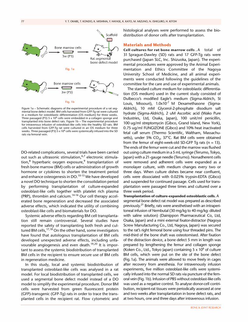
Results Transplanted collagen scaffolds still remained and amajority of GFP-positive osteoblast-like cells survived inthe scaffolds at two weeks after donor cell transplantation(Figs 2a and 2b).
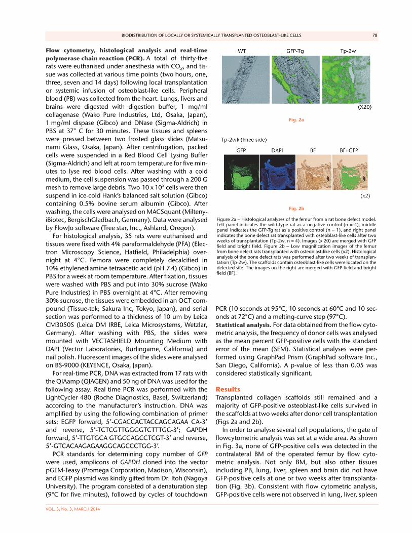
In order to analyse several cell populations, the gate offlowcytometric analysis was set at a wide area. As shownin Fig. 3a, none of GFP-positive cells was detected in thecontralateral BM of the operated femur by flow cyto-metric analysis. Not only BM, but also other tissuesincluding PB, lung, liver, spleen and brain did not haveGFP-positive cells at one or two weeks after transplanta-tion (Fig. 3b). Consistent with flow cytometric analysis,GFP-positive cells were not observed in lung, liver, spleen
Fig. 2b
Figure 2a – Histological analyses of the femur from a rat bone defect model.Left panel indicates the wild-type rat as a negative control (n = 4), middlepanel indicates the GFP-Tg rat as a positive control (n = 1), and right panelindicates the bone defect rat transplanted with osteoblast-like cells after twoweeks of transplantation (Tp-2w, n = 4). Images (x 20) are merged with GFPfield and bright field. Figure 2b – Low magnification images of the femurfrom bone defect rats transplanted with osteoblast-like cells (x2). Histologicalanalysis of the bone defect rats was performed after two weeks of transplan-tation (Tp-2w). The scaffolds contain osteoblast-like cells were located on thedefected site. The images on the right are merged with GFP field and brightfield (BF).
Fig. 2a

79 Y. T. OKABE, T. KONDO, K. MISHIMA, Y. HAYASE, K. KATO, M. MIZUNO, N. ISHIGURO, H. KITOH
BONE & JOINT RESEARCH
and brain by histological analysis (data not shown). Inaddition, there were no histological abnormalities includ-ing vascular occlusion, cell invasion or cell division oftransplanted cells in all organs tested.
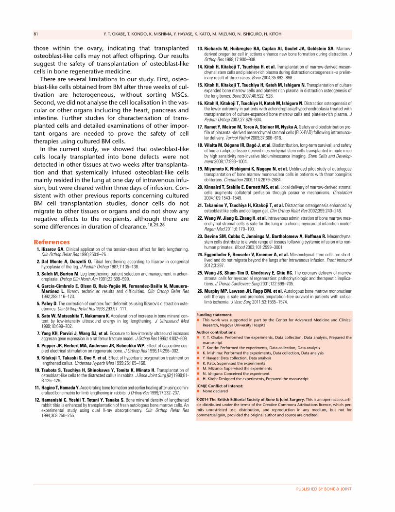
Histological examinations of various organs demon-strated that none of GFP-positive osteoblast-like cellswere detected in BM, PB, lung, liver, spleen and brainboth at one and two weeks after intravenous infusion(Fig. 4, and data not shown). By the clearance kineticexperiments, we detected approximately 0.03% and0.004% of GFP-positive cells in the lung at two hours andone day after intravenous infusion, respectively. However,these cells were cleared after three days of intravenousinfusion (Fig. 5a). GFP-positive cells were rarely detectedin liver (0.001%), PB (0.001%), BM (< 0.001%) and spleen(< 0.001%) at two hours after infusion. These cells wereno longer detectable after one day of intravenous infu-sion (Fig. 5b). It should be noted that no GFP-positivecells were detected in the brain. Histological examina-tions of the lung and liver revealed that only a few GFP-positive cells were seen in the lung at two hours afterintravenous infusion, but they completely disappeared atthree days after infusion (Fig. 5c). No vascular occlusionwas detected in lung, liver, spleen and brain for three dayspost-infusion (data not shown). Quantitative real-timePCR analysis demonstrated that the GFP gene was not
detected in any tissue of a recipient rat, including anovary (Fig. 5d). Because qPCR assays require more than100 cells to detect the target gene, this result indicatesthat GFP-positive cells residing in several tissues at twohours after transplantation were at less than 100.
Discussion This study presents evidence for the clinical safety of cul-tured osteoblast-like cells derived from BM cells in regen-erative medicine. To our knowledge, this is the first reportto show the systemic biodistribution of transplantedosteoblast-like cells in the recipient using the rat model ofa segmental bone defect. We found that the majority ofGFP-positive cells were localised at the transplanted site,without migrating to PB or other tissues. Transplantedcells survived in the transplanted site because the healingprocess occurs in the bone defect area rich in cytokinesand growth factors, where the transplanted collagen scaf-fold is located. In addition, the atelocollagen sponge hasa structure which facilitates the ready supply of nutrientsto the cells. Unfortunately, we did not demonstrate thefinal fate of these transplanted cells; however, based onthe in vitro data, we assume they differentiate into matureosteoblasts. Transplanted cells, which were differentiatedinto osteoblast-like cells for three weeks, might be suit-able to reside in the bone, although the atelocollagenscaffold could play a role in preventing diffusion ofembedded cells. Even though we did not quantify the via-bility of osteoblasts derived from transplanted cells, wepresume that these differentiated, rather than un-differ-entiated cells, have more capability to differentiate intothe osteoblasts and calcify.
We demonstrated that a small number of transplantedGFP-positive cells was detected in the lung, liver, PB, BMand spleen at two hours after intravenous injection. Pre-vious reports have shown that cultured BM cells infusedintravenously were particularly retained in reticulartissues.22,23 Contrary to these reports, Eggenhofer et al24
90
60
30
0
-30
Freq
uenc
y (%
)
GFP-Tg
(BM
)W
TTp
-1w
Tp-2
wW
TTp
-1w
Tp-2
wW
TTp
-1w
Tp-2
wW
TTp
-1w
Tp-2
wW
TTp
-1w
Tp-2
wW
TTp
-1w
Tp-2
w
BM PB Lung Liver Spleen Brain
Fig. 3b
Figure 3a – Representative flow cytometric plots showing bone marrow (BM)from the wild-type (WT) rats and GFP-Tg rats (GFP-Tg), and BM and periph-eral blood (PB) from bone defect rats that were transplanted with osteoblast-like cells after two weeks of transplantation (Tp-2w). Numbers shown in thefigure indicate the frequency of GFP-positive cells. Figure 3b – Graph show-ing percentage of GFP-positive cells in BM, PB, lung, liver, spleen and brain atone and two weeks after transplantation. WT rat was used as a negative con-trol, whereas BM of GFP-Tg rat was used as a positive control (n = 3 or 4 each).Horizontal lines represent means.
Fig. 3a
Fig. 4
Representative macroscopic images of the GFP expression in the lung (left)and liver (right) from the recipient rats at one week after intravenous infusion(i.v.-1w) of GFP-positive cells (upper panel), and from the GFP-Tg rats as apositive control (lower panel). Nuclei were stained with DAPI.

BIODISTRIBUTION OF LOCALLY OR SYSTEMICALLY TRANSPLANTED OSTEOBLAST-LIKE CELLS 80
VOL. 3, No. 3, MARCH 2014
demonstrated the short-term survival of infused culturedBM cells and a lack of distribution of viable BM cellsbeyond the lung. We showed that transplanted osteo-blast-like cells were completely cleared within three daysafter systemic infusion, even in the reticular tissues. Weassume that undifferentiated mesenchymal stem cells
(MSCs) used in previous studies tend to move to otherorgans, rather than differentiated cells. Since the differen-tiated cells were transplanted, the number of MSCs con-tained in transplanted cells was less than others and thismight provide favourable results. Notably, no GFP-positive cells were detected in genital tissues such as
2 hours day 1 day 3
0.05
0.04
0.03
0.02
0.01
0.00
Lung
% F
req
uenc
y (G
FP+ )
2 hours day 1 day 3
0.003
0.002
0.001
0.000
BM
PB
% F
req
uenc
y (G
FP+ )
Liver
Spleen
Brain
2
1.5
1
0.5
0
2
1.5
1
1
0.5
0
0.8
0.6
0.4
0.2
0
0.80.60.40.2
0
0.6
0.4
0.2
0
Lung Liver
Spleen
Ovary
Brain
GFP
(%
)
GFP
(%
)
GFP
(%
)
GFP
(%
)
GFP
(%
)
2 hoursday
1day
3 PC NC
2 hoursday
1day
3 PC NC
2 hoursday
1day
3 PC NC
2 hoursday
1day
3 PC NC
2 hoursday
1day
3 PC NC
Fig. 5d
Figure 5a – Graph showing percentage of GFP-positive cells in lung at two hours, one and three days after intravenous infusion (n = 4 or 5 each). Values arepresented as standard error of the mean (SEM). Figure 5b – Graph showing percentage of GFP-positive cells in BM, PB, liver, spleen and brain at two hours, oneand three days after intravenous infusion (n = 4 or 5 each). Values are presented as standard error of the mean (SEM). Figure 5c – Representative macroscopicimages of the GFP expression in lung (left) and liver (right) at two hours (i.v.-2h) and three days (i.v.-3d) after intravenous infusion. PBS without osteoblast-likecells was used as a negative control, whereas GFP-Tg rat was used as a positive control. Nuclei were stained with DAPI. Figure 5d – Bar graph showing quan-tification of the GFP gene by real-time PCR. Copy numbers of GFP were normal to that of GAPDH. PBS was used as a negative control (NC), whereas GFP-Tgrat was used as a positive control (PC). Values are presented as standard error of the mean (SEM).
Fig. 5c
Fig. 5bFig. 5a
81 Y. T. OKABE, T. KONDO, K. MISHIMA, Y. HAYASE, K. KATO, M. MIZUNO, N. ISHIGURO, H. KITOH
PUBLISHED BY BONE & JOINT
those within the ovary, indicating that transplantedosteoblast-like cells may not affect offspring. Our resultssuggest the safety of transplantation of osteoblast-likecells in bone regenerative medicine.
There are several limitations to our study. First, osteo-blast-like cells obtained from BM after three weeks of cul-tivation are heterogeneous, without sorting MSCs.Second, we did not analyse the cell localisation in the vas-cular or other organs including the heart, pancreas andintestine. Further studies for characterisation of trans-planted cells and detailed examinations of other impor-tant organs are needed to prove the safety of celltherapies using cultured BM cells.
In the current study, we showed that osteoblast-likecells locally transplanted into bone defects were notdetected in other tissues at two weeks after transplanta-tion and that systemically infused osteoblast-like cellsmainly resided in the lung at one day of intravenous infu-sion, but were cleared within three days of infusion. Con-sistent with other previous reports concerning culturedBM cell transplantation studies, donor cells do notmigrate to other tissues or organs and do not show anynegative effects to the recipients, although there aresome differences in duration of clearance.18,25,26
References1. Ilizarov GA. Clinical application of the tension-stress effect for limb lengthening.
Clin Orthop Relat Res 1990;250:8–26.2. Dal Monte A, Donzelli O. Tibial lengthening according to Ilizarov in congenital
hypoplasia of the leg. J Pediatr Orthop 1987;7:135–138.3. Saleh M, Burton M. Leg lengthening: patient selection and management in achon-
droplasia. Orthop Clin North Am 1991;22:589–599.4. García-Cimbrelo E, Olsen B, Ruiz-Yagüe M, Fernandez-Baíllo N, Munuera-
Martínez L. Ilizarov technique: results and difficulties. Clin Orthop Relat Res1992;283:116–123.
5. Paley D. The correction of complex foot deformities using Ilizarov's distraction oste-otomies. Clin Orthop Relat Res 1993;293:97–111.
6. Sato W, Matsushita T, Nakamura K. Acceleration of increase in bone mineral con-tent by low-intensity ultrasound energy in leg lengthening. J Ultrasound Med1999;18:699–702.
7. Yang KH, Parvizi J, Wang SJ, et al. Exposure to low-intensity ultrasound increasesaggrecan gene expression in a rat femur fracture model. J Orthop Res 1996;14:802–809.
8. Pepper JR, Herbert MA, Anderson JR, Bobechko WP. Effect of capacitive cou-pled electrical stimulation on regenerate bone. J Orthop Res 1996;14:296–302.
9. Kitakoji T, Takashi S, Ono Y, et al. Effect of hyperbaric oxygenation treatment onlengthened callus. Undersea Hyperb Med 1999;26:165–168.
10. Tsubota S, Tsuchiya H, Shinokawa Y, Tomita K, Minato H. Transplantation ofosteoblast-like cells to the distracted callus in rabbits. J Bone Joint Surg [Br] 1999;81-B:125–129.
11. Hagino T, Hamada Y. Accelerating bone formation and earlier healing after using demin-eralized bone matrix for limb lengthening in rabbits. J Orthop Res 1999;17:232–237.
12. Hamanishi C, Yoshii T, Totani Y, Tanaka S. Bone mineral density of lengthenedrabbit tibia is enhanced by transplantation of fresh autologous bone marrow cells. Anexperimental study using dual X-ray absorptiometry. Clin Orthop Relat Res1994;303:250–255.
13. Richards M, Huibregtse BA, Caplan AI, Goulet JA, Goldstein SA. Marrow-derived progenitor cell injections enhance new bone formation during distraction. JOrthop Res 1999;17:900–908.
14. Kitoh H, Kitakoji T, Tsuchiya H, et al. Transplantation of marrow-derived mesen-chymal stem cells and platelet-rich plasma during distraction osteogenesis--a prelim-inary result of three cases. Bone 2004;35:892–898.
15. Kitoh H, Kitakoji T, Tsuchiya H, Katoh M, Ishiguro N. Transplantation of cultureexpanded bone marrow cells and platelet rich plasma in distraction osteogenesis ofthe long bones. Bone 2007;40:522–528.
16. Kitoh H, Kitakoji T, Tsuchiya H, Katoh M, Ishiguro N. Distraction osteogenesis ofthe lower extremity in patients with achondroplasia/hypochondroplasia treated withtransplantation of culture-expanded bone marrow cells and platelet-rich plasma. JPediatr Orthop 2007;27:629–634.
17. Ramot Y, Meiron M, Toren A, Steiner M, Nyska A. Safety and biodistribution pro-file of placental-derived mesenchymal stromal cells (PLX-PAD) following intramuscu-lar delivery. Toxicol Pathol 2009;37:606–616.
18. Vilalta M, Dégano IR, Bagó J, et al. Biodistribution, long-term survival, and safetyof human adipose tissue-derived mesenchymal stem cells transplanted in nude miceby high sensitivity non-invasive bioluminescence imaging. Stem Cells and Develop-ment 2008;17:993–1004.
19. Miyamoto K, Nishigami K, Nagaya N, et al. Unblinded pilot study of autologoustransplantation of bone marrow mononuclear cells in patients with thromboangiitisobliterans. Circulation 2006;114:2679–2684.
20. Kinnaird T, Stabile E, Burnett MS, et al. Local delivery of marrow-derived stromalcells augments collateral perfusion through paracrine mechanisms. Circulation2004;109:1543–1549.
21. Takamine Y, Tsuchiya H, Kitakoji T, et al. Distraction osteogenesis enhanced byosteoblastlike cells and collagen gel. Clin Orthop Relat Res 2002;399:240–246.
22. Wang W, Jiang Q, Zhang H, et al. Intravenous administration of bone marrow mes-enchymal stromal cells is safe for the lung in a chronic myocardial infarction model.Regen Med 2011;6:179–190.
23. Devine SM, Cobbs C, Jennings M, Bartholomew A, Hoffman R. Mesenchymalstem cells distribute to a wide range of tissues following systemic infusion into non-human primates. Blood 2003;101:2999–3001.
24. Eggenhofer E, Benseler V, Kroemer A, et al. Mesenchymal stem cells are short-lived and do not migrate beyond the lungs after intravenous infusion. Front Immunol2012;3:297.
25. Wang JS, Shum-Tim D, Chedrawy E, Chiu RC. The coronary delivery of marrowstromal cells for myocardial regeneration: pathophysiologic and therapeutic implica-tions. J Thorac Cardiovasc Surg 2001;122:699–705.
26. Murphy MP, Lawson JH, Rapp BM, et al. Autologous bone marrow mononuclearcell therapy is safe and promotes amputation-free survival in patients with criticallimb ischemia. J Vasc Surg 2011;53:1565–1574.
Funding statement: This work was supported in part by the Center for Advanced Medicine and Clinical
Research, Nagoya University Hospital
Author contributions: Y. T. Okabe: Performed the experiments, Data collection, Data analysis, Prepared the
manuscript T. Kondo: Performed the experiments, Data collection, Data analysis K. Mishima: Performed the experiments, Data collection, Data analysis Y. Hayase: Data collection, Data analysis K. Kato: Supervised the experiments M. Mizuno: Supervised the experiments N. Ishiguro: Conceived the experiment H. Kitoh: Designed the experiments, Prepared the manuscript
ICMJE Conflict of Interest: None declared
©2014 The British Editorial Society of Bone & Joint Surgery. This is an open-access arti-cle distributed under the terms of the Creative Commons Attributions licence, which per-mits unrestricted use, distribution, and reproduction in any medium, but not forcommercial gain, provided the original author and source are credited.