comparative toxicity and biodistribution assessments in rats
TRANSCRIPT

RESEARCH Open Access
Comparative toxicity and biodistributionassessments in rats following subchronicoral exposure to copper nanoparticles andmicroparticlesIn-Chul Lee1,2, Je-Won Ko1, Sung-Hyeuk Park1, Na-Rae Shin1, In-Sik Shin1, Changjong Moon1, Je-Hein Kim3,Hyoung-Chin Kim4* and Jong-Choon Kim1*
Abstract
Background: Copper nanoparticles (Cu NPs) have great potential in electronics and biomedical fields because oftheir efficient thermodynamic and anti-microbial properties. However, their potential toxic effects and kinetic datafollowing repeated exposure are still unclear.
Methods: We evaluated the physicochemical properties of Cu NPs (25 nm) and copper microparticles (Cu MPs,14–25 μm). Comparative in vivo toxicity of Cu NPs and Cu MPs was evaluated by conducting a 28-day repeatedoral dose study at equivalent dose levels of 0, 100, 200, and 400 mg/kg/day (vehicle, 1 % hydroxypropylmethylcellulose). We determined Cu levels in the blood, tissues, urine, and feces by using inductively coupledplasma mass spectrometry.
Results: The solubility of Cu NPs and Cu MPs was 84.5 and 17.2 %, respectively, in an acidic milieu; however, theyscarcely dissolved in vehicle or intestinal milieus. The specific surface area of Cu NPs and Cu MPs was determinedto be 14.7 and 0.16 m2/g, respectively. Cu NPs exhibited a dose-dependent increase of Cu content in the bloodand tested organs, with particularly high levels of Cu in the liver, kidney, and spleen. Only for liver and kidneyincreased Cu levels were found in Cu MPs-treated rats. Cu NPs caused a dose-related increase in Cu levels in urine,whereas Cu MPs did not affect the urine Cu levels. Extremely high levels of Cu were detected in the feces of CuMPs-treated rats, whereas much lower levels were detected in the feces of Cu NPs-treated rats. A comparative invivo toxicity study showed that Cu NPs caused damages to red blood cells, thymus, spleen, liver, and kidneyat ≥200 mg/kg/days, but Cu MPs did not cause any adverse effects even at the highest dose.
Conclusions: Overall, the in vivo repeated dose toxicity study of Cu NPs and Cu MPs demonstrated that largesurface area and high solubility in physiological milieus could directly influence the toxicological responses andbiodistribution of Cu particles when administered orally. Under these experimental conditions, the no-observed-adverse-effect levels of Cu NPs and Cu MPs were determined to be 100 and ≥400 mg/kg/day, respectively.
Keywords: Copper nanoparticles, Copper microparticles, Comparative toxicity, Biodistribution
* Correspondence: [email protected]; [email protected] Animal Resource Center, Korea Research Institute of Bioscienceand Biotechnology, ChungBuk 28116, Republic of Korea1College of Veterinary Medicine BK21 Plus Project Team, Chonnam NationalUniversity, Gwangju 61186, Republic of KoreaFull list of author information is available at the end of the article
© The Author(s). 2016 Open Access This article is distributed under the terms of the Creative Commons Attribution 4.0International License (http://creativecommons.org/licenses/by/4.0/), which permits unrestricted use, distribution, andreproduction in any medium, provided you give appropriate credit to the original author(s) and the source, provide a link tothe Creative Commons license, and indicate if changes were made. The Creative Commons Public Domain Dedication waiver(http://creativecommons.org/publicdomain/zero/1.0/) applies to the data made available in this article, unless otherwise stated.
Lee et al. Particle and Fibre Toxicology (2016) 13:56 DOI 10.1186/s12989-016-0169-x
BackgroundNanomaterials are defined as materials that have at leastone dimension in the 1 to 100 nm range, which weregenerated by accidentally or engineering. The engineerednanoparticles (NPs) can be utilized in an application-specific manner by modifying their size, surface proper-ties, and shape. Thus, in recent years, remarkableprogress has been made in the area of nanotechnologyas evident from its widespread use in textile, electronics,cosmetics, and foods [1]. The physicochemical proper-ties of NPs may determinate toxicological behavior invivo by making them interact with biological systemsand be absorbed more than bulk chemicals via variousroutes [2, 3]. Hence, NPs tend to exhibit quite differenttoxicological profiles in vivo compared to the largerparticles [4–7]. Physiological conditions also influencethe interaction between biological systems and NPs andthey can determine the fate and biosafety of NPs [8].Recently, increased usage of NPs raises concerns ontheir health risks and environmental effects [9–11].Copper (Cu) is an essential element required for nor-
mal physiological functioning, including drug/xenobioticmetabolism, carbohydrate metabolism, and the antioxi-dant defense system [12, 13]. The general population isexposed to Cu through inhalation, consumption of foodand water, and dermal contact with air, water, and soilthat contains Cu. The toxicity of Cu and its compoundshas been studied for decades. A report of the availabledata has been given in the “Toxicological Profile forCopper” from the Agency for Toxic Substances andDisease Registry of the U.S. Public Health Service [14].When intake of Cu exceeds the range of biological toler-ance, it can cause adverse effects, including damage toliver, kidney, immune system, and gastrointestinaldistress [14]. Although the toxic effects of Cu and itscompounds have been studied, several studies reportedgaps concerning the risk caused by Cu in the form ofNPs [7, 15].Among the various types of nanomaterials, metal-
based NPs are used in the manufacture of hundreds ofcommercial products, and their industrial and consumerapplications are expected to increase the chances of theirexposure to the public [10, 11]. In particular, Cu-basedNPs are widely used in a variety of established and emer-ging technologies, including catalysts, solar energyconversion, and antimicrobial agents, because of theirdistinct thermophysical properties and antimicrobialactivities [16–20]. Despites widespread applications andthe growing presence of Cu-containing nanoproducts,there is only limited information on the potential risksof exposure to Cu-based NPs compared to other NPs[17, 21]. To date, many reports regarding the in vitrotoxicity of Cu-based NPs are available. Cu-based NPsinduce cytotoxic effects associated with increase of
reactive oxygen species in various cell lines, includinghuman laryngeal epithelial cells and human alveolartype-I epithelial cells [22–25]. Cu NPs showed a highertoxicity than their oxide nanoparticles (CuO NPs) inHL60 cells [26]. CuO NPs are highly toxic compared tocarbon nanotubes and other metal oxide NPs [27]. How-ever, only a few studies have explored in vivo toxicity;these studies revealed biochemical and histological alter-ations related to liver, kidney, and spleen after a singleor short-term exposure of Cu NPs [7, 15, 28, 29]. To ourknowledge, there has been no report regarding thepotential effects and biodistribution of Cu NPs followinglong-term exposure. Therefore, for further practicalapplications, it is necessary to evaluate the in vivotoxicity of Cu NPs and their biodistribution aftersubchronic exposure for the purpose of risk assessment.Herein, we investigated the in vivo toxicity of Cu NPs
and Cu microparticles (Cu MPs) following 28-dayrepeated oral dose in rats by evaluating biochemical,hematological, and histopathological parameters. Theoral route was used because gastrointestinal exposure tonanomaterials has the potential for wide public exposureto higher doses and more frequent ingestion [30, 31].Further, we investigated absorption, tissue distribution,and excretion to elucidate the main accumulation sitesand elimination routes. In this work, we report for thefirst time, to the best of our knowledge, the in vivotoxicity and biodistribution of Cu NPs by conducting arepeated dose toxicity study.
Results and discussionPhysiochemical characterization of Cu NPs and Cu MPsThe physiochemical characteristics of Cu NPs and CuMPs are summarized in Table 1. The morphology andactual size of Cu NPs and Cu MPs were characterized bytransmission electron microscopy (TEM) and scanningelectron microscopy (SEM). The morphology of NPs orMPs was spherical, and the actual size of individual par-ticles was found to be 32.7 ± 10.45 nm (300 counts) and25.3 ± 6.64 μm (100 counts), respectively (Fig. 1a–d).The purities of Cu NPs and Cu MPs were determined as98.15 and 99.06 %, respectively, using energy dispersiveX-ray spectroscopy (EDX) analysis on the same images(data not shown). The specific surface area of Cu NPsand Cu MPs was measured as 14.7 and 0.16 m2/g,respectively using the Brunauer-Emmett-Teller (BET)method. Suspension stability and surface charge reflectthe interaction of NPs with physiological milieus becauseof their large surface area/volume ratio [32, 33]. The zetapotential of the Cu NPs was 25.5 ± 0.8 mV for pH 1.5,1.32 ± 1.2 mV for pH 6.8, and -6.2 ± 0.2 mV for pH 7.8.Dynamic light scattering (DLS) measurements revealedthat Cu NPs agglomerate in intestinal (pH 7.8) andvehicle (pH 6.8) milieus with the hydrodynamic
Lee et al. Particle and Fibre Toxicology (2016) 13:56 Page 2 of 16

diameters of 334.8 ± 128.26 and 516.4 ± 116.9 nm,respectively. However, in an acidic milieu (pH 1.5), thehydrodynamic diameter of Cu NPs was not determinedbecause Cu NPs rapidly dissolved in acidic conditionsand particle numbers were not enough to determinehydrodynamic diameter. These findings suggest thetendency of Cu NPs to aggregate/agglomerate in suspen-sion or the intestinal milieu. However, the NP formpresents a greater specific surface area compared withthe same compositional particles of micro size scale thatmay cause different biological responses and modulatetheir fate in biological systems.
Dissolution of Cu NPs and Cu MPs in physiologicalconditionsThe dissolution of NPs in gastrointestinal fluids mayhelp predict uptake and blood concentrations. Therelease of toxic ions has the potential to influence thetoxicity of NPs [34–36]. Most NPs are rarely soluble atnormal physiological conditions; however, their dissol-ution can be accelerated in acidic conditions [37, 38].We evaluated the solubility of Cu NPs and Cu MPs(5 mg/mL each) under simulated gastric pH (pH 1.5),vehicle pH (pH 6.8), and intestinal pH (pH 7.8) after a24 h incubation. Cu NPs in an acidic milieu showedbluish color changes within 10 min (Fig. 1e). After moni-toring for up to 24 h, Cu NPs showed 84.5 % solubilityin an acidic milieu, whereas only minimal dissolutionwas observed in vehicle (0.65 %) and intestinal (0.09 %)conditions (Fig. 1f ). In contrast, Cu MPs showed muchlower solubility compared to Cu NPs, even in an acidicmilieu (pH 1.5, 17.2; pH 6.8, 0.04; and pH 7.8, 0.03 %). Ithas been reported that Cu NPs have high solubility in anacidic milieu with high positive charge at zeta potential
analysis [15, 39]. Compared with Cu MPs, the large spe-cific surface area of Cu NPs can lead to high reactivityand drastic interaction with hydrogen ions (H+) in agastric milieu [15]. Because the pH of gastric fluid isbetween 1.5 and 2.0, Cu NPs may be dissolved in thestomach, and dissociated Cu ions can be absorbed intothe systemic circulation when administered orally. Thedissociation of Cu NPs after gastric emptying is prohib-ited in the basic milieu of the small intestine. Thus, theresidence time in the stomach may influence the dissol-ution of Cu NPs. Cu NPs quickly dissolved in gastricmilieus, and the undissolved NPs showed a delayedretention in the stomach after 24 h in mice exposed toCu NPs, which may lead to durative interaction andpersistent Cu ion generation in vivo [15].
Absorption, distribution, and excretion of CuThe dissolution of NPs in physiological conditions andthe physicochemical features of NPs are both likely toinfluence the absorption and biological response of NPswhen administered orally [3, 35, 40]. The Cu levels inblood reflect the absorption of Cu following oral expos-ure of Cu NPs or Cu MPs. Cu levels in the blood of CuNPs-treated rats showed a dose-dependent increase andwere 4-fold higher than that of the vehicle control group(3.33 ± 0.89 μg/g vs. 0.83 ± 0.21 μg/g). In contrast, oralexposure to Cu MPs did not lead to an increase in bloodCu levels, which were not different from the vehicle con-trol group (1.16 ± 0.30 μg/g vs. 0.83 ± 0.21 μg/g) (Fig. 2a).Exposure to nano-Cu (75 mg/kg) markedly elevatesserum Cu level (3.5-fold higher) compared to a minimalincrease in Cu from the same mass of micro-Cu exposedmice at 72 h after single oral dose [15]. Consistent withthe results of a previous report, Cu NPs-treated ratsshowed 2.9-fold higher levels of Cu than that in rats ex-posed to a corresponding high dose of Cu MPs at 24 hafter the last administration. With a delayed retention inthe stomach, the higher levels of Cu in the blood of ratstreated with Cu NPs compared to Cu MPs indicate thatmore Cu ions were dissociated from Cu NPs andabsorbed into systemic circulation. Moreover, high andrapid dissolution of Cu NPs in the gastric milieusuggests that Cu NPs may be mainly absorbed as ionicforms rather than nanoparticulate states. Absorbed Cuions could enter into systemic circulation and be distrib-uted in various tissues. Generally, exposure of humanbody to Cu NPs can occur through different routes (e.g.,inhalation, ingestion, injection or physical contact).Absorbed NPs may interact with biomolecules such asproteins, nucleic acids, lipids, and even biological metab-olites [41]. Of particular importance is the absorption ofproteins on the surface of NPs and form the NP-proteincomplexes, which referred to as the NP-protein corona.The protein corona alters the size and interfacial
Table 1 Physiochemical characterization
Items Cu NPs Cu MPs
Shape Spherical Spherical
Primary sizea 32.7 ± 10.5 nm 25.3 ± 6.6 μm
Hydrodynamic size (nm)b pH 1.5 n.a n.a
pH 6.5 516 ± 116.9 n.a
pH 7.8 334 ± 128.3 n.a
Surface area (m2/g)c 14.7 0.16
Zeta potential (mV)d pH 1.5 25.5 ± 0.8 n.a
pH 6.5 1.32 ± 1.2 n.a
pH 7.8 −6.2 ± 0.2 n.a
NoteaPrimary size of Cu NPs and Cu MPs was measured by transmission electronmicroscopy (300 counts and 100 counts, respectively)bHydrodynamic size of Cu NPs was measured by a dynamic lightscattering methodcSurface area was measured by the Brunauer-Emmett-Teller method (N2 gas)dZeta potential was measured using electrophoretic light scattering methodunder pH 1.5, pH 6.5 and pH 7.8 conditionsn.a. not available
Lee et al. Particle and Fibre Toxicology (2016) 13:56 Page 3 of 16

Fig. 1 (See legend on next page.)
Lee et al. Particle and Fibre Toxicology (2016) 13:56 Page 4 of 16
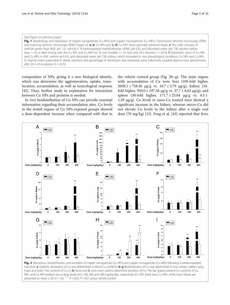
composition of NPs, giving it a new biological identity,which was determine the agglomeration, uptake, trans-location, accumulation, as well as toxicological response[42]. Thus, further study to explanation for interactionbetween Cu NPs and proteins is needed.In vivo biodistribution of Cu NPs can provide essential
information regarding their accumulation sites. Cu levelsin the tested organs of Cu NPs-exposed groups showeda dose-dependent increase when compared with that in
the vehicle control group (Fig. 2b–g). The main organswith accumulation of Cu were liver (109-fold higher,2038.1 ± 758.36 μg/g vs. 18.7 ± 5.79 μg/g), kidney (34-fold higher, 950.0 ± 197.58 μg/g vs. 27.7 ± 8.62 μg/g), andspleen (38-fold higher, 171.7 ± 23.64 μg/g vs. 4.5 ±1.59 μg/g). Cu levels in nano-Cu treated mice showed asignificant increase in the kidney, whereas micro-Cu didnot elevate Cu levels in the kidney after a single oraldose (70 mg/kg) [15]. Feng et al. [43] reported that liver,
(See figure on previous page.)Fig. 1 Morphology and dissolution of copper nanoparticles (Cu NPs) and copper microparticles (Cu MPs). Transmission electron microscopy (TEM)and scanning electron microscopy (SEM) images of (a, b) Cu NPs and (c, d) Cu MPs show generally spherical shape. e The color changes ofartificial gastric fluid (AGF; pH 1.5), vehicle (1 % hydroxypropyl methylcellulose, HPMC; pH 6.5), and deionized water (pH 7.8) solution before(top, t = 0) or after mixing with the Cu NPs and Cu MPs for 10 min (middle, t = 10 min) and 24 h (bottom, t = 24 h). f Dissolution ratios of Cu NPsand Cu MPs in AGF, vehicle (pH 6.5), and deionized water (pH 7.8) milieus, which simulated in vivo physiological conditions. Cu NPs and Cu MPs(5 mg/mL) were suspended in above solutions and percentage of dissolution was measured using inductively coupled plasma mass spectrometryafter 24 h of incubation (t = 24 h)
Fig. 2 Absorption, biodistribution, and excretion of copper nanoparticles (Cu NPs) and copper microparticles (Cu MPs) following 4 weeks-repeatedoral dose. a Systemic absorption of Cu was determined in blood Cu contents. b–g Biodistribution of Cu was determined in liver, kidney, spleen, lung,heart, and brain. The contents of Cu in (h) feces and (i) urine were used to determine excretion of Cu. The bar graphs present Cu contents of CuNPs- and Cu MPs-treated rats at dose levels of 0, 100, 200, and 400 mg/kg/day, respectively (Cu NPs, black bars; Cu MPs, white bars). Values arepresented as mean ± SD (n = 10). *, ** P < 0.05, P < 0.01 versus vehicle control
Lee et al. Particle and Fibre Toxicology (2016) 13:56 Page 5 of 16
spleen, lung, and kidney appear to be the major organs foraccumulation of Cu sulfide nanoplates after intravenousinjection. Thus, the biodistribution of Cu indicated thatCu from Cu NPs was mainly distributed in the liver, kid-ney, and spleen. Further, accumulated Cu in these organscan be a toxic reservoir based on their toxic potential.However, equivalent dose levels of Cu MPs are not onlylower than those of Cu NPs, but also showed no dose-response increase in the tested organs, except in the liver(83.1 ± 20.94 μg/g) and kidney (46.7 ± 10.14 μg/g). Thislow distribution was due to the minimal absorption rateof Cu MPs.The excretion of Cu was consistent with the absorption
and distribution patterns of Cu NPs or Cu MPs. The levelsof Cu in urine from the Cu NPs-treated group showed asignificant increase with clear dose-response when com-pared to that in the vehicle control group (12.8-foldhigher, 6.31 ± 1.59 μg/g vs. 0.49 ± 0.21 μg/g) (Fig. 2h). Incontrast, only trace Cu levels were observed in the urineof the Cu MPs-treated groups (0.60 ± 0.19 μg/g vs. 0.49 ±0.21 μg/g). Cu levels in the feces of Cu NPs treated ratsshowed clear dose-response when compared to that in thevehicle control group (115-fold higher, 26.2 ± 8.41 mg/gvs. 0.2 ± 0.09 mg/g). Cu MPs treated rats showed ex-tremely high levels of Cu in the feces, which were 2.8-foldhigher than that in Cu NPs treated-rats (73.7 ± 16.98 mg/gvs. 26.2 ± 8.41 mg/g) (Fig. 2i). Ingested Cu ions are mainlymetabolized in the liver, and the major excretory route isvia liver/bile [44]. The dissociation of Cu NPs after gastricemptying is prohibited in the basic milieu of the small in-testine, and then unabsorbed NPs are excreted as feces[15]. Thus, extremely high levels of Cu in feces suggestthat most of the absorbed Cu, dissociated from Cu NPs,or unabsorbed Cu NPs were predominantly excretedthrough feces; small amounts were excreted via the kid-ney/urine route. Most of the unabsorbed Cu MPs werealso eliminated from gastro-intestinal tracts via the feces.
Clinical signs, body weights, and food consumptionManifestations of toxicity, including anorexia, diarrhea,lethargy, and body weight loss, were observed in ratstreated with Cu NPs [28]; these manifestations were simi-lar to the effects of excessive Cu compound treatment[45–47]. In this study, treatment-related clinical signs ob-served in the high dose group of Cu NPs were consistentwith toxic manifestations observed in a previous study.The body weight and the amount of food consumption inthe high dose group of Cu NPs decreased significantlyduring the test period (Fig. 3a and b). In contrast, only testarticle-colored feces were observed in the high dose groupof Cu MPs. The body weight and food consumption of theCu MPs-treated groups showed no significant changes,even at the high dose, compared to that in the vehiclecontrol group (Fig. 3c and d).
Urinalysis, serum biochemistry, and hematologyIt has been reported that oral exposure to Cu NPs causeimbalance of acid and base by interacting with H+,resulting in metabolic alkalosis [15]. In a chronic meta-bolic alkalosis state, bicarbonate excretion ceased andled to a state of paradoxical aciduria [48]. The decreasedurine pH observed in the high dose group of Cu NPsmay be due to chronic metabolic alkalosis caused bysub-chronic exposure to Cu NPs (Additional file 1: TableS1). Other urinalysis parameters, including urine protein(PRO), occult blood (OB), leukocytes (LEU), specificgravity (SG), ketone body (KET), and nitrite (NIT), wereincreased significantly in the high dose group of Cu NPs.Hematological findings revealed that repeated exposureto Cu NPs resulted in red blood cell (RBC) destruction,which was characterized by a reduction of RBC,hemoglobin (HB), hematocrit (HCT), mean corpuscularvolume (MCV), mean corpuscular hemoglobin (MCH),and mean corpuscular hemoglobin concentration(MCHC), as well as an increase in reticulocytes (RET)(Table 2). This interpretation was well supported by in-creased yellow pigmentation in the spleen. Chronic Cuintoxication causes hemolytic anemia with diversehematological changes, including decreased RBC, HB,HCT, MCV, MCH and white blood cells (WBC) inrodents [46, 49, 50], which were consistent with theresults of this study. In addition, the changes in thehematology indicate a microcytic anemia that generallyobserved with iron deficiency. Elevated levels of Culevels have been shown to competitively inhibit ironabsorption and utilization and to be correlated withdiminution in serum iron levels [51, 52]. In the differen-tial WBC count, a dose-dependent decrease in the per-centage of lymphocytes (LYM) implied that Cu NPsmight have adverse effects on the immune system, whichwas well correlated with the reduction of cellularity seenin the thymus and spleen (Fig. 4). The increasedpercentages of neutrophils (NEU) and monocytes(MON) were thought to be related to the inflammatoryresponse of the affected organs and the decreased LYMpercentage (Table 2). These results indicated that CuNPs might affect red blood cells and immune organs(spleen and thymus). As reported previously, Cu NPscaused liver and kidney damages with biochemical alter-ations, including increased aspartate aminotransferase(AST), alanine aminotransferase (ALT), total bilirubin(TBIL), blood urea nitrogen (BUN), and creatinine(CRE) [28, 29]. With obvious changes in urinalysisparameters, Cu NPs treated rats showed a dose-relatedresponse in the increment of serum BUN, CRE, AST,ALT, TBIL, alkaline phosphatase (ALP), and lactatedehydrogenase (LDH), as well as the decrease of trigly-ceride (TG) and total protein (TP) with electrolytes dis-turbance (Table 3). These findings on the consequence
Lee et al. Particle and Fibre Toxicology (2016) 13:56 Page 6 of 16

of Cu NPs exposure proved that Cu NPs cause substan-tial damage to the liver and kidney. Collectively, Cu dis-sociated from Cu NPs mainly distributed into liver,kidney, and spleen, which caused obvious functional andstructural damage. However, the parameters of urinaly-sis, serum biochemistry, and hematology were not af-fected by repeated exposure to Cu MPs (Additional file1: Table S1, Tables 2 and 3).
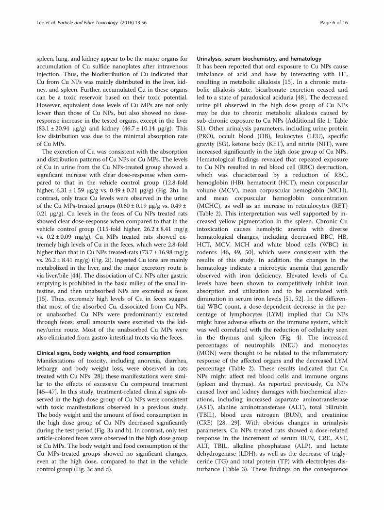
Histopathology and organ weight changesOur findings confirmed previous studies showing that asingle or short-term oral exposure of Cu NPs induces se-vere damage to the kidney and liver [7, 15, 28, 29]. Themajor histopathological findings, including mononuclearcell infiltration, dilated sinusoid, degenerated or
binucleated hepatocytes in the liver, dilated tubules, celldebris or pink or purple-colored casts in tubules, degen-erated tubular cells, and inflammatory cell infiltration inthe kidney, were observed in the rats treated with CuNPs (Table 4 and Fig. 5). Excessive Cu intake results inimpairment of both cellular and humoral immuneresponses [53]. Recently, Cu (II) chloride causes apop-tosis of splenocytes and thymocytes, especially CD4+ Tcell death [54, 55]. Exposure to nano-Cu caused dwin-dling of splenic units and reduction of lymphocytes [7].In rats treated with the high dose of Cu NPs, the spleenexhibited atrophic white pulp, decreased number of folli-cles and cellularity, and yellow pigmentation, and thethymus displayed disrupted demarcation of medulla/cor-tex, decreased cellularity, and cytoplasmic vacuolation,
Fig. 3 Body weight changes and food consumption of copper nanoparticles (Cu NPs)- and copper microparticles (Cu MPs)-treated rats following4 weeks-repeated oral dose. Repeated oral dose toxicity of Cu NPs and Cu MPs was assessed by determining (a, c) Body weight changes and(b, d) food consumption of Cu NPs- and Cu MPs-treated rats. Values are presented as mean ± SD (n = 10). *, ** P < 0.05, P < 0.01 versus vehicle control
Lee et al. Particle and Fibre Toxicology (2016) 13:56 Page 7 of 16

which was consistent with the previous studies (Fig. 4).In particular, an apparent atrophic change of follicles (Bcell area) and periarteriolar lymphoid sheath (T cell area)in the spleen and decreased cellularity in the cortex of thethymus were in agreement with the hematological findingsof our study and the results of previous studies [7, 54, 55].
The changes in organ weight included increased kidneyweight and decreased liver, spleen, and thymus weights inthe high dose group of Cu NPs (Additional file 1: TableS2). These findings were of toxicological significance, be-cause they were well supported by correlated biochemical,hematological, and histopathological changes. In contrast,
Table 2 Hematological changes in male rats treated with Cu NPs and Cu MPs following 28 days-repeated oral dose
Items Cu NPs (mg/kg/day)
0 100 200 400
No. of rats 10 10 10 10
RBC (1012/L) 7.02 ± 0.234a 6.69 ± 0.388 6.17 ± 0.346** 6.12 ± 0.612**
HB (g/dL) 16.78 ± 1.212 15.77 ± 1.668 12.70 ± 1.628** 11.80 ± 1.187**
HCT (%) 43.43 ± 3.435 42.50 ± 2.432 37.32 ± 3.179** 36.47 ± 5.747**
MCV (fl) 53.73 ± 4.388 50.09 ± 4.618 43.63 ± 9.582** 43.60 ± 4.486**
MCH (pg) 23.47 ± 1.598 21.31 ± 2.788 19.10 ± 3.306** 18.92 ± 2.099**
MCHC (g/dL) 34.68 ± 2.770 34.57 ± 3.317 33.05 ± 4.339 28.31 ± 2.198**
PLT (109/L) 1106.2 ± 104.92 1212.7 ± 125.88 1258.8 ± 166.73* 1262.4 ± 121.65*
RET (%) 2.3 ± 0.23 2.4 ± 0.51 2.7 ± 0.48 6.1 ± 2.17**
WBC (109/L) 11.2 ± 0.95 11.3 ± 2.13 8.5 ± 3.03* 8.3 ± 1.21**
NEU (%) 11.9 ± 0.95 11.2 ± 3.45 23.9 ± 5.41** 36.6 ± 8.15**
LYM (%) 82.3 ± 10.73 82.3 ± 12.79 71.7 ± 13.28 52.7 ± 20.17**
MON (%) 2.2 ± 0.39 2.2 ± 0.33 2.3 ± 0.51 3.0 ± 0.59**
EOS (%) 1.7 ± 0.39 1.7 ± 0.33 1.6 ± 0.53 2.0 ± 0.59
BAS (%) 0.7 ± 0.21 0.8 ± 0.18 0.7 ± 0.27 0.7 ± 0.19
LUC (%) 0.7 ± 0.13 0.7 ± 0.24 1.0 ± 0.19* 1.2 ± 0.25**
Items Cu MPs (mg/kg/day)
0 100 200 400
No. of rats 10 10 10 10
RBC (1012/L) 7.04 ± 0.312 7.20 ± 0.286 7.00 ± 0.403 6.96 ± 0.388
HB (g/dL) 16.65 ± 1.326 16.72 ± 1.169 15.96 ± 1.563 16.12 ± 1.971
HCT (%) 43.79 ± 4.793 43.61 ± 4.201 40.16 ± 5.776 41.63 ± 3.304
MCV (fl) 53.81 ± 5.716 52.70 ± 6.621 52.63 ± 4.184 52.34 ± 7.206
MCH (pg) 23.61 ± 3.392 24.32 ± 2.562 24.52 ± 1.397 22.93 ± 4.257
MCHC (g/dL) 34.71 ± 3.392 29.67 ± 5.561 32.09 ± 3.397 32.25 ± 4.257
PLT (109/L) 1179.7 ± 202.53 1047.4 ± 218.36 1259.8 ± 187.54 1236.5 ± 232.71
RET (%) 2.5 ± 0.58 2.6 ± 0.37 2.4 ± 0.39 2.7 ± 0.44
WBC (109/L) 11.3 ± 1.49 11.9 ± 2.48 12.3 ± 3.49 11.56 ± 2.23
NEU (%) 12.2 ± 2.23 10.8 ± 3.91 13.2 ± 4.45 11.9 ± 3.71
LYM (%) 82.9 ± 13.2 85.0 ± 22.47 82.7 ± 16.66 83.5 ± 13.09
MON (%) 2.2 ± 0.48 2.3 ± 0.31 2.5 ± 0.43 2.3 ± 0.54
EOS (%) 1.6 ± 0.42 1.5 ± 0.24 1.6 ± 0.40 1.8 ± 0.48
BAS (%) 0.7 ± 0.23 0.7 ± 0.17 0.8 ± 0.13 0.7 ± 0.21
LUC (%) 0.7 ± 0.22 0.9 ± 0.29 0.9 ± 0.29 0.9 ± 0.23
Note. RBC red blood cell, HB hemoglobin, HCT hematocrit, MCV mean corpuscular volume, MCH mean corpuscular hemoglobin, MCHC mean corpuscularhemoglobin concentration, PLT platelet, RET reticulocytes, NEU neutrophils, LYM lymphocytes, MON monocytes, EOS eosinophils, BAS basophils, LUC largeunstained cellsaValues are presented as mean ± SD*, ** P < 0.05, P < 0.01 versus vehicle control group
Lee et al. Particle and Fibre Toxicology (2016) 13:56 Page 8 of 16

Cu MPs-treated rats did not show obvious changes inhistopathology and organ weights even at the high dose(Figs. 4, 5, and Additional file 1: Table S3). The remarkablereduction in prostate and seminal vesicle weights was ob-served at the high dose of Cu NPs. Chattopadhyay et al.[56] demonstrated that male rats treated with copperchloride at 2 mg/kg/day intraperitoneally for 26 days dis-played adverse effects on testicular spermatogenesis anddevelopment of reproductive organs. Test substance-related stress in the toxicity study causes a decrease in theweights of reproductive organs, including epididymides,seminal vesicles, and prostates, but not in the testes [57].Thus, further study is needed to determine the potentialreproductive/developmental toxicity of Cu NPs because itis unclear whether decreased reproductive organ weightsare related to the anti-androgenic effects of Cu dissociated
from Cu NPs or to the stress-response phenomenon dur-ing the toxicity study.Exposure to Cu NPs can occur through various routes
(e.g., inhalation, ingestion, injection or physical contact).When administered orally, high solubility in the acidicmilieu implies that Cu NPs can be dissociated into Cuions in gastric pH conditions. Further, higher Cu levelsin blood and tissues in rats treated with Cu NPs than CuMPs indicate that absorbed Cu ions were distributed viacirculation and accumulated in various tissues, whichcan be a toxic reservoir. Consistent with the aboveresults, the toxicological study revealed that Cu NPswere more toxic than MPs of the same chemical com-position at the same mass. The dissolution of Cu NPsmay have an important role in their toxicity [7, 15]. Cuion overload caused by excessive Cu NPs administration
Fig. 4 Histopathological changes in spleen and thymus of rats treated with copper nanoparticles (Cu NPs) and copper microparticles (Cu MPs)following 4 weeks-repeated oral dose. The rats treated with Cu NPs at 400 mg/kg/day showed moderate to severe degree of atrophic whitepulp, decreased number of follicles and cellularity, and yellow pigmentation in spleen, disrupted demarcation of medulla/cortex, decreasedcellularity of medulla/cortex, and cytoplasmic vacuolation in thymus. There were no changes in spleen and thymus from rats treated withCu MPs. Hematoxylin and eosin stain
Lee et al. Particle and Fibre Toxicology (2016) 13:56 Page 9 of 16

can cause damage to their accumulation sites, especiallyliver, kidney, and spleen [15, 43]. Additionally, the tox-icity of CuO NPs was largely explained by soluble Cuions [58]. Thus, the differences in dissolution play a cru-cial role in the gap of toxicological responses betweenCu NPs and Cu MPs. In addition, the biopersistence of
NPs influences long-term toxicity and is considered tobe an important parameter needed for the risk assess-ment of NPs [36]. Therefore, further studies will be ne-cessary to investigate whether the toxic responses of CuNPs observed in this study are transient or persistentresponses.
Table 3 Serum biochemical changes in male rats treated with Cu NPs and Cu MPs following 28 days-repeated oral dose
Items Cu NPs (mg/kg/day)
0 100 200 400
No. of rats 10 10 10 10
AST (IU/L) 115.6 ± 19.32a 125.2 ± 15.04 258.4 ± 21.52** 626.6 ± 60.04**
ALT (IU/L) 23.7 ± 2.80 33.3 ± 12.95 88.4 ± 20.65** 115.8 ± 19.12**
ALP (IU/L) 129.8 ± 12.45 132.0 ± 16.08 156.4 ± 22.06* 175.7 ± 38.23**
BUN (mg/dL) 19.4 ± 2.02 20.2 ± 3.24 22.4 ± 1.39 40.6 ± 6.72**
CRE (mg/dL) 0.23 ± 0.052 0.24 ± 0.041 0.26 ± 0.058 0.38 ± 0.068**
CPK (IU/L) 519.0 ± 124.43 504.2 ± 99.57 480.5 ± 94.72 335.2 ± 94.70**
TBIL (mg/dL) 0.25 ± 0.071 0.24 ± 0.055 0.34 ± 0.036 1.07 ± 0.318**
TCHO (mg/dL) 77.6 ± 8.82 79.7 ± 8.94 78.7 ± 2.52 68.6 ± 14.36
TG (mg/dL) 142.0 ± 25.52 133.4 ± 38.73 102.3 ± 29.10* 87.3 ± 28.43**
TP (g/dL) 6.2 ± 0.21 6.1 ± 0.31 5.4 ± 0.25** 5.4 ± 0.36**
ALB (g/dL) 4.2 ± 0.36 4.0 ± 0.12 4.0 ± 0.23 4.0 ± 0.17
LDH (IU/L) 227.7 ± 79.76 268.4 ± 55.99 321.1 ± 57.63* 462.3 ± 106.3**
Na (mEq/L) 141.8 ± 0.75 140.2 ± 1.35 140.3 ± 1.21 137.8 ± 2.95**
Cl (mEq/L) 97.6 ± 2.34 96.0 ± 1.17 95.8 ± 1.53* 93.4 ± 1.14**
K (mEq/L) 4.5 ± 0.49 4.8 ± 0.28 5.1 ± 0.26 6.0 ± 1.04**
Items Cu MPs (mg/kg/day)
0 100 200 400
No. of rats 10 10 10 10
AST (IU/L) 113.7 ± 13.25 135.4 ± 29.86 138.6 ± 22.51 138.3 ± 21.89
ALT (IU/L) 24.9 ± 7.91 27.3 ± 10.83 24.7 ± 4.28 24.8 ± 5.69
ALP (IU/L) 124.7 ± 14.40 147.2 ± 20.99* 120.4 ± 18.95 122.8 ± 11.17
BUN (mg/dL) 17.3 ± 2.94 21.5 ± 4.85 21.5 ± 4.72 19.4 ± 3.67
CRE (mg/dL) 0.23 ± 0.045 0.26 ± 0.062 0.27 ± 0.058 0.24 ± 0.056
CPK (IU/L) 534.5 ± 106.82 522.7 ± 141.61 559.9 ± 80.12 516.9 ± 121.53
TBIL (mg/dL) 0.23 ± 0.045 0.24 ± 0.062 0.26 ± 0.058 0.25 ± 0.056
TCHO (mg/dL) 80.4 ± 6.41 77.7 ± 10.74 89.9 ± 15.03 77.7 ± 4.37
TG (mg/dL) 132.3 ± 20.45 139.9 ± 20.17 149.4 ± 24.16 137.6 ± 20.76
TP (g/dL) 6.4 ± 0.31 6.3 ± 0.47 6.1 ± 0.50 6.2 ± 0.16
ALB (g/dL) 4.2 ± 0.17 4.2 ± 0.27 4.3 ± 0.22 4.2 ± 0.22
LDH (IU/L) 256.8 ± 73.56 236.9 ± 61.90 298.9 ± 94.33 356.5 ± 98.59*
Na (mEq/L) 140.3 ± 1.16 141.1 ± 1.97 143.2 ± 1.14 141.6 ± 1.65
Cl (mEq/L) 97.3 ± 1.42 98.9 ± 1.52 97.6 ± 1.65 99.0 ± 1.67
K (mEq/L) 4.6 ± 0.32 5.1 ± 0.67 4.5 ± 0.36 4.6 ± 0.44
Note. AST aspartate aminotransferase, ALT alanine aminotransferase, ALP alkaline phosphatase, BUN blood urea nitrogen, CRE creatinine, CPK creatinephosphokinase, TBIL total bilirubin, TCHO total cholesterol, TG triglyceride, TP total protein, ALB albumin, LDH lactate dehydrogenase, Na sodium, Clchloride,K potassiumaValues are presented as mean ± SD*, ** P < 0.05, P < 0.01 versus vehicle control group
Lee et al. Particle and Fibre Toxicology (2016) 13:56 Page 10 of 16
MethodsTest chemicals and preparation of test chemicalsCu NPs (CAS No. 7440-50-8; 99.8 % purity) and Cu MPs(99 % purity) were purchased from SkySpring Nanomater-ials (Houston, TX, USA) and Sigma-Aldrich (St. Louis,MO, USA), respectively. The information of particle size(measured by TEM) of Cu NPs and Cu MPs was 25 and14–25 μm, respectively. Hydroxypropylmethylcellulose(HPMC, suspending vehicle) was purchased from Sig-ma-Aldrich. All other chemicals were of the highest gradecommercially available. Test chemicals were dispersedinto 1 % HPMC solution (w/v) with Milli-Q water. Particlesuspensions were made fresh every day and prepared byultrasonic dispersion (VCX130, Vibra Cell Sonics & Mate-rials, Newtown, CT, USA) on ice for 20 min (130 W,20 kHz, pulse 59/1) in agreement with recommendationsby Taurozzi et al. [59].
Physicochemical characterization and solubility of Cu NPsand Cu MPsThe primary size and morphology were measured byTEM (JEM-2100 F, JEOL, Tokyo, Japan) operating at150 kV and SEM (Zeiss EVO-MA10, Carl Zeiss SMT,Cambridge, UK) operating at 15 kV. The purity of NPs
and MPs was determined by EDX analysis on the sameimages from TEM (JEM-2100 F TEM equipped withX-MaxN 150 mm2 silicon drift detector; OxfordInstruments, UK). The samples for TEM were depositedon carbon-coated nickel grids and were air-dried over-night before analysis. The average size was obtained bymeasuring at least 100 particles using an image analyzerprogram (JEOL). The samples for SEM were dispersedon double-sided adhesive carbon tape onto an aluminumSEM stub, and then dusted to release loose particles.The specific surface area of NP and MP powder wasmeasured by the nitrogen (N2) absorption based on themultipoint BET method using an ASAP2020 (Micro-meritics, Norcross, GA, USA). In the solubility study, CuNPs and Cu MPs were incubated under three physio-logical conditions: acidic conditions using artificialgastric fluid (AGF, pH 1.5), vehicle (1 % HPMC, pH 6.5),and basic conditions using deionized water (pH 7.8) for24 h. AGF was prepared according to the previouslydescribed method [60]. In brief, 1.0 g NaCl (Affymetrix,Santa Clara, CA, USA) and 1.6 g pepsin (Sigma-Aldrich)were dissolved in 500 mL of DW and the pH of AGFwas adjusted to 1.5 using 2 N HCl (Sigma-Aldrich).Deionized water (pH 7.8) was used to simulate the basic
Table 4 Histological changes in male rats treated with Cu NPs and MPs following 28 days-repeated oral dose
Items Cu NPs (mg/kg/day) Cu MPs (mg/kg/day)
0 100 200 400 0 100 200 400
No. of rats 10 10 10 10 10 10 10 10
Liver
Mononuclear cell infiltration -(0)a -(0) +(4) ++(8) -(0) -(0) -(0) -(0)
Dilated sinusoid -(0) +(2) +(7) +(9) -(0) +(2) +(2) +(4)
Degenerated hepatocytes -(0) -(0) +(3) +(9) -(0) +(1) +(2) +(1)
Binucleated hepatocytes +(1) +(1) +(2) +(7) +(1) +(2) +(3) +(1)
Kidney
Dilated tubules -(0) +(2) ++(3) ++(8) -(0) +(1) +(4) +(3)
Cell debris in tubules -(0) -(0) ++(4) ++(8) -(0) -(0) -(0) -(0)
Purple-colored casts in tubules -(0) -(0) +(5) +++(7) -(0) -(0) -(0) -(0)
Degenerated tubular cells -(0) -(0) +(2) ++(8) -(0) -(0) -(0) -(0)
Inflammatory cell infiltration -(0) -(0) +(2) +(7) -(0) -(0) -(0) -(0)
Spleen
Atrophic white pulp -(0) -(0) +(3) ++(7) -(0) -(0) -(0) -(0)
Decreased number of follicles -(0) -(0) +(3) +++(6) -(0) -(0) -(0) -(0)
Decreased cellularity -(0) -(0) +(1) ++(7) -(0) -(0) -(0) -(0)
Yellow pigmentation -(0) -(0) +(1) +(5) -(0) -(0) -(0) -(0)
Thymus
Decreased cellularity in medulla/cortex -(0) -(0) +(2) ++(5) -(0) -(0) -(0) -(0)
Disrupted demarcation of medullar/cortex -(0) -(0) +(2) ++(4) -(0) -(0) -(0) -(0)
Cytoplasmic vacuolation -(0) -(0) -(0) +(5) -(0) -(0) -(0) -(0)a-, normal; +, mild; ++, moderate; +++, severe; (), number of case
Lee et al. Particle and Fibre Toxicology (2016) 13:56 Page 11 of 16

condition. Cu NPs and Cu MPs (5 mg/mL) were incu-bated in the above solutions for 24 h. NP- or MP-freesupernatants were collected by three rounds of centrifu-gation at 150,000 × g for 30 min [37]. The samplesweighing about 1 g were placed in 55 mL microwavedigestion vessels and digested with 10 mL of concen-trated nitric acid and 1 mL of 30 % H2O2 overnight. Thesamples were heated in a microwave digestion system(ETHOS One; Milestone, Sorisole, Italy). The microwavedigestion system condition was 40 °C for 1 min, 100 °Cfor 20 min, and 170 °C for 2 h to remove the remainingnitric acid. Afterward, the samples were allowed to cool.After the samples were completely digested and color-less, the remaining solutions were diluted with 2 % nitricacid. The degree of ionization was evaluated by deter-mining Cu63. Cu analysis of each sample was carried out
using an ICP-MS method (NexION 300X, Perkin Elmer,Waltham, MA, USA). Cu standard solutions for ICP-MScalibration were prepared at concentrations of 5, 10, 50,and 100 ng/g. The fraction of solubilized Cu ions wascalculated and expressed as a percentage by dividing themass of Cu ions by the initial mass of Cu in Cu NPs orCu MPs. The hydrodynamic diameter and zeta potentialof NPs was measured by the DLS method using ELS-8000 (Otsuka Electronics, Tokyo, Japan) equipped with a633 nm laser under above simulated physiologicalmilieus.
Animal handling and environmental conditionsMale Sprague–Dawley rats aged 7 weeks were obtainedfrom a specific pathogen-free colony at Samtako Co.(Osan, Republic of Korea). The animals were acclimated
Fig. 5 Histopathological changes in liver and kidney of rats treated with copper nanoparticles (Cu NPs) and copper microparticles (Cu MPs)following 4 weeks-repeated oral dose. Liver from rats treated with Cu NPs at 400 mg/kg/day showed mononuclear cell infiltration (closed arrows),dilated sinusoid (open arrows), degenerated hepatocytes (vacuolation; open arrowheads), and binucleated hepatocytes (closed arrowheads). The ratstreated Cu MPs at 400 mg/kg/day showed only dilated sinusoid. Kidneys from rats treated with Cu NPs showed dilated tubules (closed arrows), celldebris in tubules (closed arrowheads), pink- or purple-colored cast in tubules (open arrows), degenerated tubular cells (open arrowheads), andinflammatory cell infiltration (asterisks). The rats treated with Cu MPs did not show changes in kidney structure. Hematoxylin and eosin stain
Lee et al. Particle and Fibre Toxicology (2016) 13:56 Page 12 of 16
for 1 week before starting the experiments. The bodyweight of the animals at the beginning of the study was(220 ± 19 g). Two rats per stainless wire mesh cage werehoused in a room maintained at a temperature of 23 ±3 °C and a relative humidity of 50 ± 10 % with artificiallighting from 08:00 to 20:00 and with 13 to 18 airchanges per hour. Rats were provided tap water steril-ized by ultraviolet irradiation and commercial rodentchow (Samyang Feed, Wonju, Korea) ad libitum. TheInstitutional Animal Care and Use Committee of Chon-nam National University approved the protocols for theanimal study (approval number: CNU IACUC-YB-2014-1), and the animals were cared for in accordance withthe Guidelines for Animal Experiments of ChonnamNational University.
Experimental protocols and dose selectionThe study was carried out in compliance with theOrganization for Economic Cooperation and Develop-ment (OECD) test guideline TG407 for the testing ofchemicals [61]. As males are more susceptible to thetoxic effects of Cu NP than females [7, 15], we utilizedmale Sprague-Dawley rats for the in vivo toxicity study.A total of 80 healthy male rats were randomly assignedto eight experimental groups (n = 10). The test articleswere administered by oral gavage to rats at dose levels of100, 200, and 400 mg/kg/day, and two vehicle controlgroups were received 1 % HPMC alone. The experimen-tal doses were selected based on the results of a prelim-inary dose-range finding study. Three groups of fivemale rats were exposed to Cu NPs via oral administra-tion at doses of 50, 200, and 800 mg/kg/day for 2 weeks.At 800 mg/kg/day, the male rats displayed obviousgeneral toxicity, such as suppressed body weight gain,decreased food intake, and various clinical signs, as wellas death. At 200 mg/kg/day, Cu NPs produced a milddecrease in body weight gain and food intake. Therewere no treatment-related effects on clinical signs, bodyweights, or food intake at 50 mg/kg/day. On the basis ofthese results, 400 mg/kg/day was used as the high-dose,and the doses of 200 and 100 mg/kg/day were selectedas mid- and low-doses, respectively, using a scalingfactor of × 2. The dose levels of Cu MPs were alsoselected as 100, 200, and 400 mg/kg/day equivalent tothe dose levels of Cu NPs for comparing the toxic effectsand biodistribution. The administration volume (10 mL/kg body weight) of Cu NPs and Cu MPs was calculatedbased on the body weight of the individual animal mea-sured each week. All animals were observed twice daily(before and after treatment) throughout the study periodfor any clinical signs of toxicity and mortality. The bodyweight of each rat and the level of food consumptionwere measured prior to the beginning of treatment andonce a week during the experimental period. The
amounts of food were calculated before they were sup-plied to the cages, and the remnants were measured thenext day in order to calculate the difference, which wasregarded as daily food consumption (g/rat/day). Theweight gain was calculated by body weight on day 28 –body weight on day 0. The animals were sacrificed at24 h (test day 28) after last administration of Cu NPs orCu MPs.
Urinalysis, hematology, and clinical chemistryTo collect urine and feces, six animals per groups wereassigned to a metabolic cage for 6 h during the last weekof the test period (test day 21). Urinalysis was carriedout with fresh urine (2 mL per rats) within 1 h after col-lection to determine the urine levels of SG, pH, PRO,LEU, KET, OB, NIT, glucose, bilirubin, and urobilinogenby using the Multistix 10SG reagent strips and theClinitek Status analyzer (Bayer Healthcare, Leverkusen,Germany). To collect blood samples, the animals under-went fasting overnight before scheduled necropsy (testday 28). During the scheduled necropsy, the blood sam-ples (approximately 4 mL) were collected from the venacava under carbon dioxide anesthesia. Approximately1 mL of blood was collected in CBC bottles containingEDTA-2 K and analyzed within 1 h using an automatichematology analyzer (Bayer ADVIA 120 HematologyAnalyzer System, Leverkusen, Germany). Samples wereanalyzed for RBC (erythrocyte), HB, HCT, MCV, MCH,MCHC, platelets, WBC (leukocyte) count and the differ-ential count of WBC. A portion of the blood (about3 mL) was placed into tubes for serum separation andincubated at room temperature within 90 min. Serumsamples were collected by centrifugation at 5000 × g for10 min and evaluated with a blood chemistry autoanaly-zer (Dri-chem 4000i, Fujifilm Co., Tokyo, Japan) for thefollowing: AST, ALT, ALP, TP, BUN, CRE, TG, TBIL,glucose, albumin, total cholesterol, chloride, sodium, andpotassium within 3 h after blood collection. Afteranalysis of urine and blood, remaining urine and bloodsamples were stored immediately at −80 °C before Cuconcentration analysis.
Organ weights and histologyAll organs were removed, weighed, and examined formacroscopically visible lesions. The weights of thefollowing organs were measured: brain, thymus, heart,lung, liver, spleen, kidneys, adrenal glands, testes,seminal vesicles, prostates, and epididymides. The histo-pathological evaluation of organs and tissues wasperformed by fixing in a 10 % neutral-buffered formalinsolution for 1 week. The tissues were stained withhematoxylin and eosin for microscopic examination. Allobservations were made manually in a blinded manner
Lee et al. Particle and Fibre Toxicology (2016) 13:56 Page 13 of 16
using a light microscope with × 5, ×10, ×20, and × 40objective lenses and a × 100 oil immersion lens.
In vivo absorption, distribution, and excretion of Cu fromCu NPs or Cu MPsAfter The concentration of Cu in blood, tested organs(liver, kidney, spleen, heart, lung, and brain), urine andfeces was determined by ICP-MS. Absorption of Cu inthe Cu NP-treated rats was determined by using bloodsamples. To evaluate tissue distribution, tissue samplesfrom the liver (left part of median lobe), spleen (lefthalf ), kidney (part of left kidney), lung (part of left lobe),heart (left half ), and brain (left hemisphere) wereobtained and weighed. Excretion of Cu was measured inurine and feces. The tissues, feces (about 0.3 g) or bloodand urine (about 2 mL) samples were weighed anddigested with concentrated nitric acid and 30 % H2O2
overnight. The samples were heated in a microwavedigestion system (Milestone) at 170 °C to remove theremaining nitric acid until the samples were completelydigested and till they became colorless. Finally,remaining solutions were diluted with 2 % nitric acid toa final acid concentration of 8–12 %. All samples wereanalyzed in duplicates for elemental Cu concentration(Cu63) using ICP-MS methods (Perkin Elmer, MA,USA).
Statistical analysisThe numerical data were presented as means ± standarddeviations (SD), and all statistical comparisons were ana-lyzed by one-way analysis of variance (ANOVA) followedby Dunnett’s multiple comparison test. The urinalysisdata were rank-transformed and analyzed by the non-parametric Kruskal–Wallis H-test. If a statisticallysignificant difference was observed between groups, theMann–Whitney U-test was used to identify the groupsthat were significantly different from the vehicle controlgroup. A P value of < 0.05 was considered significant.Statistical analyses were performed using the GraphPadInStat v.3.0 (GraphPad Software, Inc., CA, USA).
ConclusionsWe described the in vivo toxicity and biodistribution ofCu NPs and Cu MPs following repeated oral exposure.The greater reactive surface area originating from itssmall size can lead to high reactivity, subsequently indu-cing rapid dissolution of Cu NPs in an acidic milieu.Thus, Cu NPs may be readily dissociated into their ionicforms in stomach compared with micro-size particles ofthe same composition. This is demonstrated by the Culevels in blood and tested organs after Cu NP or Cu MPexposure. In vivo repeated dose toxicity study demon-strated that high surface area and high solubility couldcontribute to the toxicological responses of particles by
causing Cu ion overload in their accumulation sites. CuNPs affected RBC, liver, kidney, and immune organs(spleen and thymus), as well as male accessory repro-ductive organs at ≥ 200 mg/kg/day, whereas Cu MPs didnot cause obvious changes at ≤ 400 mg/kg/day. Underthese experimental conditions, the no-observed-adverse-effect level of Cu NPs and Cu MPs was considered to be100 mg/kg/day and ≥400 mg/kg/day, respectively. Inlight of our findings, dissolution in physiological milieusinfluences absorption and biodistribution and acts as adetermination factor in the toxic responses of particlesin vivo when administered orally.
Additional file
Additional file 1: Table S1. Urinalysis findings in male rats treated withCu NPs or Cu MPs following 28 days-repeated oral dose. Table S2.Absolute and relative organ weights in male rats treated with Cu NPsfollowing 28 days-repeated oral dose. Table S3. Absolute and relative organweights in male rats treated with Cu MPs following 28 days-repeated oraldose. (DOCX 47 kb)
AbbreviationsAGF: Artificial gastric fluid; ALP: Alkaline phosphatase; ALT: Alanineaminotransferase; AST: Aspartate aminotransferase; BET: Brunauer-Emmett-Teller; BUN: Blood urea nitrogen; CRE: Creatinine; Cu MPs: Coppermicroparticles; Cu NPs: Copper nanoparticles; CuO NPs: Copper oxidenanoparticles; DLS: dynamic light scattering; EDX: Energy dispersive X-rayspectroscopy; H+: Hydrogen ion; HB: Hemoglobin; HCO3
−: Bicarbonate;HCT: Hematocrit; HPMC: Hydroxypropylmethylcellulose; ICP-MS: Inductivelycoupled plasma mass spectrometry; LDH: Lactate dehydrogenase;LEU: Leukocytes; MCH: Mean corpuscular hemoglobin; MCHC: Meancorpuscular hemoglobin concentration; MCV: Mean corpuscular volume;NIT: Nitrite; OB: Occult blood; PRO: Protein; RBC: Red blood cell;RET: Reticulocytes; SD: Standard deviation; SEM: Scanning electronmicroscopy; SG: Specific gravity; TBIL: Total bilirubin; TEM: Transmissionelectron microscopy; TG: Triglyceride; TP: Total protein;
AcknowledgementsThe animal experiment in this study was supported the Animal MedicalInstitute of Chonnam National University.
FundingThis research was supported by a grant (16182MFDS384) from Ministry ofFood and Drug Safety in 2016.
Authors’ contributionsICL and JCK designed the experimental approach and wrote the manuscript.JWK, SHP, and ISS refined the experimental protocols. ICL, NRS, and JHKperformed the animal experiments, physicochemical analyses of particlesuspensions. ICL, JWK, and JHK performed the urinalysis, hematology, andserum biochemistry. NRS and SHP performed preparation of tissue samplesfor ICP-MS analysis. Statistical analyses were done by ICL, JWK, and CM. CM,HCK, and JCK refined manuscript. All authors reviewed and approved thefinal manuscript.
Competing interestsThe authors declared that they have no competing interests.
Consent for publicationNot applicable.
Ethics approval and consent to participateNot applicable.
Lee et al. Particle and Fibre Toxicology (2016) 13:56 Page 14 of 16

Author details1College of Veterinary Medicine BK21 Plus Project Team, Chonnam NationalUniversity, Gwangju 61186, Republic of Korea. 2Natural Product ResearchCenter, Korea Research Institute of Bioscience and Biotechnology, Jeongeup56212, Republic of Korea. 3Gyeongnam Department of Environment &Toxicology, Korea Institute of Toxicology, Gyeongnam 52834, Republic ofKorea. 4Laboratory Animal Resource Center, Korea Research Institute ofBioscience and Biotechnology, ChungBuk 28116, Republic of Korea.
Received: 12 April 2016 Accepted: 17 October 2016
References1. Dowling A, Clift R, Grobert N, Hutton D, Oliver R, O’neill O, Pethica J,
Pidgeon N, Porritt J, Ryan A, Seaton A, Tendler S, Welland M, Whatmore R.Nanoscience and nanotechnology: opportunities and uncertainties. TheRoyal Society & The Royal Academy of Engineering Report London, UK.2004;61:e64.
2. Aillon KL, Xie Y, El-Gendy N, Berkland CJ, Forrest ML. Effects ofnanomaterials physicochemical properties on in vivo toxicity. Adv DrugDeliv Rev. 2009;61(6):457–66.
3. Oberdörster G, Oberdörster E, Oberdörster J. Nanotoxicology: an emergingdiscipline evolving from studies of ultrafine particles. Environ HealthPerspect. 2005;113(7):823–39.
4. Oberdörster G. Extrapolation of results from animal inhalation studies withparticles to humans? In: Mohr U, Dungworth DL, Mauderly JL, OberdörsterG, editors. Toxic and carcinogenic effects of solid particles in the respiratorytract. Washington DC: ILSI Press; 1994. p. 335–53.
5. Donaldson K, Li XY, Mac NW. Ultrafine (nanometer) particle mediated lunginjury. J Aerosol Sci. 1998;29(5–6):553–60.
6. Warheit DB, Laurence BR, Reed KL, Roach DH, Reynolds G, Webb TR.Comparative pulmonary toxicity assessment of single-wall carbonnanotubes in rats. Toxicol Sci. 2004;77(1):117–25.
7. Chen Z, Meng H, Xing GM, Chen CY, Zhao YL, Jia G, Wang TC, Yuan H, Ye C,Zhao F, Chai ZF, Zhu CF, Fang XH, Ma BC, Wan LJ. Acute toxicologicaleffects of copper nanoparticles in vivo. Toxicol Lett. 2006;163(2):109–20.
8. Yallapu MM, Chauhan N, Othman SF, Khalilzad-Sharghi V, Ebeling MC, Khan S,Jaggi M, Chauhan SC. Implications of protein corona on physic-chemical andbiological properties of magnetic nanoparticles. Biomaterials. 2015;46:1–12.
9. Giles J. Nanotechnology: What is there to fear from something so small?Nature. 2003;426:750.
10. Maynard AD, Kuempel ED. Airborne nanostructured particles andoccupational health. J Nanopart Res. 2005;7(6):587–614.
11. Agarwal M, Murugan MS, Anupama S, Rai R, Kamboj A, Sharma H, Roy S.Nanoparticles and its toxic effects: a review. Int J Curr Microbiol App Sci.2013;2(10):76–82.
12. Uauy R, Olivares M, Gonzalez M. Essentiality of copper in humans. Am J ClinNutr. 1998;67(5):952S–9.
13. Turnlund JR. Copper. In: Shils ME, Olson JA, Shike M, Ross AC, editors.Modern nutrition in health and disease. 9th ed. Baltimore: Williams andWilkins; 1999.
14. ATSDR (Agency for Toxic Substances and Disease Registry). Atlanta, GA.2004. Toxicological profile for copper. http://www.atsdr.cdc.gov/toxprofiles/tp132.pdf. Accessed 4 Apr 2016.
15. Meng H, Chen Z, Xing GM, Yuan H, Chen CY, Zhao F, Zhang CC, Zhao YL.Ultrahigh reactivity provokes nanotoxicity: explanation of oral toxicity ofnano-copper particles. Toxicol Lett. 2007;175(1–3):102–10.
16. Ebrahimnia-Bajestan E, Niazmand H, Duangthongsuk W, Wongwises S.Numerical investigation of effective parameters in convective heat transferof nanofluids flowing under a laminar flow regime. Int J Heat Mass Transfer.2011;54(19–20):4376–88.
17. Bondarenko O, Juganson K, Ivask A, Kasemets K, Mortimer M, Kahru A.Toxicity of Ag, CuO and ZnO nanoparticles to selected environmentallyrelevant test organisms and mammalian cells in vitro: a critical review. ArchToxicol. 2013;87:1181–200.
18. Esteban-Tejeda L, Malpartida F, Esteban-Cubillo A, Pecharroman C, Moya JS.Antibacterial and antifungal activity of a soda-lime glass containing coppernanoparticles. Nanotechnology. 2009;20(50):505701.
19. Ingle AP, Duran N, Rai M. Bioactivity, mechanism of action, and cytotoxicityof copper-based nanoparticles: a review. Appl Microbiol Biotechnol.2014;98:1001–9.
20. Jing X, Park JH, Peters TM, Thorne PS. Toxicity of copper oxide nanoparticlesin lung epithelial cells exposed at the air-liquid interface compared with invivo assessment. Toxicol In Vitro. 2015;29(3):502–11.
21. Kahru A, Savolainen K. Potential hazard of nanoparticles: from properties tobiological and environmental effects. Toxicology. 2010;269(2–3):89–91.
22. Fahmy B, Cormier SA. Copper oxide nanoparticles induce oxidative stressand cytotoxicity in airway epithelial cells. Toxicol In Vitro.2009;23(7):1365–71.
23. Prabhu BM, Ali SF, Murdock RC, Hussain SM, Srivatsan M. Coppernanoparticles exert size and concentration dependent toxicity onsomatosensory neurons of rat. Nanotoxicology. 2010;4(2):150–60.
24. Siddiqui MA, Alhadlaq HA, Ahmed J, Al-khedhairy AA, Musarrat J, AhamedM. Copper oxide nanoparticles induced mitochondria mediated apoptosisin human hepatocarcinoma cells. PLoS ONE. 2013;8(8):e69534. doi:10.1371/journal.pone.0069534.
25. Misra SK, Nuseibeh S, Dybowska A, Berhanu D, Tertely TD, Valsami-Jones E.Comparative study using spheres, rods and spindle-shaped nanoplateletson dispersion stability, dissolution and toxicity of CuO nanomaterials.Nanotoxicology. 2014;8(4):422–32.
26. Rodhe Y, Skoglund S, Wallinder IO, Potacova Z, Moller L. Copper-basednanoparticles induce high toxicity in leukemic HL60 cells. Toxicol in Vitro.2015;29(7):1711–9.
27. Karlsson HL, Cronholm P, Gustafsson J, Moller L. Copper oxide nanoparticlesare highly toxic: a comparison between metal oxide nanoparticles andcarbon nanotubes. Chem Res Toxicol. 2008;21(9):1726–32.
28. Lei RH, Wu CQ, Yang BH, Ma HZ, Shi C, Wang QJ, Wang QX, Yuan Y, LiaoMY. Integrated metabolomic analysis of the nano-sized copper particle-induced hepatotoxicity and nephrotoxicity in rats: A rapid in vivo screeningmethod for nanotoxicity. Toxicol Appl Pharm. 2008;232(2):292–301.
29. Manna P, Ghosh M, Ghosh J, Das J, Sil PC. Contribution of nano-copperparticles to in vivo liver dysfunction and cellular damage: role of IκBα/NF-κB,MAPKs and mitochondrial signal. Nanotoxicology. 2012;6(1):1–21.
30. Chun AL. Will the public swallow nanofood? Nat Nanotechnol.2009;4(12):790–1.
31. Rashidi L, Khosravi-Darani K. The applications of nanotechnology in foodindustry. Crit Rev Food Sci Nutr. 2011;51(8):723–30.
32. Corrigan JF, Fuhr O, Fenske D. Metal chalcogenide clusters on the borderbetween molecules and materials. Adv Mater. 2009;21:1867–71.
33. Bayat N, Lopes VR, Scholermann J, Jenson LD, Cristobal S. Vascular toxicityof ultra-small TiO2 nanoparticles and single walled carbon nanotubes invitro and in vivo. Biomaterials. 2015;63:1–13.
34. Xie Y, Williams NG, Tolic A, Chrisler WB, Teeguarden JG, Maddux BL, PoundsJG, Laskin A, Orr G. Aerosolized ZnO nanoparticles induce toxicity in alveolartype II epithelial cells at the air-liquid interface. Toxicol Sci.2012;125(2):450–61.
35. Bergin IL, Witzmann FA. Nanoparticle toxicity by the gastrointestinal route:evidence and knowledge gaps. Int Biomed Nanosci Nanotechnol.2013;3(1-2):163–210.
36. Utembe W, Potgieter K, Stefaniak AB, Gulumian M. Dissolution andbiodurability: important parameters needed for risk assessment ofnanomaterials. Part Fibre Toxicol. 2015;12:11.
37. Cho WS, Kang BC, Lee JK, Jeong J, Che JH, Seok SH. Comparativeabsorption, distribution, and excretion of titanium dioxide and zinc oxidenanoparticles after repeated oral administration. Part Fibre Toxicol.2013;10:9.
38. Adamcakova-Dodd A, Stebounova LV, Kim JS, Vorrink SU, Ault AP,O’Shaughnessy PT, Grassian VH, Thorne PS. Toxicity assessment of zincoxide nanoparticles using sub-acute and sub-chronic murine inhalationmodels. Part Fibre Toxicol. 2014;11:15.
39. Cho WS, Duffin R, Poland CA, Duschl A, Oostingh GJ, MacNee W, Bradley M,Megson IL, Donaldson K. Differential pro-inflammatory effects of metaloxide nanoparticles and their soluble ions in vitro and in vivo; zinc andcopper nanoparticles, but not their ions, recruit eosinophils to the lungs.Nanotoxicology. 2012;6(1):22–35.
40. Abbott LC, Maynard AD. Exposure assessment approaches for engineerednanomaterials. Risk Anal. 2010;30(11):1634–44.
41. Saptarshi SR, Duschl A, Lopata AL. Interaction of nanoparticles with proteins:relation to bio-reactvity of the nanoparticle. J Nanobiotechnol. 2013;11:26.
42. Rahman M, Laurent S, Tawil N, Yahia LH, Mahmoudi M. Nanoparticle andprotein corona. In: Martinac B, editor. Protein-nanoparticle interactions.Berlin: Springer; 2013. p. 21–44
Lee et al. Particle and Fibre Toxicology (2016) 13:56 Page 15 of 16

43. Feng W, Nie W, Cheng Y, Zhou X, Chen L, Qiu K, Chen Z, Zhu M, He C. Invitro and in vivo toxicity studies of copper sulfide nanoplates for potentialphotothermal applications. Nanomedicine. 2015;11(5):901–12.
44. Turnlund JR. Human whole-body copper metabolism. Am J Clin Nutr.1998;67(5):960–4.
45. Semple AB, Parry WH, Philips DE. Acute copper poisoning: an outbreaktraced to contaminated water from a corroded geyser. Lancet.1960;2(7152):700–1.
46. Winge DR, Mehra RK. Host defenses against copper toxicity. Int Rev ExpPathol. 1990;31:47–83.
47. Barceloux DG. Copper. J Toxicol Clin Toxicol. 1999;37:217–37.48. Galla JH. Metabolic alkalosis. J Am Soc Nephrol. 2000;11:369–75.49. Chung MK, Baek SS, Lee SH, Kim H, Choi K, Kim JC. Combined repeated
dose and reproductive/developmental toxicities of copper monochloride inrats. Environ Toxicol. 2009;24:315–26.
50. Al-Naimi RA, Al-Tayar NH, Alsoufi LAM, Al-Taae EHY. Hematological andbiochemical evaluation after different orally doses of copper sulfate in rats.Iraqi J Vet Med. 2013;38(1):83–91.
51. Hébert CD, Elwell MR, Travlos GS, Fitz CJ, Bucher JR. Subchronic toxicity ofcupric sulfate administered in drinking water and feed to rats and mice.Fundam Appl Toxicol. 1993;21:461–75.
52. Arredondo M, Núñez MT. Iron and copper metabolism. Mol Aspects Med.2005;26:313–27.
53. Pocino M, Baute L, Malave I. Influence of the oral administration of excesscopper on the immune response. Fundam Appl Toxicol. 1991;16:249–56.
54. Mitra S, Keswani T, Dey M, Bhattacharya S, Sarkar S, Goswami S, Ghosh N,Dutta A, Bhattacharyya A. Copper-induced immunotoxicity involves cellarrest and cell death in the spleen and thymus. Toxicology. 2012;293:78–88.
55. Mitra S, Keswani T, Ghosh N, Goswami S, Datta A, Das S, Maity S,Bhattacharyya A. Copper induced immunotoxicity promote differentialapoptotic pathways in spleen and thymus. Toxicology. 2013;306:74–84.
56. Chattopadhyay A, Sarkar M, Biswas NM. Dose-dependent effect of copperchloride on male reproductive function in immature rats. Kathmandu UnivMed J. 2005;3(4):392–400.
57. Everds NE, Snyder PW, Bailey KL, Bolon B, Creasy DM, Foley GL, Rosol TJ,Sellers T. Interpreting stress responses during routine toxicity studies: areview of the biology, impact, and assessment. Toxicol Pathol.2012;41(4):560–614.
58. Heinlaan M, Ivask A, Blinova I, Dubourguier HC, Kahru A. Toxicity ofnanosized and bulk ZnO, CuO and TiO2 to bacteria Vibrio fischeri andcrustaceans Daphnia magna and Thamnocephalus platyurus. Chemosphere.2008;71:1308–16.
59. Taurozzi JS, Hackely VA, Wiesner MR. Ultrasonic dispersion of nanoparticlesfor environmental, health and safety assessment – issues andrecommendations. Nanotoxicology. 2011;5:4.
60. Wurster DE, Burke GM, Berg MJ, Veng-Pedersen P, Schottelius DD.Phenobarbital adsorption from simulated intestinal fluid, U.S.P., andsimulated gastric fluid, U.S.P., by two activated charcoals. Pharm Res.1988;5(3):183–6.
61. OECD (Organization for Economic Cooperation and Development). Paris,France. 2008. OECD Guidelines for the testing of chemicals. Test No. 407:Repeated dose 28-day oral toxicity study in rodents. http://ntp.niehs.nih.gov/iccvam/suppdocs/feddocs/oecd/oecdtg407-2008.pdf. Accessed 5 Apr 2016.
• We accept pre-submission inquiries
• Our selector tool helps you to find the most relevant journal
• We provide round the clock customer support
• Convenient online submission
• Thorough peer review
• Inclusion in PubMed and all major indexing services
• Maximum visibility for your research
Submit your manuscript atwww.biomedcentral.com/submit
Submit your next manuscript to BioMed Central and we will help you at every step:
Lee et al. Particle and Fibre Toxicology (2016) 13:56 Page 16 of 16