finding the enemy within tumor markers in breast cancer
TRANSCRIPT

Finding the enemy within−tumor markers in breast cancerContinuing Education Seminar
Vrajesh Pandya, Ph.D.
Clinical Chemistry Fellow

Learning objectives
Briefly describe the types and characteristics of tumor markers used in clinical setting
Provide an overview of breast cancer diagnostic work-up
Elucidate the utility of breast cancer tumor markers in the clinical chemistry laboratory
1
2
3

Outline
Part-1 Basics of
breast cancer
Part-2 Diagnosis of breast cancer
Part-3 Tumor
markers in breast cancer
Part-4 Case study

Part-1Basics of Breast Cancer

What is cancer?
• Cancer
Uncontrolled cell division
Invasion of surrounding and distant tissues
Oldest description: 3000 BC Egypt
• In 2021 ~1.9 million individuals will be diagnosed with cancer
Men → 970,250 cases
Women → 927,910 cases
• About 608,570 cancer-related deaths will occur in 2021
https://www.sciencemag.org; Novikov et al, British Journal of Cancer (2021)
Tumor
Healthy
tissue
Blood vessel
1.7-million-years-old fossil − osteosarcoma

What is breast cancer?
Cancer facts and figures, ACS (2021)
https://webpath.med.utah.edu/histhtml/normal/norm005.html
Where does breast cancer develop?
https://www.cdc.gov/cancer/breast/images/breast1_566_838.jpg
Fat
Stroma
Ducts and
lobules
Symptoms?

Part-2Breast Cancer Diagnosis

Breast cancer diagnosis
Tissue differentiation
Dividing cells
Nuclear morphology
Grade
• Higher grade = more aggressive = worse outcomes
Rakha et al, BCR (2010)

Breast cancer staging
Tumor size, lymph node involvement, metastasis
cStage 0 Stage I
Stage II Stage III
Stage IV
https://pathology.jhu.edu/breast/staging-grade/
• Higher stage = extensive spread = worse outcomes

Breast cancer screening
• To identify asymptomatic disease
• Tumor markers are NOT sensitive or specific enough
• Mammography-based screening
• Benefit vs risk debatable
Unnecessary treatment
Psychological stress
• Self-exam by women
https://www.cancer.gov/types/breast/patient/breast-screening-pdq#_1

Part-3Tumor Markers in Breast Cancer

What are tumor markers?• Tumor markers (TM)
Biomarkers found in body tissues, blood, or urine that can be elevated by the presence of one or more types of cancer
Proteins, enzymes, hormones, mRNA, CTC, ctDNA
Produced by the tumor or by the body in response to the tumor
https://www.cusabio.com/c-20631.html

Types of tumor markers
Screening
Diagnostic
Prognostic
Predictive
Monitoring therapy
Preventative
Identify subclinical disease
Cancer diagnosis
Estimate survival outcomes
Therapeutic response prediction
Tumor shrinkage or cancer recurrence
Risk prediction
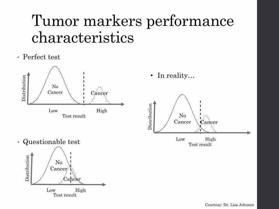
• Perfect test
• Questionable test
Low High
Dis
trib
uti
on
Test result
No
Cancer Cancer
Low High
Dis
trib
uti
on
Test result
No
Cancer
Cancer
• In reality…
Low High
Dis
trib
uti
on
Test result
No
Cancer Cancer
Tumor markers performance characteristics
Courtesy: Dr. Lisa Johnson

• The ability of a test to correctly identify the population with disease (cancer)
For tumor markers
• Good Sensitivity- Someone with cancer produces a high tumor marker result
• Poor Sensitivity- Someone with cancer produces a low/no tumor marker result
False negative results
Low High
Dis
trib
uti
on
Test result
Cancer
Sensitivity
Courtesy: Dr. Lisa Johnson
𝑆𝑁 = 𝑇𝑃/(𝑇𝑃 + 𝐹𝑁)

• The ability of a test to correctly exclude the population without disease (no cancer)
For tumor markers
• Good Specificity-Someone without cancer produces a low/no tumor marker result
• Poor Specificity- Someone without cancer produces a high tumor marker result
Increased tumor marker for benign conditions
False positive results
Low High
Dis
trib
uti
on
Test Result
No
Cancer
Specificity
Courtesy: Dr. Lisa Johnson
𝑆𝑃 = 𝑇𝑁/(𝑇𝑁 + 𝐹𝑃)

How is the cutoff determined?
https://marlin-prod.literatumonline.com/cms/attachment/34661288-1f8f-459e-b8b4-936efc49e9bc/fx1_lrg.jpg
Receiver operator characteristic curve
1- Specificity
Sen
sitiv
ity

Kaplan-Meier survival curve
Marker values ≥ cutoff
https://www.graphpad.com/support/faq/prism-3-kaplan-meier-survival-analysis/
Time (months)
Perc
en
t su
rviv
ing
• Evaluate biomarker relationship with patient outcomes
Marker values < cutoff

Breast cancer development
Normal sexual function
Hormone/GF dependent breast cancer
E
P
EGF
ER
HER2
Hormone
receptors
PR
Images prepared with biorender.comE = estrogen; P = progesterone; EGF = epidermal growth factor

Predictive biomarkers
0
20
40
60
80
TNBCImmunohistochemistry of
biopsy specimens
HER2+
HR+
% c
ase
s
ER
HER2
Hormone
receptors
PR
Images prepared with biorender.com

Breast cancer therapies
ChemotherapyTNBC
Anti-HER2 therapyHER2+
Anti-estrogen therapyHR+
Cancer cell death
Images prepared with biorender.com
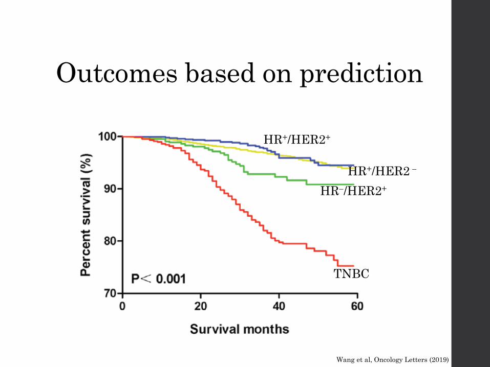
Outcomes based on prediction
TNBC
HR−/HER2+
HR+/HER2+
HR+/HER2 −
Wang et al, Oncology Letters (2019)

Breast cancer related deaths are declining
Breast
• Biomarkers → HER2
• Improved therapies → Herceptin
• Increased understanding of the diseaseCancer facts and figures, ACS (2021)

mRNA-based markers
Overa
ll s
urv
ival
(%)
Pandya et al, Oncotarget (2016)
P
BIK Low
BIK High
p<0.05
• BIK is an estrogen sensitive gene involved in apoptosis

Oncotype Dx and MammaPrint
Kwa et al, Nature Reviews Clinical Oncology (2017)
Oncotype Dx − RT-PCR
MammaPrint − Microarray

DNA-based tumor markers• BReast CAncer genes 1 and 2
• Tumor suppressor genes
Do not cause cancer
Prevent cancer
• About 1:400 individuals (0.25%) carry BRCA1/2 mutations
Family history
• Common population: ~12% women will develop breast cancer
• Women with BRCA1 mutations: 55−65%
• Women with BRCA2 mutations: ~45%
• The Angelina Jolie effect
Preventative double mastectomy
BRCA1
BRCA2
Chromosome 17 Chromosome 13
https://www.nationalbreastcancer.org/what-is-brca; https://www.verywellhealth.com/non-brca-gene-mutations-4173768

Serum-based tumor markers
• CA 15-3 and CA 27.29 Upregulated in breast cancer
Shed by epithelial cells
Stage II and III → early detection of recurrence
Stage IV → monitoring therapy response
Can be elevated in other malignancies
• Carcinoembryonic antigen (CEA) Produced by GI tissue during fetal
development
Very low levels in healthy adults
Maybe elevated in breast, colon and lung cancers
• HER2/neu by ELISA Human Epidermal Growth Factor
Receptor
• All serum-based markers are used to monitor therapy response NOT for diagnosis → poor specificity
Agarwal et al, Front. Immuno. (2018); https://www.123rf.com/photo_78436891: Contemporary practice in clinical chemistry, 4th Edition (2020)
Protein
backbone
Glycosylation
CEA
PEM

Therapy response
Rising markers with 20-30% increase may suggest treatment failure
Trends are
important!
Tietz textbook of clinical chemistry and molecular diagnostics, 6th edition (2018)

Pre-analytical considerations
• Serum or plasma are usually of choice
Recommended to store specimens at 4°C or −20 °C
• Timing of specimen collection → not too critical
Avoid collection immediately after surgery → false elevations
• Avoid testing for unfocused requests such as “tumor marker screen” or suspicion of “malignancy” from ED
• Lack of sensitivity and specificity for specific cancers should be reiterated for consults on abnormal results
Tietz textbook of clinical chemistry and molecular diagnostics, 6th edition (2018)
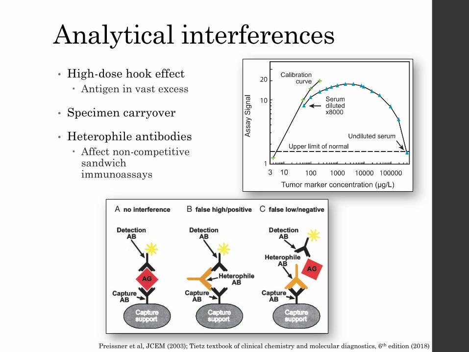
Analytical interferences
• High-dose hook effect
Antigen in vast excess
• Specimen carryover
• Heterophile antibodies
Affect non-competitive sandwich immunoassays
Preissner et al, JCEM (2003); Tietz textbook of clinical chemistry and molecular diagnostics, 6th edition (2018)

Summary of breast cancer management
Suspicious lump
Imaging studies
1° tumor biopsy
ER, PR, HER2
staining
Treatment
Monitor
treatment
response
Histology

Part-4Case Study

Case study and questions
• 38-year-old female
• She felt a peanut-sized lump in her right breast and ignored it thinking a swollen node due to a recent cold
• 3-months later the lump grew to the size of a walnut
• She had persistent headache and mood swings
• Visited her PCP who referred her to an oncologist

Case study and questions
1. What is the oncologist likely to do?A. Perform surgery to remove the tumor
B. Order a biopsy to screen for breast cancer
C. Prescribe chemotherapy
D. Perform imaging studies to visualize tumors✓
• Imaging studies followed by biopsy analysis found a primary tumor of 3.5 cm in diameter and a 1.2 cm lesion in the axillary lymph node
• Diagnosed with grade 2 stage II breast cancer
• Breast cancer subtype: Triple negative (TNBC)

2. What is the clinical team likely going to do next?A. Surgically remove the primary tumor and initiate
chemotherapy
B. Ask the patient to go home as this is not serious
C. Collect a pre-surgical serum specimen to determine the
baseline levels of CA 15-3
D. Both A and C
Case study and questions
✓
• Pre-surgical levels of CA 15-3 were 489 U/mL (RI: 0-31)
1 month later → 35 U/L
2 months later → 34 U/L
6 months later →15 U/L with no radiological abnormalities

3. What do these results likely mean?A. She never had cancer
B. Her cancer is responding to therapy and is in remission
C. The tumor is growing back
D. She will never have cancer recurrence
Case study and questions
✓
• The patient will be monitored over the next few months and put on surveillance to detect recurrent disease activity