fam i i ial hypobetal i poprotei nem ia - the journal of ... · fam i i ial hypobetal i poprotei...
TRANSCRIPT

Fam i I ial hypobetal i poprotei nem ia
MacRae E Linton,' Robert V. Fame, Jr., and Stephen G. Young Gladstone Institute of Cardiovascular Disease, Cardiovascular Research Institute, Department of Medicine, University of California, San Francisco, P.O. Box 419100, San Francisco, CA 94141-9100
Hypobetalipoproteinemia is an autosomal codominant disorder characterized by decreased or absent plasma concentrations of the apolipoprotein (apo) B-containing lipoproteins. Heterozygotes for hypobetalipoproteinemia typically have plasma concentrations of apoB and low density lipoprotein (LDL) -cholesterol that are one-fourth to one-half normal and are usually clinically asympto- matic. In the homozygous state, however, apoB and LDL- cholesterol levels are extremely low or undetectable. When the @-lipoproteins are absent, the clinical pheno- type can be severe and may include fat malabsorption, acanthocytosis, retinitis pigmentosa, and neuromuscular degeneration. This severe phenotype observed in some cases of homozygous hypobetalipoproteinemia is indistin- guishable from that of abetalipoproteinemia, a recessively inherited apoB deficiency state (1, 2). Obligate hetero- zygotes for abetalipoproteinemia have normal plasma lipid and lipoprotein levels, in contrast to familial hypobetalipoproteinemia heterozygotes.
During the past 5 years, our understanding of hypobetalipoproteinemia has been enhanced by the description of many different apoB gene mutations that cause hypobetalipoproteinemia. In this review, we sum- marize the recent progress in the molecular genetics of hypobetalipoproteinemia. We also review the history of hypobetalipoproteinemia, which has, to an extent, been lost amid the excitement of delineating the responsible apoB gene mutations. Finally, we summarize the current understanding of the metabolic abnormalities and the clinical consequences of hypobetalipoproteinemia.
EARLY DESCRIPTIONS OF PATIENTS WITH SEVERE DEFICIENCIES O F THE
@-LIPOPROTEINS
Since the early 1970s, it has been clear that the dominantly inherited disorder, familial hypobetalipopro- teinemia, is clinically and genetically distinct from the recessively inherited syndrome, abetalipoproteinemia. In the 1950s and 19609, however, the genetic and clinical differences between the two disorders had not yet been ap-
preciated, and, because the clinical and biochemical phenotype of the homozygous form of hypobetalipopro- teinemia can be identical to that of abetalipoproteinemia, the two syndromes were frequently confused or lumped together as one syndrome. For example, one of the first patients recognized to have a complete deficiency of the 8- lipoproteins was incorrectly diagnosed as having "reces- sively" inherited abetalipoproteinemia when she actually had the homozygous form of familial hypobetalipopro- teinemia (3). Whereas the clinical features and the mode of inheritance of abetalipoproteinemia were established in the 19509, a clear understanding of hypobetalipoproteine- mia as a syndrome genetically and clinically distinct from abetalipoproteinemia emerged gradually during the next two decades.
In 1950, Bassen and Komzweig (4) described an 18-year-old female who, after having been diagnosed with celiac disease in early childhood, exhibited atypical retini- tis pigmentosa, a diffuse disease of the central nervous system (thought to be a "form of Friedreich's ataxia"), and a "hitherto undescribed" malformation of the erythro- cytes. The erythrocytes had "an unusual crenated appear- ance" with "bizarre shapes, simulating small beetles, crabs and turtles." The parents of this child were first cousins, and her younger brother w a s noted to have the same red blood cell abnormality and early signs of similar retinal changes, suggesting a hereditary nature of the condition. Shortly thereafter, Singer, Fisher, and Perlstein (5) described a similar case involving a 13-year-old boy who had a history of celiac syndrome, malformed erythrocytes, ataxia, and impaired proprioception, but no retinal changes. They described the red blood cells as "thorny" and suggested the descriptive term acanthrocytes (akan- thos, thorn in Greek). In this case, the proband's parents were second cousins. Noting consanguinity and acan-
Abbreviations: M e , adenine; apo, apolipoprotein; HDL, high density lipoproteins; LDL, low density lipoproteins; IDL, intermediate density lipoproteins; SDS, sodium dodecyl sulfate; VLDL, wy low density lipoproteins; RFLP, restriction fragment length polymorphism; Lp[a], lipoprotein[a]; BMI, body mass index; apo[a], apolipoprotein[a]. 'To whom correspondence should be addressed.
Journal of Lipid h e m h Volume 34, 1993 521
by guest, on May 4, 2019
ww
w.jlr.org
Dow
nloaded from

throcytosis in the two unrelated families (4, 5 ) , the authors postulated that “this malformation of the red cells is due to a mutant recessive gene.” The term acanthrocyto- sis was later supplanted by acanthocytosis (6).
Six years later, Jampel and Falls (7) noted that the pa- tient described by Singer et al. (5) had developed retinitis pigmentosa and progressive neurologic deterioration. They also noted that the patient’s serum cholesterol level was 37 mg/dl, which was the first observation of hypocholesterolemia in the Bassen-Kornzweig syndrome. In addition, they more accurately characterized the previ- ously diagnosed childhood celiac disease as fat malabsorp- tion. Based on the marked hypocholesterolemia, they astutely concluded that “It is probable that the entire syn- drome is basically an inborn error of fat metabolism producing a harmful effect on erythrocytes and nerve cells.” In 1960, three groups-Salt et al. (3), Mabry, DiGeorge, and Auerbach (8), and Lamy et al. (9)-inde- pendently reported the absence of 0-lipoproteins from the plasma of similar patients. Salt et al. (3) suggested nam- ing this syndrome “a-6-lipoproteinemia,” a designation that supplanted “Bassen-Kornzweig syndrome.”
The patient described by Salt et al. in 1960, a 17-month-old female child, had steatorrhea and acan- thocytosis but no retinal or neurologic abnormalities (3). They demonstrated that the patient’s plasma had a com- plete absence of @-lipoproteins and decreased levels of cy- lipoproteins, with extremely low levels of cholesterol (22 mg/dl), total lipids (80 mg/dl), and phospholipids (45 mg/dl). Chylomicrons were absent from the plasma, even after a fat-rich meal. Although both the patient’s parents and her paternal grandfather clearly had 0-lipoprotein levels that were approximately one-half of normal levels, Salt et al. (3) suggested that this syndrome, “a$- lipoproteinemia,” was due to an inborn error of metabolism with a recessive mode of inheritance. In 1961, Wolff and Bauman (10) reported that, in contrast to the findings of Salt et al. (3), the parents of a 5-year-old boy with the clinical features of abetalipoproteinemia had no lipid abnormalities. Subsequent studies of abetalipopro- teinemia have confirmed the observation of Wolff and Bauman: the plasma cholesterol, triglycerides and phos- pholipids do not distinguish obligate heterozygotes for abetalipoproteinemia from normal subjects (11). In retrospect, rather than describing a case of the recessively inherited disorder, abetalipoproteinemia, Salt et al. ( 3 ) had actually described the first homozygous and heterozy- gous cases of the autosomal codominant disorder, familial hypobetalipoproteinemia. Although Salt et al. inaccurately designated the inheritance pattern in their case as reces- sive, it is noteworthy that they accurately predicted the nature of their patient’s genetic defect: “The primary gene defect appears to be an inability to form the @-lipoprotein molecule, and is probably concerned with the protein moiety (3)’’
RECOGNITION OF A LESS SEVERE DEFICIENCY
HYPOBETALIPOPROTEINEMIA OF T H E 0-LIPOPRCYI’EINS - HETEROZYGOUS
In the 1960s, several investigators noted the existence of a hypocholesterolemic syndrome that was not as severe, either in terms of clinical symptoms or in the degree of hypocholesterolemia, as abetalipoproteinemia. In general, these patients had low plasma levels of the @-lipoproteins, but not a complete deficiency, and were either asympto- matic or had neurologic symptoms. In 1966, van Buchem et al. (12) reported three brothers who had hypocholester- olemia (76-126 mg/dl) and hypotriglyceridemia (29-44 mgldl), but who did not have acanthocytosis, fat malab- sorption, neuromuscular disease, or retinitis pigmentosa. A jejunal biopsy from one of the subjects shaved no evi- dence of fat accumulation and a liver biopsy demon- strated mild steatosis. All three brothers had decreased 6-lipoproteins and were described as having “congenital 0-lipoprotein deficiency,” a term suggested by Isselbacher et al. (13) to describe abetalipoproteinemia. A fourth brother and the two children of the propositus had normal lipoprotein levels. Van Buchem et al. (12) concluded that “many degrees of this @-lipoprotein deficiency exist, even in subjects who are entirely symptom-free; it is probable that this deficiency does not occur as rarely as has been supposed. Only if traces of 0-lipoprotein are present, or if it is entirely absent, do steatorrhea, neuromuscular dis- turbances, acanthocytosis and retinal changes develop.” These authors suggested that “an autosomal recessive gene with variable penetrance is involved in this hereditary disease.”
Hypobetalipoproteinemia was first recognized as a syn- drome distinct from abetalipoproteinemia when, in 1969, Mars et al. (14) reported the occurrence of low plasma cholesterol levels in 13 of 31 individuals from three succes- sive generations of a single kindred. The propositus was a 37-year-old woman with a progressive demyelinating disorder affecting the central nervous system and a dislike for fatty foods, but no evidence of retinitis pigmentosa or steatorrhea. Analysis of plasma lipids revealed low levels of total cholesterol (79 mg/dl) and LDL-cholesterol (30 mg/dl), but a normal triglyceride level (106 mg/dl). A marked reduction in the @-lipoproteins was noted on paper electrophoresis. After fat ingestion, there was only a minimal increase in the plasma triglyceride level. A jejunal biopsy taken after a 12-h fast revealed fat droplet accumulation in the enterocytes. In addition, serum levels of vitamin A, carotene, and vitamin E were low. Twelve immediate blood relatives had low plasma cholesterol levels (mean = 110 mg/dl, range = 67-149 mg/dl) and low-to-normal triglyceride levels (mean = 38.6 mg/dl, range = 16-80 mg/dl), but were essentially asympto- matic. The red blood cells of the family members with to- tal plasma cholesterol values less than 100 mg/dl devel-
522 Journal of Lipid Research Volume 34, 1993
by guest, on May 4, 2019
ww
w.jlr.org
Dow
nloaded from

oped acanthocytosis during incubation in tissue culture media containing 10% autologous serum; however, acan- thocytes were not present on routine blood studies. From the analysis of the pedigree, Mars et al. (14) concluded that the hypocholesterolemic phenotype had an autosomal dominant mode of inheritance, and that “sufficient genetic, clinical, and biochemical differences exist between hypo-0-lipoproteinemia and a-6-lipoproteinemia to war- rant the conclusion that hypo-p-lipoproteinemia is not a ‘form fruste’ of a-0-lipoproteinemia.” They suggested naming the syndrome familial hypobetalipoproteinemia. In the same year, Richet et al. (15) described another kindred in which six family members from two successive generations had hypobetalipoproteinemia with low cholesterol levels (70-140 mg/dl) and low-to-normal triglyceride levels (20-125 mg/dl), supporting an autosomal dominant mode of inheritance.
By the early 19709, abetalipoproteinemia and familial hypobetalipoproteinemia were understood to be separate entities, both clinically and genetically. In 1972, Fredrick- son, Gotto, and Levy (16) proposed the following diagnos- tic criteria for familial hypobetalipoproteinemia: “I) LDL abnormally low but present and identifiable immuno- chemically, while concentrations of very low density lipoprotein (VLDL) and high density lipoprotein (HDL) are normal, 2) absence of diseases to which hypobetalipo- proteinemia may be secondary, and to be certain 3) detec- tion of a similar pattern in a first degree relative.” They reported that the diagnostic lipoprotein electrophoresis reveals a faint 0-lipoprotein band and that pre- betalipoproteins are usually modestly reduced (16). They concluded: “No clinical abnormalities have been consis- tently found in all the patients with hypobetalipo- proteinemia” and that patients with hypobetalipoproteinemia are close to the margin of adequate LDL but rarely will manifest pathologic changes” (16).
Five kindreds with hypobetalipoproteinemia were reported in the early 1970s that fit the criteria of Fredrick- son and co-workers (17-21). In these five kindreds, the affected family members had low plasma levels of total cholesterol (45-140 mg/dl) and low-to-normal levels of triglycerides (11-140 mg/dl). It is now clear that these cases, which fit the 1972 definition of hypobetalipoproteinemia by Fredrickson and colleagues (16), all described the het- erozygous form of hypobetalipoproteinemia and that the criteria of Fredrickson and associates apply only to this heterozygous form. At that time, no one had even specu- lated that both a homozygous and heterozygous form of this syndrome might exist.
RECOGNITION OF HOMOZYGOUS HYPOBETALIPOPROTEINEMIA
Between 1973 and 1975, Biemer and McCammon (22, 23) and Cottrill et al. (24, 25) independently reported
kindreds in which some family members had the “hypobetalipoproteinemia phenotype” and others had the “abetalipoproteinemia phenotype.” Both groups arrived at the conclusion that homozygosity for the genetic defect found in hypobetalipoproteinemia could yield a pheno- type identical to that seen in the recessive syndrome, abetalipoproteinemia. Biemer and McCammon described a 37-year-old woman who was noted to have phenotypic abetalipoproteinemia when she developed a vitamin K-deficient bleeding diathesis at parturition. The proposi- tus had severe hypocholesterolemia (31-48 mg/dl), extremely low triglyceride levels (0-11 mgldl), and un- detectable plasma levels of LDL and VLDL. In addition, she had acanthocytosis, retinitis pigmentosa, and a low se- rum carotene level (despite a normal fecal fat content). Her neurologic deficits consisted of only a minimal altera- tion of her sense of balance, decreased vibratory percep- tion, and diminished deep tendon reflexes. The newborn baby of the propositus, as well as four first-degree rela- tives, were asymptomatic and had reduced plasma levels of total cholesterol (80-124 mg/dl) and normal triglyceride levels (21-76 mg/dl); levels of LDL-cholesterol (31-63 mg/dl) were approximately a third of normal, and levels of VLDL-cholesterol were low to low-normal (3-16 mg/dl). Biemer and McCammon (23) stated: “Analysis of the data in the present family suggests that this case of abetalipoproteinemia represents the homozygous expres- sion of the same gene which when present in the heterozy- gous state results in hypobetalipoproteinemia. It is con- cluded, therefore, that this case of abetalipoproteinemia has apparently been inherited via a Merent genetic mutation than the previously reported cases of abetalipoproteinemia.”
Cottrill et al. (25) described two children from a single family who had extremely low levels of cholesterol (13-22 mg/dl), extremely low triglycerides (12-14 mg/dl), and un- detectable levels of VLDL and LDL. Antisera to LDL failed to react with their plasma. The two children had acanthocytosis, fat malabsorption, growth retardation, and intestinal and hepatic steatosis, but no neurologic or ophthalmologic abnormalities. A family study revealed three generations of subjects with the heterozygous form of hypobetalipoproteinemia occurring on both the pater- nal and maternal sides and distant consanguinity. The eight family members with heterozygous hypobetalipo- proteinemia had low levels of cholesterol (mean = 105 mg/dl, range = 41-134 mg/dl), LDL-cholesterol (mean = 37 mg/dl, range = 17-69 mg/dl), and low-normal triglyceride levels (mean = 39 mg/dl, range = 19-68 mg/dl). Cottrill et al. (25) concluded that, “It now seems probable that the absence of apoLDL in plasma can occur through at least two different genetic mechanisms. One of these would lead to the previously described form of abetalipopro- teinemia, in which there is no known phenotypic expres- sion of the heterozygous state, the second resulting from the homozygous state for the autosomal dominant dis- order hypobetalipoproteinemia.”
Linton, Farese, and Young Familial hypobetalipoproteinemia 523
by guest, on May 4, 2019
ww
w.jlr.org
Dow
nloaded from

A. Coomassie Blue Stained Gel B. MB19 C. MB47
Apo-B1W- M Y -- b -
Apo-086-
Apo-848- c 0 -
Ape-037- - -
Apo-E- 4 4
1 2 1 2 1 2
D. MB43
-0
1 2 1 = Control Chylomicrons 2 = H.J.B.-VLDL
Fig. 1. Demonstration of t w ~ abnormal apoB species, apoB-37 and apoB-86. from an apoB-37/apoB-86 heterozygote (H.J.B.) by an SDS- polyacrylamide gel and western blotting. In each panel, lane 1 s h w blood chylomicrons isolated from a normal subject after a fat-rich meal, and lane 2 shows H.J.B.’s VLDL. For each sample, 50 fig of delipidated protein was used. Panel A shows a 3-15% SDS-polyacrylamide slab gel stained with 0.1% Coomaask Brilliant Blue R-250. Panels B, C, and D show western blots using antibodies MB19, MB47, and MB43, respec- tively. Antibody MB19 binds to midues 1-56 of apoB-100 (137); anti- body MB47 binds between apoB-100 amino acids 3441 and 3569; anti- body MB43 binds betwen apoB-100 amino acids 4027-4081 (138). Panels A, B, and D were previously published (27) and arc reproduced here with the permission of the American Society of Clinical Investigation.
HYPOBETALIPOPRDI’EINEMIA HOMOZYGOTES WITH NORMOTRIGLYCERIDEMIA
In 1979, Steinberg et al. (26) reported the existence of a kindred with familial hypobetalipoproteinemia, the H.J.B. kindred, in which the homozygotes had an unusual finding- normotriglyceridemia. Three siblings of the H.J.B. kindred had extremely low LDL-cholesterol levels (3-8 mg/dl), whereas several other members had LDL- cholesterol levels that were one-fourth to one-half of nor- mal values. Steinberg et al. (26) speculated that the three siblings with extremely low levels of LDL-cholesterol were familial hypobetalipoproteinemia homozygotes, having “a double dose of a dominant allelic mutation,” and that the less severely affected subjects were heterozygotes. The three presumed homozygotes were unusual, however, be- cause unlike previously described homozygotes, they were essentially normotriglyceridemic (24-91 mg/dl), had VLDL and small amounts of apoB (< 10% normal) in their plasma, and were asymptomatic. These three sib- lings therefore demonstrated heterogeneity, both clini- cally and biochemically, in the expression of the homozy- gous form of hypobetalipoproteinemia.
The explanation for this heterogeneity in homozygotes recently became evident with the elucidation of the bio- chemical basis of hypobetalipoproteinemia. As discussed
below, hypobetalipoproteinemia can be caused by a vari- ety of apoB gene mutations. Hypobetalipoproteinemia homozygotes with apoB gene “null alleles” have absent VLDL, very low triglyceride levels, and tend to be more severely affected clinically, often appearing phenotypi- cally identical to patients with abetalipoproteinemia. In contrast, homozygotes that have mutant apoB alleles that are associated with sufficient apoB synthesis to facilitate intestinal fat absorption can have virtually normal trigly- ceride levels and are usually asymptomatic. The mutant apoB alleles in the H.J.B. kindred were of the latter type.
LINKAGE OF HYPOBETALIPOPROTEINEMIA To THE APOLIPOPROTEIN B GENE
Before the mid-l980s, it was believed that either abetalipoproteinemia or hypobetalipoproteinemia, or perhaps both disorders, were due to defects in the synthe- sis of apoB. In 1986, Young et al. (27, 28) demonstrated that hypobetalipoproteinemia was indeed associated with defects in the apoB gene. They re-examined the H.J.B. kindred (26) and documented the existence of two mutant apoB alleles, both of which resulted in hypobetalipopro- teinemia. One mutant apoB allele yielded a truncated species of apoB, apoB-37 (named according to the centile designation of Kane, Hardman, and Paulus (29)). The sec- ond apoB allele yielded significant amounts of apoB-48, but only very small amounts of a full-length apoB-100 molecule (28). Later, this second allele was shown to make small amounts of apoB-86 in addition to apoB-48 and apoB-100 (30). The three siblings in the H.J.B. kindred with extremely low cholesterol levels (thought to be homozygotes by Steinberg et al. (26)) had both mutant apoB alleles and therefore were compound heterozygotes for hypobetalipoproteinemia. Fig. 1 shows a sodium dodecyl sulfate (SDS) -polyacrylamide gel and western blots demonstrating the presence of the two abnormal apoB species (apoB-37 and apoB-86) in the VLDL of H.J.B., one of the compound heterozygotes. All of the offspring of the three compound heterozygotes inherited either the apoB-37 allele or the apoB-86 allele and had LDL-cholesterol levels about one-fourth of normal levels (28). An SDS-polyacrylamide gel of the VLDL of two apoB-37/apoB-86 compound heterozygotes, an apoB-37 heterozygote, and several apoB-86 heterozygotes is illus- trated in Fig. 2.
Shortly after the reports of Young et al. (27, 28), Lep- pert et al. (31) demonstrated the linkage of the hypobetalipo- proteinemia phenotype to the apoB gene by showing that apoB restriction fragment length polymorphisms (RFLPs) cosegregated with the hypobetalipoproteinemia pheno- type in a large family. Other apoB RFLP linkage studies demonstrated that the apoB gene was not involved in abetalipoproteinemia (32, 33) or in the chylomicron
524 Journal of Lipid Research Volume 34, 1993
by guest, on May 4, 2019
ww
w.jlr.org
Dow
nloaded from

apoB species, apoB-39, and a single nucleotide substitu- Apo-BlW- --- - mm
I tion creating a nonsense mutation in exon 25. Since these reports, a total of 25 mutations causing hypobetalipopro- A P E 8 6 - teinemia have been described (Table 1). Most of the mu- tations have been identified in heterozygotes; each muta- tion interferes with the translation of a full-length apoB-100. Nearly all of the mutations are either nonsense mutations or frameshift mutations resulting from the de-
b~ 1 2 3 4 5 6 7 8 letionof l to5 bp that createaprematurestopcodon. De- letions of 37 bp and 694 bp have been described, as has one mutation involving an intron-exon splice site (Table 1). Although we expect that a single mutation causing hypobetalipoproteinemia will be identified as a common
Allele * mutation within a particular geographical region or eth-
Apo-B48 - - APE37 - -
Subjecl 1 2 18 16 39 38 40 17
Apo-E37A11e1e ' A~E86Allele
Fig. 2. A 342% SDS-polyacrylamide dab gel of the VLDL fractions of several members of the H.J.B. kindred. The VLDL were isolated by ultracentrifugation, and 30 pg of delipidated proteins was loaded onto each lane. The gel was stained with 0.1% Coomassic Brilliant Blue R-250. Human subjects are identified by number according to the pub- lished pedigree of the H.J.B. kindred (28). Subjects 1 and 2 (lanes 1 and 2, respectively) arc apoB-37/apoB-86 compound heterozygotes. Subject 18 (lane 3) is an apoB-37 heterozygote. Subjects 16. 38, and 40 (lanes 4, 6. and 7, respectively) are apoB-86 heterozygotes; subjects 39 and 17 (lanes 5 and 8, respectively) are unaffected family members.
retention syndrome (Anderson's disease) (34), another in- herited disorder characterized by low plasma levels of the @-lipoproteins.
Although most cases of hypobetalipoproteinemia still appear to be linked to the apoB gene, it is possible that other genetic mutations could cause a similar phenotype. Recently, Hobbs et al. (35) described a dominantly in- herited cholesterol-lowering gene in a kindred with familial hypercholesterolemia and demonstrated that the apoB gene did not account for the cholesterol-lowering phenotype. The gene. responsible for this cholesterol- lowering phenotype has not yet been reported. Fazio et al. (36) have also reported a case of hypobetalipoproteinemia that was apparently unrelated to the apoB gene. Vega et al. (37) have described a subject with hypobetalipopro- teinemia whose parents did not have low plasma cholesterol levels. They postulated that the low plasma apoB level in this subject was due to enhanced LDL receptor activity, which in turn was secondary to the in- creased synthesis of bile acids.
APOLIPOPROTEIN B ,GENE MUTATIONS CAUSING HYPOBETALIPOPROTEINEMIA
In 1988, Young, Northey, and McCarthy (38) reported that a 4-bp deletion in exon 26 resulted in hypobetalipo- proteinemia that was associated with the synthesis of apoB-37. Virtually simultaneously, Collins et al. (39) described two mutations causing hypobetalipoproteine- mia: a l-bp deletion in exon 26 that yielded a truncated
. ~~
nic group, such a mutation has not yet been found. Each of the nonsense or frameshift mutations occurring
in exons 26-29 has been associated with the presence of a truncated apoB that is detectable in the plasma lipoproteins. When a truncated apoB is found in the plasma, locating the responsible mutation is usually straightforward. The length of the truncated apoB can be estimated to an accuracy of * 75 amino acids by compar- ing its migration on SDS-polyacrylamide gels with that of various size standards such as apoB-48 (2152 amino acids) and the proteolytic breakdown products, apoB-74 (3239 amino acids) and apoB-26 (1297 amino acids). In addi- tion, the reactivity of the truncated apoB with various apoB-specific monoclonal antibodies for which the epi- topes have been mapped can also aid in estimating the length of the truncated apoB (Fig. 1). After estimating the length of the truncated protein, one can exclude a large deletion or rearrangement at the apoB gene locus by Southern blot analysis and then sequence enzymatically amplified apoB genomic clones to determine the precise mutation.
Four mutations causing hypobetalipoproteinemia, all located 5' of exon 26 and predicted to form very short apoB species, have not been associated with a truncated apoB that is detectable in the plasma (Table 1). Of partic- ular interest are apoB-25 and apoB-29, neither of which could be detected in the plasma of affected heterozygotes (39, 40). Why these truncated proteins are absent from the plasma is unclear. Recently, several laboratories have transfected various lengths of apoB cDWA into cultured cells and have shown that the transfected cells are capable of synthesizing and secreting very short apoB species (e.g., apoB-18) (41-43). Presumably, the absence of apoB-25 and apoB-29 in the plasma of human subjects is due either to decreased synthesis (perhaps as a result of low levels of the mutant apoB mRNA) or to the rapid plasma catabolism of lipoproteins containing these trun- cated species.
When a truncated apoB is not detectable in the plasma, the identification of the responsible apoB mutation may be difficult; in these instances, there is no clue as to the
Linton, Fame, and Young Familial hypobctalipoproteinemia 525
by guest, on May 4, 2019
ww
w.jlr.org
Dow
nloaded from

TA
BL
E 1
. A
polip
opro
tein
B g
ene
mut
atio
ns a
ssoc
iate
d w
ith f
amili
al h
ypob
etal
ipop
rote
inem
ia
Mut
atio
n LD
L-C
holes
terol
Lev
el in
H
eter
ozyg
otes
C
omm
ents
1. A
poB
-2:
G+
T t
rans
vers
ion
in t
he f
irst b
ase o
f in
tron
5
2. A
poB
-9:
C+
T t
rans
ition
at
cDN
A n
ucle
otid
e 14
43
3. A
poB
-25:
Del
etio
n of
694
bp,
inc
ludi
ng a
ll of
exo
n 21
4. A
poB
-29:
C+
T t
rans
ition
at
cDN
A n
ucle
otid
e 41
25
5. A
poB
-31:
Del
etio
n of
cD
NA
nuc
leot
ide
4480
6. A
poB
-32:
C+
T t
rans
ition
at
cDN
A n
ucle
otid
e 45
57
7. A
poB
-32.
5: T
+G
tra
nsve
rsio
n at
cD
NA
nuc
leot
ide
4631
8. A
poB
-37:
Del
etio
n of
cD
NA
nuc
leot
ide
5391
-539
4
9. A
poB
-39:
Del
etio
n of
cD
NA
nuc
leot
ide
5591
10. A
poB
-40:
Del
etio
n of
cD
NA
nuc
leot
ides
569
3-56
94
11. A
poB
-46:
C+
T t
rans
ition
at
cDN
A n
ucle
otid
e 63
81
12. A
poB
-50:
C+
T t
rans
ition
at
cDN
A n
ucle
otid
e 69
63
13. A
poB
-52:
Del
etio
n of
apo
B w
ith c
DN
A n
ucle
otid
es 7
278-
7282
14. A
poB
-52.
8: D
elet
ion
of a
poB
cD
NA
nuc
leot
ide
7295
15. A
poB
-52.
8: D
elet
ion
of a
poB
cD
NA
nuc
leot
ide
7359
16. A
poB
-54.
8: C
+T
tra
nsiti
on a
t ap
oB c
DN
A n
ucle
otid
e 76
65
17. A
poB
-55:
C+
T t
rans
ition
at
apoB
cD
NA
nuc
leot
ide
7692
18. A
poB
-61:
Del
etio
n of
apo
B c
DN
A n
ucle
otid
es 8
525-
8561
19. A
poB
-67:
Del
etio
n of
apo
B c
DN
A n
ucle
otid
e 93
27
20. A
poB
-75:
Del
etio
n of
apo
B c
DN
A n
ucle
otid
e 10
366
21. A
poB
-82:
C+
A t
rans
vers
ion
at a
poB
cD
NA
nuc
leot
ide
1141
1
22. A
poB
-83:
C
+A
tra
nsve
rsio
n at
apo
B c
DN
A n
ucle
otid
e 11
458
23. A
poB
-86:
Del
etio
n of
cD
NA
nuc
leot
ide
1184
0
24. A
poB
-87:
Del
etio
n of
cD
NA
nuc
leot
ide
1203
2
25. A
poB
-89:
Del
etio
n of
cD
NA
nuc
leot
ide
1230
9
mg/
dl
38
16
NA
NA
29 +
7
31
23
31 f
12
NA
49
74 f
19
NA
49 f
22
38
33
49 +
14
69 f
12
45
42 f
15
48 f
11
61
50 f
18
31 f
15
62
74
Alm
ost
cert
ainl
y in
terf
eres
with
pro
per m
RN
A s
plic
ing.
No
apoB
det
ecta
ble
in t
he p
lasm
a (4
4).
Pred
icte
d to
yie
ld a
n ap
oB sp
ecie
s con
tain
ing
41 1
amin
o ac
ids,
but
non
e w
as d
etec
tabl
e in
the
plas
ma
(44)
. Pr
edic
ted
to y
ield
a tr
unca
ted
apoB
spe
cies
con
tain
ing
1085
amin
o ac
ids,
but
non
e w
as d
etec
tabl
e in
the
pla
sma
(40)
.
Pred
icte
d to
yie
ld a
trun
cate
d ap
oB sp
ecie
s con
tain
ing
1305
amin
o ac
ids,
but
non
e w
as d
etec
tabl
e in
pla
sma
(39)
.
Apo
B-3
2 (1
425
amin
o ac
ids)
is p
rese
nt i
n H
DL
and
in
the
d >
1.2
1 g/
ml
frac
tion
(56)
.
Apo
B-3
2 (14
49
amin
o ac
ids)
is p
rese
nt i
n L
DL
, H
DL
, and
the
d >
1.2
1 g/
ml
frac
tion
(84)
.
Apo
B-3
2.5
(147
3 am
ino
acid
s) is
pre
sent
in
HD
L a
nd
the
d >
1.2
1 g/
ml
frac
tion
(55)
.
Apo
B-3
7 (1
728
amin
o ac
ids)
is p
rese
nt i
n V
LD
L, L
DL
, and
HD
L
(27,
28,
38,
117
).
Apo
B-3
9 (1
799
amin
o ac
ids)
is p
rese
nt i
n V
LD
L a
nd L
DL
(3
9).
Apo
B-4
0 (18
29
amin
o ac
ids)
is p
rese
nt i
n V
LD
L, L
DL
, and
HD
L (7
5,
134)
.
Apo
B-4
6 (20
57
amin
o ac
ids)
is p
rese
nt i
n V
LD
L, L
DL
, and
HD
L
(54)
.
Apo
B-5
0 (2
251
amin
o ac
ids)
is p
rese
nt i
n V
LD
L (
76,
79).
Apo
B-5
2 (23
61
amin
o ac
ids)
is p
rese
nt i
n V
LD
L a
nd L
DL
(1
35).
Apo
B-5
2.8
(239
5 am
ino
acid
s) is
pre
sent
in
VL
DL
and
LD
L (
55).
Apo
B-5
2.8
(239
5 am
ino
acid
s) is
pre
sent
in
VL
DL
and
LD
L (
55).
Apo
B-5
4.8
(248
5 am
ino
acid
s) is
pre
sent
in
VL
DL
and
LD
L (
57).
Apo
B-5
5 (2
492
amin
o ac
ids)
is t
he s
ame
size
as
apoB
-54.
8 on
SD
S-po
lyac
ryla
mid
e ge
ls (
96).
Apo
B-6
1 (2
784
amin
o ac
ids)
is p
rese
nt i
n V
LD
L a
nd L
DL
(4
9).
Apo
B-6
7 (3
040
amin
o ac
ids)
is p
rese
nt i
n V
LD
L a
nd L
DL
(83
).
Apo
B (3
386
amin
o ac
ids)
is p
rese
nt i
n V
LD
L a
nd L
DL
(8
6).
Apo
B-8
2 (3
733
amin
o ac
ids)
is p
rese
nt p
rim
arily
in V
LD
L; t
race
am
ount
s ar
e pr
esen
t in
IDL
. Id
en-
tifie
d in
an
Afr
o-A
mer
ican
sub
ject
(55)
.
Apo
B-8
3 (3
749 a
min
o ac
ids)
is p
rese
nt p
rim
arily
in V
LD
L; tr
ace
amou
nts
are
pres
ent
in I
DL
(85)
.
Apo
B-8
6 (3
896
amin
o ac
ids)
is p
rese
nt i
n V
LD
L a
nd L
DL
(30
).
Apo
B-8
7 (3
978
amin
o ac
ids)
is p
rese
nt in
VL
DL
and
LD
L; bind
s w
ith in
crea
sed
atfin
ity to
the
LD
L
rece
ptor
(77
, 13
6).
Apo
B-8
9 (4
039 a
min
o ac
ids)
is p
rese
nt in
VL
DL
and
LD
L; bi
nds
with
incr
ease
d af
iinity
to th
e L
DL
re
cept
or (
75,
119,
120
, 13
4).
NA
, no
t av
aila
ble.
by guest, on May 4, 2019www.jlr.orgDownloaded from
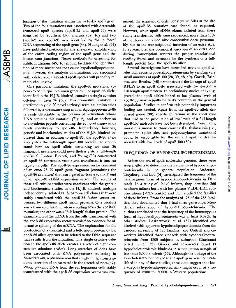
location of the mutation within the -43-kb apoB gene. Two of the four mutations not associated with detectable truncated apoB species (apoB-25 and apoB-29) were identified by Southern blot analysis (39, 40) and two (apoB-2 and apoB-9) were identified by “brute force” DNA sequencing of the apoB gene (44). Huang et al. (44) have published methods for the enzymatic amplification of the entire coding region of the apoB gene and the intron-exon junctions. Newer methods for screening for subtle mutations (45, 46) should facilitate the identifica- tion of more mutations that cause hypobetalipoproteine- mia; however, the analysis of mutations not associated with a detectable truncated apoB species will probably re- main challenging.
One particular mutation, the apoB-86 mutation, ap- pears to be unique in human genetics. The apoB-86 allele, identified in the H.J.B. kindred, contains a single cytosine deletion in exon 26 (30). This frameshift mutation is predicted to yield 20 novel carboxyl-terminal amino acids followed by a premature stop codon. Apolipoprotein B-86 is easily detectable in the plasma of individuals whose DNA contains this mutation (Fig. 2), and an antiserum to a synthetic peptide containing the 20 novel amino acids binds specifically to apoB-86. Remarkably, however, genetic and biochemical studies of the H.J.B. kindred in- dicated that, in addition to apoB-86, the apoB-86 allele also yields the full-length apoB-100 protein. To under- stand how an apoB allele containing an exon 26 frameshift mutation could nevertheless yield a full-length apoB-100, Linton, Pierotti, and Young (30) constructed an apoB-86 expression vector and transfected it into rat hepatoma cells. The apoB-86 expression vector consisted of an exon 26-29 apoB gene fragment (containing the apoB-86 mutation) that was ligated in-frame to the 3‘ end of an apoB-18 cDNA expression vector. The results of these cell culture studies were consistent with the genetic and biochemical studies in the H.J.B. kindred: multiple independently isolated rat hepatoma cell clones that were stably transfected with the apoB-86 fusion vector ex- pressed two different apoB fusion proteins. One product was a truncated fusion protein resulting from the apoB-86 mutation; the other was a “full-length” fusion protein. The examination of the cDNA from the cells transformed with the apoB-86 expression vector revealed no evidence for al- ternative splicing of the mRNA. The explanation for the production of a truncated and a full-length protein by the apoB-86 allele appears to be related to the DNA sequence that results from the mutation. The single cytosine dele- tion in the apoB-86 allele creates a stretch of eight con- secutive adenines (Ades). Long stretches of Ades have been associated with RNA polymerase stuttering in Escherichia coli, a phenomenon that results in the transcrip- tional insertion of an extra Ade in the stretch of Ades (47). When genomic DNA from the rat hepatoma cells stably transformed with the apoB-86 expression vector was exa-
mined, the sequence of eight consecutive Ades at the site of the apoB-86 mutation was found, as expected. However, when apoB cDNA clones isolated from these stably transformed cells were sequenced, more than 10% of the clones contained nine consecutive Ades, presuma- bly due to the transcriptional insertion of an extra Ade. It appears that the occasional insertion of an extra Ade during transcription restores the proper translational reading frame and accounts for the synthesis of a full- length protein from the apoB-86 allele.
Several investigators have identified mutant apoB al- leles that cause hypobetalipoproteinemia by yielding very small amounts of apoB-100 (28, 39, 48, 49). Gavish, Brin- ton, and Breslow (48) demonstrated the linkage of apoB RFLPs to an apoB allele associated with low levels of a full-length apoB protein. In preliminary studies, they sug- gested that apoB alleles linked to low plasma levels of apoB-100 may actually be fairly common in the general population. Studies to confirm this potentially important finding are needed. Apart from the apoB-86 allele dis- cussed above (30), specific mutations in the apoB gene that lead to the production of low levels of a full-length apoB-100 molecule have not been described. Presumably, mutations similar to those causing 0 + thalassemia (Le., promoter, splice site, and polyadenylation mutations) could be responsible for the mutant apoB alleles as- sociated with low levels of apoB-100 (50).
FREQUENCY OF HYPOBETALIPOPROTEINEMIA
Before the era of apoB molecular genetics, there were several efforts to determine the frequency of hypobetalipo- proteinemia in the general population. Andersen, Trojaborg, and Lou (51) investigated the frequency of the heterozygous form of hypobetalipoproteinemia in Den- mark. In a study of 10,440 infants, they identified 266 newborn infants born with low plasma VLDL-LDL con- centrations ( < 2.5 centile) and then studied the families of these infants. From the analysis of 176 of the 266 fami- lies, they documented that 9 had three-generation Men- delian inheritance of hypobetalipoproteinemia. The authors concluded that the frequency of the heterozygous form of hypobetalipoproteinemia was at least 0.09%. In other studies, Laskarzewski et al. (52) identified one kindred with apparent hypobetalipoproteinemia from the random screening of 125 families, and Cottrill and co- workers identified three kindreds with hypobetalipopro- teinemia from 1200 subjects in suburban Cincinnati (cited in ref. 52). Glueck and co-workers found 13 hypocholesterolemic kindreds in a population sample of less than 6,000 kindreds (53). Although the linkage of the low-cholesterol phenotype to the apoB gene was not estab- lished in any of these studies, these data suggest that het- erozygous hypobetalipoproteinemia might occur at a fre- quency of 1/500 to 1/1,000 in Western populations.
Linton, Fame, and Young Familial hypobetalipoproteinemia 527
by guest, on May 4, 2019
ww
w.jlr.org
Dow
nloaded from

The frequency of apoB gene mutations causing trun- cated apoBs and hypobetalipoproteinemia is not known. In our laboratory, we screened the lipoprotein fractions from approximately 75 healthy adults with total plasma cholesterol levels less than 120 mg/dl using SDS- polyacrylamide gels and identified two familial hypobetalipo- proteinemia heterozygotes with truncated apoBs (apoB-46 and apoB-82) (54, 55). While studying the apoB-46 kindred, we identified an apoB-31 mutation in the spouse of an apoB-46 heterozygote (56). Wagner et al. (57) used immunoblots of plasma to screen for apoB trun- cations in 525 healthy subjects with total cholesterol levels that were less than the tenth percentile and identified four apoB alleles yielding truncated apoBs. From these studies, it appears that truncated apoBs are not particularly rare in healthy subjects with low plasma cholesterol levels. These data probably underestimate the true frequency of apoB gene mutations in hypocholesterolemic subjects, however, because mutations that did not yield a truncated protein would not be detected by the methods used. Properly addressing the true frequency in which apoB gene mutations cause hypocholesterolemia would require performing family studies and showing the linkage of the apoB gene to the low cholesterol phenotype.
CLINICAL P H E N W P E IN RELATION TO SPECIFIC APOB MUTATIONS
Null-allele homozygotes
Hypobetalipoproteinemia patients who are homozy- gous for “null-alleles” (Le., who make no detectable apoB) are phenotypically similar to abetalipoproteinemia pa- tients and may have malabsorption, neurologic disease, and hematologic abnormalities as prominent clinical fea- tures. Fat malabsorption is often the presenting finding (3, 25, 44,58-60). Studies have shown that approximately 30-40% of the fat consumed by homozygotes is not ab- sorbed (1, 25, 61) and that chylomicronemia is not ob- served after fat ingestion (1, 3, 23, 25). Because of the fat malabsorption, the plasma levels of the fat-soluble vita- mins A and E are low (3, 23, 25, 44, 62). One null-allele homozygote, who was 37 years old at diagnosis, denied having fatty food intolerance or steatorrhea and had a normal fecal fat content (23). The serum carotene level was low, however, and when she was given a diet contain- ing 100 g of fat per day she developed nausea, vomiting, and diarrhea (63).
The neurologic disease that has been observed in null- allele homozygotes has been similar to that observed in abetalipoproteinemia, but appears to be milder in severity. Abetalipoproteinemia subjects can develop a progressive neurologic syndrome that includes areflexia, impaired proprioception, ataxia, dysarthria, muscle
weakness, kyphoscoliosis, and ophthalmoparesis (64); if not treated, these patients can be crippled by their third decade. Biemer and McCammon (23) originally sug- gested that the neurologic problems associated with homozygous hypobetalipoproteinemia might be milder than those seen in abetalipoproteinemia, based on their description of a 37-year-old female homozygote whose only abnormal findings were retinitis pigmentosa and ab- sent deep tendon reflexes. A number of reports support the impression that hypobetalipoproteinemia homozygotes may have milder neurologic disease (3, 25, 44, 58, 59, 65, 66) than patients with abetalipoproteinemia. At the time of their diagnosis, four of these subjects (ages 0.5-6 years) had no abnormal neurologic signs (3, 25, 58). Two other homozygous subjects, ages 10 (44) and 11 (59) had only absent deep tendon reflexes when they were diagnosed. One 47-year-old female homozygote had only a mild sen- sory polyneuropathy with resultant sensory ataxia (65). This is not to say, however, that homozygous hypobetalipo- proteinemia cannot be a severe and crippling disease, as illustrated by an 11-year-old male homozygote who had severe ataxia, dysarthria, impaired position and vibratory sensation, and absent deep tendon reflexes (44).
Retinitis pigmentosa, a progressive degenerative dis- ease of the retina that is associated with vision loss and is commonly seen in abetalipoproteinemia, has been described in a number of hypobetalipoproteinemia homozygotes (23, 65, 67, 68). One 37-year-old homozygote had severe retinitis pigmentosa at the time she was diag- nosed (23). The patient described by Salt et al. (3) had de- veloped mild pigmentary retinopathy at age 5 despite vitamin A supplementation (68). At age 7, treatment with large oral doses of vitamin E was initiated, and an exami- nation at 31 years of age showed no abnormalities of reti- nal function (69). The onset of the retinopathy in homo- zygotes varies, but often symptoms do not occur until adulthood; there was no evidence of retinitis pigmentosa in the younger homozygous subjects, who were diagnosed at ages ranging from 5 months to 11 years (3, 25, 59).
Red blood cell acanthocytosis is commonly seen when the 0-lipoproteins are absent from the plasma and has been observed in nearly all of the hypobetalipoproteine- mia homozygotes. The markedly abnormal erythrocyte forms are thought to be caused by an abnormal mem- brane lipid content. Biochemical studies of the acantho- cytic membranes in abetalipoproteinemia have demon- strated a decreased amount of linoleic acid, an increase in sphingomyelin content, and an increase in the sphin- gomyelinAecithin molar ratio (70-72). The altered sphin- gomyelinAecithin ratio is believed to cause changes in erythrocyte membrane fluidity (70). Similar changes in the lipid content of erythrocyte membranes have been ob- served in hypobetalipoproteinemia homozygotes (73, 74). In addition to acanthocytosis, prothrombin deficiency,
528 Journal of Lipid Research Volume 34, 1993
by guest, on May 4, 2019
ww
w.jlr.org
Dow
nloaded from

due to vitamin K deficiency, has been observed in homozygotes (3, 23, 25). As mentioned above, a 37-year- old woman was diagnosed as having homozygous hypobetalipoproteinemia after she presented with acute hemorrhagic diathesis at parturition (23).
The histologic findings in homozygous subjects are similar to those seen in abetalipoproteinemia and include the accumulation of fat droplets in the intestinal epithelium (1, 3, 25, 61, 62) and the liver (25, 61, 62). There are two reports of hepatic fibrosis occurring in homozygous subjects (61, 62); however, in one report, the hepatic fibrosis was attributed to therapy with medium- chain triglycerides (61), and in the other it was thought to be due to toxicity from vitamin A therapy (62).
Normotriglyceridemic homozygotes
To date, twelve persons from six different kindreds have been described who are homozygotes (or compound het- erozygotes) and whose lipoproteins contain small amounts of apoB-100 or a truncated apoB (26, 28, 39, 49, 75-77). In general, the total plasma cholesterol levels in these homozygotes are as low as the levels found in null allele homozygotes, ranging from 25 to 75 mgldl(75, 76). The LDL-cholesterol levels in these subjects have ranged from 0 to 21 mg/dl, and HDL-cholesterol levels have varied, ranging from low levels (20 mg/dl) to relatively high levels (77 mg/dl) (28). The lipid parameter that dis- tinguishes these subjects from null allele homozygotes is their triglyceride levels, which are essentially normal. Laboratory studies have revealed mild acanthocytosis in several subjects (26, 78) and mild fat absorption in one subject (26). Of the twelve normotriglyceridemic homozygotes, ten have been asymptomatic. A compound heterozygote who has an apoB-39 allele and an allele yielding trace amounts of apoB-100 was “under investiga- tion for fat malabsorption.” (39)
Of the homozygous patients with detectable apoB syn- thesis, by far the most severely affected was an 8-year-old girl who was homozygous for an apoB-50 defect (76, 79). This patient had both apoB-48 and apoB-50 in her VLDL fraction, but had virtually undetectable plasma levels of LDL. The total plasma cholesterol and triglycerides were 25 mg/dl and 30 mg/dl, respectively. Her plasma triglycer- ides increased to 240 mg/dl after a fat-rich meal (76). At the time of her initial evaluation at age eight, she was retarded (having a mental age of 2 to 3 years) and had a wide-based ataxic gait. Although she had no evidence of fat malabsorption and had a normal small bowel biopsy, she had nearly undetectable levels of plasma tocopherols (0.1 mgldl). Her plasma carotene level was low (31 pg/dl), but her plasma vitamin A level was normal. She was treated with 400 mg/day of dl-alpha-tocopherol and her serum tocopherol levels normalized. No new neurological symptoms developed, and her ataxia improved greatly (76). Although the syndrome exhibited by this patient was
designated “normotriglyceridemic abetalipoproteinemia“ when it w a s first described (76), it is now recognized as a homozygous form of hypobetalipoproteinemia.
Heterozygotes
Because the heterozygous form of hypobetalipopro- teinemia is more common than the homozygous form, there is more known about the plasma lipoproteins. Het- erozygotes typically have total plasma cholesterol levels of 90-140 mg/dl and LDL cholesterol levels of 30-50 mg/dl (Table 1). Among subjects who have truncated apoBs, it has not been possible to discern a relationship between the length of the truncated apoB and the plasma cholesterol levels. For example, apoB-37 heterozygotes and apoB-86 heterozygotes had the same mean plasma LDL-cholesterol level (28). The low cholesterol levels in heterozygotes are present from birth, and the diagnosis of hypobetalipoproteinemia can be made from umbilical cord blood (51, 80, 81). There has been one reported case of a female heterozygote who was also heterozygous for an LDL receptor defect (82). Interestingly, the two muta- tions appeared to have offset each other, resulting in a normal plasma lipid profile (total cholesterol, 187 mg/dl; triglycerides, 77 mg/dl; HDL cholesterol, 47 mg/dl; and LDL cholesterol, 125 mg/dl).
Welty et al. (83) have reported that apoB-67 heter- ozygotes had significantly higher mean HDL-cholesterol levels (75 mg/dl) compared with unaffected family mem- bers (55 mg/dl). Significantly higher HDL-cholesterol levels were also observed in affected members of the apoB-46 kindred (54). Subjects heterozygous for the apoB-31 (56), apoB-32 (84), apoB-54.8 (57), and apoB-83 (85) mutations had higher average HDL-cholesterol levels than unaffected kindred members, although this finding did not achieve statistical significance in these studies. Glueck et al. (53) found only a small increase in HDL- cholesterol levels in heterozygotes from multiple kindreds, and HDL-cholesterol levels were not elevated in apoB-75 (86) or apoB-61 (49) heterozygotes. Thus, whether apoB gene defects that cause hypobetalipoproteinemia have the secondary effect of raising plasma HDL cholesterol levels is currently unknown and will require the study of more affected kindreds.
Plasma triglyceride levels of heterozygotes have tended to be lower than those of unaffected family members (54, 57, 83, 86). For example, the mean fasting triglyceride levels in the apoB-67 heterozygotes (13 mg/dl) was significantly lower than that of unaffected kindred mem- bers (77 mg/dl). Two studies have analyzed the issue of whether heterozygotes have lower lipoprotein[a] (Lp[a]) levels than unaffected family members. Lontie et al. (87) studied two small kindreds with phenotypic hypobetalipo- proteinemia (linkage of the phenotype to the apoB gene was not established) and concluded that hypobetalipopro- teinemia did not appear to alter plasma Lp[a] levels. In
Linton, Farese, and Young Familial hypobetalipopmteinemia 529
by guest, on May 4, 2019
ww
w.jlr.org
Dow
nloaded from

contrast, Hegele et al. (88) found that heterozygotes with hypobetalipoproteinemia (identified by phenotype and apoB gene linkage studies) had significantly lower mean Lp[a] levels (10.9 mg/dl) than those of normal subjects (19.1 mg/dl).
Before the era of apoB molecular genetics, most investi- gators emphasized that hypobetalipoproteinemia heter- ozygotes were clinically asymptomatic (16). However, there were a few reports of heterozygotes who had symp- toms that could have been related to hypobetalipopro- teinemia. For example, fat malabsorption was reported in a heterozygote (89); however, the information provided did not permit a firm diagnosis of familial hypobetalipo- proteinemia. Mars et al. (14) reported a heterozygote who had an intestinal biopsy that showed fat accumulation. In addition, heterozygotes were reported who had vitamin E deficiency (14, 89, 90) and an abnormal vitamin A ab- sorption test (21). Like fat malabsorption, neurologic symp- toms were considered rare in heterozygotes, although a number of heterozygotes with neurological problems were reported (14, 18, 20, 51, 89-95). The neurologic disorders described in these reports are quite disparate and, for the most part, strikingly dissimilar to the spinocerebellar de- generation and peripheral neuropathy seen in abetalipo- proteinemia. The disorders include a progressive demye- linating disorder with relapses suggestive of multiple scle- rosis (14), polyneuropathy probably due to Guillain-Barr6 syndrome (18), psychomotor retardation (20), infantile spasms (93), X-linked spinal muscular atrophy (Kennedy's syndrome) (94), cerebellar ataxia due to olivopontocere- bellar atrophy (95), and dysfunction in the basal ganglia and corticospinal tracts (51). In four reports (89-92) the findings of spinocerebellar degeneration and peripheral neuropathy were consistent with abnormalities seen in abetalipoproteinemia; however, the family studies did not allow a firm diagnosis of familial hypobetalipopro- teinemia in three of these cases (89, 91, 92). It seems possi- ble that the association of such a disparate group of neu- rologic disorders with the heterozygous state of familial hypobetalipoproteinemia was coincidental. It is interest- ing to note, however, that three heterozygotes with progressive ataxia had extremely low levels of vitamin E (14, 89, 90). Although heterozygotes do not routinely de- velop retinitis pigmentosa, two cases have been described (37, 96). In one of these two cases (37), linkage of the low cholesterol phenotype to the apoB gene was not estab- lished. In the other case (96), the atypical retinitis pig- mentosa was thought to be unrelated to hypobetalipopro- teinemia.
Since the era of apoB molecular genetics, we know of no heterozygotes who have had symptoms that resulted from hypobetalipoproteinemia. The affected subjects in the apoR-67 kindred had a lower body mass index (BMI) than that found in the unaffected family members; however, the heterozygotes had no symptoms of fat
malabsorption (83). No such difference in BMI was ob- served in the apoB-54.8 and apoB-75 kindreds (57, 86). Cholelithiasis appeared to be more common in the affected members of the apoB-83 kindred (85), although this finding did not achieve statistical significance.
Not only are heterozygotes relatively free of symptoms, they may actually be protectea from developing coronary atherosclerotic disease. Kahn and Glueck (97) reported the virtual absence of atherosclerosis in a 76-year-old with hypobetalipoproteinemia. Also, in a study of 13 kindreds with hypobetalipoproteinemia, Glueck et al. (53) found significantly less coronary artery morbidity and mortality among the first-degree relatives of hypobetalipoproteine- mia probands than among first-degree relatives of 73 nor- molipidemic spouse controls. Noninvasive studies of the cardiovascular system (e.g., carotid ultrasound studies or exercise-thallium myocardial perfusion studies) compar- ing subjects with hypobetalipoproteinemia to appropriate control subjects are needed.
CHARACTERIZKI'ION OF THE PLASMA LIPOPROTEINS IN HYPOBE'IALIPOPROTEINEMIA
When truncated apoBs are present in the plasma of hypobetalipoproteinemia heterozygotes, they are invaria- bly found in very low concentrations. In the subjects exa- mined by our laboratory, the estimated total plasma con- centration of the truncated apoB has never exceeded 5 to 10 mg/dl and is probably more commonly less than 3 mg/dl. This concentration is usually less than 10% of the amount of apoB-100 expected from a normal allele. When present, the truncated apoB species can often be detected most easily in the VLDL fraction because apoB breakdown products are rare in freshly isolated VLDL and because the relative proportion of the truncated apoB to apoB-100 is almost al- ways highest in this fraction. For example, in apoB-46, apoB-52.8, apoB-67, apoB-82, and apoB-83 heterozygotes, we have estimated that the truncated apoB accounts for 20-30% of the total apoB in the VLDL. Although each of these truncated apoBs is also present in the LDL frac- tion, they account for only -5-1096 of the total apoB in the LDL. Apolipoprotein B-83, which constitutes - 20% of the apoB in the VLDL of heterozygotes, is undetectable in the LDL fraction (85). There appears to be no relation- ship between the length of the truncated apoB and the to- tal amount of the truncated apoB in the plasma. Whereas apoB-31 is present in the plasma in very low levels, apoB-37, apoB-46, apoB-52.8, and apoB-67 are present in the plasma in approximately equal concentrations (all in somewhat greater amounts than apB-31). Apolipoprotein B-82 and apoB-83 are present in significantly lower amounts than the other truncated apoBs. This may be due to the fact that apoB-82 and apoB-83 are removed from the plasma rapidly
530 Journal of Lipid Research Volume 34, 1993
by guest, on May 4, 2019
ww
w.jlr.org
Dow
nloaded from

because they contain the portion of the apoB molecule that is thought to interact with the LDL receptor (98).
A direct relationship appears to exist between the buoyant density of the lipoproteins containing truncated apoBs and the length of the truncated apoB (99). For ex- ample, two of the shortest apoBs, apoB-31 and apoB-32.5, are found only in the HDL and the d > 1.21 g/ml fraction and are not detectable in the VLDL or LDL fractions (55, 56). Apolipoprotein B-32 is found in a similar distribu- tion, except that small amounts of apoB-32 are seen in the LDL fraction (84). Apolipoprotein B-37 and all truncated proteins longer than apoB-37 are easily detectable in the VLDL. For both apoB-37 and apoB-46, our laboratory has examined the different VLDL subfractions and found that both of these truncated apoBs (as well as apoB-48) were found in large amounts in all subfractions, including the most buoyant (Sr> 100) subfraction. Interestingly, the density distribution of apoB-46 differs significantly from the distribution of the intestinally derived apoB-48. Whereas both apoB-48 and apoB-46 are present in the VLDL, only apoB-46 is detectable in the LDL fraction (54, 100). Whether this difference is due to different in- trinsic metabolic properties or instead reflects the differ- ent sites of synthesis for apoB-48 (intestine) and apoB-% (liver and intestine), or a combination of both factors, is unknown.
In nearly all instances that a’truncated apoB is detecta- ble in the VLDL, the truncated apoB can also be detected in smaller, denser particles. In the plasma of many hetero- zygotes (e.g., those with apoB-37, apoB-46, apoB-67), most of the truncated apoB is in the higher density (LDL or HDL) particles (even though the amount of the trun- cated apoB, relative to apoB-100, is lower in the higher density fractions than in the VLDL fraction). The density of these smaller particles appears to be inversely related to the length of the truncated apoB. For example, apart from the apoB-37 found in the VLDL, virtually all of the re- maining apoB-37 in plasma is contained in small particles in the HDL density range (27, 28). Most apoB-46 parti- cles not contained in the VLDL are found in the dense subfractions of LDL, with small amounts detectable in
the HDL (54). Similarly, most of the apoB-75 and apoB-89 particles not contained in the VLDL are found within the LDL fraction, where they have a slightly higher density than the apoB-100-containing LDL parti- cles (75, 86). We believe that it is likely that most trun- cated apoBs (except for very short ones, e.g., apoB-31) are secreted as .VLDL and then metabolized to smaller, denser “remnant” particles, just as most apoB-100 is secreted on VLDL and then metabolized to LDL (101).
The inverse relationship between the length of the apoB species and the buoyant density of lipoproteins containing the truncated apoB species undoubtedly is due to intrinsic properties of the apoB molecule; presumably, longer apoB proteins contain more lipid binding regions, which are widely dispersed throughout apoB-100 (102). During the past several years, our laboratory (41) and others (42, 43) have confirmed this clinical observation in tissue culture studies. In these studies, truncated apoB species were ex- pressed in hepatoma cells by transfecting the cells with various apoB cDNA apoB expression vectors. Each of the studies demonstrated an inverse relationship between the length of the truncated apoB produced by the transfected cells and the buoyant density of the apoB-containing lipoprotein. In general, the truncated apoBs produced by the cell lines closely resemble the smaller, higher density “remnantn particles found in the plasma of affected heter- ozygotes. For example, the densities of apoB-31 and apoB-37 particles produced by transfected hepatoma cells (Fig. 3) were virtually identical to the peak densities of apoB-31 and apoB-37 particles obtained from heter- ozygotes (-1.20 g/ml and -1.15 g/ml, respectively).
In heterozygotes who have apoB truncations longer than apoB-48, the amount of apoB-48, relative to apoB-100, in the VLDL fraction is invariably increased. For example, in apoB-86 heterozygotes, the amount of apoB-48 (relative to apoB-100) is significantly increased when compared with the VLDL of unaffected family members (Fig. 2). Similar observations have been made for apoB-67 and apoB-83 heterozygotes (83, 85). Presumably the explanation for the relative increase in VLDL apoB-48 in these instances is that heterozygotes
Fig. 3. Density distribution of the apoB-31- and apoB-37-containing lipoproteins secreted by rat hepa- Ap0-B31- * . - a * e * toma cells stably transformed with apoB-31 and apoB-37 cDNA aprusion wcton (41). Dulbecco’s modified Eagle’s medium containing 10% fetal calf se- rum was placed on the stably transformed cells for 24 h. The medium was then concentrated to 1 ml by an Amicon filter and adiusted to d 1.21 dml. The
Ap0-B37-
Fractions - 1 2 3 4 5 6 7 8 g 10 11 12 13 14 medium was then fraAonated by dirontkuous salt gradient ultracentrifugation (27), and fractions were
Density - ? O ” , ’ .?, ? .?, G ? ’ ’ ’ ’ ’ ’ then assessed by western blotting using the apoB- (g/ml) 6 o 8 P 0 3 ;*s ’%’$*%*$ *% % specific monoclonal antibody 1D1.
Linton, Fame, and Young Familial hypobetalipoproteinemia 531
by guest, on May 4, 2019
ww
w.jlr.org
Dow
nloaded from

have two apoB alleles producing apoB-48 in the intestine, but only one normal apoB allele in the liver producing apoB-100. In agreement with this explanation, an in- creased amount of apoB-48, relative to apoB-100, is not apparent in the VLDL from an apoB-37 heterozygote (Fig. 2).
Lipoprotein[a] has been detected in the plasma from several hypobetalipoproteinemia heterozygotes who have different apoB truncations (85). Our laboratory, in col- laboration with Dr. Angelo Scanu’s laboratory, has ana- lyzed the Lp[a] from these individuals in an attempt to determine what region of the apoB protein is involved in binding to apolipoprotein[a] (apo[a]). We examined the lipoproteins from apoB-46 heterozygotes and found that, when present, their Lp[a] contained apoB-100, but not apoB-46 (103). We also examined the lipoproteins from an apoB-371apoB-86 compound heterozygote for the presence of Lp[a]. In this subject, the VLDL (reduced with 0-mercapto- ethanol) contained a significant amount of apo[a] (Fig. 4). The nonreduced HDL fraction contained a substan- tial amount of Lp[a], but no apoB-100 or apoB-86. On reduction with P-mercaptoethanol, the HDL-Lp[a] con- tained apoB-100, but no apoB-86 (Fig. 4). This finding suggests the possibility that apoB-86 cannot form Lp[a] particles. We have observed a similar finding in the HDL- Lp[a] from an apoB-83 heterozygote (85). Coleman et al. (104) reported that the apoB-100 in LDL contains two un- bound cysteines that are accessible to labeling with a fluorescent sulfhydryl probe (Cys-3734 and Cys-4190). From these data, they suggested that one of these two cys- teines may be involved in the binding of apoB-100 to apo[a]. Apolipoprotein B-86 (3896 amino acids in length) contains Cys-3734 but not Cys-4190. Because the reduced Lp[a] from the apoB-37/apoB-86 compound heterozygote contained apoB-100 but not apoB-86, it seems possible that Cys-4190, rather than Cys-3734, may be involved in the binding of apoB to apo[a]. We do not, however, be- lieve that these data allow definitive conclusions regarding the attachment site of apo[a]. It is possible, for example, that apo[a] binds to apoB-86 but that apoB-86-apo[a] particles are very rapidly catabolized, and are therefore absent from the plasma.
Fasting chylomicronemia has been observed in several subjects with hypobetalipoproteinemia (16, 26, 105). For example, the proband of the H.J.B. kindred, an apoB-371apoB-86 compound heterozygote, had chylomi- cronemia after a 48-h fast (26). The VLDL from this sub- ject had a large amount of triglyceride relative to apoB. It is possible that this subject had triglyceride accumula- tion in intestinal enterocytes, leading to delayed chylo- micron production, which persisted even while fasting. Alternatively, it is possible that the apoB synthesis rates in the liver and the intestine in this subject are diminished, and that apoB-containing particles produced in either or-
gan are always engorged with triglycerides. If this were true, many nascent apoB-containing lipoprotein particles might be of chylomicron size, whether they were produced in the intestine or in the liver. We suspect that a careful examination of the fasting VLDL from subjects with hypobetalipoproteinemia, including heterozygotes, might reveal a skewed distribution toward larger particles. In fact, Steinberg et al. (26) observed a skewed distribu- tion toward larger VLDL particles in H.J.B., a compound heterozygote. In addition, Pullinger et al. (49) recently ex- amined the VLDL of a compound heterozygote by elec- tron microscopy and found a preponderance of large par- ticles, compared with VLDL from normal subjects.
The effects of hypobetalipoproteinemia on the HDL particles have been incompletely characterized and are not well understood. In patients with abetalipoproteine- mia (106) and in familial hypobetalipoproteinemia homozygotes who make no apoB (107), the HDL particles tend to be large and buoyant (HDL2) and enriched in apoE. Presumably, the increase in HDLz occurs in these subjects owing to the absence of apoB-containing particles to serve as acceptors for transfer of HDL-cholesteryl ester. In contrast, however, familial hypobetalipoproteinemia homozygotes whose plasma contains low levels of apoB-100 or low levels of truncated apoBs have an enrich- ment in small, dense HDL3 and a virtual absence of HDL, (27, 75, 76). The unexpected increase in HDL3 is difficult to explain, but conceivably could relate to altered VLDL metabolism. It is possible that VLDL synthesis
Fig. 4. A silver-stained 3-12% SDS-polyacrylamide slab gel demon- strating the presence of Lp[a] in H.J.B.’s lipoproteins. Lane 1 shows - 5 p g of H.J.B.’s delipidated VLDL proteins, reduced with 3% 8- mercaptoethanol; lane 2, 20 p g of H.J.B.’s delipidated HDL proteins (nonreduced); lane 3, 20 pg of HJ.B.’s HDL proteins, reduced with 3% 8-mercaptoethanol. Western blots using a rabbit antiserum to recom- binant apo[a] were used to identify Lp[a] and apo[a].
532 Journal of Lipid Research Volume 34, 1993
by guest, on May 4, 2019
ww
w.jlr.org
Dow
nloaded from

rates are low in these subjects; if so, there might be a decreased rate of VLDL catabolism and a diminished flux of VLDL surface components, including free cholesterol, to HDL. This would result in decreased cholesteryl ester formation in the HDL particles, and a decreased conver- sion of HDLs to HDLZ. There are few data on HDL com- position in hypobetalipoproteinemia heterozygotes. Welty et al. (83) noted an increase in HDL3 in two apoB-67 het- erozygotes, whereas Lontie et al. (87) noted a relative in- crease in HDLZ in heterozygotes.
A striking finding that was noted in the early studies of abetalipoproteinemia is that there is a selective decrease in the more cationic forms of apoC-I11 (apoC-IIIo and apoC-1111) in the plasma of affected subjects (1, 108). The identical finding has been observed in the plasma of rats fed orotic acid, a compound that blocks VLDL secretion from hepatocytes (109). Kane and Have1 (1) have sug- gested that apoC-1110 and apoC-1111 are normally secreted on VLDL, whereas ap0C-111~ can be secreted on HDL. A selective decrease in apoC-1111 has also been noted in hypobetalipoproteinemia homozygotes (26, 59, 76). One study has reported that hypobetalipoproteine- mia heterozygotes have an overall decrease in the plasma levels of apoC-I1 and apoC-111, but no selective decrease in apoC-1111 (87).
LIPOPROTEIN METABOLISM IN HYPOBETALIPOPROTEINEMIA
Although the number of apoB gene mutations known to cause hypobetalipoproteinemia has grown rapidly, the metabolic basis for the hypocholesterolemia that results from these mutations has until recently remained largely unknown. In early studies, Levy et al. (17) and Sigurds- son, Nicoll, and Lewis (110) performed lZ5I-labeled LDL turnover studies in several hypobetalipoproteinemia het- erozygotes. In these studies, the fractional catabolic rate of LDL was normal, leading to the conclusion that LDL levels were low because of low production rates. Converse et al. (111) performed 12Y-labeled LDL turnover studies in a patient with atypical retinitis pigmentosa and hypobetalipoproteinemia (subject JR) and concluded that the patient had half-normal LDL production rates. This patient was later found to be heterozygous for an apoB mutation yielding apoB-55 (96). Because truncated apoB species in the plasma of heterozygotes are present in very low plasma concentrations (99) it seems quite likely that the 125I-labeled LDL used in the turnover study contained primarily apoB-100 and that the finding of low LDL syn- thesis rate reflected low levels of apoB-100 production. Steinberg et al. (26) performed a lZ5I-labeled LDL turn- over study on H.J.B., a compound heterozygote, and found that the LDL fractional catabolic rate was at the upper range of normal. They concluded that H.J.B.’s
strikingly low plasma LDL-cholesterol levels ( - 1 mg/dl) must be due to a very low LDL production rate.
In recent years, it has become apparent that many sub- jects with hypobetalipoproteinemia have truncated apoBs in their plasma. We believe that understanding the reason for the low plasma concentrations of truncated apoBs is the key to understanding how these mutations cause hypocholesterolemia (99, 100). Presumably, the low plasma levels of truncated apoBs must result from diminished production rates of lipoproteins containing the truncated apoBs, enhanced clearance rates of these lipoproteins, or both. Because the various truncated apoBs contain different domains of the apoB protein (e.g., the LDL-receptor binding region), it is reasonable to ex- pect that the mechanism may vary between different trun- cations.
Initially, our laboratory considered the possibility that lipoproteins containing truncated apoBs might be secreted inefficiently from cells. To test this idea, we in- troduced two naturally occurring mutations, the apoB-31 and apoB-37 mutations, into apoB cDNA expression vec- tors and generated stably transformed rat hepatoma cell lines producing human apoB-31 and apoB-37. The newly synthesized apoB-31 and apoB-37 were rapidly and com- pletely secreted from cells (41). Because these cultured hepatoma cells make few lipoproteins at the size and den- sity of VLDL, these results must not be overinterpreted; these findings suggest, however, that hepatocytes are probably capable of efficiently secreting lipoproteins con- taining the truncated forms of apoB.
Although these cell culture experiments appear to make a secretion defect unlikely, they allow no conclusions regarding the actual rate of synthesis of truncated apoBs in subjects who have these mutations. There are several reasons to suspect that the synthesis of truncated apoBs in human subjects may in fact be low. First, many of the mu- tations causing truncated apoBs are nonsense mutations; nonsense mutations in several other genes have been shown to result in low mRNA levels and diminished pro- tein synthesis rates (112-114). Because most subjects with hypobetalipoproteinemia are heterozygotes and are asymptomatic, liver tishe for assessing apoB mRNA levels has not been readily accessible. Ip the only study addressing this issue, Ross et al. (65) demonstrated markedly reduced hepatic apoB mRNA levels in a hypobetalipoproteinemia homozygote in whom no apoB was detectable in the plasma. The apoB mutation has not yet been identified in this subject, however, and it is possi- ble that this subject has a mutation other than a nonsense mutation that is responsible for the low apoB mRNA levels. A second reason to suspect low synthesis rates for truncated apoBs is the observation that an apoB-50 homozygote (76) and an apoB-37IapoB-86 compound heterozygote (26) had almost no detectable plasma apoC- 1110 and apoC-111,. Similar deficiencies in apoC-IIIo and
Linton, Farese, and Young Familial hypobetalipoproteinemia 533
by guest, on May 4, 2019
ww
w.jlr.org
Dow
nloaded from
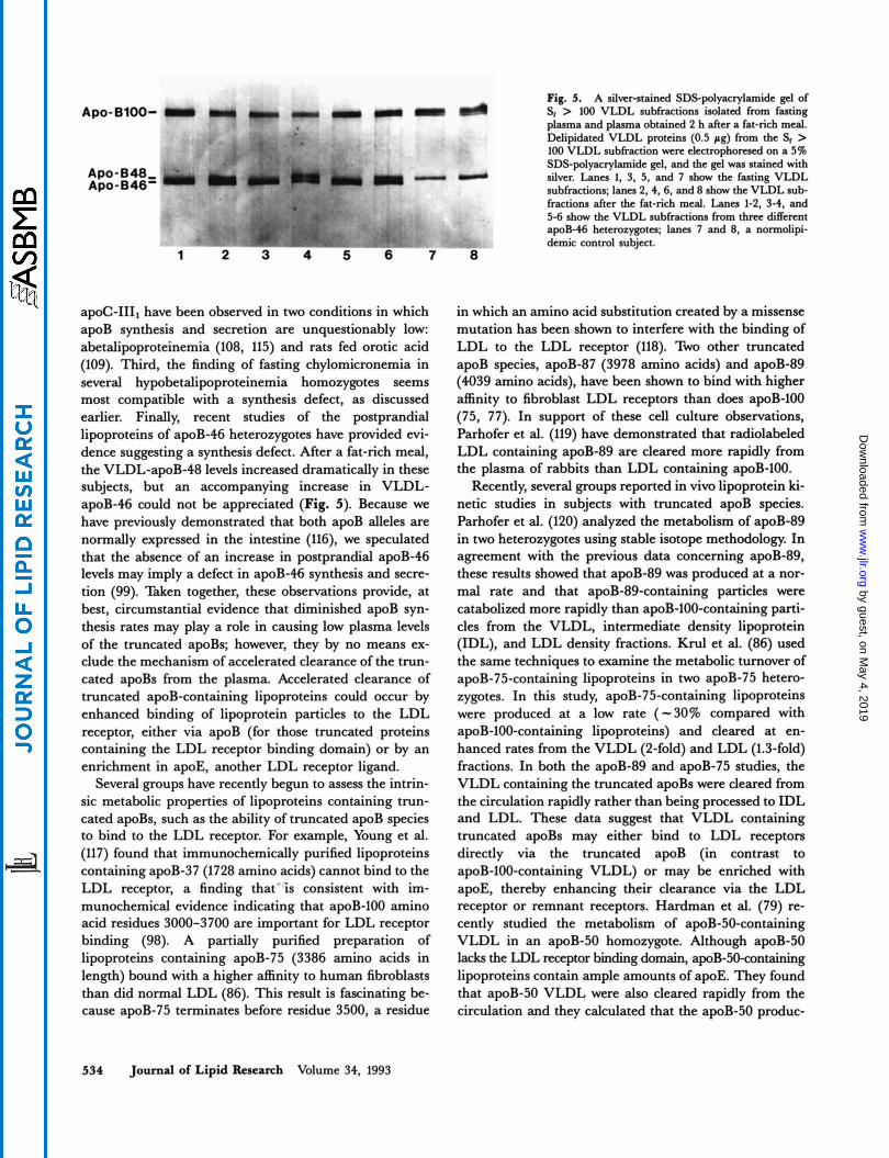
Fig. 5. A silveratained SDSpolyacrylamide gel of Sr > 100 VLDL subfractions isolated from fasting plasma and plasma obtained 2 h after a fat-rich meal. Delipidated VLDL proteins (0.5 pg) from the Sr > 100 VLDL subfraction were electrophoresed on a 5% SDS-polyacrylamide gel. and the gel was stained with silver. Lana 1, 3. 5, and 7 show the fasting VLDL subfractions; lanes 2,4,6, and 8 show the VLDL sub- fractions after the fat-rich meal. Lana 1-2, 3-4, and 5-6 show the VLDL subfractions from thm different apoB-46 heterozygotes; lanes 7 and 8, a normolipi- demic control subject.
A ~ o - 8100-
A po- B 48 - Apo-646'
1 2 3 4 5 6
a&-111, have been observed in two conditions in which apoB synthesis and secretion are unquestionably low: abetalipoproteinemia (108, 115) and rats fed orotic acid (109). Third, the finding of fasting chylomicronemia in several hypobetalipoproteinemia homozygotes seems most compatible with a synthesis defect, as discussed earlier. Finally, recent studies of the postprandial lipoproteins of apoB-46 heterozygotes have provided evi- dence suggesting a synthesis defect. After a fat-rich meal, the VLDL-apoB-48 levels increased dramatically in these subjects, but an accompanying increase in VLDL- apoB-46 could not be appreciated (Fig. 5). Because we have previously demonstrated that both apoB alleles are normally expressed in the intestine (116), we speculated that the absence of an increase in postprandial apoB-46 levels may imply a defect in apoB-46 synthesis and secre- tion (99). Taken together, these observations provide, at best, circumstantial evidence that diminished apoB syn- thesis rates may play a role in causing low plasma levels of the truncated apoBs; however, they by no means ex- clude the mechanism of accelerated clearance of the trun- cated apoBs from the plasma. Accelerated clearance of truncated apoB-containing lipoproteins could occur by enhanced binding of lipoprotein particles to the LDL receptor, either via apoB (for those truncated proteins containing the LDL receptor binding domain) or by an enrichment in apoE, another LDL receptor ligand.
Several groups have recently begun to assess the intrin- sic metabolic properties of lipoproteins containing trun- cated apoBs, such as the ability of truncated apoB species to bind to the LDL receptor. For example, Young et al. (117) found that immunochemically purified lipoproteins containing apoB-37 (1728 amino acids) cannot bind to the LDL receptor, a finding that'is consistent with im- munochemical evidence indicating that apoB-100 amino acid residues 3000-3700 are important for LDL receptor binding (98). A partially purified preparation of lipoproteins containing apoB-75 (3386 amino acids in length) bound with a higher affinity to human fibroblasts than did normal LDL (86). This result is fascinating be- cause apoB-75 terminates before residue 3500, a residue
in which an amino acid substitution created by a missense mutation has been shown to interfere with the binding of LDL to the LDL receptor (118). Two other truncated apoB species, apoB-87 (3978 amino acids) and apoB-89 (4039 amino acids), have been shown to bind with higher affinity to fibroblast LDL receptors than does apoB-100 (75, 77). In support of these cell culture observations, Parhofer et al. (119) have demonstrated that radiolabeled LDL containing apoB-89 are cleared more rapidly from the plasma of rabbits than LDL containing apoB-100.
Recently, several groups reported in vivo lipoprotein ki- netic studies in subjects with truncated apoB species. Parhofer et al. (120) analyzed the metabolism of apoB-89 in two heterozygotes using stable isotope methodology. In agreement with the previous data concerning apoB-89, these results showed that apoB-89 was produced at a nor- mal rate and that apoB-89-containing particles were catabolized more rapidly than apoB-100-containing parti- cles from the VLDL, intermediate density lipoprotein (IDL), and LDL density fractions. Krul et al. (86) used the same techniques to examine the metabolic turnover of apB-75-containing lipoproteins in two apoB-75 hetero- zygotes. In this study, apoB-75-containing lipoproteins were produced at a low rate (-30% compared with apoB-100-containing lipoproteins) and cleared at en- hanced rates from the VLDL (2-fold) and LDL (1.3-fold) fractions. In both the apoB-89 and apoB-75 studies, the VLDL containing the truncated apoBs were cleared from the circulation rapidly rather than being processed to IDL and LDL. These data suggest that VLDL containing truncated apoBs may either bind to LDL receptors directly via the truncated apoB (in contrast to apoB-100-containing VLDL) or may be enriched with apoE, thereby enhancing their clearance via the LDL receptor or remnant receptors. Hardman et al. (79) re- cently studied the metabolism of apoB-50-containing VLDL in an apoB-50 homozygote. Although apoB-50 lacks the LDL receptor binding domain, apoB-50-containing lipoproteins contain ample amounts of apoE. They found that apoB-50 VLDL were also cleared rapidly from the circulation and they calculated that the apoB-50 produc-
534 Journal of Lipid hearch Volume 34, 1993
by guest, on May 4, 2019
ww
w.jlr.org
Dow
nloaded from

tion rate was probably normal. In contrast, a stable iso- tope study on an apoB-54.8 heterozygote revealed a low apoB-54.8 production rate (121). Finally, data from a sta- ble isotope kinetic study in an apoB-31 heterozygote indi- cated that apoB-31 has a markedly diminished rate of production and a probably reduced clearance rate (121).
A common theme that is apparent from the initial in vivo kinetic studies is that the truncated apoBs disappear rapidly from the VLDL fraction, and there is decreased conversion of the VLDL to higher density lipoproteins. It is tempting to speculate that this finding may result from an increased apoE content of VLDL particles containing a truncated apoB, leading to rapid apoE-mediated clear- ance of these particles similar to that of apoB-48-containing chylomicrons. It is even possible that the expression of the hypobetalipoproteinemia phenotype may, with some mu- tations, be entirely dependent upon enhanced apoE- mediated clearance of lipoproteins containing truncated apoBs. However, important differences among the muta- tions have also emerged from the initial kinetic studies. For example, the effects of different mutations on apoB production rates have been variable. Clearly, more meta- bolic studies on the known kindreds are needed to under- stand how the various apoB mutations cause low trun- cated apoB levels. A thorough metabolic understanding will probably also require the identification of more kindreds and an even broader range of mutations. For ex- ample, it would be interesting to know whether mutations at the far 3‘ end of the apoB gene might yield a milder phenotype (i.e., a less marked reduction in cholesterol levels). Would an apoB-95 or an apoB-99 mutation cause hypobetalipoproteinemia?
The recently developed techniques of gene targeting may provide an alternative approach for investigating the metabolic consequences of apoB nonsense mutations. Gene targeting provides a means of targeting specific mu- tations into genes in a cultured cell line by selecting for the homologous recombination of a DNA vector with the cognate gene (122). We recently showed that it is possible to use these techniques to modify an apoB allele in the hu- man hepatoma cell line, HepG2 (123). In this study, we inactivated, or “knocked out,” one of the three functioning apoB alleles in HepG2 cells. Recently, techniques were developed that in theory make it possible to target more subtle point mutations into a gene of interest (124, 125). Using this approach, it should be possible to introduce a nonsense mutation into one of the apoB alleles in HepG2 cells, enabling one to then determine the effect of this mu- tation on apoB mRNA levels and protein synthesis rates from this allele. Maeda and co-workers were recently suc- cessful in using gene-targeting techniques to interrupt exon 26 of the mouse apoB gene in mouse embryonic stem cells (N. Maeda, personal communication). These targeted pleuripotent stem cells were then used to gener- ate heterozygous and homozygous mice that have the exon 26 mutation. Homozygotes for this targeted muta-
tion have low plasma levels of cholesterol and apoB, and their lipoproteins contain apoB-48 and a truncated apoB, apoB-70; apoB-100 is absent from the plasma. In collabo- ration with Dr. Maeda’s laboratory, we have demonstrated that the homozygous mice have fasting chylomicronemia, just like that observed in an apoB-37IapoB-86 compound heterozygote (H. J.B.). Undoubtedly, the thorough study of these mice will yield fresh insights into the metabolism of truncated apoBs.
Interestingly, hypobetalipoproteinemia heterozygotes frequently do not have apoB-100 plasma levels that are one-half normal, as might be expected; but instead, as discussed above, they often have apoB-100 levels that are one-fourth to one-third normal. Like the truncated apoBs, the less-than-expected levels of apoB-100 could result from one of two mechanisms: the diminished production of apoB-100-containing lipoproteins or the en- hanced clearance of these particles from the plasma. En- hanced clearance might result from either the presence of a better LDL receptor ligand on the particle (i.e., apoE enrichment) or from the “up-regulation” of LDL receptors due to the low plasma cholesterol levels that occur in hypobetalipoproteinemia. The mechanism involved in the less-than-half normal apoB-100 levels in heterozygotes is just beginning to be addressed by in vivo kinetic studies, and currently little data exist. Apolipoprotein B-100 production rates were measured, however, in one subject from the apoB-75 kindred who had a plasma LDL cholesterol level that was 37 % of the level of control family members and a total plasma apoB that was 17% of control values (86). In this subject, the apoB-100 production rate (11.6 mg/kg per day) was in the normal range, but the proportion of VLDL-apoB-100 converted to LDL appeared to be diminished, suggesting that enhanced clearance of the apoB-100-containing VLDL from the plasma resulted in the low plasma levels. More data will be required to resolve this issue.
Little metabolic turnover data exist for the other apolipoproteins in subjects with familial hypobetalipopro- teinemia. Malmendier et al. (126) used radioiodinated tracer studies in a kindred with hypobetalipoproteinemia to try to determine the mechanism that produces the low plasma levels of apoC-I1 and apoC-111 that are found in this syndrome. Their results show that the low levels of these C apolipoproteins result from both diminished production rates and increased catabolic rates. Their modeling suggested that these apolipoproteins, like apoB-75 and apoB-89 discussed earlier, are removed from the plasma directly from the VLDL pool.
STEROL METABOLISM IN HYPOBETALIPOPROTEINEMIA
Because familial hypobetalipoproteinemia subjects have low LDL cholesterol levels, it would be reasonable to
Linton, Farese, and Young Familial hypobetalipoproteinemia 535
by guest, on May 4, 2019
ww
w.jlr.org
Dow
nloaded from

postulate that they would have substantially increased rates of total body cholesterol biosynthesis to compensate for the diminished cholesterol delivery to peripheral cells. This issue was addressed in a subject with homozygous hypobetalipoproteinemia (in whom apoB was undetecta- ble in the plasma), and the results were similar to those obtained from studying abetalipoproteinemia subjects: total body cholesterol biosynthesis was only slightly to moderately increased over control values. Illingworth et al. (59) used sterol balance techniques to study a subject with homozygous hypobetalipoproteinemia (who had no detectable apoB) and found total body cholesterol synthe- sis to be modestly elevated (-2 x normal) to an extent that was roughly equivalent to the degree of malabsorp- tion of biliary cholesterol. More recently, Illingworth, Pappu, and Gregg (127) used measurements of the daily excretion of urinary mevalonic acid, an intermediate in cholesterol biosynthesis, as a measurement of total body cholesterol synthesis. In this study, they examined the same subject with homozygous hypobetalipoproteinemia whom they had previously examined in the sterol balance studies, as well as two other homozygous subjects, and found that urinary mevalonic acid excretion was approxi- mately twice that of control subjects. Why are total body cholesterol synthesis rates not more markedly elevated in subjects with homozygous hypobetalipoproteinemia or abetalipoproteinemia? Presumably the inability to deliver cholesterol to peripheral tissues by LDL is compensated for by the increased delivery of cholesterol by apoE- enriched HDL particles (107, 128). In hypobetalipopro- teinemia heterozygotes, neither intestinal cholesterol ab- sorption nor total body cholesterol biosynthesis rates have been examined. It would be interesting to determine whether a single normal apoB allele is sufficient for nor- mal lipid absorption from the intestine.
Adrenal and ovarian steroidogenesis, which requires cholesterol as a substrate, appears to be normal in sub- jects with phenotypic abetalipoproteinemia except when demands are quite high. The hypobetalipoproteinemia homozygotes identified by Illingworth, Kenny, and Or- woll (129), as well as subjects with abetalipoproteinemia, have been shown to have normal rates of cortisol produc- tion under basal conditions but an impaired steroidogenic response to chronic stimulation with adrenocorticotropic hormone. Two hypobetalipoproteinemia heterozygotes were also tested in this study and were found to have normal cortisol production in response to adrenocorticotropic hor- mone. A hypobetalipoproteinemia homozygote had markedly reduced serum progesterone levels during the luteal phase of the menstrual cycle (129). During this phase, the corpus luteum is highly steroidogenic (producing 30-40 mg of progesterone/day), and apparently the amount of choles- terol provided for steroidogenesis was insufficient to meet the demand. Despite this, there have been several reports of successful pregnancies both in abetalipoproteinemia
subjects (23, 130) and in homozygous hypobetalipopro- teinemia subjects (including the subject that was shown to have low progesterone levels) (23, 28, 69, 131), suggesting that there is sufficient cholesterol delivery for the steroido- genesis needed to maintain pregnancy.
TREATMENT OF HYPOBETALIPOPROTEINEMIA
Null-allele homozygotes
Recommendations for the treatment of “null-allele’’ homozygotes are based on the larger clinical experience of treating individuals with abetalipoproteinemia. Dietary fat restriction and vitamin replacement therapy have been the cornerstones of treatment (101). Although the dietary restriction of triglycerides containing long-chain fatty acids may be useful in treating symptomatic steatorrhea in infancy, the use of medium-chain triglycerides as a ca- loric substitute for long-chain fatty acids is controversial because two children have developed hepatic fibrosis dur- ing a course of medium-chain triglycerides supplementa- tion (59, 61). One authority recommends supplementing the diet with essential fatty acids (5 g of corn oil or safflower oil per day) and encouraging the patient to in- clude as much fat in their diets as they can tolerate (H. J. Kayden, personal communication). The ability to ab- sorb fat increases with age, and adults with abetalipopro- teinemia can usually tolerate diets containing 60-70 g of fat per day.
Dietary supplementation of fat-soluble vitamins is ex- tremely important in homozygotes. Large doses of vita- min E (100-300 mg/kg per day) are needed and have been shown to normalize adipose tissue levels of vitamin E (132), although serum levels often remain low (59, 133). Treatment with large doses of vitamin E appears to be quite effective in preventing the neurologic complications in homozygotes (44, 58, 59, 66, 68, 69, 132). In fact, the 11-year-old girl noted to have absent reflexes by Illing- worth et al. (59) was treated with high doses of vitamins E and A and, at age 16, she was able to participate on her high school track team. The patient originally described by Salt et al. (3) began treatment with vitamin E at 8 years of age; 36 years later, she had no evidence of neuro- logic abnormalities (69).
In general, most patients with phenotypic abetalipopro- teinemia do not develop problems related to vitamin A or vitamin K deficiencies (H. J. Kayden, personal communi- cation). Low serum levels of vitamin A (25) and carotene (23, 25, 76) have been noted in homozygous hypobetalipo- proteinemia, however, and abnormalities in scotopic vi- sion have been shown to respond to vitamin A therapy in abetalipoproteinemia (133). Therefore, it is reasonable to supplement these patients with a water-soluble prepara- tion of vitamin A; the restoration of normal serum levels
536 Journal of Lipid Research Volume 34, 1993
by guest, on May 4, 2019
ww
w.jlr.org
Dow
nloaded from

requires high doses (200-400 IUfkg per day) (133). I n ad- dition, vitamin K supplements should be provided if bruising, bleeding, or hypoprothrombinemia are present.
Normotriglyceridemic homozygotes and heterozygotes
The majority of hypobetalipoproteinemia heterozygotes and normotriglyceridemic compound heterozygotes are asymptomatic. However, there have been several reports of heterozygous subjects who have had neurologic abnor- malities similar to those seen in abetalipoproteinemia (89-92). Because serum vitamin E levels may not reflect tissue vitamin E levels and because vitamin E therapy ap- pears to be innocuous, it would be reasonable to supple- ment normotriglyceridemic homozygotes and hetero- zygotes (especially those with very low lipid values) with 200 mg/day of vitamin E.
Genetic counseling
Perhaps because familial hypobetalipoproteinemia has been considered to be a rare and relatively benign condi- tion, the role of genetic counseling in this disorder has not been discussed. After identifying a subject with hypobetalipoproteinemia, family studies should be under- taken to identify all affected individuals in the kindred, and the heterozygous subjects should be made aware of the nature of the disorder. For the heterozygote, the prin- cipal consideration is the unlikely event of a mating with another heterozygote, because one-quarter of their offspring will inherit two defective apoB alleles and could be severely affected. In the case of this rare scenario, ap- propriate studies should be undertaken to determine the haplotypes of the mutant apoB alleles and the nature of their respective apoB gene defects, and the heterozygotes should be referred to a qualified medical geneticist for counseling. OM
We thank Drs. G. Fless and A. Scanu for helpful discussions and experiments on Lp[a] in subjects with apoB-46 and apoB-86, and K. Starzel and S. Richmond for manuscript preparation. This work was supported by National Institutes of Health Grant No. 41633, an American Heart Association, California Affiliate, Fellowship Award (to MFL), and a National Institutes of Health General Clinical Research Center Clinical Associate Physician Award (to RVF).
Manuscript received 27 Ochber 1992 and in revised form 4 December 1992
REFERENCES
1. Kane, J. P., and R. J. Havel. 1989. Disorders of the biogen- esis and secretion of lipoproteins containing the B apolipoproteins. In The Metabolic Basis of Inherited Dis- ease. 6th edition. c . R. Scriver, A. L. Beaudet, W. S. Sly, and D. Valle, editors. McGraw-Hill, New York. 1139-1164.
2. Wetterau, J. R., L. P. Aggerbeck, M-E. Bouma, C. Eisen- berg, A. Munck, M. Hermier, J. Schmitz, G. Gay, D. J. Rader, and R. E. Gregg. 1992. Absence of microsomal triglyceride transfer protein in individuals with abetalipoproteinemia. Science. 258: 999-1001.
3. Salt, H. B., 0. H. Wolff, J. K. Lloyd, A. S. Fosbrooke, A. H. Cameron, and D. V. Hubble. 1960. On having no beta- lipoprotein. A syndrome comprising a-beta-lipoproteinaemia, acanthocytosis, and steatorrhoea. Lancet. 2: 325-329.
4. Bassen, F. A., and A. L. Kornzweig. 1950. Malformation of the erythrocytes in a case of atypical retinitis pigmentosa.
5. Singer, K., B. Fisher, and M. A. Perlstein. 1952. Acan- throcytosis. A genetic erythrocytic malformation. Blood. 7:
6. Druez, G. 1959. Un nouveau cas d’acanthocytose. Dysmor- phie trythrocytaire congknitale avec rttinite, troubles ner- veux et stigmates dtgkntratifs. Rev. Hmzutol. 14: 3-11.
7. Jampel, R. S., and H. F. Falls. 1958. Atypical retinitis pig- mentosa, acanthrocytosis, and heredodegenerative neu- romuscular disease. AMA Arch. Ophthalmol. 59: 818-820.
8. Mabry, C. C., A. M. DiGeorge, andV. H. Auerbach. 1960. Studies concerning the defect in a patient with acanthrocy- tosis. Clin. Res. 8: 371 (Abstract).
9. Lamy, M., J. Frtzal, J. Polonovski, and J. R7p 1960. Ihb- sence congnitale de 0-lipoprottines. Societe de Biologie.
10. Wolff, J. A., and W. A. Bauman. 1961. Studies concerning acanthocytosis: a new genetic syndrome with absent beta lipoprotein. Am. J. Dk. Child. 102: 478-479.
11. Herbert, P. N., G. Assmann, A. M. Gotto, Jr., and D. S. Fredrickson. 1983. Familial lipoprotein deficiency: abetalipoproteinemia, hypobetalipoproteinemia, and Tang- ier disease. In The Metabolic Basis of Inherited Disease. 5th ed. J. B. Stanbury, J. B. Wyngaarden, D. S. Fredrick- son, J. L. Goldstein, and M. S. Brown, editors. McGraw- Hill, New York. 589-621.
12. van Buchem, F. S. P., G. Pol, J. de Gier, C. J. E Bottcher, and C. Pries. 1966. Congenital /3-lipoprotein deficiency. Am. J. Med. 40: 794-804.
13. Isselbacher, K. J., R. Scheig, G. R. Plotkin, and J. B. Caulfield. 1964. Congenital 0-lipoprotein deficiency: an hereditary disorder involving a defect in the absorption and transport of lipids. Medicine. 43: 347-361.
14. Mars, H., L. A. Lewis, A. L. Robertson, Jr., A. Butkus, and G. H. Williams, J . 1969. Familial hypo-/3- lipoproteinemia. A genetic disorder of lipid metabolism with nervous system involvement. Am. J. Med. 46: 886-900.
15. Richet, G., H. Durepaire, L. Hartmann, M-P. Ollier, J. Polonovski, and B. Maitrot. 1969. Hypolipoprottintmie familiale asymptomatique prtdominant sur les beta- lipoprottines. Presse Med. 77: 2045-2048.
16. Fredrickson, D. S., A. M. Gotto, and R. I. Levy. 1972. Familial lipoprotein deficiency. (Abetalipoproteinemia, hypobetalipoproteinemia, and Tangier disease). In The Metabolic Basis of Inherited Disease. 3rd edition. J. B. Stanbury, J. B. Wyngaarden, and D. S. Fredrickson, edi- tors. McGraw-Hill, New York. 493-530.
17. Levy, R. I., T. Langer, A. M. Gotto, and D. S. Fredrickson. 1970. Familial hypobetalipoproteinemia, a defect in lipoprotein synthesis. Clin. Res. 18: 539 (Abstract).
18. Mawatari, S., H. Iwashita, and Y. Kuroiwa. 1972. Familial hypo-P-lipoproteinaemia. J. “34rol. Sci. 16: 93-101.
19. Fosbrooke, A,, S. Choksey, and B. Wharton. 1973. Familial hypo-0-lipoproteinaemia. Arch. Dis. Child. 48: 729-732.
Blood. 5: 381-387.
577-591.
3440 1974-1978.
Linton, Furese, and Young Familial hypobetalipoproteinemia 537
by guest, on May 4, 2019
ww
w.jlr.org
Dow
nloaded from

20. Brown, B. J., L. A. Lewis, and R. D. Mercer. 1974. Familial hypobetalipoproteinemia: report of a case with psychomo- tor retardation. Pediatrics. 54: 111-113.
21. Rerabek, J., and H. Eliasch. 1973. A contribution to the knowledge of the hypo-@-lipoproteinemia. Acto Med. Scand.
22. Biemer, J. J., and R. E. McCammon. 1973. Abetalipopro- teinemia and hypobetalipoproteinemia within the same fa- mily. Ann. Clin. Lab. Sci. 3: 308 (Abstract).
23. Biemer, J. J., and R. E. McCammon. 1975. The genetic relationship of abetalipoproteinemia and hypobetalipopro- teinemia: a report of the occurrence of both diseases within the same family. J. Lab. Clin. Med. 85: 556-565.
24. Cottrill, C., C. J. Glueck, J. Partin, V. Leuba, D. Puppione, and W. V. Brown. 1973. Familial homozygous hypobetali- poproteinemia. Circulation. 48: IV-15 (Abstract).
25. Cottrill, C., C. J. Glueck, V. Leuba, E Millett, D. Puppi- one, and W. V. Brown. 1974. Familial homozygous hypobetalipoproteinemia. Metabolism. 23: 779-791.
26. Steinberg, D., S. M. Grundy, H. Y. I. Mok, J. D. Turner, D. B. Weinstein, W. V. Brown, and J. J. Albers. 1979. Metabolic studies in an unusual case of asymptomatic familial hypobetalipoproteinemia with hypoalphalipopro- teinemia and fasting chylomicronemia. J. Clin. Invest. 64:
27. Young, S. G., S. J. Bertics, L. K. Curtiss, and J. L. Witz- tum. 1987. Characterization of an abnormal species of apolipoprotein B, apolipoprotein B-37, associated with familial hypobetalipoproteinemia. J. Clin. Invest. 79:
28. Young, S. G., S. J. Bertics, L. K. Curtiss, B. W. Dubois, and J. L. Witztum. 1987. Genetic analysis of a kindred with familial hypobetalipoproteinemia. Evidence for two separate gene defects: one associated with an abnormal apolipoprotein B species, apolipoprotein B-37; and a se- cond associated with low plasma concentrations of apolipoprotein B-100. J. Clin. Invest. 79: 1842-1851.
29. Kane, J. P., D. A. Hardman, and H. E. Paulus. 1980. Het- erogeneity of apolipoprotein B: isolation of a new species from human chylomicrons. Proc. Natl. Acad. Sci. USA. 77:
30. Linton, M. E, V. Pierotti, and S. G. Young. 1993. Reading frame restoration with an apoB gene frameshift mutation. Proc. Natl. Acad. Sci. USA. In press.
31. Leppert, M., J. L. Breslow, L. Wu, S. Hasstedt, P. OCon- nell, M. Lathrop, R. R. Williams, R. White, and J-M. Lalouel. 1988. Inference of a molecular defect of apolipoprotein B in hypobetalipoproteinemia by linkage analysis in a large kindred. J. Clin. Invest. 82: 847-851.
32. Talmud, P. J., J. K. Lloyd, D. P. R. Muller, D. R. Collins, J. Scott, and S. Humphries. 1988. Genetic evidence from two families that the apolipoprotein B gene is not involved in abetalipoproteinemia. J. Clin. Invest. 82: 1803-1806.
33. Huang, L.-S., P. A. Janne, J. de Graaf, M. Cooper, R. J. Decklebaum, H. Kayden, and J. L. Breslow, 1990. Exclu- sion of linkage between the human apolipoprotein B gene and abetalipoproteinemia. Am. J. Hum. Genet. 46: 1141-1148.
34. Pessah, M., P. Benlian, I. Beucler, N. Loux, J. Schmitz, C. Junien, and R. Infante. 1991. Anderson’s disease: genetic exclusion of the apolipoprotein-B gene in two families. J Clin. Inuest. 87: 367-370.
35. Hobbs, H. H., E. Leitersdorf, C. C. Leffert, D. R. Cryer, M. S. Brown, and J. L. Goldstein. 1989. Evidence for a dominant gene that suppresses hypercholesterolemia in a family with defective low density lipoprotein receptors. J Clin. Znuest. 84: 656-664.
194: 379-385.
292-301.
1831-1841.
2465-2469.
36. Fazio, S., A. Sidoli, A. Vivenzio, A. Maietta, S. Giampaoli, A. Menotti, R. Antonini, G. Urbinati, F. E. Baralle, and G. Ricci. 1991. A form of familial hypobetalipoproteinae- mia not due to a mutation in the apolipoprotein B gene. J. Intern. Med. 229: 41-41.
37. Vega, G. L., K. von Bergmann, S. M. Grundy, W. Beltz, C. Jahn, and C. East. 1987. Increased. catabolism of VLDL-apolipoprotein B and synthesis of bile acids in a case of hypobetalipoproteinemia. Metabolism. 36: 262-269.
38. Young, S. G., S. T. Northey, and B. J. McCarthy. 1988. Low plasma cholesterol levels caused by a short deletion in the apolipoprotein B gene. Science. 241: 591-593.
39. Collins, D. R., T. J. Knott, R. J. Pease, L. M. Powell, S. C. Wallis, S. Robertson, C. R. Pullinger, R. M. Milne, Y. L. Marcel, S. E. Humphries, P. J. Talmud, J. K. Lloyd, N. E. Miller, D. Muller, and J. Scott. 1988. Truncated variants of apolipoprotein B cause hypobetalipoproteinaemia. Nucleic Acids Res. 16: 8361-8375.
40. Huang, L. S., M. E. Ripps, S. H. Korman, R. J. Deckel- baum, and J. L. Breslow. 1989. Hypobetalipoproteinemia due to an apolipoprotein B gene exon 21 deletion derived by Alu-Alu recombination. J. Biol. Chem. 264: 11394-11400.
41. Yao, Z., B. D. Blackhart, M. E Linton, S. M. Taylor, S. G. Young, and B. J. McCarthy. 1991. Expression of carboxyl- terminally truncated forms of human apolipoprotein B in rat hepatoma cells. Evidence that the length of apolipoprotein B has a major effect on the buoyant density of the secreted lipoproteins. J. Bzol. Chem. 266: 3300-3308.
42. Herscovitz, H., M. Hadzopoulou-Cladaras, M. T. Walsh, C. Cladaras, V. I. Zannis, and D. M. Small. 1991. Expres- sion, secretion, and lipid-binding characterization of the N- terminal 17% of apolipoprotein B. Pmc. Natl. Acad. Sci.
43. Graham, D. L., T. J. Knott, T. C. Jones, R. J. Pease, C. R. Pullinger, and J. Scott. 1991. Carboxyl-terminal truncation of apolipoprotein B results in gradual loss of the ability to form buoyant lipoproteins in cultured human and rat liver cell lines. 3iochmirt~y. 3 0 5616-5621.
44. Huang, L-S., H. Kayden, R. J. Sokol, and J. L. Breslow. 1991. ApoB gene nonsense and splicing mutations in a com- pound heterozygote for familial hypobetalipoproteinemia. J Lipid Res. 32: 1341-1348.
45. Orita, M., H. Iwahana, H. Kanazawa, K. Hayashi, and T. Sekiya. 1989. Detection of polymorphisms of human DNA by gel electrophoresis as single-strand conformation poly- morphisms. Proc. Natl. Acad. Sci. USA. 86: 2766-2770.
46. Thigpen, A. E., D. L. Davis, A. Milatovich, B. B. Men- donca, J. Imperato-McGinley, J. E. Griffin, u. Francke, J. D. Wilson, and D. W. Russell. 1992. Molecular genetics of steroid 5a-reductase 2 deficiency. J. Clin. Inuest. 90:
47. Wagner, L. A., R. B. Weiss, R. Driscoll, D. S. Dunn, and R. F. Gesteland. 1990. Transcriptional slippage occurs dur- ing elongation at runs of adenine or thymine in Escherichia coli. Nucleic Acids Res. 18: 3529-3535.
48. Gavish, D., E. A. Bripton, and J. L. Breslow. 1989. Herita- ble allele-specific differences in amounts of apoB and low- density lipoproteins in plasma. Science. 244 72-76.
49. Pullinger, C. R., E. Hillas, D. A. Hardman, G. C. Chen, J. M. Naya-Vigne, J. A. Iwasa, R. L. Hamilton, J-M. Lalouel, R. R. Williams, and J. P. Kane. 1992. Two apolipoprotein B gene defects in a kindred with hypobetalipoproteinemia, one of which results in a trun- cated variant, apoB-61, in VLDL and LDL. J Lipid Res 33: 699-710.
50. Weatherall, D. J. 1986. The thalassemias: molecular patho- genesis, Zn Hemoglobin: Molecular, Genetic, and Chemical
USA. 88: 7313-7317.
799-809.
538 Journal of Lipid Research Volume 34, 1993
by guest, on May 4, 2019
ww
w.jlr.org
Dow
nloaded from

Aspects. H. Bunn and B. G. Forget, editors. WB Saunders Company, Philadelphia. 223-321.
51. Andersen, G. E., W. Trojaborg, and H. C. Lou. 1979. A clinical and neurophysical investigation of a Danish kindred with heterozygous familial hypobetalipoproteinemia. Acta Pncdiat,: Scand. 68: 155-159.
52. Laskarzewski, P. M., P. Khoury, J. A. Morrison, K. Kelly, M. J. Mellies, and C. J. Glueck. 1982. Prevalence of familial hyper- and hypolipoproteinemias: The Princeton School District Family Study. Metabolism. 31: 558-577.
53. Glueck, C. J., P. S. Gartside, M. J. Mellies, and P. M. Steiner. 1977. Familial hypobetalipoproteinemia: studies in 13 kindreds. Zans. Assoc. Am. Physicians. 90: 184-203.
54. Young, S. G., S. T. Hubl, D. A. Chappell, R. S. Smith, F. Claiborne, S. M. Snyder, and J. F. Terdiman. 1989. Familial hypobetalipoproteinemia associated with a mutant species of apolipoprotein B (B46). N. Engl. J. Med. 320:
55. Young, S. G., C. R. Pullinger, B. R. Zysow, H. Hofmann- Radvani, M. F. Linton, R. V. Farese, Jr., J. F. Terdiman, S. M. Snyder, S. M. Grundy, G. L. Vega, M. J. Malloy, and J. P. Kane. 1993. Four new mutations in the apolipoprotein B gene causing hypobetalipoproteinemia, including two different frameshift mutations that yield truncated apolipoprotein B proteins of identical length. J. Lipid Rex.
56. Young, S. G., S. T. Hubl, R. S. Smith, S. M. Snyder, and J. F. Terdiman. 1990. Familial hypobetalipoproteinemia caused by a mutation in the apolipoprotein B gene that results in a truncated species of apolipoprotein B (B-31). A unique mutation that helps to define the portion of the apolipoprotein B molecule required for the formation of buoyant, triglyceride-rich lipoproteins. J. Clin. Invest. 85:
57. Wagner, R. D., E. S. Krul, J. Tang, K. G. Parhofer, K. Garlock, P. Talmud, and G. Schonfeld. 1991. ApoB-54.8, a truncated apolipoprotein found primarily in VLDL, is as- sociated with a nonsense mutation in the apoB gene and hypobetalipoproteinemia. J. Lipid Res. 32: 1001-1011.
58. Berger, G. M. B., G. Brown, H. E. Henderson, and F. Bon- nici. 1983. Apolipoprotein B detected in the plasma of a pa- tient with homozygous hypobetalipoproteinaemia: implica- tions for aetiology. J. Med. Genet. 20: 189-195.
59. Illingworth, D. R., W. E. Connor, N. R. M. Buist, B. M. Jhaveri, D. S. Lin, and M. P. McMurry. 1979. Sterol balance in abetalipoproteinemia: studies in a patient with homozygous familial hypobetalipoproteinemia. Metabolism.
60. Gay, G., M. Pessah, M. E. Bouma, J. F. Roche, J. P. Ay- mard, I. Beucler, L. P. Aggerbeck, avd R. Infante. 1990. UypobCtalipoprotCinEmie familiale. Etude familiale de 4 cas. Rev. Med. Interne. 11: 273-279.
61. Partin, J. S., J. C. Partin, W. K. Schubert, and A. J. McA- dams. 1974. Liver ultrastructure in abetalipoproteinemia: evolution of micronodular cirrhosis. Gartroentemlou. 67: 107-118.
62. Scoazec, J-Y., M-E. Bouma, J-F. Roche, D. Blache, N. Ver- thier, G. Feldmann, and G. Gay. 1992. Liver fibrosis in a patient with familial homozygous hypobetalipoproteinae- mia: possible role of vitamin supplementation. Gut. 33:
63. Herbert, P. N., A. M. Gotto, and D. S. Fredrickson. 1978. Familial lipoprotein deficiency. (Abetalipoproteinemia, hypobetalipoproteinemia, and Tangier disease). In The Metabolic Basis of Inherited Disease. 4th edition. J. B.
1604-1610.
34: 501-507.
933-942.
28: 1152-1160.
414-417.
Stanbury, J. B. Wyngaarden, and D. S. Fredrickson, edi- tors. McGraw-Hill, New York. 551-558.
64. Schwartz, J. F., L. P. Rowland, H. Eder, P. A. Marks, E. F. Osserman, E. Hirschberg, and H. Anderson. 1963. Bassen- Kornzweig syndrome: deficiency of serum @-lipoprotein. A neuromuscular disorder resembling Friedreich’s ataxia, as- sociated with steatorrhea, acanthocytosis, retinitis pigmen- tosa, and a disorder of lipid metabolism. Anh. Neurol. 8:
65. Ross, R. S., R. E. Gregg, S. W. Law, J. C. Monge, S. M. Grant, K. Higuchi, T. J. Triche, J. Jefferson, and H. B. Brewer, Jr. 1988. Homozygous hypobetalipoproteinemia: a disease distinct from abetalipoproteinemia at the molecular 1evel.J. Clin. Invest. 81: 590-595.
66. Muller, D. P. R., J. K. Lloyd, and A. C. Bird. 1977. Long- term management of abetalipoproteinaemia. Arch. Dis. Child 52: 209-214.
67. Wolff, 0. H., J. K. Lloyd, and E. L. Tonks. 1964. A-beta- lipoproteinaemia with special reference to the visual defect. Exp. Eye Res. 3: 439.
68. Runge, P., D. P. R. Muller, J. McAllister, D. Calver, J. K. Lloyd, and D. Taylor. 1986. Oral vitamin E supplements can prevent the retinopathy of abetalipoproteinaemia. Bt: J. Ophthalmol. 70: 166-173.
69. Lloyd, J. K. 1990. The importance of vitamin E in human nutrition. Acta Pncdiatt: Scand, 79: 6-11.
70. Cooper, R. A,, J. R. Durocher, and M. H. Leslie. 1977. Decreased fluidity of red cell membrane lipids in abetalipoproteinemia. J. Clin. Invest. 6 0 115-121.
71. Iida, H., Y. Takashima, S. Maeda, T. Sekiya, M. Kawade, M. Kawamura, Y. Okano, and Y. Nozawa. 1984. Altera- tions in erythrocyte membrane lipids in abetalipoproteine- mia: phospholipid and fatty acyl composition. Biochem. Med.
72. Ways, P., C. F. Reed, D. J. Hanahan, D. Dong, S. Palmer, M. Murphy, and G. Roberts. 1963. Red-cell and plasma lipids in acanthocytosis. J. Clin. Invest. 42: 1248-1260.
73. Shacklady, M. M., E. M. Djardjouras, and J. K. Lloyd. 1968. Red-cell lipids in familial alphalipoprotein deficiency (Tangier disease). Lancet. 2: 151-153.
74. Biemer, J. J. 1980. Acanthocytosis - biochemical and phys- iological considerations. Ann. Clin. Lab. Sci. 1 0 238-249.
75. Krul, E. S., M. Kinoshita, P. Talmud, S. E. Humphries, S. Turner, A. C. Goidberg, K. Cook, E. Boerwinkle, and G. Schonfeld. 1989. Two distinct truncated apolipoprotein B species in a kindred with hypobetalipoproteinemia. Arterio-
76. Malloy, M. J., J. P. Kane, D. A. Hardman, R. L. Hamil- ton, and K. B. Dalal. 1981. Normotriglyceridemic abetalipoproteinemia. Absence of the B-100 apolipoprotein. J. Clin. Invest. 67: 1441-1450.
77. Gabelli, C., G. Baggio, C. Bilato, S. Martini, L. Previato, Y. Marcel, A. Corsini, and G. Crepaldi. 1989. Identifica- tion of a new apoB variant associated with hypobetalipoproteinemia. Circulation. 80: 11-276 (Abstract).
78. Gabelli, C. 1992. The lipoprotein metabolism of apolipoprotein B mutants. Cum Opin. Lipidol. 3: 208-214.
79. Hardman, D. A., C. R. Pullinger, R. L. Hamilton, J. P. Kane, and M. J. Malloy. 1991. Molecular and metabolic ba- sis for the metabolic disorder normotriglyceridemic abetalipoproteinemia. J. Clin. Invest. 88: 1722-1729.
80. Stein, E. A. 1977. Familial hypo-@-lipoproteinemia. A fa- mily detected by cord blood tests. Am. J. &. Child. 131:
81. Glueck, C. J., R. C. Tsang, M. J. Mellies, R. W. Fallat, and
438-454.
32: 79-87.
~Cl~OsiS. 9: 856-868.
1363-1365.
Linton, Farese, and Young Familial hypobetalipoproteinemia 539
by guest, on May 4, 2019
ww
w.jlr.org
Dow
nloaded from
P. M. Steiner. 1976. Neonatal familial hypobeta- lipoproteinemia. Metabolism. 25: 611-614.
82. Emi, M., R. M. Hegele, P. N. Hopkins, L. L. Wu, R. Plaetke, R. R. Williams, and J-M. Lalouel. 1991. Effects of three genetic loci in a pedigree with multiple lipoprotein phenotypes. Arteriosclm Thmmb. 11: 1349-1355.
83. Welty, F. K., S. T. Hubl, V. R. Pierotti, and S. G. Young. 1991. A truncated species of apolipoprotein B (B67) in a kindred with familial hypobetalipoproteinemia. J. Clin. In- vest. 87: 1748-1754.
84. McCormick, S. P. A., A. P. Fellowes, T. A. Walmsley, and P. M. George. 1992. Apolipoprotein B-32: a new truncated mutant of human apolipoprotein B capable of forming par- ticles in the low density lipoprotein range. Biochim. Biophys.
85. Farese, R. V., Jr., A. Garg, V. R. Pierotti, G. L. Vega, and S. G. Young. 1992. A truncated species of apolipoprotein B, B-83, associated with hypobetalipoproteinemia. J Lipid
86. Krul, E. S., K. G. Parhofer, P. H. R. Barrett, R. D. Wagner, and G. Schonfeld. 1992. ApoB-75, a truncation of apolipoprotein B associated with familial hypobetalipopro- teinemia: genetic and kinetic studies. J. Lipid Res. 33:
87. Lontie, J-F., C. L. Malmendier, C. Serougne, D. Y. Dubois, C. Dachet, J. Ferezou, and D. Matht. 1990. Plasma lipids, lipoproteins and apolipoproteins in two kindreds of hypobetalipoproteinemia. Atherosclemsis. 83:
88. Hegele, R. A,, S. Sutherland, M. Robertson, L. Wu, M. Emi, P. N. Hopkins, R. R. Williams, and J-M. Lalouel. 1991. The effect of genetic determinants of low density lipoprotein levels on lipoprotein [a]. Clin. Inues~. Med. 14:
89. Scott, B. B., J. P. Miller, and M. S. Losowsky. 1979. Hypobetalipoproteinaemia- a variant of the Bassen- Kornzweig syndrome. Gut. 20: 163-168.
90. Tsuji, S., T. Uozumi, Y. Ito, A. Ohnishi, and Y. Murai. 1990. Serial somatosensory evoked potentials in a patient with familial hypobetalipoproteinemia, and vitamin E deficiency. Jpn. J. Med. 29: 628-632.
91. Critchley, E. M. R., D. B. Clark, and A. Wikler. 1968. Acanthocytosis and neurological disorder without be- talipoproteinemia. Arch. Neuml. 18: 134-160.
92. Aggerbeck, L. P., J. P. McMahon, and A. M. Scanu. 1974. Hypobetalipoproteinemia: clinical and biochemical descrip- tion of a new kindred with ‘Friedreich‘s ataxia.’ Neurology.
93. Tamir, I . , 0. Levtow, D. Lotan, C. Legum, D. Heldenberg, and B. Werbin. 1976. Further observations on familial hypobetalipoproteinaemia. Clin. Genet. 9: 149-155.
94. Warner, C. L., S. Servidei, D. J. Lange, E. Miller, R. E. Lovelace, and L. P. Rowland. 1990. X-linked spinal muscu- lar atrophy (Kennedy’s syndrome). A kindred with hypobetalipoproteinemia. Arch. Neurol. 47: 1117-1120.
95. Araki, W., S. Hirose, Y. Mimori, S. Nakamura, J. Kimura, K. Ohno, and T. Shimada. 1991. Familial hypobetalipopro- teinaemia complicated by cerebellar ataxia and steatocystoma multiplex. J Intern. Med. 229: 197-199.
96. Talmud, P. J., C. Converse, E. Krul, L. Huq, G. G. McIl- waine, J. J. Series, P. Boyd, G. Schonfeld, A. Dunning, and S. Humphries. 1992. A novel truncated apolipoprotein B (apoB-55) in a patient with familial hypobetalipoproteine- mia and atypical retinitis pigmentosa. Clin. Genet. 42:
Acta. 1138: 290-296.
Res. 33: 569-577.
1037-1050..
187-196.
146-152.
24: 1051-1063.
62-70.
97. Kahn, J. A., and C. J. Glueck. 1978. Familial hypobetalipoproteinemia. Absence of atherosclerosis in a postmortem study. J. Am. Med. Assoc. 240: 47-48.
98. Milne, R., R. Thkolis, Jr., R. Maurice, R. J. Pease, P. K. Weech, E. Rassart, J-C. Fruchart, J. Scott, and Y. L. Mar- cel. 1989. The use of monoclonal antibodies to localize the low density lipoprotein receptor-binding domain of apolipoprotein B. J. Biol. Chem. 264: 19754-19760.
99. Young, S. G. 1990. Recent progress in understanding apolipoprotein B. Circulation. 82: 1574-1594.
100. Farese, R. V., Jr., M. F. Linton, and S. G. Young. 1992. Apolipoprotein B gene mutations affecting cholesterol levels. J. Intern. Med. 231: 643-652.
101. Havel, R. J., and J. P. Kane. 1989. Structure and metabolism of plasma lipoproteins. In The Metabolic Basis of Inherited Disease, 6th Edition. C. R. Scriver, .A. L. Beaudet, W. S. Sly, and D. Valle, editors. McGraw-Hill, New York. 1129-1138.
102. De Loof, H., M. Rosseneu, C-Y. Yang, W-H. Li, A. M. Gotto, Jr., and L. Chan. 1987. Human apolipoprotein B: analysis of internal repeats and homology with other apolipoproteins. J. Lipid Res. 28: 1455-1465.
103. Scanu, A. M., and G. M. Fless. 1990. Lipoprotein[a]. Het- erogeneity and biological relevance. J Clin. Invest. 85:
104. Coleman, R. D., T. W. Kim, A. M. Gotto, Jr., and C-Y. Yang. 1990. Determination of cysteine on low-density lipoproteins using the fluorescent probe, 5-iodoacetamidofluoresceine. Biochim. Biophys. Acta. 1037:
105. Huet, G., M. C. Dieu, A. Martin, G. Grard, J. M. Bard, P. Fossati, and P. Degand. 1991. Heterozygous hypobetalipoproteinemia with fasting chylomicronemia. Clin. Chem. 37: 296-300.
106. Gurdon, J. B., and M. P. Wickens. 1983. The use ofXenoptls oocytes for the expression of cloned genes. Methods Enzymol.
107. Illingworth, D. R., N. A. Alam, E. E. Sundberg, F. C. Hagemenas, and D. L. Layman. 1983. Regulation of low density lipoprotein receptors by plasma lipoproteins from patients with abetalipoproteinemia. Pmc. Natl. Acad. Sci.
108. Gotto, A. M., R. I. Levy, K. John, and D. S. Fredrickson. 1971. On the protein defect in abetalipoproteinemia. N. Engl. J . Med. 284: 813-818.
109. Henderson, L., P. Herbert, H. Windmueller, and R. Krauss. 1974. The C-apolipoproteins of the orotic acid rat: a model for abetalipoproteinemia. Circulation. 50: 111-114 (Abstract).
110. Sigurdsson, G., A. Nicoll, and B. Lewis. 1977. Turnover of apolipoprotein-B in two subjects with familial hypobetalipoproteinemia. Metabolism. 26: 25-30.
111. Converse, C. A,, W. A. Keegan, L. Huq, J. Series, M. Cas- lake, T. McLachlan, C. J. Packard, and J. Sheperd. 1989. Further epidemiological studies on lipid metabolism in reti- nitis pigmentosa. In Inherited and Environmentally In- duced Retinal Degenerations. Alan R. Liss, Inc., New York. 39-47.
112. Kadowaki, T., H. Kadowaki, M. M. Rechler, M. Serrano- Rios, J. Roth, P. Gorden, and S. I. Taylor. 1990. Five mu- tant alleles of the insulin receptor gene in patients with genetic forms of insulin resistance. J. Clin. Invest. 86:
113. Ritchie, H. H., M. R. Hughes, E. T. Thompson, P. J. Mal- loy, Z. Hochberg, D. Feldman, J. W. Pike, and B. W.
1709-1715.
129-132.
101: 370-386.
USA. 80: 3475-3479.
254-264.
540 Journal of Lipid Research Volume 34, 1993
by guest, on May 4, 2019
ww
w.jlr.org
Dow
nloaded from

OMalley. 1989. An ochre mutation in the vitamin D recep- tor gene causes hereditary 1,25-dihydroxyvitamin DS- resistant rickets in three families. Pmc. Natl. A c d . Sci. USA. 86: 9783-9787.
114. Chelly, J., H. Gilgenkrantz, M. Lambert, G. Hamard, P. Chafey, D. Rtcan, P. Katz, A. de la Chapelle, M. Koenig, I. B. Ginjaar, M. Fardeau, F. Tomt, A. Kahn, and J-C. Kaplan. 1990. Effect of dystrophin gene deletions on mRNA levels and processing in Duchenne and Becker mus- cular dystrophies. Cell. 63: 1239-1248.
115. Scanu, A. M., L. P. Aggerbeck, A. W. Kruski, C. T. Lim, and H. J. Kayden. 1974. A study of the abnormal lipoproteins in abetalipoproteinemia. J. Clin. Invest. 53:
116. Young, S. G., S. J. Bertics, T. M. Scott, B. W. Dubois, L. K. Curtiss, and J. L. Witztum. 1986. Parallel expression of the MB19 genetic polymorphism in apoprotein B-100 and apoprotein B-48. Evidence that both apoproteins are products of the same gene. J. Biol. Chem. 261: 2995-2998.
117. Young, S. G., E P. Peralta, B. W. Dubois, L. K. Curtiss, J. K. Boyles, and J. L. Witztum. 1987. Lipoprotein B37, a naturally occurring lipoprotein containing the amino- terminal portion of apolipoprotein B-100, does not bind to the apolipoprotein B,E (low density lipoprotein) receptor. J. Biol. Chem. 262: 16604-16611.
118. Soria, L. E, E. H. Ludwig, H. R. G. Clarke, G. L. Vega, S. M. Grundy, and B. J. McCarthy. 1989. Association be- tween a specific apolipoprotein B mutation and familial defective apolipoprotein B-100. Pmc. Natl. Acad. Sci. USA.
119. Parhofer, K. G., A. Daugherty, M. Kinoshita, and G. Schonfeld. 1990. Enhanced clearance from plasma of low density lipoproteins containing a truncated apolipoprotein, apoB-89. J. Lipid Res. 31: 2001-2007.
120. Parhofer, K. G., P. H. R. Barrett, D. M. Bier, and G. Schonfeld. 1992. Lipoproteins containing the truncated apolipoprotein, apoB-89, are cleared from human plasma more rapidly than apoB-100-containing lipoproteins in vivo. J. Clin. Invest. 89: 1931-1937.
121. Barrett, P. H. R., K. G. Parhofer, S. G. Young, and G. Schonfeld. 1992. Apolipoproteins B-31 and B-54.8: trunca- tions associated with reduced apoB production rates. Circu- lation. 86: 1-209 (Abstract).
122. Capecchi, M. R. 1989. Altering the genome by homologous recombination. Science. 244: 1288-1292.
123. Farese, R. V., Jr., L. M. Flynn, and S. G. Young. 1992. Modification of the apolipoprotein B gene in HepG2 cells by gene targeting. J. Clin. Invest. 90: 256-261.
124. Hasty, P., R. Ramirez-Solis, R. Krumlauf, and A. Bradley. 1991. Introduction of a subtle mutation into the HOX-2.6 lo- cus in embryonic stem cells. Nature. 350: 243-246.
125. Valancius, V., and 0. Smithies. 1991. Testing an “in-out’’ targeting procedure for making subtle genomic modifica- tions in mouse embryonic stem cells. Mol. Cell. Biol. 11:
440-453.
86: 587-591.
1402-1408. 126. Malmendier, C. L., C. Delcroix, J-E Lontie, and D. Y.
Dubois. 1991. Apolipoprotein C-I1 and C-I11 metabolism in a kindred of familial hypobetalipoproteinemia. Metabolism. 40: 45-50.
127. Illingworth, D. R., A. S. Pappu, and R. E. Gregg. 1989. In- creased urinary mevalonic acid excretion in patients with abetalipoproteinemia and homozygous hypobetalipopro- teinemia. Atherosclerosis. 76: 21-27.
128. Gibson, J. C., A. Rubinstein, W. V. Brown, H. N. Gins- berg, H. Greten, R. Norum, and H. Kayden. 1985. ApoE- containing lipoproteins in low or high density lipoprotein deficiency. Arteriosclerosis. 5: 371-380.
129. Illingworth, D. R., T. A. Kenny, and E. S. Orwoll. 1982. Adrenal function in heterozygous and homozygous hypobetalipoproteinemia. J Clin. Endocrinol. Metab. 54:
130. Kayden, H. J. 1980. Is essential fatty acid deficiency part of the syndrome of abetalipoproteinemia? Nut% Rev. 38:
131. Parker, C. R., Jr., D. R. Illingworth, J. Bissonnette, and B. R. Carr. 1986. Endocrine changes during pregnancy in a patient with homozygous familial hypobetalipoproteine- mia. N Engl. J. Med. 314: 557-560.
132. Kayden, H. J., and M. G. Traber. 1985. Clinical, nutri- tional and biochemical consequences of apolipoprotein B deficiency. Adv. Exb. Med. Biol. 201: 67-81.
133. Illingworth, D. R., W. E. Connor, and R. G. Miller. 1980. Abetalipoproteinemia. Report of two cases and a review of therapy. Arch. Neuml. 37: 659-662.
134. Talmud, P., L. King-Underwood, E. Krul, G. Schonfeld, and S. Humphries. 1989. The molecular basis of truncated forms of apolipoprotein B in a kindred with compound het- erozygous hypobetalipoproteinemia. J. Lipid Res. 30:
135. Groenewegen, W. A., E. S. Krul, and G. Schonfeld. 1992. Deletion ambiguities in a novel apolipoprotein B trunca- tion, apoB-52. Circulation. 86: 1-690 (Abstr.).
136. Tennyson, G. E., C. Gabelli, G. Baggio, C. Bilato, and H. B. Brewer, Jr. 1990. Molecular defect in the apolipoprotein B gene in a patient with hypobetalipopro- teinemia and three distinct apoB species. Clin. Res. 38: 482A (Abstract).
137. Xiong, W., E. Zsigmond, A. M. Gotto, Jr., K. Y. Lei, and L. Chan. 1991. Locating a low density lipoprotein-targeting domain of human apolipoprotein B-100 by expressing a minigene construct in transgenic mice. J. Biol. Chem. 266:
138. Pease, R. J., R. W. Miline, W. K. Jessup, A. Law, P. Provost, J-C. Fruchart, R. T. Dean, Y. L. Marcel, and J. Scott. 1990. Use of bacterial expression cloning to localize the epitopes for a series of monoclonal antibodies against apolipoprotein B-100. J. Biol. Chem. 265: 553-568.
27-33.
244-246.
1773-1779.
20893-20898.
Linton, Farese, and Young Familial hypobetalipoproteinemia 541
by guest, on May 4, 2019
ww
w.jlr.org
Dow
nloaded from