extra-renal lesion caused by dioctophyma renale eggs in an ... · renal lesion caused by...
TRANSCRIPT
1031
Int. J. Morphol.,28(4):1031-1034, 2010.
Extra-Renal Lesion Caused by Dioctophyma renaleEggs in an Erratic Cycle in a Dog
Lesión Extra Renal Causada por los Huevos de Dioctophyma renaleen un Ciclo Errático en un Perro
*Luciana da Silva Lemos; *Alessa Siqueira de Oliveira dos Santos; *Ana Bárbara Freitas Rodrigues;**Maria Laudelina Vieira Seródio Goulart; *Luciano Grillo de Almeida & *Leonardo Serafim da Silveira
LEMOS, L. S.; SANTOS, A. S. O.; RODRIGUES, A. B. F.; GOULART, M. L. V. S.; ALMEIDA, L. G. & SILVEIRA, L. S. Extra-renal lesion caused by Dioctophyma renale eggs in an erratic cycle in a dog. Int. J. Morphol., 28(4):1031-1034, 2010.
SUMMARY: A dog with multiple infection by D. renale in the abdominal cavity presented granular peritonitis with giant cellsphagocytizing D. renale eggs. Hepatic and phrenic serositis associated to numerous eggs of the parasite immersed in fibrinous processwere observed. Lungs presented D. renale eggs in the parenchyma, mononucleated cell infiltrates, edema, hemorrhage, congestion,atelectasia, emphysema, and thromboembolism. D. renale eggs were detected inside the center-lobular veins, auricular cavities, andsuperficial venous bed of the heart. These findings characterized an atypical dissemination pathway of eggs in erratic cycle.
KEY WORDS: Egg; Dioctophyma renale; Erratic cycle; Extrarenal parasitism; Canine.
INTRODUCTION
Dioctophyma renale is a cosmopolitan parasiteknown as giant kidney worm. The parasite infests severalmammal species, even man (Acha & Szyfres, 2003; Fortes,1997). D. renale is often described in dogs (Osborne et al.,1969; Amato et al., 1976; Kommers et al., 1999; Leite etal., 2005; Nakagawa et al., 2007). The parasite is commonlydiagnosed in the right kidney and in the abdominal cavity(Osborne et al.; Amato et al.; Kommers et al.; Leite et al.)but it may parasitize both kidneys (Augusto Filho et al.,1999). Also, D. renale may be diagnosed only in the abdo-minal cavity. Other organs may also be affected, like theliver (Gargili et al., 2002), stomach and mesenterium (Mi-randa et al., 1992), causing perforations, hemorrhage andperitonitis (Osborne et al.; Monteiro et al., 2003).
The lesions caused by D. renale depend on thelocation in the body where the parasite is diagnosed(Kommers et al.). More specifically, in the kidney one ormore worms (Amato et al.; Kano et al., 2003) cause thecompressive destruction of the parenchyma (Leite et al.;Nakagawa et al.) and lead to the thickening of renal capsuledue to the development of fibrosis (Leite et al.). The clinical
expression of the parasitosis by D. renale is unspecific(Osborne et al.; Kommers et al.; Kano et al.; Monteiro etal.), or may even be absent (Osborne et al.). If only onekidney is infected, the renal function is preserved by thecontralateral kidney (Kano et al.; Leite et al.; Monteiro etal.). Anemia (Kano et al.), hematuria, progressive weightloss (Kommers et al.; Nakagawa et al.) and abdominaldistension may be signs associated to D. renale parasitosis(Kano et al.).
The parasite’s preference for renal sites favors itscycle, which is long and complex. The cycle involves severalhosts; eggs are eliminated through urinary ways of theinfected host and then ingested by intermediate hosts, aquaticoligochaetes, which are ingested by paratenic hosts (fish andfrogs). Definitive hosts and/or terminal hosts are infectedby ingestion of contaminated oligochaetes or paratenic hosts(Fortes). Characteristically, D. renale eggs are large,elliptical, brown, thick-shelled, wrinkled, and are roughlybetween 60 and 80mm in length and 39 and 46mm indiameter (Osborne et al.). D. renale diagnosis is normallyreached with casual necropsy findings, during surgical
* Laboratório de Sanidade Animal - Setor de Morfologia e Anatomia Patológica, Universidade Estadual do Norte Fluminense Darcy Ribeiro, Brasil.** Médica Veterinaria.
1032
procedures, in tests in urinary sediment and ascetic liquid(Kommers et al.; Leite et al.), and via radiographic andultrasound investigation (Costa et al., 2004). Asanatomopathological findings of D. renale in dogs arerestricted to renal lesions, the aim of this study was to des-cribe extra-renal lesions in tissue samples collected from adog with peritonitis caused by multiple D. renale infestation.
MATERIAL AND METHOD
The study material consisted of tissue samples froma young, 7-kg mixed-breed dog (Canis familiaris) goneastray in the vicinities of Muriaé River, municipality ofItaperuna, state of Rio de Janeiro, Brazil. The animal
presented cachexia, abdominal distension, anemia,hematuria.
The dog was treated with serum and antibioticstherapy and a blood transfusion, in a treatment period of 14days, after which it died. A necropsy was conducted, afterwhich the cardiorespiratory tract, mediastinic lymph nodes,liver and respective lymph nodes as well as part of thediaphragm were kept in formaldehyde 10% in totum. Viscerawere inspected and sliced in several parts, including vascularcoagula, and subsequently sectioned, mounted on slides andstained with hematoxylin/eosin (HE) for visualization in alight microscope. Egg morphometry was carried out using adigital morphometry software (Microimage Processing Soft-ware Dn-200M) with 20 eggs sectioned longitudinally.
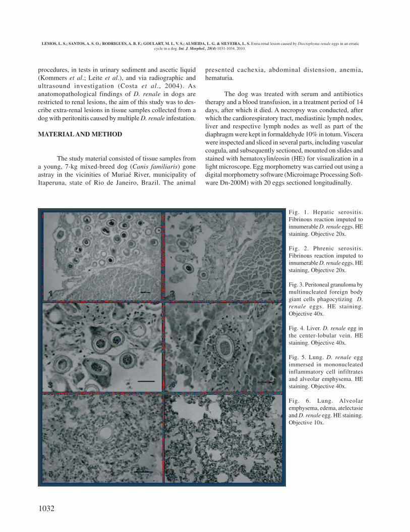
Fig. 1. Hepatic serositis.Fibrinous reaction imputed toinnumerable D. renale eggs. HEstaining. Objective 20x.
Fig. 2. Phrenic serositis.Fibrinous reaction imputed toinnumerable D. renale eggs. HEstaining, Objective 20x.
Fig. 3. Peritoneal granuloma bymultinucleated foreign bodygiant cells phagocytizing D.renale eggs. HE staining.Objective 40x.
Fig. 4. Liver. D. renale egg inthe center-lobular vein. HEstaining. Objective 40x.
Fig. 5. Lung. D. renale eggimmersed in mononucleatedinflammatory cell infiltratesand alveolar emphysema. HEstaining. Objective 40x.
Fig. 6. Lung. Alveolaremphysema, edema, atelectasieand D. renale egg. HE staining.Objective 10x.
LEMOS, L. S.; SANTOS, A. S. O.; RODRIGUES, A. B. F.; GOULART, M. L. V. S.; ALMEIDA, L. G. & SILVEIRA, L. S. Extra-renal lesion caused by Dioctophyma renale eggs in an erraticcycle in a dog. Int. J. Morphol., 28(4):1031-1034, 2010.
1033
RESULTS
The incision in the abdominal cavity revealed a consi-derable amount of sanguinolent liquid, pale viscera, and signsof peritonitis, evinced by the accumulation of fibrin in thevisceral and parietal peritoneum and by adhered areas in themesenterium. Eleven adult nematodes identified as D. renalewere found between intestinal loops. The right kidney wasabsent, indicating destruction by the worms. The inspectionof the material by light microscopy confirmed theaccumulation of fibrin in the liver and in the abdominal sideof the diaphragm. Histopathological investigations revealedhepatic (Fig. 1) and phrenic serositis (Fig. 2). The eggs foundwere thick-shelled, irregularly shaped, chestnut brown in co-lor and measured approximately 63x40mm. In severalmicroscopy fields eggs were involved in phagocytosis processrepresented by multinucleated foreign body giant cells (Fig.3). The hepatic parenchyma sections revealed congestion, thepresence of eggs inside center-lobular veins (Fig. 4), andthickening of the portal connective tissue.
The microscopy of lung samples revealed D. renaleeggs were surrounded by mononucleated cell infiltrates (Fig.5), destructuration of the histoarchitecture of lungs due to ede-ma, hemorrhage, congestion, alveolar emphysema,thromboembolism, and D. renale eggs scattered in theparenchyma (Fig. 6), characterizing pneumonia. Eggs wereobserved in both auricular cavities and in the superficial venousbed of the heart. No eggs were found in the hepatic hilus or inmediastinic lymph nodes.
DISCUSSION
The anatomopathology of dogs infected with D. renaleis commonly restricted to renal lesions (Amato et al.; Kommerset al.; Leite et al.; Nakagawa et al.), as the association withperitonitis and the effects on other organs are less frequentlyobserved events (Amato et al.; Kano et al.; Monteiro et al.).Since the diagnosis of parasitosis is in most cases reachedincidentally (Osborne et al.; Kommers et al.), thehistopathological investigation of other organs as well as thephysiopathology of peritonitis caused by D. renale has beenthe subject of limited research. This may be explained by thefact that the parasitosis by D. renale is symptomaticallyunspecific (Osborne et al.; Kommers et al.; Kano et al.;Monteiro et al.), and often occurs parenthetically to caninedistemper, Hepatozoon sp, Ehrlichia sp (Porfirio et al., 2004),and leptospirosis (Costa et al.).
The peritonitis diagnosed in the present study was
caused by worms and eggs, which led to irritation of the organserous membrane and accumulation of fibrin, starting theinflammatory process in the peritoneum and the adherence ofintestinal loops and mesenterium. The event may be explainedby the fact that any noxious stimulus causing a lesion in theperitoneal mesothelium is enough to trigger an inflammatoryresponse (Cotran et al., 1989), similar to that occurring in anyother part of the organism and characterized by hyperemia,exudation, concentration of phagocytes, and localaccumulation of fibrin. The inflammation in the peritoneummay be classified as primary, secondary or tertiary in relationto its origin being peritonitis (Wittmann et al., 1996;Zimmermann et al., 2006). In the present study, peritonitis byD. renale was classed as primary, as the process was of diffuseappearance in the peritoneal cavity, no hollow viscera wasperforated, and no signs of bacterial presence were observed.Multiple infection, with females in their egg-laying stages andabundance of eggs in the peritoneal cavity characterized aninflammatory response that evolved into granular processrepresented by the amount of phagocytes that united to formforeign body giant cells in an attempt to isolate D. renale eggs.These eggs are inert, insoluble structures, and difficult todigeste — all of which are characteristics shared withSchistosoma mansoni eggs, also capable to inducegranulomatous response (Atta et al., 1981; Lambertucci et al.,2005). Dogs are considered terminal hosts, uncommonly oraccidentally infected (Acha & Szyfres), since they often hostone single parasite in the right kidney (Amato et al; AugustoFilho et al.; Kommers et al.; Monteiro et al.). In the naturalcycle of D. renale, adult worms of both sexes are necessary inthe kidneys to produce fertile eggs, whose disseminationpathway is the urinary tract (Osborne et al.).
In the animal studied in this case report, the erraticcycle was characterized by multiple infection in the abdomi-nal cavity. Even though worms of both sexes and a largenumber of eggs were present, neither worm nor egg had thedissemination pathway clear to reach the environment. Therecords of D. renale in lung parenchyma and in the cardiacand hepatic vascular bed suggest that worms laid eggs in thecirculatory system, an atypical dissemination pathway of eggsin their erratic cycle. The D. renale eggs are capable of inducinggranular peritonitis and pneumonia, an observation so farunpublished in the literature reviewed. The findings of thepresent study emphasize the importance of more detailedanatomopathological investigations about the systemicinvolvement caused by the parasite.
ACKNOWLEDGMENTS
The collaboration of Helaíne Haddad Simões Ma-chado.
LEMOS, L. S.; SANTOS, A. S. O.; RODRIGUES, A. B. F.; GOULART, M. L. V. S.; ALMEIDA, L. G. & SILVEIRA, L. S. Extra-renal lesion caused by Dioctophyma renale eggs in an erraticcycle in a dog. Int. J. Morphol., 28(4):1031-1034, 2010.

1034
REFERENCES
Acha, P. N. & Szyfres, B. Zoonoses and communicable diseasescommom to man and animais. Washington, OPAS, 2003.
Amato, J. F. R.; Grisi, L. & Rosa, V. L. M. Reunião dos casosbrasileiros de dioctofimose canina, com registro de mais altaintensidade de infecção por Dioctophyma renale (Goeze, 1782).Rev. Brasil. Biol, 36:117-22, 1976.
Atta, A. M.; Magalhães, L. A. & Alcântara, G. P. EsquistossomoseMansônica. I. evolução do quadro patológico: análiseparasitológica, hematológica e histopatológica. Rev. Saúde Pú-blica, 15:72-92, 1981.
Augusto Filho, O.; Araújo, W. N.; Paes, A. C. & Mamprim, M. J.Dioctofimíase canina bilateral com vários parasitas. Relato decaso. Seminário Brasileiro de Parasitologia Veterinária, XI. Sal-vador, Colégio Brasileiro de Parasitologia Veterinária, 1999.
Costa, P. R. S.; Neto, N. M. A.; Oliveira, D. M. C.; Vasconcelos, R. S.& Menezes, F. M. Dioctofimose e leptospirose em um cão. Clí-nica Veterinária, 51:48-50, 2004.
Cotran, R. S.; Kumar, V. & Robbins, S. L. Robbins Pathologic Basisof Disease. 4th ed. Philadelphia, W. B. Saunders Company, 1989.pp.65-71.
Fortes, E. Parasitologia Veterinária. 3rd ed. São Paulo, Ícone, 1997.
Gargili, A.; Firat, I.; Toparlak, M. & Çetinkaya, H. First case reportof Dioctophyme renale (Goeze, 1782) in a dog in Istanbul, Turkey.Turk. J. Vet. Anim. Sci., 26:1189-91, 2002.
Kano, F. S.; Shimada, M. T.; Suzuki, S. N.; Osaki, S. C.; Menarim, B.C.; Ruthes, F. R. V. & Laidane Filho, M. A. Ocorrência dedioctofimose em dois cães no município de Guarapuava-PR.Ciências Agrárias, 24:177-80, 2003.
Kommers, D. G.; Ilha, M. R. S. & Barros, C. S. L: Dioctofimose emcães: 16 casos. Ciência Rural, 29:517-22, 1999.
Lambertucci, J. R.; Moreira, R. F. & Barbosa, A. J. A. Nódulopulmonar solitário causado pelo Schistosoma mansoni em pa-ciente com carcinoma medular da tireóide. Rev. Soc. Bras. Med.Trop., 38(6):536-7, 2005.
Leite, L. C.; Círio, S. M.; Diniz, J. M. F.; Luz, E.; Navarro-Silva, M.A.; Silva, A. W. C.; Leite, S .C.; Zadorosnei, A. C.; Musiat, K.C.; Veronesi, E. M. & Pereira, C. C. Lesões anatomopatológicospresentes na infecção por Dioctophyma renale (Goeze, 1782) emcães domésticos (Canis Familiares, Linnaeus, 1758). Arch. Vet.Sci., 10:95-101, 2005.
Miranda, M. A.; Benigno, R. N. M.; Galvão, G. R. & Oliveira, S. A.L. Dioctophyme renale (Goeze, 1782): localização ectópica e altaintensidade parasitária em Canis familiares do Pará – Brasil. Arq.Brás. Med. Vet. Zoot., 44:151-3, 1992.
Monteiro, S. G.; Sallis, E. S. & Stainki, D. R. Infecção natural portrinta e quatro helmintos da espécie Dioctophyma renale (Goeze,1782) em um cão. Rev. Fac. Zootec. Vet. Agro. Uruguaiana, 9:29-32, 2003.
Nakagawa, T. L. D. R.; Bracarense, A. P. F. R. L.; Reis, A. C. F.;Yamamura, M. H. & Headley, S. A. Giant kidney Word(Dioctophyma renale) infections in dogs from Northen Paraná,Brazil. Vet. Parasitol., 145:366-70, 2007.
Osborne, C. A.; Stevens, J. B. & Hanlon, G. F. Dioctophyma renalein a dog. J. Am. Vet. Med. Assoc., 155:605-20, 1969.
Porfirio, L. C.; Carvalho, G. D.; Massariol, P. B.; Afonso, T. Z.; Ma-chado, J. P. & Masseno, A. P. B. Infecção concomitante porDioctophyma renale, Hepatozoon sp., Ehrlichia sp. e cinomoseem cão. Veterinária Ser., 1:16-22, 2004.
Wittmann, D. H.; Schein, M. & Condon, R. E. Management ofsecondary peritonitis. Ann. Surg., 224:10-8, 1996.
Zimmermann, M.; Raiser, A. G.; Mazzanti, A.; Lopes, S. T. A. &Salbego, F. Z. Peritonite em cães. Ciência Rural, 36:1655-63,2006.
Correspondence to:
Luciana da Silva Lemos
Laboratório de Sanidade Animal
Setor de Morfologia e Anatomia Patológica
Universidade Estadual do Norte Fluminense Darcy Ribeiro
(UENF)
Av. Alberto Lamego 2000
Horto, Campos dos Goytacazes
BRASIL
Email: [email protected]
LEMOS, L. S.; SANTOS, A. S. O.; RODRIGUES, A. B. F.; GOULART, M. L. V. S.; ALMEIDA, L. G. & SILVEIRA, L. S. Lesión extra renalcausada por los huevos de Dioctophyma renale en un ciclo errático en un perro. Int. J. Morphol., 28(4):1031-1034, 2010.
RESUMEN: Un perro con infección múltiple por D. renale en la cavidad abdominal presentó peritonitis granulomatosa con células gigantesfagocitando huevos de D. renale. Se pudo observar serositis hepática y frénica asociada a numerosos huevos del parásito inmersos en un procesofibrinoso. Los pulmones presentaron huevos de D. renale en el parénquima, infiltrados de células mononucleares, edema, hemorragia, congestión,atelectasia, enfisema y tromboembolismo. Huevos de D. renale fueron detectados en el interior de la vena centro lobular, cavidades auriculares ylechos venosos superficiales del corazón. Estos hallazgos caracterizan una vía de diseminación atípica de los huevos en ciclo errático.
PALABRAS CLAVE: Huevo; Dioctophima renale; Ciclo errático; Parasitismo extrarenal; Canino.
Received: 18-05-2010
Accepted: 29-08-2010
LEMOS, L. S.; SANTOS, A. S. O.; RODRIGUES, A. B. F.; GOULART, M. L. V. S.; ALMEIDA, L. G. & SILVEIRA, L. S. Extra-renal lesion caused by Dioctophyma renale eggs in an erraticcycle in a dog. Int. J. Morphol., 28(4):1031-1034, 2010.