european journal of radiology open - core.ac.uk fileeuropean journal of radiology open 3 (2016)...
TRANSCRIPT
Sl
TKa
6b
c
d
e
f
g
a
ARAA
KRLLC
1
ldcud
C
h20
European Journal of Radiology Open 3 (2016) 67–73
Contents lists available at ScienceDirect
European Journal of Radiology Open
jou r n al hom epage: www.elsev ier .com/ locate /e j ro
tandard-dose vs. low-dose CT protocols in the evaluation of localizedung lesions: Capability for lesion characterization—iLEAD study�
akeshi Kuboa,∗, Yoshiharu Ohnob, Daisuke Takenakac, Mizuki Nishinod, Shiva Gautame,azuro Sugimurab, Hans Ulrich Kauczor f, Hiroto Hatabug, iLEAD study group
Department of Diagnostic Imaging and Nuclear Medicine, Kyoto University Graduate School of Medicine, 54 Shogoin Kawahara-cho, Sakyo-ku, Kyoto06-8507, JapanDepartment of Radiology, Kobe University Graduate School of Medicine, 7-5-2 Kusunoki-cho, Chuo-ku, Kobe 650-0017, JapanDepartment of Radiology, Hyogo Cancer Center, 13-70 Kitaouji-cho, Akashi, 673-8558, JapanDepartment of Radiology, Dana Farber Cancer Institute, 44 Binney Street, Boston, MA 02115, United StatesDepartments of Radiology, Beth Israel Deaconess Medical Center, 330 Brookline Avenue, Boston, MA 02215, United StatesDiagnostic and Interventional Radiology, University Clinic Heidelberg, Im Neuenheimer Feld 110, D-69120 Heidelberg, GermanyDepartment of Radiology, Brigham and Women’s Hospital, 75 Francis Street, Boston, MA 02115, United States
r t i c l e i n f o
rticle history:eceived 23 February 2016ccepted 6 March 2016vailable online 24 March 2016
eywords:adiation dose reductionung nodulesung masseshest CT
a b s t r a c t
Objective: To determine the lesion characterization capability by low dose CT for localized lung lesions incomparison with standard dose CT.Subjects and methods: Approval for this study was granted by our Institutional Review Board. Fifty-twoconsecutive patients (36 males and 16 females, median age of 71 years.) who had CT examinations forevaluation of lung lesions comprise the study population. Two chest CT scans were performed withcurrent time product of 50 and 150 mAs at 120 kVp, with the same scan length with a 16 detector-rowCT scanner. Three readers evaluated 52 target lesions and assigned an overall impression score to eachtarget lesion, using a 5 point scale from 1 (definitely benign) to 5 (definitely malignant). Six features ofthe lesions including lesion type, margin characteristics, calcification, lobulation, speculation, and pleuralindentation were also reported with 5-point scales. The weighted kappa analyses and receiver operatingcharacteristic analysis were used for analysis.Results: The mean kappa value between low-and standard-dose CT was 0.82 for overall impression of
the lesions, showing almost perfect agreement. Area under the curve of low-dose CT (Az = 0.74) had nosignificant difference from that of standard-dose CT (Az = 0.74) (p = 0.61). The kappa values for six lesionfeatures ranged from 0.45 to 0.83, showing moderate to almost perfect agreement.Conclusion: Lesion characterization capability by low-dose CT images was comparable to that by standard-dose CT images and therefore sufficient for evaluation of localized lung lesions.© 2016 The Authors. Published by Elsevier Ltd. This is an open access article under the CC BY-NC-ND
. Introduction
Low dose CT examinations are currently employed to detectung nodules and to find treatable lung cancer cases [1–8]. Lowose CT is much more effective as a screening process than
hest X-ray, but the drawback is that low dose CT examinationsncover many nodules or larger localized lesion which should beifferentiated from lung cancers. The nodules or larger localized� The study was supported by a research grant from Toshiba Medical Systemsorporation.∗ Corresponding author.
E-mail address: [email protected] (T. Kubo).
ttp://dx.doi.org/10.1016/j.ejro.2016.03.002352-0477/© 2016 The Authors. Published by Elsevier Ltd. This is an open access article
/).
license (http://creativecommons.org/licenses/by-nc-nd/4.0/).
lesions that are found by low dose screening CT may be furtherevaluated with standard dose CT, for more detailed assessment ofthe lesion characteristics. If the lesion was judged to require furtherfollow-up, repeated use of standard dose CT may be needed. Thus,screening for malignant tumor may require additional CT exam-inations, elevating the cumulative radiation dose to the patient.The repeated use of CT examination is of concern as CT examina-tions are reportedly significant causes of malignant tumor [9–11]. Iflesion characteristics can be evaluated satisfactorily with low doseCT, low dose CT examinations can be used for initial assessment
and follow-up examinations, contributing to reduction of radiationexposure to the patients with localized lung lesions. Comparison oflesion characteristics assessed by readers between low dose CT andunder the CC BY-NC-ND license (http://creativecommons.org/licenses/by-nc-nd/4.

6 al of Radiology Open 3 (2016) 67–73
si
c1m
2
aiserwtit
2
ssdtaoinwfiawg5nfy3
2
sJbwctegt2dtusC
2
Cs
Fig. 1. (a, standard dose image; b, low dose image) 75 year-old-male (body weight46 kg) who had a chest CT examination for a suspected lung tumor in the right upperlobe. Visualization of a tumor with partially irregular margin and spicula (arrow) isclear in both images.
8 T. Kubo et al. / European Journ
tandard dose CT may determine whether low dose CT is adequaten these settings.
The purpose of this study is to determine whether low-dosehest CT with 50 mAs can substitute standard-dose chest CT with50 mAs to evaluate localized lesions and assess the likelihood ofalignancy of the lesions.
. Subjects and methods
Institutional Review Board of the participating institutionspproved this study. To build an image database containing local-zed lung lesions or nodules, patients who were referred foruspected nodular lung lesions or masses were prospectivelynrolled in the study. In compliance with the study protocol autho-ized by the Institutional Review Board, written informed consentsere obtained from all patients prior to study participation. Collec-
ion and review of the patient data, including medical record andmage data, was conducted according to the protocol authorized byhe review board.
.1. Subjects
52 patients with lung nodules or masses participated in thetudy protocol. After the completion of the enrollment into thetudy, one of the authors (Y.O.) selected patients with clinicaliagnoses of the nodular lung lesions or masses, by reviewinghe medical records, the results of the histological examinationsnd the available chest CT and chest X-ray films, including onesbtained prior to the participation into the study. First, the authordentified the lesion for which the patients were referred for diag-osis and treatment. Then the clinical diagnoses of those lesionsere determined as follows: if the patient had histologically con-rmed diagnosis of that lesion, the clinical diagnosis was the sames histological diagnosis. For lesions without histological diagnoses,hen the lesion was followed up for more than 2 years with no
rowth, the clinical diagnosis of the lesion was benign lesion. Thus,2 lesions in 52 patients were identified for which the clinical diag-oses of the determined. The patients consist of 36 male and 16
emale patients with an age range of 56–84 years (median; 71ears). The body mass indices of the patients range from 14.3 to5.7 with a mean of 22.8.
.2. CT examinations
All CT examinations were performed on a 16 detector-row CTcanner (Aquilion 16; Toshiba Medical Systems, Otawara, Tochigi,apan). The patients who were registered in the study underwentoth standard dose CT and low dose CT examinations. Whole chestas scanned with a single breath hold. The two scans had the same
raniocaudal coverage and field of view but two different currentime product settings (50 mAs and 150 mAs). Other scan param-ters were the same for both scans: peak tube voltage of 120 kV,antry speed of 0.5 s per rotation, slice collimation 0.5 mm × 16,able feed 7.5 mm/rotation, pitch factor 0.94. A series of contiguous
mm-thick images was reconstructed from each of two projectionata sets (150 mAs and 50 mAs) using a standard lung reconstruc-ion algorithm (FC 51, Figs. 1–4 ). The images were reconstructedsing a three-dimensional adaptive filter (BOOST 3D) to lessen thetreak artifacts which tend to be prominent especially in low doseT images [12–14].
.3. Image analysis
To assess the lesion characterization capability of low doseT and determine the feasibility of substituting low-dose CT fortandard-dose CT, comparison of low dose images and standard
Fig. 2. (a, standard dose image; b, low dose image) 69 year old male (body weight56 kg) who underwent preoperative CT examination for a lung tumor in the rightupper lobe. Peripheral ground-glass opacity (arrow) and spicula (arrowhead) areappreciated clearly in both low dose and standard dose images.

T. Kubo et al. / European Journal of Radiology Open 3 (2016) 67–73 69
Fig. 3. (a, standard dose image; b, low dose image) 67 year-old male (body weight77 kg) who had a chest CT examination for evaluation of a suspected nodule in therg
diwtis
jtglrTbAbt1ttieasocS(
Fig. 4. (a, standard dose image; b, low dose image) 67 year-old female (body weight55 kg) who had a chest CT examination for evaluation of a suspected mass in the
ight middle lobe which turned out to be lung cancer. A well-demarcated ground-lass nodule is finely visualized with vascular structures inside the nodule.
ose images was conducted in two ways: (1) the readers’ overallmpressions of the lesions (the likelihood of malignant or benign)
ere compared between low dose CT and standard dose CT pro-ocols and (2) the individual CT features of the lesions that weredentified by the readers were compared between low dose CT andtandard dose CT protocols.
One of the authors (Y.O.), who selected the eligible study sub-ects, reviewed all the images acquired for the study to identify thearget lesion for which the clinical diagnoses were made. One tar-et lesion was determined for each patient. The author compiled aist of target lesions to be analyzed by readers. The target lesionsanged from 7 to 82 mm in diameter, with mean diameter of 28 mm.here are 104 (52 × 2) target lesions, each of 52 unique lesionseing imaged with both standard-dose and low-dose methods.ll 104 images containing target lesions were presented to threeoard-certified chest radiologists (H.H., H.K. and N.M.)for evalua-ion. Readers were offered a list of the locations of target lesions on04 image series, so that all readers can correctly identify the samearget lesion in a given patient. The radiologists were not working athe site of patient enrollment were selected as readers for unbiasednterpretation of images. The readers have more than 10 years ofxperience as thoracic radiologists. The images were anonymizednd presented in a random order and readers were not informed ofcan protocol of individual image series. The evaluation was made
n the diagnostic grade LCD monitors of a picture archiving andommunications systems viewer (TPC-7200G3, Toshiba Medicalystems). The readers evaluated images with fixed window settingslung, window level/width = −500/1500; soft tissue = 50/350).left lower lobe. A mixed ground-glass nodule with internal web-like structures isequally visualized in both images. The tumor was removed surgically and confirmedto be a lung cancer.
First, readers looked at the designated target lesion on the listto assess the features of the lesions. Correct identification of tar-get lesions by the individual readers was confirmed on site by oneof the authors (Y.O.) who prepared the list of analyzable lesions.The type of the lesion was reported with a 5-point score (1, purelyground-glass attenuation lesion; 2, predominantly ground-glassattenuation lesion; 3, mixed solid and ground-glass attenuationlesion; 4, predominantly solid lesion; 5, purely solid lesion). Thereaders also reported conspicuity of selected CT findings of thelesions to assess the consistency CT feature recognition by the read-ers. The readers reported with a 5-point lesion feature scores theconspicuity of the five commonly-used CT lesion features includ-ing (1) margin characteristics, (2) calcification, (3) lobulation, (4)spiculation and (5) pleural indentation. The scoring criteria for CTfeatures are shown in Table 1. The readers evaluated these CT fea-tures independently. Then, the readers independently reported anoverall impression on the lesion characteristics (i.e., the likelihoodof being a benign or a malignant lesion). The overall impressionscores were assigned using a 5 point scale (1, definitely benign; 2,probably benign; 3, equivocal; 4, probably malignant; 5, definitelymalignant).
2.4. Statistical analysis
The consistency of lesion characterization by the readers wasassessed by comparing overall impression scores between lowdose CT and standard dose CT images using McNemar’s test and
70 T. Kubo et al. / European Journal of Radiology Open 3 (2016) 67–73
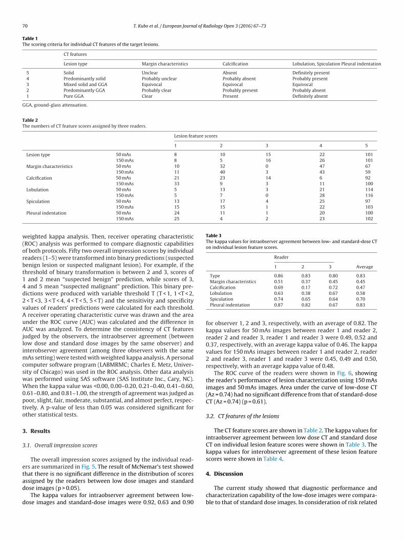
Table 1The scoring criteria for individual CT features of the target lesions.
CT features
Lesion type Margin characteristics Calcification Lobulation, Spiculation Pleural indentation
5 Solid Unclear Absent Definitely present4 Predominantly solid Probably unclear Probably absent Probably present3 Mixed solid and GGA Equivocal Equivocal Equivocal2 Predominantly GGA Probably clear Probably present Probably absent1 Pure GGA Clear Present Definitely absent
GGA, ground-glass attenuation.
Table 2The numbers of CT feature scores assigned by three readers.
Lesion feature scores
1 2 3 4 5
Lesion type 50 mAs 8 10 15 22 101150 mAs 8 5 16 26 101
Margin characteristics 50 mAs 10 32 0 47 67150 mAs 11 40 3 43 59
Calcification 50 mAs 21 23 14 6 92150 mAs 33 9 3 11 100
Lobulation 50 mAs 5 13 3 21 114150 mAs 5 7 0 28 116
Spiculation 50 mAs 13 17 4 25 9715 1 22 10311 1 20 1004 2 23 102
w(orbt14d2vAuAjlimcswW0pto
3
3
etad
d
Table 3The kappa values for intraobserver agreement between low- and standard-dose CTon individual lesion feature scores.
Reader
1 2 3 Average
Type 0.86 0.83 0.80 0.83Margin characteristics 0.51 0.37 0.45 0.45Calcification 0.69 0.17 0.72 0.47
150 mAs 15
Pleural indentation 50 mAs 24
150 mAs 25
eighted kappa analysis. Then, receiver operating characteristicROC) analysis was performed to compare diagnostic capabilitiesf both protocols. Fifty two overall impression scores by individualeaders (1–5) were transformed into binary predictions (suspectedenign lesion or suspected malignant lesion). For example, if thehreshold of binary transformation is between 2 and 3, scores of
and 2 mean “suspected benign” prediction, while scores of 3, and 5 mean “suspected malignant” prediction. This binary pre-ictions were produced with variable threshold T (T < 1, 1 <T < 2,
< T <3, 3 < T < 4, 4 < T < 5, 5 < T) and the sensitivity and specificityalues of readers’ predictions were calculated for each threshold.
receiver operating characteristic curve was drawn and the areander the ROC curve (AUC) was calculated and the difference inUC was analyzed. To determine the consistency of CT features
udged by the observers, the intraobserver agreement (betweenow dose and standard dose images by the same observer) andnterobserver agreement (among three observers with the same
As setting) were tested with weighted kappa analysis. A personalomputer software program (LABMRMC; Charles E. Metz, Univer-ity of Chicago) was used in the ROC analysis. Other data analysisas performed using SAS software (SAS Institute Inc., Cary, NC).hen the kappa value was <0.00, 0.00–0.20, 0.21–0.40, 0.41–0.60,
.61–0.80, and 0.81–1.00, the strength of agreement was judged asoor, slight, fair, moderate, substantial, and almost perfect, respec-ively. A p-value of less than 0.05 was considered significant forther statistical tests.
. Results
.1. Overall impression scores
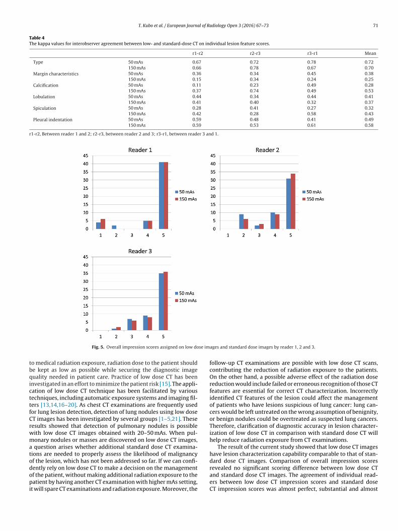
The overall impression scores assigned by the individual read-rs are summarized in Fig. 5. The result of McNemar’s test showedhat there is no significant difference in the distribution of scores
ssigned by the readers between low dose images and standardose images (p > 0.05).The kappa values for intraobserver agreement between low-ose images and standard-dose images were 0.92, 0.63 and 0.90
Lobulation 0.63 0.38 0.67 0.58Spiculation 0.74 0.65 0.64 0.70Pleural indentation 0.87 0.82 0.67 0.83
for observer 1, 2 and 3, respectively, with an average of 0.82. Thekappa values for 50 mAs images between reader 1 and reader 2,reader 2 and reader 3, reader 1 and reader 3 were 0.49, 0.52 and0.37, respectively, with an average kappa value of 0.46. The kappavalues for 150 mAs images between reader 1 and reader 2, reader2 and reader 3, reader 1 and reader 3 were 0.45, 0.49 and 0.50,respectively, with an average kappa value of 0.48.
The ROC curve of the readers were shown in Fig. 6, showingthe reader’s performance of lesion characterization using 150 mAsimages and 50 mAs images. Area under the curve of low-dose CT(Az = 0.74) had no significant difference from that of standard-doseCT (Az = 0.74) (p = 0.61).
3.2. CT features of the lesions
The CT feature scores are shown in Table 2. The kappa values forintraobserver agreement between low dose CT and standard doseCT on individual lesion feature scores were shown in Table 3. Thekappa values for interobserver agreement of these lesion featurescores were shown in Table 4.
4. Discussion
The current study showed that diagnostic performance andcharacterization capability of the low-dose images were compara-ble to that of standard dose images. In consideration of risk related
T. Kubo et al. / European Journal of Radiology Open 3 (2016) 67–73 71
Table 4The kappa values for interobserver agreement between low- and standard-dose CT on individual lesion feature scores.
r1-r2 r2-r3 r3-r1 Mean
Type 50 mAs 0.67 0.72 0.78 0.72150 mAs 0.66 0.78 0.67 0.70
Margin characteristics 50 mAs 0.36 0.34 0.45 0.38150 mAs 0.15 0.34 0.24 0.25
Calcification 50 mAs 0.11 0.23 0.49 0.28150 mAs 0.37 0.74 0.49 0.53
Lobulation 50 mAs 0.44 0.34 0.44 0.41150 mAs 0.41 0.40 0.32 0.37
Spiculation 50 mAs 0.28 0.41 0.27 0.32150 mAs 0.42 0.28 0.58 0.43
Pleural indentation 50 mAs 0.59 0.48 0.41 0.49150 mAs 0.59 0.53 0.61 0.58
r1-r2, Between reader 1 and 2; r2-r3, between reader 2 and 3; r3-r1, between reader 3 and 1.
se im
tbqicttfCrwmatodopi
Fig. 5. Overall impression scores assigned on low do
o medical radiation exposure, radiation dose to the patient shoulde kept as low as possible while securing the diagnostic imageuality needed in patient care. Practice of low dose CT has been
nvestigated in an effort to minimize the patient risk [15]. The appli-ation of low dose CT technique has been facilitated by variousechniques, including automatic exposure systems and imaging fil-ers [13,14,16–20]. As chest CT examinations are frequently usedor lung lesion detection, detection of lung nodules using low doseT images has been investigated by several groups [1–5,21]. Theseesults showed that detection of pulmonary nodules is possibleith low dose CT images obtained with 20–50 mAs. When pul-onary nodules or masses are discovered on low dose CT images,
question arises whether additional standard dose CT examina-ions are needed to properly assess the likelihood of malignancyf the lesion, which has not been addressed so far. If we can confi-ently rely on low dose CT to make a decision on the management
f the patient, without making additional radiation exposure to theatient by having another CT examination with higher mAs setting,t will spare CT examinations and radiation exposure. Moreover, the
ages and standard dose images by reader 1, 2 and 3.
follow-up CT examinations are possible with low dose CT scans,contributing the reduction of radiation exposure to the patients.On the other hand, a possible adverse effect of the radiation dosereduction would include failed or erroneous recognition of those CTfeatures are essential for correct CT characterization. Incorrectlyidentified CT features of the lesion could affect the managementof patients who have lesions suspicious of lung cancer: lung can-cers would be left untreated on the wrong assumption of benignity,or benign nodules could be overtreated as suspected lung cancers.Therefore, clarification of diagnostic accuracy in lesion character-ization of low dose CT in comparison with standard dose CT willhelp reduce radiation exposure from CT examinations.
The result of the current study showed that low dose CT imageshave lesion characterization capability comparable to that of stan-dard dose CT images. Comparison of overall impression scoresrevealed no significant scoring difference between low dose CT
and standard dose CT images. The agreement of individual read-ers between low dose CT impression scores and standard doseCT impression scores was almost perfect, substantial and almost
72 T. Kubo et al. / European Journal of Radiology Open 3 (2016) 67–73
F n (bei fferen
pmbRm
dadltsvhCtdidcp
hCals
ig. 6. Receiver operating characteristic (ROC) curves of the lesion characterizatiomages and broken lines represent the curves for 150 mAs images. No significant di
erfect for reader 1–3, respectively, while the interobserver agree-ent was moderate, except for the fair agreement (kappa = 0.37)
etween reader 1 and 2 on 50 mAs images. At the same time,OC analysis showed no significant difference in diagnostic perfor-ance between low dose CT images and standard dose CT images.Our results suggest that the low dose CT is an adequate initial
ecision tool for lung cancers. The decisions on the lesion foundt low dose CT images are as reliable as those made on stan-ard dose CT images. If the lesions are likely to be benign, the
esion does not need additional standard CT examination for bet-er visualization. For selected cases where lung cancer is stronglyuspected, standard dose CT examinations, preferably with intra-enous contrast, may be appropriate to examine the findings in theilum and mediastinum. The result also indicates that low doseT exams are adequate tool for follow up examinations becausehe nodule characteristics can be appreciated properly with lowose CT images. The use of standard dose CT for follow-up exam-
nations may be unnecessary. This study showed that radiationose reduction is possible both in screening and follow-up settings,ontributing wise use of reduced dose CT examinations to improveatient safety.
Comparison of individual CT feature scores also demonstratedigh degree of agreement between low dose CT and standard dose
T images. Agreement in lesion type and pleural indentation waslmost perfect, with a mean kappa value more than 0.80. Corre-ation of scores was substantial for the presence or absence ofpiculation. Other CT feature scores, lobulation, calcification andnign vs. malignant) for three readers. Solid lines represent the curves for 50 mAsce was found in area under the curve (AUC) between 50 mAs and 150 mAs images.
margin characteristics had lower kappa scores, average kappa valueranging from 0.45 to 0.58, the agreement of scores being moderate.
The study showed lower agreement rate in the detection of cal-cification, being 0.17 for one of the readers. A possible explanationfor this low agreement rate is that artifactual high CT value pixelscould simulate microcalcifications. Lung images are reconstructedwith high-frequency reconstruction algorithms and objects tend tobe encircled with pixels with high CT values due to edge enhance-ment. This effect occasionally adds seemingly high-dense spotsin the lesions, especially when the image is contaminated withheavy spotty noises as in low dose images. Hence, exclusive useof lung images in this study could be responsible for this lowerrate of agreement on calcification assessment. If soft tissue imageswere also available for review at the time of the reading experi-ment, the inconsistent recognition of calcification could be avoided.Despite lower score agreement regarding some CT features includ-ing margin characteristics, calcification and lobulation, the overallimpression of the lesion made by the radiologists was highly con-sistent, with almost equal receiver operation curve characteristics.The present study showed that radiation dose reduction did notaffect lesion characterization capability despite the image qualitydegradation.
The current study has a couple of limitations. First, majority of
lesions that are included in this study are relatively large (>2 cm),mostly solid. Diagnostic accuracy of smaller nodules (e.g. <10 mm)or nodules showing ground-glass attenuation by low dose CT is notfully evaluated in this study. Dedicated study may be needed that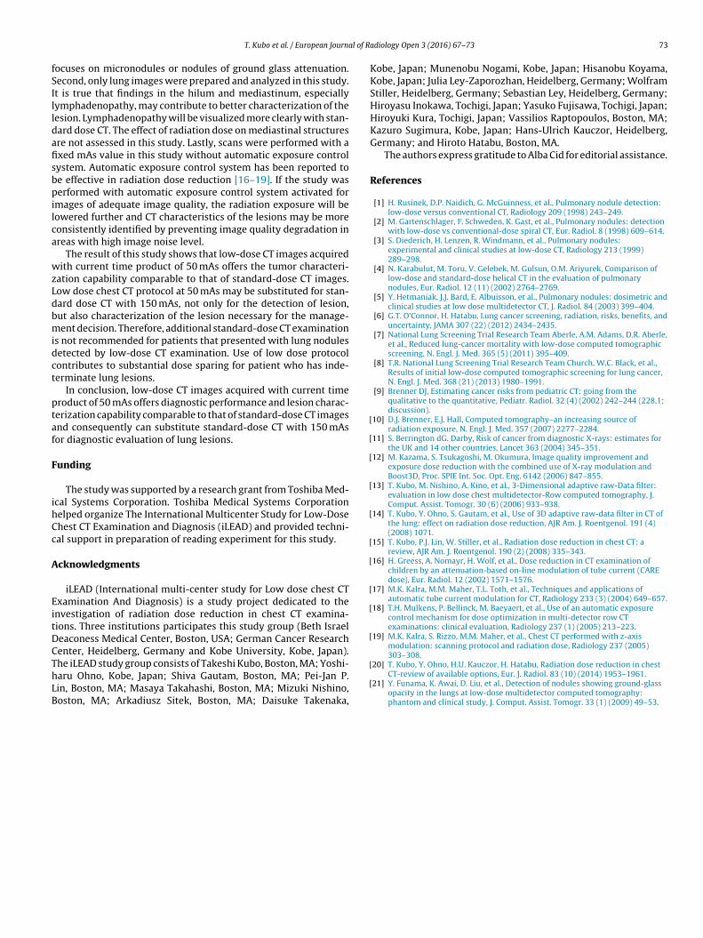
l of Ra
fSIlldafisbpilca
wzLdbmidct
ptaf
F
ihCc
A
EitDCThLB
[
[
[
[
[
[
[
[
[
[
T. Kubo et al. / European Journa
ocuses on micronodules or nodules of ground glass attenuation.econd, only lung images were prepared and analyzed in this study.t is true that findings in the hilum and mediastinum, especiallyymphadenopathy, may contribute to better characterization of theesion. Lymphadenopathy will be visualized more clearly with stan-ard dose CT. The effect of radiation dose on mediastinal structuresre not assessed in this study. Lastly, scans were performed with axed mAs value in this study without automatic exposure controlystem. Automatic exposure control system has been reported toe effective in radiation dose reduction [16–19]. If the study waserformed with automatic exposure control system activated for
mages of adequate image quality, the radiation exposure will beowered further and CT characteristics of the lesions may be moreonsistently identified by preventing image quality degradation inreas with high image noise level.
The result of this study shows that low-dose CT images acquiredith current time product of 50 mAs offers the tumor characteri-
ation capability comparable to that of standard-dose CT images.ow dose chest CT protocol at 50 mAs may be substituted for stan-ard dose CT with 150 mAs, not only for the detection of lesion,ut also characterization of the lesion necessary for the manage-ent decision. Therefore, additional standard-dose CT examination
s not recommended for patients that presented with lung nodulesetected by low-dose CT examination. Use of low dose protocolontributes to substantial dose sparing for patient who has inde-erminate lung lesions.
In conclusion, low-dose CT images acquired with current timeroduct of 50 mAs offers diagnostic performance and lesion charac-erization capability comparable to that of standard-dose CT imagesnd consequently can substitute standard-dose CT with 150 mAsor diagnostic evaluation of lung lesions.
unding
The study was supported by a research grant from Toshiba Med-cal Systems Corporation. Toshiba Medical Systems Corporationelped organize The International Multicenter Study for Low-Dosehest CT Examination and Diagnosis (iLEAD) and provided techni-al support in preparation of reading experiment for this study.
cknowledgments
iLEAD (International multi-center study for Low dose chest CTxamination And Diagnosis) is a study project dedicated to thenvestigation of radiation dose reduction in chest CT examina-ions. Three institutions participates this study group (Beth Israeleaconess Medical Center, Boston, USA; German Cancer Researchenter, Heidelberg, Germany and Kobe University, Kobe, Japan).
he iLEAD study group consists of Takeshi Kubo, Boston, MA; Yoshi-aru Ohno, Kobe, Japan; Shiva Gautam, Boston, MA; Pei-Jan P.in, Boston, MA; Masaya Takahashi, Boston, MA; Mizuki Nishino,oston, MA; Arkadiusz Sitek, Boston, MA; Daisuke Takenaka,[
[
diology Open 3 (2016) 67–73 73
Kobe, Japan; Munenobu Nogami, Kobe, Japan; Hisanobu Koyama,Kobe, Japan; Julia Ley-Zaporozhan, Heidelberg, Germany; WolframStiller, Heidelberg, Germany; Sebastian Ley, Heidelberg, Germany;Hiroyasu Inokawa, Tochigi, Japan; Yasuko Fujisawa, Tochigi, Japan;Hiroyuki Kura, Tochigi, Japan; Vassilios Raptopoulos, Boston, MA;Kazuro Sugimura, Kobe, Japan; Hans-Ulrich Kauczor, Heidelberg,Germany; and Hiroto Hatabu, Boston, MA.
The authors express gratitude to Alba Cid for editorial assistance.
References
[1] H. Rusinek, D.P. Naidich, G. McGuinness, et al., Pulmonary nodule detection:low-dose versus conventional CT, Radiology 209 (1998) 243–249.
[2] M. Gartenschlager, F. Schweden, K. Gast, et al., Pulmonary nodules: detectionwith low-dose vs conventional-dose spiral CT, Eur. Radiol. 8 (1998) 609–614.
[3] S. Diederich, H. Lenzen, R. Windmann, et al., Pulmonary nodules:experimental and clinical studies at low-dose CT, Radiology 213 (1999)289–298.
[4] N. Karabulut, M. Toru, V. Gelebek, M. Gulsun, O.M. Ariyurek, Comparison oflow-dose and standard-dose helical CT in the evaluation of pulmonarynodules, Eur. Radiol. 12 (11) (2002) 2764–2769.
[5] Y. Hetmaniak, J.J. Bard, E. Albuisson, et al., Pulmonary nodules: dosimetric andclinical studies at low dose multidetector CT, J. Radiol. 84 (2003) 399–404.
[6] G.T. O’Connor, H. Hatabu, Lung cancer screening, radiation, risks, benefits, anduncertainty, JAMA 307 (22) (2012) 2434–2435.
[7] National Lung Screening Trial Research Team Aberle, A.M. Adams, D.R. Aberle,et al., Reduced lung-cancer mortality with low-dose computed tomographicscreening, N. Engl. J. Med. 365 (5) (2011) 395–409.
[8] T.R. National Lung Screening Trial Research Team Church, W.C. Black, et al.,Results of initial low-dose computed tomographic screening for lung cancer,N. Engl. J. Med. 368 (21) (2013) 1980–1991.
[9] Brenner DJ, Estimating cancer risks from pediatric CT: going from thequalitative to the quantitative, Pediatr. Radiol. 32 (4) (2002) 242–244 (228,1;discussion).
10] D.J. Brenner, E.J. Hall, Computed tomography–an increasing source ofradiation exposure, N. Engl. J. Med. 357 (2007) 2277–2284.
11] S. Berrington dG. Darby, Risk of cancer from diagnostic X-rays: estimates forthe UK and 14 other countries, Lancet 363 (2004) 345–351.
12] M. Kazama, S. Tsukagoshi, M. Okumura, Image quality improvement andexposure dose reduction with the combined use of X-ray modulation andBoost3D, Proc. SPIE Int. Soc. Opt. Eng. 6142 (2006) 847–855.
13] T. Kubo, M. Nishino, A. Kino, et al., 3-Dimensional adaptive raw-Data filter:evaluation in low dose chest multidetector-Row computed tomography, J.Comput. Assist. Tomogr. 30 (6) (2006) 933–938.
14] T. Kubo, Y. Ohno, S. Gautam, et al., Use of 3D adaptive raw-data filter in CT ofthe lung: effect on radiation dose reduction, AJR Am. J. Roentgenol. 191 (4)(2008) 1071.
15] T. Kubo, P.J. Lin, W. Stiller, et al., Radiation dose reduction in chest CT: areview, AJR Am. J. Roentgenol. 190 (2) (2008) 335–343.
16] H. Greess, A. Nomayr, H. Wolf, et al., Dose reduction in CT examination ofchildren by an attenuation-based on-line modulation of tube current (CAREdose), Eur. Radiol. 12 (2002) 1571–1576.
17] M.K. Kalra, M.M. Maher, T.L. Toth, et al., Techniques and applications ofautomatic tube current modulation for CT, Radiology 233 (3) (2004) 649–657.
18] T.H. Mulkens, P. Bellinck, M. Baeyaert, et al., Use of an automatic exposurecontrol mechanism for dose optimization in multi-detector row CTexaminations: clinical evaluation, Radiology 237 (1) (2005) 213–223.
19] M.K. Kalra, S. Rizzo, M.M. Maher, et al., Chest CT performed with z-axismodulation: scanning protocol and radiation dose, Radiology 237 (2005)303–308.
20] T. Kubo, Y. Ohno, H.U. Kauczor, H. Hatabu, Radiation dose reduction in chestCT-review of available options, Eur. J. Radiol. 83 (10) (2014) 1953–1961.
21] Y. Funama, K. Awai, D. Liu, et al., Detection of nodules showing ground-glassopacity in the lungs at low-dose multidetector computed tomography:phantom and clinical study, J. Comput. Assist. Tomogr. 33 (1) (2009) 49–53.