epidemiological impact of myocarditis
TRANSCRIPT

Journal of
Clinical Medicine
Review
Epidemiological Impact of Myocarditis
Ainoosh Golpour 1, Dimitri Patriki 2, Paul J. Hanson 3, Bruce McManus 3 and Bettina Heidecker 1,*
�����������������
Citation: Golpour, A.; Patriki, D.;
Hanson, P.J.; McManus, B.; Heidecker,
B. Epidemiological Impact of
Myocarditis. J. Clin. Med. 2021, 10,
603. https://doi.org/10.3390/
jcm10040603
Academic Editor: Andrea Frustaci
Received: 13 January 2021
Accepted: 1 February 2021
Published: 5 February 2021
Publisher’s Note: MDPI stays neutral
with regard to jurisdictional claims in
published maps and institutional affil-
iations.
Copyright: © 2021 by the authors.
Licensee MDPI, Basel, Switzerland.
This article is an open access article
distributed under the terms and
conditions of the Creative Commons
Attribution (CC BY) license (https://
creativecommons.org/licenses/by/
4.0/).
1 Campus Benjamin Franklin, Charite Universitätsmedizin Berlin, 12203 Berlin, Germany;[email protected]
2 Department of Medicine, Cantonal Hospital of Baden, 15005 Baden, Switzerland; [email protected] Department of Pathology & Laboratory Medicine, University of British Columbia, Vancouver,
BC V5K0A1, Canada; [email protected] (P.J.H.); [email protected] (B.M.)* Correspondence: [email protected]
Abstract: Myocarditis is an inflammatory disease of the heart muscle with a wide range of potentialetiological factors and consequently varying clinical patterns across the world. In this review, weaddress the epidemiology of myocarditis. Myocarditis was considered a rare disease until intensifiedresearch efforts in recent decades revealed its true epidemiological importance. While it remains achallenge to determine the true prevalence of myocarditis, studies are underway to obtain betterapproximations of the proportions of this disease. Nowadays, the prevalence of myocarditis hasbeen reported from 10.2 to 105.6 per 100,000 worldwide, and its annual occurrence is estimated atabout 1.8 million cases. This wide range of reported cases reflects the uncertainty surrounding thetrue prevalence and a potential underdiagnosis of this disease. Since myocarditis continues to bea significant public health issue, particularly in young adults in whom myocarditis is among themost common causes of sudden cardiac death, improved diagnostic and therapeutic proceduresare necessary. This manuscript aims to summarize the current knowledge on the epidemiologyof myocarditis, new diagnostic approaches and the current epidemiological impact of the COVID-19 pandemic.
Keywords: myocarditis; epidemiology; incidence and prevalence; myocarditis associated withCOVID-19; etiology; diagnosis; regional differences
1. Introduction
Myocarditis is a heterogeneous disease with a variety of symptoms ranging from mildchest discomfort to cardiogenic shock. As it often mimics other common disorders, such ascoronary artery disease (CAD), diagnosis may be challenging [1]. Over the years, variousdiagnostic tests have evolved to identify patients suffering from myocarditis. Nowadays, adiagnosis of myocarditis includes clinical, laboratory, imaging and histological parameters.While the gold standard for diagnosis of myocarditis is endomyocardial biopsy (EMB) [2,3],cardiac magnetic resonance imaging (CMR) is considered a non-invasive alternative inpatients with suspected myocarditis [3–5]. New diagnostic algorithms and tools helpus better understand its true proportions and indicate a potential underestimation ofthe prevalence of myocarditis. This is reflected in the discrepancy within the publisheddata about the prevalence of myocarditis ranging from 10.2 to 105.6 per 100,000 peopleworldwide [6]. The potentially severe complications range from heart failure to suddencardiac death [3].
Most cases of myocarditis are caused by infectious agents, toxic substances, drugsor autoimmune disorders [7]. Hence, it is increasingly recognized that myocarditis is aninflammatory condition of the myocardium triggered by various factors rather than adistinct cardiovascular disease.
This review aims to shed light on the worldwide epidemiological impact of myocardi-tis and provide perspective on the apparent contribution of heart damage associated withCOVID-19 observed during the ongoing pandemic.
J. Clin. Med. 2021, 10, 603. https://doi.org/10.3390/jcm10040603 https://www.mdpi.com/journal/jcm

J. Clin. Med. 2021, 10, 603 2 of 15
2. Etiology of Myocarditis2.1. Infection
Acute lymphocytic myocarditis is the most common type of myocarditis and is fre-quently caused by pathogens, such as adenoviruses, after a respiratory infection [8,9]. Astudy including 12,747 routine autopsies over a period of 10 years in a general populationrevealed that lymphocytic myocarditis was present in 1.06% of the cases [10].
However, the reported incidence of lymphocytic myocarditis confirmed by EMB variesin the literature, in part due to differences in diagnostic sensitivity of methods used forhistology or sample collection.
In 1989, Chow and colleagues determined that it required up to 17 right ventricularbiopsy specimens to detect 79% of cases with myocarditis—a number that is unrealisticto achieve in the clinical setting [11]. Thus, it is expected that when standard biopsytechniques are used, a low sensitivity of detection will be achieved. This is, of course,a different issue than the usually patchy nature of myocarditis detected, which will beaddressed below.
In 2010, Dec described the incidence of lymphocytic myocarditis to be 55% amongcases of biopsy-proven myocarditis [12]. A subsequent study identified lymphocyticmyocarditis in most of the patients (95.5%) diagnosed with myocarditis based on EMB. Inthat study, 564 out of 1752 patients diagnosed with myocarditis received left ventricularEMB, and 1118 of them received biventricular EMB. Biventricular EMB increased diagnosticsensitivity in this study [13]. The high degree of variability in diagnosis of myocarditisvia EMB may be attributable to intra/interobserver error, but is more likely a reflection ofthe heterogeneous, focal patchy nature of the disease. Hence, in contrast to post-mortemgross observation, EMB would require an unrealistic sample size of biopsy pieces to detectregions of inflammation and injury that would accurately represent the true prevalence ofmyocarditis among patients with clinical symptoms of the disease.
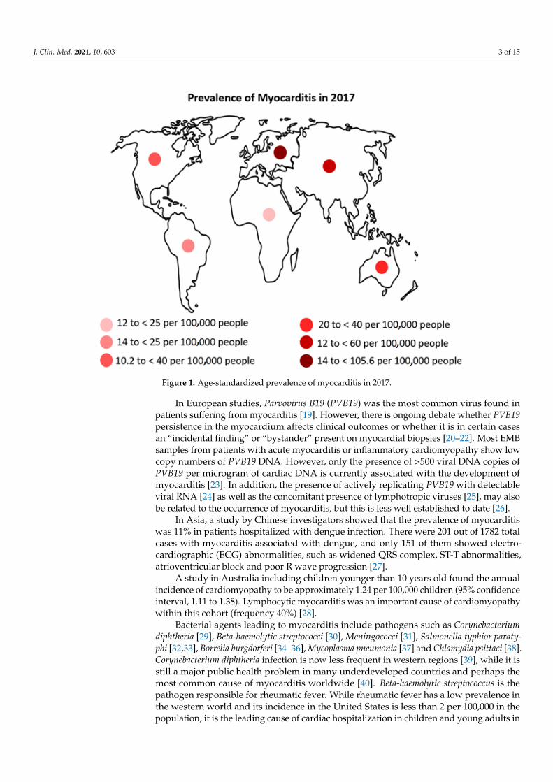
It is generally difficult to determine the true incidence of myocarditis because, on theone hand, as described above, the disease has a patchy nature, and on the other hand, EMB,the gold standard for the diagnosis of myocarditis, is not frequently performed becauseof its invasive character [2,3]. In 1986, Dallas criteria were established with the aim ofproviding a standardized histopathological categorization that could be helpful for thediagnosis of myocarditis using EMB [14]. However, interpretation of the histological data,as well as sampling errors of EMB itself, further impedes final diagnosis [4,5]. Nowadays,electroanatomic mapping (EAM) is an emerging alternative approach for the diagnosisof myocarditis, which could lead to the improvement of sensitivity and the reduction infalse-negative rates in endomyocardial biopsy (EMB) [15]. Another non-invasive diagnosticalternative is cardiac magnetic resonance (CMR) imaging. However, access to CMR maybe limited, and the lack of financial coverage of CMR by many insurance companiesstill creates a challenge in many countries. Differences in the availability and expertiseof diagnostic tools and procedures in the various regions may also have contributed todifferences in the reported prevalence (Figure 1).
According to the World Health Organization (WHO), in the period from 1975 to 1985,Coxsackievirus B3 (CVB3, an enterovirus) was the most common virus causing myocardialinjury worldwide (34.6 per 1000 cases), followed by influenza B virus (17.4 per 1000 cases),influenza A (11.7 per 1000 cases), Coxsackie virus A (9.1 per 1000 cases) and cytomegalovirus(CMV) (8.0 per 1000 cases) [16]. Bowles and colleagues as well as Martin and co-workersdemonstrated that adenoviruses were predominant causes of myocarditis in children [17,18].
J. Clin. Med. 2021, 10, 603 3 of 15J. Clin. Med. 2021, 10, x FOR PEER REVIEW 3 of 16
Figure 1. Age‐standardized prevalence of myocarditis in 2017.
According to the World Health Organization (WHO), in the period from 1975 to 1985,
Coxsackievirus B3 (CVB3, an enterovirus) was the most common virus causing myocardial
injury worldwide (34.6 per 1000 cases), followed by influenza B virus (17.4 per 1000 cases),
influenza A (11.7 per 1000 cases), Coxsackie virus A (9.1 per 1000 cases) and cytomegalovirus
(CMV) (8.0 per 1000 cases) [16]. Bowles and colleagues as well as Martin and co‐workers
demonstrated that adenoviruses were predominant causes of myocarditis in children [17],
[18].
In European studies, Parvovirus B19 (PVB19) was the most common virus found in
patients suffering from myocarditis [19]. However, there is ongoing debate whether
PVB19 persistence in the myocardium affects clinical outcomes or whether it is in certain
cases an “incidental finding” or “bystander” present on myocardial biopsies [20–22]. Most
EMB samples from patients with acute myocarditis or inflammatory cardiomyopathy
show low copy numbers of PVB19 DNA. However, only the presence of >500 viral DNA
copies of PVB19 per microgram of cardiac DNA is currently associated with the
development of myocarditis [23]. In addition, the presence of actively replicating PVB19
with detectable viral RNA [24] as well as the concomitant presence of lymphotropic
viruses [25], may also be related to the occurrence of myocarditis, but this is less well
established to date [26].
In Asia, a study by Chinese investigators showed that the prevalence of myocarditis
was 11% in patients hospitalized with dengue infection. There were 201 out of 1782 total
cases with myocarditis associated with dengue, and only 151 of them showed
electrocardiographic (ECG) abnormalities, such as widened QRS complex, ST‐T
abnormalities, atrioventricular block and poor R wave progression [27].
A study in Australia including children younger than 10 years old found the annual
incidence of cardiomyopathy to be approximately 1.24 per 100,000 children (95%
confidence interval, 1.11 to 1.38). Lymphocytic myocarditis was an important cause of
cardiomyopathy within this cohort (frequency 40%) [28].
Figure 1. Age-standardized prevalence of myocarditis in 2017.
In European studies, Parvovirus B19 (PVB19) was the most common virus found inpatients suffering from myocarditis [19]. However, there is ongoing debate whether PVB19persistence in the myocardium affects clinical outcomes or whether it is in certain casesan “incidental finding” or “bystander” present on myocardial biopsies [20–22]. Most EMBsamples from patients with acute myocarditis or inflammatory cardiomyopathy show lowcopy numbers of PVB19 DNA. However, only the presence of >500 viral DNA copies ofPVB19 per microgram of cardiac DNA is currently associated with the development ofmyocarditis [23]. In addition, the presence of actively replicating PVB19 with detectableviral RNA [24] as well as the concomitant presence of lymphotropic viruses [25], may alsobe related to the occurrence of myocarditis, but this is less well established to date [26].
In Asia, a study by Chinese investigators showed that the prevalence of myocarditiswas 11% in patients hospitalized with dengue infection. There were 201 out of 1782 totalcases with myocarditis associated with dengue, and only 151 of them showed electro-cardiographic (ECG) abnormalities, such as widened QRS complex, ST-T abnormalities,atrioventricular block and poor R wave progression [27].
A study in Australia including children younger than 10 years old found the annualincidence of cardiomyopathy to be approximately 1.24 per 100,000 children (95% confidenceinterval, 1.11 to 1.38). Lymphocytic myocarditis was an important cause of cardiomyopathywithin this cohort (frequency 40%) [28].
Bacterial agents leading to myocarditis include pathogens such as Corynebacteriumdiphtheria [29], Beta-haemolytic streptococci [30], Meningococci [31], Salmonella typhior paraty-phi [32,33], Borrelia burgdorferi [34–36], Mycoplasma pneumonia [37] and Chlamydia psittaci [38].Corynebacterium diphtheria infection is now less frequent in western regions [39], while it isstill a major public health problem in many underdeveloped countries and perhaps themost common cause of myocarditis worldwide [40]. Beta-haemolytic streptococcus is thepathogen responsible for rheumatic fever. While rheumatic fever has a low prevalence inthe western world and its incidence in the United States is less than 2 per 100,000 in thepopulation, it is the leading cause of cardiac hospitalization in children and young adults in

J. Clin. Med. 2021, 10, 603 4 of 15
the age group of 5–25 years in developing countries [41]. Chlamydia is estimated to presentwith minimally symptomatic myocarditis in 5% to 15% of cases [38].
2.2. Toxicity or Hypersensitivity Reaction
Toxic myocarditis may be triggered by numerous agents. Prescribed drugs such asdobutamine, phenytoin [42], antibiotics (e.g., ampicillin, azithromycin, cephalosporinsand tetracyclines), psychiatric medications (tricyclic antidepressants, benzodiazepinesand clozapine) [43], recreational/illicit drugs (e.g., methamphetamine or cocaine) [44],heavy metals (copper, lead and arsenicals) and antineoplastic agents (e.g., anthracyclines,cyclophosphamide, 5-fluorouracil and tyrosine kinase inhibitors) are known etiologies ofmyocarditis [43]. With the increasing use of Immune-Checkpoint Inhibitors (ICIs) to treata variety of cancers, reports of lethal myocarditis as potential adverse effects of therapyhave increased. Mahmood and colleagues showed that the prevalence of myocarditisin patients treated with ICIs from 2014 to 2017 was 1.14%, with myocarditis developingapproximately 34 days after the start of treatment with ICIs [45]. It was also found thatincidence of myocarditis was higher in patients treated with ICI as compared to patientstreated with other drugs that were included in the Vigibase database, a unique WHOglobal database including more than 16 million individual case safety reports submitted bynational pharmacovigilance centers since 1967 [46].
Myocarditis was also reported as a hypersensitivity reaction during smallpox vaccina-tion in the 1950s and 1960s [47,48]. The frequency of myocarditis was dependent on thestrain used to produce the vaccine, and the method for detecting myocarditis. However, itstrue incidence was unknown. In the United States Military Dryvax vaccination program,which was a large-scale smallpox vaccination program, the incidence of myocarditis wasestimated to be 0.01% or about one in 10,000 [48,49].
2.3. Autoimmunity
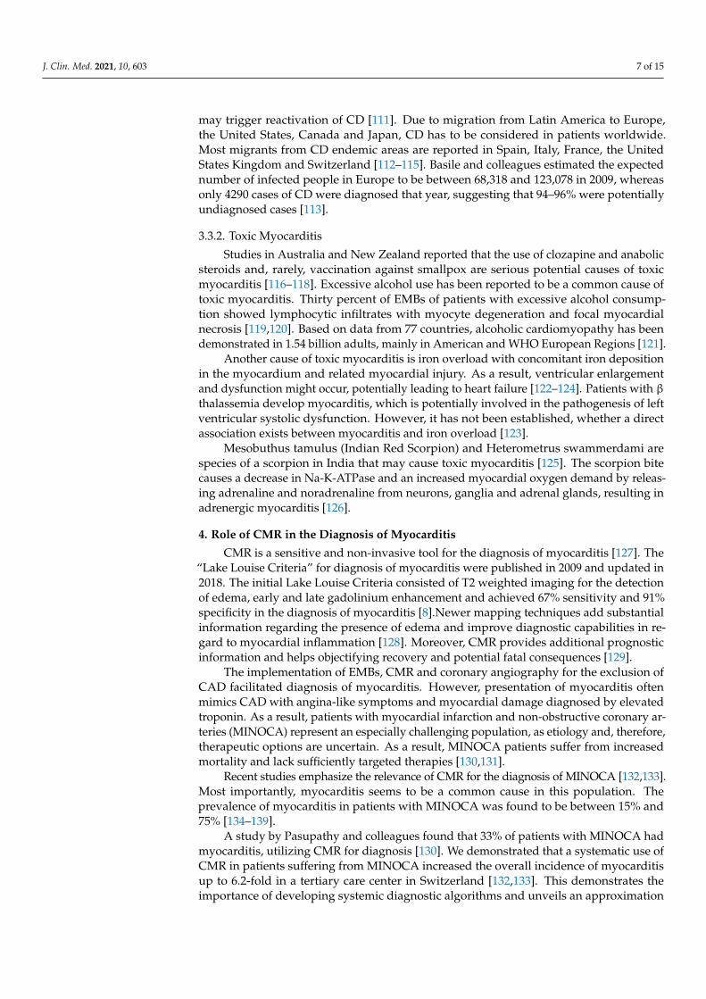
Autoimmunity has been increasingly recognized as one of the main factors sustain-ing inflammation and disease progression in myocarditis. Pathogens, such as viruses,may initiate autoimmune mechanisms that lead to inflammatory cardiomyopathy andmyocarditis [50–52]. We recently showed that by the time of symptom onset of myocarditis,no relevant viral infection was detectable in patients with myocarditis when interrogatedby full virome sequencing for all known vertebrate viruses. Importantly, this study alsoincluded myocardial tissue samples from patients with giant cell myocarditis (Figure 2).No relevant viral infection could be detected in those samples [53].
Additionally, systemic autoimmune diseases may involve the heart and manifest asmyocarditis [54,55]. In that regard, it is estimated that cardiac involvement is present in2% to 5% of patients with systemic sarcoidosis [56–59]. A study in Finland showed thatbetween 1988 and 2012, the number of cases of cardiac sarcoidosis increased significantly,with the prevalence of cardiac sarcoidosis being 2.2 per 100,000 people in 2012 [60].
In patients with systemic lupus erythematosus (SLE), myocarditis may be a life-threatening manifestation. Although in clinical studies SLE myocarditis was found in onlyabout 9% of patients with SLE [61–64], post-mortem analyses reported a high prevalenceof 57%, indicating a high prevalence of subclinical disease [65–67]. SLE is most common inwomen in their 20s and 30s, and the severity as well as the clinical manifestations of thedisease differ between early and late onset [68–70]. Specifically, it has been reported thatpatients diagnosed with SLE at an older age are more likely to develop a cardiovasculardisease than patients diagnosed with SLE at a younger age [71]. However, it is noticeablethat lupus myocarditis occurs more frequently in young people. While the prevalence inpatients diagnosed with SLE before the age of 18 is 1%, the prevalence in elderly individualsis 0.3% [72].

J. Clin. Med. 2021, 10, 603 5 of 15
J. Clin. Med. 2021, 10, x FOR PEER REVIEW 5 of 16
Figure 2. Histology and immunohistochemistry of a patient with giant cell myocarditis. Images show a lesion of giant cell
myocarditis (magnification 4×–40×). Masson’s trichrome staining (panels 1–4), arrows point out a giant cell and cells in the
process of forming a giant cell.
Additionally, systemic autoimmune diseases may involve the heart and manifest as
myocarditis [54,55]. In that regard, it is estimated that cardiac involvement is present in
2% to 5% of patients with systemic sarcoidosis [56–59]. A study in Finland showed that
between 1988 and 2012, the number of cases of cardiac sarcoidosis increased significantly,
with the prevalence of cardiac sarcoidosis being 2.2 per 100,000 people in 2012 [60].
In patients with systemic lupus erythematosus (SLE), myocarditis may be a life‐
threatening manifestation. Although in clinical studies SLE myocarditis was found in only
about 9% of patients with SLE [61–64], post‐mortem analyses reported a high prevalence
of 57%, indicating a high prevalence of subclinical disease [65–67]. SLE is most common
in women in their 20s and 30s, and the severity as well as the clinical manifestations of the
disease differ between early and late onset [68–70]. Specifically, it has been reported that
patients diagnosed with SLE at an older age are more likely to develop a cardiovascular
disease than patients diagnosed with SLE at a younger age [71]. However, it is noticeable
that lupus myocarditis occurs more frequently in young people. While the prevalence in
patients diagnosed with SLE before the age of 18 is 1%, the prevalence in elderly individ‐
uals is 0.3% [72].
Giant cell myocarditis is among the most aggressive forms of myocarditis and may
be associated with autoimmune diseases, such as SLE, Sjögren’s syndrome, vasculitis, ul‐
cerative colitis and polymyositis. Patients with giant cell myocarditis continue to have
poor prognosis despite maximal therapy [73–76]. The epidemiology of giant cell myocar‐
ditis has not been investigated comprehensively given its low incidence. An autopsy
study, including 377,841 autopsy cases, found the incidence of giant cell myocarditis to be
0.007% [77,78]. In comparison, the incidence of other types of myocarditis was considera‐
bly higher (0.11%) [78]. Another subtype of myocarditis in the context of systemic auto‐
immune disease is eosinophilic myocarditis, which is estimated to be present in 50%–
Figure 2. Histology and immunohistochemistry of a patient with giant cell myocarditis. Images show a lesion of giant cellmyocarditis (magnification 4×–40×). Masson’s trichrome staining (panels 1–4), arrows point out a giant cell and cells in theprocess of forming a giant cell.
Giant cell myocarditis is among the most aggressive forms of myocarditis and maybe associated with autoimmune diseases, such as SLE, Sjögren’s syndrome, vasculitis,ulcerative colitis and polymyositis. Patients with giant cell myocarditis continue to havepoor prognosis despite maximal therapy [73–76]. The epidemiology of giant cell my-ocarditis has not been investigated comprehensively given its low incidence. An autopsystudy, including 377,841 autopsy cases, found the incidence of giant cell myocarditis to be0.007% [77,78]. In comparison, the incidence of other types of myocarditis was considerablyhigher (0.11%) [78]. Another subtype of myocarditis in the context of systemic autoimmunedisease is eosinophilic myocarditis, which is estimated to be present in 50–60% of cases ofperipheral eosinophilia [79,80]. It has been shown that male sex hormones may act in aproinflammatory way, while female sex hormones protect against myocyte infectivity andreduce potentially harmful myocardial inflammatory response. Accordingly, eosinophilicmyocarditis also occurs most commonly in males [81].
3. Epidemiology of Myocarditis3.1. Prevalence of Myocarditis
Patients suffering from myocarditis are mostly male (82%), and young adults (averageage: men: 40 ± 16; women: 40 ± 17) [82,83].
A study including 195 countries estimated 1.80 million (95% uncertainty interval (UI)1.64 to 1.98) cases of myocarditis worldwide in 2017. The global number of deaths causedby myocarditis in 2017 was estimated to be around 46,486 (95% UI 39,709 to 51,824), andthe highest age-standardized death rate was found in Oceania (2.6 (95% UI 2.0 to 3.4)per 100,000 people), most likely due to insufficient health resources in this region. Thehigh-income Asia-Pacific region demonstrates the highest age-standardized prevalenceof myocarditis (45.6 (95% UI 41.1 to 50.1) per 100,000 people). The prevalence rates ofmyocarditis varied between 10.2 (95% UI 9.0 to 11.4) per 100,000 people in Chile to 105.6

J. Clin. Med. 2021, 10, 603 6 of 15
(95% UI 90.8 to 120.8) per 100,000 people in Albania (Figure 1). Across all 194 countries,prevalence rates for myocarditis differed by a factor of 10.4 and mortality rates by a factorof 43.9 in 2017. Between 1990 and 2017, age-standardized rates for myocarditis declined,while global prevalence and death rates increased significantly [84].
In several studies, myocarditis was found in 1.4–63% of EMBs from patients withunexplained congestive heart failure, unexplained ventricular arrhythmias or “primary”atrial fibrillation [85–88].Inflammatory changes were found in 22 of 35 patients with idio-pathic congestive cardiomyopathy, suggesting that persistent myocardial inflammationmay be associated with dilated cardiomyopathy (DCM) [88]. In line with those findings,myocardial inflammation is present in 13% to 27% of patients suffering from idiopathicDCM [89]. Moreover, observations from Western Europe and North America indicate thatbetween 10% and 50% of acute DCM cases are likely to be associated with myocarditis [90].
3.2. Sex-Specific Differences
There is evidence for sex-specific differences in myocarditis regarding clinical, lab-oratory and pathophysiological features. Myocarditis is diagnosed more commonly inmen than in women. Moreover, men are more likely to experience a severe trajectoryof myocarditis, while women have a significantly lower risk of death or heart transplan-tation [91]. From 1990 to 2017, a greater decrease in age-standardized prevalence andmortality rates for myocarditis was observed in women vs. men [84]. It is important tonote that clinical presentation appears to be more subtle in women than in men, leadingto a potential underdiagnosis of myocarditis in women [82]. Additionally, male subjectsshowed a higher incidence of CVB3-induced myocarditis with a more severe clinical coursethan females in murine models [92]. Differences in the innate immune response to CVB3between men and women could explain this phenomenon [93,94]. The latest advances incellular and molecular biology have shown that both direct viral and immune-mediatedinjury are involved in the pathogenesis of enteroviral myocarditis [95]. While males presentwith increased γδ T cells; increased TLR4+ CD11b+ inflammation, including macrophages,neutrophils, mast cells and DCs; and an increased Th1 response, females present withprotective Th2 response, increased B cells, more inhibitory Tim-3+ CD4+ T cells and moreT regulatory cells [96,97].
3.3. Regional Differences in Myocarditis3.3.1. Infectious Myocarditis
Frequent infectious causes for myocarditis in Asia between 1966 and 2000 were associ-ated with Diphtheria (Afghanistan and India) [98], typhoid fever [99] and viral infections,such as CVB3 [100] and Chikungunya. Hepatitis C virus was suggested to be a significantcause of myocarditis in Japan, as its genomic material was detected in several cardiomy-opathies [101–103]. Studies from Australia and New Zealand revealed regional differenceswith CVB3 and enterovirus 71 epidemics [104,105]. In Mexico, Central and South America,measles, meningococcal meningitis, human immunodeficiency virus (HIV), dengue fever anddiphtheria were characteristic etiologies of myocarditis, whereby Chagas disease (CD)was excluded from the survey [90]. Reports from Africa addressed HIV [106], peripartalcardiomyopathy and occasionally infections, such as trypanosomiasis [107] and shigellosisas some of the most common causes of myocarditis.
CD, induced by Trypanosoma cruzi, is a major cause of myocarditis in Latin America. Itis estimated that 6–8 million people [108] are infected with Trypanosoma cruzi worldwide,with almost 11,000 deaths annually. EMBs showed myocardial inflammation in about 60%of CD patients [109]. Less than 1% of patients with acute CD have a severe course with acutemyocarditis, pericardial effusion and/or meningoencephalitis, while the acute phase of CDis asymptomatic or includes non-specific clinical features [110]. In addition, while somenewborns with congenital CD are asymptomatic, others may present with myocarditis,meningoencephalitis or hepatosplenomegaly. In particular, patients with AIDS who havebeen exposed to Trypanosoma cruzi are at risk for myocarditis, since immunosuppression
J. Clin. Med. 2021, 10, 603 7 of 15
may trigger reactivation of CD [111]. Due to migration from Latin America to Europe,the United States, Canada and Japan, CD has to be considered in patients worldwide.Most migrants from CD endemic areas are reported in Spain, Italy, France, the UnitedStates Kingdom and Switzerland [112–115]. Basile and colleagues estimated the expectednumber of infected people in Europe to be between 68,318 and 123,078 in 2009, whereasonly 4290 cases of CD were diagnosed that year, suggesting that 94–96% were potentiallyundiagnosed cases [113].
3.3.2. Toxic Myocarditis
Studies in Australia and New Zealand reported that the use of clozapine and anabolicsteroids and, rarely, vaccination against smallpox are serious potential causes of toxicmyocarditis [116–118]. Excessive alcohol use has been reported to be a common cause oftoxic myocarditis. Thirty percent of EMBs of patients with excessive alcohol consump-tion showed lymphocytic infiltrates with myocyte degeneration and focal myocardialnecrosis [119,120]. Based on data from 77 countries, alcoholic cardiomyopathy has beendemonstrated in 1.54 billion adults, mainly in American and WHO European Regions [121].
Another cause of toxic myocarditis is iron overload with concomitant iron depositionin the myocardium and related myocardial injury. As a result, ventricular enlargementand dysfunction might occur, potentially leading to heart failure [122–124]. Patients with β
thalassemia develop myocarditis, which is potentially involved in the pathogenesis of leftventricular systolic dysfunction. However, it has not been established, whether a directassociation exists between myocarditis and iron overload [123].
Mesobuthus tamulus (Indian Red Scorpion) and Heterometrus swammerdami arespecies of a scorpion in India that may cause toxic myocarditis [125]. The scorpion bitecauses a decrease in Na-K-ATPase and an increased myocardial oxygen demand by releas-ing adrenaline and noradrenaline from neurons, ganglia and adrenal glands, resulting inadrenergic myocarditis [126].
4. Role of CMR in the Diagnosis of Myocarditis
CMR is a sensitive and non-invasive tool for the diagnosis of myocarditis [127]. The“Lake Louise Criteria” for diagnosis of myocarditis were published in 2009 and updated in2018. The initial Lake Louise Criteria consisted of T2 weighted imaging for the detectionof edema, early and late gadolinium enhancement and achieved 67% sensitivity and 91%specificity in the diagnosis of myocarditis [8].Newer mapping techniques add substantialinformation regarding the presence of edema and improve diagnostic capabilities in re-gard to myocardial inflammation [128]. Moreover, CMR provides additional prognosticinformation and helps objectifying recovery and potential fatal consequences [129].
The implementation of EMBs, CMR and coronary angiography for the exclusion ofCAD facilitated diagnosis of myocarditis. However, presentation of myocarditis oftenmimics CAD with angina-like symptoms and myocardial damage diagnosed by elevatedtroponin. As a result, patients with myocardial infarction and non-obstructive coronary ar-teries (MINOCA) represent an especially challenging population, as etiology and, therefore,therapeutic options are uncertain. As a result, MINOCA patients suffer from increasedmortality and lack sufficiently targeted therapies [130,131].
Recent studies emphasize the relevance of CMR for the diagnosis of MINOCA [132,133].Most importantly, myocarditis seems to be a common cause in this population. Theprevalence of myocarditis in patients with MINOCA was found to be between 15% and75% [134–139].
A study by Pasupathy and colleagues found that 33% of patients with MINOCA hadmyocarditis, utilizing CMR for diagnosis [130]. We demonstrated that a systematic use ofCMR in patients suffering from MINOCA increased the overall incidence of myocarditisup to 6.2-fold in a tertiary care center in Switzerland [132,133]. This demonstrates theimportance of developing systemic diagnostic algorithms and unveils an approximation

J. Clin. Med. 2021, 10, 603 8 of 15
of the true epidemiologic significance of myocarditis. Certainly, as mentioned above, adefinitive diagnosis can only be made with EMB.
From 2020 onwards, the current NSTEMI guidelines recommend CMR in all patientswith a first diagnosis of MINOCA. This may lead to higher numbers of reported cases ofmyocarditis in the future.
5. Updates on SARS-CoV-2 Infection and COVID-19 Association with Myocarditis
SARS-CoV-2 infection and its resultant clinical manifestation COVID-19 rapidly spreadall over the world in 2020. Interestingly, the first studies reported cardiac injury, defined byincreased levels of troponin, in 22.2–31% of COVID-19 patients. Troponin elevation wasassociated with severe COVID-19 infection, increased morbidity and mortality [140–142].These results were later confirmed in a larger case series of 5700 patients [143]. Moreover,increased serum troponin levels were associated with higher in-hospital mortality [144,145].However, most early studies neither included specified characteristics of patients withincreased troponin nor further cardiac imaging or EMB. As a result, the cause of troponinelevation remains uncertain. As myocarditis was already discussed to be a potentialcomplication of middle east respiratory syndrome (MERS) and other coronaviruses, a linkbetween COVID-19 and myocarditis seems rational [7,146]. Nevertheless, data on thistopic are still quite preliminary.
There are numerous case reports associating COVID-19 and myocarditis [147–150]. Arecent study by Puntmann and colleagues detected cardiac involvement in 78 patients (78%)and persistent myocarditis in 60 patients (60%) by CMR after COVID-19 infection [151].Patients that had recently recovered from COVID-19 presented lower left ventricularejection fraction, higher left ventricular volumes and raised native T1 and T2 than healthycontrols and risk-factor matched controls. In total, 78 patients (78%) revealed abnormalitiesin CMR, such as raised myocardial native T1 (n = 73), raised myocardial native T2 (n = 60),myocardial late gadolinium enhancement (n = 32) or pericardial enhancement (n = 22).Pre-existing conditions, severity and overall course of the acute disease and time sinceoriginal diagnosis had no impact on the results [151]. These data have to be interpretedwith caution, since definitive diagnosis of myocarditis via EMB was only obtained in asubset of patients.
A CMR study of 26 athletes with COVID-19 with mild or no symptoms revealedfindings suggestive for myocarditis in 4 of them (15%) and late gadolinium accumulationwithout T2 elevation indicating previous myocardial injury in 8 other athletes (30.8%) [152].
A recent autopsy study by Halushka et al. reported a much lower incidence ofmyocarditis in patients who died from COVID-19. Out of 277 autopsies, only 20 showedsigns of myocarditis, which represents only 0.07% of patients who died from COVID-19 inthis cohort [153].
Other cardiovascular complications appear to be more predominant in patients withCOVID-19. Thrombotic events, arrhythmias as well as aggravation of an underlying heartdisease may also result in myocardial damage, potentially worsening prognosis [154]. Anautopsy study reported that only 5 out of 68 (7%) fatal cases of COVID-19 were associatedwith myocardial damage and circulatory failure [155].
Moreover, it has been suggested that underlying cardiovascular disease may promotea more severe course of COVID-19 infection [141,142,155–158]. In this context, a meta-analysis of 1527 COVID-19 patients showed that 17.1% of these patients were hypertensiveand 16.4% had pre-existing heart disease [159]. In another study involving 44,672 COVID-19 patients, the fatality rate in patients with cardiovascular disease was found to be fivetimes higher than in patients without underlying cardiovascular disease [157].
Another recent study including 47 heart transplant recipients showed that there was ahigh rate of death after infection with SARS-CoV-2 in this patient group compared withthe general population. The higher death rates were attributable to comorbidities. Noevidence of myocardial injury was found in any of these patients, suggesting that cardiacinvolvement by COVID-19 may be rare among cardiac transplant recipients [160].
J. Clin. Med. 2021, 10, 603 9 of 15
In summary, the association of COVID-19 and acute cardiac injury has been repeatedlyreported and is associated with poor outcomes. Data on myocarditis in patients withCOVID-19 have been somewhat controversial, although the overall consensus is thatmyocarditis is still rare in this patient population. Whether myocarditis will reveal itself tobe causative or a bystander in the development of complications of COVID-19 remains anarea of research. Indeed, with regard to the proportions of the COVID-19 pandemic, wespeculate that there may be a wave of cardiac post-COVID-19 complications in the longrun [161].
6. Conclusions
The prevalence of myocarditis has been estimated to range from 10.2 to 105.6 per100,000 people worldwide, with relevant regional differences influenced by a variety ofpathogens, as well as locally available diagnostics procedures. Based on studies performedin 2017, the annual prevalence of myocarditis is about 1.8 million worldwide. Whilewe slowly approximate the true incidence of myocarditis, its true proportions remainunknown.
In this context, the establishment of CMR as a standard for the diagnosis of myocarditisprovides the possibility of rapid detection of myocarditis before complications mightoccur. Although myocarditis is associated with high morbidity and mortality [162–165],clinical practice recommendations remain vague due to the lack of data from large clinicaltrials [166]. The limited awareness of this disease leads to an even deeper knowledge gapthat we urgently need to fill. Considering the appearance of novel pathogens, such asSARS-CoV2, an increasing expansion of pathogens, such as CD, through globalization incombination with improved diagnostic tools, we expect a further increase in the incidenceof myocarditis over the next decade. International collaborations will be necessary to makeprogress in developing effective therapies for this increasing patient population.
Funding: This research was funded by the German Research Foundation (DFG) and the Open AccessPublication Fund of Charité – Universitätsmedizin Berlin.
Institutional Review Board Statement: Not applicable.
Informed Consent Statement: Not applicable.
Conflicts of Interest: Bettina Heidecker, MD, is an inventor on patents that use RNA for diagnosisof myocarditis.
References1. Sagar, S.; Liu, P.P.; Cooper, L.T., Jr. Myocarditis. Lancet 2012, 379, 738–747. [CrossRef]2. Cooper, L.T.; Baughman, K.L.; Feldman, A.M.; Frustaci, A.; Jessup, M.; Kuhl, U.; Levine, G.N.; Narula, J.; Starling, R.C.; Towbin, J.
The role of endomyocardial biopsy in the management of cardiovascular disease: A scientific statement from the American HeartAssociation, the American College of Cardiology, and the European Society of Cardiology Endorsed by the Heart Failure Societyof America and the Heart Failure Association of the European Society of Cardiology. Eur. Heart J. 2007, 28, 3076–3093.
3. Bozkurt, B.; Colvin, M.; Cook, J.; Cooper, L.T.; Deswal, A.; Fonarow, G.C.; Francis, G.S.; Lenihan, D.; Lewis, E.F.; McNamara,D.M. Current diagnostic and treatment strategies for specific dilated cardiomyopathies: A scientific statement from the AmericanHeart Association. Circulation 2016, 134, e579–e646. [CrossRef]
4. Shanes, J.; Ghali, J.; Billingham, M.; Ferrans, V.; Fenoglio, J.; Edwards, W.; Tsai, C.; Saffitz, J.; Isner, J.; Furner, S. Interobservervariability in the pathologic interpretation of endomyocardial biopsy results. Circulation 1987, 75, 401–405. [CrossRef]
5. Hauck, A.J.; Kearney, D.L.; Edwards, W.D. Evaluation of Postmortem Endomyocardial Biopsy Specimens from 38 Patients withLymphocytic Myocarditis: Implications for Role of Sampling Error; Mayo Clinic Proceedings; Elsevier: Amsterdam, The Netherlands,1989.
6. Pollack, A.; Kontorovich, A.R.; Fuster, V.; Dec, G.W. Viral myocarditis—diagnosis, treatment options, and current controversies.Nat. Rev. Cardiol. 2015, 12, 670. [CrossRef]
7. Tschöpe, C.; Ammirati, E.; Bozkurt, B.; Caforio, A.L.; Cooper, L.T.; Felix, S.B.; Hare, J.M.; Heidecker, B.; Heymans, S.; Hübner, N.Myocarditis and inflammatory cardiomyopathy: Current evidence and future directions. Nat. Rev. Cardiol. 2020, 1–25. [CrossRef]
8. Friedrich, M.G.; Sechtem, U.; Schulz-Menger, J.; Holmvang, G.; Alakija, P.; Cooper, L.T.; White, J.A.; Abdel-Aty, H.; Gutberlet, M.;Prasad, S. Cardiovascular magnetic resonance in myocarditis: A JACC White Paper. J. Am. Coll. Cardiol. 2009, 53, 1475–1487.[CrossRef] [PubMed]

J. Clin. Med. 2021, 10, 603 10 of 15
9. Baboonian, C.; McKenna, W. Eradication of viral myocarditis: Is there hope? J. Am. Coll. Cardiol. 2003, 42, 473–476. [CrossRef]10. Gravanis, M.; Sternby, N. Incidence of myocarditis. A 10-year autopsy study from. Arch Pathol. Lab. Med. 1991, 115, 390–392.
[PubMed]11. Chow, L.H.; Radio, S.J.; Sears, T.D.; Mcmanus, B.M. Insensitivity of right ventricular endomyocardial biopsy in the diagnosis of
myocarditis. J. Am. Coll. Cardiol. 1989, 14, 915–920. [CrossRef]12. Dec, G. Epidemiology and Prognosis of Myocarditis and Dilated Cardiomyopathy: Predictive Value of Clinical Parameters and
Biopsy Findings. In Inflammatory Cardiomyopathy (DCMi); Springer: Berlin/Heidelberg, Germany, 2010.13. Chimenti, C.; Frustaci, A. Contribution and risks of left ventricular endomyocardial biopsy in patients with cardiomyopathies: A
retrospective study over a 28-year period. Circulation 2013, 128, 1531–1541. [CrossRef]14. Aretz, T. Myocarditis. A histopathologic definition and classification. Am. J. Cardiovasc. Pathol. 1986, 1, 3–14.15. Vaidya, V.R.; Abudan, A.A.; Vasudevan, K.; Shantha, G.; Cooper, L.T.; Kapa, S.; Noseworthy, P.A.; Cha, Y.M.; Asirvatham, S.J.;
Deshmukh, A.J. The efficacy and safety of electroanatomic mapping-guided endomyocardial biopsy: A systematic review. J.Interv. Card Electrophysiol. 2018, 53, 63–71. [CrossRef]
16. Grist, N. Epidemiology of viral infections of the heart. Viral Infect. Heart 1993, 88, 23–31.17. Bowles, N.E.; Ni, J.; Kearney, D.L.; Pauschinger, M.; Schultheiss, H.-P.; McCarthy, R.; Hare, J.; Bricker, J.T.; Bowles, K.R.; Towbin,
J.A. Detection of viruses in myocardial tissues by polymerase chain reaction: Evidence of adenovirus as a common cause ofmyocarditis in children and adults. J. Am. Coll. Cardiol. 2003, 42, 466–472. [CrossRef]
18. Martin, A.B.; Webber, S.; Fricker, F.J.; Jaffe, R.; Demmler, G.; Kearney, D.; Zhang, Y.-H.; Bodurtha, J.; Gelb, B.; Ni, J. Acutemyocarditis. Rapid diagnosis by PCR in children. Circulation 1994, 90, 330–339. [CrossRef] [PubMed]
19. Pankuweit, S.; Moll, R.; Baandrup, U.; Portig, I.; Hufnagel, G.; Maisch, B. Prevalence of the parvovirus B19 genome inendomyocardial biopsy specimens. Hum. Pathol. 2003, 34, 497–503. [CrossRef]
20. Schenk, T.; Enders, M.; Pollak, S.; Hahn, R.; Huzly, D. High prevalence of human parvovirus B19 DNA in myocardial autopsysamples from subjects without myocarditis or dilative cardiomyopathy. J. Clin. Microbiol. 2009, 47, 106–110. [CrossRef] [PubMed]
21. Lotze, U.; Egerer, R.; Glück, B.; Zell, R.; Sigusch, H.; Erhardt, C.; Heim, A.; Kandolf, R.; Bock, T.; Wutzler, P. Low level myocardialparvovirus B19 persistence is a frequent finding in patients with heart disease but unrelated to ongoing myocardial injury. J. Med.Virol. 2010, 82, 1449–1457. [CrossRef]
22. Koepsell, S.A.; Anderson, D.R.; Radio, S.J. Parvovirus B19 is a bystander in adult myocarditis. Cardiovasc. Pathol. 2012, 21,476–481. [CrossRef]
23. Bock, C.-T.; Klingel, K.; Kandolf, R. Human parvovirus B19–associated myocarditis. N. Engl. J. Med. 2010, 362, 1248–1249.[CrossRef] [PubMed]
24. Kuhl, U.; Lassner, D.; Dorner, A.; Rohde, M.; Escher, F.; Seeberg, B.; Hertel, E.; Tschope, C.; Skurk, C.; Gross, U. A distinctsubgroup of cardiomyopathy patients characterized by transcriptionally active cardiotropic erythrovirus and altered cardiac geneexpression. Basic Res. Cardiol. 2013, 108, 372. [CrossRef]
25. Bock, C.-T.; Düchting, A.; Utta, F.; Brunner, E.; Sy, B.T.; Klingel, K.; Lang, F.; Gawaz, M.; Felix, S.B.; Kandolf, R. Molecularphenotypes of human parvovirus B19 in patients with myocarditis. World J. Cardiol. 2014, 6, 183. [CrossRef] [PubMed]
26. Van Linthout, S.; Elsanhoury, A.; Klein, O.; Sosnowski, M.; Miteva, K.; Lassner, D.; Abou-El-Enein, M.; Pieske, B.; Kühl, U.;Tschöpe, C. Telbivudine in chronic lymphocytic myocarditis and human parvovirus B19 transcriptional activity. ESC Heart Fail.2018, 5, 818–829. [CrossRef]
27. Li, Y.; Hu, Z.; Huang, Y.; Li, J.; Hong, W.; Qin, Z.; Tong, Y.; Li, J.; Lv, M.; Li, M.; et al. Characterization of the Myocarditis duringthe worst outbreak of dengue infection in China. Medicine 2016, 95, e4051. [CrossRef]
28. Nugent, A.W.; Daubeney, P.E.; Chondros, P.; Carlin, J.B.; Cheung, M.; Wilkinson, L.C.; Davis, A.M.; Kahler, S.G.; Chow, C.;Wilkinson, J.L. The epidemiology of childhood cardiomyopathy in Australia. N. Engl. J. Med. 2003, 348, 1639–1646. [CrossRef]
29. Kanungo, R.; Vijayalakshmi, N.; Nalini, P.; Bhattacharya, S. Diphtheria due to non-toxigenic Corynebacterium diphtheriae: Areport of two cases. Indian J. Med. Microbiol. 2002, 20, 50. [PubMed]
30. Putterman, C.; Caraco, Y.; Shalit, M. Acute nonrheumatic perimyocarditis complicating streptococcal tonsillitis. Cardiology 1991,78, 156–160. [CrossRef] [PubMed]
31. Aung, M.; Raith, E.; Williams, E.; Burrell, A.J. Severe meningococcal serogroup W sepsis presenting as myocarditis: A case reportand review of literature. J. Intensive Care Soc. 2019, 20, 182–186. [CrossRef]
32. Baysal, K.; Sancak, R.; Ozturk, F.; Uysal, S.; Gurses, N. Cardiac involvement due to Salmonella typhi infections in children. Ann.Trop. Paediatr. 1998, 18, 23–25. [CrossRef] [PubMed]
33. Malik, A.S. Complications of bacteriologically confirmed typhoid fever in children. J. Trop. Pediatrics 2002, 48, 102–108. [CrossRef]34. van der Linde, M.R.; Crijns, H.J.; Lie, K.I. Transient complete AV block in Lyme disease: Electrophysiologic observations. Chest
1989, 96, 219–221. [CrossRef]35. Van der Linde, M. Lyme carditis: clinical characteristics of 105 cases. Scand. J. Infect. Dis. Suppl. 1991, 77, 81–84.36. Klein, J.; Stanek, G.; Bittner, R.; Horvat, R.; Holzinger, C.; Glogar, D. Lyme borreliosis as a cause of myocarditis and heart muscle
disease. Eur. Heart J. 1991, 12, (Suppl. D). 73–75. [CrossRef] [PubMed]37. Kulkarni, R.; Oberoi, M.; Oliver, T. A Rare Case of Myocarditis, Intracardiac Thrombus And Embolic Stroke Caused By
Mycoplasma Pneumoniae. J. Card. Fail. 2020, 26, S62. [CrossRef]38. Odeh, M.; Oliven, A. Chlamydial infections of the heart. Eur. J. Clin. Microbiol. Infect. Dis. 1992, 11, 885–893. [CrossRef] [PubMed]
J. Clin. Med. 2021, 10, 603 11 of 15
39. Havaldar, P. Diphtheria in the eighties: Experience in a south Indian district hospital. J. Indian Med. Assoc. 1992, 90, 155.40. Gwaltney, J.; Mandell, G.; Douglas, R., Jr. Principles and Practices of Infectious Diseases; Elsevier: Amsterdam, The Netherlands,
1989; pp. 651–656.41. Ledford, D.K. Immunologic aspects of vasculitis and cardiovascular disease. JAMA 1997, 278, 1962–1971. [CrossRef]42. Kodliwadmath, A. Phenytoin-induced Stevens–Johnson syndrome with myocarditis: A rare case report. Int. Med. Case Rep. J.
2017, 10, 229. [CrossRef]43. Noël, M.-C.; Powell, V.; Burton, L.; Panda, R.; Remington, G. Clozapine-related myocarditis and rechallenge: A case series and
clinical review. J. Clin. Psychopharmacol. 2019, 39, 380–385. [CrossRef]44. Paratz, E.D.; Cunningham, N.J.; MacIsaac, A.I. The cardiac complications of methamphetamines. HeartLung Circ. 2016, 25,
325–332. [CrossRef]45. Mahmood, S.S.; Fradley, M.G.; Cohen, J.V.; Nohria, A.; Reynolds, K.L.; Heinzerling, L.M.; Sullivan, R.J.; Damrongwatanasuk, R.;
Chen, C.L.; Gupta, D. Myocarditis in patients treated with immune checkpoint inhibitors. J. Am. Coll. Cardiol. 2018, 71, 1755–1764.[CrossRef]
46. Salem, J.-E.; Manouchehri, A.; Moey, M.; Lebrun-Vignes, B.; Bastarache, L.; Pariente, A.; Gobert, A.; Spano, J.-P.; Balko,J.M.; Bonaca, M.P. Cardiovascular toxicities associated with immune checkpoint inhibitors: An observational, retrospective,pharmacovigilance study. Lancet Oncol. 2018, 19, 1579–1589. [CrossRef]
47. Halsell, J.S.; Riddle, J.R.; Atwood, J.E.; Gardner, P.; Shope, R.; Poland, G.A.; Gray, G.C.; Ostroff, S.; Eckart, R.E.; Hospenthal,D.R. Myopericarditis following smallpox vaccination among vaccinia-naive US military personnel. JAMA 2003, 289, 3283–3289.[CrossRef]
48. Eckart, R.E.; Love, S.S.; Atwood, J.E.; Arness, M.K.; Cassimatis, D.C.; Campbell, C.L.; Boyd, S.Y.; Murphy, J.G.; Swerdlow, D.L.;Collins, L.C. Incidence and follow-up of inflammatory cardiac complications after smallpox vaccination. J. Am. Coll. Cardiol. 2004,44, 201–205. [CrossRef]
49. Poland, G.A.; Grabenstein, J.D.; Neff, J.M. The US smallpox vaccination program: A review of a large modern era smallpoxvaccination implementation program. Vaccine 2005, 23, 2078–2081. [CrossRef]
50. Root-Bernstein, R.; Fairweather, D. Unresolved issues in theories of autoimmune disease using myocarditis as a framework. J.Biol. 2015, 375, 101–123. [CrossRef]
51. Root-Bernstein, R.; Fairweather, D. Complexities in the relationship between infection and autoimmunity. Curr. Allergy AsthmaRep. 2014, 14, 407. [CrossRef]
52. Fairweather, D.; Kaya, Z.; Shellam, G.; Berry, C.; Rose, N. From Infection to Autoimmunity. J. Autoimmun. 2001, 16, 175–186.[CrossRef] [PubMed]
53. Heidecker, B.; Williams, S.H.; Jain, K.; Oleynik, A.; Patriki, D.; Kottwitz, J.; Berg, J.; Garcia, J.A.; Baltensperger, N.; Lovrinovic, M.Virome Sequencing in Patients With Myocarditis. Circ. Heart Fail. 2020, 13, e007103. [CrossRef] [PubMed]
54. Kühl, U.; Noutsias, M.; Seeberg, B.; Schultheiss, H. Immunohistological evidence for a chronic intramyocardial inflammatoryprocess in dilated cardiomyopathy. Heart 1996, 75, 295–300. [CrossRef]
55. Mahon, N.G.; Madden, B.P.; Caforio, A.L.; Elliott, P.M.; Haven, A.J.; Keogh, B.E.; Davies, M.J.; McKenna, W.J. Immunohistologicevidence of myocardial disease in apparently healthy relatives of patients with dilated cardiomyopathy. J. Am. Coll. Cardiol. 2002,39, 455–462. [CrossRef]
56. Kim, J.S.; Judson, M.A.; Donnino, R.; Gold, M.; Cooper, L.T., Jr.; Prystowsky, E.N.; Prystowsky, S. Cardiac sarcoidosis. Am. Heart J.2009, 157, 9–21. [CrossRef] [PubMed]
57. Biggs, R.; Patel, B.; Martinez, M.W.; McCambridge, M.; Kim, S. Cardiac Sarcoidosis Mimicking Arrhythmogenic Right VenticularDysplasia. Heart Rhythm Case Rep. 2017, 3, 418–421.
58. Blauwet, L.A.; Cooper, L.T. Idiopathic giant cell myocarditis and cardiac sarcoidosis. Heart Fail. Rev. 2013, 18, 733–746. [CrossRef]59. Baughman, R.P.; Teirstein, A.S.; Judson, M.A.; Rossman, M.D.; Yeager, H., Jr.; Bresnitz, E.A.; DePALO, L.; Hunninghake, G.;
Iannuzzi, M.C.; Johns, C.J. Clinical characteristics of patients in a case control study of sarcoidosis. Am. J. Respir. Crit. Care Med.2001, 164, 1885–1889. [CrossRef]
60. Kandolin, R.; Lehtonen, J.; Airaksinen, J.; Vihinen, T.; Miettinen, H.; Ylitalo, K.; Kaikkonen, K.; Tuohinen, S.; Haataja, P.; Kerola,T. Cardiac sarcoidosis: Epidemiology, characteristics, and outcome over 25 years in a nationwide study. Circulation 2015, 131,624–632. [CrossRef]
61. Brigden, W.; Bywaters, E.G.; Lessof, M.H.; Ross, I.P. The heart in systemic lupus erythematosus. Br. Heart J. 1960, 22, 1–16.[CrossRef]
62. Dubois, E.L.; Tuffanelli, D.L. Clinical Manifestations of Systemic Lupus Erythematosus: Computer Analysis of 520 Cases. JAMA1964, 190, 104–111. [CrossRef]
63. Hejtmancik, M.R.; Wright, J.C.; Quint, R.; Jennings, F.L. The cardiovascular manifestations of systemic lupus erythematosus. Am.Heart J. 1964, 68, 119–130. [CrossRef]
64. Badui, E.; Garcia-Rubi, D.; Robles, E.; Jimenez, J.; Juan, L.; Deleze, M.; Diaz, A.; Mintz, G. Cardiovascular Manifestations inSystemic Lupus Erythematosus. Prospective Study of 100 Patients. Angiology 1985, 36, 431–441. [CrossRef] [PubMed]
65. Griffith, G.C.; Vural, I.L. Acute and subacute disseminated lupus erythematosus; A correlation of clinical and postmortemfindings in eighteen cases. Circulation 1951, 3, 492–500. [CrossRef] [PubMed]
J. Clin. Med. 2021, 10, 603 12 of 15
66. Harvey, A.M.; Shulman, L.E.; Tumulty, P.A.; Conley, C.L.; Schoenrich, E.H. Systemic lupus erythematosus: Review of the literatureand clinical analysis of 138 cases. Medicine 1954, 33, 291–437. [CrossRef]
67. Kong, T.Q.; Kellum, R.E.; Haserick, J.R. Clinical diagnosis of cardiac involvement in systemic lupus erythematosus. A correlationof clinical and autopsy findings in thirty patients. Circulation 1962, 26, 7–11. [CrossRef] [PubMed]
68. Amador-Patarroyo, M.J.; Rodriguez-Rodriguez, A.; Montoya-Ortiz, G. How does age at onset influence the outcome of autoim-mune diseases? Autoimmune Dis. 2012, 2012, 251730. [CrossRef] [PubMed]
69. Hersh, A.O.; von Scheven, E.; Yazdany, J.; Panopalis, P.; Trupin, L.; Julian, L.; Katz, P.; Criswell, L.A.; Yelin, E. Differencesin long-term disease activity and treatment of adult patients with childhood-and adult-onset systemic lupus erythematosus.Arthritis Care Res. 2009, 61, 13–20. [CrossRef]
70. Alonso, M.; Martinez-Vazquez, F.; de Teran, T.D.; Miranda-Filloy, J.; Dierssen, T.; Blanco, R.; Gonzalez-Juanatey, C.; Llorca, J.;Gonzalez-Gay, M. Late-onset systemic lupus erythematosus in Northwestern Spain: Differences with early-onset systemic lupuserythematosus and literature review. Lupus 2012, 21, 1135–1148. [CrossRef] [PubMed]
71. Merola, J.F.; Bermas, B.; Lu, B.; Karlson, E.W.; Massarotti, E.; Schur, P.H.; Costenbader, K.H. Clinical manifestations and survivalamong adults with (SLE) according to age at diagnosis. Lupus 2014, 23, 778–784. [CrossRef]
72. Chang, J.C.; Xiao, R.; Mercer-Rosa, L.; Knight, A.M.; Weiss, P.F. Child-onset systemic lupus erythematosus is associated with ahigher incidence of myopericardial manifestations compared to adult-onset disease. Lupus 2018, 27, 2146–2154. [CrossRef]
73. Levin, M.-D.; Zoet-Nugteren, S.; Markusse, H. Myocarditis and primary Sjogren’s syndrome. Lancet 1999, 354, 128–129. [CrossRef]74. Busteed, S.; Sparrow, P.; Molloy, C.; Molloy, M. Myocarditis as a prognostic indicator in systemic lupus erythematosus. Postgrad.
Med. J. 2004, 80, 366–367. [CrossRef]75. Cihakova, D.; Rose, N.R. Pathogenesis of myocarditis and dilated cardiomyopathy. Adv. Immunol. 2008, 99, 95–114.76. Cooper, L.T., Jr.; Berry, G.J.; Shabetai, R. Idiopathic giant-cell myocarditis-natural history and treatment. Multicenter Giant Cell
Myocarditis Study Group Investigators. N. Engl. J. Med. 1997, 336, 1860–1866. [CrossRef] [PubMed]77. Okada, R.; Wakafuji, S. Myocarditis in autopsy. Heart Vessel. 1985, 1, 23–29. [CrossRef]78. Wakafuji, S.; Okada, R. Twenty Year Autopsy Statistics of Myocarditis Incidence in Japan: The 10th conference on the 10th
conference on prevention for rheumatic fever and rheumatic heart disease. Jpn. Circ. J. 1986, 50, 1288–1293. [CrossRef] [PubMed]79. Bracamonte-Baran, W.; Ciháková, D. Cardiac Autoimmunity: Myocarditis. In The Immunology of Cardiovascular Homeostasis and
Pathology; Springer: Berlin/Heidelberg, Germany, 2017; pp. 187–221.80. Aggarwal, H.K.; Jain, D.; Kaverappa, V.; Jain, P.; Kumar, A.; Yadav, S. Idiopathic hypereosinophilic syndrome presenting as severe
Loeffler’s endocarditis. Arq Bras. Cardiol. 2013, 100, e43–e46. [CrossRef] [PubMed]81. Blauwet, L.A.; Cooper, L.T. Myocarditis. Prog. Cardiovasc. Dis. 2010, 52, 274–288. [CrossRef]82. Patriki, D.; Kottwitz, J.; Berg, J.; Landmesser, U.; Lüscher, T.F.; Heidecker, B. Clinical Presentation and Laboratory Findings in
Men Versus Women with Myocarditis. J. Womens Health (Larchmt) 2020, 29, 193–199. [CrossRef]83. Caforio, A.L.; Pankuweit, S.; Arbustini, E.; Basso, C.; Gimeno-Blanes, J.; Felix, S.B.; Fu, M.; Heliö, T.; Heymans, S.; Jahns, R.;
et al. Current state of knowledge on aetiology, diagnosis, management, and therapy of myocarditis: A position statement ofthe European Society of Cardiology Working Group on Myocardial and Pericardial Diseases. Eur. Heart J. 2013, 34, 2636–2648.[CrossRef]
84. Dai, H.; Lotan, D.; Much, A.A.; Younis, A.; Lu, Y.; Bragazzi, N.L.; Wu, J. Global, regional, and national burden of myocarditis andcardiomyopathy, 1990–2017. medRxiv 2020. [CrossRef]
85. Chow, L.C.; Dittrich, H.C.; Shabetai, R. Endomyocardial biopsy in patients with unexplained congestive heart failure. Ann. Intern.Med. 1988, 109, 535–539. [CrossRef]
86. Vignola, P.A.; Aonuma, K.; Swaye, P.S.; Rozanski, J.J.; Blankstein, R.L.; Benson, J.; Gosselin, A.J.; Lister, J.W. Lymphocyticmyocarditis presenting as unexplained ventricular arrhythmias: Diagnosis with endomyocardial biopsy and response toimmunosuppression. J. Am. Coll. Cardiol. 1984, 4, 812–819. [CrossRef]
87. Frustaci, A.; Caldarulo, M.; Buffon, A.; Bellocci, F.; Fenici, R.; Melina, D. Cardiac biopsy in patients with “primary” atrialfibrillation: Histologic evidence of occult myocardial diseases. Chest 1991, 100, 303–306. [CrossRef]
88. Zee-Cheng, C.-S.; Tsai, C.C.; Palmer, D.C.; Codd, J.E.; Pennington, D.G.; Williams, G.A. High incidence of myocarditis byendomyocardial biopsy in patients with idiopathic congestive cardiomyopathy. J. Am. Coll. Cardiol. 1984, 3, 63–70. [CrossRef]
89. Latham, R.D.; Mulrow, J.P.; Virmani, R.; Robinowitz, M.; Moody, J.M. Recently diagnosed idiopathic dilated cardiomyopathy:Incidence of myocarditis and efficacy of prednisone therapy. Am. Heart J. 1989, 117, 876–882. [CrossRef]
90. Cooper, L.T., Jr.; Keren, A.; Sliwa, K.; Matsumori, A.; Mensah, G.A. The global burden of myocarditis: Part 1: A systematicliterature review for the Global Burden of Diseases, Injuries, and Risk Factors 2010 study. Glob. Heart 2014, 9, 121–129. [CrossRef]
91. McNamara, D.M.; Starling, R.C.; Cooper, L.T.; Boehmer, J.P.; Mather, P.J.; Janosko, K.M.; Gorcsan, J.; Kip, K.E.; Dec, G.W.;Investigators, I. Clinical and demographic predictors of outcomes in recent onset dilated cardiomyopathy: Results of the IMAC(Intervention in Myocarditis and Acute Cardiomyopathy)-2 study. J. Am. Coll. Cardiol. 2011, 58, 1112–1118. [CrossRef] [PubMed]
92. Fairweather, D.; Frisancho-Kiss, S.; Rose, N.R. Viruses as adjuvants for autoimmunity: Evidence from Coxsackievirus-inducedmyocarditis. Rev. Med. Virol 2005, 15, 17–27. [CrossRef] [PubMed]
93. Frisancho-Kiss, S.; Nyland, J.F.; Davis, S.E.; Frisancho, J.A.; Barrett, M.A.; Rose, N.R.; Fairweather, D. Sex differences incoxsackievirus B3-induced myocarditis: IL-12Rβ1 signaling and IFN-γ increase inflammation in males independent from STAT4.Brain Res. 2006, 1126, 139–147. [CrossRef] [PubMed]

J. Clin. Med. 2021, 10, 603 13 of 15
94. Fairweather, D.; Cooper, L.T., Jr.; Blauwet, L.A. Sex and gender differences in myocarditis and dilated cardiomyopathy. Curr.Probl. Cardiol. 2013, 38, 7–46. [CrossRef]
95. Esfandiarei, M.; McManus, B.M. Molecular biology and pathogenesis of viral myocarditis. Annu. Rev. Pathol. Mech. Dis. 2008, 3,127–155. [CrossRef] [PubMed]
96. Frisancho-Kiss, S.; Nyland, J.F.; Davis, S.E.; Barrett, M.A.; Gatewood, S.J.; Njoku, D.B.; Cihakova, D.; Silbergeld, E.K.; Rose,N.R.; Fairweather, D. Cutting edge: T cell Ig mucin-3 reduces inflammatory heart disease by increasing CTLA-4 during innateimmunity. J. Immunol. 2006, 176, 6411–6415. [CrossRef]
97. Frisancho-Kiss, S.; Davis, S.E.; Nyland, J.F.; Frisancho, J.A.; Cihakova, D.; Barrett, M.A.; Rose, N.R.; Fairweather, D. Cutting edge:Cross-regulation by TLR4 and T cell Ig mucin-3 determines sex differences in inflammatory heart disease. J. Immunol. 2007, 178,6710–6714. [CrossRef] [PubMed]
98. Singh, M.; Saidali, A.; Bakhtiar, A.; Arya, L. Diphtheria in Afghanistan—review of 155 cases. J. Trop. Med. Hyg. 1985, 88, 373–376.99. Salaki, A.; Samik, W.M.; Sugeng, B. Electrocardiographic pattern in children suffering from typhoid fever. Paediatr. Indones. 1985,
25, 131.100. Paul, F.M.; Yin-Murphy, M. Coxsackie B. virus infection in Singapore children. J. Trop. Pediatr. 1985, 31, 96–100. [CrossRef]101. Matsumori, A. Hepatitis C virus infection and cardiomyopathies. Am. Heart Assoc. 2005, 96, 144–147. [CrossRef]102. Matsumori, A.; Ohashi, N.; Hasegawa, K.; Sasayama, S.; Eto, T.; Imaizumi, T.; Izumi, T.; Kawamura, K.; Kawana, M.; Kimura, A.
Hepatitis C virus infection and heart diseases. Jpn. Circ. J. 1998, 62, 389–391. [CrossRef]103. Matsumori, A.; Yutani, C.; Ikeda, Y.; Kawai, S.; Sasayama, S. Hepatitis C virus from the hearts of patients with myocarditis and
cardiomyopathy. Lab. Investig. 2000, 80, 1137–1142. [CrossRef] [PubMed]104. Craig, M.; Vale, T.; Robertson, P.; Rawlinson, W.; Gould, B. Enterovirus 71 infection in Australian expatriate children following an
outbreak in Malaysia. J. Paediatr. Child. Health 1999, 35, 107–108.105. Daley, A.; Isaacs, D.; Dwyer, D.; Gilbert, G. A cluster of cases of neonatal coxsackievirus B meningitis and myocarditis. J. Paediatr.
Child. Health 1998, 34, 196–198. [CrossRef]106. Mayosi, B.M. Contemporary trends in the epidemiology and management of cardiomyopathy and pericarditis in sub-Saharan
Africa. Heart 2007, 93, 1176–1183. [CrossRef] [PubMed]107. Poltera, A.; Cox, J.; Owor, R. Pancarditis affecting the conducting system and all valves in human African trypanosomiasis. Heart
1976, 38, 827–837. [CrossRef] [PubMed]108. Schofield, C.J.; Jannin, J.; Salvatella, R. The future of Chagas disease control. Trends Parasitol. 2006, 22, 583–588. [CrossRef]
[PubMed]109. Mady, C.; Pereira-Barretto, A.C.; Ianni, B.M.; Lopes, E.A.; Pileggi, F. Right ventricular endomyocardial biopsy in undetermined
form of Chagas’ disease. Angiology 1984, 35, 755–759. [CrossRef]110. Maguire, J. Trypanosoma. Infectious Diseases, 2nd ed.; Lippincott Williams Wilkins: Philadephia, PA, USA, 2004; pp. 2327–2334.111. Sartori, A.M.C.; Lopes, M.H.; Caramelli, B.; Duarte, M.I.S.; Pinto, P.L.d.S.; Neto, V.A.; Shikanai-Yasuda, M.A. Simultaneous
occurrence of acute myocarditis and reactivated Chagas’ disease in a patient with AIDS. Clin. Infect. Dis. 1995, 21, 1297–1299.[CrossRef] [PubMed]
112. Strasen, J.; Williams, T.; Ertl, G.; Zoller, T.; Stich, A.; Ritter, O. Epidemiology of Chagas disease in Europe: Many calculations, littleknowledge. Clin. Res. Cardiol. 2014, 103, 1–10. [CrossRef]
113. Basile, L.; Jansa, J.; Carlier, Y.; Salamanca, D.D.; Angheben, A.; Bartoloni, A.; Seixas, J.; Van Gool, T.; Cañavate, C.; Flores-Chávez,M. Chagas disease in European countries: The challenge of a surveillance system. Eurosurveillance 2011, 16, 19968. [CrossRef]
114. Schmunis, G.A.; Yadon, Z.E. Chagas disease: A Latin American health problem becoming a world health problem. Acta Trop.2010, 115, 14–21. [CrossRef]
115. Schmunis, G.A. Epidemiology of Chagas disease in non endemic countries: The role of international migration. Memórias Do Inst.Oswaldo Cruz 2007, 102, 75–86. [CrossRef]
116. Karjalainen, J.; Heikkilä, J.; Nieminen, M.S.; Jalanko, H.; Kleemola, M.; Lapinleimu, K.; Sahi, T. Etiology of mild acute infectiousmyocarditis: Relation to clinical features. Acta Med. Scand. 1983, 213, 65–73. [CrossRef] [PubMed]
117. Ronaldson, K.J.; Taylor, A.J.; Fitzgerald, P.B.; Topliss, D.J.; Elsik, M.; McNeil, J.J. Diagnostic characteristics of clozapine-inducedmyocarditis identified by an analysis of 38 cases and 47 controls. J. Clin. Psychiatry 2010, 71, 976. [CrossRef]
118. Kerroumi, A.; Majdoub, K.; Guennoun, Z.; Doghmi, N.; Cherti, M. Myocarditis Associated With Anabolic Steroid Abuse Reportof Two Cases. IOSR J. Dent. Med Sci. 2019, 18, 55–61.
119. Vasiljevic, J.D.; Kanjuh, V.; Seferovic, P.; Sesto, M.; Stojsic, D.; Olsen, E.G. The incidence of myocarditis in endomyocardial biopsysamples from patients with congestive heart failure. Am. Heart J. 1990, 120, 1370–1377. [CrossRef]
120. Wilke, A.; Kaiser, A.; Ferency, I.; Maisch, B. Alcohol and myocarditis. Herz 1996, 21, 248–257. [PubMed]121. Manthey, J.; Rehm, J. Mortality from Alcoholic Cardiomyopathy: Exploring the Gap between Estimated and Civil Registry Data.
J. Clin. Med. 2019, 8, 1137. [CrossRef]122. Grisaru, D.; Rachmilewitz, E.A.; Mosseri, M.; Gotsman, M.; Lafair, J.S.; Okon, E.; Goldfarb, A.; Hasin, Y. Cardiopulmonary
assessment in beta-thalassemia major. Chest 1990, 98, 1138–1142. [CrossRef] [PubMed]123. Kremastinos, D.T.; Tiniakos, G.; Theodorakis, G.N.; Katritsis, D.G.; Toutouzas, P.K. Myocarditis in β-thalassemia major: A cause
of heart failure. Circulation 1995, 91, 66–71. [CrossRef]

J. Clin. Med. 2021, 10, 603 14 of 15
124. Kremastinos, D.; Toutouzas, P.; Vyssoulis, G.; Venetis, C.; Avgoustakis, D. Iron overload and left ventricular performance in betathalassemia. Acta Cardiol. 1984, 39, 29–40.
125. Bawaskar, H.; Bawaskar, P. Scorpion sting. J. Assoc. Physicians India 1998, 46, 388–392. [CrossRef]126. Rahav, G.; Weiss, A.T. Scorpion sting-induced pulmonary edema: Scintigraphic evidence of cardiac dysfunction. Chest 1990, 97,
1478–1480. [CrossRef] [PubMed]127. Members, W.C.; Hundley, W.G.; Bluemke, D.A.; Finn, J.P.; Flamm, S.D.; Fogel, M.A.; Friedrich, M.G.; Ho, V.B.; Jerosch-Herold, M.;
Kramer, C.M. ACCF/ACR/AHA/NASCI/SCMR 2010 expert consensus document on cardiovascular magnetic resonance: Areport of the American College of Cardiology Foundation Task Force on Expert Consensus Documents. Circulation 2010, 121,2462–2508. [CrossRef]
128. Ferreira, V.M.; Schulz-Menger, J.; Holmvang, G.; Kramer, C.M.; Carbone, I.; Sechtem, U.; Kindermann, I.; Gutberlet, M.; Cooper,L.T.; Liu, P. Cardiovascular magnetic resonance in nonischemic myocardial inflammation: Expert recommendations. J. Am. Coll.Cardiol. 2018, 72, 3158–3176. [CrossRef] [PubMed]
129. Gräni, C.; Eichhorn, C.; Bière, L.; Murthy, V.L.; Agarwal, V.; Kaneko, K.; Cuddy, S.; Aghayev, A.; Steigner, M.; Blankstein, R.Prognostic value of cardiac magnetic resonance tissue characterization in risk stratifying patients with suspected myocarditis. J.Am. Coll. Cardiol. 2017, 70, 1964–1976. [CrossRef] [PubMed]
130. Pasupathy, S.; Air, T.; Dreyer, R.P.; Tavella, R.; Beltrame, J.F. Systematic review of patients presenting with suspected myocardialinfarction and nonobstructive coronary arteries. Circulation 2015, 131, 861–870. [CrossRef]
131. Maddox, T.M.; Ho, P.M.; Roe, M.; Dai, D.; Tsai, T.T.; Rumsfeld, J.S. Utilization of secondary prevention therapies in patients withnonobstructive coronary artery disease identified during cardiac catheterization: Insights from the National Cardiovascular DataRegistry Cath-PCI Registry. Circ. Cardiovasc. Qual. Outcomes 2010, 3, 632–641. [CrossRef] [PubMed]
132. Heidecker, B.; Ruedi, G.; Baltensperger, N.; Gresser, E.; Kottwitz, J.; Berg, J.; Manka, R.; Landmesser, U.; Lüscher, T.F.; Patriki, D.Systematic use of cardiac magnetic resonance imaging in MINOCA led to a five-fold increase in the detection rate of myocarditis:A retrospective study. Swiss Med. Wkly. 2019, 149, w20098. [CrossRef]
133. Patriki, D.; Gresser, E.; Manka, R.; Emmert, M.Y.; Lüscher, T.F.; Heidecker, B. Approximation of the incidence of myocarditis bysystematic screening with cardiac magnetic resonance imaging. JACC Heart Fail. 2018, 6, 573–579. [CrossRef]
134. Gerbaud, E.; Harcaut, E.; Coste, P.; Erickson, M.; Lederlin, M.; Labèque, J.N.; Perron, J.M.; Cochet, H.; Dos Santos, P.; Durrieu-Jaïs,C. Cardiac magnetic resonance imaging for the diagnosis of patients presenting with chest pain, raised troponin, and unobstructedcoronary arteries. Int. J. Cardiovasc. Imaging 2012, 28, 783–794. [CrossRef]
135. Mahmoudi, M.; Harden, S.; Abid, N.; Peebles, C.; Nicholas, Z.; Jones, T.; Mckenzie, D.; Curzen, N. Troponin-positive chestpain with unobstructed coronary arteries: Definitive differential diagnosis using cardiac MRI. Br. J. Radiol. 2012, 85, e461–e466.[CrossRef]
136. Panovský, R.; Borová, J.; Pleva, M.; Feitová, V.; Novotný, P.; Kincl, V.; Holecek, T.; Meluzín, J.; Sochor, O.; Štepánová, R. Theunique value of cardiovascular magnetic resonance in patients with suspected acute coronary syndrome and culprit-free coronaryangiograms. BMC Cardiovasc. Disord. 2017, 17, 170. [CrossRef]
137. Assomull, R.G.; Lyne, J.C.; Keenan, N.; Gulati, A.; Bunce, N.H.; Davies, S.W.; Pennell, D.J.; Prasad, S.K. The role of cardiovascularmagnetic resonance in patients presenting with chest pain, raised troponin, and unobstructed coronary arteries. Eur. Heart J. 2007,28, 1242–1249. [CrossRef]
138. Kawecki, D.; Morawiec, B.; Monney, P.; Pellaton, C.; Wojciechowska, C.; Jojko, J.; Basiak, M.; Przywara-Chowaniec, B.; Fournier, S.;Nowalany-Kozielska, E.; et al. Diagnostic contribution of cardiac magnetic resonance in patients with acute coronary syndromeand culprit-free angiograms. Med. Sci. Monit 2015, 21, 171–180.
139. Leurent, G.; Langella, B.; Fougerou, C.; Lentz, P.A.; Larralde, A.; Bedossa, M.; Boulmier, D.; Le Breton, H. Diagnostic contributionsof cardiac magnetic resonance imaging in patients presenting with elevated troponin, acute chest pain syndrome and unobstructedcoronary arteries. Arch. Cardiovasc. Dis. 2011, 104, 161–170. [CrossRef] [PubMed]
140. Huang, C.; Wang, Y.; Li, X.; Ren, L.; Zhao, J.; Hu, Y.; Zhang, L.; Fan, G.; Xu, J.; Gu, X. Clinical features of patients infected with2019 novel coronavirus in Wuhan, China. Lancet 2020, 395, 497–506. [CrossRef]
141. Zhou, F.; Yu, T.; Du, R.; Fan, G.; Liu, Y.; Liu, Z.; Xiang, J.; Wang, Y.; Song, B.; Gu, X. Clinical course and risk factors for mortality ofadult inpatients with COVID-19 in Wuhan, China: A retrospective cohort study. Lancet 2020, 395, 1054–1062. [CrossRef]
142. Wang, D.; Hu, B.; Hu, C.; Zhu, F.; Liu, X.; Zhang, J.; Wang, B.; Xiang, H.; Cheng, Z.; Xiong, Y. Clinical characteristics of 138hospitalized patients with 2019 novel coronavirus–infected pneumonia in Wuhan, China. JAMA 2020, 323, 1061–1069. [CrossRef]
143. Richardson, S.; Hirsch, J.S.; Narasimhan, M.; Crawford, J.M.; McGinn, T.; Davidson, K.W. The Northwell COVID-19 ResearchConsortium. Presenting Characteristics, Comorbidities, and Outcomes Among 5700 Patients Hospitalized With COVID-19 in theNew York City Area. JAMA 2020, 323, 2052–2059. [CrossRef]
144. Shi, S.; Qin, M.; Shen, B.; Cai, Y.; Liu, T.; Yang, F.; Gong, W.; Liu, X.; Liang, J.; Zhao, Q.; et al. Association of Cardiac Injury WithMortality in Hospitalized Patients With COVID-19 in Wuhan, China. JAMA Cardiol. 2020, 5, 802–810. [CrossRef]
145. Arentz, M.; Yim, E.; Klaff, L.; Lokhandwala, S.; Riedo, F.X.; Chong, M.; Lee, M. Characteristics and Outcomes of 21 Critically IllPatients With COVID-19 in Washington State. JAMA 2020, 323, 1612–1614. [CrossRef]
146. Alhogbani, T. Acute myocarditis associated with novel Middle east respiratory syndrome coronavirus. Ann. Saudi Med. 2016, 36,78–80. [CrossRef]

J. Clin. Med. 2021, 10, 603 15 of 15
147. Trogen, B.; Gonzalez, F.J.; Shust, G.F. COVID-19-associated myocarditis in an adolescent. Pediatric Infect. Dis. J. 2020, 39, e204–e205.[CrossRef]
148. Doyen, D.; Moceri, P.; Ducreux, D.; Dellamonica, J. Myocarditis in a patient with COVID-19: A cause of raised troponin and ECGchanges. Lancet 2020, 395, 1516. [CrossRef]
149. Kim, I.-C.; Kim, J.Y.; Kim, H.A.; Han, S. COVID-19-related myocarditis in a 21-year-old female patient. Eur. Heart J. 2020, 41, 1859.[CrossRef] [PubMed]
150. Zeng, J.-H.; Liu, Y.-X.; Yuan, J.; Wang, F.-X.; Wu, W.-B.; Li, J.-X.; Wang, L.-F.; Gao, H.; Wang, Y.; Dong, C.-F. First case of COVID-19complicated with fulminant myocarditis: A case report and insights. Infection 2020, 48, 773–777. [CrossRef] [PubMed]
151. Puntmann, V.O.; Carerj, M.L.; Wieters, I.; Fahim, M.; Arendt, C.; Hoffmann, J.; Shchendrygina, A.; Escher, F.; Vasa-Nicotera, M.;Zeiher, A.M. Outcomes of cardiovascular magnetic resonance imaging in patients recently recovered from coronavirus disease2019 (COVID-19). JAMA Cardiol. 2020, 5, 1265–1273. [CrossRef] [PubMed]
152. Rajpal, S.; Tong, M.S.; Borchers, J.; Zareba, K.M.; Obarski, T.P.; Simonetti, O.P.; Daniels, C.J. Cardiovascular magnetic resonancefindings in competitive athletes recovering from COVID-19 infection. JAMA Cardiol. 2020, 6, 116–118. [CrossRef]
153. Halushka, M.K.; Vander Heide, R.S. Myocarditis is rare in COVID-19 autopsies: Cardiovascular findings across 277 post-mortemexaminations. Cardiovasc. Pathol. 2020, 50, 107300. [CrossRef]
154. Babapoor-Farrokhran, S.; Gill, D.; Walker, J.; Rasekhi, R.T.; Bozorgnia, B.; Amanullah, A. Myocardial injury and COVID-19:Possible mechanisms. Life Sci. 2020, 253, 117723. [CrossRef] [PubMed]
155. Ruan, Q.; Yang, K.; Wang, W.; Jiang, L.; Song, J. Clinical predictors of mortality due to COVID-19 based on an analysis of data of150 patients from Wuhan, China. Intensive Care Med. 2020, 46, 846–848. [CrossRef]
156. Murthy, S.; Gomersall, C.D.; Fowler, R.A. Care for critically ill patients with COVID-19. JAMA 2020, 323, 1499–1500. [CrossRef]157. Wu, Z.; McGoogan, J.M. Characteristics of and important lessons from the coronavirus disease 2019 (COVID-19) outbreak in
China: Summary of a report of 72 314 cases from the Chinese Center for Disease Control and Prevention. JAMA 2020, 323,1239–1242. [CrossRef]
158. Trump, S.; Lukassen, S.; Anker, M.S.; Chua, R.L.; Liebig, J.; Thürmann, L.; Corman, V.M.; Binder, M.; Loske, J.; Klasa, C.; et al.Hypertension delays viral clearance and exacerbates airway hyperinflammation in patients with COVID-19. Nat. Biotechnol. 2020.[CrossRef]
159. Li, B.; Yang, J.; Zhao, F.; Zhi, L.; Wang, X.; Liu, L.; Bi, Z.; Zhao, Y. Prevalence and impact of cardiovascular metabolic diseases onCOVID-19 in China. Clin. Res. Cardiol. 2020, 109, 531–538. [CrossRef] [PubMed]
160. Bottio, T.; Bagozzi, L.; Fiocco, A.; Nadali, M.; Caraffa, R.; Bifulco, O.; Ponzoni, M.; Lombardi, C.M.; Metra, M.; Russo, C.F.; et al.COVID-19 in Heart Transplant Recipients: A Multicenter Analysis of the Northern Italian Outbreak. JACC Heart Fail. 2021, 9,52–61. [CrossRef] [PubMed]
161. Davis, H.E.; Assaf, G.S.; McCorkell, L.; Wei, H.; Low, R.J.; Re’em, Y.; Redfield, S.; Austin, J.P.; Akrami, A. Characterizing LongCOVID in an International Cohort: 7 Months of Symptoms and Their Impact. medRxiv 2020. [CrossRef]
162. Dec, G.W., Jr.; Palacios, I.F.; Fallon, J.T.; Aretz, H.T.; Mills, J.; Lee, D.C.; Johnson, R.A. Active myocarditis in the spectrum ofacute dilated cardiomyopathies: Clinical features, histologic correlates, and clinical outcome. N. Engl. J. Med. 1985, 312, 885–890.[CrossRef] [PubMed]
163. Mason, J.W.; O’connell, J.B.; Herskowitz, A.; Rose, N.R.; McManus, B.M.; Billingham, M.E.; Moon, T.E.; Investigators, M.T.T. Aclinical trial of immunosuppressive therapy for myocarditis. N. Engl. J. Med. 1995, 333, 269–275. [CrossRef]
164. McCarthy, R.E.; Boehmer, J.P.; Hruban, R.H.; Hutchins, G.M.; Kasper, E.K.; Hare, J.M.; Baughman, K.L. Long-term outcome offulminant myocarditis as compared with acute (nonfulminant) myocarditis. N. Engl. J. Med. 2000, 342, 690–695. [CrossRef]
165. Grogan, M.; Redfield, M.M.; Bailey, K.R.; Reeder, G.S.; Gersh, B.J.; Edwards, W.D.; Rodeheffer, R.J. Long-term outcome of patientswith biopsy-proved myocarditis: Comparison with idiopathic dilated cardiomyopathy. J. Am. Coll. Cardiol. 1995, 26, 80–84.[CrossRef]
166. MH, F.G.; Fonarow, G.; Givertz, M.; Hollenberg, S.; Lindenfeld, J.; Masoudi, F.; McBride, P. 2017 ACC/AHA/HFSA FocusedUpdate of the 2013 ACCF/AHA Guideline for the Management of Heart Failure. Circulation 2017, 136, e137–e161.