cardiovascular magnetic resonance in myocarditis: a … · cardiovascular magnetic resonance in...
TRANSCRIPT
CiMGHANPi
CaCafmC
B
I
Imt(mg
F†§MDLC#IS
Journal of the American College of Cardiology Vol. 53, No. 17, 2009© 2009 by the American College of Cardiology Foundation ISSN 0735-1097/09/$36.00Published by Elsevier Inc. doi:10.1016/j.jacc.2009.02.007
JACC Whit
ardiovascular Magnetic Resonancen Myocarditis: A JACC White Paperatthias G. Friedrich, MD,* Udo Sechtem, MD,‡ Jeanette Schulz-Menger, MD,§odtfred Holmvang, MD,� Pauline Alakija, MD,† Leslie T. Cooper, MD,¶ James A. White, MD,#assan Abdel-Aty, MD,§ Matthias Gutberlet, MD,** Sanjay Prasad, MD,††nthony Aletras, PHD,‡‡ Jean-Pierre Laissy, MD,§§ Ian Paterson, MD,� �
eil G. Filipchuk, MD,* Andreas Kumar, MD,* Matthias Pauschinger, MD,¶¶eter Liu, MD,## for the International Consensus Group on Cardiovascular Magnetic Resonance
n Myocarditis
ardiovascular magnetic resonance (CMR) has become the primary tool for noninvasivessessment of myocardial inflammation in patients with suspected myocarditis. The Internationalonsensus Group on CMR Diagnosis of Myocarditis was founded in 2006 to achieve consensus
mong CMR experts and develop recommendations on the current state-of-the-art use of CMRor myocarditis. The recommendations include indications for CMR in patients with suspectedyocarditis, CMR protocol standards, terminology for reporting CMR findings, and diagnosticMR criteria for myocarditis (i.e., “Lake Louise Criteria”).
e Paper
dae
ovMgmp
diovasa; ‡Deikum#Lonany; †and; §rtmento, Cannsus G
ackground: Myocarditis
ncidence and Etiology
n this paper, myocarditis is defined as inflammation ofyocardial tissue. Myocarditis has been reported in up
o 12% of young adults presenting with sudden death1–4) and is an important underlying etiology of otheryocardial diseases such as dilated (5) and arrhythmo-
enic right ventricular (6) cardiomyopathy. The inci-
rom the *Department of Cardiac Sciences and Radiology, Stephenson CarDepartment of Pathology, University of Calgary, Calgary, Alberta, CanadFranz-Volhard-Klinik, Charité Universitätsmedizin Berlin, HELIOS-Klinassachusetts; ¶Mayo Clinic College of Medicine, Rochester, Minnesota;iagnostic and Interventional Radiology, University Leipzig, Leipzig, Germung and Blood Institute, National Institutes of Health, Bethesda, Marylardiology, University of Alberta, Edmonton, Alberta, Canada; ¶¶Depa#Toronto General Hospital, Max Bell Research Centre, Toronto, Ontarimaging, Inc., Calgary, Alberta, Canada. One of the meetings of the Conse
olutions Canada and from Berlex Canada Inc. None of the sponsors was involveManuscript received September 3, 2008; revised manuscript received January 31ence of nonfatal myocarditis is likely greater thanctually diagnosed, mostly as a result of the challenges ofstablishing the diagnosis in standard clinical settings.
Infectious disease accounts for most cases in previ-usly healthy patients typically either because of a directiral infection or post-viral immune-mediated reaction.
yocardial inflammation, however, also may be trig-ered by reversible and/or irreversible toxic, ischemic, orechanical injury, drug-related inflammation, trans-
lant rejection, or other immune reactions.
cular MR Centre at the Libin Cardiovascular Institute of Alberta, and thepartment of Cardiology, Robert-Bosch-Krankenhaus, Stuttgart, Germany;Berlin-Buch, Berlin, Germany; �Massachusetts General Hospital, Boston,don Health Sciences Centre, London, Ontario, Canada; **Department of†Royal Brompton Hospital, London, United Kingdom; ‡‡National Heart,§Department of Radiology, Hôpital Bichat, Paris, France; � �Division ofof Cardiology, Klinikum Nürnberg Süd, Nürnberg, Germany; and the
ada. Dr. Friedrich is a shareholder and advisor for Circle Cardiovascularroup was in part supported by nonrestricted grants from Siemens Medical
d in the writing process., 2009, accepted February 3, 2009.
P
Tdswalgs
DM
Cdiad
Hapnpfp
Vwntdm
Ewcbatliid
BddmpcmTm
Bchnmc
tsfcrsiul
s(clsFm(p
hBwm
IAbmfelmv
isNlsp
1476 Friedrich et al. JACC Vol. 53, No. 17, 2009JACC White Paper: CMR in Myocarditis April 28, 2009:1475–87
athogenesis and Pathology
he pathogenetic features of myocarditis are reviewed inetail elsewhere (7). After the initial injury, local andystemic immune responses activate cytokines and B cellsith subsequent edema, additional myocyte injury, and
utoantibody production. Although the molecular and cel-ular pathophysiology may differ between different etiolo-ies, cellular infiltration, edema, necrosis, and (in latertages) fibrotic scars are common features.
iagnostic Approaches toyocarditis and Their Limitations
urrently, no single clinical or imaging finding confirms theiagnosis of myocarditis with absolute certainty. Rather, an
ntegrated synopsis, including history, clinical assessment,nd noninvasive test results, should be used to diagnose theisease and guide treatment.
istory and physical exam. Although of limited specificity,careful history and thorough clinical assessment have to
recede further diagnostic tests. Patients may appear almostormal, may have nonspecific symptoms, but also mayresent with features of acute myocardial infarction or heartailure with hemodynamic compromise. Physical exams ofatients with myocarditis are often unremarkable.
entricular functional analysis. Although many patientsith myocarditis have regional or global wall motion ab-ormalities (8–10), dysfunction is not specific to inflamma-ion, and its sensitivity is limited (9,11–13). Biventricularysfunction in myocarditis, however, was found to be theain predictor of death and transplantation (14).
lectrocardiogram (ECG). The ECG findings associatedith myocarditis may include ST-segment and T-wave
hanges, Q waves, atrioventricular block, and bundle-ranch block. Arrhythmias such as ventricular tachycardiand ventricular fibrillation occur. The diagnostic value ofhe ECG in myocarditis, however, is limited. Aside from aow specificity, either ST-segment elevation or T-wavenversion is present as the most sensitive ECG criterionn �50% of patients, even during the first weeks of theisease (15).
iomarkers. Depending on the severity and time of testinguring the course of disease, serum biomarkers of myocar-ial injury such as creatine kinase, creatine kinase-yocardial band, and troponin may be increased. When
resent, the magnitude of increase as well as the time tolearance is similar to that of a small- to medium-sizedyocardial infarction and indicates more severe disease.he prevalence of an increased troponin T in biopsy-proven
yocarditis, however, is only 35% to 45% (16). miopsy. Endomyocardial biopsy (EMB) is a widely ac-epted method for diagnosing myocarditis, based uponistopathology, immunohistology, and molecular tech-iques to identify viral genomes. A Joint Scientific State-ent of several professional societies on its use in various
linical scenarios has been published (17).Some limitations of EMB have to be considered. First,
he sensitivity of EMB is limited as the result of so-calledampling error (18–21). Second, severe complications (per-oration, tamponade) occur in 0.1% to 0.5%, and the overallomplication rate is 6% (17). Third, substantial debate existsegarding diagnostic criteria for analyzing myocardial tissuepecimens (22). The utility of the Dallas criteria (23), withnflammatory infiltration and associated myocyte necrosisncharacteristic for an ischemic event as disease markers, isimited by poor interobserver agreement (24,25).
Immunohistochemistry has a greater sensitivity thantandard histopathology for the diagnosis of myocarditis26,27), and immunohistology protocols and evaluationriteria have been proposed (10,28). Cost, availability, andimited standardization, however, have limited the wide-pread use of immunohistology and viral genome analysis.inally, in adults, the recommended indications for endo-yocardial biopsy are confined to patients with heart failure
17) and, therefore, EMB is not recommended in manyatients with myocarditis.In summary, history, clinical exam, ECG, and serology
ave an unsatisfactory diagnostic accuracy in myocarditis.iopsy, including immunohistochemistry, remains theidely accepted standard, but may not be appropriate forany patients, especially those with less severe disease.
maging Modalities Other Than CMRdetailed review of noninvasive imaging in myocarditis can
e found elsewhere (29). Ultrasound studies of the heart inyocarditis typically are performed to visualize associated
unctional abnormalities, wall thickness, and pericardialffusion (8,30). The diagnostic value of echocardiography isimited by the fact that many patients with less severe
yocarditis have a normal echocardiogram and the highlyariable echocardiographic findings lack specificity (8).
111Indium antimyosin antibody and 67gallium nuclearmaging have been used to diagnose myocarditis (31). Thepecificity of these approaches, however, is very limited (32).uclear medicine techniques also are hampered by the
imited availability of tracers mentioned previously, poorpatial resolution, and radiation issues. In current clinicalractice, nuclear medicine is used only rarely to diagnose
yocarditis.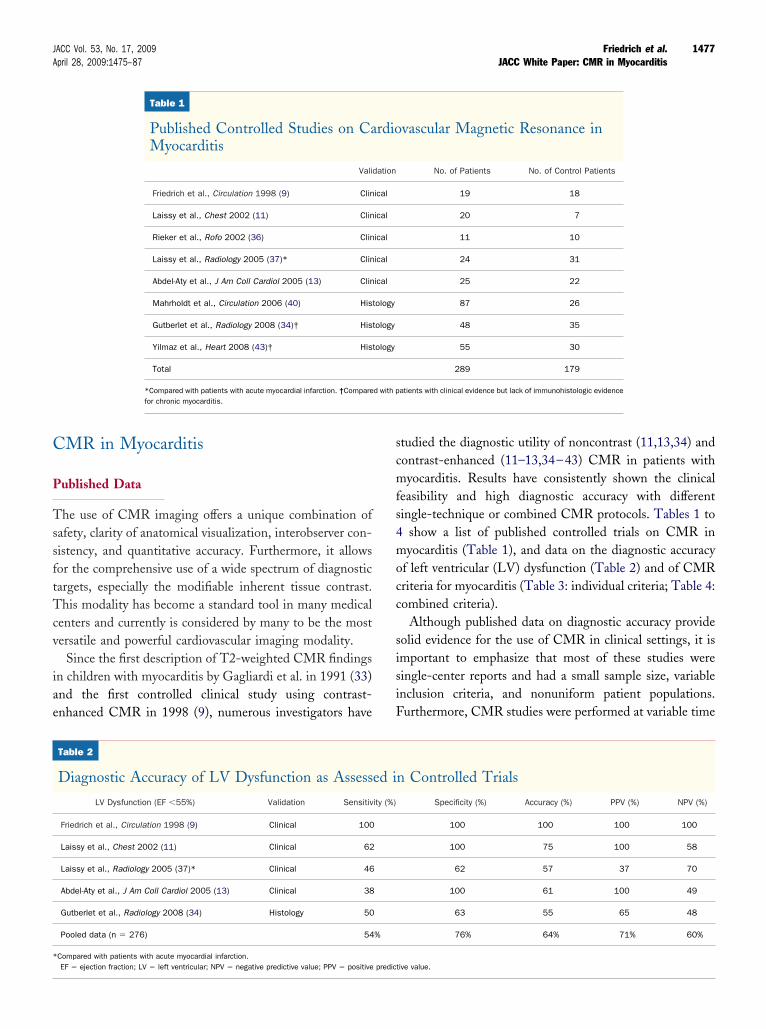
C
P
TssftTcv
iae
scmfs4mocc
sisiF
*
1477JACC Vol. 53, No. 17, 2009 Friedrich et al.April 28, 2009:1475–87 JACC White Paper: CMR in Myocarditis
MR in Myocarditis
ublished Data
he use of CMR imaging offers a unique combination ofafety, clarity of anatomical visualization, interobserver con-istency, and quantitative accuracy. Furthermore, it allowsor the comprehensive use of a wide spectrum of diagnosticargets, especially the modifiable inherent tissue contrast.his modality has become a standard tool in many medical
enters and currently is considered by many to be the mostersatile and powerful cardiovascular imaging modality.
Since the first description of T2-weighted CMR findingsn children with myocarditis by Gagliardi et al. in 1991 (33)nd the first controlled clinical study using contrast-nhanced CMR in 1998 (9), numerous investigators have
Table 1
Published Controlled Studies on CaMyocarditis
Vali
Friedrich et al., Circulation 1998 (9) Clin
Laissy et al., Chest 2002 (11) Clin
Rieker et al., Rofo 2002 (36) Clin
Laissy et al., Radiology 2005 (37)* Clin
Abdel-Aty et al., J Am Coll Cardiol 2005 (13) Clin
Mahrholdt et al., Circulation 2006 (40) His
Gutberlet et al., Radiology 2008 (34)† His
Yilmaz et al., Heart 2008 (43)† His
Total
*Compared with patients with acute myocardial infarction. †Comparefor chronic myocarditis.
Table 2
Diagnostic Accuracy of LV Dysfunction as Assess
LV Dysfunction (EF �55%) Validation Sensitiv
Friedrich et al., Circulation 1998 (9) Clinical 100
Laissy et al., Chest 2002 (11) Clinical 62
Laissy et al., Radiology 2005 (37)* Clinical 46
Abdel-Aty et al., J Am Coll Cardiol 2005 (13) Clinical 38
Gutberlet et al., Radiology 2008 (34) Histology 50
Pooled data (n � 276) 54
Compared with patients with acute myocardial infarction.
EF � ejection fraction; LV � left ventricular; NPV � negative predictive value; PPV � positive predictudied the diagnostic utility of noncontrast (11,13,34) andontrast-enhanced (11–13,34–43) CMR in patients withyocarditis. Results have consistently shown the clinical
easibility and high diagnostic accuracy with differentingle-technique or combined CMR protocols. Tables 1 to
show a list of published controlled trials on CMR inyocarditis (Table 1), and data on the diagnostic accuracy
f left ventricular (LV) dysfunction (Table 2) and of CMRriteria for myocarditis (Table 3: individual criteria; Table 4:ombined criteria).
Although published data on diagnostic accuracy provideolid evidence for the use of CMR in clinical settings, it ismportant to emphasize that most of these studies wereingle-center reports and had a small sample size, variablenclusion criteria, and nonuniform patient populations.urthermore, CMR studies were performed at variable time
vascular Magnetic Resonance in
No. of Patients No. of Control Patients
19 18
20 7
11 10
24 31
25 22
87 26
48 35
55 30
289 179
atients with clinical evidence but lack of immunohistologic evidence
n Controlled Trials
Specificity (%) Accuracy (%) PPV (%) NPV (%)
100 100 100 100
100 75 100 58
62 57 37 70
100 61 100 49
63 55 65 48
76% 64% 71% 60%
rdio
dation
ical
ical
ical
ical
ical
tology
tology
tology
d with p
ed i
ity (%)
%
tive value.
pc
natcdcapb
rdcpan
D
Dntm
Fvafs
hml
PiAp
ns(spdbnossFiaiset
nA
A
1478 Friedrich et al. JACC Vol. 53, No. 17, 2009JACC White Paper: CMR in Myocarditis April 28, 2009:1475–87
oints after disease onset, used different imaging diagnosticriteria, and mostly did not include biopsy for confirmation.
Furthermore, the specificity was mostly compared withormal control patients or those with myocardial infarctionnd not to other heart diseases with similar clinical presen-ation, such as acute coronary syndrome or other secondaryardiomyopathies. Current data do not allow for a clearefinition of the diagnostic accuracy of CMR in variouslinical, histological, and immunohistochemical subgroups,nd data from larger (multicenter) trials with standardizedrotocols comparing comprehensive CMR studies toiopsy-derived criteria are lacking.The prognostic value of CMR criteria for myocarditis
emains to be defined. In a small study, increased myocar-ial early gadolinium enhancement ratio at 4 weeks afterlinical onset of the disease was associated with an impairedrognosis regarding functional recovery and symptoms after3-year follow-up (44). Confirmative studies on the prog-ostic value of the various parameters are required.
iagnostic Targets of CMR in Myocarditis
ifferent from other diagnostic modalities, targets for CMRot only include functional and morphological abnormali-ies but also tissue pathology as diagnostic features ofyocardial inflammation.
unctional abnormalities. The CMR assessment of rightentricular and LV function is very reproducible and thusllows for identifying, quantifying, and following even mildunctional abnormalities, if present. In patients with more
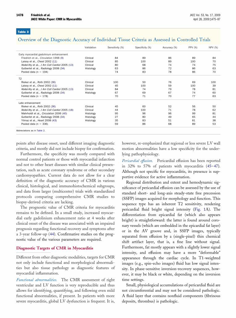
Table 3
Overview of the Diagnostic Accuracy of Individua
Validation Sensit
Early myocardial gadolinium enhancementFriedrich et al., Circulation 1998 (9) ClinicalLaissy et al., Chest 2002 (11) ClinicalAbdel-Aty et al., J Am Coll Cardiol 2005 (13) ClinicalGutberlet et al., Radiology 2008 (34) HistologyPooled data (n � 194)
T2Rieker et al., Rofo 2002 (36) Clinical 1Laissy et al., Chest 2002 (11) ClinicalAbdel-Aty et al., J Am Coll Cardiol 2005 (13) ClinicalGutberlet et al., Radiology 2008 (34) HistologyPooled data (n � 178)
Late enhancementRieker et al., Rofo 2002 (36) ClinicalAbdel-Aty et al., J Am Coll Cardiol 2005 (18) ClinicalMahrholdt et al., Circulation 2006 (40) HistologyGutberlet et al., Radiology 2008 (34) HistologyYilmaz et al., Heart 2008 (43) HistologyPooled data (n � 336)
bbreviations as in Table 2.
evere myocarditis, global LV dysfunction is frequent. It is, d
owever, re-emphasized that regional or less severe LV wallotion abnormalities have a low specificity for the under-
ying pathophysiology.
ericardial effusion. Pericardial effusion has been reportedn 32% to 57% of patients with myocarditis (45–47).lthough not specific for myocarditis, its presence is sup-ortive evidence for active inflammation.Regional distribution and extent and hemodynamic sig-
ificance of pericardial effusion can be assessed by the use oftandard short- and long-axis steady-state free precessionSSFP) images acquired for morphology and function. Thisequence type has an inherent T2 sensitivity, renderingericardial fluid bright signal intensity (Fig. 1A). Theifferentiation from epicardial fat (which also appearsright) is straightforward: the latter is found around coro-ary vessels (which are embedded in the epicardial fat layer)r in the AV groove and, in SSFP images, typicallyeparated from effusion by a (single-pixel) thin chemicalhift artifact layer, that is, a fine line without signal.urthermore, fat mostly appears with a slightly lower signal
ntensity, and effusion may have a more “deformable”ppearance through the cardiac cycle. In T1-weightedmages (e.g., spin-echo images) fluid has low signal inten-ity. In phase-sensitive inversion-recovery sequences, how-ver, it may be black or white, depending on the inversionime settings.
Small, physiological accumulations of pericardial fluid areot circumferential and may not be considered pathologic.
fluid layer that contains nonfluid components (fibrinous
ssue Criteria as Assessed in Controlled Trials
) Specificity (%) Accuracy (%) PPV (%) NPV (%)
89 86 89 84100 89 100 7068 74 74 7586 72 86 6383 78 86 70
50 76 69 100100 59 100 3974 79 78 8169 67 74 6071 70 77 63
60 52 56 50100 71 78 6296 96 99 8180 49 65 4483 51 81 3886 68 89 53
l Ti
ivity (%
8485806374
0045846770
454495273559
eposits, thrombus) is pathologic.
Mtddowtcr
T
Gcochs
EiWlmtp
wgesse
pitidoni
wfcrirsi
dwmeccp
Hetta
L
1479JACC Vol. 53, No. 17, 2009 Friedrich et al.April 28, 2009:1475–87 JACC White Paper: CMR in Myocarditis
orphological abnormalities. A transient increase of wallhickness during myocarditis was first described in echocar-iography studies (48) and may serve as a supportive findinguring follow-up. A decrease of LV mass during the coursef uncomplicated myocarditis was found to be associatedith edema as assessed by T2-weighted CMR (49). A
ransient increase of LV volumes has been observed in theourse of myocarditis (9) and may also may serve asetrospective, supportive evidence for recent myocarditis.
issue Characterization With CMR
iven the unique potential of CMR to visualize tissuehanges, this area is of special interest. As outlined previ-usly, expected tissue pathology in active myocarditis in-ludes intracellular and interstitial edema, capillary leakage,yperemia, and, in more severe cases, cellular necrosis andubsequent fibrosis (50).
dema. An important hallmark of inflammatory cell injurys the increased permeability of cellular membranes.
hereas initial membrane defects are of a functional nature,eading to Na� influx and subsequent intracellular edema, a
ore severe injury allows for a net efflux of water andransmembranous leakage of larger molecules such as tro-onin, eventually leading to loss of cellular functions.T2-weighted imaging sensitively detects tissue edema
ith the long T2 of water-bound protons as the contrast-enerating mechanism, resulting in a high signal intensity ofdematous tissue (Fig. 1C). Triple inversion recovery turbopin echo sequences with inversion pulses for fat and blooduppression (51) provide excellent contrast between regional
Table 4
Overview of the Diagnostic Accuracy of Several C
Validation Sensit
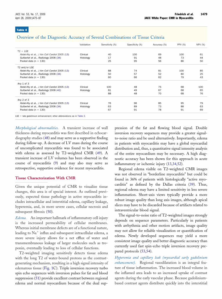
T2 � LGEAbdel-Aty et al., J Am Coll Cardiol 2005 (13) ClinicalGutberlet et al., Radiology 2008 (34) HistologyPooled data (n � 130)
T2 and/or LGEAbdel-Aty et al., J Am Coll Cardiol 2005 (13) ClinicalGutberlet et al., Radiology 2008 (34) HistologyPooled data (n � 130)
Any 1 of 3Abdel-Aty et al., J Am Coll Cardiol 2005 (13) Clinical 1Gutberlet et al., Radiology 2008 (42) HistologyPooled data (n � 130)
Any 2 of 3Abdel-Aty et al., J Am Coll Cardiol 2005 (13) ClinicalGutberlet et al., Radiology 2008 (34) HistologyPooled data (n � 130)
GE � late gadolinium enhancement; other abbreviations as in Table 2.
dema and normal myocardium because of the dual sup- b
ression of the fat and flowing blood signal. Doublenversion recovery sequences may provide a greater signal-o-noise ratio and be used alternatively. Importantly, edeman patients with myocarditis may have a global myocardialistribution and, thus, a quantitative signal intensity analysisf the entire myocardium may be necessary. A high diag-ostic accuracy has been shown for this approach in acute
nflammatory or ischemic injury (13,34,52).Regional edema visible on T2-weighted CMR images
as not observed in “borderline myocarditis” but could beound in 36% of patients with histologically “active myo-arditis” as defined by the Dallas criteria (39). Thus,egional edema may have a limited sensitivity in less severenflammation. Short-axis views typically provide a moreobust image quality than long axis images, although apicallices may have to be discarded because of artifacts related tontraventricular blood signal.
The signal-to-noise ratio of T2-weighted images stronglyepends on sequence parameters. Particularly in patientsith arrhythmia and other motion artifacts, image qualityay not allow for reliable visualization or quantification of
dema. Newly developed sequences may yield a moreonsistent image quality and better diagnostic accuracy thanurrently used fast spin-echo triple inversion recovery pre-ared protocols (53,54).
yperemia and capillary leak (myocardial early gadoliniumnhancement). Regional vasodilatation is an integral fea-ure of tissue inflammation. The increased blood volume inhe inflamed area leads to an increased uptake of contrastgents during the early vascular phase. Because gadolinium-
inations of Tissue Criteria
) Specificity (%) Accuracy (%) PPV (%) NPV (%)
100 69 100 6191 48 73 4495 56 86 50
74 81 100 8557 52 80 2566 62 79 43
48 75 68 10049 67 68 6548 70 68 76
96 85 95 7989 73 88 6391 78 91 69
omb
ivity (%
401725
885060
008188
766367
ased contrast agents distribute quickly into the interstitial
smsaap
(id
wc(
vvsm
N
dunc
tvvfl
(sSfiileasnsseh
(ReD
1480 Friedrich et al. JACC Vol. 53, No. 17, 2009JACC White Paper: CMR in Myocarditis April 28, 2009:1475–87
pace after administration, this phase lasts for the firstinutes after the contrast bolus. Contrast-enhanced fast
pin-echo T1-weighted MR during this time can be used tossess experimentally induced myocardial hyperemia (55)nd to detect muscular inflammation (56). Accordingly, the
Figure 1
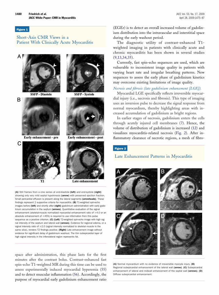
Short-Axis CMR Views in aPatient With Clinically Acute Myocarditis
A) Still frames from a cine series at end-diastole (left) and end-systole (right)howing only very mild septal hypokinesis (arrow) with preserved ejection fraction.mall pericardial effusion is present along the lateral segments (arrowheads). Thesendings represent 2 supportive criteria for myocarditis. (B) T1-weighted spin-echomages before (left) and shortly after (right) gadolinium administration with early gado-inium accumulation in the septum (arrows). Quantitative evaluation of the signalnhancement (skeletal-muscle normalized myocardial enhancement ratio of �4.0 or anbsolute enhancement of �45%) is required to use information from this pulseequence as a positive criterion. (C) (Left) T2-weighted spin-echo image with high sig-al intensity of the septum and lateral wall (arrows). Evidence for regional edema, or aignal intensity ratio of �2.0 (signal intensity normalized to skeletal muscle in theame slice), renders T2 findings positive. (Right) Late enhancement image withoutvidence for significant delay of gadolinium washout. The thin subepicardial layer ofigh signal intensity in the inferolateral region represents fat.
urpose of myocardial early gadolinium enhancement ratio
EGEr) is to detect an overall increased volume of gadolin-um distribution into the intravascular and interstitial spaceuring the early washout period.The diagnostic utility of contrast-enhanced T1-
eighted imaging in patients with clinically acute andhronic myocarditis has been shown in several studies9,13,34,35).
Currently, fast spin-echo sequences are used, which areulnerable to inconsistent image quality in patients witharying heart rate and irregular breathing patterns. Newequences to assess the early phase of gadolinium kineticsay overcome existing limitations of image quality.
ecrosis and fibrosis (late gadolinium enhancement [LGE]).Myocardial LGE specifically reflects irreversible myocar-
ial injury (i.e., necrosis and fibrosis). This type of imagingses an inversion pulse to decrease the signal response fromormal myocardium, thereby highlighting areas with in-reased accumulation of gadolinium as bright regions.
In earlier stages of necrosis, gadolinium enters the cellshrough acutely injured cell membranes (7). Hence, theolume of distribution of gadolinium is increased (12) andisualizes myocarditis-related necrosis (Fig. 2). After in-ammatory clearance of necrotic regions, a mesh of fibro-
Figure 2
Late Enhancement Patterns in Myocarditis
A) Normal myocardium with no evidence of irreversible myocyte injury. (B)egional subepicardial enhancement of the lateral wall (arrow). (C) Subepicardialnhancement of lateral and midwall enhancement of the septal wall (arrows). (D)iffuse subepicardial enhancement.
cvgwt
hfcdL(d(ie
toDsm
atinsdna
Ccccaatdt2m
Ia
I
As
mmcmpam
b(utdC
T
Titeidi
dat(ciwp
L(a
dgae
T
Rrtarp
1481JACC Vol. 53, No. 17, 2009 Friedrich et al.April 28, 2009:1475–87 JACC White Paper: CMR in Myocarditis
ytes with a large interstitial component replaces formerlyiable tissue, again increasing the volume of distribution foradolinium into this extracellular space during the lateashout period. Thus, the late sequelae of inflammatory
issue damage also can be observed by LGE.Microscopic (57), animal (58), and clinical (59) studies
ave confirmed the role of LGE imaging as s gold standardor in vivo detection of irreversible myocardial injury asso-iated with myocardial infarction. In patients with myocar-itis, several studies have demonstrated a high specificity ofGE for the detection of such injury in myocarditis
12,13,37,38,40). The regional distribution of injury asefined by LGE not only allows differentiating ischemicwith mandatory subendocardial involvement) from non-schemic injury (60), but also may indicate the underlyingtiology of the nonischemic insult (61).
As a potential limitation, LGE showed a variable sensi-ivity to detect active or chronic inflammation, dependingn the selection of patients (12,13,34,39,40,43). Using theallas criteria, De Cobelli et al. (39) found LGE to be less
ensitive in “borderline” myocarditis (44%) than in “active”yocarditis (84%).One reason may be that active myocarditis may not
lways lead to large-enough regions of necrotic myocyteso be visually detectable, given the pixel size in CMRmages. This contrasts with the situation in ischemicecrosis for which LGE has been shown to be highlyensitive. Therefore, LGE may be insensitive for theetection of symptomatic myocarditis with limited oronfocal irreversible injury. More studies are needed toddress this issue.
ombined use of tissue pathology markers. Two studies haveompared all 3 tissue-based markers as well as variousombinations of these. Abdel-Aty et al. (13) used combinedlinical criteria for active myocarditis, whereas Gutberlet etl. (34) assessed patients with chronic myocarditis, validatedgainst histopathological criteria of myocardial inflamma-ion. In both studies, the approach with the best overalliagnostic accuracy was found by the combined use of all 3issue-based CMR parameters, with the presence of at least
positive criteria defining the CMR study as positive foryocarditis (Tables 3 and 4).
ndications, Procedure,nd Protocol of CMR
ndications for CMR
CMR study should only be performed if patients are
ymptomatic, if there is sufficient clinical evidence for Syocarditis, and if the CMR result will likely affect clinicalanagement. Thus, it is generally indicated in patients with
urrent or persisting symptoms, evidence for significantyocardial injury, and suspected viral etiology. CMR is of
otential use in patients with chest pain, elevated troponin,nd normal coronary arteries, where it was shown to identifyyocarditis in more than 30% of patients (62).Additional indications may exist for subjects with possi-
le myocarditis being exposed to strenuous physical exercisee.g., professional athletes) or for patients with otherwisenexplained new ECG findings consistent with myocardi-is, even in the absence of symptoms suggestive of myocar-itis. Table 5 lists recommended criteria for requesting aMR study in patients with suspected myocarditis.
he CMR Procedure
he patient should be monitored throughout the session,ncluding ECG, blood pressure, breathing, and O2 satura-ion. Furthermore, communication to the patient should bensured by the use of intercom devices. A physician trainedn cardiac resuscitation should be available. As for all cardiaciagnostic modalities, drugs and equipment for immediatenterventions should be within reach.
Typically, patients are examined in a supine position. Aedicated cardiac phased-array surface coil should be used tocquire functional images. It is very important to emphasizehat for all sequences used to analyze signal intensityqualitatively or quantitatively), either a signal intensityorrection algorithm or the body coil should be used. Thenhomogeneous sensitivity field of surface coils may other-ise lead to false negative (inferolateral wall) or falseositive (septum) results.The coverage of the heart should allow for assessing all 17
V segments according to published recommendations63). Images of the apex may be of insufficient image qualitynd may have to be excluded.
Published data on contrast-enhanced CMR in myocar-itis mostly have been obtained with the use of gadoliniumadopentetate dimeglumine and, thus, recommendationsre only valid for this substance or compounds with anquivalent pharmacokinetic profile.
he CMR Protocol
ecommended imaging parameters and detailed protocolecommendations are provided in the Online Appendix ofhis article. CMR sequences generally will be ECG-gatednd performed by the use of breath-hold protocols. Theseecommendations are based on the current evidence asublished in peer-reviewed literature as of January 2009.
ome of the currently recommended sequences have distinct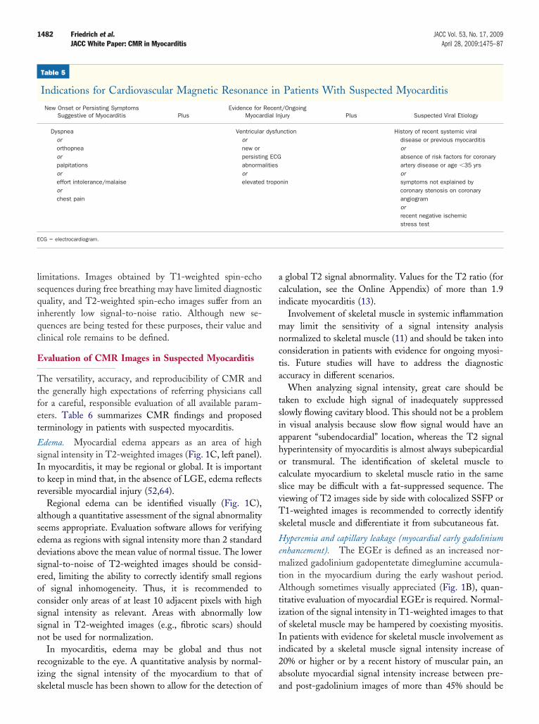
lsqiqc
E
Ttfet
EsItr
asedseocssn
ris
aci
mncta
tsiahocsvTs
HemtAtioIi2a
E
1482 Friedrich et al. JACC Vol. 53, No. 17, 2009JACC White Paper: CMR in Myocarditis April 28, 2009:1475–87
imitations. Images obtained by T1-weighted spin-echoequences during free breathing may have limited diagnosticuality, and T2-weighted spin-echo images suffer from annherently low signal-to-noise ratio. Although new se-uences are being tested for these purposes, their value andlinical role remains to be defined.
valuation of CMR Images in Suspected Myocarditis
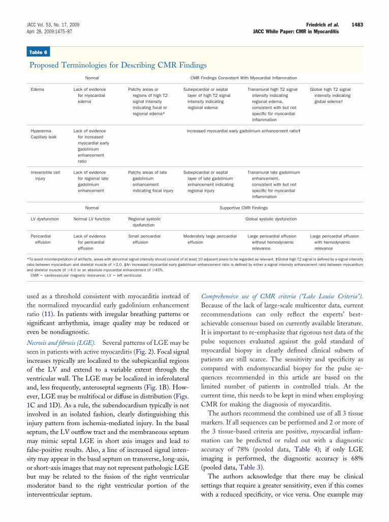
he versatility, accuracy, and reproducibility of CMR andhe generally high expectations of referring physicians callor a careful, responsible evaluation of all available param-ters. Table 6 summarizes CMR findings and proposederminology in patients with suspected myocarditis.
dema. Myocardial edema appears as an area of highignal intensity in T2-weighted images (Fig. 1C, left panel).n myocarditis, it may be regional or global. It is importanto keep in mind that, in the absence of LGE, edema reflectseversible myocardial injury (52,64).
Regional edema can be identified visually (Fig. 1C),lthough a quantitative assessment of the signal abnormalityeems appropriate. Evaluation software allows for verifyingdema as regions with signal intensity more than 2 standardeviations above the mean value of normal tissue. The lowerignal-to-noise of T2-weighted images should be consid-red, limiting the ability to correctly identify small regionsf signal inhomogeneity. Thus, it is recommended toonsider only areas of at least 10 adjacent pixels with highignal intensity as relevant. Areas with abnormally lowignal in T2-weighted images (e.g., fibrotic scars) shouldot be used for normalization.In myocarditis, edema may be global and thus not
ecognizable to the eye. A quantitative analysis by normal-zing the signal intensity of the myocardium to that of
Table 5
Indications for Cardiovascular Magnetic Resonanc
New Onset or Persisting SymptomsSuggestive of Myocarditis Plus
Evidence forMyoca
Dyspneaororthopneaorpalpitationsoreffort intolerance/malaiseorchest pain
Ventriculaornew orpersistinabnormaorelevated
CG � electrocardiogram.
keletal muscle has been shown to allow for the detection of a
global T2 signal abnormality. Values for the T2 ratio (foralculation, see the Online Appendix) of more than 1.9ndicate myocarditis (13).
Involvement of skeletal muscle in systemic inflammationay limit the sensitivity of a signal intensity analysis
ormalized to skeletal muscle (11) and should be taken intoonsideration in patients with evidence for ongoing myosi-is. Future studies will have to address the diagnosticccuracy in different scenarios.
When analyzing signal intensity, great care should beaken to exclude high signal of inadequately suppressedlowly flowing cavitary blood. This should not be a problemn visual analysis because slow flow signal would have anpparent “subendocardial” location, whereas the T2 signalyperintensity of myocarditis is almost always subepicardialr transmural. The identification of skeletal muscle toalculate myocardium to skeletal muscle ratio in the samelice may be difficult with a fat-suppressed sequence. Theiewing of T2 images side by side with colocalized SSFP or1-weighted images is recommended to correctly identify
keletal muscle and differentiate it from subcutaneous fat.
yperemia and capillary leakage (myocardial early gadoliniumnhancement). The EGEr is defined as an increased nor-alized gadolinium gadopentetate dimeglumine accumula-
ion in the myocardium during the early washout period.lthough sometimes visually appreciated (Fig. 1B), quan-
itative evaluation of myocardial EGEr is required. Normal-zation of the signal intensity in T1-weighted images to thatf skeletal muscle may be hampered by coexisting myositis.n patients with evidence for skeletal muscle involvement asndicated by a skeletal muscle signal intensity increase of0% or higher or by a recent history of muscular pain, anbsolute myocardial signal intensity increase between pre-
Patients With Suspected Myocarditis
t/Ongoingjury Plus Suspected Viral Etiology
nction
nin
History of recent systemic viraldisease or previous myocarditisorabsence of risk factors for coronaryartery disease or age �35 yrsorsymptoms not explained bycoronary stenosis on coronaryangiogramorrecent negative ischemicstress test
e in
Recenrdial In
r dysfu
g ECGlities
tropo
nd post-gadolinium images of more than 45% should be
utrse
Nsiovae1iismfsobmi
CBraIpmpcqlcC
mtmai(
s
*ra
1483JACC Vol. 53, No. 17, 2009 Friedrich et al.April 28, 2009:1475–87 JACC White Paper: CMR in Myocarditis
sed as a threshold consistent with myocarditis instead ofhe normalized myocardial early gadolinium enhancementatio (11). In patients with irregular breathing patterns orignificant arrhythmia, image quality may be reduced orven be nondiagnostic.
ecrosis and fibrosis (LGE). Several patterns of LGE may beeen in patients with active myocarditis (Fig. 2). Focal signalncreases typically are localized to the subepicardial regionsf the LV and extend to a variable extent through theentricular wall. The LGE may be localized in inferolateralnd, less frequently, anteroseptal segments (Fig. 1B). How-ver, LGE may be multifocal or diffuse in distribution (Figs.C and 1D). As a rule, the subendocardium typically is notnvolved in an isolated fashion, clearly distinguishing thisnjury pattern from ischemia-mediated injury. In the basaleptum, the LV outflow tract and the membraneous septumay mimic septal LGE in short axis images and lead to
alse-positive results. Also, a line of increased signal inten-ity may appear in the basal septum on transverse, long-axis,r short-axis images that may not represent pathologic LGEut may be related to the fusion of the right ventricularoderator band to the right ventricular portion of the
Table 6
Proposed Terminologies for Describing CMR Fin
Normal
Edema Lack of evidencefor myocardialedema
Patchy areas orregions of high T2signal intensityindicating focal orregional edema*
Sublainre
HyperemiaCapillary leak
Lack of evidencefor increasedmyocardial earlygadoliniumenhancementratio
In
Irreversible cellinjury
Lack of evidencefor regional lategadoliniumenhancement
Patchy areas of lategadoliniumenhancementindicating focal injury
Sublaere
Normal
LV dysfunction Normal LV function Regional systolicdysfunction
Pericardialeffusion
Lack of evidencefor pericardialeffusion
Small pericardialeffusion
Moe
To avoid misinterpretation of artifacts, areas with abnormal signal intensity should consist of at latio between myocardium and skeletal muscle of �2.0. ‡An increased myocardial early gadolinnd skeletal muscle of �4.0 or an absolute myocardial enhancement of �45%.CMR � cardiovascular magnetic resonance; LV � left ventricular.
nterventricular septum. w
omprehensive use of CMR criteria (“Lake Louise Criteria”).ecause of the lack of large-scale multicenter data, current
ecommendations can only reflect the experts’ best-chievable consensus based on currently available literature.t is important to re-emphasize that rigorous test data of theulse sequences evaluated against the gold standard ofyocardial biopsy in clearly defined clinical subsets of
atients are still scarce. The sensitivity and specificity asompared with endomyocardial biopsy for the pulse se-uences recommended in this article are based on theimited number of patients in controlled trials. At theurrent time, this needs to be kept in mind when employingMR for making the diagnosis of myocarditis.The authors recommend the combined use of all 3 tissuearkers. If all sequences can be performed and 2 or more of
he 3 tissue-based criteria are positive, myocardial inflam-ation can be predicted or ruled out with a diagnostic
ccuracy of 78% (pooled data, Table 4); if only LGEmaging is performed, the diagnostic accuracy is 68%pooled data, Table 3).
The authors acknowledge that there may be clinicalettings that require a greater sensitivity, even if this comes
s
indings Consistent With Myocardial Inflammation
dial or septalhigh T2 signal
y indicatingl edema
Transmural high T2 signalintensity indicatingregional edema,consistent with but notspecific for myocardialinflammation
Global high T2 signalintensity indicatingglobal edema†
d myocardial early gadolinium enhancement ratio‡
dial or septallate gadolinium
ement indicatingl injury
Transmural late gadoliniumenhancement,consistent with but notspecific for myocardialinflammation
Supportive CMR Findings
Global systolic dysfunction
ly large pericardial Large pericardial effusionwithout hemodynamicrelevance
Large pericardial effusionwith hemodynamicrelevance
adjacent pixels to be regarded as relevant. †Global high T2 signal is defined by a signal intensityhancement ratio is defined by either a signal intensity enhancement ratio between myocardium
ding
CMR F
epicaryer oftensitgiona
crease
epicaryer ofnhancgiona
derateffusion
east 10ium en
ith a reduced specificity, or vice versa. One example may
bpcrcwtC
F
Tmemat
pnbwutputIpa
R
Tq
*ccbmmi
A
1484 Friedrich et al. JACC Vol. 53, No. 17, 2009JACC White Paper: CMR in Myocarditis April 28, 2009:1475–87
e the use of CMR to assess patients with a high pre-testrobability or children with suspected inflammation afterardiac transplantation. It is re-emphasized that both refer-ing physicians and CMR readers should use the reportedriteria as part of a comprehensive diagnostic approach,hich also includes clinical, functional, and other informa-
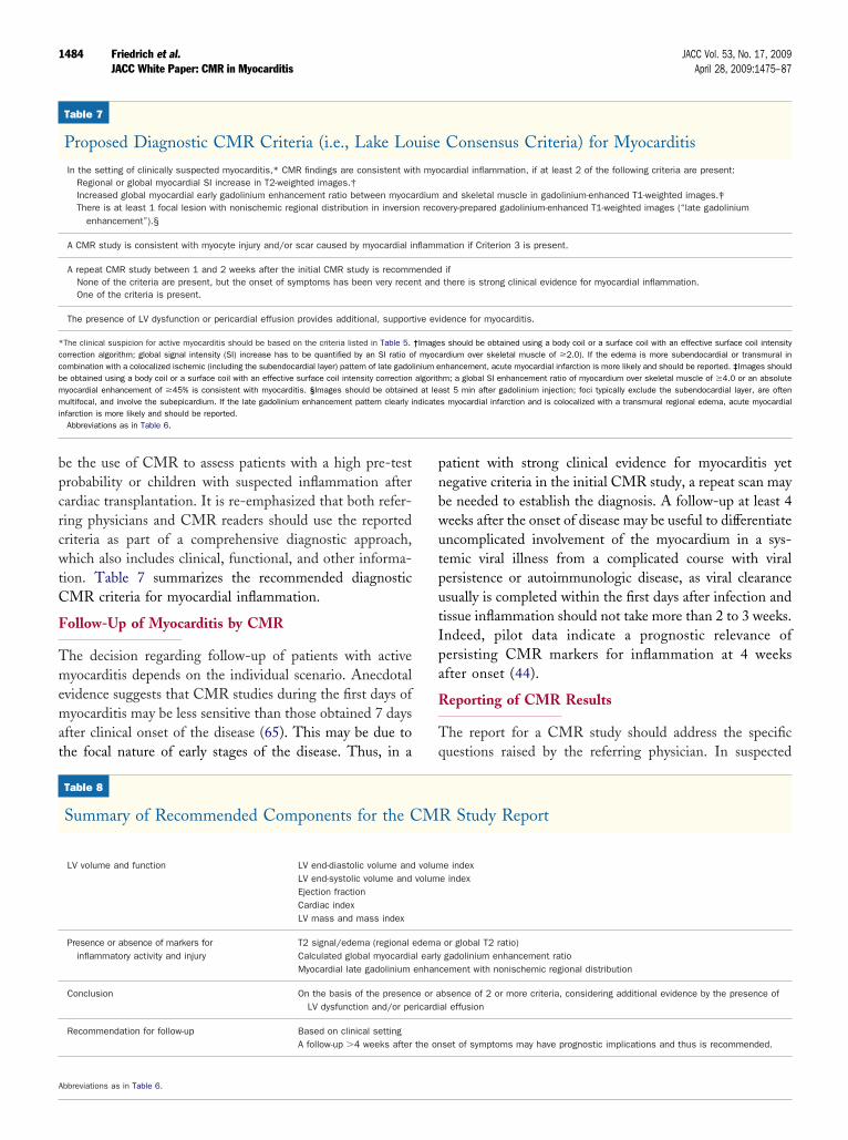
ion. Table 7 summarizes the recommended diagnosticMR criteria for myocardial inflammation.
ollow-Up of Myocarditis by CMR
he decision regarding follow-up of patients with activeyocarditis depends on the individual scenario. Anecdotal
vidence suggests that CMR studies during the first days ofyocarditis may be less sensitive than those obtained 7 days
fter clinical onset of the disease (65). This may be due tohe focal nature of early stages of the disease. Thus, in a
Table 7
Proposed Diagnostic CMR Criteria (i.e., Lake Lo
In the setting of clinically suspected myocarditis,* CMR findings are consistent wiRegional or global myocardial SI increase in T2-weighted images.†Increased global myocardial early gadolinium enhancement ratio between myocaThere is at least 1 focal lesion with nonischemic regional distribution in inversio
enhancement”).§
A CMR study is consistent with myocyte injury and/or scar caused by myocardial i
A repeat CMR study between 1 and 2 weeks after the initial CMR study is recommNone of the criteria are present, but the onset of symptoms has been very receOne of the criteria is present.
The presence of LV dysfunction or pericardial effusion provides additional, support
The clinical suspicion for active myocarditis should be based on the criteria listed in Table 5.orrection algorithm; global signal intensity (SI) increase has to be quantified by an SI ratio oombination with a colocalized ischemic (including the subendocardial layer) pattern of late gadole obtained using a body coil or a surface coil with an effective surface coil intensity correctionyocardial enhancement of �45% is consistent with myocarditis. §Images should be obtaineultifocal, and involve the subepicardium. If the late gadolinium enhancement pattern clearly i
nfarction is more likely and should be reported.Abbreviations as in Table 6.
Table 8
Summary of Recommended Components for the C
LV volume and function LV end-diastolic volume andLV end-systolic volume andEjection fractionCardiac indexLV mass and mass index
Presence or absence of markers forinflammatory activity and injury
T2 signal/edema (regional eCalculated global myocardiaMyocardial late gadolinium
Conclusion On the basis of the presencLV dysfunction and/or pe
Recommendation for follow-up Based on clinical settingA follow-up �4 weeks after
bbreviations as in Table 6.
atient with strong clinical evidence for myocarditis yetegative criteria in the initial CMR study, a repeat scan maye needed to establish the diagnosis. A follow-up at least 4eeks after the onset of disease may be useful to differentiatencomplicated involvement of the myocardium in a sys-emic viral illness from a complicated course with viralersistence or autoimmunologic disease, as viral clearancesually is completed within the first days after infection andissue inflammation should not take more than 2 to 3 weeks.ndeed, pilot data indicate a prognostic relevance ofersisting CMR markers for inflammation at 4 weeksfter onset (44).
eporting of CMR Results
he report for a CMR study should address the specificuestions raised by the referring physician. In suspected
Consensus Criteria) for Myocarditis
cardial inflammation, if at least 2 of the following criteria are present:
and skeletal muscle in gadolinium-enhanced T1-weighted images.‡very-prepared gadolinium-enhanced T1-weighted images (“late gadolinium
ation if Criterion 3 is present.
ifthere is strong clinical evidence for myocardial inflammation.
idence for myocarditis.
s should be obtained using a body coil or a surface coil with an effective surface coil intensityrdium over skeletal muscle of �2.0). If the edema is more subendocardial or transmural innhancement, acute myocardial infarction is more likely and should be reported. ‡Images shouldm; a global SI enhancement ratio of myocardium over skeletal muscle of �4.0 or an absolutest 5 min after gadolinium injection; foci typically exclude the subendocardial layer, are oftens myocardial infarction and is colocalized with a transmural regional edema, acute myocardial
R Study Report
e indexe index
or global T2 ratio)gadolinium enhancement ratio
cement with nonischemic regional distribution
bsence of 2 or more criteria, considering additional evidence by the presence ofal effusion
set of symptoms may have prognostic implications and thus is recommended.
uise
th myo
rdiumn reco
nflamm
endednt and
ive ev
†Imagef myocainium ealgorithd at leandicate
M
volumvolum
demal earlyenhan
e or aricardi
the on
mac
oTTpd
bcTi
rott
F
Tntcdtt
1485JACC Vol. 53, No. 17, 2009 Friedrich et al.April 28, 2009:1475–87 JACC White Paper: CMR in Myocarditis
yocarditis, this will usually include the inflammatoryctivity, LV function, and other information such as peri-ardial effusion, cardiac index, and extent of scarring.
There was consensus that for the time being the presencer absence of the 3 criteria, if acquired, should be reported.he report summary should include components as listed inable 8. The report should relate quantitative values toublished reference values. References may be cited aseemed appropriate.It is important to be aware that CMR, like myocardial
iopsy, depicts the patient’s status at one point in time andannot characterize acute, chronic, or relapsing forms.hese attributes are based on the clinical course rather than
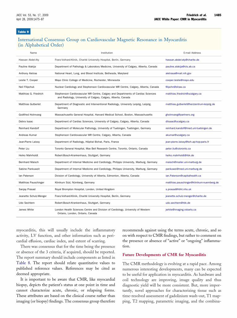
Table 9
International Consensus Group on Cardiovascular(in Alphabetical Order)
Name Institution
Hassan Abdel-Aty Franz-Volhard-Klinik, Charité University Hospital, Ber
Pauline Alakija Department of Pathology & Laboratory Medicine, Un
Anthony Aletras National Heart, Lung, and Blood Institute, Bethesda
Leslie T. Cooper Mayo Clinic College of Medicine, Rochester, Minnes
Neil Filipchuk Nuclear Cardiology and Stephenson Cardiovascular
Matthias G. Friedrich Stephenson Cardiovascular MR Centre, Calgary andand Radiology, University of Calgary, Calgary, Albe
Matthias Gutberlet Department of Diagnostic and Interventional RadioloGermany
Godtfred Holmvang Massachusetts General Hospital, Harvard Medical S
Debra Isaac Department of Cardiac Sciences, University of Calga
Reinhard Kandolf Department of Molecular Pathology, University of Tu
Andreas Kumar Stephenson Cardiovascular MR Centre, Calgary, Alb
Jean-Pierre Laissy Department of Radiology, Hôpital Bichat, Paris, Fran
Peter Liu Toronto General Hospital, Max Bell Research Centre
Heiko Mahrholdt Robert-Bosch-Krankenhaus, Stuttgart, Germany
Bernhard Maisch Department of Internal Medicine and Cardiology, Ph
Sabine Pankuweit Department of Internal Medicine and Cardiology, Ph
Ian Paterson Division of Cardiology, University of Alberta, Edmont
Matthias Pauschinger Klinikum Süd, Nürnberg, Germany
Sanjay Prasad Royal Brompton Hospital, London, United Kingdom
Jeanette Schulz-Menger Franz-Volhard-Klinik, Charité University Hospital, Ber
Udo Sechtem Robert-Bosch-Krankenhaus, Stuttgart, Germany
James White London Health Sciences Centre and Division of CarOntario, London, Ontario, Canada
maging (or biopsy) findings. The consensus group therefore p
ecommends against using the terms acute, chronic, and son with respect to CMR findings, but rather to comment onhe presence or absence of “active” or “ongoing” inflamma-ion.
uture Developments of CMR for Myocarditis
he CMR methodology is evolving at a rapid pace. Amongumerous interesting developments, many can be expectedo be useful for application in myocarditis. As hardware andoil technology are improving, image quality and thusiagnostic yield will be more consistent. But, more impor-antly, novel approaches for characterizing tissue such asime-resolved assessment of gadolinium wash-out, T1 map-
gnetic Resonance in Myocarditis
E-mail Address
rmany [email protected]
y of Calgary, Alberta, Canada [email protected]
land [email protected]
ntre, Calgary, Alberta, Canada [email protected]
rtments of Cardiac Sciencesanada
iversity Leipzig, Leipzig, [email protected]
, Boston, Massachusetts [email protected]
lgary, Alberta, Canada [email protected]
n, Tuebingen, Germany [email protected]
anada [email protected]
nto, Ontario, Canada [email protected]
University, Marburg, Germany [email protected]
University, Marburg, Germany [email protected]
erta, Canada [email protected]
rmany [email protected]
, University of Western [email protected]
Ma
lin, Ge
iversit
, Mary
ota
MR Ce
Departa, C
gy, Un
chool
ry, Ca
ebinge
erta, C
ce
, Toro
ilipps
ilipps
on, Alb
lin, Ge
diology
ing, T2 mapping, parametric imaging, and the combina-
ti
CTpwaitw
hdaFdCathbd
ATdb
ah
RFCeCu
R
1
1
1
1
1
1
1
1
1
1
2
2
2
2
2
2
2
2
2
2
1486 Friedrich et al. JACC Vol. 53, No. 17, 2009JACC White Paper: CMR in Myocarditis April 28, 2009:1475–87
ion of imaging criteria with seromarkers will likely furtherncrease the utility of CMR.
onclusionshis work provides recommendations on the use of CMR asart of a comprehensive diagnostic approach in patientsith suspected myocardial inflammation. The use of CMR
ppears suitable to identify patients with significant ongoingnflammation, which may be especially important for pa-ients with recurrent or persisting symptoms and in patientsith new onset heart failure.On the basis of published data, we propose a compre-
ensive CMR protocol to determine the extent and regionalistribution of reversible and irreversible myocardial injury,s well as to detect functional and other abnormalities.urthermore, we suggest consensus criteria providing evi-ence for or against myocardial inflammation based onMR findings. We are aware that these recommendations
re based on limited data and that not all centers will be ableo apply all components of the suggested protocol. Newardware, software, and contrast agent techniques mayecome available to further improve diagnostic and proce-ural efficiency of CMR in myocarditis.
cknowledgmentshe entire Consensus Group actively participated in theiscussion that resulted in the recommendations; the mem-ers are listed in Table 9.The authors thank Drs. Debra Isaac, Sabine Pankuweit,
nd Oliver Strohm for their careful review of the paper andelpful suggestions.
eprint requests and correspondence: Dr. Matthias G.riedrich, Stephenson Cardiovascular MR Centre at the Libinardiovascular Institute of Alberta, Department of Cardiac Sci-
nces and Radiology, University of Calgary, 1403 29th Street NW,algary, Alberta T2N 2T9, Canada. E-mail: [email protected].
EFERENCES
1. Fabre A, Sheppard MN. Sudden adult death syndrome and othernon-ischaemic causes of sudden cardiac death. Heart 2006;92:316–20.
2. Doolan A, Langlois N, Semsarian C. Causes of sudden cardiac deathin young Australians. Med J Aust 2004;180:110–2.
3. Puranik R, Chow CK, Duflou JA, Kilborn MJ, McGuire MA. Suddendeath in the young. Heart Rhythm 2005;2:1277–82.
4. Virmani R, Burke AP, Farb A. Sudden cardiac death. CardiovascPathol 2001;10:211–8.
5. Kawai C. From myocarditis to cardiomyopathy: mechanisms ofinflammation and cell death: learning from the past for the future.Circulation 1999;99:1091–100.
6. Calabrese F, Basso C, Carturan E, Valente M, Thiene G. Arrhyth-mogenic right ventricular cardiomyopathy/dysplasia: is there a role forviruses? Cardiovasc Pathol 2006;15:11–7.
7. Liu PP, Mason JW. Advances in the understanding of myocarditis.Circulation 2001;104:1076–82.
8. Pinamonti B, Alberti E, Cigalotto A, et al. Echocardiographicfindings in myocarditis. Am J Cardiol 1988;62:285–91.
9. Friedrich MG, Strohm O, SchulzMenger J, Marciniak H, Luft FC,Dietz R. Contrast media-enhanced magnetic resonance imagingvisualizes myocardial changes in the course of viral myocarditis.Circulation 1998;97:1802–9.
0. Maisch B, Portig I, Ristic A, Hufnagel G, Pankuweit S. Definition ofinflammatory cardiomyopathy (myocarditis): on the way to consensus.A status report. Herz 2000;25:200–9.
1. Laissy JP, Messin B, Varenne O, et al. MRI of acute myocarditis: acomprehensive approach based on various imaging sequences. Chest2002;122:1638–48.
2. Mahrholdt H, Goedecke C, Wagner A, et al. Cardiovascular magneticresonance assessment of human myocarditis: a comparison to histologyand molecular pathology. Circulation 2004;109:1250–8.
3. Abdel-Aty H, Boye P, Zagrosek A, et al. Diagnostic performance ofcardiovascular magnetic resonance in patients with suspected acutemyocarditis: comparison of different approaches. J Am Coll Cardiol2005;45:1815–22.
4. Caforio AL, Calabrese F, Angelini A, et al. A prospective study ofbiopsy-proven myocarditis: prognostic relevance of clinical and aetio-pathogenetic features at diagnosis. Eur Heart J 2007;28:1326–33.
5. Morgera T, Di Lenarda A, Dreas L, et al. Electrocardiography ofmyocarditis revisited: clinical and prognostic significance of electro-cardiographic changes. Am Heart J 1992;124:455–67.
6. Lauer B, Niederau C, Kuhl U, et al. Cardiac troponin T in patientswith clinically suspected myocarditis. J Am Coll Cardiol 1997;30:1354–9.
7. Cooper LT, Baughman KL, Feldman AM, et al. The role ofendomyocardial biopsy in the management of cardiovascular disease: ascientific statement from the American Heart Association, the Amer-ican College of Cardiology, and the European Society of Cardiology.Endorsed by the Heart Failure Society of America and the HeartFailure Association of the European Society of Cardiology. J Am CollCardiol 2007;50:1914–31.
8. Chow LH, Radio SJ, Sears TD, McManus BM. Insensitivity of rightventricular endomyocardial biopsy in the diagnosis of myocarditis.J Am Coll Cardiol 1989;14:915–20.
9. Hauck AJ, Kearney DL, Edwards WD. Evaluation of postmortemendomyocardial biopsy specimens from 38 patients with lymphocyticmyocarditis: implications for role of sampling error. Mayo Clin Proc1989;64:1235–45.
0. Shirani J, Freant LJ, Roberts WC. Gross and semiquantitativehistologic findings in mononuclear cell myocarditis causing suddendeath, and implications for endomyocardial biopsy. Am J Cardiol1993;72:952–7.
1. Feldman AM, McNamara D. Medical progress: myocarditis. N EnglJ Med 2000;343:1388–98.
2. Baughman KL. Diagnosis of myocarditis: death of Dallas criteria.Circulation 2006;113:593–5.
3. Aretz HT, Billingham ME, Edwards WD, et al. Myocarditis. Ahistopathologic definition and classification. Am J Cardiovasc Pathol1987;1:3–14.
4. Hahn EA, Hartz VL, Moon TE, et al. The Myocarditis TreatmentTrial: design, methods and patients enrollment. Eur Heart J 1995; 16Suppl O:162–7.
5. Shanes JG, Ghali J, Billingham ME, et al. Interobserver variability inthe pathologic interpretation of endomyocardial biopsy results. Circu-lation 1987;75:401–5.
6. Herskowitz A, Ahmed-Ansari A, Neumann DA, et al. Induction ofmajor histocompatibility complex antigens within the myocardium ofpatients with active myocarditis: a nonhistologic marker of myocardi-tis. J Am Coll Cardiol 1990;15:624–32.
7. Angelini A, Crosato M, Boffa GM, et al. Active versus borderlinemyocarditis: clinicopathological correlates and prognostic implications.Heart 2002;87:210–5.
8. Richardson P, McKenna W, Bristow M, et al. Report of the 1995World Health Organization/International Society and Federation ofCardiology Task Force on the Definition and Classification of Car-diomyopathies. Circulation 1996;93:841–2.
9. Skouri HN, Dec GW, Friedrich MG, Cooper LT. Noninvasive
imaging in myocarditis. J Am Coll Cardiol 2006;48:2085–93.
3
3
3
3
3
3
3
3
3
3
4
4
4
4
4
4
4
4
4
4
5
5
5
5
5
5
5
5
5
5
6
6
6
6
6
6
Kc
F
1487JACC Vol. 53, No. 17, 2009 Friedrich et al.April 28, 2009:1475–87 JACC White Paper: CMR in Myocarditis
0. Felker GM, Boehmer JP, Hruban RH, et al. Echocardiographicfindings in fulminant and acute myocarditis. J Am Coll Cardiol2000;36:227–32.
1. Sarda L, Colin P, Boccara F, et al. Myocarditis in patients with clinicalpresentation of myocardial infarction and normal coronary angio-grams. J Am Coll Cardiol 2001;37:786–92.
2. Dec GW, Palacios I, Yasuda T, et al. Antimyosin antibody cardiacimaging: its role in the diagnosis of myocarditis. J Am Coll Cardiol1990;16:97–104.
3. Gagliardi MG, Bevilacqua M, Di Renzi P, Picardo S, Passariello R,Marcelletti C. Usefulness of magnetic resonance imaging for diagnosisof acute myocarditis in infants and children, and comparison withendomyocardial biopsy. Am J Cardiol 1991;68:1089–91.
4. Gutberlet M, Spors B, Thoma T, et al. Suspected chronic myocarditisat cardiac MR: diagnostic accuracy and association with immunohis-tologically detected inflammation and viral persistence. Radiology2008;246:401–9.
5. Roditi GH, Hartnell GC, Cohen MC. MRI changes in myocarditis—evaluation with spin echo, cine MR angiography and contrast en-hanced spin echo imaging. Clin Radiol 2000;55:752–8.
6. Rieker O, Mohrs O, Oberholzer K, Kreitner KF, Thelen M. CardiacMRI in suspected myocarditis. Rofo Fortschr Geb Rontgenstr NeuenBildgeb Verfahr 2002;174:1530–6.
7. Laissy JP, Hyafil F, Feldman LJ, et al. Differentiating acute myocardialinfarction from myocarditis: diagnostic value of early- and delayed-perfusion cardiac MR imaging. Radiology 2005;237:75–82.
8. Ingkanisorn WP, Paterson DI, Calvo KR, et al. Cardiac magneticresonance appearance of myocarditis caused by high dose IL-2:similarities to community-acquired myocarditis. J Cardiovasc MagnReson 2006;8:353–60.
9. De Cobelli F, Pieroni M, Esposito A, et al. Delayed gadolinium-enhanced cardiac magnetic resonance in patients with chronic myo-carditis presenting with heart failure or recurrent arrhythmias. J AmColl Cardiol 2006;47:1649–54.
0. Mahrholdt H, Wagner A, Deluigi CC, et al. Presentation, patterns ofmyocardial damage, and clinical course of viral myocarditis. Circula-tion 2006;114:1581–90.
1. Schulz-Menger J, Wassmuth R, Abdel-Aty H, et al. Patterns ofmyocardial inflammation and scarring in sarcoidosis as assessed bycardiovascular magnetic resonance. Heart 2006;92:399–400.
2. Yelgec NS, Dymarkowski S, Ganame J, Bogaert J. Value of MRI inpatients with a clinical suspicion of acute myocarditis. Eur Radiol2007;17:2211–7.
3. Yilmaz A, Mahrholdt H, Athanasiadis A, et al. Coronary vasospasm asthe underlying cause for chest pain in patients with PVB19-myocarditis. Heart 2008;94:1456–63.
4. Wagner A, Schulz-Menger J, Dietz R, Friedrich MG. Longtermfollow-up of patients with acute myocarditis by magnetic resonanceimaging. Magma 2003;16:17–20.
5. Ammann P, Naegeli B, Schuiki E, et al. Long-term outcome of acutemyocarditis is independent of cardiac enzyme release. Int J Cardiol2003;89:217–22.
6. Carniel E, Sinagra G, Bussani R, et al. Fatal myocarditis: morphologicand clinical features. Ital Heart J 2004;5:702–6.
7. Karjalainen J, Heikkila J. “Acute pericarditis”: myocardial enzymerelease as evidence for myocarditis. Am Heart J 1986;111:546–52.
8. Hiramitsu S, Morimoto S, Kato S, et al. Transient ventricular wallthickening in acute myocarditis: a serial echocardiographic and his-topathologic study. Jpn Circ J 2001;65:863–6.
9. Zagrosek A, Wassmuth R, Abdel-Aty H, Rudolph A, Dietz R,Schulz-Menger J. Relation between myocardial edema and myocardial
mass during the acute and convalescent phase of myocarditis—a CMRstudy. J Cardiovasc Magn Reson 2008;10:19. m0. Kishimoto C, Hiraoka Y. Clinical and experimental studies in myo-carditis. Curr Opin Cardiol 1994;9:349–56.
1. Simonetti OP, Kim RJ, Fieno DS, et al. An improved MR imagingtechnique for the visualization of myocardial infarction. Radiology2001;218:215–23.
2. Abdel-Aty H, Zagrosek A, Schulz-Menger J, et al. Delayed enhance-ment and T2-weighted cardiovascular magnetic resonance imagingdifferentiate acute from chronic myocardial infarction. Circulation2004;109:2411–6.
3. Kellman P, Aletras AH, Mancini C, McVeigh ER, Arai AE.T2-prepared SSFP improves diagnostic confidence in edema imagingin acute myocardial infarction compared to turbo spin echo. MagnReson Med 2007;57:891–7.
4. Aletras AH, Kellman P, Derbyshire JA, Arai AE. ACUT2E TSE-SSFP: a hybrid method for T2-weighted imaging of edema in theheart. Magn Reson Med 2008;59:229–35.
5. Miller DD, Holmvang G, Gill JB, et al. MRI detection of myocardialperfusion changes by gadolinium-DTPA infusion during dipyridamolehyperemia. Magn Reson Med 1989;10:246–55.
6. Paajanen H, Brasch RC, Schmiedl U, Ogan M. Magnetic resonanceimaging of local soft tissue inflammation using gadolinium-DTPA.Acta Radiol 1987;28:79–83.
7. Rehwald WG, Fieno DS, Chen EL, Kim RJ, Judd RM. Myocardialmagnetic resonance imaging contrast agent concentrations after re-versible and irreversible ischemic injury. Circulation 2002;105:224–9.
8. Kim RJ, Fieno DS, Parrish TB, et al. Relationship of MRI delayedcontrast enhancement to irreversible injury, infarct age, and contractilefunction. Circulation 1999;100:1992–2002.
9. Kim RJ, Wu E, Rafael A, et al. The use of contrast-enhancedmagnetic resonance imaging to identify reversible myocardial dysfunc-tion. N Engl J Med 2000;343:1445–53.
0. Codreanu A, Djaballah W, Angioi M, et al. Detection of myocarditisby contrast-enhanced MRI in patients presenting with acute coronarysyndrome but no coronary stenosis. J Magn Reson Imaging 2007;25:957–64.
1. Mahrholdt H, Wagner A, Judd RM, Sechtem U, Kim RJ. Delayedenhancement cardiovascular magnetic resonance assessment of non-ischaemic cardiomyopathies. Eur Heart J 2005;26:1461–74.
2. Assomull RG, Lyne JC, Keenan N, et al. The role of cardiovascularmagnetic resonance in patients presenting with chest pain, raisedtroponin, and unobstructed coronary arteries. Eur Heart J 2007;28:1242–9.
3. Cerqueira MD, Weissman NJ, Dilsizian V, et al. Standardizedmyocardial segmentation and nomenclature for tomographic imagingof the heart: a statement for healthcare professionals from the CardiacImaging Committee of the Council on Clinical Cardiology of theAmerican Heart Association. Circulation 2002;105:539–42.
4. Aletras AH, Tilak GS, Natanzon A, et al. Retrospective determinationof the area at risk for reperfused acute myocardial infarction withT2-weighted cardiac magnetic resonance imaging: histopathologicaland displacement encoding with stimulated echoes (DENSE) func-tional validations. Circulation 2006;113:1865–70.
5. Friedrich MG, Strohm O, Schulz-Menger J, Marciniak H, Luft FC,Dietz R. Noninvasive diagnosis of acute myocarditis by contrast-enhanced magnetic resonance imaging—response to the author. Cir-culation 1999;99:459–460.
ey Words: cardiovascular magnetic resonance y myocarditis yonsensus.
APPENDIX
or the recommended imaging parameters and detailed protocol recom-
endations, please see the online version of this article.