myocarditis - ju medicine•myocarditis is an inflammatory disease of the cardiac muscle. ......
TRANSCRIPT
Myocarditis
By : Nader Alaridah MD,PhD
Background
• Myocarditis is an inflammatory disease of the cardiac muscle.
• Histologically, It is described as an inflammatory infiltrate of the myocardium with necrosis and/or degeneration of adjacent myocytes.
• There are multiple aetiologies including viral, bacterial, parasitic, fungal, allergic, eosinophilic, granulomatous, toxic, and post-viral immune-mediated response, infiltrative etc..
• Myocarditis usually manifests in an otherwise healthy person and can result in rapidly progressive (and often fatal) heart failure and arrhythmia.
Dallas criteria
• Active myocarditis: the presence of an inflammatory infiltrate of the myocardium with necrosis and/or degeneration of adjacent myocytes not typical of the ischemic damage associated with coronary artery disease (CAD).
• Borderline myocarditis: the presence of an inflammatory infiltrate of the myocardium without necrosis or degeneration of adjacent myocytes.
Epidemiology
No racial predilection exists.
No sex predilection exists in humans, but there is some indication in laboratory animals that the disease may be more aggressive in males than in females.
Patients are usually fairly young. The median age of patients affected with lymphocytic myocarditis is 42 years.
Younger patients, especially newborns and infants, immunocompromised patients may be more susceptible to myocarditis.
Mortality/Morbidity
With suspected coxsackievirus B, the mortality rate is higher in newborns (75%) than in older infants and children (10-25%).
Complete recovery of ventricular function has been reported in as many as 50% of patients.
Some patients develop chronic myocarditis (ongoing or resolving) and/or dilated cardiomyopathy and may eventually require cardiac transplantation.
Etiology
• Myocarditis is probably caused by a wide variety of infectious organisms, autoimmune disorders, and exogenous agents, with genetic and environmental predisposition.
• Coxsackie B virus is most often associated with myocarditis. It is a member of the picornavirus family and the enterovirus genus, and is closely related to other enteroviruses such as echovirus, poliovirus, and rhinovirus. Most adults have at some time been infected with this cardiotropic virus.
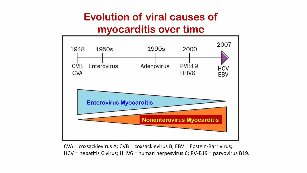
Enterovirus Myocarditis
Nonenterovirus Myocarditis
Evolution of viral causes of
myocarditis over time
CVA = coxsackievirus A; CVB = coxsackievirus B; EBV = Epstein-Barr virus;HCV = hepatitis C virus; HHV6 = human herpesvirus 6; PV-B19 = parvovirus B19.
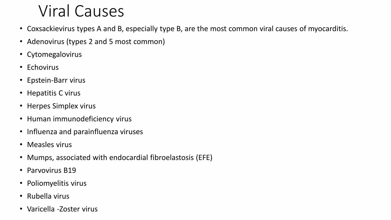
Viral Causes• Coxsackievirus types A and B, especially type B, are the most common viral causes of myocarditis.
• Adenovirus (types 2 and 5 most common)
• Cytomegalovirus
• Echovirus
• Epstein-Barr virus
• Hepatitis C virus
• Herpes Simplex virus
• Human immunodeficiency virus
• Influenza and parainfluenza viruses
• Measles virus
• Mumps, associated with endocardial fibroelastosis (EFE)
• Parvovirus B19
• Poliomyelitis virus
• Rubella virus
• Varicella -Zoster virus
Enteroviruses
• Enteroviruses are picornaviruses that are extremely small RNA viruses, naked capsid virions with icosahedral symmetry.
• The coxsackieviruses, echoviruses, and other enteroviruses are widespread throughout the world.
• Coxsackieviruses type A (4,16), type B (1, 2,3,4,5) cause myocarditis.
• Their name is derived from their ability to infect intestinal tract epithelial and lymphoid tissues and shed into the feces, but do not commonly cause gastrointestinal diseases.
Coxsackieviruses
• Coxsackie B viruses are estimated to be responsible for at least 50% of the cases of infection-caused heart diseases.
• For reasons yet unknown, the cardiac disease caused by this virus mainly occurs in middle-aged men, with onset occurring, on average, around age 42 years.
• The cardiac disease becomes apparent about two weeks after exposure to the virus.
Transmission
• Humans are the major natural host for coxsackieviruses.
• Person-to-person.
• fecal–oral transmission.
Other Rare Causes of Heart Infection
• Bacterial Causes
• - Diphtheria - Myocarditis
• - Psittacosis (Chlamydia psittaci) - Endocarditis
• - Q fever (Coxiella burnetii) - Pericarditis, myocarditis, and endocarditis. Endocarditis is frequently associated with purpuric rash, renal insufficiency, stroke, and heart failure.
• - Typhus (Rickettsia spp) - Myocarditis
• Parasitic Causes
• - Chagas' Disease (Trypanosoma cruzi) - Myocarditis • - Trichinosis (Trichinella spiralis) - Myocarditis
• - Amebiasis ( Entameba histolytica) - Pericarditis
• - Trypanosomiasis (Trypanosoma brucei rhodesiense or T b gambiense) -Myocarditis
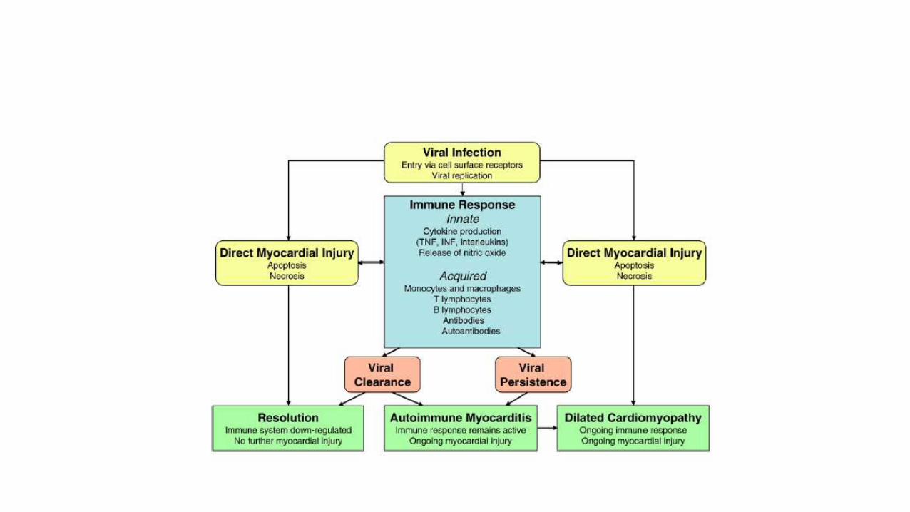
PATHOGENESIS
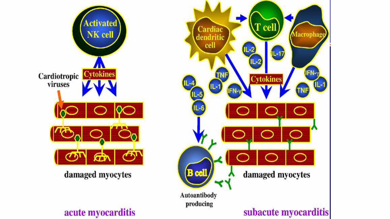
• Both direct viral-induced myocyte damage and post-viral immune inflammatory reactions contribute to myocyte damage and necrosis
• Inflammatory lesions and the necrotic process may persist for months, although the viruses only replicate in the heart for at most two or three weeks after infection
• Evidence from experimental models has incriminated cytokines such as interleukin-1 and TNF, oxygen free radicals and microvascular changes as contributory pathogenic factors
PATHOGENESIS
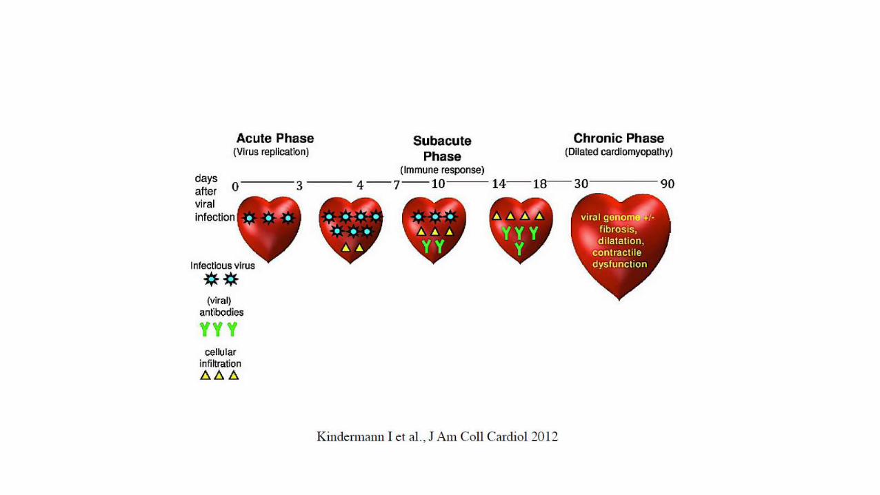
• Three phases:
Viral Replication
Autoimmune injury
Dilated cardiomyopathy
PATHOGENESIS Phase I: Viral Infection and Replication
• Viruses like coxsackievirus B cause an infectious phase, which lasts 7-10 days, and is characterized by active viral replication.
• Virus infection directly contributes to cardiac tissue destruction by cleaving the cytoskeleton protein dystrophin, leading to a disruption of the dystrophin-glycoprotein complex causing the release of antigenic intracellular components such as myosin into the bloodstream.
PATHOGENESIS Phase II: Autoimmunity and injury
• The local release of cytokines, such as interleukin-1, interleukin-2, interleukin-6, tumor necrosis factor (TNF), and nitric oxide may play a role in determining the T-cell reaction and the subsequent degree of autoimmune perpetuation.
• These cytokines may also cause reversible depression of myocardial contractility without causing cell death.
PATHOGENESIS Phase II: Autoimmunity and injury
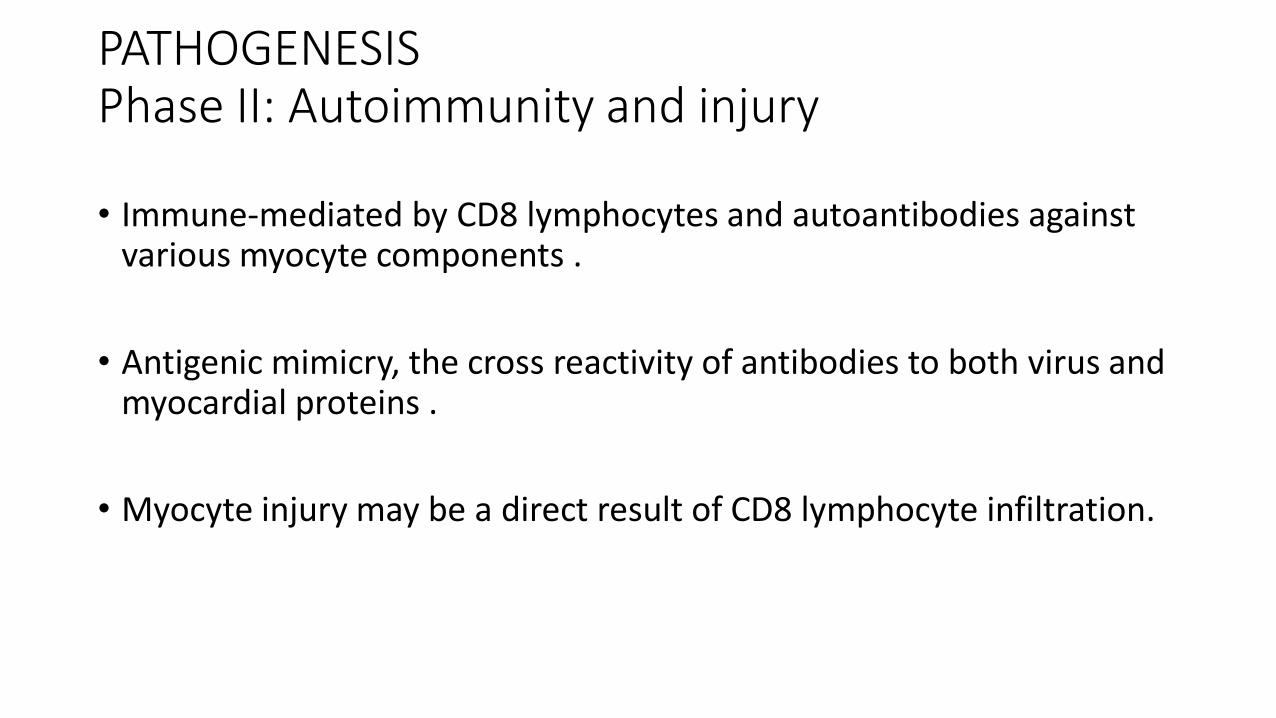
• Immune-mediated by CD8 lymphocytes and autoantibodies against various myocyte components .
• Antigenic mimicry, the cross reactivity of antibodies to both virus and myocardial proteins .
• Myocyte injury may be a direct result of CD8 lymphocyte infiltration.
PATHOGENESIS Phase III: Dilated Cardiomyopathy (DCM)
• Viruses may also directly cause myocyte apoptosis.
• During the autoimmune phase, cytokines activate the matrix metalloproteinase, such as gelatinase, collagenases, and elastases.
• In later stages of immune activation, cytokines play a leading role in adverse remodelling and progressive heart failure.
• Cardiomyopathy develops despite the absence of viral proliferation but is correlated with elevated levels of cytokines such as TNF.

Stages of Viral Myocardium InfectionNew England Journal of Medicine 343:1391 2000
Clinical presentation
• Clinical presentation varies considerably.
• In mild forms, there are few or no symptoms.
• In severe cases, patients may present with acute cardiac decompensation and progress to death.
Clinical manifestations
• A variety of cardiac symptoms can be induced by myocarditis.
• Chest pain may occur, usually due to concomitant pericarditis.
• Excessive fatigue or decreased exercise ability may be the initial sign of myocardial dysfunction .
• Since both ventricles are generally involved, patients develop biventricular failure .
• Patients present with signs of right ventricular failure such as hepatomegaly, and peripheral edema .
• If there is predominant left ventricular involvement, the patient may present with the symptoms of pulmonary congestion: dyspnea, orthopnea, rales, and, in severe cases, acute pulmonary edema.

Coxsackie Virus Clinical Manifestations
• The early symptoms of the coxsackie -induced cardiac myopathy include some generalized viral symptoms-fever, fatigue, malaise-with the addition of chest pain.
• As the virus enters the heart cells, the immune system attacks and damages both infected and normal heart cells; the affected individual feels severe fatigue when there is significant impairment of heart function.
• In most cases, the disease is resolved spontaneously without any treatment, though some permanent heart damage may have occurred
Coxsackie Virus Clinical Manifestations
• In about 20% of the cases, there can be progressive disease or recurrence of symptoms; the heart damage can be extensive, causing arrhythmias, weakened left ventricular functions, and, in the worst cases, heart failure requiring heart transplantation.
• In these severe cases, cardiac disease progression persists after the virus is long gone; the immune system continues to damage the heart.
Physical Findings
• Signs of diminished cardiac output, such as tachycardia, weak pulse, cool extremities, decreased capillary refill, and pale or mottled skin may be present.
• Heart sounds may be muffled, especially in the presence of pericarditis.
• Hepatomegaly may be present in younger children.
• Rales may be heard in older children.
• Jugular venous distention and edema of the lower extremities may be present.
Diagnostics: Expanded Criteria for Diagnosis of Myocarditis
• Category I: Clinical Symptoms
Clinical heart failure
Fever
Viral prodrome
Fatigue Dyspnea on exertion
Chest pain
Palpitations
Pre-syncope or syncope
Category II: Evidence of Cardiac Structural or Functional Perturbation in the absence of Regional Coronary Ischemia Echocardiography evidence
Regional wall motion abnormalitiesCardiac dilation Regional cardiac hypertrophy
Troponin releaseHigh sensitivity (>0.1 ng/mL)
Positive indium In 111 antimyosin scintigraphy and
Normal coronary angiography or
Absence of reversible ischemia by coronary distribution on perfusion scan
Category III: Cardiac Magnetic Resonance Imaging
Increased myocardial T2 signal on inversion recovery sequence
Delayed contrast enhancement after gadolinium-DTPA infusion
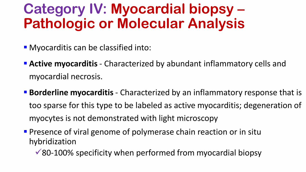
Category IV: Myocardial biopsy –Pathologic or Molecular Analysis
Myocarditis can be classified into:
Active myocarditis - Characterized by abundant inflammatory cells and
myocardial necrosis.
Borderline myocarditis - Characterized by an inflammatory response that is
too sparse for this type to be labeled as active myocarditis; degeneration of
myocytes is not demonstrated with light microscopy
Presence of viral genome of polymerase chain reaction or in situ hybridization
80-100% specificity when performed from myocardial biopsy
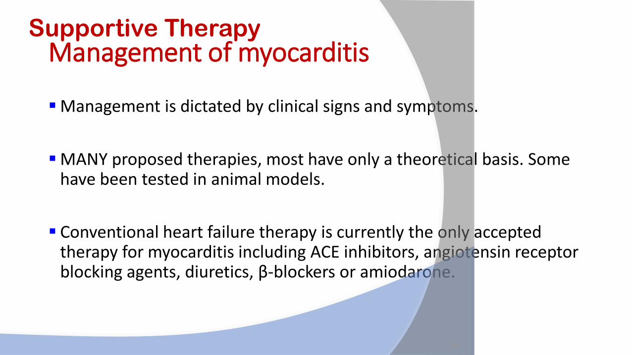
Management of myocarditis
Management is dictated by clinical signs and symptoms.
MANY proposed therapies, most have only a theoretical basis. Some have been tested in animal models.
Conventional heart failure therapy is currently the only accepted therapy for myocarditis including ACE inhibitors, angiotensin receptor blocking agents, diuretics, β-blockers or amiodarone.
32
Supportive Therapy
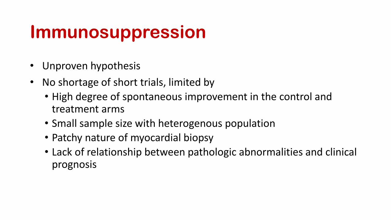
Immunosuppression
• Unproven hypothesis
• No shortage of short trials, limited by
• High degree of spontaneous improvement in the control and treatment arms
• Small sample size with heterogenous population• Patchy nature of myocardial biopsy
• Lack of relationship between pathologic abnormalities and clinical prognosis
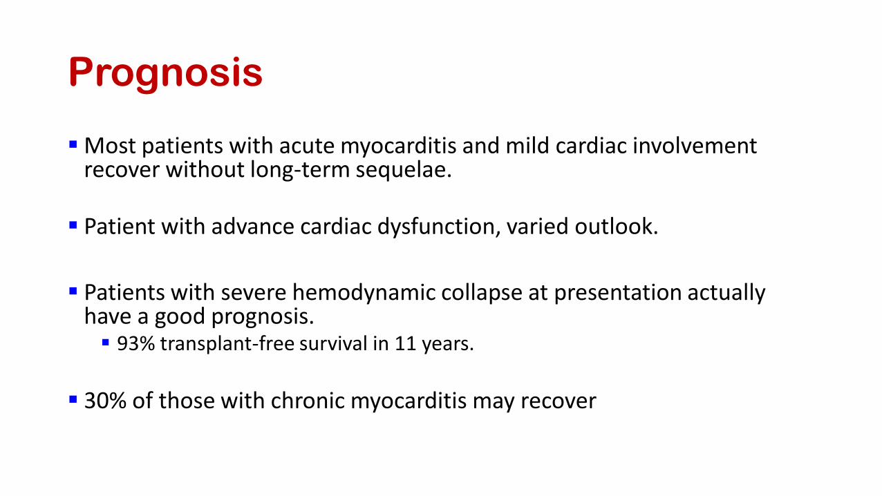
Prognosis
Most patients with acute myocarditis and mild cardiac involvement recover without long-term sequelae.
Patient with advance cardiac dysfunction, varied outlook.
Patients with severe hemodynamic collapse at presentation actually have a good prognosis. 93% transplant-free survival in 11 years.
30% of those with chronic myocarditis may recover