embryonic origin of the gnathostome vertebral...
TRANSCRIPT
on June 27, 2018http://rspb.royalsocietypublishing.org/Downloaded from
rspb.royalsocietypublishing.org
ResearchCite this article: Criswell KE, Coates MI, Gillis
JA. 2017 Embryonic origin of the gnathostome
vertebral skeleton. Proc. R. Soc. B 284:
20172121.
http://dx.doi.org/10.1098/rspb.2017.2121
Received: 22 September 2017
Accepted: 24 October 2017
Subject Category:Evolution
Subject Areas:developmental biology, evolution
Keywords:vertebral skeleton, skate, somite, notochord,
vertebrae, evolution
Author for correspondence:Katharine E. Criswell
e-mail: [email protected]
& 2017 The Authors. Published by the Royal Society under the terms of the Creative Commons AttributionLicense http://creativecommons.org/licenses/by/4.0/, which permits unrestricted use, provided the originalauthor and source are credited.
Embryonic origin of the gnathostomevertebral skeleton
Katharine E. Criswell1,2,3, Michael I. Coates1 and J. Andrew Gillis2,3
1Department of Organismal Biology and Anatomy, University of Chicago, Chicago, IL, USA2Department of Zoology, University of Cambridge, Cambridge, UK3Marine Biological Laboratory, Woods Hole, MA, USA
KEC, 0000-0002-4004-0192; MIC, 0000-0003-2843-1075
The vertebral column is a key component of the jawed vertebrate (gnathos-
tome) body plan, but the primitive embryonic origin of this skeleton remains
unclear. In tetrapods, all vertebral components (neural arches, haemal arches
and centra) derive from paraxial mesoderm (somites). However, in teleost
fishes, vertebrae have a dual embryonic origin, with arches derived from
somites, but centra formed, in part, by secretion of bone matrix from the
notochord. Here, we test the embryonic origin of the vertebral skeleton in
a cartilaginous fish (the skate, Leucoraja erinacea) which serves as an out-
group to tetrapods and teleosts. We demonstrate, by cell lineage tracing,
that both arches and centra are somite-derived. We find no evidence of cel-
lular or matrix contribution from the notochord to the skate vertebral
skeleton. These findings indicate that the earliest gnathostome vertebral skel-
eton was exclusively of somitic origin, with a notochord contribution arising
secondarily in teleosts.
1. IntroductionThe presence of vertebrae is a defining feature of the vertebrate body plan. A
vertebral skeleton may consist of a series of paired neural arches that cover
the spinal cord, paired haemal arches that enclose the caudal artery and vein,
and, in many jawed vertebrates (gnathostomes), a series of centra that replace
the notochord as the predominant support structure. Vertebral centra are
highly variable in terms of morphology and tissue composition, and likely
evolved independently in many different gnathostome lineages, including tet-
rapods, teleost fishes and cartilaginous fishes [1]. This apparent evolutionary
convergence raises questions about the embryonic origin of vertebral skeletal
elements across gnathostomes.
In tetrapods, all components of the vertebral skeleton derive from somites:
transient, bilateral blocks of segmented paraxial mesoderm that form dorsally
within the embryonic trunk. Somites are partitioned into dorsal and ventral
subdivisions that give rise to trunk connective tissue and musculature (‘dermo-
myotome’) and skeletal tissues (‘sclerotome’), respectively. Cell lineage
tracing experiments using chick–quail chimaeras [2–5] and fluorescein–
dextran injections or grafts from GFP-transgenic donor embryos in axolotl [6]
have shown a fully somitic origin of the vertebral skeleton in these taxa, with
somite-derived cells recovered in developing arches and nascent cartilage of
the centra.
Conversely, in teleost ray-finned fishes, the vertebral skeleton appears to
have a dual embryonic origin, with contributions from both paraxial mesoderm
and the notochord. Teleost vertebral centra consist of an inner layer (the chor-
dacentrum) and an outer layer, both composed of bone that forms by
intramembranous ossification [7]. The chordacentrum of teleosts forms first,
by secretion of bone matrix proteins (e.g. SPARC, type I collagen) from ‘chordo-
blast’ cells that reside within the notochord epithelium [8–10]. In zebrafish,
sc
ce
nc
ol
at
olat il
ne
ha
hsp
na
nsp(a) (a¢)
(b¢)
(b¢)(a¢)
(b)
il
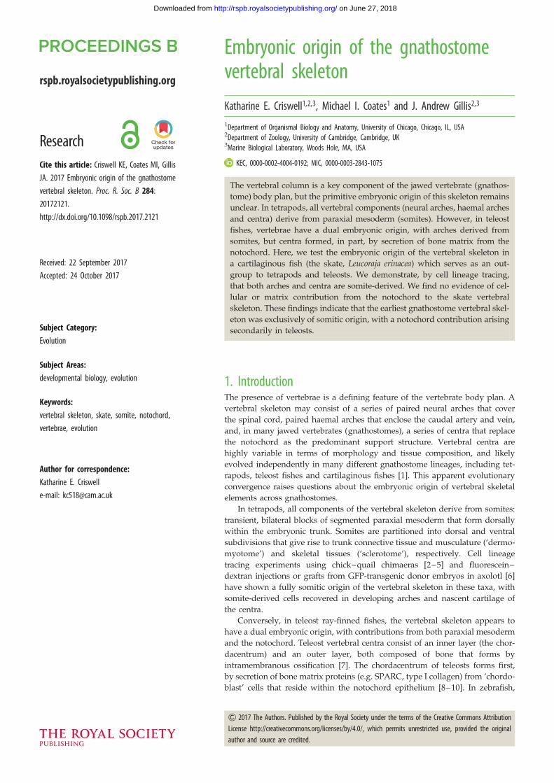
Figure 1. (a) Cross section through a skate caudal vertebra (stained with Masson’s trichrome); (a0), magnified cross section illustrating the three layers of thecentrum; (b) schematic illustrating the components and tissues of the skate vertebra; (b0) schematic of the tri-layered centrum. at, areolar tissue; ce, centrum;ha, haemal arch; hsp, haemal spine; il, inner layer of the centrum; na, neural arch; nc, notochord; ne, notochord epithelium; nsp, neural spine; ol, outer layerof the centrum; sc, spinal cord. Scale bar, 200 mm.
rspb.royalsocietypublishing.orgProc.R.Soc.B
284:20172121
2
on June 27, 2018http://rspb.royalsocietypublishing.org/Downloaded from
in vitro assays have shown that cultured notochord cells have
the capacity to secrete bone matrix, and ablation experiments
have demonstrated that in the absence of notochord, chorda-
centra fail to form [11]. Teleost chordacentra are subsequently
surrounded by a relatively late-developing layer of paraxial
mesoderm-derived membrane bone [7,12]. Additionally, zeb-
rafish mutants with somite patterning defects possess
normally developing chordacentra, but exhibit profound
neural and haemal arch defects, indicating the likely paraxial
mesodermal origin of arch tissues [11,13,14].
To determine whether the dual origin of vertebral centra is
a teleost-specific feature of the vertebral skeleton, or a general
feature for gnathostomes that has been lost in tetrapods, data
on the embryonic origin of vertebrae from an outgroup to the
bony fishes (i.e. Osteichthyes: the group that includes tetrapods
and teleosts) are needed. Cartilaginous fishes (Chondrichthyes:
sharks, skates, rays and holocephalans) occupy a key phyloge-
netic position as the sister group to the bony fishes, and data
from this lineage may therefore be used to help infer primitive
developmental conditions for the last common ancestor of
gnathostomes. We have previously shown that vertebrae in
the little skate (Leucoraja erinacea) each consist of a dorsal
neural spine, two sets of dorsal cartilages that enclose the
spinal cord (neural and intercalary arches), a single haemal
arch and spine extending ventrally, and a tri-layered centrum
(figure 1) [15]. Here, we use somite and notochord fate map-
ping experiments, as well as mRNA in situ hybridization for
genes encoding skeletal matrix proteins, to test the embryonic
origin of the skate vertebral skeleton. We show that all com-
ponents of the skate vertebral skeleton derive from paraxial
mesoderm, with no evidence for cellular or matrix contri-
butions from the notochord. When considered alongside data
from bony fishes, our findings point to a general and probably
primitive paraxial mesodermal origin of the vertebrate column
in jawed vertebrates.
2. Material and methods(a) Somite fate mappingLeucoraja erinacea embryos were obtained from the Marine Bio-
logical Laboratory (MBL) in Woods Hole, MA, and kept in a
flow-through sea table at approximately 168C until S24. A flap
was cut in the egg case using a razor blade, and the embryo
and yolk were transferred to a Petri dish. Embryos were anaesth-
etized in a solution of MS-222 (100 mg l21 ethyl 3-aminobenzoate
methanesulfonate—Sigma-Aldrich) in seawater. CellTracker CM-
DiI (Thermofisher) (5 mg ml21 in ethanol) was diluted 1 : 10 in
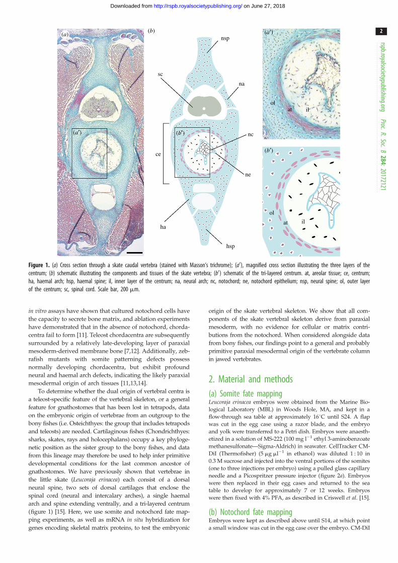
0.3 M sucrose and injected into the ventral portions of the somites
(one to three injections per embryo) using a pulled glass capillary
needle and a Picospritzer pressure injector (figure 2a). Embryos
were then replaced in their egg cases and returned to the sea
table to develop for approximately 7 or 12 weeks. Embryos
were then fixed with 4% PFA, as described in Criswell et al. [15].
(b) Notochord fate mappingEmbryos were kept as described above until S14, at which point
a small window was cut in the egg case over the embryo. CM-DiI
(a)
(b)
(c)
window
yolk
shellgraft
Figure 2. Microinjection of skate embryos with CM-DiI. CM-DiI labelling of(a) somites at S24 (three somites are highlighted with dashed lines) and(b) notochord progenitor cells at S14 (with the ‘notochord triangle’ of Ballardet al. [16] outlined). (c) Sealing of a windowed skate egg with donor eggshell. Scale bars, 200 mm.
rspb.royalsocietypublishing.orgProc.R.Soc.B
284:20172121
3
on June 27, 2018http://rspb.royalsocietypublishing.org/Downloaded from
was microinjected into the notochord triangle as described above
(figure 2b). The window was then sealed with donor eggshell
and Krazy GlueTM gel (figure 2c), and eggs were returned to
the sea table to develop for an additional 16–18 weeks prior to
fixation (as described in Criswell et al. [15]).
(c) Validation of CM-DiI injection placementTo verify the correct placement of CM-DiI injections, three
somite-injected embryos were fixed immediately post-injection,
and three notochord-injected embryos were fixed 5 days post-
injection (dpi). Embryos were fixed in 4% paraformaldehyde in
PBS overnight at 48C, rinsed 3 � 15 min in PBS and stained
with DAPI at 1 mg ml21 overnight at room temperature.
Somite-injected embryos were imaged on a Zeiss lightsheet
microscope and notochord-injected embryos were imaged on
Zeiss lightsheet or LSM 780 confocal microscopes.
(d) Histology and mRNA in situ hybridizationCM-DiI-labelled L. erinacea embryos were embedded in paraffin
wax and sectioned at 8 mm thickness as described in O’Neill
et al. [17] for histological analysis. Prior to embedding, embryos
were demineralized in 10% EDTA (ethylenediaminetetraacetic
acid) for 14 days. Histochemical staining was performed follow-
ing the Masson’s trichrome protocol of Witten and Hall [18].
In situ hybridization experiments for Col1a1 (GenBank accession
number MG017616) and SPARC (GenBank accession number
MG017615) were performed on sections as described in O’Neill
et al. [17], with modifications according to Gillis et al. [19].
3. Results(a) Somitic contribution to all components of the skate
vertebral skeletonTo test for somitic contribution to the skate vertebral skeleton,
we microinjected CM-DiI into ventral portions of the somites
(i.e. the presumptive sclerotome—figure 3a) of skate embryos
at stage (S) 24 (Ballard et al. [16]). Focal labelling of the
somites (with no notochordal contamination) was confirmed
by light sheet microscopy, in embryos fixed immediately
post-injection (figure 3b; n ¼ 3). By 50–52 dpi (S31), spin-
dle-shaped cells of the developing areolar tissue of
the centrum surround the notochord, and preskeletal
mesenchyme has condensed around the neural tube and
caudal artery and vein. In all embryos analysed at this
stage (n ¼ 5), CM-DiI was recovered in the spindle-shaped
cells of the developing areolar tissue (figure 3c), indicating
their somitic origin.
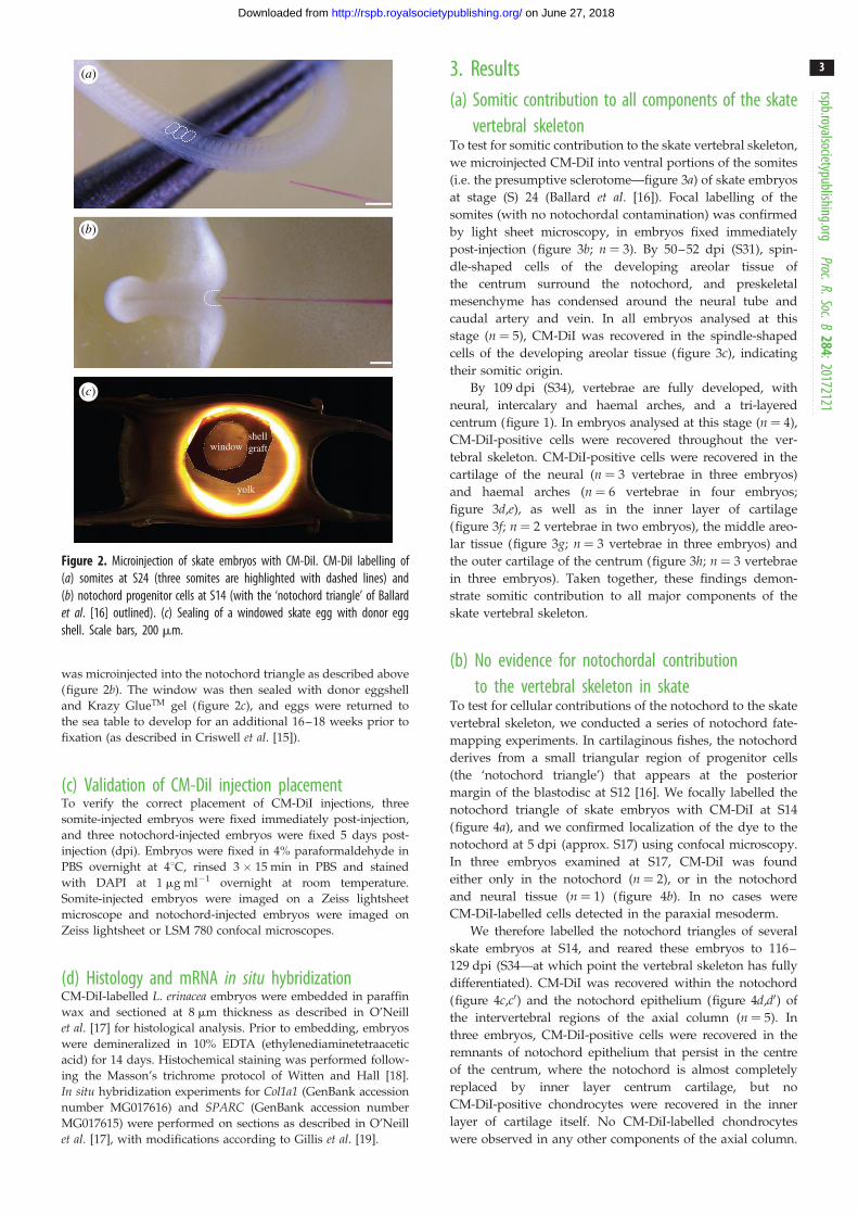
By 109 dpi (S34), vertebrae are fully developed, with
neural, intercalary and haemal arches, and a tri-layered
centrum (figure 1). In embryos analysed at this stage (n ¼ 4),
CM-DiI-positive cells were recovered throughout the ver-
tebral skeleton. CM-DiI-positive cells were recovered in the
cartilage of the neural (n ¼ 3 vertebrae in three embryos)
and haemal arches (n ¼ 6 vertebrae in four embryos;
figure 3d,e), as well as in the inner layer of cartilage
(figure 3f; n ¼ 2 vertebrae in two embryos), the middle areo-
lar tissue (figure 3g; n ¼ 3 vertebrae in three embryos) and
the outer cartilage of the centrum (figure 3h; n ¼ 3 vertebrae
in three embryos). Taken together, these findings demon-
strate somitic contribution to all major components of the
skate vertebral skeleton.
(b) No evidence for notochordal contributionto the vertebral skeleton in skate
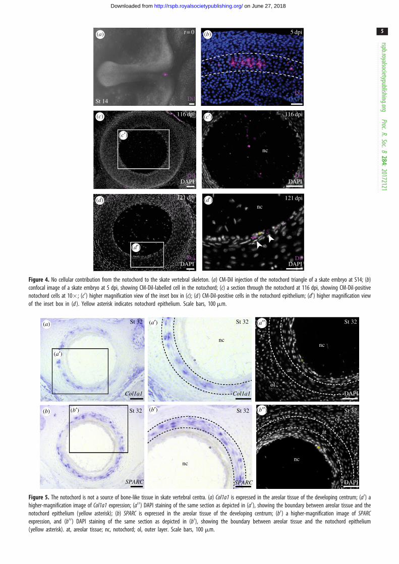
To test for cellular contributions of the notochord to the skate
vertebral skeleton, we conducted a series of notochord fate-
mapping experiments. In cartilaginous fishes, the notochord
derives from a small triangular region of progenitor cells
(the ‘notochord triangle’) that appears at the posterior
margin of the blastodisc at S12 [16]. We focally labelled the
notochord triangle of skate embryos with CM-DiI at S14
(figure 4a), and we confirmed localization of the dye to the
notochord at 5 dpi (approx. S17) using confocal microscopy.
In three embryos examined at S17, CM-DiI was found
either only in the notochord (n ¼ 2), or in the notochord
and neural tissue (n ¼ 1) (figure 4b). In no cases were
CM-DiI-labelled cells detected in the paraxial mesoderm.
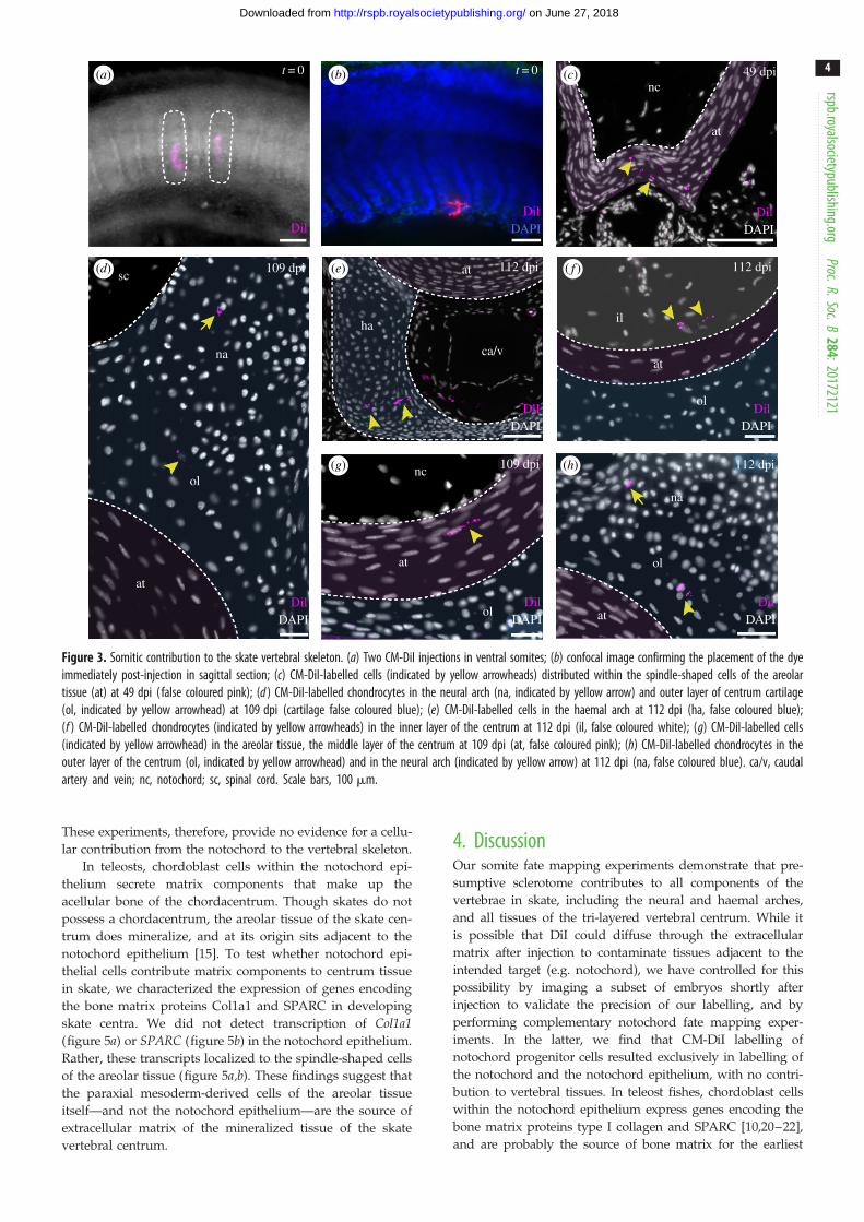
We therefore labelled the notochord triangles of several
skate embryos at S14, and reared these embryos to 116–
129 dpi (S34—at which point the vertebral skeleton has fully
differentiated). CM-DiI was recovered within the notochord
(figure 4c,c0) and the notochord epithelium (figure 4d,d0) of
the intervertebral regions of the axial column (n ¼ 5). In
three embryos, CM-DiI-positive cells were recovered in the
remnants of notochord epithelium that persist in the centre
of the centrum, where the notochord is almost completely
replaced by inner layer centrum cartilage, but no
CM-DiI-positive chondrocytes were recovered in the inner
layer of cartilage itself. No CM-DiI-labelled chondrocytes
were observed in any other components of the axial column.
109 dpi 112 dpi 112 dpi
ca/v
sc
na
ol
ol
ol
ol
na
nc
at
at
at
ha
at
at
il
t = 0t = 0 49 dpinc
at
DilDil
DAPI
DilDAPI
DilDAPI
DilDAPI
DilDAPI
DilDAPI
109 dpi 112 dpi
DilDAPI
(a) (b) (c)
(d) (e) ( f )
(g) (h)
Figure 3. Somitic contribution to the skate vertebral skeleton. (a) Two CM-DiI injections in ventral somites; (b) confocal image confirming the placement of the dyeimmediately post-injection in sagittal section; (c) CM-DiI-labelled cells (indicated by yellow arrowheads) distributed within the spindle-shaped cells of the areolartissue (at) at 49 dpi ( false coloured pink); (d ) CM-DiI-labelled chondrocytes in the neural arch (na, indicated by yellow arrow) and outer layer of centrum cartilage(ol, indicated by yellow arrowhead) at 109 dpi (cartilage false coloured blue); (e) CM-DiI-labelled cells in the haemal arch at 112 dpi (ha, false coloured blue);(f ) CM-DiI-labelled chondrocytes (indicated by yellow arrowheads) in the inner layer of the centrum at 112 dpi (il, false coloured white); (g) CM-DiI-labelled cells(indicated by yellow arrowhead) in the areolar tissue, the middle layer of the centrum at 109 dpi (at, false coloured pink); (h) CM-DiI-labelled chondrocytes in theouter layer of the centrum (ol, indicated by yellow arrowhead) and in the neural arch (indicated by yellow arrow) at 112 dpi (na, false coloured blue). ca/v, caudalartery and vein; nc, notochord; sc, spinal cord. Scale bars, 100 mm.
rspb.royalsocietypublishing.orgProc.R.Soc.B
284:20172121
4
on June 27, 2018http://rspb.royalsocietypublishing.org/Downloaded from
These experiments, therefore, provide no evidence for a cellu-
lar contribution from the notochord to the vertebral skeleton.
In teleosts, chordoblast cells within the notochord epi-
thelium secrete matrix components that make up the
acellular bone of the chordacentrum. Though skates do not
possess a chordacentrum, the areolar tissue of the skate cen-
trum does mineralize, and at its origin sits adjacent to the
notochord epithelium [15]. To test whether notochord epi-
thelial cells contribute matrix components to centrum tissue
in skate, we characterized the expression of genes encoding
the bone matrix proteins Col1a1 and SPARC in developing
skate centra. We did not detect transcription of Col1a1(figure 5a) or SPARC (figure 5b) in the notochord epithelium.
Rather, these transcripts localized to the spindle-shaped cells
of the areolar tissue (figure 5a,b). These findings suggest that
the paraxial mesoderm-derived cells of the areolar tissue
itself—and not the notochord epithelium—are the source of
extracellular matrix of the mineralized tissue of the skate
vertebral centrum.
4. DiscussionOur somite fate mapping experiments demonstrate that pre-
sumptive sclerotome contributes to all components of the
vertebrae in skate, including the neural and haemal arches,
and all tissues of the tri-layered vertebral centrum. While it
is possible that DiI could diffuse through the extracellular
matrix after injection to contaminate tissues adjacent to the
intended target (e.g. notochord), we have controlled for this
possibility by imaging a subset of embryos shortly after
injection to validate the precision of our labelling, and by
performing complementary notochord fate mapping exper-
iments. In the latter, we find that CM-DiI labelling of
notochord progenitor cells resulted exclusively in labelling of
the notochord and the notochord epithelium, with no contri-
bution to vertebral tissues. In teleost fishes, chordoblast cells
within the notochord epithelium express genes encoding the
bone matrix proteins type I collagen and SPARC [10,20–22],
and are probably the source of bone matrix for the earliest
t = 0
St 14 DilDil
DAPI
DilDAPI
DilDAPI
DilDAPI
DilDAPI
121 dpi
nc
nc
*
5 dpi
116 dpi116 dpi
121 dpi
(a) (b)
(c) (c¢)
(c¢)
(d) (d¢)
(d¢)
Figure 4. No cellular contribution from the notochord to the skate vertebral skeleton. (a) CM-DiI injection of the notochord triangle of a skate embryo at S14; (b)confocal image of a skate embryo at 5 dpi, showing CM-DiI-labelled cell in the notochord; (c) a section through the notochord at 116 dpi, showing CM-DiI-positivenotochord cells at 10�; (c0) higher magnification view of the inset box in (c); (d ) CM-DiI-positive cells in the notochord epithelium; (d0) higher magnification viewof the inset box in (d ). Yellow asterisk indicates notochord epithelium. Scale bars, 100 mm.
(a) (a¢)
(a¢)
(a¢¢)
(b¢¢)(b¢)(b¢)(b)
St 32
St 32
nc
*
SPARC SPARC
St 32
St 32 St 32
St 32
DAPI
DAPI
ncnc
nc
at
*
*
at
ol
ol
*
Col1a1 Col1a1
Figure 5. The notochord is not a source of bone-like tissue in skate vertebral centra. (a) Col1a1 is expressed in the areolar tissue of the developing centrum; (a0) ahigher-magnification image of Col1a1 expression; (a00) DAPI staining of the same section as depicted in (a0), showing the boundary between areolar tissue and thenotochord epithelium (yellow asterisk); (b) SPARC is expressed in the areolar tissue of the developing centrum; (b0) a higher-magnification image of SPARCexpression, and (b00) DAPI staining of the same section as depicted in (b0), showing the boundary between areolar tissue and the notochord epithelium(yellow asterisk). at, areolar tissue; nc, notochord; ol, outer layer. Scale bars, 100 mm.
rspb.royalsocietypublishing.orgProc.R.Soc.B
284:20172121
5
on June 27, 2018http://rspb.royalsocietypublishing.org/Downloaded from
lamprey elasmobranch teleost salamander bird
ccc
sclerotome derived notochord contribution
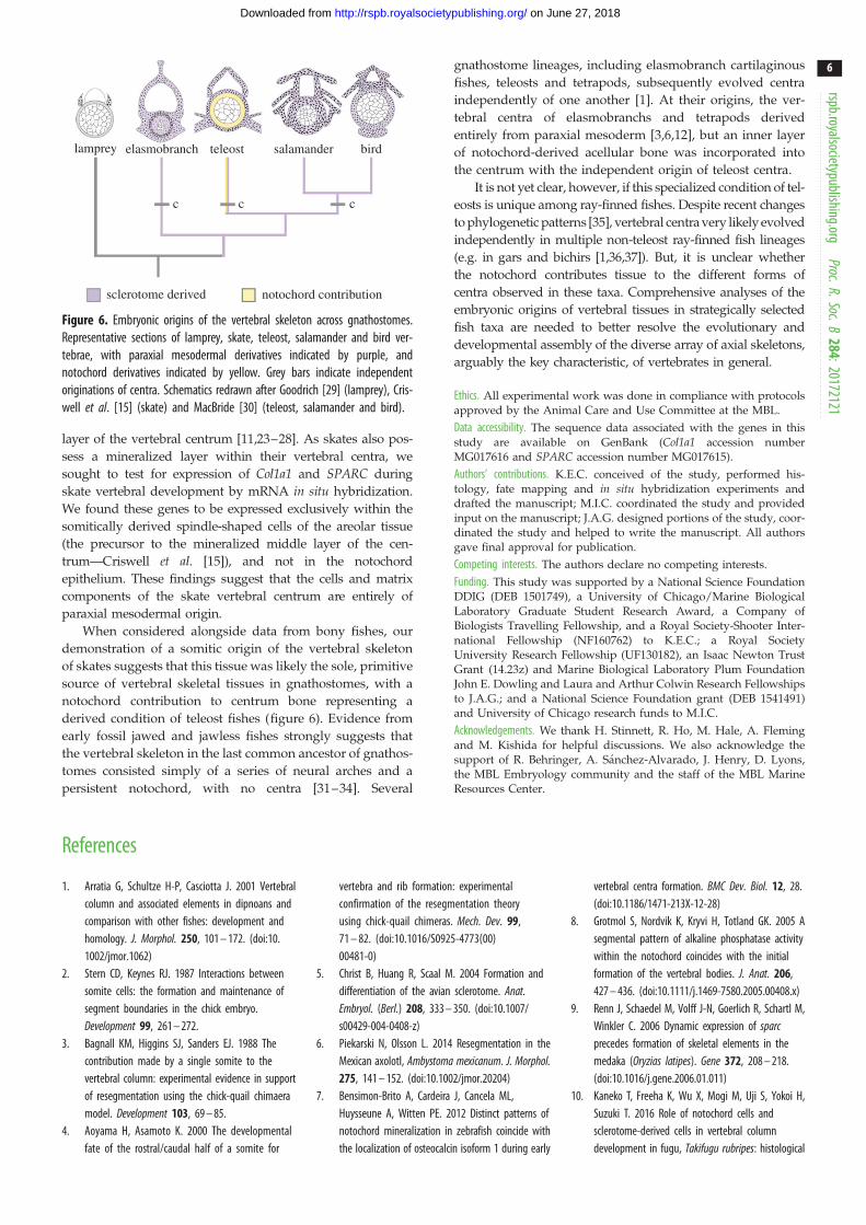
Figure 6. Embryonic origins of the vertebral skeleton across gnathostomes.Representative sections of lamprey, skate, teleost, salamander and bird ver-tebrae, with paraxial mesodermal derivatives indicated by purple, andnotochord derivatives indicated by yellow. Grey bars indicate independentoriginations of centra. Schematics redrawn after Goodrich [29] (lamprey), Cris-well et al. [15] (skate) and MacBride [30] (teleost, salamander and bird).
rspb.royalsocietypublishing.orgProc.R.Soc.B
284:20172121
6
on June 27, 2018http://rspb.royalsocietypublishing.org/Downloaded from
layer of the vertebral centrum [11,23–28]. As skates also pos-
sess a mineralized layer within their vertebral centra, we
sought to test for expression of Col1a1 and SPARC during
skate vertebral development by mRNA in situ hybridization.
We found these genes to be expressed exclusively within the
somitically derived spindle-shaped cells of the areolar tissue
(the precursor to the mineralized middle layer of the cen-
trum—Criswell et al. [15]), and not in the notochord
epithelium. These findings suggest that the cells and matrix
components of the skate vertebral centrum are entirely of
paraxial mesodermal origin.
When considered alongside data from bony fishes, our
demonstration of a somitic origin of the vertebral skeleton
of skates suggests that this tissue was likely the sole, primitive
source of vertebral skeletal tissues in gnathostomes, with a
notochord contribution to centrum bone representing a
derived condition of teleost fishes (figure 6). Evidence from
early fossil jawed and jawless fishes strongly suggests that
the vertebral skeleton in the last common ancestor of gnathos-
tomes consisted simply of a series of neural arches and a
persistent notochord, with no centra [31–34]. Several
gnathostome lineages, including elasmobranch cartilaginous
fishes, teleosts and tetrapods, subsequently evolved centra
independently of one another [1]. At their origins, the ver-
tebral centra of elasmobranchs and tetrapods derived
entirely from paraxial mesoderm [3,6,12], but an inner layer
of notochord-derived acellular bone was incorporated into
the centrum with the independent origin of teleost centra.
It is not yet clear, however, if this specialized condition of tel-
eosts is unique among ray-finned fishes. Despite recent changes
to phylogenetic patterns [35], vertebral centra very likely evolved
independently in multiple non-teleost ray-finned fish lineages
(e.g. in gars and bichirs [1,36,37]). But, it is unclear whether
the notochord contributes tissue to the different forms of
centra observed in these taxa. Comprehensive analyses of the
embryonic origins of vertebral tissues in strategically selected
fish taxa are needed to better resolve the evolutionary and
developmental assembly of the diverse array of axial skeletons,
arguably the key characteristic, of vertebrates in general.
Ethics. All experimental work was done in compliance with protocolsapproved by the Animal Care and Use Committee at the MBL.
Data accessibility. The sequence data associated with the genes in thisstudy are available on GenBank (Col1a1 accession numberMG017616 and SPARC accession number MG017615).
Authors’ contributions. K.E.C. conceived of the study, performed his-tology, fate mapping and in situ hybridization experiments anddrafted the manuscript; M.I.C. coordinated the study and providedinput on the manuscript; J.A.G. designed portions of the study, coor-dinated the study and helped to write the manuscript. All authorsgave final approval for publication.
Competing interests. The authors declare no competing interests.
Funding. This study was supported by a National Science FoundationDDIG (DEB 1501749), a University of Chicago/Marine BiologicalLaboratory Graduate Student Research Award, a Company ofBiologists Travelling Fellowship, and a Royal Society-Shooter Inter-national Fellowship (NF160762) to K.E.C.; a Royal SocietyUniversity Research Fellowship (UF130182), an Isaac Newton TrustGrant (14.23z) and Marine Biological Laboratory Plum FoundationJohn E. Dowling and Laura and Arthur Colwin Research Fellowshipsto J.A.G.; and a National Science Foundation grant (DEB 1541491)and University of Chicago research funds to M.I.C.
Acknowledgements. We thank H. Stinnett, R. Ho, M. Hale, A. Flemingand M. Kishida for helpful discussions. We also acknowledge thesupport of R. Behringer, A. Sanchez-Alvarado, J. Henry, D. Lyons,the MBL Embryology community and the staff of the MBL MarineResources Center.
References
1. Arratia G, Schultze H-P, Casciotta J. 2001 Vertebralcolumn and associated elements in dipnoans andcomparison with other fishes: development andhomology. J. Morphol. 250, 101 – 172. (doi:10.1002/jmor.1062)
2. Stern CD, Keynes RJ. 1987 Interactions betweensomite cells: the formation and maintenance ofsegment boundaries in the chick embryo.Development 99, 261 – 272.
3. Bagnall KM, Higgins SJ, Sanders EJ. 1988 Thecontribution made by a single somite to thevertebral column: experimental evidence in supportof resegmentation using the chick-quail chimaeramodel. Development 103, 69 – 85.
4. Aoyama H, Asamoto K. 2000 The developmentalfate of the rostral/caudal half of a somite for
vertebra and rib formation: experimentalconfirmation of the resegmentation theoryusing chick-quail chimeras. Mech. Dev. 99,71 – 82. (doi:10.1016/S0925-4773(00)00481-0)
5. Christ B, Huang R, Scaal M. 2004 Formation anddifferentiation of the avian sclerotome. Anat.Embryol. (Berl.) 208, 333 – 350. (doi:10.1007/s00429-004-0408-z)
6. Piekarski N, Olsson L. 2014 Resegmentation in theMexican axolotl, Ambystoma mexicanum. J. Morphol.275, 141 – 152. (doi:10.1002/jmor.20204)
7. Bensimon-Brito A, Cardeira J, Cancela ML,Huysseune A, Witten PE. 2012 Distinct patterns ofnotochord mineralization in zebrafish coincide withthe localization of osteocalcin isoform 1 during early
vertebral centra formation. BMC Dev. Biol. 12, 28.(doi:10.1186/1471-213X-12-28)
8. Grotmol S, Nordvik K, Kryvi H, Totland GK. 2005 Asegmental pattern of alkaline phosphatase activitywithin the notochord coincides with the initialformation of the vertebral bodies. J. Anat. 206,427 – 436. (doi:10.1111/j.1469-7580.2005.00408.x)
9. Renn J, Schaedel M, Volff J-N, Goerlich R, Schartl M,Winkler C. 2006 Dynamic expression of sparcprecedes formation of skeletal elements in themedaka (Oryzias latipes). Gene 372, 208 – 218.(doi:10.1016/j.gene.2006.01.011)
10. Kaneko T, Freeha K, Wu X, Mogi M, Uji S, Yokoi H,Suzuki T. 2016 Role of notochord cells andsclerotome-derived cells in vertebral columndevelopment in fugu, Takifugu rubripes: histological
rspb.royalsocietypublishing.orgProc.R.Soc.B
284:20172121
7
on June 27, 2018http://rspb.royalsocietypublishing.org/Downloaded from
and gene expression analyses. Cell Tissue Res. 366,37 – 49. (doi:10.1007/s00441-016-2404-z)
11. Fleming A, Keynes R, Tannahill D. 2004 A centralrole for the notochord in vertebral patterning.Development 131, 873 – 880. (doi:10.1242/dev.00952)
12. Morin-Kensicki EM, Melancon E, Eisen JS. 2002Segmental relationship between somites andvertebral column in zebrafish. Development 129,3851 – 3860.
13. Van Eeden FJ et al. 1996 Mutations affecting somiteformation and patterning in the zebrafish, Daniorerio. Development 123, 153 – 164.
14. Fleming A, Keynes RJ, Tannahill D. 2001 The role ofthe notochord in vertebral column formation.J. Anat. 199, 177 – 180. (doi:10.1017/S0021878201008044)
15. Criswell KE, Coates MI, Gillis JA. 2017 Embryonicdevelopment of the axial column in the little skate,Leucoraja erinacea. J. Morphol. 278, 300 – 320.(doi:10.1002/jmor.20637)
16. Ballard WW, Mellinger J, Lechenault H. 1993A series of normal stages for development ofScyliorhinus canicula, the lesser spotted dogfish(Chondrichthyes: Scyliorhinidae). J. Exp. Zool. 267,318 – 336. (doi:10.1002/jez.1402670309)
17. O’Neill P, McCole RB, Baker CVH. 2007 A molecularanalysis of neurogenic placode and cranial sensoryganglion development in the shark, Scyliorhinuscanicula. Dev. Biol. 304, 156 – 181. (doi:10.1016/j.ydbio.2006.12.029)
18. Witten PE, Hall BK. 2003 Seasonal changes in thelower jaw skeleton in male Atlantic salmon (Salmosalar L.): remodelling and regression of the kypeafter spawning. J. Anat. 203, 435 – 450. (doi:10.1046/j.1469-7580.2003.00239.x)
19. Gillis JA, Modrell MS, Northcutt RG, Catania KC, LuerCA, Baker CVH. 2012 Electrosensory ampullary
organs are derived from lateral line placodes incartilaginous fishes. Development 139, 3142 – 3146.(doi:10.1242/dev.084046)
20. Thisse B et al. 2001 Expression of the zebrafishgenome during embryogenesis (NIH R01 RR15402).Zfin Direct Data Submiss.
21. Rotllant J, Liu D, Yan Y-L, Postlethwait JH,Westerfield M, Du S-J. 2008 Sparc (osteonectin)functions in morphogenesis of the pharyngealskeleton and inner ear. Matrix. Biol. 27, 561 – 572.(doi:10.1016/j.matbio.2008.03.001)
22. Wang S, Furmanek T, Kryvi H, Krossøy C, TotlandGK, Grotmol S, Wargelius A. 2014 Transcriptomesequencing of Atlantic salmon (Salmo salar L.)notochord prior to development of thevertebrae provides clues to regulation ofpositional fate, chordoblast lineage andmineralisation. BMC Genomics 15, 141. (doi:10.1186/1471-2164-15-141)
23. Ramanujam SG. 1929 The study of the developmentof the vertebral column in teleosts, as shown in thelife-history of the herring. J. Zool. 99, 365 – 414.(doi:10.1111/j.1469-7998.1929.tb07696.x)
24. Mookerjee HK, Mitra GN, Mazumdar SR. 1940The development of the vertebral column of aviviparous teleost, Lebistes reticulatus. J. Morphol.67, 241 – 269. (doi:10.1002/jmor.1050670203)
25. Laerm J. 1976 The development, function, anddesign of amphicoelous vertebrae in teleost fishes.Zool. J. Linn. Soc. 58, 237 – 254. (doi:10.1111/j.1096-3642.1976.tb00830.x)
26. Grotmol S, Kryvi H, Nordvik K, Totland GK. 2003Notochord segmentation may lay down thepathway for the development of the vertebralbodies in the Atlantic salmon. Anat. Embryol. (Berl.)207, 263 – 272. (doi:10.1007/s00429-003-0349-y)
27. Nordvik K, Kryvi H, Totland GK, Grotmol S. 2005The salmon vertebral body develops through
mineralization of two preformed tissues that areencompassed by two layers of bone. J. Anat. 206,103 – 114. (doi:10.1111/j.1469-7580.2005.00372.x)
28. Renn J, Buttner A, To TT, Chan SJH, Winkler C. 2013A col10a1:nlGFP transgenic line displays putativeosteoblast precursors at the medaka notochordalsheath prior to mineralization. Dev. Biol. 381,134 – 143. (doi:10.1016/j.ydbio.2013.05.030)
29. Goodrich E. 1930 Studies on the structure anddevelopment of vertebrates. London, UK: DoverPublications.
30. MacBride EW. 1932 Recent work on thedevelopment of the vertebral column. Biol Rev 7,108 – 148. (doi:10.1111/j.1469-185X.1962.tb01038.x)
31. Gardiner BG, Miles RS. 1994 Eubrachythoracidarthrodires from Gogo, Western Australia.Zool. J. Linn. Soc. 112, 443 – 477. (doi:10.1111/j.1096-3642.1994.tb00331.x)
32. Janvier P. 1996 Early vertebrates. Oxford, UK:Clarendon Press.
33. Long JA, Trinajstic K, Young GC, Senden T. 2008Live birth in the Devonian period. Nature 453,650 – 652. (doi:10.1038/nature06966)
34. Johanson Z, Trinajstic K, Carr R, Ritchie A. 2013Evolution and development of the synarcual in earlyvertebrates. Zoomorphology 132, 95 – 110. (doi:10.1007/s00435-012-0169-9)
35. Giles S, Xu G-H, Near TJ, Friedman M. 2017 Earlymembers of ‘living fossil’ lineage imply later originof modern ray-finned fishes. Nature 549, 265 – 268.(doi:10.1038/nature23654)
36. Laerm J. 1979 The origin and homology of thechondrostean vertebral centrum. Can. J. Zool. 57,475 – 485. (doi:10.1139/z79-058)
37. Laerm J. 1982 The origin and homology of theneopterygian vertebral centrum. J. Paleontol. 56,191 – 202.