elbow trauma: managing challenging clinical …galeazzi monteggia forearm fractures mugr monteggia...
TRANSCRIPT
2/26/2018
1
ELBOW TRAUMA: MANAGING CHALLENGING CLINICAL SEQUELLAE
Jane Fedorczyk , PT, PhD, CHT
Certified Hand Therapist
Professor‐ University of South Florida
Matthew Lazinski, PT, DPT, OCS
Board Certified Clinical Specialist in Orthopedics
Assistant Professor‐ University of South Florida
Disclosure
Matt Lazinski, PT, DPT, OCS
Nothing to Disclose
Jane Fedorczyk, PT, PhD, CHT
Royalties, Elsevier
Consultant, MedRisk
2/26/2018
2
Objectives
• Describe anatomy and biomechanics of the elbow complex including the course of the peripheral nerves.
• Employ clinical examination techniques to identify peripheral nerve injuries and other causes of movement dysfunction.
• Formulate a plan of care with targeted interventions to reduce movement dysfunction and optimize functional outcomes.
• Develop concepts for future clinical research investigation.
ANATOMY AND BIOMECHANICS
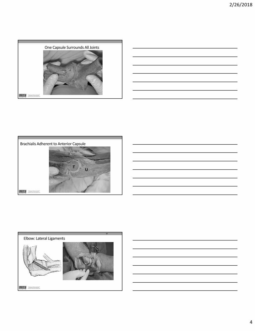
Elbow Complex: 3 Articulations
• Ulnohumeral
• Radiohumeral
• Superior or Proximal Radioulnar Joint
H
RU
CT
RHO
2/26/2018
3
Elbow Complex: Osteology“Lock and Key” Configuration Primary to Stability
Articular Configuration Adds to Stability
8
Elbow Most Stable in Flexion
2/26/2018
4
One Capsule Surrounds All Joints
TU
Brachialis Adherent to Anterior Capsule
12
Elbow: Lateral Ligaments
2/26/2018
5
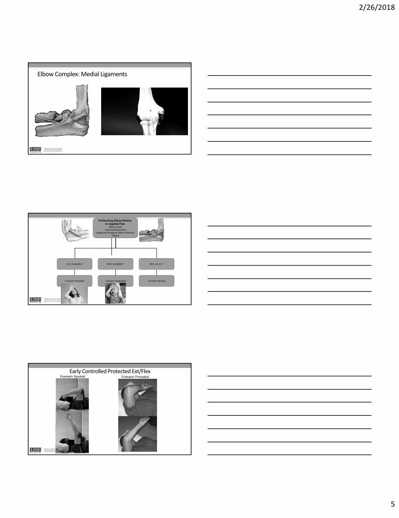
Elbow Complex: Medial Ligaments
Performing Elbow Motion in Sagittal Plan
Elbow InjuryFracture/Dislocation
Ligament Disruption With or Without Repair
LCL Instability? MCL Instability? MCL & LCL?
Forearm Pronated Forearm Supinated Forearm Neutral
Early Controlled Protected Ext/Flex Forearm Neutral Forearm Pronated
2/26/2018
6
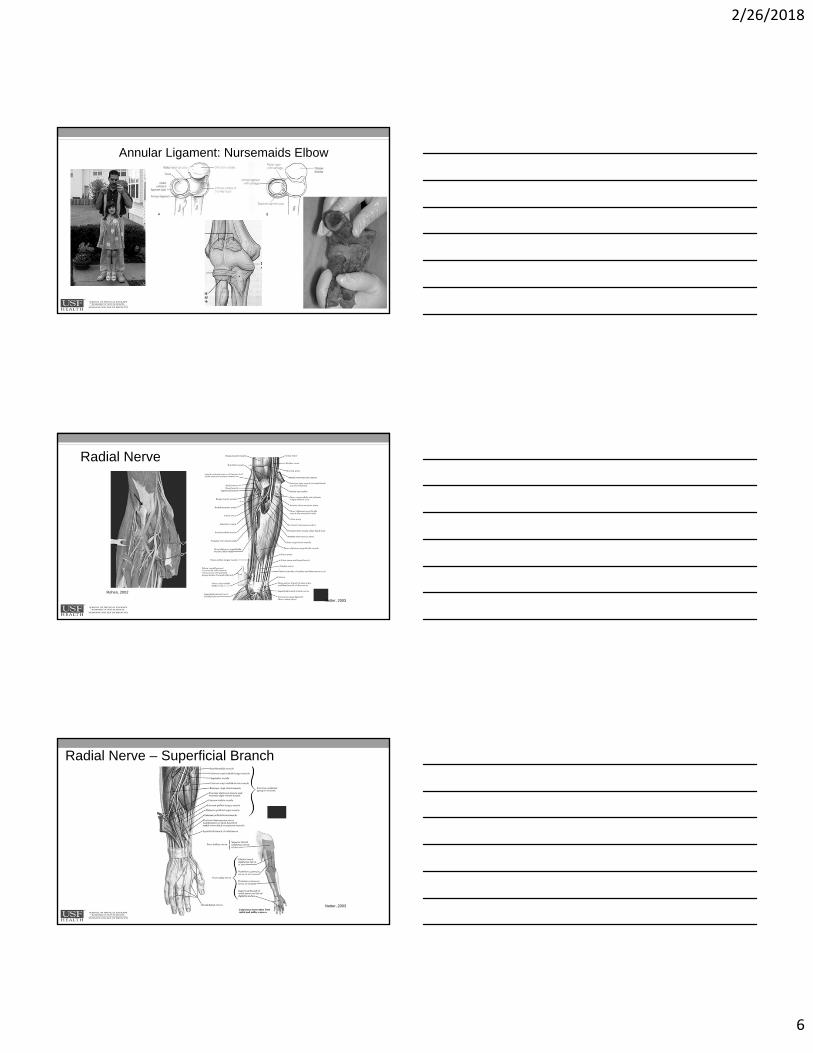
Annular Ligament: Nursemaids Elbow
Radial Nerve
Rohen, 2002
Netter, 2003
Radial Nerve – Superficial Branch
Netter, 2003
2/26/2018
7
Radial Nerve – Deep Branch
Rohen, 2002
Netter, 2003
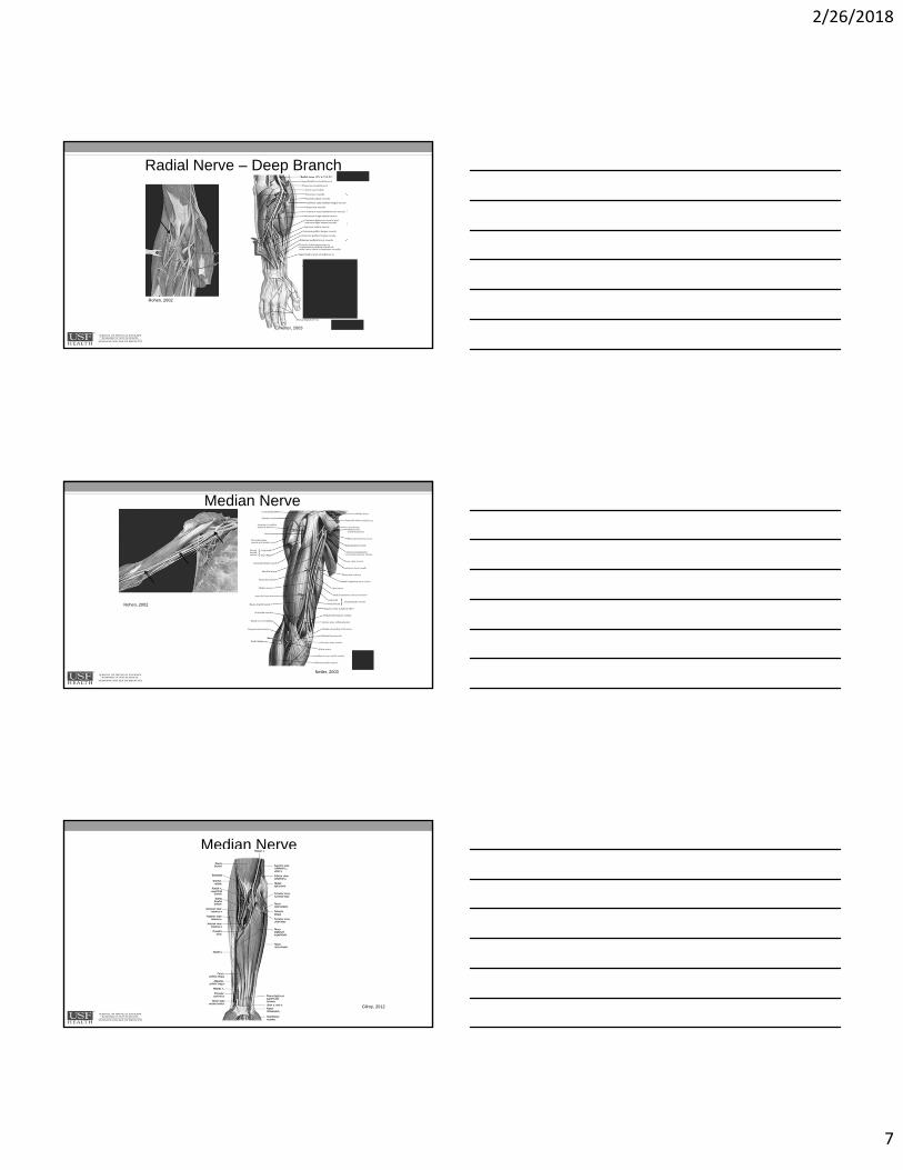
Median Nerve
Rohen, 2002
Netter, 2003
Median Nerve
Gilroy, 2012
2/26/2018
8
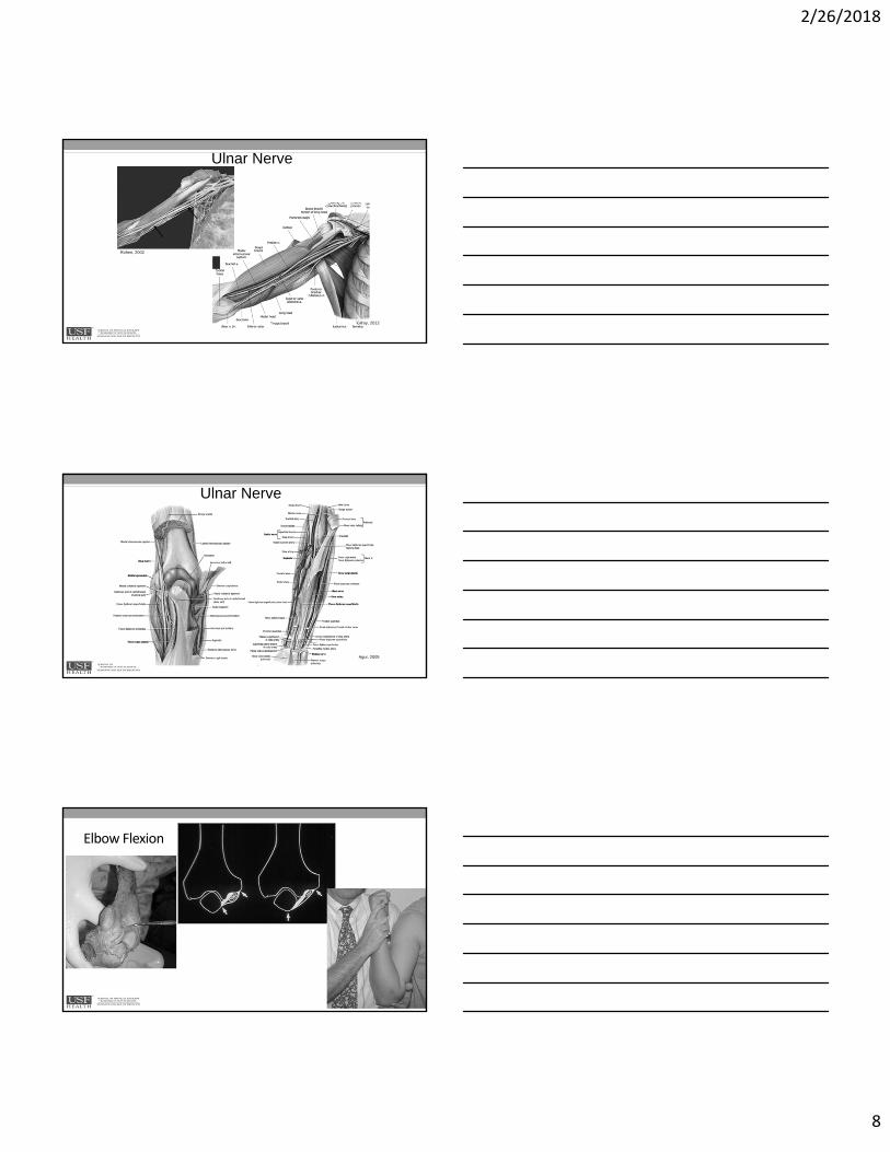
Ulnar Nerve
Rohen, 2002
Gilroy, 2012
Ulnar Nerve
Agur, 2005
Elbow Flexion
2/26/2018
9
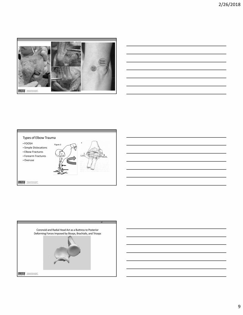
TricepsTendon
PostR/C
Types of Elbow Trauma
• FOOSH
• Simple Dislocations
• Elbow Fractures
• Forearm Fractures
• Overuse
Coronoid and Radial Head Act as a Buttress to Posterior Deforming Forces Imposed by Biceps, Brachialis, and Triceps
27
2/26/2018
10
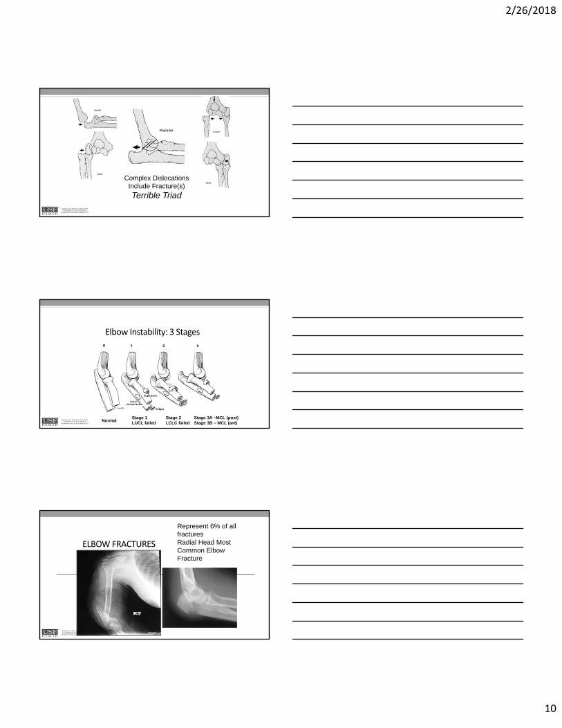
Complex Dislocations Include Fracture(s)
Terrible Triad
Elbow Instability: 3 Stages
Stage 1LUCL failedNormal
Stage 2 LCLC failed
Stage 3A –MCL (post)Stage 3B – MCL (ant)
ELBOW FRACTURES
Represent 6% of all fracturesRadial Head Most Common Elbow Fracture
2/26/2018
11
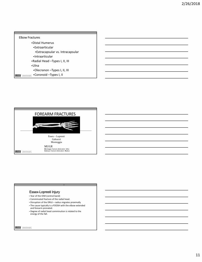
•Distal Humerus
•Extraarticular
•Extracapsular vs. Intracapsular
•Intraarticular
•Radial Head –Types I, II, III
•Ulna
•Olecranon –Types I, II, III
•Coronoid –Types I, II
Elbow Fractures
Essex – LoprestiGaleazziMonteggia
FOREARM FRACTURES
MUGRMonteggia fracture-dislocation: UlnaGaleazzi fracture-dislocation: Radius
• Tear of the IOM (central band)
• Comminuted fracture of the radial head.
• Disruption of the DRUJ – radius migrates proximally
• The cause typically is a FOOSH with the elbow extended and forearm pronated.
• Degree of radial head comminution is related to the energy of the fall.
33
2/26/2018
12
34
35
Acute:
Radial head replacement is recommended.
Reconstruction of the IOM?
+ TFCC repair
Chronic:Ulnar-shortening osteotomy (“leveling” of DRUJ)
TFCC repair to restore DRUJ stability
Radial head replacement?
Reconstruct the IOM ?
Treatment
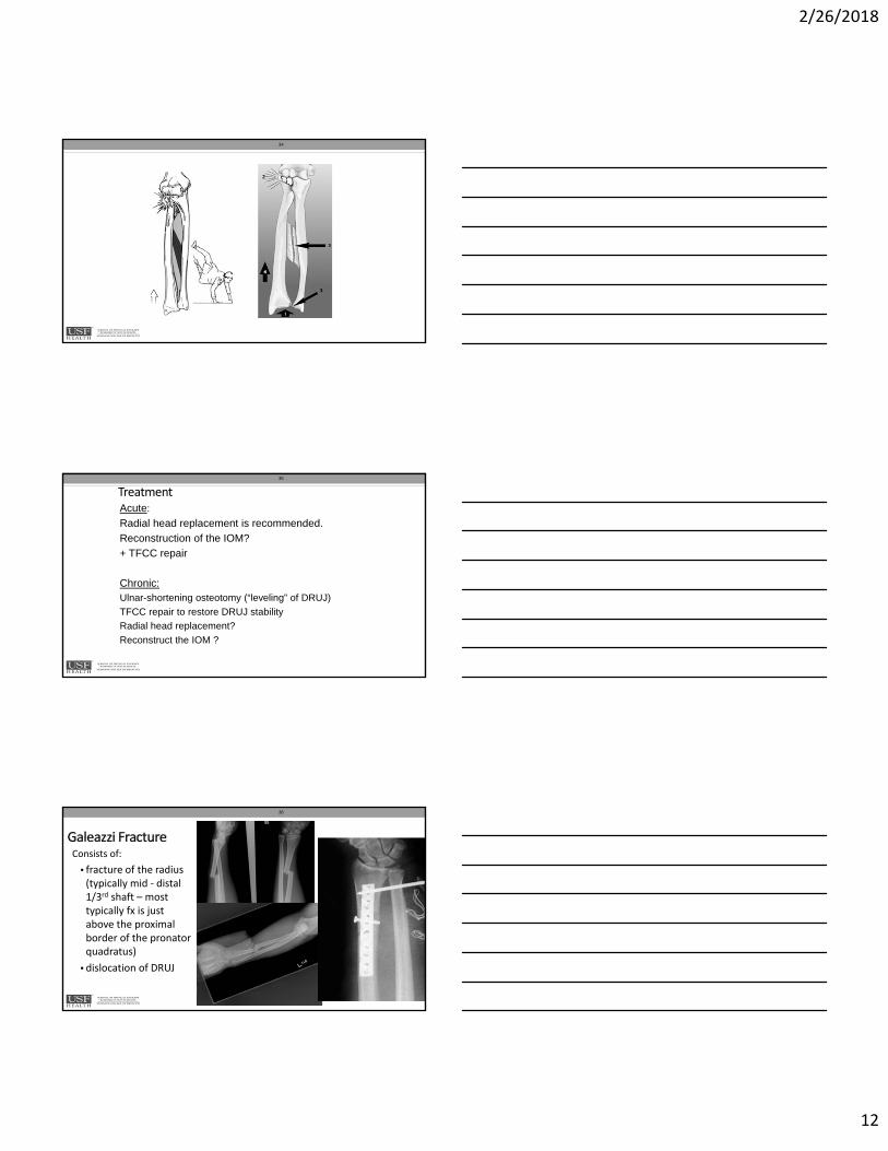
Consists of:
• fracture of the radius (typically mid ‐ distal 1/3rd shaft – most typically fx is just above the proximal border of the pronatorquadratus)
• dislocation of DRUJ
36
2/26/2018
13
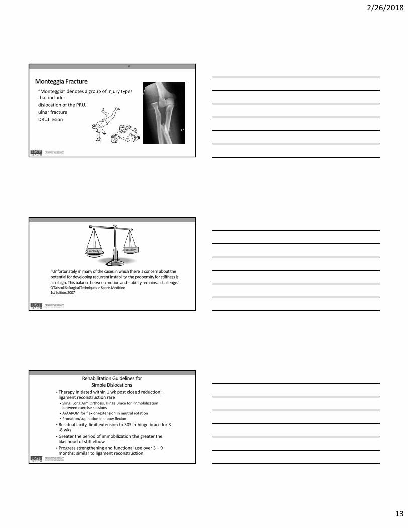
“Monteggia” denotes a group of injury types that include:
dislocation of the PRUJ
ulnar fracture
DRUJ lesion
37
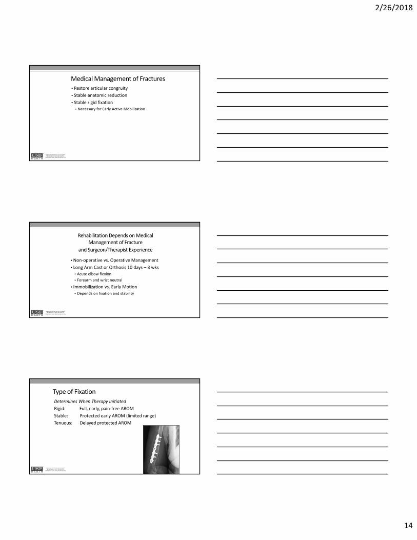
mobility stability
pain
“Unfortunately, in many of the cases in which there is concern about the potential for developing recurrent instability, the propensity for stiffness is also high. This balance between motion and stability remains a challenge.”O’Driscoll S: Surgical Techniques in Sports Medicine1st Edition, 2007
Rehabilitation Guidelines for Simple Dislocations
• Therapy initiated within 1 wk post closed reduction; ligament reconstruction rare• Sling, Long Arm Orthosis, Hinge Brace for immobilization between exercise sessions
• A/AAROM for flexion/extension in neutral rotation
• Pronation/supination in elbow flexion
• Residual laxity, limit extension to 30º in hinge brace for 3 ‐8 wks
• Greater the period of immobilization the greater the likelihood of stiff elbow
• Progress strengthening and functional use over 3 – 9 months; similar to ligament reconstruction
2/26/2018
14
Medical Management of Fractures• Restore articular congruity
• Stable anatomic reduction
• Stable rigid fixation
• Necessary for Early Active Mobilization
Rehabilitation Depends on Medical Management of Fracture
and Surgeon/Therapist Experience
• Non‐operative vs. Operative Management
• Long Arm Cast or Orthosis 10 days – 8 wks
• Acute elbow flexion
• Forearm and wrist neutral
• Immobilization vs. Early Motion
• Depends on fixation and stability
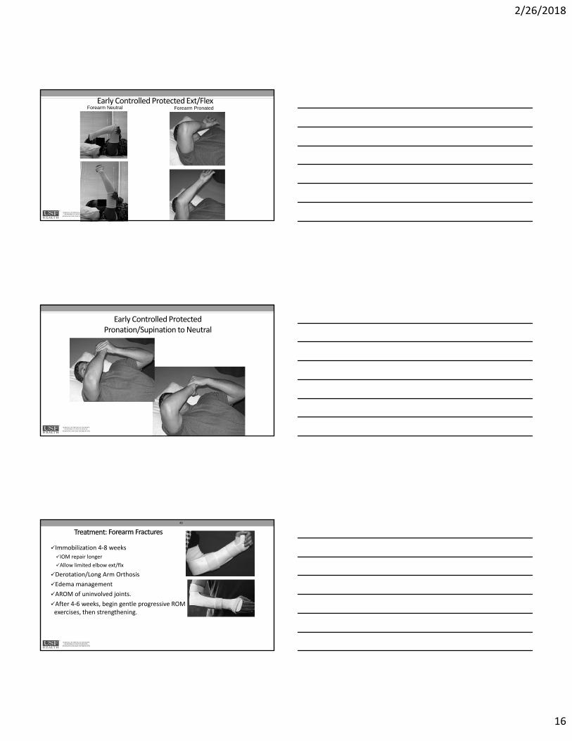
Type of Fixation
Determines When Therapy Initiated
Rigid: Full, early, pain‐free AROM
Stable: Protected early AROM (limited range)
Tenuous: Delayed protected AROM
2/26/2018
15
Protective Orthoses: Fractures and/or Dislocations
Phase I: Inflammatory (0‐2wks)
• Protective Orthosis or Cast
• Pain Modulation
• Edema Management
• AROM/fracture/fixation
• Maintain ROM to Noninvolved
Joints
• Monitor for Complications
Performing Elbow Motion in Sagittal Plan
Elbow InjuryFracture/Dislocation
Ligament Disruption With or Without Repair
LCL Instability? MCL Instability? MCL & LCL?
Forearm Pronated Forearm Supinated Forearm Neutral
2/26/2018
16
Early Controlled Protected Ext/Flex Forearm Neutral Forearm Pronated
Early Controlled Protected Pronation/Supination to Neutral
48
Immobilization 4‐8 weeks
IOM repair longer
Allow limited elbow ext/flx
Derotation/Long Arm Orthosis
Edema management
AROM of uninvolved joints.
After 4‐6 weeks, begin gentle progressive ROM exercises, then strengthening.
2/26/2018
17
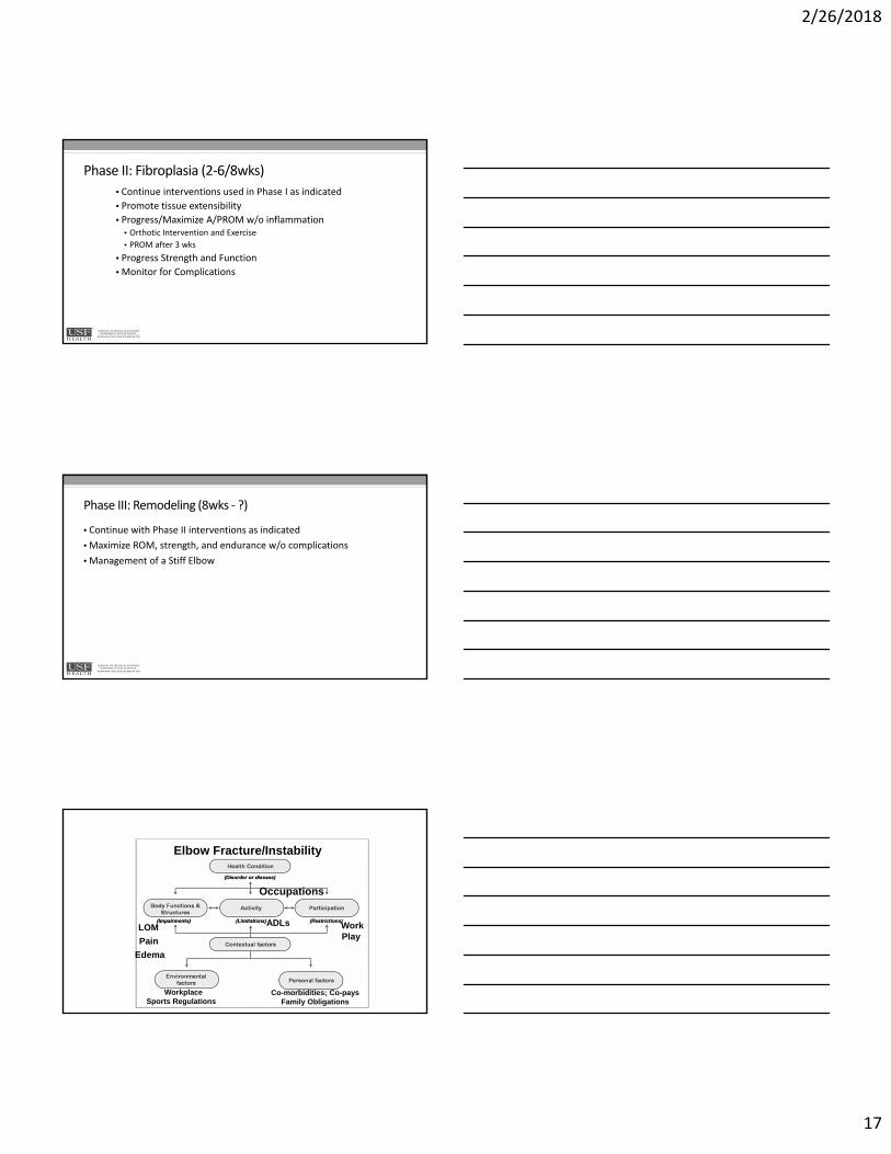
Phase II: Fibroplasia (2‐6/8wks)
• Continue interventions used in Phase I as indicated
• Promote tissue extensibility
• Progress/Maximize A/PROM w/o inflammation • Orthotic Intervention and Exercise
• PROM after 3 wks
• Progress Strength and Function
• Monitor for Complications
Phase III: Remodeling (8wks ‐ ?)
• Continue with Phase II interventions as indicated
• Maximize ROM, strength, and endurance w/o complications
• Management of a Stiff Elbow
Pain
LOM
Edema
ADLs WorkPlay
WorkplaceSports Regulations
Co-morbidities; Co-paysFamily Obligations
Occupations
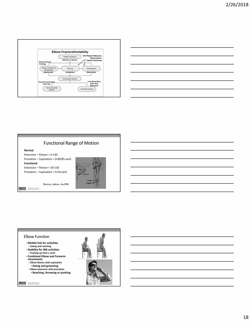
Elbow Fracture/Instability
2/26/2018
18
Physical Exam Findings
Self Report Measure, Observation,
Patient Interview
Low Bone Mass Falls RiskEducation
Hazards Associatedwith Falls
Elbow Fracture/Instability
Functional Range of Motion
Normal
Extension – Flexion = 0‐140
Pronation – Supination = 0‐80/85 each
Functional
Extension – Flexion = 30‐130
Pronation – Supination = 0‐50 each
Morrey, Askew, An,1981
Elbow Function
• Mobile link for activities• Eating and reaching
• Stability for WB activities• Pushing up from a chair
• Combined Elbow and Forearm movements• Elbow flexion with supination
• Eating and grooming• Elbow extension with pronation
• Reaching, throwing or pushing
2/26/2018
19
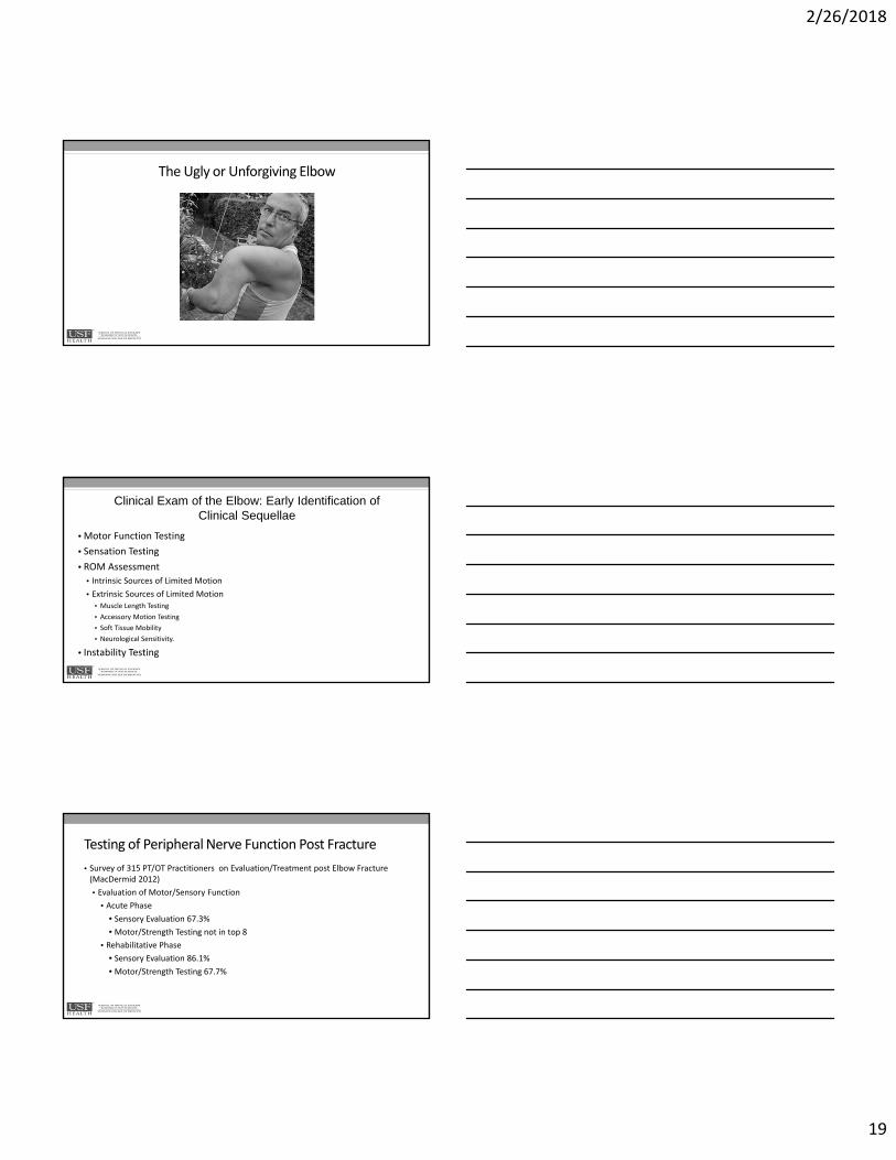
The Ugly or Unforgiving Elbow
• Motor Function Testing
• Sensation Testing
• ROM Assessment
• Intrinsic Sources of Limited Motion
• Extrinsic Sources of Limited Motion• Muscle Length Testing
• Accessory Motion Testing
• Soft Tissue Mobility
• Neurological Sensitivity.
• Instability Testing
Clinical Exam of the Elbow: Early Identification of Clinical Sequellae
Testing of Peripheral Nerve Function Post Fracture
• Survey of 315 PT/OT Practitioners on Evaluation/Treatment post Elbow Fracture (MacDermid 2012)
• Evaluation of Motor/Sensory Function
• Acute Phase
• Sensory Evaluation 67.3%
• Motor/Strength Testing not in top 8
• Rehabilitative Phase
• Sensory Evaluation 86.1%
• Motor/Strength Testing 67.7%
2/26/2018
20
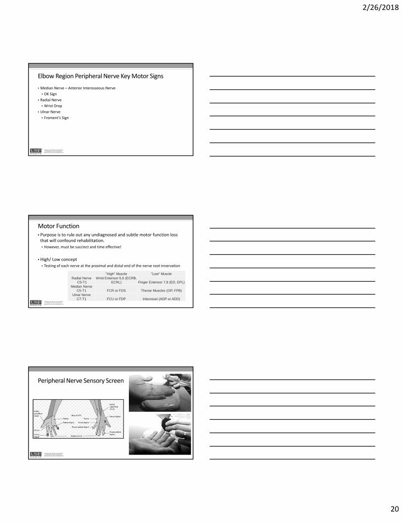
Elbow Region Peripheral Nerve Key Motor Signs
• Median Nerve – Anterior Interosseous Nerve
• OK Sign
• Radial Nerve
• Wrist Drop
• Ulnar Nerve
• Froment’s Sign
Motor Function• Purpose is to rule out any undiagnosed and subtle motor function loss that will confound rehabilitation.
• However, must be succinct and time effective!
• High/ Low concept
• Testing of each nerve at the proximal and distal end of the nerve root innervation
"High" Muscle "Low" MuscleRadial Nerve
C5-T1Wrist Extensor-5,6 (ECRB,
ECRL) Finger Extensor 7,8 (ED, EPL)Median Nerve
C5-T1 FCR or FDS Thenar Muscles (OP, FPB)Ulnar Nerve
C7-T1 FCU or FDP Interossei (ADP or ADD)
Peripheral Nerve Sensory Screen
X
X
X
2/26/2018
21
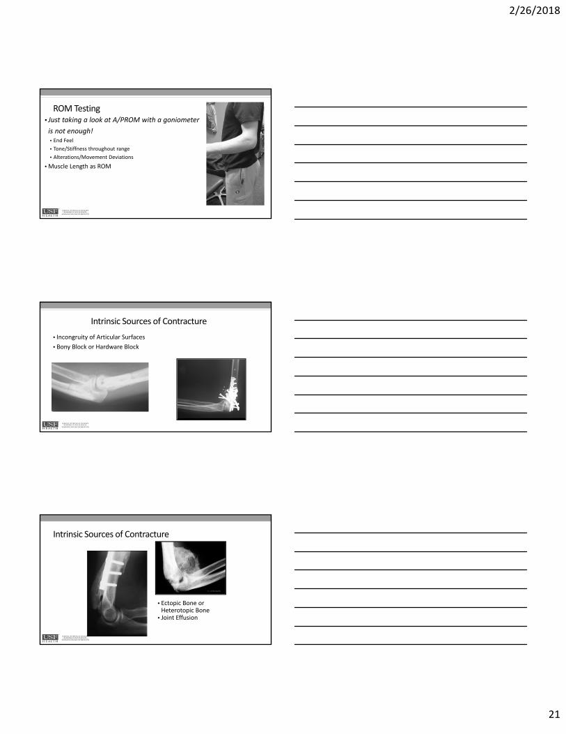
ROM Testing• Just taking a look at A/PROM with a goniometer
is not enough!• End Feel
• Tone/Stiffness throughout range
• Alterations/Movement Deviations
• Muscle Length as ROM
Intrinsic Sources of Contracture
• Incongruity of Articular Surfaces
• Bony Block or Hardware Block
Intrinsic Sources of Contracture
• Ectopic Bone or Heterotopic Bone
• Joint Effusion
2/26/2018
22
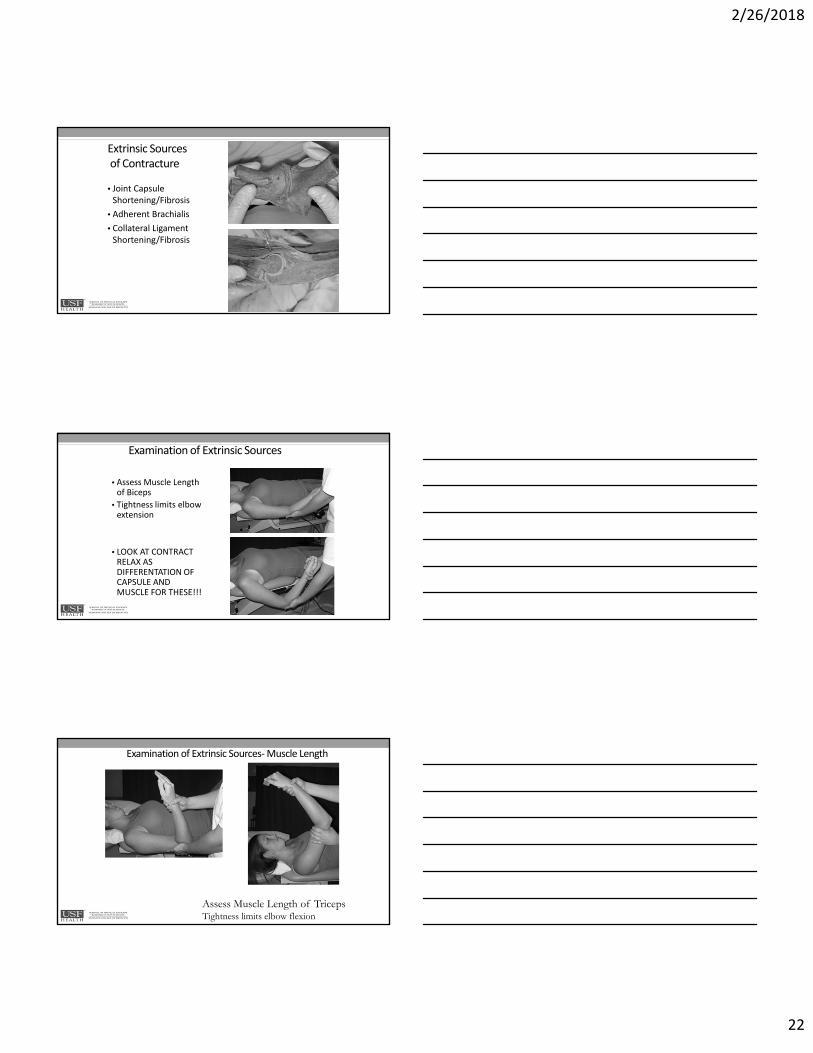
Extrinsic Sourcesof Contracture
• Joint Capsule Shortening/Fibrosis
• Adherent Brachialis
• Collateral Ligament Shortening/Fibrosis
Examination of Extrinsic Sources
• Assess Muscle Length of Biceps
• Tightness limits elbow extension
• LOOK AT CONTRACT RELAX AS DIFFERENTATION OF CAPSULE AND MUSCLE FOR THESE!!!
Examination of Extrinsic Sources‐Muscle Length
Assess Muscle Length of TricepsTightness limits elbow flexion
2/26/2018
23
Movement Patterns of the Elbow
Osteokinematic Motions
• Elbow Flexion/Extension‐• Thru Capitulum and Trochlea
• Increased PROM over AROM
• Elbow Pronation/Supination
• Thru both Proximal and Distal Radioulnar joints‐ spin thru Radial Head
• Limitation in PROM due to muscle, connective tissues
Coupled/Combined Motions
• Flexion‐ Humeral Adduction
• Extension‐ Humeral Abduction
• Supination‐ Adduction, external rotation of Ulna
• Pronation‐ Abduction, internal rotation of Ulna, positive ulnar variance
Force Couples During Elbow ROM
Flexion/Extension
• Biceps and Triceps are antagonists• Increased flexion force over extension
• ECRL can be flexor with forearm pronated
Pronation/Supination
• Supinators stronger than pronators• Pronation more easily compensated
• Shoulder abduction, medial rotation
• Overuse of pronator teres w/ wrist extension
• Possible rotation of ulna
• Supinator more active with increased elbow flexion
• Otherwise primary supinator is biceps tendon
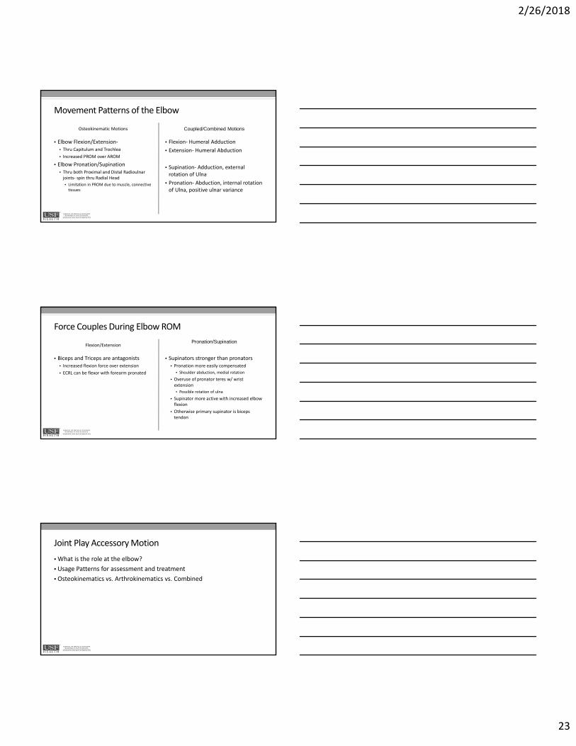
Joint Play Accessory Motion
• What is the role at the elbow?
• Usage Patterns for assessment and treatment
• Osteokinematics vs. Arthrokinematics vs. Combined
2/26/2018
24
Cases focused on clinical examination
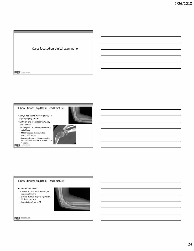
Elbow Stiffness s/p Radial Head Fracture
• 20 y/o male with history of FOOSH injury playing soccer
• MD visit one week later w/ X‐ray and CT scan
• Findings of 1.8‐2mm displacement of radial head
• Mild Displaced CommuniatedCoronoid Fracture
• Conservative care‐ 90 degree splint for one week, then wean off/ MD visit 4 weeks
Elbow Stiffness s/p Radial Head Fracture
• 4 weeks Follow Up
• patient in splint for all 4 weeks, no movement in sling
• Limited ROM 10 degrees supination , 95 flexion per MD
• Immediate referral to PT
2/26/2018
25
Radial Fracture Case
• Subjective Complaints
• Stiffness in elbow, concerned about moving elbow
• No Prior History of Etiology of Elbow
• Moderate sleeping dysfunction
• Observation: Moderate guarding of right elbow, no swing in gait pattern
• No focal edema noted
• Outcome Measure: Patient Rated Elbow Evaluation
• Pain 21 points, Activity 39 points, Usual Activity 24 points: Total 42%
• Pain level‐ 6/10 with flexion, extension, supination movement
• Functional deficits‐ eating, dressing combing hair
Patient Rated Elbow Evaluation
• 3 subscales
• Pain‐ 50%
• Function‐ 50%
• Specific Activities
• Usual Activities
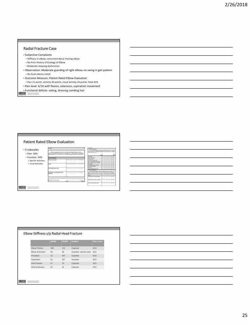
Elbow Stiffness s/p Radial Head Fracture
AROM PROM Endfeel Pain Level
Elbow Flexion 108 113 Capsular 4/10
Elbow Extension 38 36 Guarded- anterior pain 5/10
Pronation 32 N/T Guarded 5/10
Supination 52 N/T Guarded 5/10
Wrist Flexion 67 70 Capsular 3/10
Wrist Extension 54 61 Capsular 2/10
2/26/2018
26
Elbow Stiffness s/p Radial Head Fracture‐ Findings
• Muscle length
• Lacking 10 degrees for biceps, 5 degrees for triceps over available range
• Strength Testing:
• Elbow flexion, pronation, supination N/T
• Elbow extension 3+/5 in available
• Wrist flexion, extension 4/5 without pain
• Grip 43lbs
• Median, Radial, Ulnar High/Low 5/5
• Structure Specific Testing
• Negative OK, Negative Froments, + Ulnar nerve compression for parathesia
• Sensation‐ Normal 2.83 all peripheral nerves
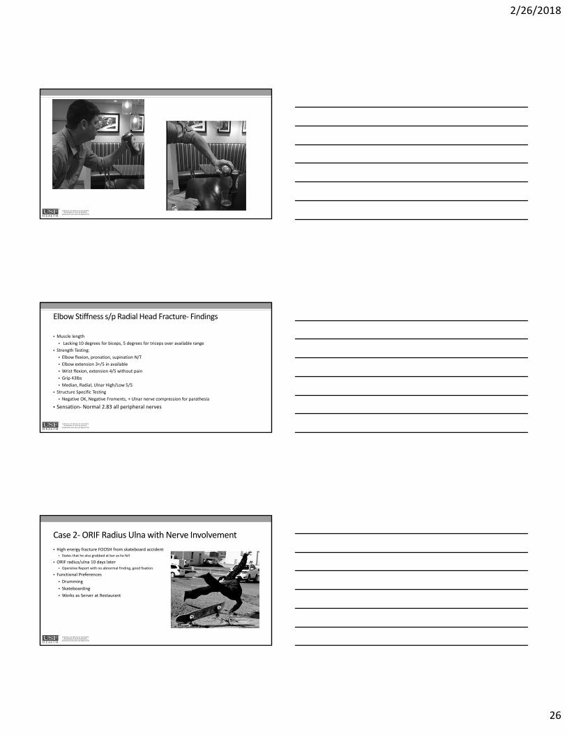
Case 2‐ORIF Radius Ulna with Nerve Involvement
• High energy fracture FOOSH from skateboard accident• States that he also grabbed at bar as he fell
• ORIF radius/ulna 10 days later• Operative Report with no abnormal Finding, good fixation.
• Functional Preferences
• Drumming
• Skateboarding
• Works as Server at Restaurant
2/26/2018
27
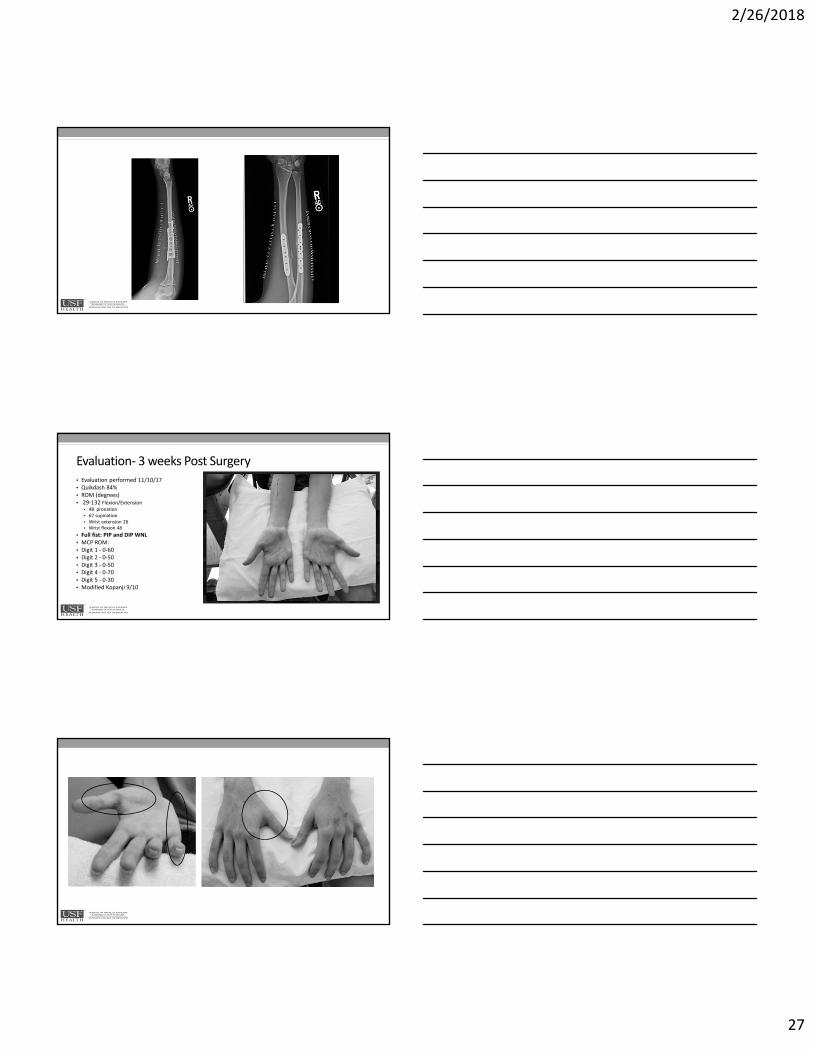
Evaluation‐ 3 weeks Post Surgery• Evaluation performed 11/10/17• Quikdash 84%• ROM (degrees)• 29‐132 Flexion/Extension
• 48 pronation• 67 supination• Wrist extension 26• Wrist flexion 48
• Full fist: PIP and DIP WNL• MCP ROM:• Digit 1 ‐ 0‐60• Digit 2 ‐ 0‐50• Digit 3 ‐ 0‐50• Digit 4 ‐ 0‐70• Digit 5 ‐ 0‐30• Modified Kopanji 9/10
2/26/2018
28
ORIF Examination Continued •
• Sensation:• Monofilament Testing (Semmes Weinstein)• Right Hand:• ‐ 3.61 over Thenar Eminence • ‐ Third Finger Palmar Sensation• 3.61 palmar aspect of 3rd finger proximal phalanx• ‐ Sensation present 4.31 at middle, distal phalanx phalanx
• ‐ 3.61 over thenar eminence• ‐ Dorsal Aspect: all 3.61 over digit 1‐5 distal phalanx
• Left Hand WNL
• Moving 2‐point discrimination (m2pd):• 2 mm across all distal R finger tips
• Functional MMT Testing:
• RUE LUE
• Grip Dynamometer 10lbs R, 100 L
• Key Pinch 5 lbs R, 19 L.
• Reflexes:
• Biceps: 1+ Bilateral
• Brachioradialis: 1+ L , R N/T
• Triceps 2/6 L, N/T R
Targeted Interventions to Manage Clinical Sequellaeand Maximize Patient Outcomes
Elbow Fracture Survey – Interventions Performed
• Acute Phase
• HEP 99%
• Education 93%
• Active ROM 86%
• AAROM ROM 75%
• Rehabilitative Phase:
• AROM/ PROM 95%+
• Mobilization with Movement 67%
• Manual Therapy not surveyed
2/26/2018
29
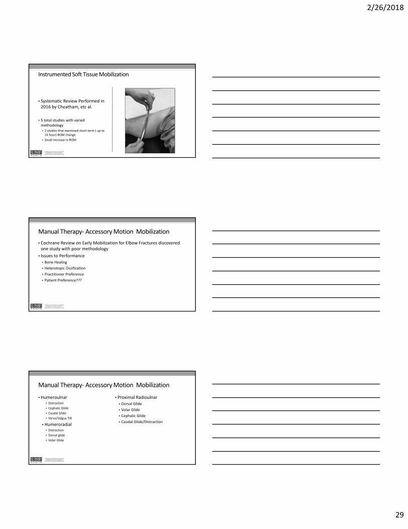
Instrumented Soft Tissue Mobilization
• Systematic Review Performed in 2016 by Cheatham, etc al.
• 5 total studies with varied methodology
• 2 studies that examined short term ( up to 24 hour) ROM change
• Small increase in ROM
Manual Therapy‐Accessory Motion Mobilization
• Cochrane Review on Early Mobilization for Elbow Fractures discovered one study with poor methodology
• Issues to Performance
• Bone Healing
• Heterotopic Ossification
• Practitioner Preference
• Patient Preference???
Manual Therapy‐Accessory Motion Mobilization
• Humeroulnar• Distraction
• Cephalic Glide
• Caudal Glide
• Varus/Valgus Tilt
• Humeroradial• Distraction
• Dorsal glide
• Volar Glide
• Proximal Radioulnar
• Dorsal Glide
• Volar Glide
• Cephalic Glide
• Caudal Glide/Distraction
2/26/2018
30
Neural Mobilization
Systematic Review and Meta‐Analysis‐ JOSPT 2017 Basson et. Al
Lateral Epicondylalgia‐ 3 studies total
• 1 study with lateral cervical glides found to be effective
• 2 other studies unclear, high bias
Cubital Tunnel Syndrome
• 1 study‐ no significant difference compared to control group and elbow brace
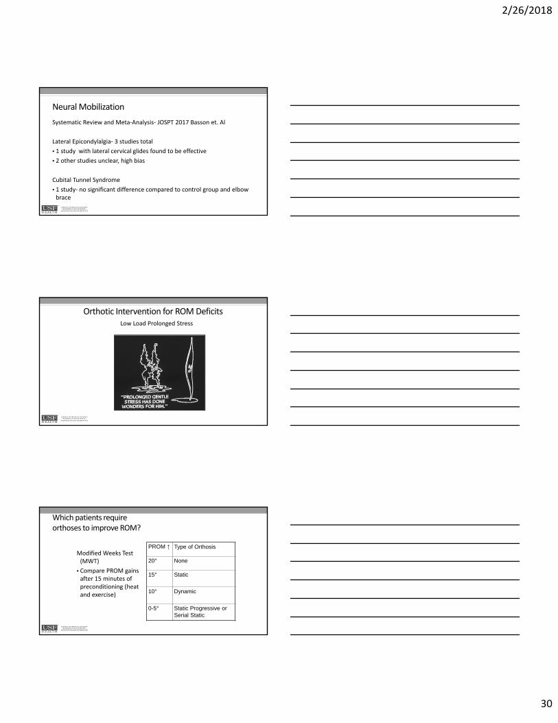
Orthotic Intervention for ROM DeficitsLow Load Prolonged Stress
Which patients require orthoses to improve ROM?
Modified Weeks Test (MWT)
• Compare PROM gains after 15 minutes of preconditioning (heat and exercise)
PROM ↑ Type of Orthosis
20° None
15° Static
10° Dynamic
0-5° Static Progressive or Serial Static
2/26/2018
31
Which patients require orthotic intervention to improve ROM?
• Compare PROM gains after 15 minutes of preconditioning (heat and exercise)
PROM ↑ Orthotic Intervention
15 - 20° Keep going with therex; manual techniques; maybe static orthosisto maintain motion
0 - 10° Mobilizing Orthosis: Serial static; static progressive; dynamic
Jane’s Modification
Considerations for Orthoses Selection
• End Feel
• Degree of Contracture
• Therapists’ Experience
• Patient Compliance
• Refer to stiffness presentations
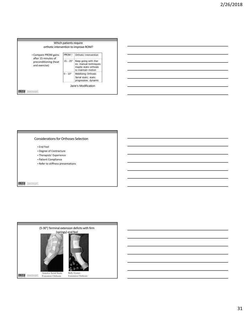
(5‐30°) Terminal extension deficits with firm (springy) end feel
Belly Gutter Extension Orthosis
Anterior Serial Static Extension Orthosis
2/26/2018
32
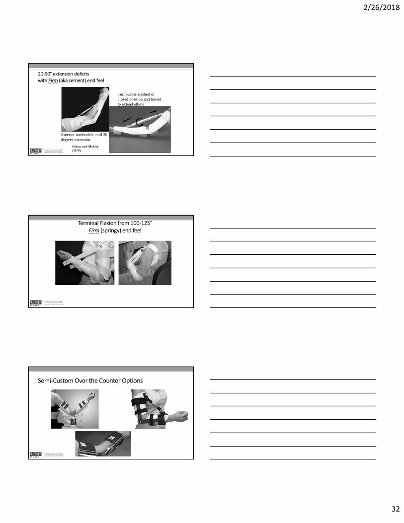
20‐90° extension deficits with Firm (aka cement) end feel
Anterior turnbuckle until 20 degrees extension
Turnbuckle applied in closed position and turned to extend elbow
Green and McCoy (1979)
Terminal Flexion from 100‐125°Firm (springy) end feel
Semi‐Custom Over the Counter Options
2/26/2018
33
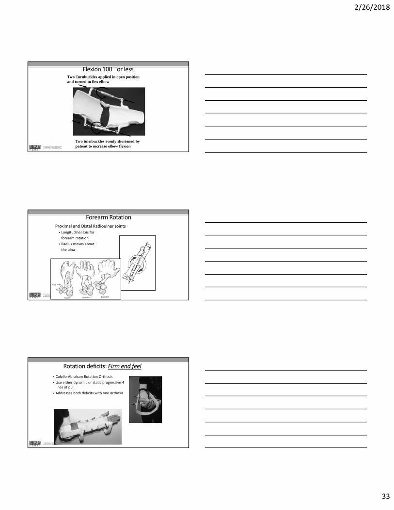
Flexion 100 ° or less
Two turnbuckles evenly shortened by patient to increase elbow flexion
Two Turnbuckles applied in open position and turned to flex elbow
Forearm RotationProximal and Distal Radioulnar Joints
• Longitudinal axis for
forearm rotation
• Radius moves about
the ulna
Rotation deficits: Firm end feel
• Colello‐Abraham Rotation Orthosis
• Use either dynamic or static progressive 4 lines of pull
• Addresses both deficits with one orthosis
2/26/2018
34
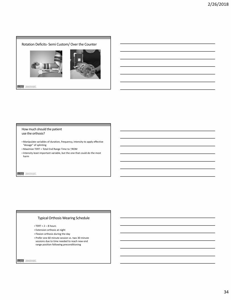
Rotation Deficits‐ Semi Custom/ Over the Counter
How much should the patientuse the orthosis?
• Manipulate variables of duration, frequency, intensity to apply effective “dosage” of splinting
• Maximize TERT = Total End Range Time to ↑ROM
• Intensity least important variable, but the one that could do the most harm
Typical Orthosis Wearing Schedule
• TERT = 2 – 8 hours
• Extension orthosis at night
• Flexion orthosis during the day
• Prefer one 60 minute session vs. two 30 minute sessions due to time needed to reach new end range position following preconditioning
2/26/2018
35
How do you know if the orthosis is effective or harmful?
Effective
• ROM improves
• Measurements taken after preconditioning
• Adjust variables to progress ROM increases especially duration and frequency
Harmful
• Tissues Reactive
• Pain
• Loss of motion
• Sign of inflammation
• Edema
• Some joints more susceptible than others
• Rest tissues for a few days
When do you discontinue using the orthosis?
Good news: ROM goals achieved
Bad news: ROM plateau
Splint not tolerated
Joint inflammation present
Cases focused on interventions
2/26/2018
36
Elbow Stiffness Case
• Patient preference to not have any more braces/splints on Day #1
• Young, active patient but with considerable apprehension in movement patterns.
• Low/Moderate Irritability levels
• Patient leaving for Winter Break in 5 weeks
• Fair Psychosocial Status Visit #1, increased quickly
Which patients require orthotic intervention to improve ROM?
• Compare PROM gains after 15 minutes of preconditioning (heat and exercise)
PROM ↑ Orthotic Intervention
15 - 20° Keep going with therex; manual techniques; maybe static orthosis to maintain motion
0 - 10° Mobilizing Orthosis: Serial static; static progressive; dynamic
Jane’s Modification
Elbow Stiffness s/p Radial Head Fracture
• Plan of Care 2x week for 5 weeks
• Leaving for Winter Break for 4 weeks
• First 5 visits treatment plan
• AROM within pain free range.
• Low Load long duration stretching 15 min w/ Heat into Extension
• Muscle Length Stretching/Mobilization
• IASTM to biceps and triceps, NO IASTM over radial head
• Pain science/ cognitive training
2/26/2018
37
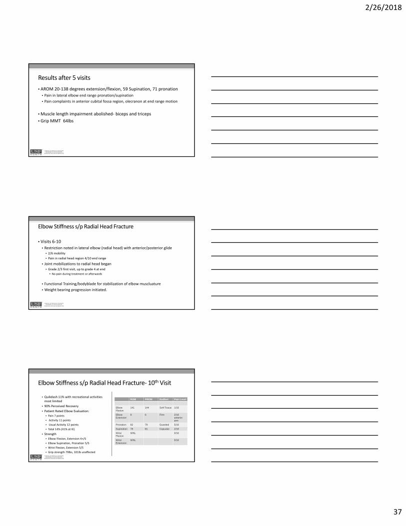
Results after 5 visits
• AROM 20‐138 degrees extension/flexion, 59 Supination, 71 pronation
• Pain in lateral elbow end range pronation/supination
• Pain complaints in anterior cubital fossa region, olecranon at end range motion
• Muscle length impairment abolished‐ biceps and triceps
• Grip MMT 64lbs
Elbow Stiffness s/p Radial Head Fracture
• Visits 6‐10
• Restriction noted in lateral elbow (radial head) with anterior/posterior glide
• 2/6 mobility
• Pain in radial head region 4/10 end range
• Joint mobilizations to radial head began
• Grade 2/3 first visit, up to grade 4 at end
• No pain during treatment or afterwards
• Functional Training/bodyblade for stabilization of elbow muscluature
• Weight bearing progression initiated.
Elbow Stiffness s/p Radial Head Fracture‐ 10th Visit
• Quikdash 11% with recreational activities most limited
• 90% Perceived Recovery
• Patient Rated Elbow Evaluation:
• Pain 7 points
• Activity 11 points
• Usual Activity 12 points
• Total 14% (41% at IE)
• Strength
• Elbow Flexion, Extension 4+/5
• Elbow Supination, Pronation 5/5
• Wrist Flexion, Extension 5/5
• Grip strength 79lbs, 101lb unaffected
ROM PROM Endfeel Pain Level
Elbow Flexion
141 144 Soft Tissue 1/10
Elbow Extension
8 6 Firm 2/10 anterior arm
Pronation 82 79 Guarded 5/10
Supination 79 81 Capualar 2/10
Wrist Flexion
WNL 0/10
Wrist Extension
WNL 0/10
2/26/2018
38
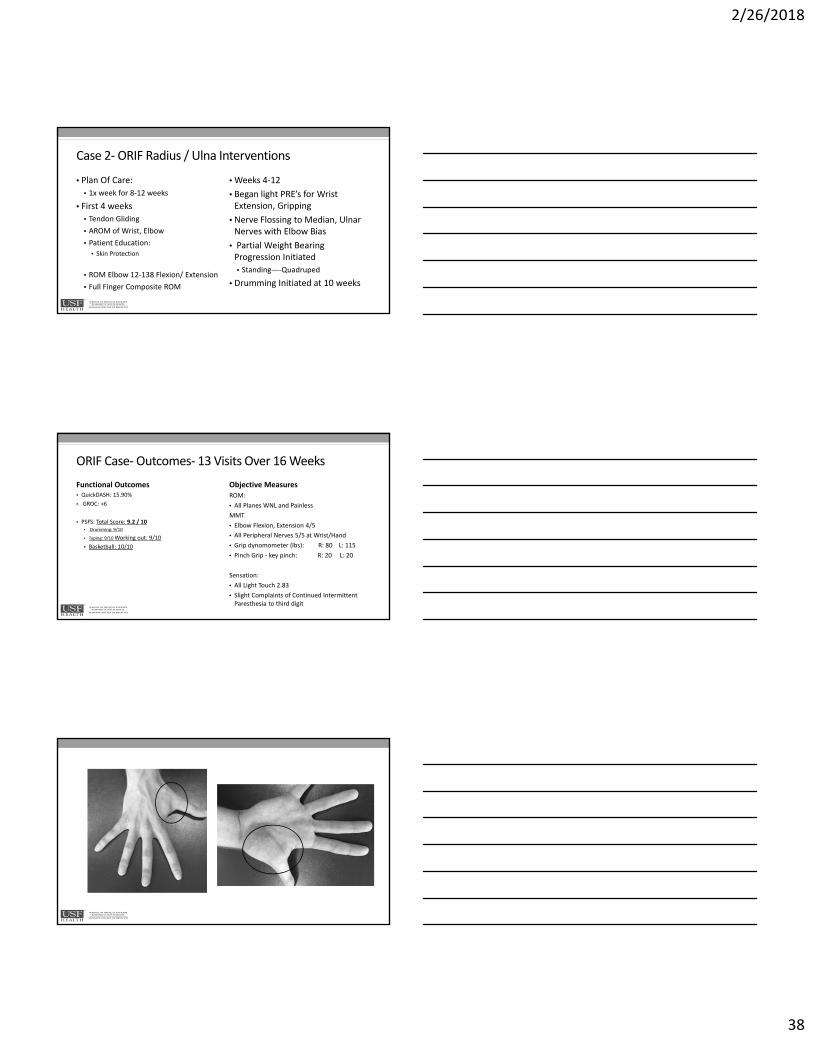
Case 2‐ORIF Radius / Ulna Interventions
• Plan Of Care:
• 1x week for 8‐12 weeks
• First 4 weeks
• Tendon Gliding
• AROM of Wrist, Elbow
• Patient Education:
• Skin Protection
• ROM Elbow 12‐138 Flexion/ Extension
• Full Finger Composite ROM
• Weeks 4‐12
• Began light PRE’s for Wrist Extension, Gripping
• Nerve Flossing to Median, Ulnar Nerves with Elbow Bias
• Partial Weight Bearing Progression Initiated
• Standing‐‐‐‐Quadruped
• Drumming Initiated at 10 weeks
ORIF Case‐Outcomes‐ 13 Visits Over 16 Weeks
Functional Outcomes• QuickDASH: 15.90%
• GROC: +6
• PSFS: Total Score: 9.2 / 10• Drumming: 9/10
• Typing: 9/10 Working out: 9/10
• Basketball: 10/10
Objective MeasuresROM:
• All Planes WNL and Painless
MMT
• Elbow Flexion, Extension 4/5
• All Peripheral Nerves 5/5 at Wrist/Hand
• Grip dynomometer (lbs): R: 80 L: 115
• Pinch Grip ‐ key pinch: R: 20 L: 20
Sensation:
• All Light Touch 2.83
• Slight Complaints of Continued Intermittent Paresthesia to third digit
2/26/2018
39
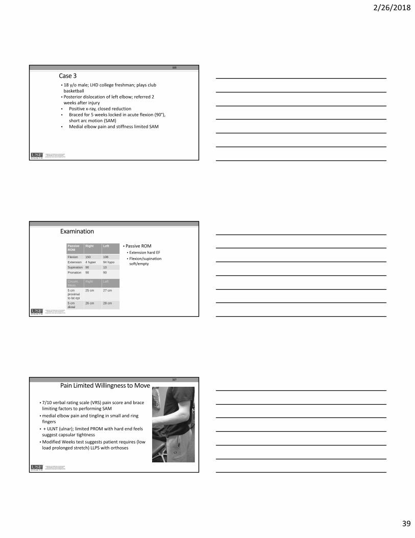
Case 3 • 18 y/o male; LHD college freshman; plays club basketball
• Posterior dislocation of left elbow; referred 2 weeks after injury
• Positive x‐ray, closed reduction• Braced for 5 weeks locked in acute flexion (90°),
short arc motion (SAM)• Medial elbow pain and stiffness limited SAM
115
Examination
• Passive ROM
• Extension hard EF
• Flexion/supination soft/empty
Passive ROM
Right Left
Flexion 150 106
Extension 4 hyper 94 hypo
Supination 90 10
Pronation 90 90
Circum.Meas.
Right Left
5 cm proximal to lat epi
25 cm 27 cm
5 cm distal
26 cm 28 cm
Pain Limited Willingness to Move
• 7/10 verbal rating scale (VRS) pain score and brace limiting factors to performing SAM
• medial elbow pain and tingling in small and ring fingers
• + ULNT (ulnar); limited PROM with hard end feels suggest capsular tightness
• Modified Weeks test suggests patient requires (low load prolonged stretch) LLPS with orthoses
117
2/26/2018
40
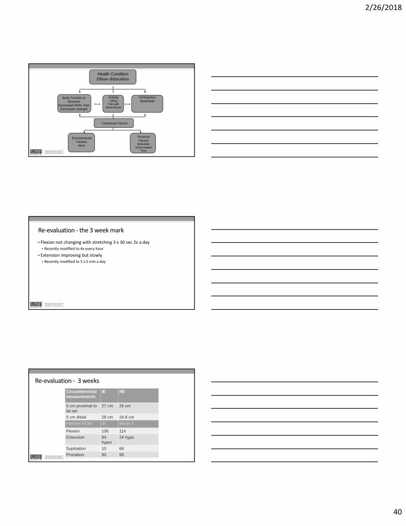
Health ConditionElbow dislocation
Body Function & Structure
Decreased ROM, PainDecreased strength
ActivityLifting
Pain with elbow flexion
ParticipationBasketball
Contextual Factors
Environmental Factors
Mom
Personal Factors
MotivationGood support
Time
Re‐evaluation ‐ the 3 week mark
• Flexion not changing with stretching 3 x 30 sec 2x a day
• Recently modified to 4x every hour
• Extension improving but slowly
• Recently modified to 5 x 5 min a day
Re‐evaluation ‐ 3 weeks
Circumferential measurements
IE RE
5 cm proximal to lat epi
27 cm 26 cm
5 cm distal 28 cm 24.8 cm
Passive ROM IE Week 3
Flexion 106 114
Extension 94 hyper
24 hypo
Supination 10 66
Pronation 90 90
2/26/2018
41
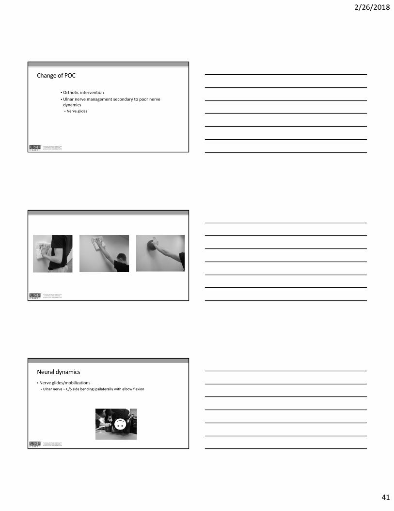
Change of POC
• Orthotic intervention
• Ulnar nerve management secondary to poor nerve dynamics
• Nerve glides
Neural dynamics
• Nerve glides/mobilizations
• Ulnar nerve – C/S side bending ipsilaterally with elbow flexion
2/26/2018
42
Which patients require orthotic intervention to improve ROM?
• Compare PROM gains after 15 minutes of preconditioning (heat and exercise)
PROM ↑ Orthotic Intervention
15 - 20° Keep going with therex; manual techniques; maybe static orthosisto maintain motion
0 - 10° Mobilizing Orthosis: Serial static; static progressive; dynamic
Jane’s Modification
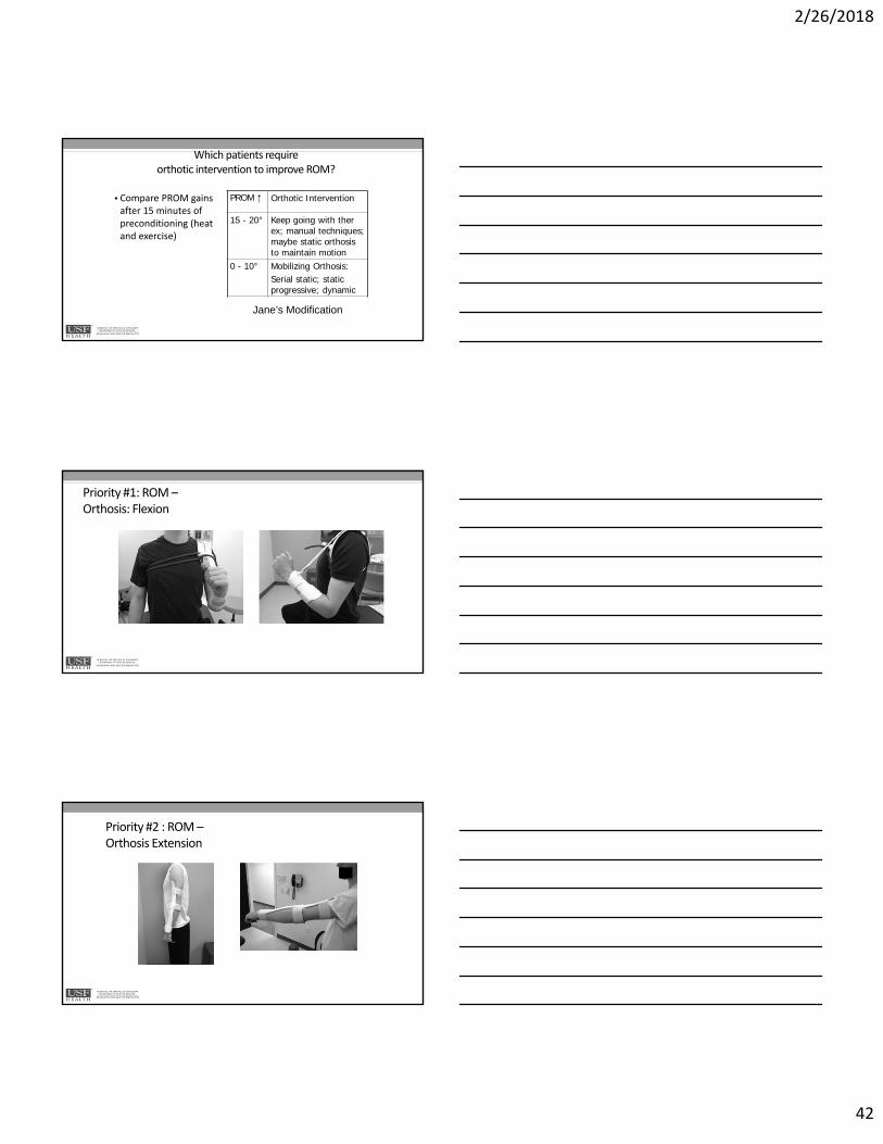
Priority #1: ROM –Orthosis: Flexion
Priority #2 : ROM –Orthosis Extension
2/26/2018
43
Other impairments
• Edema
• KT tape (used for pain too)
• Strengthening
• functional activity; no more than 3‐5#
• 2 months – total arm strengthening w/ wts
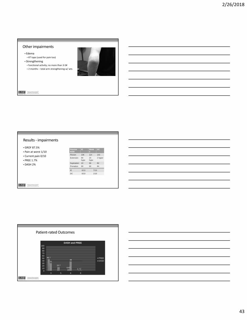
Results ‐ impairments
• GROF 87.5%
• Pain at worst 1/10
• Current pain 0/10
• PREE 1.7%
• DASH 2%Pain Current Worst
IE 0/10 7/10
DC 0/10 1/10
Passive ROM
IE Week 3
DC
Flexion 106 114 142
Extension 94 hypo
24 hypo
2 hyper
Supination 10 66 82
Pronation 90 90 84
Patient‐rated Outcomes
46.7
15.76.67
1.7
3138
20
102030405060708090
100
3 5 6 8
DASH and PREE
PREE
DASH

2/26/2018
44
Summary Comments • Experience matters for managing elbow trauma and identifying clinical sequellae.
• Access to imaging and communication with surgeon essential.
• Elbow stiffness does not discriminate with patient’s age.
• Nerve injuries may need to be treated coincidentally.
• Future investigations need to validate our clinical observations.
• Items within the tests and measures field need to be
included within the outcomes registry.
mobilitystability
Safe Travels
May Your Journey Lead to Many Rainbows
Questions?
2/26/2018
45
References
Cheatham, SW Lee M, Cain M. Baker R. The Efficacy of Instrument assisted soft tissue massage: a Systematic Review. J Can Chiropr Assoc 2016 60(3): 200‐211.
Basson A, Olivier B, Ellis R. Coppieters M, Stewart A, Mudi W. The Effectiveness of Neural Mobilization for Neuromusculoskeletal Conditions: A Systematic Review and Meta‐analysis. JOSPT 2017; 47 (9). 593‐615.
MacDermid JC, Vincent JI, Kieffer L, Kieffer A, Demiatte J. A Survey of Practice Patterns for Rehabilitation Post Elbow Fracture. The Open Orthopedics Journal 2012 429‐439.
Gradl G, Jupiter JB: Current concepts review ‐ fractures in the region of the elbow. Acta Chir Orthop Traumatol Cech. 2012;79(3):203‐12.
Harding P, Rasekaba T, Smirneos L, Holland AE: Early mobilization for elbow fractures in adults. Cochrane Database Syst Rev. 2011 Jun 15;(6).
Lucado AM, Li Z. Static progressive splinting to improve wrist stiffness after distal radius fracture: a prospective, case series study. Physiother Theory Pract. 2009 May;25(4):297‐309.
McGrath Ms, Ulrich SD, Bonutti PM, Marker DR, Johanssen HR, Mont MA: Static progressive splinting forrestorationof rotational motion of the forearm. J Hand Ther. 2009 Jan‐Mar; 22(1): 3‐9.
•
References
Skirven TM, Osterman AL, Fedorczyk JM and Amadio PC (eds): Rehabilitation of the Hand and Upper Extremity, 6th edition, Mosby, St. Louis, 2011.
JC MacDermid. Vincent JI, Kieffer L, Kieffer A, Demaiter J, MacIntosh S. A Survey of Practice Patterns for Rehabilitation Post Elbow Fracture. The Open Orthopaedics Journal 2012; 6, 429‐439
Chemama B, Bonnevialle N, Peter O, Mansat P. Terrible triad injury of the elbow: How to improve outcomes?. Orthopaedics & Traumatology: Surgery & Research. 2010; 96(2) 147‐154.
Rehim SA, Maynard MA, Sebastian SJ, Chung KC. Monteggia Fracture Dislocations: A Historical Review. The Journal of Hand Surgery. 2014; 39(7): 1384‐1394.
Anakwe, RE, Middleton, Scott D.; Jenkins, PJ.; More D. Patient‐Reported Outcomes After Simple Dislocation of the Elbow. Journal of Bone & Joint Surgery. 2011; 93(13): 1220‐1226.
Smith, MV., Calfee RP.; Baumgarten, Keith M.; More R. Upper Extremity‐Specific Measures of Disability and Outcomes in Orthopaedic Surgery. Journal of Bone & Joint Surgery. 2012; 94(3):277‐285.