elbow ct: positioning - wrong wayanatomy technical foph bbff monteggia galeazzi essex-lop. fracture...
TRANSCRIPT

Anatomy Technical FOPH BBFF Monteggia Galeazzi Essex-Lop. Fat Pads Peds Elbow Supracond. Lat. condyle Med. epicon. 4
© 2013 Ken L Schreibman, PhD/MD www.schreibman.info
Elbow & Forearm Trauma ASRT @ RSNA
18 of 99
Elbow CT: Positioning
We don’t scan patients with their elbow down at their side Excess radiation across torso X-ray scatter decreases res.
NEVER scan patients with their elbow laying on their abdomen Elbow moves with
patient breathing Makes it impossible
to get good reformats Instead get terrible
images like this
- WRONG WAY
CT Scout

Anatomy Technical FOPH BBFF Monteggia Galeazzi Essex-Lop. Fat Pads Peds Elbow Supracond. Lat. condyle Med. epicon. 4
© 2013 Ken L Schreibman, PhD/MD www.schreibman.info
Elbow & Forearm Trauma ASRT @ RSNA
19 of 99
Elbow CT: Positioning - RIGHT WAY Elbow over head! Avoids torso radiation Avoids scatter from torso
Supine = OK Prone/Decubitus = OK
Optimal Position Elbow straight Head bent out of field
However the patient is most comfortable

Anatomy Technical FOPH BBFF Monteggia Galeazzi Essex-Lop. Fat Pads Peds Elbow Supracond. Lat. condyle Med. epicon. 4
© 2013 Ken L Schreibman, PhD/MD www.schreibman.info
Elbow & Forearm Trauma ASRT @ RSNA
20 of 99
Elbow CT: Positioning - BEST WAY
S,Y 30yoM CT Scout
Straight arm & elbow Perpendicular to
CT scanning plane Easier reformatting Yields highest
resolution reformats
Sagittal Reformat
Coronoid Fracture

Anatomy Technical FOPH BBFF Monteggia Galeazzi Essex-Lop. Fat Pads Peds Elbow Supracond. Lat. condyle Med. epicon. 4
© 2013 Ken L Schreibman, PhD/MD www.schreibman.info
Elbow & Forearm Trauma ASRT @ RSNA
21 of 99
Elbow CT: Positioning This is the position to avoid!
with arm parallel to CT scanning plane
- With a cast…
CT Scout T,B 65yoF
Sagittal Reformat
Streak artifacts from scanning
length of forearm limits resolution
Anatomy Technical FOPH BBFF Monteggia Galeazzi Essex-Lop. Fat Pads Peds Elbow Supracond. Lat. condyle Med. epicon. 4
© 2013 Ken L Schreibman, PhD/MD www.schreibman.info
Elbow & Forearm Trauma ASRT @ RSNA
22 of 99
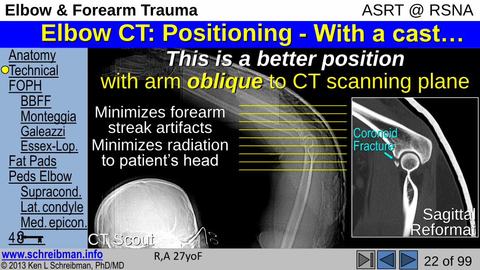
Elbow CT: Positioning This is a better position
with arm oblique to CT scanning plane
- With a cast…
CT Scout R,A 27yoF
Sagittal Reformat
Coronoid Fracture
Minimizes forearm streak artifacts
Minimizes radiation to patient’s head

Anatomy Technical FOPH BBFF Monteggia Galeazzi Essex-Lop. Fat Pads Peds Elbow Supracond. Lat. condyle Med. epicon. 4
© 2013 Ken L Schreibman, PhD/MD www.schreibman.info
Elbow & Forearm Trauma ASRT @ RSNA
23 of 99
Elbow CT: Reformat in 6 planes
3 planes relative to distal humerus 3 planes relative to proximal forearm Off Sagittal reference image:
1) Axial to Humerus 2) Axial to Forearm 3) Coronal to Radius 4) Coronal to Ulna
Off Axial Humerus reference: 5) Parallel to Inter-Epicondyle Line 6) Perpendicular to Epicondyle Line
If elbow is straight, only need 3!
UW 2-page Protocol Sheets
www.radiology.wisc.edu
Positioning Reformatting Reformatting